Article

Synthesis and Study of the Effect of Ba2+ Cations Substitution with Sr2+ Cations on Structural and Optical Properties of Ba2−xSrxZnWO6 Double Perovskite Oxides (x = 0.00, 0.25, 0.50, 0.75, 1.00)

1
Department of Physics, Faculty of Science and Technology, Al Neelain University, Khartoum 13314, Sudan
2
Research Chair in Laser Diagnosis of Cancers, Department of Physics and Astronomy, College of Science, King Saud University, Riyadh 11451, Saudi Arabia
3
Department of Physics, Faculty of Education and Applied Science, Hajjah University, Hajjah 1729, Yemen
*
Author to whom correspondence should be addressed.
Materials 2017, 10(5), 469; https://doi.org/10.3390/ma10050469
Submission received: 13 February 2017 / Revised: 20 April 2017 / Accepted: 21 April 2017 / Published: 28 April 2017

Abstract

:
The effect of Sr2+ substitution on the morphology, crystal structure, and optical properties of double perovskite oxide Ba2−xSrxZnWO6 (x = 0.00, 0.25, 0.50, 0.75, 1.00) were investigated. Scanning electronic microscopy demonstrated that all samples have similar microstructure morphology but differ in the range of grain sizes. X-ray diffraction measurements indicated that these materials crystallize in a (Fm-3m) cubic crystal structure, and also confirmed the tolerance factor. Rietveld analysis revealed that the lattice parameter decreased from 8.11834 to 8.039361 Å when the substitution of Ba2+ with Sr2+ cations increased from zero to 100%. Fourier transform infrared (FTIR) and Raman spectroscopies displayed a symmetric stretching vibration of WO6 octahedra at 825 cm−1, and an anti-symmetric stretching mode of WO6 was observed by FTIR at 620 cm−1. A strong peak at 420 cm−1 was also observed in the Raman spectra and is due to the W–O–W bending vibration modes. UV-Vis diffuse reflectance spectroscopy was carried out for the series, and the band gap energy decreased from 3.27 eV for Ba2ZnWO6 to 3.02 and 3.06 eV for Ba1.75Sr0.25ZnWO6 and Ba1.5Sr0.5ZnWO6, respectively. The excitation and emission photoluminescence properties were investigated at room temperature.

1. Introduction

Many researchers are interested in double perovskite oxides that consist of transition metals [1]. These materials represent a large part of material science research because of the diversity in their physical and chemical characteristics, and their diverse applications [1,2,3,4,5,6], such as light harvesting (LaNiMnO6) [2], ferroelectrics (Pb2Mn0.6Co0.4WO6) [5], Multiferroic (Bi2NiMnO6, Bi2FeCrO6) [6], superconductivity (Sr2YRu0.95Cu0.05O6) [7], magneto resistance (Sr2FeMoO6) [8], dielectric resonators (Ca2AlTaO6, SrAlTaO6) [9], and photo-catalysis (Cs2BiAgCl6) [10].
A variety of devices and methods have previously been used to find and characterize new double perovskite compounds at high temperatures. Zaraq et al. [1] synthesized SrCaCoTeO6 and SrCaNiTeO6 compounds and used X-ray diffraction (XRD) and scanning electron microscopy (SEM) to describe their crystal structure and phase transition. Chufeng Lau et al. [2] used XRD, photoelectron spectroscopy, and UV-Vis-NIR spectroscopy to describe the LaNiMnO6 compound and studied the possibility for its solar cell applications. Paiva et al. [3] used the PANalytical diffractometer and Solartron 1260 impedance analyzer to study the structure and microwave properties of Sr3WO6, which is used in Bluetooth and mobile system devices for microwave telecommunications through wireless antennae. By utilizing the conventional solid-state ceramic route, BiCu2VO6 and BiCa2VO6 powders, which are used in low-temperature co-fired ceramic applications, were prepared and examined using XRD, SEM, and the TE01δ shielded cavity method with a network analyzer (8720ES) and temperature chamber (Delta 9023) to characterize their structure and microwave dielectric behaviors. Orlandi et al. [5] utilized the solid-state reaction route to synthesize Pb2Mn0.6Co0.4WO6, and used XRD with the SQUID MPMS Quantum Design magnetometer in order to investigate its crystal and magnetic structure. In addition, the perovskite compound can be used in biomedical applications. LaNiMnO6 nanoparticles were synthesized using the co-precipitation method and were characterized by XRD using a vibration magnetometer (PPMS-9, Quantum design), Transmission electron microscopy (TEM), and UV-VIS spectroscopy to investigate the structure, magnetic, and adsorption of bovine serum albumin applications. The nanoparticles displayed good adsorption performance in the bovine serum albumin proteins. The Double-perovskite La2NiMnO6 (LNMO) nanoparticles are potential carriers for large biomolecules, which have wide use in biomedical applications.
The general chemical formula of double perovskite oxide is expressed as AA′BB′O6, and the crystal structure of AA′BB′O6 consists of the exchange sites of BO6 and B′O6 octahedra across the corners of the network connection. The A and A′ atoms exist in the space between the BO6 and B′O6 octahedra, and can be any element from groups 1 and 2 of the periodic table, especially rare earth elements, while the B and B′ cations can be any transition element [1,11].
The double-perovskite oxide compounds have a very high flexibility in crystal structure and chemical composition, where it is possible to vaccinate or replace the A-sites and B-sites cations with the continuation of the octahedra network connection [1,11] such as Sr2FeMo1−xWxO6 (where 0 ≤ x ≤ 1) [12], Ba2Mg1−xCaxWO6 (where 0.0 ≤ x ≤ 0.15) [13], Ca3WO6:Dy3+ [14], Sr2MWO6 (where M = Co, Ni) [15], and Sr2Ca1−2xEuxNaxMoO6 [16]. In this study, we use XRD, SEM, Fourier transform infrared (FTIR) spectroscopy, photoluminescence, and UV-Vis diffuse reflectance to study the structure and optical properties of the Ba2−xSrxZnWO6 double perovskite series (x = 0.00, 0.25, 0.50, 0.75, 1.00) and discuss the effect of Ba2+ cation substitution with Sr2+ cations in series behavior.

2. Results and Discussion

2.1. Structural Characterization

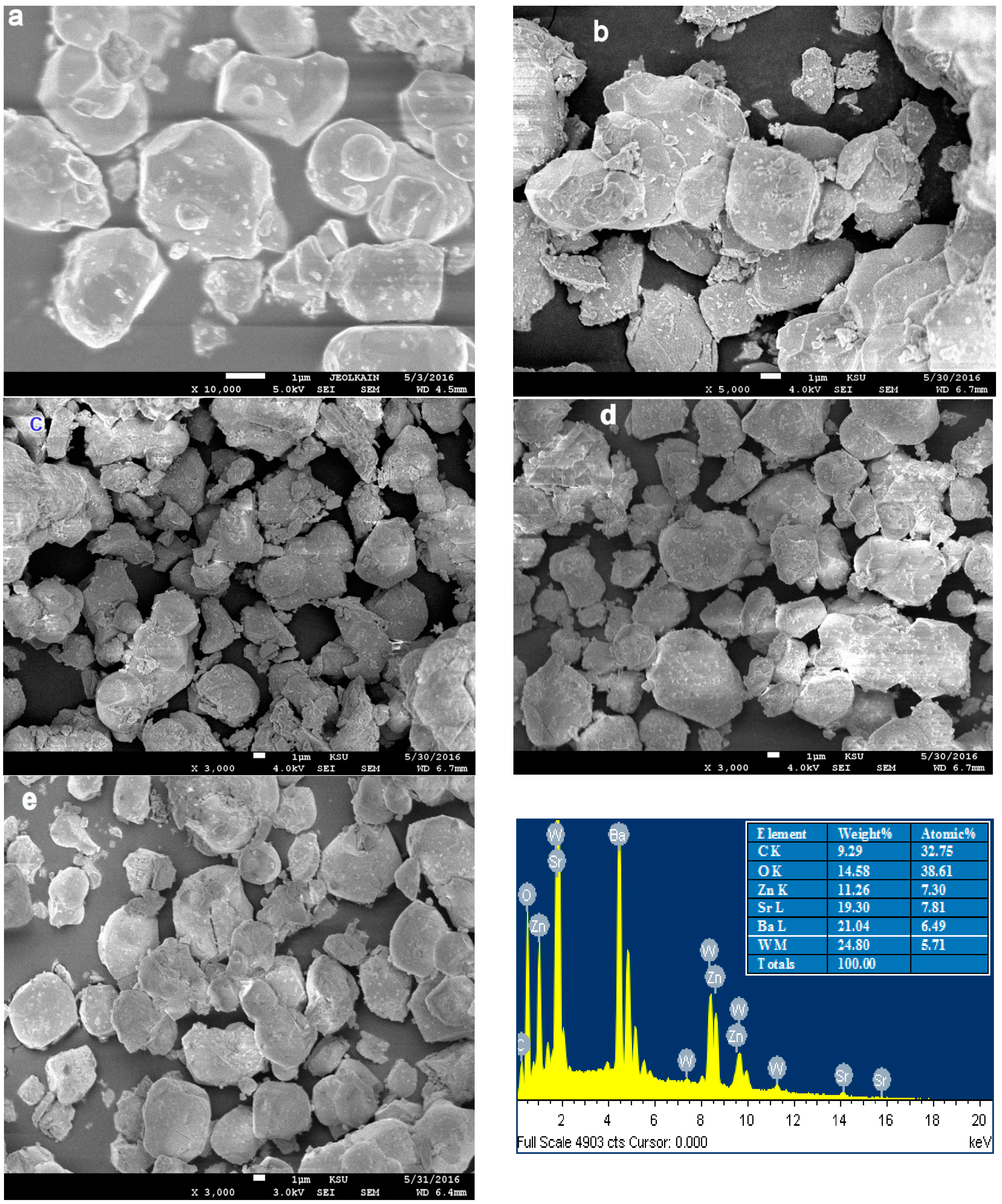
2.1.1. Scanning Electron Microscopy

The ESM images of the Ba2−xSrxZnWO6 (x = 0.00, 0.25, 0.50, 0.75, 1.00) series are displayed in Figure 1a–e. The morphologies of all samples are identical and they appear to be highly homogeneous with no impurities. It is observed that, in all samples, the size of the particles is large and they are aggregated in groups, which is due to the higher preparation temperature. Chunfeng Lan et al. [2] observed an equivalent effect of temperature in the morphology of La2NiMnO6 double perovskite oxide. Furthermore, the images reveal the presence of fine fragments that are produced during the preparation grinding. Each of the samples has grains of various sizes, i.e., Ba2ZnWO6 has 1–3 μm grains, Ba1.75Sr0.25ZnWO6 has 1–5 μm grains, Ba1.5Sr0.5ZnWO6 has 1.5–4 μm grains, Ba1.25Sr0.75ZnWO6 has 2–8 μm grains, and BaSrZnWO6 has 2–7 μm grains. An Energy-dispersive X-ray spectroscopy (EDX) analysis is conducted with each sample using the SEM images. Figure 1e presents the energy dispersive X-ray spectrum from the element that formed the BaSrZnWO6 sample. All EDX graphs confirm that all samples contain elements of the raw material preparation composition and a proportion approximating the input quantities to configure each sample with a small error, which refers to the homogeneity and crystal purity.

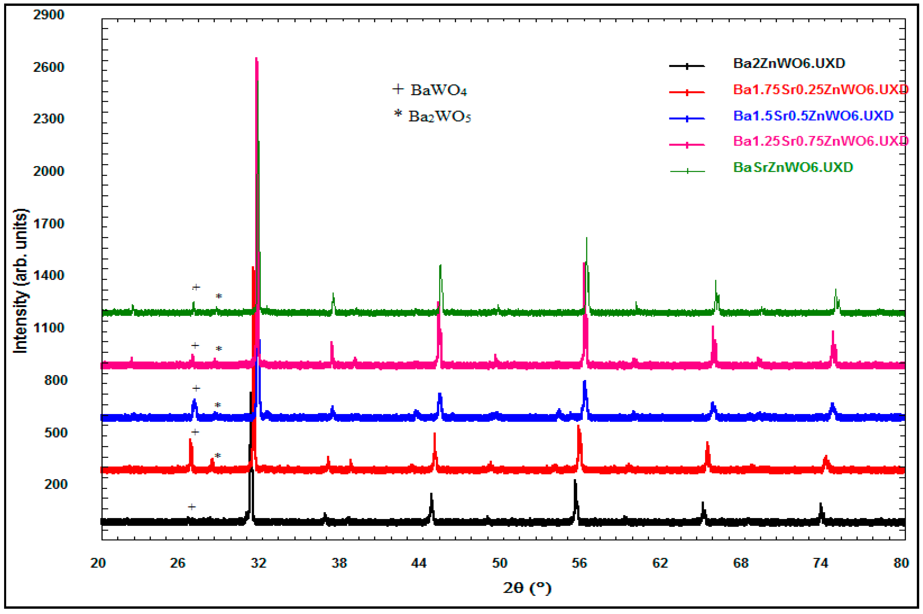
2.1.2. X-ray Powder Diffraction

The X-ray diffraction data of the perovskite oxide compounds are essential in determining the crystalline structure of the samples in terms of the Bravais lattice, atomic position, lattice parameter, and space group. Many studies refer to the importance of the study of material structure since they govern the other properties of the materials [17]. The XRD of the Ba2−xSrxZnWO6 (x = 0.00, 0.25, 0.50, 0.75, 1.00) double perovskite oxide series prepared by the solid-state reaction is shown in Figure 2. The BaWO4 and Ba2WO5 phases displayed as minor peaks at low intensity in the XRD pattern shown in Figure 1 are attributed to the impurities in the Ba2−xSrxZnWO6 (x = 0.00, 0.25, 0.50, 0.75, 1.00) structure around 26° and 28° [18]. A plus sign and a star are used to depict them in Figure 1 when 2 θ is around 26° and 28° for BaWO4 and Ba2WO5, respectively. The XRD data of each sample in the series are refined by the Rietveld method using the FullProf program. Table 1 displays the atom coordinates of all the samples obtained in the (Fm-3m) cubic crystal structure. Figure 3 shows the XRD refinement of BaSrZnWO6, which is represented by a (Fm-3m) cubic structure with lattice parameters a = b = c = 8.039361 Å and α = β = γ = 90°. Identical results are obtained for Ba2ZnWO6 using single-crystal X-ray and neutron diffraction [19]. Furthermore, the Ba2−xSrxMgTeO6 series was found in a (Fm-3m) cubic crystal structure [20]. The crystallite size was calculated from the Full width at half maximum (FWHMs) at the major peaks at (220) for the Ba2−xSrxZnWO6 (x = 0.00, 0.25, 0.50, 0.75, 1.00) double perovskite series using the Scherer equation [21], which was observed to vary between 47.41 and 105.9 nm for the samples.
D = 0.94   λ β cos θ ,
where D is the crystalline size, λ is the wavelength (1.5405 Å), β is the full width at half maximum, and θ is the diffraction angle. The tolerance factor was found to be between 0.998 and 1.007, which can be calculated by
t = ( 1 ( x 2 ) r A ` ) + x 2 r A ` ` + r o 2     ( r B ` 2 + r B ` ` 2 + r o )
Table 2 displays the tolerance factor and parameter of the crystal structure of Ba2−xSrxZnWO6 (x = 0.00, 0.25, 0.50, 0.75, 1.00) using the Rietveld method of refinement. The unit cell volume decreases with an increasing substitution as a result of the larger ionic radius of the Ba2+ cation than that of Sr2+.

2.2. Optical Studies

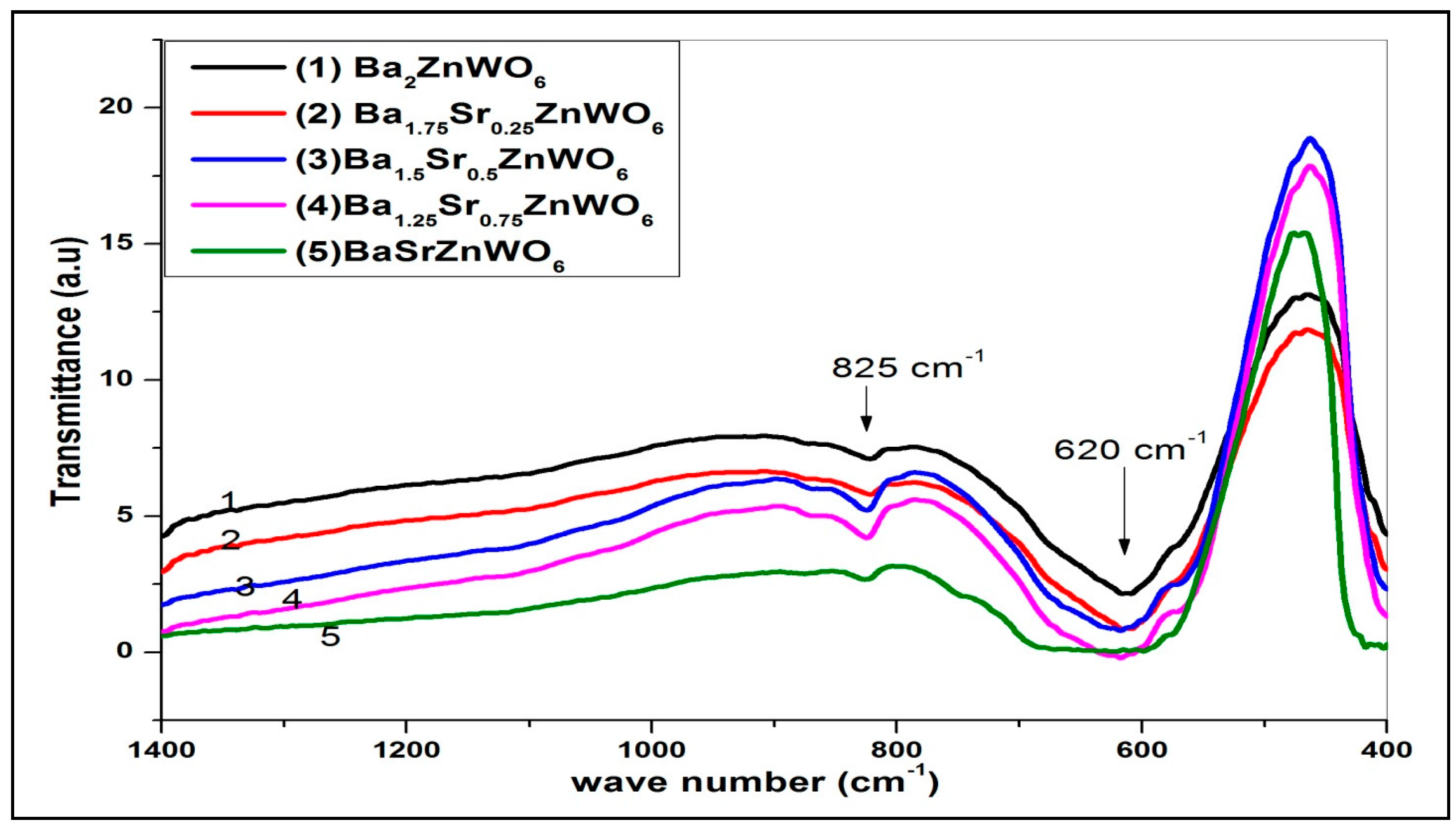
2.2.1. FTIR Spectroscopy

The FTIR spectra identify the crystal structure of the perovskite structure materials that have characteristic absorption bands in the 850–400 cm−1 region [22]. The strong high-energy anti-symmetric stretching mode of the WO6 octahedral displayed at 620 cm−1 is due to the higher charge of the tungsten cations. The symmetric stretching vibration of the WO6 octahedral appears as a high-intensity band at about 825 cm−1. Figure 4 shows the transmittance of the Ba2−xSrxZnWO6 double perovskite series versus wave number, and all the samples confirm the molecular bands on the perovskite oxide structure [17].

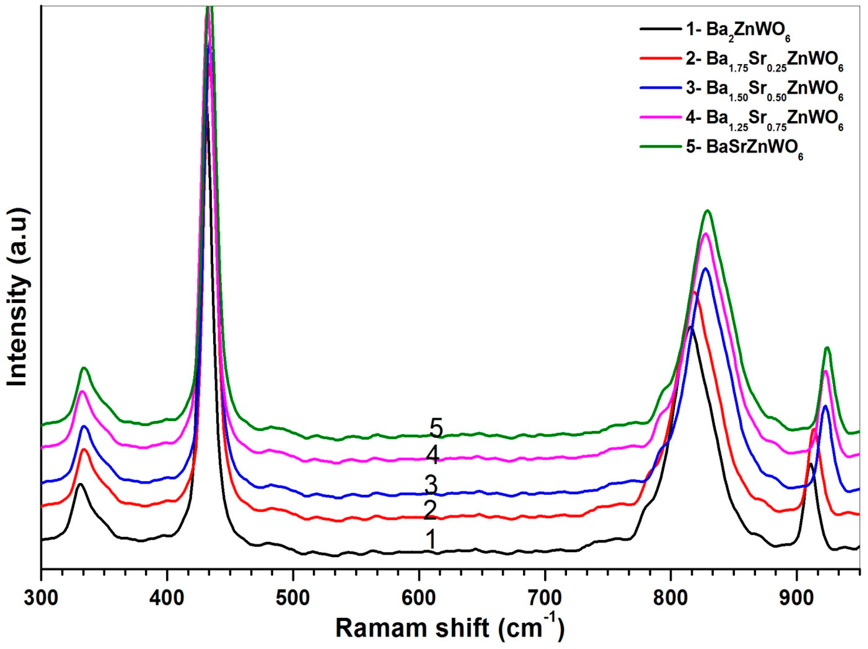
2.2.2. Raman Spectroscopy

The Raman spectra of the samples are shown in Figure 5 for Ba2−xSrxZnWO2 (x = 0.00, 0.25, 0.50, 0.75, 1.00). For all samples, the Raman modes are classified into two types of lattice vibration—the W–O–W bending vibration in the 200–500 cm−1 region and the W–O stretching mode between 700 and 950 cm−1. This result was observed in many studies on double perovskite oxides [22,23]. Ba has a larger ionic radius (149 Å) than that of Sr (132 Å). When the Ba substitution increases, the effect of the Ba radius is reflected by a decrease in both the bending and stretching modes of the W–O bonds. A redshift in the Raman energy is also observed.

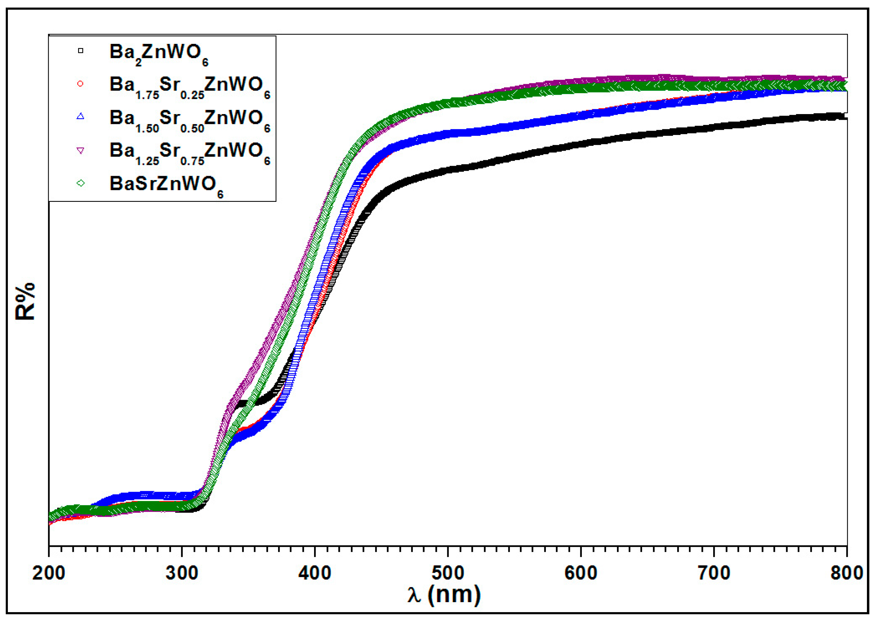
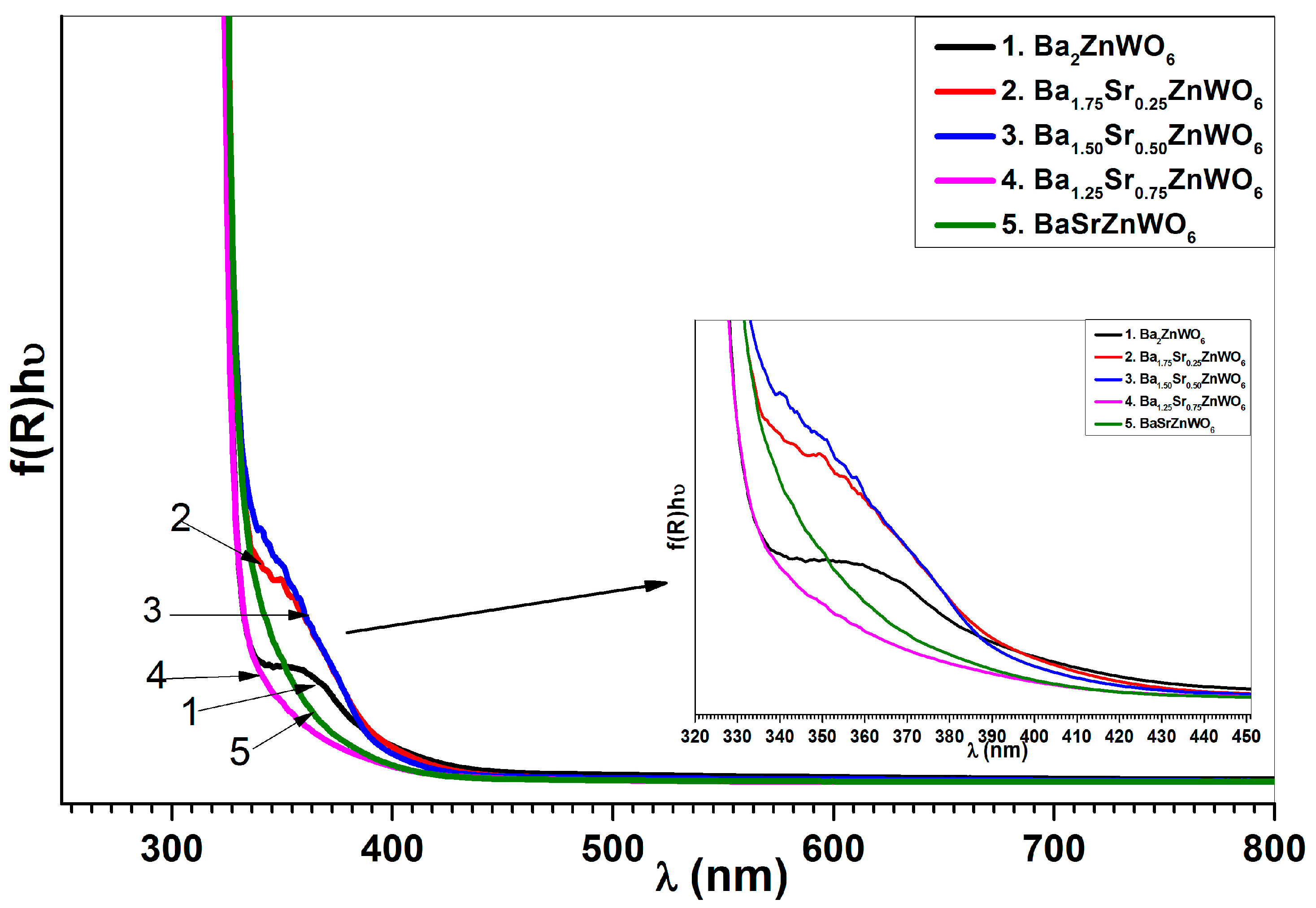
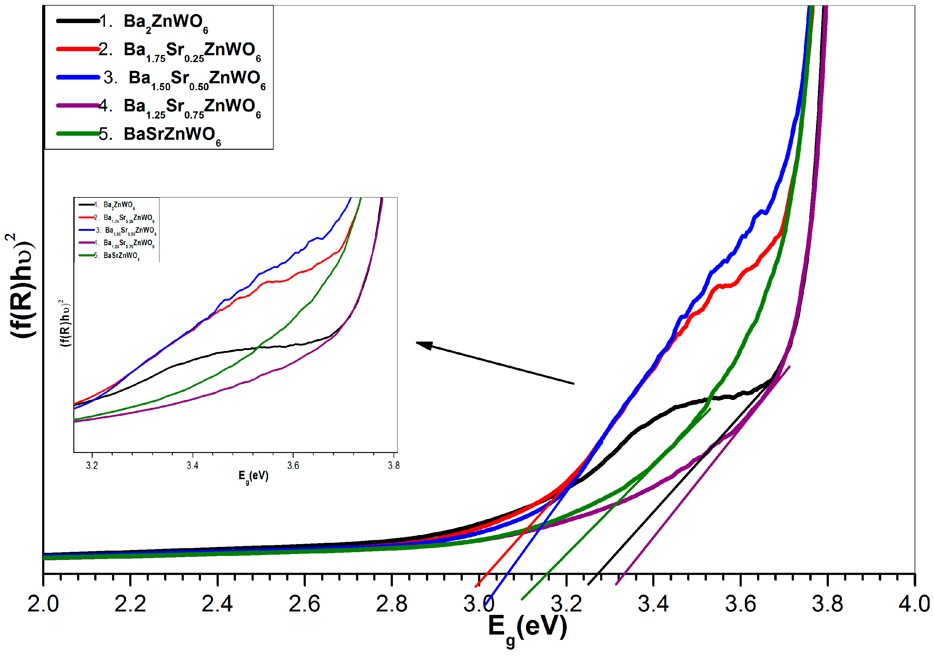
2.2.3. UV-Visible Diffuse Reflectance Spectroscopy

Figure 6 presents the diffuse reflectance spectrum of the Ba2−xSrxZnWO6 series at room temperature in the 200–800 nm range. The strong absorption band observed at 300–450 nm refers to the absorption edge in tungsten due to the charge transfer transition of W 6 + O 2 in the lattice from the highest filled molecular orbital 2p of oxygen to the lowest empty molecular orbital 5d of tungsten [14,19,24]. The absorption band has a blue shift with an increasing substitution ratio of Ba2+ with Sr2+ cations.
The absorption coefficient can be calculated for the Ba2−xSrxZnWO6 series from the diffuse reflectance data using the Kubelka–Munk function [25]:
F ( R ) = α s = ( 1 R ) 2 2 R
where F ( R ) is the KM function, α is the absorption coefficient, s is the scattering coefficient, and R is the reflection coefficient. The absorbance (f(R)) in relation to wavelength is shown in Figure 7. The absorbance can be used to observe the absorption edge for the samples that have values of 380, 410, 405, 370, and 389 nm for Ba2ZnWO6, Ba1.75Sr0.25ZnWO6, Ba1.5Sr5ZnWO6, Ba1.25Sr0.75ZnWO6, and BaSrZnWO6, respectively. The band gap energy of the series samples was calculated from the absorption edge [19] according to the relationship Eg = 1240/λ (λ is the absorption edge wavelength and Eg is the band gap [26]). In addition, the band gap energy was calculated for the samples using the Tauc plot [18], as shown in Figure 8, according to Equation (4).
( F ( R ) h ν ) ) n = A ( h ν E g )
where h ν is the incident photon energy, A is a proportional constant, E g is the band gap energy, and n takes values of 2 or 0.5 for indirect and direct transitions, respectively. Table 3 illustrates the bang gap energy according to the absorption edge and Tauc plot. The results of UV–vis diffuse reflectance and the optical energy gap of the sample series indicate that they can be classified as semiconductor materials [19,27].
In the case of complete substitution of Ba2+ with Sr2+, the material of Sr2ZnWO6 has a monoclinic (P21/n) crystal structure [28,29] with 3.8 eV band gap energy [29].

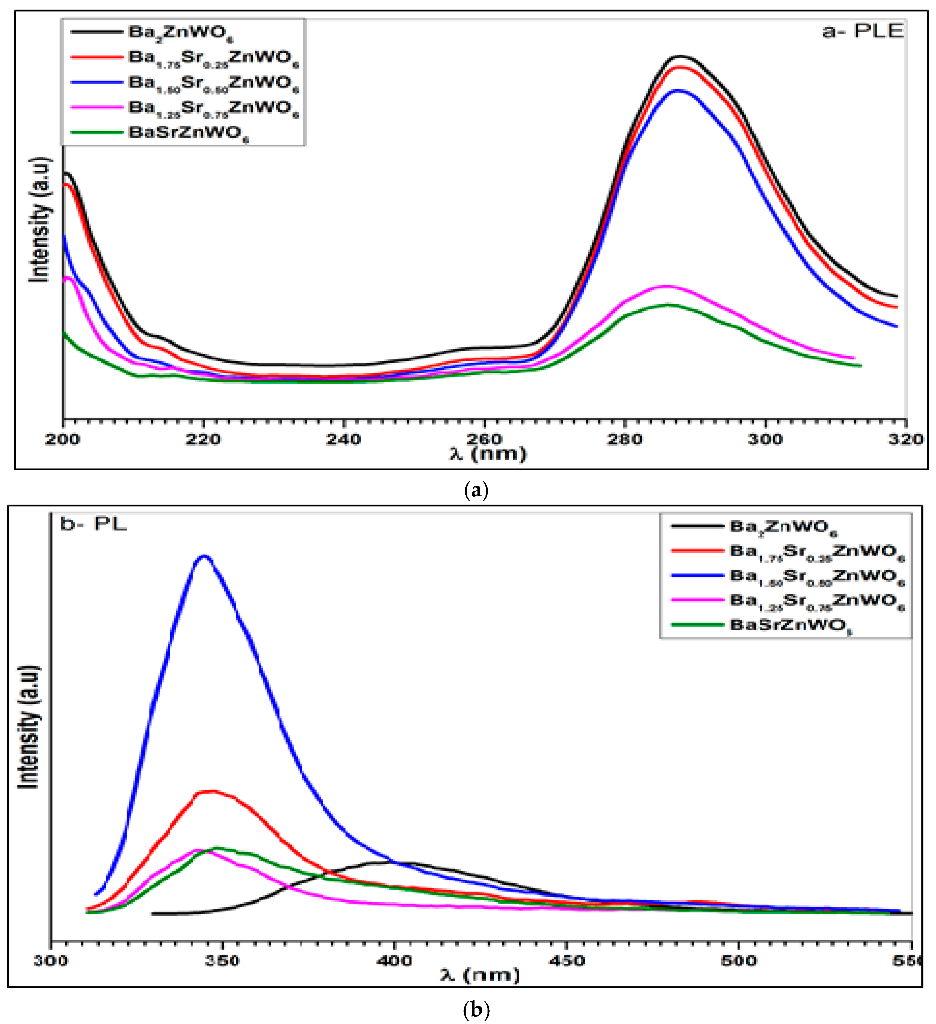
2.2.4. Photoluminescence Spectroscopy

Figure 9 shows the excitation and photoluminescence emission spectra of the Ba2−xSrxZnWO6 (x = 0.00, 0.25, 0.50, 0.75, 1.00) double perovskite oxide series. The excitation spectra shown in Figure 9a were collected when λem = 380 nm for Ba2ZnWO6, λem = 346 nm for Ba1.75Sr0.25ZnWO6, λem = 344 nm for Ba1.5Sr0.5ZnWO6, λem = 343 nm for Ba1.25Sr0.75ZnWO6, and λem = 349 nm for BaSrZnWO6. A broad band was observed between 260 and 320 nm, resulting from the electronic excitation of the O (2p) orbital-W (5d) orbital in octahedral WO6 [14,28]. In addition, the excitation peaks of the samples decrease with the increase in the Ba2+ ratio substitution of Sr2+ cations. The photoluminescence emission of the samples was investigated at λex = 290 nm for Ba2ZnWO6, λex = 288 nm for Ba1.75Sr0.25ZnWO6, λex = 287 nm for Ba1.5Sr0.5ZnWO6, and λex = 386 nm for Ba1.25Sr0.75ZnWO6 and BaSrZnWO6 displayed a spectral emission spread between 320 and 450 nm. Bugaris et al. [24] found a complimentary result where the emission peak of Ba2ZnWO6 displays a maximum at 539 nm when λex = 380 nm. In addition, there is a decrease in excitation intensity peaks with an increase in substitution. The photoluminescence (PL) of Ba2ZnWO6 has an emission spectrum peak at 380 nm and a FWHM of 70 nm. Similarly, Ba1.75Sr0.25ZnWO6, Ba1.5Sr0.5ZnWO6, Ba1.25Sr0.75ZnWO6, and BaSrZnWO6 have peaks at 345, 344, 343, and 342 nm with FWHMs of 40, 40, 40, and 50 nm, respectively. From the peaks of photoluminescence emission, the indirect band gap energy was calculated using the E = hc/λ equation for the series that was found to be 3.26, 3.5, 3.6, 3.6 and 3.65 eV for Ba2ZnWO6 Ba1.75Sr0.25ZnWO6, Ba1.5Sr0.5ZnWO6, Ba1.25Sr0.75ZnWO6, and BaSrZnWO6, respectively [30].

3. Materials and Methods

3.1. Samples Preparation

Ba2−xSrxZnWO6 (where x = 0.00, 0.25, 0.50, 0.75, 1.00) was synthesized using the solid-state interaction method from BaCO3 (barium carbonite), WO3 (tungsten trioxide), ZnO (zinc oxide), and NiO (nickel oxide) powders mixed in stoichiometric proportions according to the following chemical equation.
( 2 x ) BaCO 3 + x SrCO 3 + ZnO + WO 3   Ba 2 x Sr x ZnWO 6 + 2 CO 2
Initially, the materials were used as purchased from Alfa Acer with a purity of 99.99%. Several different treatments of the samples were conducted to achieve the crystal structure. The raw materials were mixed and ground in an agate mortar with acetone, kept in crucibles, and subsequently heated in air at 800 °C for 12 h twice. The sample pellets were prepared in a round shape and heated at 1000 °C twice, following which the same procedure was repeated at 1200 °C. Between the steps for the heating treatment, the sample was ground for 2 h with acetone to increase the homogeneity of the sample at a rate of 10 °C per minute during the heating and cooling process.

3.2. Sample Characterization

A Jeol JSM-6360 (JEOL Inc. Peabody, MA, USA) and high-resolution Stereo Scan LEO 440 SEM (LEO, Austin, TX, USA) were used to investigate the morphology and determine the homogeneity of the samples, as well as to obtain crystal-scale crystallization. At room temperature, the XRD data were recorded with a Bruker: D8 Advance (Bruker-Axs, Madison, WI, USA) using CuKα radiation (λ = 1.5406 Å) with a nickel filter. At 40 kV and 40 mA, data were collected for 2θ at 0.02-step sizes and 5-s count times in the 20°–80° range. The XRD data were analyzed using the Rietveld refinement method with the FullProf Suite program [31]. The crystalline size (D) [32] was calculated using the Debi–Scherer equation for all samples. At room temperature, the transmittance mode was investigated for all samples using the Satellite FTIR 5000 (ARCoptix S.A, Neuchatel, Switzerland) (with a wavelength range of 400–4000 cm−1) [33], where the important bands and peaks of the perovskite structure can be assigned. Using FTIR spectroscopy collected using the KBr pellet method, the material was mixed with KBr at 1:100 ratios for the FTIR measurement in the range of 400–2000 cm−1. The Raman spectra were collected in Raman HR (Stellarnet Inc., Tampa, FL, USA), using the high resolution Raman spectrometer with a range of 200–2200 cm−1 at 785 nm with a resolution of 4 cm−1. The Raman probe attaches to the laser via FC/APC and the spectrometer via SMA 905, and has integrated Raman filters and optics with a working distance to the sample of 4.5 mm, configured for the 785 nm laser. A UV–Vis spectrophotometer (UV-2550, Shimadzu, Chiyoda-ku, Tokyo) using BaSO4 as a reference is used to calculate the UV–Vis diffuse reflectance spectrum at room temperature. In addition, the UV–Vis reflectance spectrum is converted to absorbance using the Kubella–Munk method to estimate the edge of absorption and band gap of the Ba2−xSrxZnWO6 double perovskite powder series. A Perkin Elmer LS55 fluorescence spectrometer (Perkin-Elmer, Wokingham, UK) was used to investigate the emission and excitation of the Ba2−xSrxZnWO6 double perovskite series at room temperature.

4. Conclusions

The Ba2−xSrxZnWO6 (x = 0.00, 0.25, 0.50, 0.75, 1.00) double perovskite series was prepared by the solid-state reaction technique. In addition, techniques such as X-ray diffraction, scanning electronic microscopy, Fourier transform infrared spectra, Raman spectra, UV-Vis diffuse reflectance, photoluminescence excitation, and emission spectra were investigated. SEM revealed that the prepared Ba2−xSrxZnWO6 samples crystallized at micrometer scales, where the crystal structure of the samples was determined by XRD as a cubic Fm-3m space group. The lattice parameter decreased with an increase in the proportion of substitution from x = 0 to 1, where the result of FTIR also confirmed the double perovskite structure. The Raman spectra of W–O bonds indicated a systematic decrease and re-shifting with an increasing Ba substitution. Strong UV-Vis absorption was found between 350 and 410 nm and smaller optical bandgap energy, 3.02 eV, was found for Ba1.75Sr0.25ZnWO6 compared to the Sr-free sample. The excitation photoluminescence spectra displayed broad bands between 260 and 320 nm, which were assigned to the charge transfer band of Ba2−xSrxZnWO6. Ba2−xSrxZnWO6 displays photoluminescence in the near-UV and visible region. This feature makes Ba2−xSrxZnWO6 a potential semiconductor in optoelectronics applications.

Acknowledgments

The authors are grateful to the Deanship of Scientific Research, King Saud University for funding through Vice Deanship of Scientific Research Chairs.

Author Contributions

Y.A.A. and M.S.A., involved designing of the work, data analysis and interpretation. A.A.E. contributed data analysis and writing of the manuscript. E.M.M. helped to improve the quality of the manuscript.

Conflicts of Interest

The authors declare no conflict of interest.

References

  1. Zaraq, A.; Orayech, B.; Faik, A.; Igartua, J.; Jouanneaux, A.; El Bouari, A. High temperature induced phase transitions in SrCaCoTeO6 and SrCaNiTeO6 ordered double perovskites. Polyhedron 2016, 110, 119–124. [Google Scholar] [CrossRef]
  2. Lan, C.; Zhao, S.; Xu, T.; Ma, J.; Hayase, S.; Ma, T. Investigation on structures, band gaps, and electronic structures of lead free La2NiMnO6 double perovskite materials for potential application of solar cell. J. Alloys Compd. 2016, 655, 208–214. [Google Scholar] [CrossRef]
  3. Paiva, D.; Silva, M.; Sombra, A.; Fechine, P. Dielectric investigation of the Sr3WO6 double perovskite at RF/microwave frequencies. RSC Adv. 2016, 6, 42502–42509. [Google Scholar] [CrossRef]
  4. Xie, H.D.; Chen, C.; Xi, H.H.; Tian, R.; Wang, X.C. Synthesis, low temperature co-firing, and microwave dielectric properties of two ceramics BiM2VO6 (M = Cu, Ca). Ceram. Int. 2016, 42, 989–995. [Google Scholar] [CrossRef]
  5. Orlandi, F.; Righi, L.; Mezzadri, F.; Manuel, P.; Khalyavin, D.D.; Delmonte, D.; Pernechele, C.; Cabassi, R.; Bolzoni, F.; Solzi, M.; et al. Improper Ferroelectric Contributions in the Double Perovskite Pb2Mn0.6Co0.4WO6 System with a Collinear Magnetic Structure. Inorg. Chem. 2016, 55, 4381–4390. [Google Scholar] [CrossRef] [PubMed]
  6. Shimakawa, Y.; Azuma, M.; Ichikawa, N. Multiferroic compounds with double-perovskite structures. Materials 2011, 4, 153–168. [Google Scholar] [CrossRef]
  7. DeMarco, M.; Blackstead, H.; Dow, J.D.; Wu, M.; Chen, D.Y.; Chen, D.F.; Haka, M.; Steve, T.; Joel, F. Magnetic phase transition in superconducting Sr2YRu0.95Cu0.05O6 observed by the 99 Ru Mössbauer effect. Phys. Rev. B 2000, 62, 14301–14303. [Google Scholar] [CrossRef]
  8. Harnagea, L.; Jurca, B.; Berthet, P. Low-field magnetoresistance up to 400 K in double perovskite Sr2FeMoO6 synthesized by a citrate route. J. Solid State Chem. 2014, 211, 219–226. [Google Scholar] [CrossRef]
  9. Gorodea, I.; Goanta, M.; Toma, M. Impact of A cation size of double perovskite A2AlTaO6 (A = Ca, Sr, Ba) on dielectric and catalytic properties. J. Alloys Compd. 2015, 632, 805–809. [Google Scholar] [CrossRef]
  10. Djerdj, I.; Popović, J.; Mal, S.; Weller, T.; Nuskol, M.; Jagličić, Z.S.; Damir, P.; Christian, S.; Pascal, V.; Roland, M.; et al. Aqueous Sol–Gel Route toward Selected Quaternary Metal Oxides with Single and Double Perovskite-Type Structure Containing Tellurium. Cryst. Growth Des. 2016, 16, 2535–2541. [Google Scholar] [CrossRef]
  11. Wang, Z.L.; Kang, Z.C. Perovskite and Related Structure Systems. In Functional and Smart Materials: Functional and Smart Materials; Springer: New York, NY, USA, 1998; Volume 3, pp. 93–149. [Google Scholar]
  12. Iranmanesh, M.; Lingg, M.; Stir, M.; Hulliger, J. Sol gel and ceramic synthesis of Sr2FeMo1−xWxO6 (0 ≤ x ≤ 1) double perovskites series. RSC Adv. 2016, 6, 42069–42075. [Google Scholar] [CrossRef]
  13. Wu, J.Y.; Bian, J.J. Structure stability and microwave dielectric properties of double perovskite ceramics Ba2Mg1−xCaxWO6 (0.0 ≤ x ≤ 0.15). Ceram. Int. 2012, 38, 3217–3225. [Google Scholar] [CrossRef]
  14. Xu, D.; Yang, Z.; Sun, J.; Gao, X.; Du, J. Synthesis and luminescence properties of double-perovskite white emitting phosphor Ca3WO6:Dy3+. J. Mater. Sci. Mater. Electron. 2016, 27, 8370–8377. [Google Scholar] [CrossRef]
  15. Tian, S.; Zhao, J.; Qiao, C.; Ji, X.; Jiang, B. Structure and properties of the ordered double perovskites Sr2MWO6 (M = Co, Ni) by sol-gel route. Mater. Lett. 2006, 60, 2747–2750. [Google Scholar] [CrossRef]
  16. Xia, Z.; Sun, J.; Du, H.; Chen, D.; Sun, J. Luminescence properties of double-perovskite Sr2Ca1−2xEuxNaxMoO6 red-emitting phosphors prepared by the citric acid-assisted sol–gel method. J. Mater. Sci. 2010, 45, 1553–1559. [Google Scholar] [CrossRef]
  17. Elbadawi, A.A.; Yassin, O.; Siddig, M.A. Effect of the Cation Size Disorder at the A-Site on the Structural Properties of SrAFeTiO6 Double Perovskites (A = La, Pr or Nd). J. Mater. Sci. Chem. Eng. 2015, 3, 21–29. [Google Scholar]
  18. Manoun, B.; Ezzahi, A.; Benmokhtar, S.; Ider, A.; Lazor, P.; Bih, L.; Igartuae, J.M. X-ray diffraction and Raman spectroscopy studies of temperature and composition induced phase transitions in Ba2−xSrxZnWO6 (0 ≤ x ≤ 2) double perovskite oxides. J. Alloys Compd. 2012, 533, 43–52. [Google Scholar] [CrossRef]
  19. Bugaris, D.E.; Hodges, J.P.; Huq, A.; Zur-Loye, H.C. Crystal growth, structures, and optical properties of the cubic double perovskites Ba2MgWO6 and Ba2ZnWO6. J. Solid State Chem. 2011, 184, 2293–2298. [Google Scholar] [CrossRef]
  20. Tamraoui, Y.; Manoun, B.; Mirinioui, F.; Haloui, R.; Lazor, P. X-ray diffraction and Raman spectroscopy studies of temperature and composition induced phase transitions in Ba2−xSrxMgTeO6 (0 ≤ x ≤ 2). J. Alloys Compd. 2014, 603, 86–94. [Google Scholar] [CrossRef]
  21. Mostafa, M.F.; Ata-Allah, S.S.; Youssef, A.A.A.; Refai, H.S. Electric and AC magnetic investigation of the manganites La0.7Ca0.3Mn0.96In0.04xAl(1−x)0.04O3; (0.0 ≤ x ≤ 1.0). J. Magn. Magn. Mater. 2008, 320, 344–353. [Google Scholar] [CrossRef]
  22. Liegeois-Duyckaerts, M.; Tarte, P. Vibrational studies of molybdates, tungstates and related c compounds—III. Ordered cubic perovskites A2BIIBVIO6. Spectrochim. Acta Part A Mol. Spectrosc. 1974, 30, 1771–1786. [Google Scholar] [CrossRef]
  23. Baldinozzi, G.; Sciau, P.; Bulou, A. Raman study of the structural phase transition in the ordered perovskite Pb2MgWO6. J. Phys. Condens. Matter 1995, 7, 8109. [Google Scholar] [CrossRef]
  24. Xiao, N.; Shen, J.; Xiao, T.; Wu, B.; Luo, X.; Li, L.; Wang, Z.; zhou, X. Sr2CaWxMo1−xO6:Eu3+, Li+: An emission tunable phosphor through site symmetry and excitation wavelength. Mater. Res. Bull. 2015, 70, 684–690. [Google Scholar] [CrossRef]
  25. Dutta, A.; Mukhopadhyay, P.; Sinha, T.; Shannigrahi, S.; Himanshu, A.; Sen, P.; Bandyopadhyay, S.K. Sr2SmNbO6 perovskite: Synthesis, characterization and density functional theory calculations. Mater. Chem. Phys. 2016, 179, 55–64. [Google Scholar] [CrossRef]
  26. Chen, F.; Niu, C.; Yang, Q.; Li, X.; Zeng, G. Facile synthesis of visible-light-active BiOI modified Bi2MoO6 photocatalysts with highly enhanced photocatalytic activity. Ceram. Int. 2016, 42, 2515–2525. [Google Scholar] [CrossRef]
  27. Kittel, C.; Holcomb, D.F. Introduction to solid state physics. Am. J. Phys. 1967, 35, 547–548. [Google Scholar] [CrossRef]
  28. Manoun, B.; Igartua, J.M.; Lazor, P.; Ezzahi, A. High temperature induced phase transitions in Sr2ZnWO6 and Sr2CoWO6 double perovskite oxides: Raman spectroscopy as a tool. J. Mol. Struct. 2012, 1029, 81–85. [Google Scholar] [CrossRef]
  29. Eng, H.W. The Crystal and Electronic Structures of Oxides Containing d0 Transition Metals in Octahedral Coordination. Ph.D. Thesis, The Ohio State University, Columbus, OH, USA, 2003. [Google Scholar]
  30. Klein, P.; Nwagwu, U.; Edgar, J.H.; Freitas, J.A., Jr. Photoluminescence investigation of the indirect band gap and shallow impurities in icosahedral B12As2. J. Appl. Phys. 2012, 112, 013508. [Google Scholar] [CrossRef]
  31. Suryanarayana, C.; Norton, M.G. X-rays and Diffraction. In X-ray Diffraction; Springer: New York, NY, USA, 1998; Volume 1, pp. 3–19. [Google Scholar]
  32. Alsabah, Y.A.; Elbadawi, A.A.; Mustafa, E.M.; Siddig, M.A. The Effect of Replacement of Zn2+ Cation with Ni2+ Cation on the Structural Properties of Ba2Zn1−xNixWO6 Double Perovskite Oxides (X = 0, 0.25, 0.50, 0.75, 1). J. Mater. Sci. Chem. Eng. 2016, 4, 61–70. [Google Scholar]
  33. Kavitha, V.T.; Jose, R.; Ramakrishna, S.; Wariar, P.R.S.; Koshy, J. Combustion synthesis and characterization of Ba2NdSbO6 nanocrystalsm. Bull. Mater. Sci. 2011, 34, 661–665. [Google Scholar] [CrossRef]
Figure 1. (a) SEM image of Ba2ZnWO6; (b) SEM image of Ba1.75Sr0.25ZnWO6; (c) SEM image of Ba1.5Sr0.5ZnWO6; (d) SEM image of Ba1.25Sr0.75ZnWO6; (e) SEM image and EDX spectroscopy of BaSrZnWO6.
Materials 10 00469 g001
Figure 2. X-ray powder diffraction of Ba2−xSrxZnWO6 series.
Materials 10 00469 g002
Figure 3. Refined XRD patterns of the BaSrZnWO6.
Materials 10 00469 g003
Figure 4. The Fourier transform infrared (FTIR) spectra of the Ba2−xSrxZnWO6 double perovskite series.
Materials 10 00469 g004
Figure 5. The Raman shift spectra of the Ba2−xSrxZnWO6 double perovskite series.
Materials 10 00469 g005
Figure 6. The diffuse reflection spectrum for Ba2−xSrxZnWO6 (x = 0.00, 0.25, 0.5, 0.75, and 1.00) double perovskite series.
Materials 10 00469 g006
Figure 7. Absorbance versus the wavelength of Ba2−xSrxZnWO6 series.
Materials 10 00469 g007
Figure 8. Indirect band gap plot for Ba2−xSrxZnWO6 (x = 0.00, 0.25, 0.5, 0.75, and 1.00) series.
Materials 10 00469 g008
Figure 9. Photoluminescence excitation (a) and emission (b) spectrum of Ba2−xSrxZnWO6 series.
Materials 10 00469 g009
Table 1. Atom coordinates of Ba2−xSrxZnWO6 (x = 0.00, 0.25, 0.50, 0.75, 1.00) double perovskite series following Rietveld refinement of X-ray powder diffraction.
Cation/AnionCoordinatesBa2ZnWO6Ba1.75Sr0.25ZnWO6Ba1.5Sr0.5ZnWO6Ba1.25Sr0.75ZnWO6BaSrZnWO6
Ba+2X0.25000.25000.25000.25000.2500
Y0.25000.25000.25000.25000.2500
Z0.25000.25000.25000.25000.2500
Sr+2 ---------0.25000.25000.25000.2500
---------0.25000.25000.25000.2500
---------0.25000.25000.25000.2500
Zn+2X0.5000.5000.5000.5000.500
Y0.5000.5000.5000.5000.500
Z0.5000.5000.5000.5000.500
W+6X0.000.000.000.000.00
Y0.000.000.000.000.00
Z0.000.000.000.000.00
O1−2/O2−2X0.239900.258150.244140.225560.24414
Y00000
Z00000
Table 2. The tolerance factor and the parameter of crystal structure of Ba2−xSrxZnWO6 (x = 0.00, 0.25, 0.50, 0.75, 1.00) following Rietveld method refinement.
Empirical FormulaBa2ZnWO6Ba1.75Sr0.25ZnWO6Ba1.5Sr0.5ZnWO6Ba1.25Sr0.75ZnWO6BaSrZnWO6
Space groupFm-3mFm-3mFm-3mFm-3mFm-3m
α (Å)8.118348.1006798.0768698.0608428.039361
α/β/γ9090909090
V3)535.0590531.57465526.9001523.7707519.5751
D (nm)105.0978.3847.4188.0384.59
T1.0071.000.9920.9900.998
RWP9.789.718.8713.611.3
RP8.7510.28.1510.310.8
χ 2 1.77381.8861.6812.8132.356
Table 3. Illustration of band gap energy according to the absorption edge and Tauc plot for direct transition.
Bang Gap EnergyBa2ZnWO6Ba1.75Sr0.25ZnWO6Ba1.5Sr0.5ZnWO6Ba1.25Sr0.75ZnWO6BaSrZnWO6
Cut-off wavelength (nm)380410405370389
Eg (eV) by cutoff wavelength3.263.023.063.353.18
Eg (eV) by Tauc plot3.273.023.073.343.16

