Nanoheterostructures (NHS) and Their Applications in Nanomedicine: Focusing on In Vivo Studies


Abstract
:1. Introduction
2. Diagnostic Imaging in Nanomedicine
2.1. Bioimaging Techniques
2.1.1. Magnetic Resonance Imaging
- (i)
- Presence of two magnetic domains that cooperate to enhance the particle efficacy (increase in r2 value). This approach is generally typical of alloys for single-domain particles, but core@shell heterostructures based on two different magnetic materials have also been successfully prepared.
- (ii)
- Presence of magnetic and metallic domains: their combination leads to higher MRI performance.
- (iii)
- Presence of domains fulfilling different tasks: the magnetic ones serve for MRI, whereas the second material (metallic or semiconductor) accomplishes a distinct function. In the latter case, the system would be multifunctional; there is, however, the risk that the second domain may lead to a decrease of the MRI performance, owing to interferences with the magnetic field.
2.1.2. Computed Tomography
2.1.3. Luminescence Imaging
2.1.4. Photoacoustic and Photothermal Imaging
2.2. Nanoheterostructures for Diagnostic Imaging
2.2.1. Magnetic/Metallic Nanoheterostructures
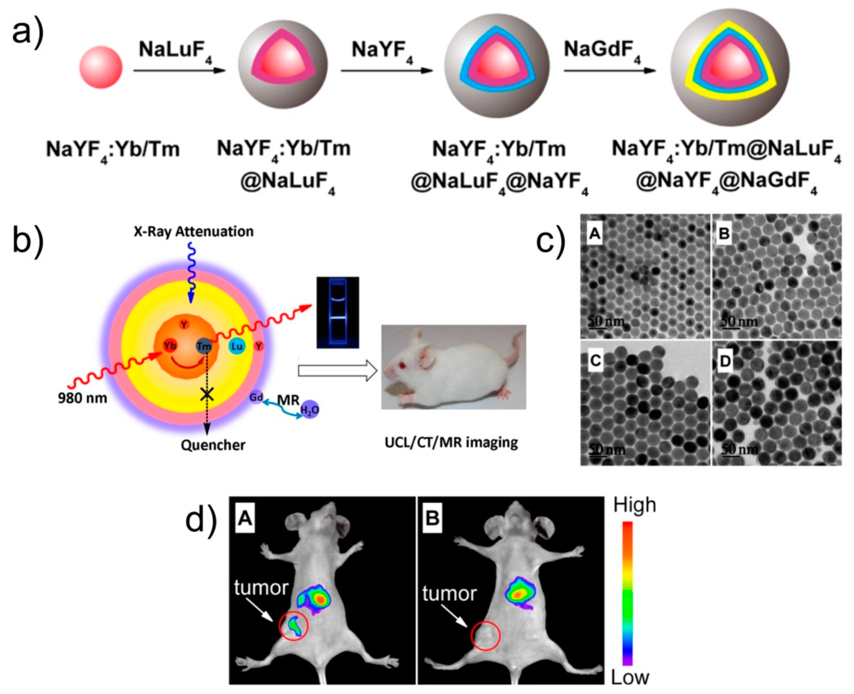
2.2.2. Luminescent Nanoheterostructures
3. Therapy and Theranostics in Nanomedicine
3.1. Therapeutic Techniques
3.2. Nanoheterostructures for Therapy and Theranostics
4. Pharmacokinetics of the Nanoheterostructures
5. Discussion
6. Conclusions
Funding
Acknowledgments
Conflicts of Interest
Abbreviations
| AC | Alternated magnetic field |
| ADME | Absorption, distribution, metabolism, and excretion |
| CT | Computed Tomography |
| FDA | Food and Drug Administration |
| FITC | Fluorescein IsoThioCyanate |
| IONP | Iron Oxide NanoParticle |
| IL | Interleukin |
| LRP | Lipoprotein Receptor related Protein |
| mAb | Monoclonal AntiBody |
| MRI | Magnetic Resonance Imaging |
| NHS | Nanoheterostructures |
| NIR | Near-InfraRed |
| NP | NanoParticle |
| QD | Quantum Dot |
| PA | PhotoAcoustic |
| PDT | PhotoDynamic Therapy |
| PEG | Poly(Ethylene Glycol) |
| PET | Positron Emission Tomography |
| PT | PhotoThermal |
| PTT | PhotoThermal Therapy |
| ROS | Reactive Oxygen Species |
| SAR | Specific Absorption Rate |
| SPECT | Single-Photon Emission Computed Tomography |
| UC | UpConversion |
| UCL | UpConversion Luminescence |
| UV | UltraViolet |
| 8-OHdG | 8-hydroxy-20-deoxysuanosine |
References
- Cuenca, A.G.; Jiang, H.B.; Hochwald, S.N.; Delano, M.; Cance, W.G.; Grobmyer, S.R. Emerging Implications of Nanotechnology on Cancer Diagnostics and Therapeutics. Cancer 2006, 107, 459–466. [Google Scholar] [CrossRef]
- Brigger, I.; Dubernet, C.; Couvreur, P. Nanoparticles in Cancer Therapy and Diagnosis. Adv. Drug Deliv. Rev. 2002, 54, 631–651. [Google Scholar] [CrossRef]
- Neoh, K.G.; Kang, E.T. Functionalization of Inorganic Nanoparticles with Polymers for Stealth Biomedical Applications. Polym. Chem. 2011, 2, 747–759. [Google Scholar] [CrossRef]
- Medintz, I.L.; Uyeda, H.T.; Goldman, E.R.; Mattoussi, H. Quantum Dot Bioconjugates for Imaging, Labelling and Sensing. Nat. Mater. 2005, 4, 435–446. [Google Scholar] [CrossRef] [PubMed]
- Di Corato, R.; Quarta, A.; Piacenza, P.; Ragusa, A.; Figuerola, A.; Buonsanti, R.; Cingolani, R.; Manna, L.; Pellegrino, T. Water Solubilization of Hydrophobic Nanocrystals by Means of Poly(Maleic Anhydride-Alt-1-Octadecene). J. Mater. Chem. 2008, 18, 1991–1996. [Google Scholar] [CrossRef]
- Lewinski, N.; Colvin, V.; Drezek, R. Cytotoxicity of Nanoparticles. Small 2008, 4, 26–49. [Google Scholar] [CrossRef]
- Arami, H.; Khandhar, A.; Liggitt, D.; Krishnan, K.M. In Vivo Delivery, Pharmacokinetics, Biodistribution and Toxicity of Iron Oxide Nanoparticles. Chem. Soc. Rev. 2015, 44, 8576–8607. [Google Scholar] [CrossRef]
- Kiessling, F.; Mertens, M.E.; Grimm, J.; Lammers, T. Nanoparticles for Imaging: Top or Flop? Radiology 2014, 273, 10–28. [Google Scholar] [CrossRef]
- Lartigue, L.; Alloyeau, D.; Kolosnjaj-Tabi, J.; Javed, Y.; Guardia, P.; Riedinger, A.; Péchoux, C.; Pellegrino, T.; Wilhelm, C.; Gazeau, F. Biodegradation of Iron Oxide Nanocubes: High-Resolution in Situ Monitoring. ACS Nano 2013, 7, 3939–3952. [Google Scholar] [CrossRef]
- Khanna, P.; Ong, C.; Bay, B.H.; Baeg, G.H. Nanotoxicity: An Interplay of Oxidative Stress, Inflammation and Cell Death. Nanomaterials 2015, 5, 1163–1180. [Google Scholar] [CrossRef] [Green Version]
- Sharifi, S.; Behzadi, S.; Laurent, S.; Laird Forrest, M.; Stroeve, P.; Mahmoudi, M. Toxicity of Nanomaterials. Chem. Soc. Rev. 2012, 41, 2323–2343. [Google Scholar] [CrossRef] [PubMed]
- Schladt, T.D.; Shukoor, M.I.; Schneider, K.; Tahir, M.N.; Natalio, F.; Ament, I.; Becker, J.; Jochum, F.D.; Weber, S.; Kohler, O.; et al. Au@MnO Nanoflowers: Hybrid Nanocomposites for Selective Dual Functionalization and Imaging. Angew. Chem.-Int. Ed. 2010, 49, 3976–3980. [Google Scholar] [CrossRef] [PubMed]
- Jiang, J.; Gu, H.W.; Shao, H.L.; Devlin, E.; Papaefthymiou, G.C.; Ying, J.Y. Manipulation Bifunctional Fe3O4-Ag Heterodimer Nanoparticles for Two-Photon Fluorescence Imaging and Magnetic Manipulation. Adv. Mater. 2008, 20, 4403–4407. [Google Scholar] [CrossRef]
- Xu, C.J.; Wang, B.D.; Sun, S.H. Dumbbell-Like Au-Fe3O4 Nanoparticles for Target-Specific Platin Delivery. J. Am. Chem. Soc. 2009, 131, 4216–4217. [Google Scholar] [CrossRef] [PubMed]
- Liu, J.; Zhang, W.; Zhang, H.L.; Yang, Z.Y.; Li, T.R.; Wang, B.D.; Huo, X.; Wang, R.; Chen, H.T. A Multifunctional Nanoprobe Based on Au-Fe3O4 Nanoparticles for Multimodal and Ultrasensitive Detection of Cancer Cells. Chem. Commun. 2013, 49, 4938–4940. [Google Scholar] [CrossRef] [PubMed]
- Wolfbeis, O.S. An Overview of Nanoparticles Commonly Used in Fluorescent Bioimaging. Chem. Soc. Rev. 2015, 44, 4743–4768. [Google Scholar] [CrossRef] [PubMed]
- Kobayashi, H.; Ogawa, M.; Alford, R.; Choyke, P.L.; Urano, Y. New Strategies for Fluorescent Probe Design in Medical Diagnostic Imaging. Chem. Rev. 2010, 110, 2620–2640. [Google Scholar] [CrossRef] [PubMed] [Green Version]
- Ros, P.R.; Freeny, P.C.; Harms, S.E.; Seltzer, S.E.; Davis, P.L.; Chan, T.W.; Stillman, A.E.; Muroff, L.R.; Runge, V.M.; Nissenbaum, M.A.; et al. Hepatic Mr-Imaging with Ferumoxides—A Multicenter Clinical-Trial of the Safety and Efficacy in the Detection of Focal Hepatic-Lesions. Radiology 1995, 196, 481–488. [Google Scholar] [CrossRef]
- Pasternak, J.J.; Williamson, E.E. Clinical Pharmacology, Uses, and Adverse Reactions of Iodinated Contrast Agents: A Primer for the Non-Radiologist. Mayo Clin. Proc. 2012, 87, 390–402. [Google Scholar] [CrossRef]
- Na, H.B.; Song, I.C.; Hyeon, T. Inorganic Nanoparticles for Mri Contrast Agents. Adv. Mater. 2009, 21, 2133–2148. [Google Scholar] [CrossRef]
- Mahmoudi, M.; Hosseinkhani, H.; Hosseinkhani, M.; Boutry, S.; Simchi, A.; Journeay, W.S.; Subramani, K.; Laurent, S. Magnetic Resonance Imaging Tracking of Stem Cells in Vivo Using Iron Oxide Nanoparticles as a Tool for the Advancement of Clinical Regenerative Medicine. Chem. Rev. 2010, 111, 253–280. [Google Scholar] [CrossRef] [PubMed]
- Laurent, S.; Forge, D.; Port, M.; Roch, A.; Robic, C.; Vander Elst, L.; Muller, R.N. Magnetic Iron Oxide Nanoparticles: Synthesis, Stabilization, Vectorization, Physicochemical Characterizations, and Biological Applications. Chem. Rev. 2008, 108, 2064–2110. [Google Scholar] [CrossRef] [PubMed]
- Cormode, D.P.; Naha, P.C.; Fayad, Z.A. Nanoparticle Contrast Agents for Computed Tomography: A Focus on Micelles. Contrast Media Mol. Imaging 2014, 9, 37–52. [Google Scholar] [CrossRef] [PubMed]
- Shilo, M.; Reuveni, T.; Motiei, M.; Popovtzer, R. Nanoparticles as Computed Tomography Contrast Agents: Current Status and Future Perspectives. Nanomedicine 2012, 7, 257–269. [Google Scholar] [CrossRef]
- Jakhmola, A.; Anton, N.; Vandamme, T.F. Inorganic Nanoparticles Based Contrast Agents for X-ray Computed Tomography. Adv. Healthc. Mater. 2012, 1, 413–431. [Google Scholar] [CrossRef]
- Frangioni, J.V. In Vivo near-Infrared Fluorescence Imaging. Curr. Opin. Chem. Biol. 2003, 7, 626–634. [Google Scholar] [CrossRef] [PubMed]
- Wegner, K.D.; Hildebrandt, N. Quantum Dots: Bright and Versatile in Vitro and in Vivo Fluorescence Imaging Biosensors. Chem. Soc. Rev. 2015, 44, 4792–4834. [Google Scholar] [CrossRef] [PubMed]
- Wang, Y.; Hu, R.; Lin, G.; Roy, I.; Yong, K.-T. Functionalized Quantum Dots for Biosensing and Bioimaging and Concerns on Toxicity. ACS Appl. Mater. Interfaces 2013, 5, 2786–2799. [Google Scholar] [CrossRef]
- Ho, Y.-P.; Leong, K.W. Quantum Dot-Based Theranostics. Nanoscale 2010, 2, 60–68. [Google Scholar] [CrossRef]
- Qiu, P.; Zhou, N.; Chen, H.; Zhang, C.; Gao, G.; Cui, D. Recent Advances in Lanthanide-Doped Upconversion Nanomaterials: Synthesis, Nanostructures and Surface Modification. Nanoscale 2013, 5, 11512–11525. [Google Scholar] [CrossRef]
- Park, Y.I.; Lee, K.T.; Suh, Y.D.; Hyeon, T. Upconverting Nanoparticles: A Versatile Platform for Wide-Field Two-Photon Microscopy and Multi-Modal in Vivo Imaging. Chem. Soc. Rev. 2015, 44, 1302–1317. [Google Scholar] [CrossRef] [PubMed]
- Eliseeva, S.V.; Bunzli, J.-C.G. Lanthanide Luminescence for Functional Materials and Bio-Sciences. Chem. Soc. Rev. 2010, 39, 189–227. [Google Scholar] [CrossRef] [PubMed]
- Wu, X.; Chen, G.; Shen, J.; Li, Z.; Zhang, Y.; Han, G. Upconversion Nanoparticles: A Versatile Solution to Multiscale Biological Imaging. Bioconjugate Chem. 2015, 26, 166–175. [Google Scholar] [CrossRef] [PubMed]
- Yu, J.; Nguyen, H.N.Y.; Steenbergen, W.; Kim, K. Recent Development of Technology and Application of Photoacoustic Molecular Imaging toward Clinical Translation. J. Nucl. Med. 2018, 59, 1202–1207. [Google Scholar] [CrossRef] [PubMed]
- Zeng, L.; Ma, G.; Lin, J.; Huang, P. Photoacoustic Probes for Molecular Detection: Recent Advances and Perspectives. Small 2018, 14, 1800782. [Google Scholar] [CrossRef] [PubMed]
- Anker, J.N.; Hall, W.P.; Lyandres, O.; Shah, N.C.; Zhao, J.; Van Duyne, R.P. Biosensing with Plasmonic Nanosensors. Nat. Mater. 2008, 7, 442–453. [Google Scholar] [CrossRef] [PubMed]
- Zharov, V.P.; Lapotko, D.O. Photothermal Imaging of Nanoparticles and Cells. IEEE J. Sel. Top. Quantum Electron. 2005, 11, 733–751. [Google Scholar] [CrossRef]
- Kumagai, M.; Sarma, T.K.; Cabral, H.; Kaida, S.; Sekino, M.; Herlambang, N.; Osada, K.; Kano, M.R.; Nishiyama, N.; Kataoka, K. Enhanced in Vivo Magnetic Resonance Imaging of Tumors by Pegylated Iron-Oxide-Gold Core-Shell Nanoparticles with Prolonged Blood Circulation Properties. Macromol. Rapid Commun. 2010, 31, 1521–1528. [Google Scholar] [CrossRef] [PubMed]
- Lee, N.; Cho, H.R.; Oh, M.H.; Lee, S.H.; Kim, K.; Kim, B.H.; Shin, K.; Ahn, T.-Y.; Choi, J.W.; Kim, Y.-W.; et al. Multifunctional Fe3O4/Taox Core/Shell Nanoparticles for Simultaneous Magnetic Resonance Imaging and X-Ray Computed Tomography. J. Am. Chem. Soc. 2012, 134, 10309–10312. [Google Scholar] [CrossRef]
- Ahn, S.H.; Lee, N.; Choi, C.; Shin, S.W.; Han, Y.; Park, H.C. Feasibility Study of Fe3O4/Taox Nanoparticles as a Radiosensitizer for Proton Therapy. Phys. Med. Biol. 2018, 63, 114001. [Google Scholar] [CrossRef]
- Li, F.F.; Zhi, D.B.; Luo, Y.F.; Zhang, J.Q.; Nan, X.; Zhang, Y.J.; Zhou, W.; Qiu, B.S.; Wen, L.P.; Liang, G.L. Core/Shell Fe3O4/Gd2O3 Nanocubes as T-1-T-2 Dual Modal Mri Contrast Agents. Nanoscale 2016, 8, 12826–12833. [Google Scholar] [CrossRef] [PubMed]
- Tang, W.; Zhen, Z.P.; Yang, C.; Wang, L.N.; Cowger, T.; Chen, H.M.; Todd, T.; Hekmatyar, K.; Zhao, Q.; Hou, Y.L.; et al. Fe5 C2 Nanoparticles with High Mri Contrast Enhancement for Tumor Imaging. Small 2014, 10, 1245–1249. [Google Scholar] [CrossRef]
- Yang, M.; Cheng, K.; Qi, S.B.; Liu, H.G.; Jiang, Y.X.; Jiang, H.; Li, J.B.; Chen, K.; Zhang, H.M.; Cheng, Z. Affibody Modified and Radiolabeled Gold-Iron Oxide Hetero-Nanostructures for Tumor Pet, Optical and Mr Imaging. Biomaterials 2013, 34, 2796–2806. [Google Scholar] [CrossRef] [PubMed]
- Zhu, J.; Zhang, B.; Tian, J.; Wang, J.Q.; Chong, Y.; Wang, X.; Deng, Y.Y.; Tang, M.H.; Li, Y.G.; Ge, C.C.; et al. Synthesis of Heterodimer Radionuclide Nanoparticles for Magnetic Resonance and Single-Photon Emission Computed Tomography Dual-Modality Imaging. Nanoscale 2015, 7, 3392–3395. [Google Scholar] [CrossRef] [PubMed]
- Zhu, J.; Lu, Y.J.; Li, Y.G.; Jiang, J.; Cheng, L.; Liu, Z.; Guo, L.; Pan, Y.; Gu, H.W. Synthesis of Au-Fe3O4 Heterostructured Nanoparticles for in Vivo Computed Tomography and Magnetic Resonance Dual Model Imaging. Nanoscale 2014, 6, 199–202. [Google Scholar] [CrossRef] [PubMed]
- Efremova, M.V.; Naumenko, V.A.; Spasova, M.; Garanina, A.S.; Abakumov, M.A.; Blokhina, A.D.; Melnikov, P.A.; Prelovskaya, A.O.; Heidelmann, M.; Li, Z.A.; et al. Magnetite-Gold Nanohybrids as Ideal All-in-One Platforms for Theranostics. Sci. Rep. 2018, 8, 11295. [Google Scholar] [CrossRef] [PubMed]
- Cheng, K.; Yang, M.; Zhang, R.P.; Qin, C.X.; Su, X.H.; Cheng, Z. Hybrid Nanotrimers for Dual T-1 and T-2-Weighted Magnetic Resonance Imaging. ACS Nano 2014, 8, 9884–9896. [Google Scholar] [CrossRef]
- Kim, D.; Yu, M.K.; Lee, T.S.; Park, J.J.; Jeong, Y.Y.; Jon, S. Amphiphilic Polymer-Coated Hybrid Nanoparticles as Ct/Mri Dual Contrast Agents. Nanotechnology 2011, 22, 155101. [Google Scholar] [CrossRef] [PubMed]
- Xie, J.; Zhang, F.; Aronova, M.; Zhu, L.; Lin, X.; Quan, Q.M.; Liu, G.; Zhang, G.F.; Choi, K.Y.; Kim, K.; et al. Manipulating the Power of an Additional Phase: A Flower-Like Au-Fe3O4 Optical Nanosensor for Imaging Protease Expressions in Vivo. ACS Nano 2011, 5, 3043–3051. [Google Scholar] [CrossRef]
- Sun, X.L.; Huang, X.L.; Guo, J.X.; Zhu, W.L.; Ding, Y.; Niu, G.; Wang, A.; Kiesewetter, D.O.; Wang, Z.L.; Sun, S.H.; et al. Self-Illuminating Cu-64-Doped CdSe/ZnS Nanocrystals for in Vivo Tumor Imaging. J. Am. Chem. Soc. 2014, 136, 1706–1709. [Google Scholar] [CrossRef]
- Guo, W.; Sun, X.; Jacobson, O.; Yan, X.; Min, K.; Srivatsan, A.; Niu, G.; Kiesewetter, D.O.; Chang, J.; Chen, X. Intrinsically Radioactive [64Cu]Cuins/Zns Quantum Dots for Pet and Optical Imaging: Improved Radiochemical Stability and Controllable Cerenkov Luminescence. ACS Nano 2015, 9, 488–495. [Google Scholar] [CrossRef] [PubMed]
- Xia, A.; Gao, Y.; Zhou, J.; Li, C.; Yang, T.; Wu, D.; Wu, L.; Li, F. Core–Shell NaYF4:Yb3+,Tm3+@Fexoy Nanocrystals for Dual-Modality T2-Enhanced Magnetic Resonance and Nir-to-Nir Upconversion Luminescent Imaging of Small-Animal Lymphatic Node. Biomaterials 2011, 32, 7200–7208. [Google Scholar] [CrossRef] [PubMed]
- Zhu, X.; Zhou, J.; Chen, M.; Shi, M.; Feng, W.; Li, F. Core–Shell Fe3O4@Naluf4:Yb,Er/Tm Nanostructure for Mri, Ct and Upconversion Luminescence Tri-Modality Imaging. Biomaterials 2012, 33, 4618–4627. [Google Scholar] [CrossRef] [PubMed]
- Shen, S.; Guo, X.; Wu, L.; Wang, M.; Wang, X.; Kong, F.; Shen, H.; Xie, M.; Ge, Y.; Jin, Y. Dual-Core@Shell-Structured Fe3O4-NaYF4@TiO2 Nanocomposites as a Magnetic Targeting Drug Carrier for Bioimaging and Combined Chemo-Sonodynamic Therapy. J. Mater. Chem. B 2014, 2, 5775–5784. [Google Scholar] [CrossRef]
- Park, Y.I.; Kim, H.M.; Kim, J.H.; Moon, K.C.; Yoo, B.; Lee, K.T.; Lee, N.; Choi, Y.; Park, W.; Ling, D.; et al. Theranostic Probe Based on Lanthanide-Doped Nanoparticles for Simultaneous in Vivo Dual-Modal Imaging and Photodynamic Therapy. Adv. Mater. 2012, 24, 5755–5761. [Google Scholar] [CrossRef] [PubMed]
- Zeng, L.; Xiang, L.; Ren, W.; Zheng, J.; Li, T.; Chen, B.; Zhang, J.; Mao, C.; Li, A.; Wu, A. Multifunctional Photosensitizer-Conjugated Core-Shell Fe3O4@NaYF4:Yb/Er Nanocomplexes and Their Applications in T2-Weighted Magnetic Resonance/Upconversion Luminescence Imaging and Photodynamic Therapy of Cancer Cells. RSC Adv. 2013, 3, 13915–13925. [Google Scholar] [CrossRef]
- Ni, D.L.; Zhang, J.W.; Bu, W.B.; Xing, H.Y.; Han, F.; Xiao, Q.F.; Yao, Z.W.; Chen, F.; He, Q.J.; Liu, J.N.; et al. Dual-Targeting Upconversion Nanoprobes across the Blood-Brain Barrier for Magnetic Resonance/Fluorescence Imaging of Intracranial Glioblastoma. ACS Nano 2014, 8, 1231–1242. [Google Scholar] [CrossRef] [PubMed]
- Wang, Z.; Zhang, P.; Yuan, Q.H.; Xu, X.; Lei, P.P.; Liu, X.L.; Su, Y.; Dong, L.L.; Feng, J.; Zhang, H.J. Nd3+-Sensitized Naluf4 Luminescent Nanoparticles for Multimodal Imaging and Temperature Sensing under 808 Nm Excitation. Nanoscale 2015, 7, 17861–17870. [Google Scholar] [CrossRef]
- Shen, J.W.; Yang, C.X.; Dong, L.X.; Sun, H.R.; Gao, K.; Yan, X.P. Incorporation of Computed Tomography and Magnetic Resonance Imaging Function into NaYF4:Yb/Tm Upconversion Nanoparticles for in Vivo Trimodal Bioimaging. Anal. Chem. 2013, 85, 12166–12172. [Google Scholar] [CrossRef]
- Sun, Y.; Zhu, X.J.; Peng, J.J.; Li, F.Y. Core-Shell Lanthanide Upconversion Nanophosphors as Four-Modal Probes for Tumor Angiogenesis Imaging. ACS Nano 2013, 7, 11290–11300. [Google Scholar] [CrossRef]
- Zhou, T.; Wu, B.Y.; Xing, D. Bio-Modified Fe3O4 Core/Au Shell Nanoparticles for Targeting and Multimodal Imaging of Cancer Cells. J. Mater. Chem. 2012, 22, 470–477. [Google Scholar] [CrossRef]
- Gao, J.; Zhang, W.; Huang, P.; Zhang, B.; Zhang, X.; Xu, B. Intracellular Spatial Control of Fluorescent Magnetic Nanoparticles. J. Am. Chem. Soc. 2008, 130, 3710–3711. [Google Scholar] [CrossRef] [PubMed]
- Wang, S.; Jarrett, B.R.; Kauzlarich, S.M.; Louie, A.Y. Core/Shell Quantum Dots with High Relaxivity and Photoluminescence for Multimodality Imaging. J. Am. Chem. Soc. 2007, 129, 3848–3856. [Google Scholar] [CrossRef]
- Resch-Genger, U.; Grabolle, M.; Cavaliere-Jaricot, S.; Nitschke, R.; Nann, T. Quantum Dots Versus Organic Dyes as Fluorescent Labels. Nat. Meth. 2008, 5, 763–775. [Google Scholar] [CrossRef] [PubMed]
- Jamieson, T.; Bakhshi, R.; Petrova, D.; Pocock, R.; Imani, M.; Seifalian, A.M. Biological Applications of Quantum Dots. Biomaterials 2007, 28, 4717–4732. [Google Scholar] [CrossRef] [PubMed]
- Guo, W.; Yang, W.; Wang, Y.; Sun, X.; Liu, Z.; Zhang, B.; Chang, J.; Chen, X. Color-Tunable Gd-Zn-Cu-in-S/ZnS Quantum Dots for Dual Modality Magnetic Resonance and Fluorescence Imaging. Nano Res. 2014, 7, 1581–1591. [Google Scholar] [CrossRef] [PubMed]
- Cui, S.; Yin, D.; Chen, Y.; Di, Y.; Chen, H.; Ma, Y.; Achilefu, S.; Gu, Y. In Vivo Targeted Deep-Tissue Photodynamic Therapy Based on near-Infrared Light Triggered Upconversion Nanoconstruct. ACS Nano 2013, 7, 676–688. [Google Scholar] [CrossRef]
- Zeng, L.; Luo, L.; Pan, Y.; Luo, S.; Lu, G.; Wu, A. In Vivo Targeted Magnetic Resonance Imaging and Visualized Photodynamic Therapy in Deep-Tissue Cancers Using Folic Acid-Functionalized Superparamagnetic-Upconversion Nanocomposites. Nanoscale 2015, 7, 8946–8954. [Google Scholar] [CrossRef]
- Harada, Y.; Ogawa, K.; Irie, Y.; Endo, H.; Feril, L.B., Jr.; Uemura, T.; Tachibana, K. Ultrasound Activation of TiO2 in Melanoma Tumors. J. Control. Release 2011, 149, 190–195. [Google Scholar] [CrossRef]
- Nel, A.E.; Madler, L.; Velegol, D.; Xia, T.; Hoek, E.M.V.; Somasundaran, P.; Klaessig, F.; Castranova, V.; Thompson, M. Understanding Biophysicochemical Interactions at the Nano-Bio Interface. Nat. Mater. 2009, 8, 543–557. [Google Scholar] [CrossRef]
- Chithrani, B.D.; Chan, W.C.W. Elucidating the Mechanism of Cellular Uptake and Removal of Protein-Coated Gold Nanoparticles of Different Sizes and Shapes. Nano Lett. 2007, 7, 1542–1550. [Google Scholar] [CrossRef]
- Delehanty, J.B.; Mattoussi, H.; Medintz, I.L. Delivering Quantum Dots into Cells: Strategies, Progress and Remaining Issues. Anal. Bioanal. Chem. 2008, 393, 1091–1105. [Google Scholar] [CrossRef] [PubMed]
- Walkey, C.D.; Olsen, J.B.; Guo, H.; Emili, A.; Chan, W.C.W. Nanoparticle Size and Surface Chemistry Determine Serum Protein Adsorption and Macrophage Uptake. J. Am. Chem. Soc. 2012, 134, 2139–2147. [Google Scholar] [CrossRef] [PubMed]
- Monopoli, M.P.; Aberg, C.; Salvati, A.; Dawson, K.A. Biomolecular Coronas Provide the Biological Identity of Nanosized Materials. Nat. Nano 2012, 7, 779–786. [Google Scholar] [CrossRef] [PubMed]
- Wang, B.; He, X.; Zhang, Z.; Zhao, Y.; Feng, W. Metabolism of Nanomaterials in Vivo: Blood Circulation and Organ Clearance. Acc. Chem. Res. 2013, 46, 761–769. [Google Scholar] [CrossRef]
- Kuang, X.-Y.; Liu, H.; Hu, W.-Y.; Shao, Y.-Z. Hydrothermal Synthesis of Core-Shell Structured TbPO4:Ce3+@TbPO4:Gd3+ Nanocomposites for Magnetic Resonance and Optical Imaging. Dalton Trans. 2014, 43, 12321–12328. [Google Scholar] [CrossRef]
- Fan, W.P.; Shen, B.; Bu, W.B.; Chen, F.; Zhao, K.L.; Zhang, S.J.; Zhou, L.P.; Peng, W.J.; Xiao, Q.F.; Xing, H.Y.; et al. Rattle-Structured Multifunctional Nanotheranostics for Synergetic Chemo-/Radiotherapy and Simultaneous Magnetic/Luminescent Dual-Mode Imaging. J. Am. Chem. Soc. 2013, 135, 6494–6503. [Google Scholar] [CrossRef]
- Hu, J.S.; Zhan, S.P.; Wu, X.F.; Hu, S.G.; Wu, S.B.; Liu, Y.X. Core/Shell Upconversion Nanoparticles with Intense Fluorescence for Detecting Doxorubicin in Vivo. RSC Adv. 2018, 8, 21505–21512. [Google Scholar] [CrossRef]
- Johannsen, M.; Gneueckow, U.; Thiesen, B.; Taymoorian, K.; Cho, C.H.; Waldofner, N.; Scholz, R.; Jordan, A.; Loening, S.A.; Wust, P. Thermotherapy of Prostate Cancer Using Magnetic Nanoparticles: Feasibility, Imaging, and Three-Dimensional Temperature Distribution. Eur. Urol. 2007, 52, 1653–1662. [Google Scholar] [CrossRef]
- van Landeghem, F.K.H.; Maier-Hauff, K.; Jordan, A.; Hoffmann, K.T.; Gneveckow, U.; Scholz, R.; Thiesen, B.; Bruck, W.; von Deimling, A. Post-Mortem Studies in Glioblastoma Patients Treated with Thermotherapy Using Magnetic Nanoparticles. Biomaterials 2009, 30, 52–57. [Google Scholar] [CrossRef]
- Mornet, S.; Vasseur, S.; Grasset, F.; Duguet, E. Magnetic Nanoparticle Design for Medical Diagnosis and Therapy. J. Mater. Chem. 2004, 14, 2161–2175. [Google Scholar] [CrossRef]
- Roti Roti, J.L. Cellular Responses to Hyperthermia (40–46 °C): Cell Killing and Molecular Events. Int. J. Hyperth. 2008, 24, 3–15. [Google Scholar] [CrossRef]
- Hergt, R.; Dutz, S.; Müller, R.; Zeisberger, M. Magnetic Particle Hyperthermia: Nanoparticle Magnetism and Materials Development for Cancer Therapy. J. Phys. Condens. Matter 2006, 18, S2919–S2934. [Google Scholar] [CrossRef]
- Dolmans, D.; Fukumura, D.; Jain, R.K. Photodynamic Therapy for Cancer. Nat. Rev. Cancer 2003, 3, 380–387. [Google Scholar] [CrossRef] [PubMed]
- Huang, X.; El-Sayed, M.A. Plasmonic Photo-Thermal Therapy (Pptt). Alex. J. Med. 2011, 47, 1–9. [Google Scholar] [CrossRef]
- Jaque, D.; Martinez Maestro, L.; del Rosal, B.; Haro-Gonzalez, P.; Benayas, A.; Plaza, J.L.; Martin Rodriguez, E.; Garcia Sole, J. Nanoparticles for Photothermal Therapies. Nanoscale 2014, 6, 9494–9530. [Google Scholar] [CrossRef]
- Wang, J.; Zhou, Z.G.; Wang, L.; Wei, J.; Yang, H.; Yang, S.P.; Zhao, J.M. CoFe2O4@MnFe2O4/Polypyrrole Nanocomposites for in Vitro Photothermal/Magnetothermal Combined Therapy. RSC Adv. 2015, 5, 7349–7355. [Google Scholar] [CrossRef]
- Liu, Y.M.; Yang, K.; Cheng, L.; Zhu, J.; Ma, X.X.; Xu, H.; Li, Y.G.; Guo, L.; Gu, H.W.; Liu, Z. Pegylated FePt@Fe2O3 Core-Shell Magnetic Nanoparticles: Potential Theranostic Applications and in Vivo Toxicity Studies. Nanomed.-Nanotechnol. Biol. Med. 2013, 9, 1077–1088. [Google Scholar] [CrossRef]
- Zhou, Z.G.; Sun, Y.A.; Shen, J.C.; Wei, J.; Yu, C.; Kong, B.; Liu, W.; Yang, H.; Yang, S.P.; Wang, W. Iron/Iron Oxide Core/Shell Nanoparticles for Magnetic Targeting Mri and near-Infrared Photothermal Therapy. Biomaterials 2014, 35, 7470–7478. [Google Scholar] [CrossRef]
- Wang, M.F.; Deng, K.R.; Lu, W.; Deng, X.R.; Li, K.; Shi, Y.S.; Ding, B.B.; Cheng, Z.Y.; Xing, B.G.; Han, G.; et al. Rational Design of Multifunctional Fe@Gamma-Fe2O3@ H-TiO2 Nanocomposites with Enhanced Magnetic and Photoconversion Effects for Wide Applications: From Photocatalysis to Imaging-Guided Photothermal Cancer Therapy. Adv. Mater. 2018, 30, 1706747. [Google Scholar] [CrossRef]
- Fan, Z.; Shelton, M.; Singh, A.K.; Senapati, D.; Khan, S.A.; Ray, P.C. Multifunctional Plasmonic Shell-Magnetic Core Nanoparticles for Targeted Diagnostics, Isolation, and Photothermal Destruction of Tumor Cells. ACS Nano 2012, 6, 1065–1073. [Google Scholar] [CrossRef]
- Ju, Y.M.; Zhang, H.L.; Yu, J.; Tong, S.Y.; Tian, N.; Wang, Z.Y.; Wang, X.B.; Su, X.T.; Chu, X.; Lin, J.; et al. Monodisperse Au-Fe2c Janus Nanoparticles: An Attractive Multifunctional Material for Triple-Modal Imaging-Guided Tumor Photothermal Therapy. ACS Nano 2017, 11, 9239–9248. [Google Scholar] [CrossRef] [PubMed]
- Hu, Y.; Wang, R.Z.; Wang, S.G.; Ding, L.; Li, J.C.; Luo, Y.; Wang, X.L.; Shen, M.W.; Shi, X.Y. Multifunctional Fe3O4 @ Au Core/Shell Nanostars: A Unique Platform for Multimode Imaging and Photothermal Therapy of Tumors. Sci. Rep. 2016, 6, 28325. [Google Scholar] [CrossRef] [PubMed]
- Tian, Q.W.; Hu, J.Q.; Zhu, Y.H.; Zou, R.J.; Chen, Z.G.; Yang, S.P.; Li, R.W.; Su, Q.Q.; Han, Y.; Liu, X.G. Sub-10 Nm Fe3O4@Cu2-Xs Core-Shell Nanoparticles for Dual-Modal Imaging and Photothermal Therapy. J. Am. Chem. Soc. 2013, 135, 8571–8577. [Google Scholar] [CrossRef]
- Chen, M.; Tang, S.H.; Guo, Z.D.; Wang, X.Y.; Mo, S.G.; Huang, X.Q.; Liu, G.; Zheng, N.F. Core-Shell Pd@Au Nanoplates as Theranostic Agents for in-Vivo Photoacoustic Imaging, Ct Imaging, and Photothermal Therapy. Adv. Mater. 2014, 26, 8210–8216. [Google Scholar] [CrossRef] [PubMed]
- Li, J.; Wang, W.J.; Zhao, L.; Rong, L.; Lan, S.J.; Sun, H.C.; Zhang, H.; Yang, B. Hydroquinone-Assisted Synthesis of Branched Au-Ag Nanoparticles with Polydopamine Coating as Highly Efficient Photothermal Agents. Acs Appl. Mater. Interfaces 2015, 7, 11613–11623. [Google Scholar] [CrossRef] [PubMed]
- Lv, R.C.; Yang, P.P.; Chen, G.Y.; Gai, S.L.; Xu, J.T.; Prasad, P.N. Dopamine-Mediated Photothermal Theranostics Combined with up-Conversion Platform under near Infrared Light. Sci. Rep. 2017, 7, 13562. [Google Scholar] [CrossRef] [PubMed]
- Li, J.C.; Hu, Y.; Yang, J.; Wei, P.; Sun, W.J.; Shen, M.W.; Zhang, G.X.; Shi, X.Y. Hyaluronic Acid-Modified Fe3O4@Au Core/Shell Nanostars for Multimodal Imaging and Photothermal Therapy of Tumors. Biomaterials 2015, 38, 10–21. [Google Scholar] [CrossRef] [PubMed]
- Chudasama, B.; Vala, A.; Andhariya, N.; Upadhyay, R.V.; Mehta, R.V. Enhanced Antibacterial Activity of Bifunctional Fe3O4-Ag Core-Shell Nanostructures. Nano Res. 2009, 2, 955–965. [Google Scholar] [CrossRef]
- Huo, D.; He, J.; Li, H.; Yu, H.P.; Shi, T.T.; Feng, Y.H.; Zhou, Z.Y.; Hu, Y. Fabrication of Au@Ag Core-Shell Nps as Enhanced Ct Contrast Agents with Broad Antibacterial Properties. Colloid Surf. B-Biointerfaces 2014, 117, 29–35. [Google Scholar] [CrossRef]
- Huo, D.; Ding, J.; Cui, Y.X.; Xia, L.Y.; Li, H.; He, J.; Zhou, Z.Y.; Wang, H.W.; Hu, Y. X-Ray Ct and Pneumonia Inhibition Properties of Gold-Silver Nanoparticles for Targeting Mrsa Induced Pneumonia. Biomaterials 2014, 35, 7032–7041. [Google Scholar] [CrossRef] [PubMed]
- Nikoobakht, B.; El-Sayed, M.A. Preparation and Growth Mechanism of Gold Nanorods (Nrs) Using Seed-Mediated Growth Method. Chem. Mater. 2003, 15, 1957–1962. [Google Scholar] [CrossRef]
- Hu, B.; Wang, N.; Han, L.; Chen, M.L.; Wang, J.H. Core-Shell-Shell Nanorods for Controlled Release of Silver That Can Serve as a Nanoheater for Photothermal Treatment on Bacteria. Acta Biomater. 2015, 11, 511–519. [Google Scholar] [CrossRef] [PubMed]
- Hamidi, M.; Azadi, A.; Rafiei, P.; Ashrafi, H. A Pharmacokinetic Overview of Nanotechnology-Based Drug Delivery Systems: An Adme-Oriented Approach. Crit. Rev. Ther. Drug Carr. Syst. 2013, 30, 435–467. [Google Scholar] [CrossRef]
- Aula, S.; Lakkireddy, S.; Jamil, K.; Kapley, A.; Swamy, A.V.N.; Lakkireddy, H.R. Biophysical, Biopharmaceutical and Toxicological Significance of Biomedical Nanoparticles. RSC Adv. 2015, 5, 47830–47859. [Google Scholar] [CrossRef]
- Owens, D.E.; Peppas, N.A. Opsonization, Biodistribution, and Pharmacokinetics of Polymeric Nanoparticles. Int. J. Pharm. 2006, 307, 93–102. [Google Scholar] [CrossRef] [PubMed]
- Li, M.; Zou, P.; Tyner, K.; Lee, S. Physiologically Based Pharmacokinetic (Pbpk) Modeling of Pharmaceutical Nanoparticles. AAPS J. 2017, 19, 26–42. [Google Scholar] [CrossRef]
- Montagne, A.; Toga, A.W.; Zlokovic, B.V. Blood-Brain Barrier Permeability and Gadolinium Benefits and Potential Pitfalls in Research. JAMA Neurol. 2016, 73, 13–14. [Google Scholar] [CrossRef]
- FDA Drug Safety Communication: FDA warns that gadolinium-based contrast agents (GBCAs) are retained in the body; requires new class warnings. Available online: www.fda.gov/Drugs/DrugSafety/ucm589213.htm (accessed on 20th December 2018).
- Di Corato, R.; Espinosa, A.; Lartigue, L.; Tharaud, M.; Chat, S.; Pellegrino, T.; Menager, C.; Gazeau, F.; Wilhelm, C. Magnetic Hyperthermia Efficiency in the Cellular Environment for Different Nanoparticle Designs. Biomaterials 2014, 35, 6400–6411. [Google Scholar] [CrossRef]
- Di Corato, R.; Béalle, G.; Kolosnjaj-Tabi, J.; Espinosa, A.; Clément, O.; Silva, A.K.A.; Ménager, C.; Wilhelm, C. Combining Magnetic Hyperthermia and Photodynamic Therapy for Tumor Ablation with Photoresponsive Magnetic Liposomes. ACS Nano 2015, 9, 2904–2916. [Google Scholar] [CrossRef]
- Espinosa, A.; Di Corato, R.; Kolosnjaj-Tabi, J.; Flaud, P.; Pellegrino, T.; Wilhelm, C. Duality of Iron Oxide Nanoparticles in Cancer Therapy: Amplification of Heating Efficiency by Magnetic Hyperthermia and Photothermal Bimodal Treatment. ACS Nano 2016, 10, 2436–2446. [Google Scholar] [CrossRef] [PubMed]
- Cheng, L.-C.; Jiang, X.; Wang, J.; Chen, C.; Liu, R.-S. Nano-Bio Effects: Interaction of Nanomaterials with Cells. Nanoscale 2013, 5, 3547–3569. [Google Scholar] [CrossRef] [PubMed]
- Manke, A.; Wang, L.; Rojanasakul, Y. Mechanisms of Nanoparticle-Induced Oxidative Stress and Toxicity. Biomed. Res. Int. 2013, 942916. [Google Scholar] [CrossRef]
- Gonzalez, L.; Lison, D.; Kirsch-Volders, M. Genotoxicity of Engineered Nanomaterials: A Critical Review. Nanotoxicology 2008, 2, 252–273. [Google Scholar] [CrossRef]
- Fu, P.P.; Xia, Q.; Hwang, H.-M.; Ray, P.C.; Yu, H. Mechanisms of Nanotoxicity: Generation of Reactive Oxygen Species. J. Food Drug Anal. 2014, 22, 64–75. [Google Scholar] [CrossRef] [PubMed]
- Zhu, X.; Hondroulis, E.; Liu, W.; Li, C.-Z. Biosensing Approaches for Rapid Genotoxicity and Cytotoxicity Assays Upon Nanomaterial Exposure. Small 2013, 9, 1821–1830. [Google Scholar] [CrossRef] [PubMed]
- Girgis, E.; Khalil, W.K.B.; Emam, A.N.; Mohamed, M.B.; Rao, K.V. Nanotoxicity of Gold and Gold–Cobalt Nanoalloy. Chem. Res. Toxicol. 2012, 25, 1086–1098. [Google Scholar] [CrossRef]
- Reidy, B.; Haase, A.; Luch, A.; Dawson, K.; Lynch, I. Mechanisms of Silver Nanoparticle Release, Transformation and Toxicity: A Critical Review of Current Knowledge and Recommendations for Future Studies and Applications. Materials 2013, 6, 2295. [Google Scholar] [CrossRef]
- Juzenas, P.; Chen, W.; Sun, Y.-P.; Coelho, M.A.N.; Generalov, R.; Generalova, N.; Christensen, I.L. Quantum Dots and Nanoparticles for Photodynamic and Radiation Therapies of Cancer. Adv. Drug Deliv. Rev. 2008, 60, 1600–1614. [Google Scholar] [CrossRef]
- Li, K.G.; Chen, J.T.; Bai, S.S.; Wen, X.; Song, S.Y.; Yu, Q.; Li, J.; Wang, Y.Q. Intracellular Oxidative Stress and Cadmium Ions Release Induce Cytotoxicity of Unmodified Cadmium Sulfide Quantum Dots. Toxicol. Vitr. 2009, 23, 1007–1013. [Google Scholar] [CrossRef]
- Gnach, A.; Lipinski, T.; Bednarkiewicz, A.; Rybka, J.; Capobianco, J.A. Upconverting Nanoparticles: Assessing the Toxicity. Chem. Soc. Rev. 2015, 44, 1561–1584. [Google Scholar] [CrossRef] [PubMed]
- Stern, S.T.; Adiseshaiah, P.P.; Crist, R.M. Autophagy and Lysosomal Dysfunction as Emerging Mechanisms of Nanomaterial Toxicity. Part. Fibre Toxicol. 2012, 9, 1–17. [Google Scholar] [CrossRef]
- Kroemer, G.; Jaattela, M. Lysosomes and Autophagy in Cell Death Control. Nat. Rev. Cancer 2005, 5, 886–897. [Google Scholar] [CrossRef]


| Heterostructure (Materials) | In Vivo Imaging Technique | Type | Average TEM Size (nm) | DLS/ Z-potential | Surface Coating | Targeting Moiety | Animal Model | Ref. | |||||
|---|---|---|---|---|---|---|---|---|---|---|---|---|---|
| MR | CT | PA | PL em. | SPECT | PET | ||||||||
| Magnetic/metallic nanoheterostructures | |||||||||||||
| Fe3O4@Au | x | Core/Shell | 12 | -/0.49 ± 0.12 mV | PEG | - | Mice bearing a subcutaneous C26 colon cancer | [38] | |||||
| Fe3O4@TaOx | x | x | Core/Shell | 10 | 21nm/- | RITC- functionalized silane and PEG-silane | - | Rats bearing xenograft tumors | [39,40] | ||||
| Fe3O4@Gd2O3 | x | Core/Shell | 9 | - | Dopamine | - | Sprague Dawley rats | [41] | |||||
| Fe5C2@Fe3O4 | x | Core/Shell | 23 | 35nm/- | DSPE-PEG-COOH | c(RGDyK) peptide | U87MG tumor bearing mice | [42] | |||||
| Au-Fe3O4 labelled with [64Cu] | x | x | Heterodimers | 17 | 24.4 ± 2 nm/ −28.6 ± 1.8mV | LPA-mPEG-2000/ LPA-PEG-2000-NOTA | Anti-EGFR affibody protein | Mice bearing EGFR positive A431 tumors | [43] | ||||
| Fe3O4-Ag125I | x | Heterodimers | 23 | - | mPEG-LA | - | Kunming mice | [44] | |||||
| Au-Fe3O4 | x | x | Heterodimers | 25 | - | TMAOH | - | Rabbits | [45] | ||||
| Fe3O4@Au | x | Core/Shell | 30 | 121 ± 5 nm/ −19.1 ± 3.3 mV | DSPE-PEG-COOH | - | 4T1-GFP bearing mice | [46] | |||||
| Au-Pt-IONP | x | x | x | Heterotrimer | 25 | 26.8 ± 0.9 nm/ –16.4 ± 1.2 mV | dopamine terminated PEG5k | - | HT-29 tumor bearing mice | [47] | |||
| Au@Fe3O4 | x | x | Flower-like | 25 | 30.4 ± 8 nm/- | Amphiphilic polymer-PEG | - | hepatoma-bearing mice | [48] | ||||
| Au@Fe3O4 | Flower-like | 20 | - | TDOPA/MMP/NIRF dye Cy5.5 and PEG5000-SH | - | Mice bearing a SCC-7 tumor | [49] | ||||||
| Luminescent nanoheterostructures | |||||||||||||
| [64Cu] doped CdSe/ZnS QDs | x | x | Core/shell | 6 | 18.5 ± 0.9 nm/- | Amine-PEG-thiol | - | U87MG glioblastoma bearing mice | [50] | ||||
| [64Cu] doped CuIn@ZnS QDs | x | x | Core/shell | 6 | 23 nm/- | Methoxy-PEG-Thiol | GSH | U87MG glioblastoma bearing mice | [51] | ||||
| NaYF4:Yb3+,Tm3+@Fe3O4 | x | x | Core/shell | 30 | - | Dopamine | - | Lymphatic system of nude mice | [52] | ||||
| Fe3O4@NaLuF4 | x | x | x | Core/shell | 300 | - | - | - | Nude mice bearing xenograft (HeLa cells) tumors | [53] | |||
| Fe3O4-NaYF4@TiO2 | x | Core/shell | 250 | -/−35.4 mV | hyaluronic acid | - | Nude mice bearing xenograft (S180 cells) tumors | [54] | |||||
| NaYF4:Yb,Er/NaGdF4 | x | x | Core/shell | 42 | 62.6 nm/- | PEG-phospholipids | - | Nude mice bearing a U87MG tumor | [55] | ||||
| Fe3O4@NaYF4:Yb/Er | x | x | Core/shell | 50 | 138 nm/ +35.1 mV | 3-APTES and PEG | Folic acid | Nude mice bearing xenograft MCF7-derived tumor | [56] | ||||
| NaYF4:Yb/Tm/Gd@NaGdF4 | x | x | Core/Shell | 19 | 47.9 ± 2.2 nm/ 30.8 ± 0.5 mV | NH2-PEG5k-SH | Angiopep-2 | human breast adenocarcinoma tumor bearing mice | [57] | ||||
| NaLuF4:Gd/Yb/Er@NaLuF4:Yb@NaLuF4:Nd/Yb@NaLuF4 | x | x | Core/Shell | 42 | - | PEG-conjugated phospholipid | - | Kunming mice | [58] | ||||
| NaYF4:Yb/Tm@NaLuF4 @NaYF4@NaGdF4 | x | x | x | Core/Shell | 38 | 55 nm/ −21 mV | Polyacrylic acid | Folic acid | Nude mice bearing Hela tumors | [59] | |||
| NaLuF4:Yb,Tm@NaGdF4(153Sm) | x | x | x | x | Core/Shell | 21 | 23 nm/- | Citric acid | - | KB tumor grafted in nude mice | [60] | ||
| Heterostructure (Materials) | Type | Imaging Technique | Therapeutic Activity | Average TEM Size (nm) | DLS/Z-potential | Surface Coating | Targeting Moiety | Animal Model | Ref. | |||||
|---|---|---|---|---|---|---|---|---|---|---|---|---|---|---|
| MR | CT | PA | UCL | MH | PTT | DD | ||||||||
| CoFe2O4@MnFe2O4 | Core/shell | x | x | 70 | 110 nm/- | PPy/PVA | - | - | [87] | |||||
| FePt@Fe2O3 | Core/shell | x | x | 7 | 51 nm/- | PEG | Folic Acid | Balb/c mice, 4T1 tumor | [88] | |||||
| Fe@Fe3O4 | Core/shell | x | x | 13.4 | 103 ± 1.1 nm/- | PEG | - | Hypodermic xenograft HeLa tumor | [89] | |||||
| Fe@y-Fe2O3@H-TiO2 | Core/shell | x | x | x | 300 | -/−8 mV | PEG | - | Mice bearing H22 tumors | [90] | ||||
| Fe3O4@Au | Core/shell | x | 40 | 55 ± 3 nm/- | PEG | Cy3-modified S6 aptamer | - | [91] | ||||||
| Au@Fe2C Janus NPs | Dumbell | x | x | x | x | 12 | 33.83 nm/- | PEG | ZHER2:342 | Nude mice bearing the MDA-MB-231 tumor | [92] | |||
| Fe3O4@Au Nanostar | Core/Shell | x | x | x | x | 150 | 224.2 ± 4.9 nm/+14.4 ± 0.2mV | Carboxy-PEG | Folic acid | Mice bearing xenograft HeLa tumors | [93] | |||
| Fe3O4@Cu2−xS | Core/Shell | x | x | 8.5 | 26 nm/- | Polymaleic anhydride | - | Mice bearing xenograft HeLa tumors | [94] | |||||
| Pd@Au Nanoplates | Core/Shell | x | x | x | 30 | 60 nm/−5 mV | PEG | - | Balb/c mice bearing 4T1 | [95] | ||||
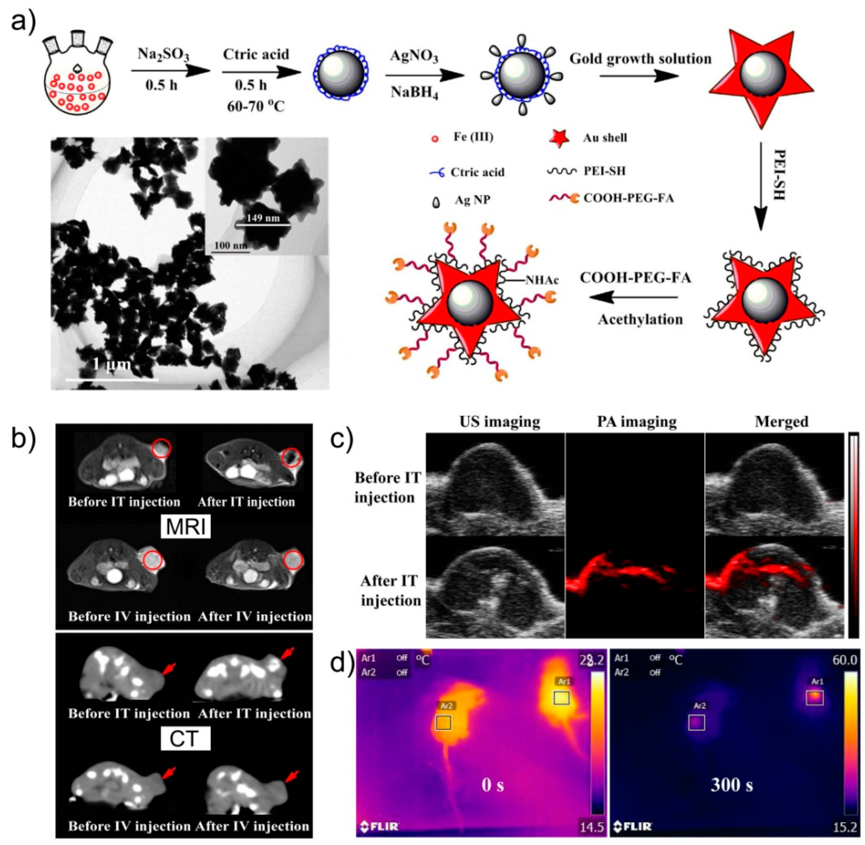
| Au@Ag | Core/Shell | x | 135 | - | Polydopamine | - | [96] | |||||||
| NaYF4:Yb/Er/Tm@NaGdF4 | Core/Shell | x | x | x | 45 | 79.1 nm/- | Silica | - | Nude mice bearing xenograft HeLa tumor | [77] | ||||
| NaGdF4:Yb/Er@NaGdF4:Yb | Core/Shell | x | x | x | 35 | - | Silica/Dopamine | - | Balb/c mice with subcutaneous U14 tumor | [97] | ||||
© 2019 by the authors. Licensee MDPI, Basel, Switzerland. This article is an open access article distributed under the terms and conditions of the Creative Commons Attribution (CC BY) license (http://creativecommons.org/licenses/by/4.0/).
Share and Cite
Quarta, A.; Piccirillo, C.; Mandriota, G.; Di Corato, R. Nanoheterostructures (NHS) and Their Applications in Nanomedicine: Focusing on In Vivo Studies. Materials 2019, 12, 139. https://doi.org/10.3390/ma12010139
Quarta A, Piccirillo C, Mandriota G, Di Corato R. Nanoheterostructures (NHS) and Their Applications in Nanomedicine: Focusing on In Vivo Studies. Materials. 2019; 12(1):139. https://doi.org/10.3390/ma12010139
Chicago/Turabian StyleQuarta, Alessandra, Clara Piccirillo, Giacomo Mandriota, and Riccardo Di Corato. 2019. "Nanoheterostructures (NHS) and Their Applications in Nanomedicine: Focusing on In Vivo Studies" Materials 12, no. 1: 139. https://doi.org/10.3390/ma12010139