Design Techniques to Optimize the Scaffold Performance: Freeze-dried Bone Custom-made Allografts for Maxillary Alveolar Horizontal Ridge Augmentation


 ,
,  ,
,  ,
, 
 and
and 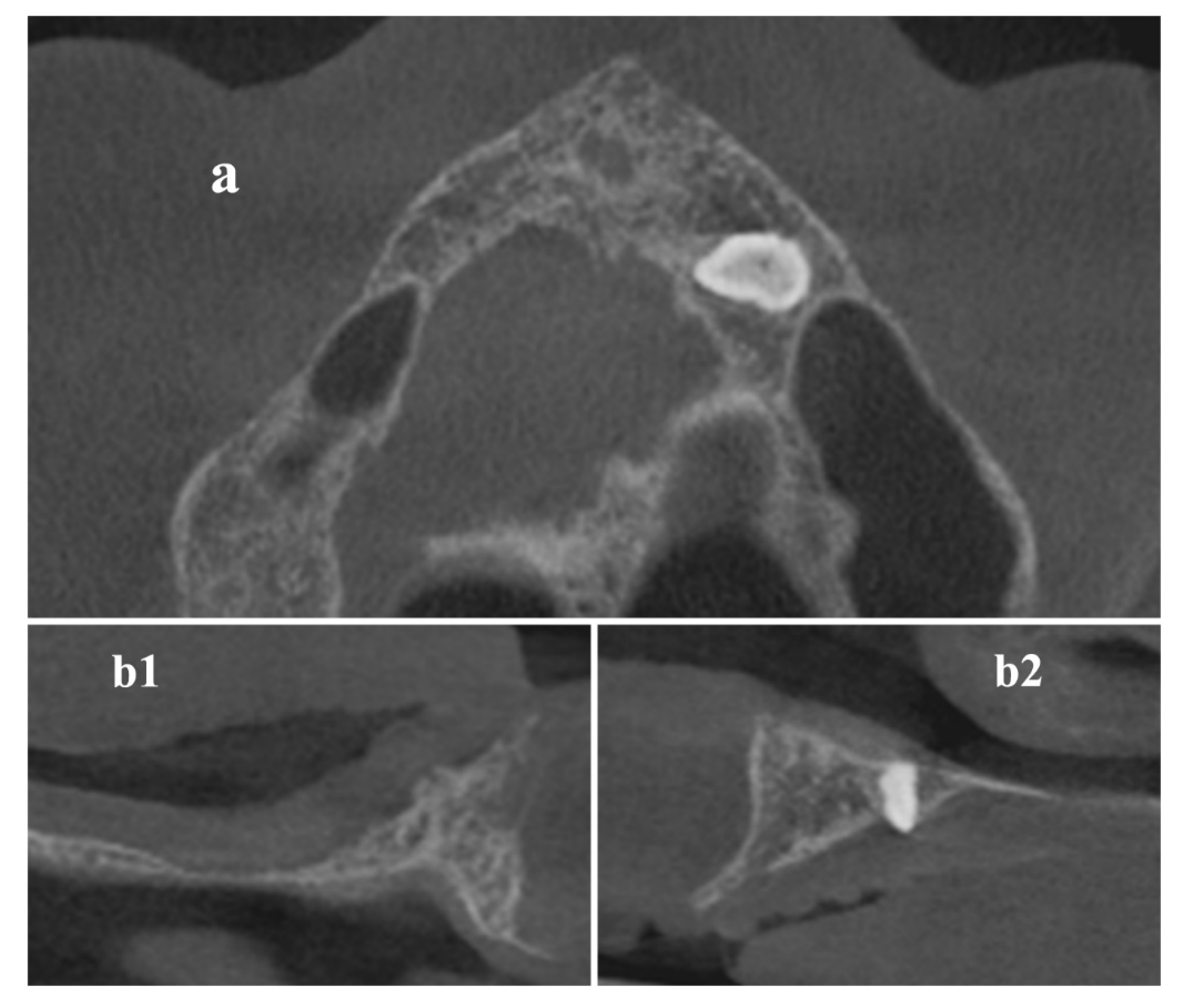
Abstract
1. Introduction
2. Materials and Methods
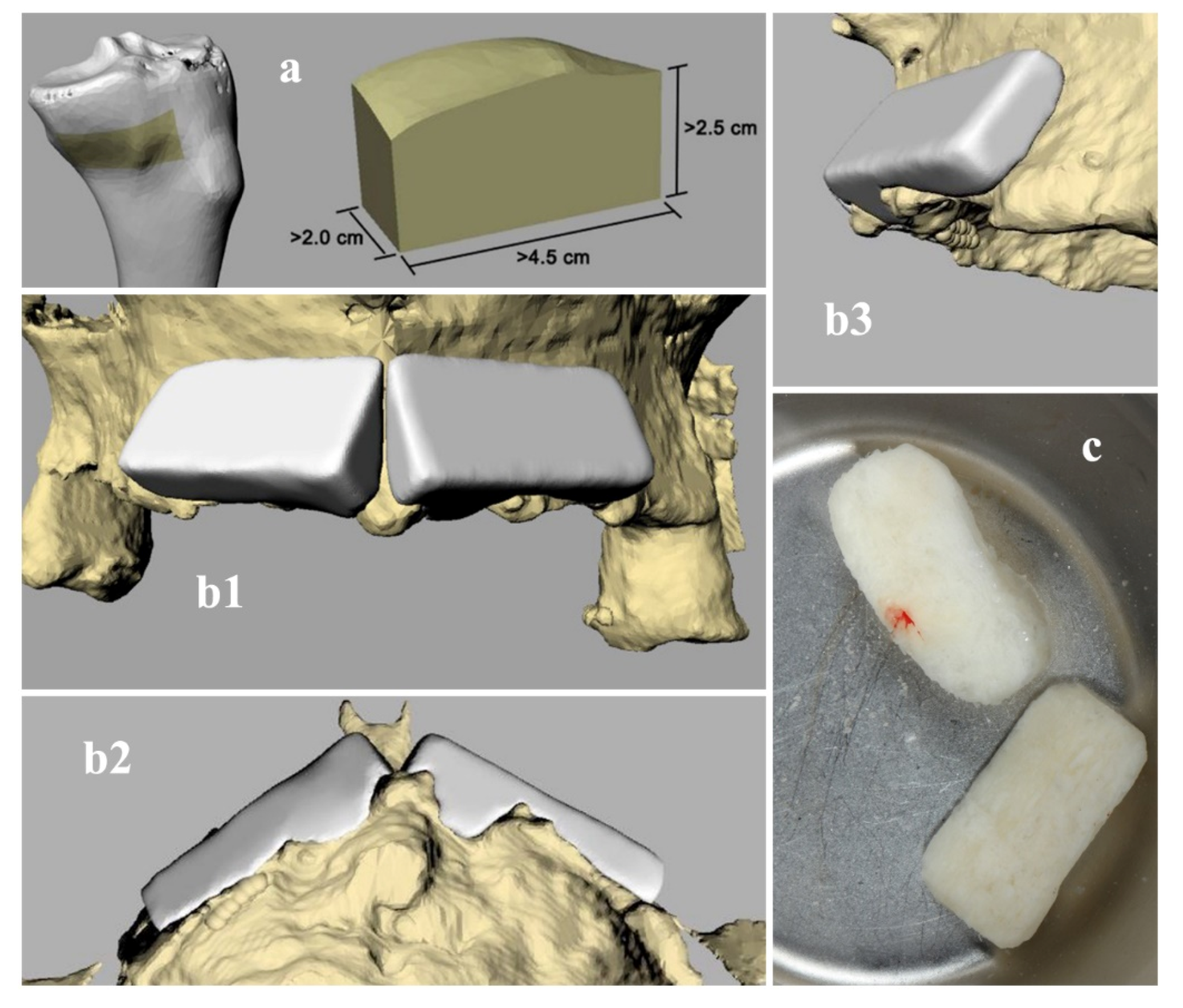
Graft Sample Blocks
Graft Design
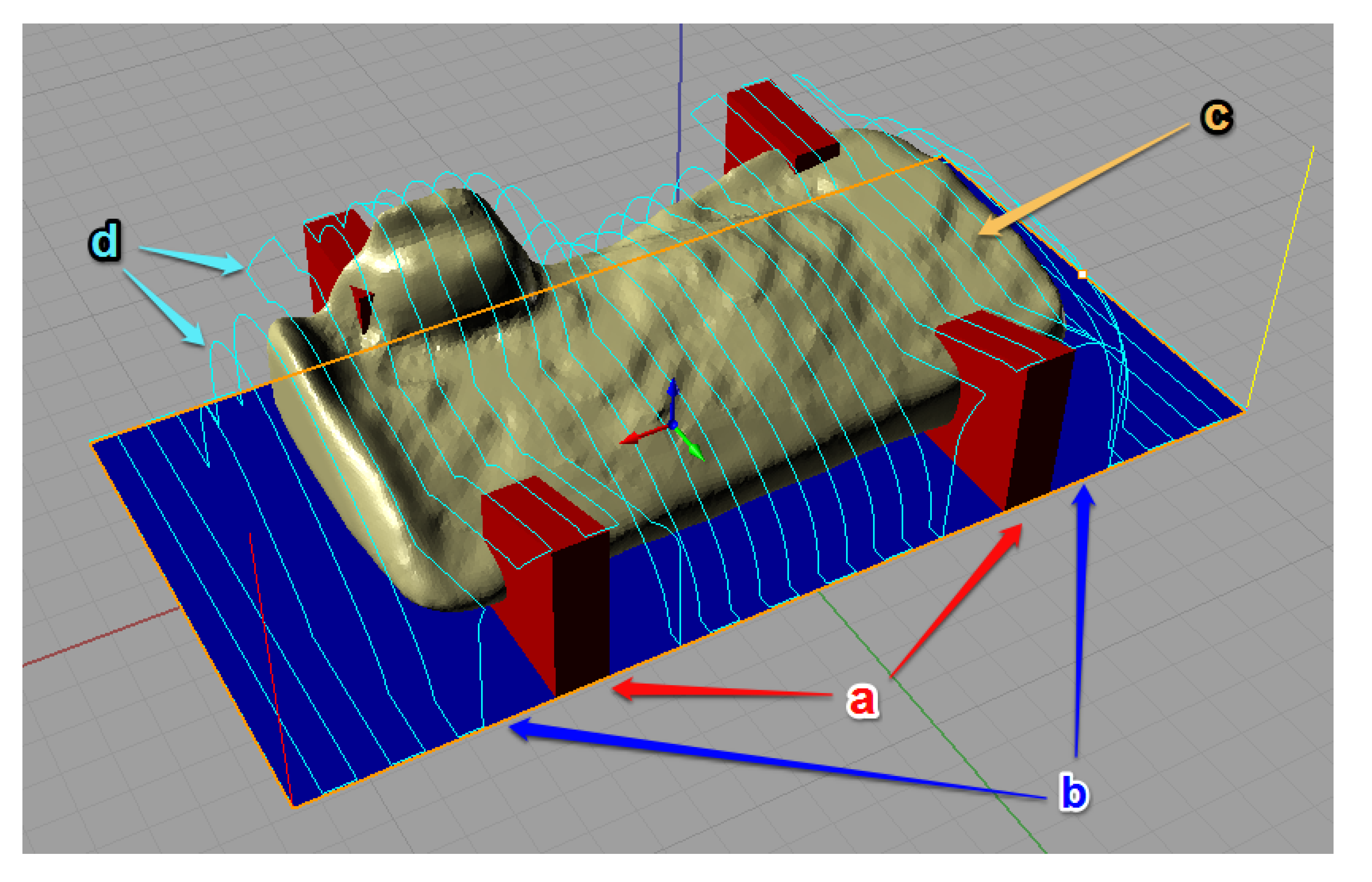
Trajectory Planning and Graft Manufacturing
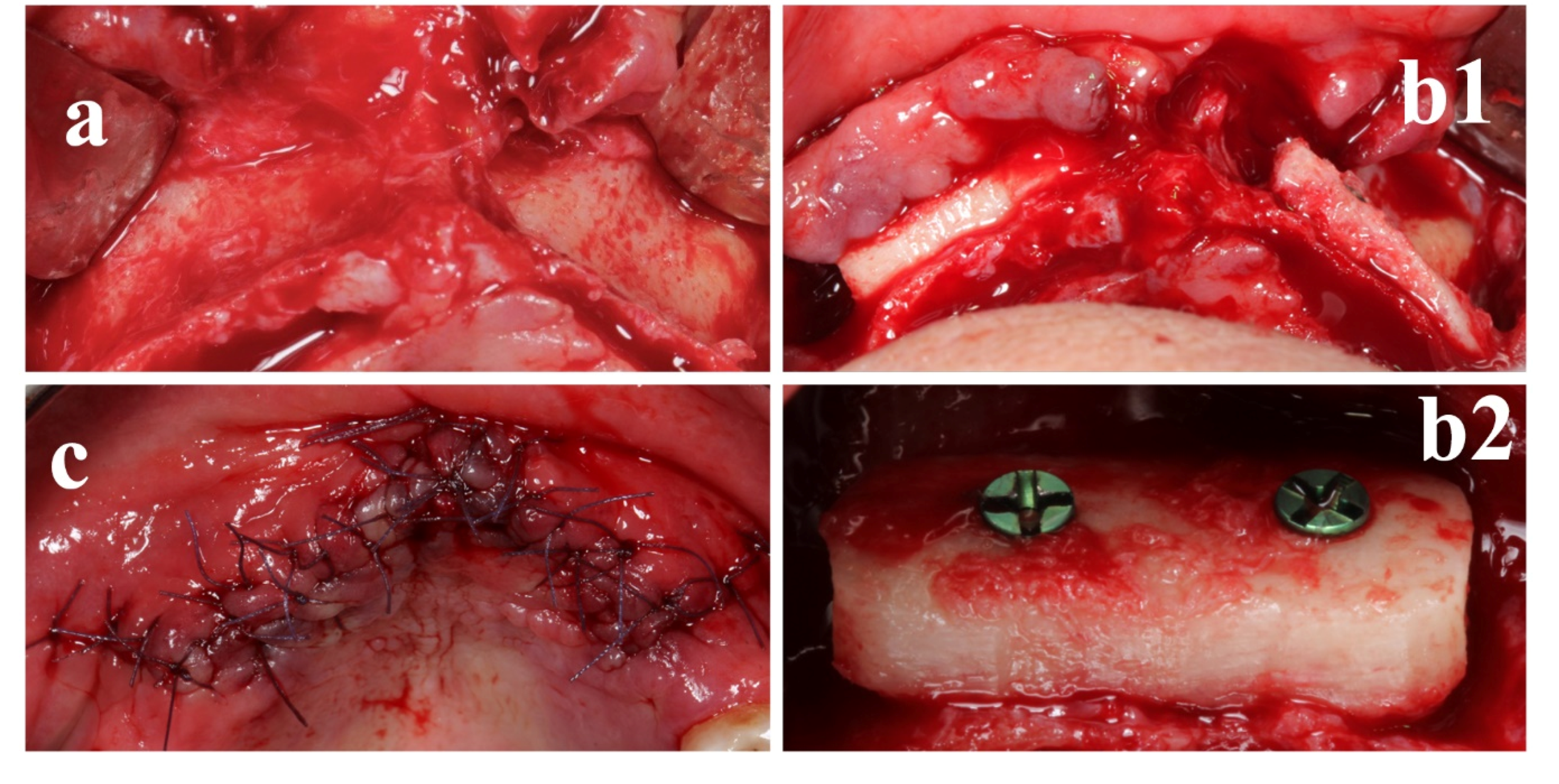
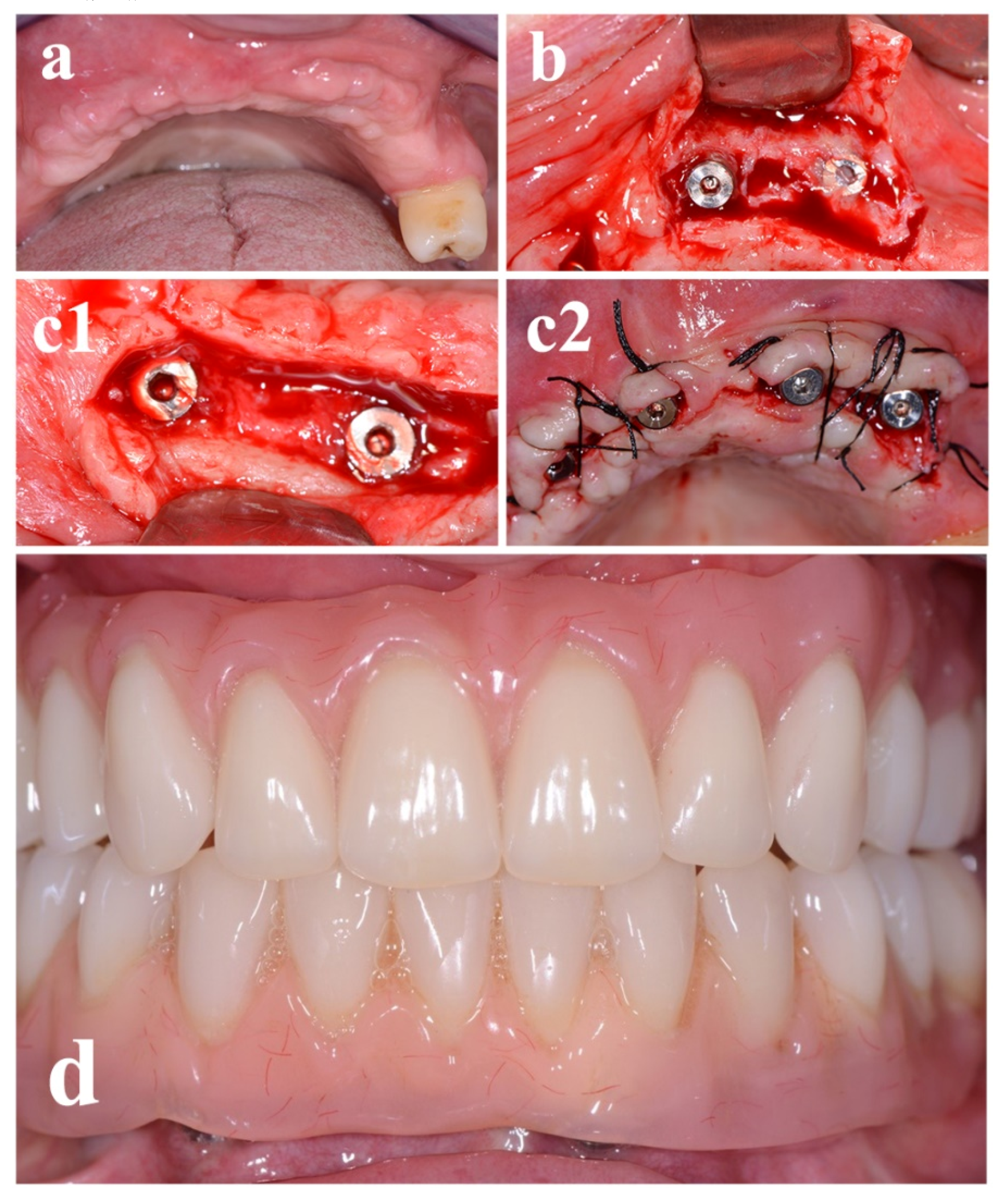
Surgical Procedure
Statistical Evaluation
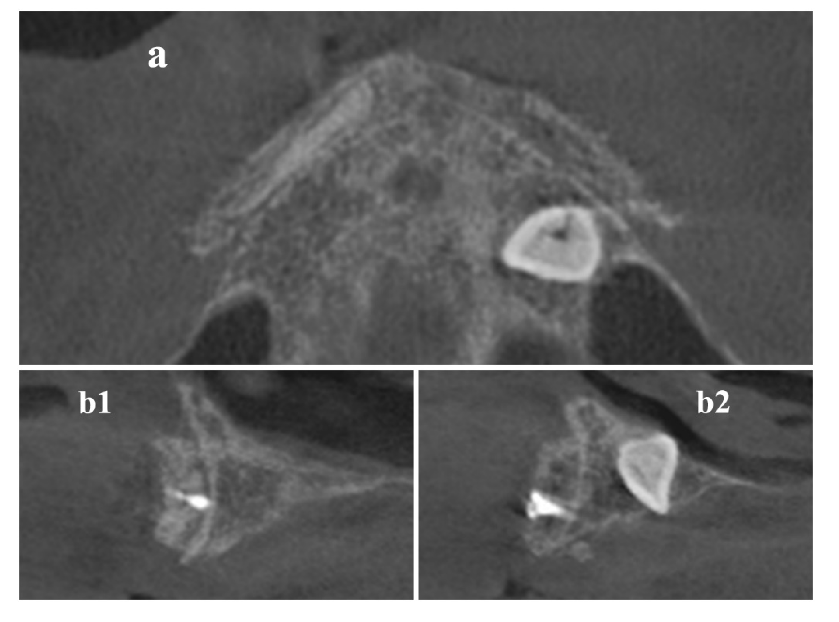
3. Results
4. Discussion
5. Conclusions
Author Contributions
Funding
Conflicts of Interest
References
- Barone, A.; Covani, U. Maxillary alveolar ridge reconstruction with nonvascularized autogenous block bone: Clinical results. J. Oral Maxillofac. Surg. 2007, 65, 2039–2046. [Google Scholar] [CrossRef] [PubMed]
- Cawood, J.I.; Howell, R.A. A classification of the edentulous jaws. Int. J. Oral Maxillofac. Surg. 1988, 17, 232–236. [Google Scholar] [CrossRef]
- Cawood, J.I.; Howell, R.A. Reconstructive preprosthetic surgery. I. Anatomical considerations. Int. J. Oral Maxillofac. Surg. 1991, 20, 75–82. [Google Scholar] [CrossRef]
- Leonetti, J.A.; Koup, R. Localized maxillary ridge augmentation with a block allograft for dental implant placement: Case reports. Implant. Dent. 2003, 12, 217–226. [Google Scholar] [CrossRef]
- Burchardt, H. Biology of bone transplantation. Orthop. Clin. N. Am. 1987, 18, 187–196. [Google Scholar]
- Schultze-Mosgau, S.; Schliephake, H.; Schultze-Mosgau, S.; Neukam, F.W. Soft tissue profile changes after autogenous iliac crest onlay grafting for the extremely atrophic maxilla. J. Oral Maxillofac. Surg. 2000, 58, 971–975. [Google Scholar] [CrossRef]
- Misch, C.M.; Misch, C.E.; Resnik, R.R.; Ismail, Y.H. Reconstruction of maxillary alveolar defects with mandibular symphysis grafts for dental implants: A preliminary procedural report. Int. J. Oral Maxillofac. Implant. 1992, 7, 360–366. [Google Scholar]
- Hardin, C.K. Banked bone. Otolaryngol. Clin. N. Am. 1994, 27, 911–925. [Google Scholar]
- Simpson, D.; Kakarala, G.; Hampson, K.; Steele, N.; Ashton, B. Viable cells survive in fresh frozen human bone allografts. Acta Orthop. 2007, 78, 26–30. [Google Scholar] [CrossRef]
- Perrott, D.H.; Smith, R.A.; Kaban, L.B. The use of fresh frozen allogeneic bone for maxillary and mandibular reconstruction. Int. J. Oral Maxillofac. Surg. 1992, 21, 260–265. [Google Scholar] [CrossRef]
- Gocke, D.J. Tissue donor selection and safety. Clin. Orthop. Relat. Res. 2005, 17–21. [Google Scholar] [CrossRef] [PubMed]
- Venet, L.; Perriat, M.; Mangano, F.G.; Fortin, T. Horizontal ridge reconstruction of the anterior maxilla using customized allogeneic bone blocks with a minimally invasive technique—A case series. BMC Oral Health 2017, 17, 146. [Google Scholar] [CrossRef] [PubMed]
- De Vos, W.; Casselman, J.; Swennen, G.R. Cone-beam computerized tomography (CBCT) imaging of the oral and maxillofacial region: A systematic review of the literature. Int. J. Oral Maxillofac. Surg. 2009, 38, 609–625. [Google Scholar] [CrossRef] [PubMed]
- Lumetti, S.; Consolo, U.; Galli, C.; Multinu, A.; Piersanti, L.; Bellini, P.; Manfredi, E.; Corinaldesi, G.; Zaffe, D.; Macaluso, G.M.; et al. Fresh-frozen bone blocks for horizontal ridge augmentation in the upper maxilla: 6-month outcomes of a randomized controlled trial. Clin. Implant. Dent. Relat. Res. 2014, 16, 116–123. [Google Scholar] [CrossRef]
- Tissues‘ Banks in Italy. Available online: http://www.trapianti.salute.gov.it/trapianti/dettaglioContenutiCnt.jsp?lingua=italiano&area=cnt&menu=chiSiamo&sottomenu=rete&id=237 (accessed on 27 January 2020).
- Fedorov, A.; Beichel, R.; Kalpathy-Cramer, J.; Finet, J.; Fillion-Robin, J.C.; Pujol, S.; Bauer, C.; Jennings, D.; Fennessy, F.; Sonka, M.; et al. 3D Slicer as an image computing platform for the Quantitative Imaging Network. Magn. Reson. Imaging 2012, 30, 1323–1341. [Google Scholar] [CrossRef]
- Kikinis, R.; Pieper, S.D.; Vosburgh, K.G. 3D Slicer: A Platform for Subject-Specific Image Analysis, Visualization, and Clinical Support. In Intraoperative Imaging and Image-Guided Therapy; Jolesz, F.A., Ed.; Springer: New York, NY, USA, 2014; pp. 277–289. [Google Scholar] [CrossRef]
- Matsiushevich, K.; Belvedere, C.; Leardini, A.; Durante, S. Quantitative comparison of freeware software for bone mesh from DICOM files. J. Biomech. 2019, 84, 247–251. [Google Scholar] [CrossRef]
- Krause, W.R. Orthogonal bone cutting: Saw design and operating characteristics. J. Biomech. Eng. 1987, 109, 263–271. [Google Scholar] [CrossRef]
- Van Isacker, T.; Cornu, O.; Barbier, O.; Dufrane, D.; de Gheldere, A.; Delloye, C. Bone Autografting, Allografting and Banking. In European Surgical Orthopaedics and Traumatology: The LEFORT Textbook; Bentley, G., Ed.; Springer: Berlin/Heidelberg, Germany, 2014; pp. 77–90. [Google Scholar] [CrossRef]
- Mangano, F.; Macchi, A.; Shibli, J.A.; Luongo, G.; Iezzi, G.; Piattelli, A.; Caprioglio, A.; Mangano, C. Maxillary ridge augmentation with custom-made CAD/CAM scaffolds. A 1-year prospective study on 10 patients. J. Oral Implantol. 2014, 40, 561–569. [Google Scholar] [CrossRef]
- Pereira, E.; Messias, A.; Dias, R.; Judas, F.; Salvoni, A.; Guerra, F. Horizontal Resorption of Fresh-Frozen Corticocancellous Bone Blocks in the Reconstruction of the Atrophic Maxilla at 5 Months. Clin. Implant. Dent. Relat. Res. 2015, 17 (Suppl. S2), e444–e458. [Google Scholar] [CrossRef]
- Luongo, F.; Mangano, F.G.; Macchi, A.; Luongo, G.; Mangano, C. Custom-Made Synthetic Scaffolds for Bone Reconstruction: A Retrospective, Multicenter Clinical Study on 15 Patients. Biomed. Res. Int. 2016, 2016, 5862586. [Google Scholar] [CrossRef]
- Yen, H.H.; Stathopoulou, P.G. CAD/CAM and 3D-Printing Applications for Alveolar Ridge Augmentation. Curr. Oral Health Rep. 2018, 5, 127–132. [Google Scholar] [CrossRef] [PubMed]
- Nguyen, H.; Cassady, A.I.; Bennett, M.B.; Gineyts, E.; Wu, A.; Morgan, D.A.; Forwood, M.R. Reducing the radiation sterilization dose improves mechanical and biological quality while retaining sterility assurance levels of bone allografts. Bone 2013, 57, 194–200. [Google Scholar] [CrossRef] [PubMed]
- Nguyen, H.; Morgan, D.A.; Forwood, M.R. Sterilization of allograft bone: Effects of gamma irradiation on allograft biology and biomechanics. Cell Tissue Bank. 2007, 8, 93–105. [Google Scholar] [CrossRef] [PubMed]
- Kao, S.T.; Scott, D.D. A review of bone substitutes. Oral Maxillofac. Surg. Clin. N. Am. 2007, 19, 513–521. [Google Scholar] [CrossRef] [PubMed]
- Naidu, P. Allografts in Periodontal Regeneration. Madr. J. Case Rep. Stud. 2019, 3, 121–125. [Google Scholar] [CrossRef]
- Mellonig, J.T.; Bowers, G.M.; Bright, R.W.; Lawrence, J.J. Clinical evaluation of freeze-dried bone allografts in periodontal osseous defects. J. Periodontol. 1976, 47, 125–131. [Google Scholar] [CrossRef]
- Grover, V.; Kapoor, A.; Malhotra, R.; Sachdeva, S. Bone allografts: A review of safety and efficacy. Indian J. Dent. Res. 2011, 22, 496. [Google Scholar] [CrossRef]
- Delloye, C.; Cornu, O.; Druez, V.; Barbier, O. Bone allografts: What they can offer and what they cannot. J. Bone Jt. Surg. Br. 2007, 89, 574–579. [Google Scholar] [CrossRef]
- Delloye, C. Tissue allografts and health risks. Acta Orthop. Belg. 1994, 60 (Suppl. S1), 62–67. [Google Scholar]
- Cornu, O.; Banse, X.; Docquier, P.L.; Luyckx, S.; Delloye, C. Effect of freeze-drying and gamma irradiation on the mechanical properties of human cancellous bone. J. Orthop. Res. 2000, 18, 426–431. [Google Scholar] [CrossRef]
- Dziedzic-Goclawska, A.; Kaminski, A.; Uhrynowska-Tyszkiewicz, I.; Stachowicz, W. Irradiation as a safety procedure in tissue banking. Cell Tissue Bank. 2005, 6, 201–219. [Google Scholar] [CrossRef] [PubMed]
- Holt, D.J.; Grainger, D.W. Demineralized bone matrix as a vehicle for delivering endogenous and exogenous therapeutics in bone repair. Adv. Drug Deliv. Rev. 2012, 64, 1123–1128. [Google Scholar] [CrossRef] [PubMed]
- Urist, M.R.; Strates, B.S. Bone morphogenetic protein. J. Dent. Res. 1971, 50, 1392–1406. [Google Scholar] [CrossRef] [PubMed]
- Barone, A.; Varanini, P.; Orlando, B.; Tonelli, P.; Covani, U. Deep-frozen allogeneic onlay bone grafts for reconstruction of atrophic maxillary alveolar ridges: A preliminary study. J. Oral Maxillofac. Surg. 2009, 67, 1300–1306. [Google Scholar] [CrossRef]
- Blume, O.; Hoffmann, L.; Donkiewicz, P.; Wenisch, S.; Back, M.; Franke, J.; Schnettler, R.; Barbeck, M. Treatment of Severely Resorbed Maxilla Due to Peri-Implantitis by Guided Bone Regeneration Using a Customized Allogenic Bone Block: A Case Report. Materials (Basel) 2017, 10, 1213. [Google Scholar] [CrossRef]
- Motamedian, S.R.; Khojaste, M.; Khojasteh, A. Success rate of implants placed in autogenous bone blocks versus allogenic bone blocks: A systematic literature review. Ann. Maxillofac. Surg. 2016, 6, 78–90. [Google Scholar] [CrossRef]
- Makvandi, P.; Ali, G.W.; Della Sala, F.; Abdel-Fattah, W.I.; Borzacchiello, A. Hyaluronic acid/corn silk extract based injectable nanocomposite: A biomimetic antibacterial scaffold for bone tissue regeneration. Mater. Sci. Eng. C Mater. Biol. Appl. 2020, 107, 110195. [Google Scholar] [CrossRef]


© 2020 by the authors. Licensee MDPI, Basel, Switzerland. This article is an open access article distributed under the terms and conditions of the Creative Commons Attribution (CC BY) license (http://creativecommons.org/licenses/by/4.0/).
Share and Cite
Grassi, F.R.; Grassi, R.; Vivarelli, L.; Dallari, D.; Govoni, M.; Nardi, G.M.; Kalemaj, Z.; Ballini, A. Design Techniques to Optimize the Scaffold Performance: Freeze-dried Bone Custom-made Allografts for Maxillary Alveolar Horizontal Ridge Augmentation. Materials 2020, 13, 1393. https://doi.org/10.3390/ma13061393
Grassi FR, Grassi R, Vivarelli L, Dallari D, Govoni M, Nardi GM, Kalemaj Z, Ballini A. Design Techniques to Optimize the Scaffold Performance: Freeze-dried Bone Custom-made Allografts for Maxillary Alveolar Horizontal Ridge Augmentation. Materials. 2020; 13(6):1393. https://doi.org/10.3390/ma13061393
Chicago/Turabian StyleGrassi, Felice Roberto, Roberta Grassi, Leonardo Vivarelli, Dante Dallari, Marco Govoni, Gianna Maria Nardi, Zamira Kalemaj, and Andrea Ballini. 2020. "Design Techniques to Optimize the Scaffold Performance: Freeze-dried Bone Custom-made Allografts for Maxillary Alveolar Horizontal Ridge Augmentation" Materials 13, no. 6: 1393. https://doi.org/10.3390/ma13061393
APA StyleGrassi, F. R., Grassi, R., Vivarelli, L., Dallari, D., Govoni, M., Nardi, G. M., Kalemaj, Z., & Ballini, A. (2020). Design Techniques to Optimize the Scaffold Performance: Freeze-dried Bone Custom-made Allografts for Maxillary Alveolar Horizontal Ridge Augmentation. Materials, 13(6), 1393. https://doi.org/10.3390/ma13061393