
2D Materials Nanoarchitectonics for 3D Structures/Functions


1
Research Center for Materials Nanoarchitectonics (MANA), National Institute for Materials Science (NIMS), 1-1 Namiki, Tsukuba 305-0044, Ibaraki, Japan
2
Graduate School of Frontier Sciences, The University of Tokyo, 5-1-5 Kashiwanoha, Kashiwa 277-8561, Chiba, Japan
Materials 2024, 17(4), 936; https://doi.org/10.3390/ma17040936
Submission received: 20 January 2024
/
Revised: 9 February 2024
/
Accepted: 9 February 2024
/
Published: 17 February 2024
(This article belongs to the Special Issue Materials Nanoarchitectonics for Membranes, Surfaces, Self-Assembly, and Biomimetics)
Abstract
:It has become clear that superior material functions are derived from precisely controlled nanostructures. This has been greatly accelerated by the development of nanotechnology. The next step is to assemble materials with knowledge of their nano-level structures. This task is assigned to the post-nanotechnology concept of nanoarchitectonics. However, nanoarchitectonics, which creates intricate three-dimensional functional structures, is not always easy. Two-dimensional nanoarchitectonics based on reactions and arrangements at the surface may be an easier target to tackle. A better methodology would be to define a two-dimensional structure and then develop it into a three-dimensional structure and function. According to these backgrounds, this review paper is organized as follows. The introduction is followed by a summary of the three issues; (i) 2D to 3D dynamic structure control: liquid crystal commanded by the surface, (ii) 2D to 3D rational construction: a metal–organic framework (MOF) and a covalent organic framework (COF); (iii) 2D to 3D functional amplification: cells regulated by the surface. In addition, this review summarizes the important aspects of the ultimate three-dimensional nanoarchitectonics as a perspective. The goal of this paper is to establish an integrated concept of functional material creation by reconsidering various reported cases from the viewpoint of nanoarchitectonics, where nanoarchitectonics can be regarded as a method for everything in materials science.
1. Introduction
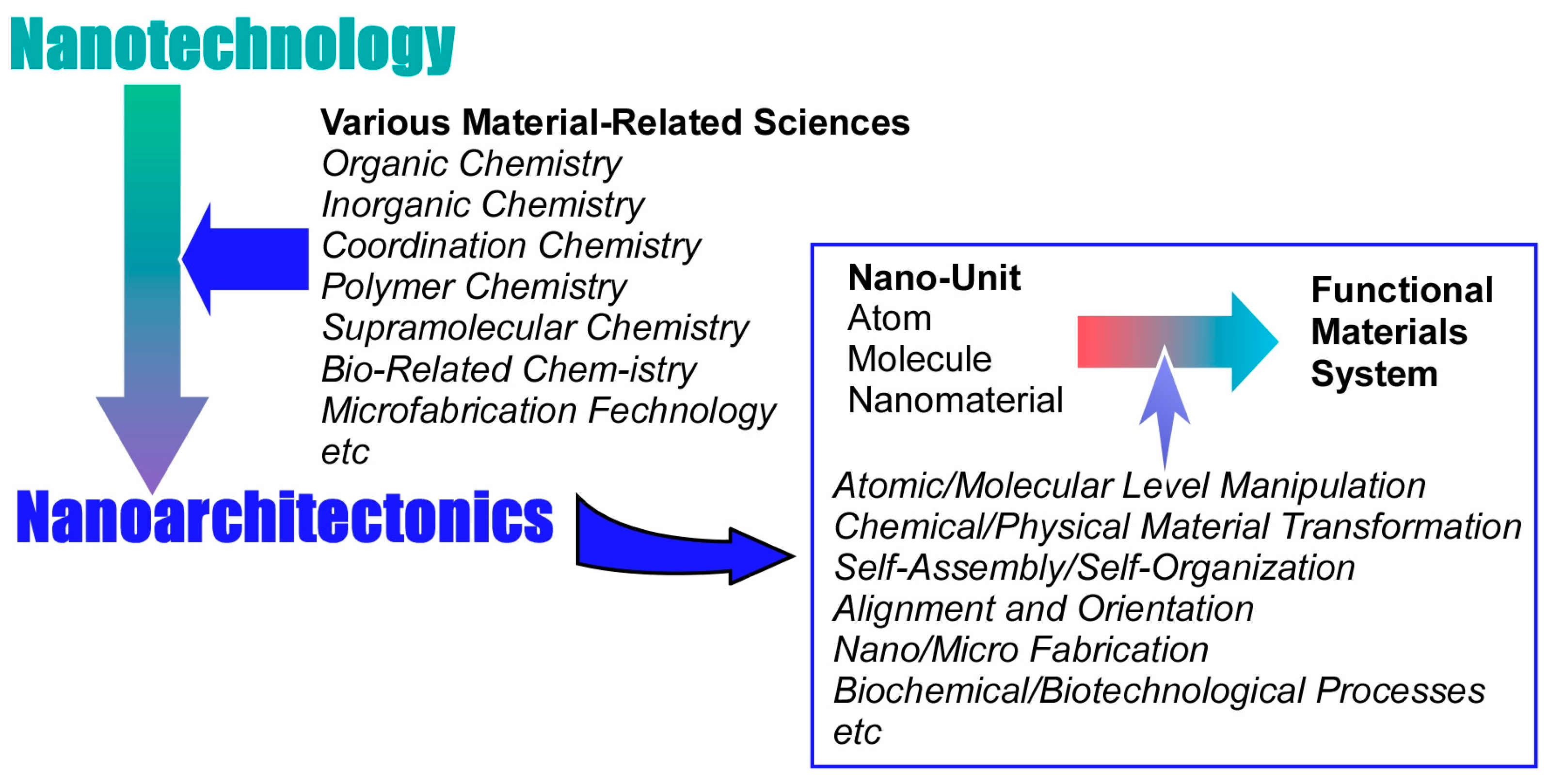
The development of new technologies such as information technology, device technology, and medical technology is supporting the development of society. However, materials chemistry, which has been steadily developing since the 20th century, is also indispensable [1,2]. Useful materials are essential to fulfill important parts of the later technologies. Such developments in materials chemistry solve a variety of existing problems. Energy production [3,4,5,6,7,8,9,10,11,12,13,14,15,16,17,18], energy storage [19,20,21,22,23,24,25,26,27,28,29,30], environmental remediation [31,32,33,34,35,36,37,38,39,40], carbon neutral strategies [41,42,43,44,45,46], detection of hazardous substances [47,48,49,50,51,52], bio-related sensing [53,54,55,56,57,58], technologies leading to medicine [59,60,61,62,63,64,65,66,67,68,69], and the creation of device materials to support them [70,71,72,73,74,75] are among the many demands that depend on the development of materials chemistry. Effective function is not determined solely by the properties of the substances themselves. Their functions depend much on what kind of structure a substance takes or what kind of internal structure it takes. With the progress of materials chemistry, such matters have been gradually elucidated. This has been greatly accelerated by the development of nanotechnology. As is still the case in current research, nanotechnology makes it possible to observe nanostructures from the atomic and molecular levels [76,77,78,79,80,81,82,83,84,85,86,87,88,89]. Accordingly, it has become clear that superior material functions are derived from precisely controlled nanostructures. The next step is to assemble materials with knowledge of their nano-level structures. This task is assigned to the post-nanotechnology concept of nanoarchitectonics (Figure 1) [90].
Nanotechnology was founded by Richard Feynman in the middle of the 20th century [91,92]. In the early 21st century, nanoarchitectonics was proposed by Masakazu Aono [93,94] as a successor to Feynman’s work. The goal of nanoarchitectonics is to establish a methodology for building functional material systems from nano-units such as atoms, molecules, and nanomaterials [95,96,97]. Rather than a completely new concept, it is more of an integration of existing concepts. In other words, it is a fusion of nanotechnology with various material-related sciences and peripheral fields (organic chemistry, inorganic chemistry, coordination chemistry, polymer chemistry, supramolecular chemistry, bio-related chemistry, microfabrication technology, etc.). The following elemental technologies for building functional material systems from nano-units can be considered: atomic/molecular-level manipulation, chemical/physical material transformation including organic synthesis, self-assembly/self-organization, alignment and orientation by application of external fields and energy, nano/microfabrication, and biochemical/biotechnological processes [98]. These are selected and combined as appropriate. Compared to self-assembly, which is a single-equilibrium process, it is often a multistep process. Accordingly, nanoarchitectonics is well suited to creating asymmetric and hierarchical structures [99]. In addition, the underlying nanoscale interactions often involve uncertainties such as thermal fluctuations, stochastic distributions, and quantum effects. Therefore, the various effects are harmonized rather than simply added together [100].
The above principles are general and Independent of the type of materials and their functions. Nanoarchitectonics will be universally applicable to a wide variety of material systems. Originally, all matter is composed of units of atoms and molecules. Therefore, the concept of nanoarchitectonics, which is the architecture of matter from atoms and molecules, can be the creation of all matter. Like the ultimate theory of everything in physics [101], it could become a method for everything [102,103], an integrated concept for synthesizing functional materials in materials science. In fact, many papers advocating nanoarchitectonics have been published in recent years. The field ranges from basic to applied sciences. It includes material synthesis [104,105,106,107,108,109,110,111,112,113,114], creation of specific structures [115,116,117,118,119,120,121,122,123,124,125], organization of structures [126,127,128,129,130,131,132,133,134,135,136,137,138], exploration of basic physical phenomena [139,140,141,142,143,144,145,146,147,148,149,150], basic biochemistry [151,152,153,154,155,156,157,158,159,160,161,162,163], catalysis [164,165,166,167,168,169,170,171,172,173,174,175], environmental remediation [176,177,178,179,180,181,182,183,184,185,186,187], sensors [188,189,190,191,192,193,194,195,196,197], devices [198,199,200,201,202,203,204,205], energy generation [206,207,208,209,210,211,212,213,214], energy storage [215,216,217,218,219,220,221,222], drug delivery [223,224,225,226,227], cellular control [228,229,230,231], and biomedical applications [232,233,234,235,236,237,238,239,240,241,242]. Since nanoarchitectonics is a comprehensive concept, there are also many approaches that do not advocate nanoarchitectonics but have the same effect as nanoarchitectonics.
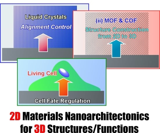
However, nanoarchitectonics, which creates intricate three-dimensional functional structures, is not always easy. Two-dimensional nanoarchitectonics based on reactions and arrangements at the surface may be an easier target to tackle. A better methodology would be to define a two-dimensional structure and then develop it into a three-dimensional structure and function. It is a structure development prescription from interface to bulk. Such an approach can be seen in several existing research examples. A typical approach is the interface-based nano thin film fabrication technique. One typical approach is the Langmuir–Blodgett (LB) method [243,244,245,246,247,248,249]. A thin film at the monolayer level is first prepared at a two-dimensional liquid interface, such as a water surface. If these films are sequentially transferred onto a substrate, a three-dimensional, multilayered structure is formed. Layer-by-layer (LbL) assembly is a simpler and more versatile method than the LB method, although its structural control is more ambiguous [250,251,252,253,254,255,256]. Based on specific interactions; thin films are sequentially deposited on a substrate to obtain a variety of thin film structures. If this technique is performed on colloidal templates, it is possible to fabricate three-dimensional thin-film capsules. These techniques are widely studied as a powerful method to convert two-dimensional nanoarchitectonics into three-dimensional structures. However, the resulting structures are still at the thin-film level and may be described as somewhat thicker two-dimensional nanoarchitectonics. Other than these methods, nanoarchitectonics conversion from two dimensional to three dimensional has to be considered.
This review aims to discuss other possibilities of methodologies that expand from some two-dimensional nanoarchitectonics to three-dimensional structures and functions. Three typical examples are presented (Figure 2) that fall under this research orientation, regardless of what is advocated for nanoarchitectonics. The first is the control of bulk materials through nanoarchitectonics of two-dimensional surfaces. This is illustrated by the example of controlling the orientation of a bulk liquid crystal by changing the structure of a very thin surface layer. The second is rational structural architecture from two dimensional to three dimensional. Several examples of metal–organic frameworks (MOFs) and covalent organic frameworks (COFs) from two-dimensional control to three-dimensional architecture are given as examples for this purpose. The third category is the control of cells by surfaces. Cells originally have a cascading signal-generated growth mechanism [257,258,259]. Contact with a two-dimensionally nanoarchitectonized interface can induce advanced functions of the cell. This can be introduced as amplification from two-dimensional nanoarchitectonics to three-dimensional function.
According to the above backgrounds, this review paper is organized as follows. This introduction is followed by a summary of the three issues introduced in the previous paragraphs. They are the following sections: (i) 2D to 3D dynamic structure control: liquid crystal commanded by the surface; (ii) 2D to 3D rational construction: MOF and COF; (iii) 2D to 3D functional amplification: cells regulated by the surface. In addition, this review summarizes the important aspects of the ultimate three-dimensional nanoarchitectonics as a perspective. The goal of this paper is to establish an integrated concept of functional material creation by reconsidering various reported cases from the viewpoint of nanoarchitectonics. Nanoarchitectonics can be regarded as a method for everything in materials science.
2. 2D to 3D Dynamic Structure Control: Liquid Crystal Commanded by the Surface
Liquid crystals are materials that can flexibly change their structures while maintaining a certain degree of orientation and other organizing ability [260,261,262]. In Ichimura’s review article on the optical control of liquid crystals [263], the behavior of liquid crystals incorporating photochromic molecules is discussed. Photochromic molecules are usually embedded in various matrices in both fundamental and practical research. In such cases, the structural transformation of the photochromic guest molecule alters the reversible properties of the matrix, the liquid crystal. The orientation of the host molecules and residues that act as the matrix is changed. Broadly speaking, photoaligned liquid crystal systems fall into two types. The first type consists of liquid crystal molecules doped with photochromic units. The light-induced structure of a few photochromic chromophores results in the reorientation of the majority of matrix liquid crystal molecules. This leads to the generation of large optical anisotropy. Another type of liquid crystal system reflects the photoalignment state of photochromic molecules attached to the substrate surface into the bulk liquid crystal layer. This type of structural change in the surface, in which minute structural changes on the surface dictate structural changes in many of the molecular layers above it, is called a command surface [264,265]. This technique demonstrates the effective application of photochromic units to fabricate liquid crystal photoresponsive systems. It also opens the way to the control of photoalignment of liquid crystals, even with irreversible photochemistry. This concept is not only performed by photochromic molecules. It can be a methodology to expand broadly two-dimensional nanoarchitectonics to three-dimensional liquid crystal functionality. It can also be an effective method for fabricating new types of optical elements and devices for photonics applications.
One of the pioneering examples of the command surface concept can be found in the report of Seki, Ichimura, and co-workers [266]. In this study, LB films of side-chain azobenzene amphiphilic polymers were used as photochromic command layers. The photoisomerization of the command layer was used to control the reversible homeotropic planar photochemical orientation of nematic liquid crystals. As a nanoarchitectonics strategy for the command surface, it is important that the azobenzene photochromic unit is separated from the background, the poly(vinyl alcohol) backbone, by a methylene spacer of appropriate length. With this molecular design, a single azobenzene monolayer is sufficient to induce a change in thick liquid crystal orientation. For LB nanoarchitectonics, the vertical transfer method of preparation is more advantageous. The vertical transfer method produced more homogeneously aligned LB films and the liquid crystal molecules were oriented parallel to the immersion direction. When linearly polarized UV light was irradiated, the liquid crystalline molecules reoriented in the direction perpendicular to the polarization plane. In other words, the trans–cis isomerization of the highly photoreactive azobenzene photochromic units in the polymer LB layer successfully commanded the orientation between the homeotropic and planar modes of the nematic liquid crystal.
The orientation of liquid crystal molecules is largely governed by the degree of freedom of the surfaces with which they come into contact and other factors. As in the above example, the photoresponsive layer on the surface with freedom serves as the command layer of the side-chain liquid crystal polymer film. Various studies have been conducted to understand the interfacial behavior of these liquid crystal molecules. For example, the photoalignment behavior of nematic liquid crystals on azobenzene polymer films has been the subject of research. Hara, Seki, and co-workers have investigated the optical orientation behavior of side-chain liquid crystalline azobenzene polymer films with a thickness of approximately 400 nm and a 35 μm-thick low-molecular-weight nematic liquid crystal, 4′-pentyl-4-cyanobiphenyl in situ at the interface (Figure 3) [267]. In addition to polarized light optical microscopy observations, small-angle X-ray scattering measurements were used to evaluate the structure inside the liquid crystal-injected sandwich cell. This technique has provided new insights into the behavior of liquid crystal molecules in the vicinity of alignment films on solid substrates. For example, they have succeeded in detecting the selective and time-series structuring and orientation of mesogens at the interface in the formation of smectic layers. A highly ordered smectic liquid crystalline phase is induced by the hybridization of the mesogens of azobenzene polymers and 4′-pentyl-4-cyanobiphenyls. A cooperative hybrid highly ordered smectic liquid crystal phase is formed by weak electron transfer at interfacial contact. Such analysis is important to understand how slight structural changes at the two-dimensional interface are reflected in the three-dimensional structure. Direct X-ray observations in intact liquid crystal cell systems provide useful information on the driving mechanism. It can be a powerful tool in industrial applications in terms of liquid crystal device design. It should also expand the possibilities in terms of practical applications.
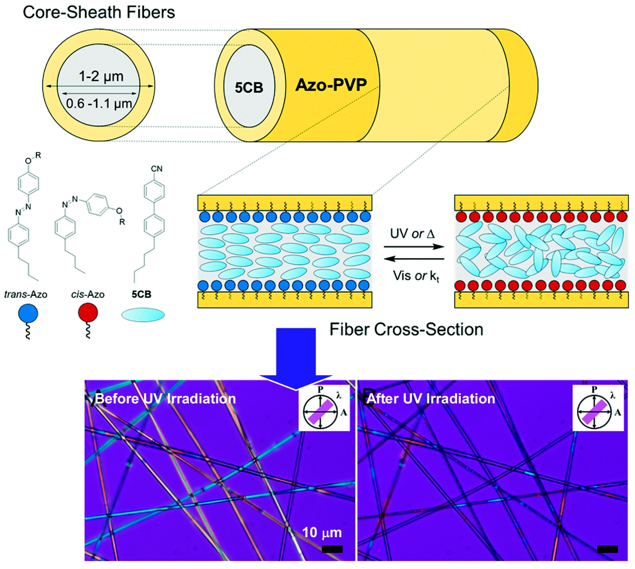
Lundin and co-workers used electrospinning to fabricate core–sheath nanofibers with photochromic and liquid crystalline components (Figure 4) [268]. The core–sheath nanofibers consist of a polyvinylpyrrolidone sheath doped with a photochromic azobenzene surfactant and a low-molecular-weight nematic liquid crystal core. By incorporating the azobenzene surfactant into the polymer sheath, the nematic-to-isotropic transition temperature of the liquid crystal core could be photochemically controlled. In other words, ultraviolet (UV) irradiation reduced the phase transition temperature. At high surfactant content, the temperature of the photo-induced phase transition was reduced to below room temperature. It allows “turning on” and “turning off” the birefringence of the nanofibers upon UV irradiation and does not require external heating. Photoisomerization of azobenzene surfactants at the poly(vinylpyrrolidone)/liquid crystal interface causes surface-induced disordering of the liquid crystal core. Therefore, UV irradiation causes a change from planar-axis orientation to random orientation. In the case of visible light irradiation, the opposite change is expected to occur. Thus, the photochromic nature of this core–sheath nanofiber system is the result of the difference in the compatibility of the cis- and trans-isomers with the liquid crystal matrix at the liquid crystal/polymer interface. Similar to the command surface of LB films described above, the incorporation of azobenzene into the polymer sheath of liquid-crystalline-core nanofibers allows molecular changes to be reflected in the three-dimensional properties of the liquid crystal.
The optical control of liquid crystal molecules as an example of how two-dimensional nanoarchitectonics through molecular design and orientation is reflected in the properties of three-dimensional materials. Not only LB-type monolayers, but also polymer layers with a core-shell structure act as a command surface. The molecular-level phenomena of structural and orientation changes on the command surface are amplified into three-dimensional material properties such as liquid crystal orientation and phase transitions. The key to this process is molecular phenomena at the interface. The control of the interface leads to the amplification of functional structures from two to three dimensions.
3. 2D to 3D Rational Construction: MOF and COF
The synthetic approaches of metal–organic frameworks (MOFs) [269,270,271,272,273,274,275,276,277,278,279] and covalent organic frameworks (COFs) [280,281,282,283,284] are methods for rationally building structures from molecular units and ions. These structures are often formed as two-dimensional structures in interfacial environments, although they can also be obtained in three-dimensional structures such as crystals. As a rational nanoarchitectonics from two dimensions to three dimensions, material designs based on MOFs and COFs are beneficial. In this section, several approaches to three-dimensional architecting from two-dimensional MOFs and COFs are exemplified.
Structural evolution from two dimensions to three dimensions is a useful technique for tuning mechanical properties. For example, developing strategies to improve the structural robustness of COFs has been recognized as very important. Yu, Zhang and co-workers reported a method to rationally design and synthesize crosslinked COFs in which the two-dimensional COF layers are covalently fixed and linked by poly(ethylene glycol) (PEG) three-dimensional or alkyl chains (Figure 5) [285]. It is a bottom-up synthetic strategy to structuralize layered structures of two-dimensional COFs using monomers linked by flexible PEG or alkyl chains. The two-dimensional layered structure was converted into a quasi- three-dimensional accumulation framework via covalent crosslinking, where vertical crosslayer bonding is dominant. All synthesized crosslinked COFs were highly crystalline and porous. In particular, they exhibited robust structural stability that surpassed that of typical two-dimensional COFs. While simple assemblies of two-dimensional COFs were easily exfoliated into nanosheets by sonication, pulverization, and water treatment, three-dimensional crosslinked COFs maintained their ordered framework even after such treatments due to the crosslinking effect. The structural stability while inheriting high crystallinity and porosity provides the interlayer stability that is extremely necessary for advanced applications such as heterogeneous catalytic reactions and proton/ion transport. A high application potential of quasi-three-dimensional COFs is expected.
Three-dimensional COFs are of interest as a structurally stable group of materials with their inherent large number of open sites and pore confinement effects. However, it is not easy to generate an entangled three-dimensional network formed from multiple two-dimensional layers inclined toward each other. Ma, Li, and co-workers have successfully synthesized a new three-dimensional COF based on a two-dimensional network with interpenetrations (Figure 6) [286]. The structures were formed by [3+2]imine condensation reactions using triangular knots and linear linkers. Specifically, they were formed by [3+2]imine condensation reactions using 1,3,5-triformylbenzene and 2,3,5,6-tetramethyl-1,4-phenylenediamine. The range of strategies for achieving three-dimensional COFs was broadened by mutual coordination. Such examples demonstrate that structurally complex extended frameworks can be obtained from simple molecules and can be used for nanoarchitectonics. They enrich synthetic strategies for three-dimensional COFs and greatly expand the range of COF materials.
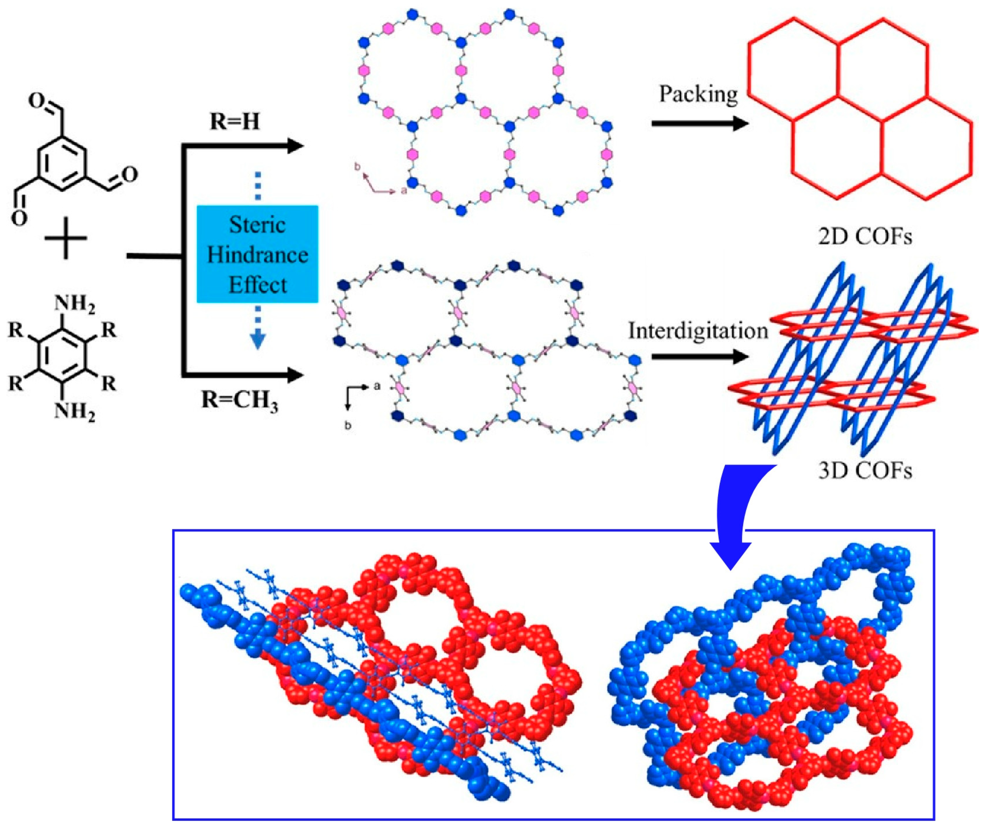
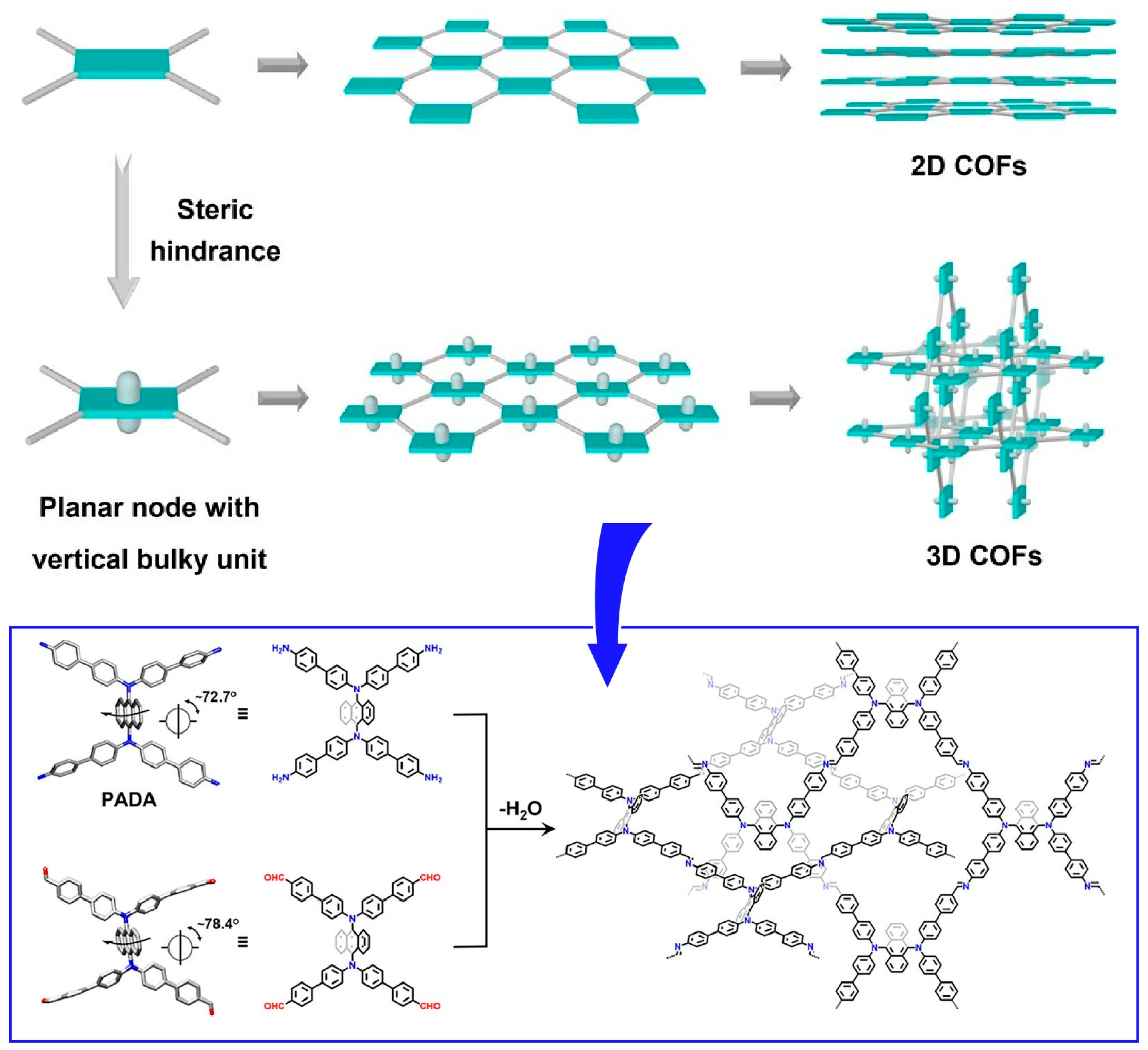
Gui, Sun, and Wang and co-workers devised a method to form three-dimensional COFs by introducing steric hindrance to molecular blocks that inhibit π–π stacking, as a method for two-dimensional COFs to intertwine with each other (Figure 7) [287]. In this approach, highly crystalline COFs are synthesized starting from rationally designed precursors containing longitudinally bulky anthracene units. Structurally, the presence of anthracene groups outside the C2h symmetry plane strongly inhibits π–π interactions. As a result, the formation of square entanglement is promoted. Furthermore, the fluorescence synthesized here can be used as a sensor to detect trace amounts of antibiotics in water. It is promising to construct three-dimensional COFs by entanglement of two-dimensional layers from precursors with bulky groups in the vertical direction of the skeleton shown in this example. This strategy would open the door for the design and synthesis of many entangled COFs.
COFs can selectively interact with biomolecules due to their large surface area and well-defined pores. Their properties lend themselves to highly sensitive and selective sensing methods. Pseudo-three-dimensional COF nanosheets have shown applications in virus detection, etc. Parvin et al. synthesized pseudo-three-dimensional COF nanosheets by [2+2]imine condensation reaction between building blocks of p-phenylenediamine and 2,5-furandicarbonaldehyde [288]. Then, trend-based detection of biomolecules, including COVID-19 virus, was demonstrated (Figure 8). During the synthesis process, a two-dimensional sheet was initially formed, which was later converted into a stable pseudo-three-dimensional structure by changing the bond angles. Furthermore, exfoliation techniques were used to produce nanosheets with reduced π stacking. The exfoliation of COFs into nanosheets provides highly porous structures. The pseudo-three-dimensional COF nanosheet created in this research functions as an adsorbent for biomolecular probes. In addition, it also functions as an acceptor to quench the fluorescence of Texas Red dye-labeled probes. This enabled sensitive and selective fluorescence-based detection of biomolecules, including COVID-19 virus, with a low detection limit of 2 picomoles. Pseudo-three-dimensional COF nanosheets exhibited advantages over conventional graphene oxide, including large surface area, pore structure, specific channel structure, multidimensionality, and stability. Since no catalyst is used in the synthesis process, simpler and more cost-effective production methods can be employed. It also does not require complex and time-consuming procedures such as RT-PCR. These materials have potential applications in the detection of various diseases, electronic devices, and membrane separations.
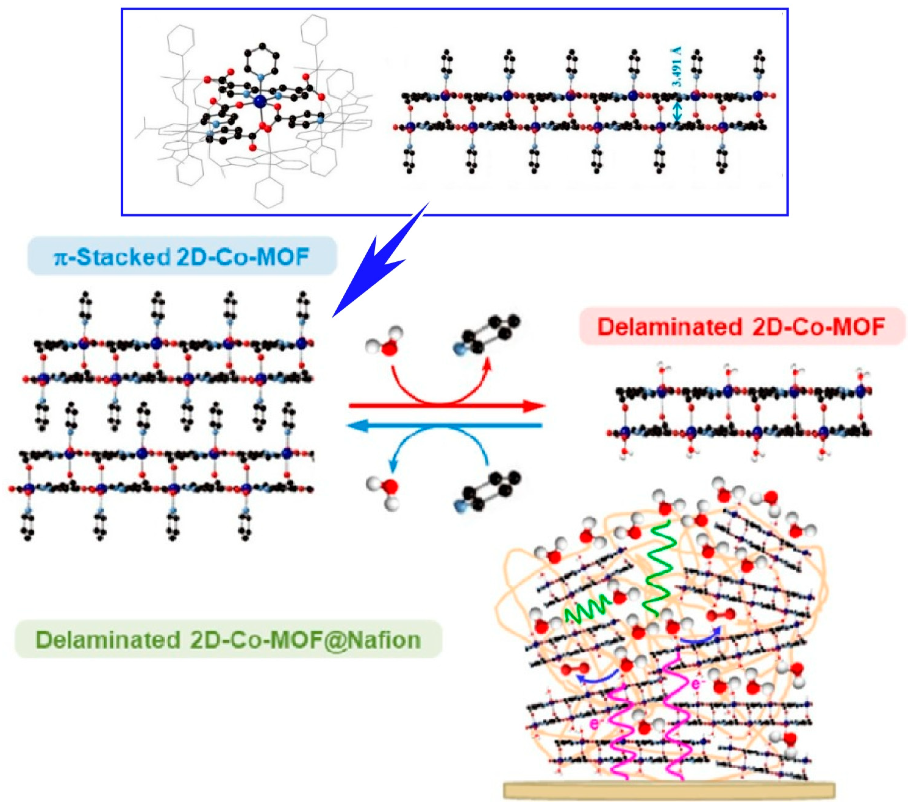
Oña-Burgos and co-workers have developed a stable structure consisting of two pairs of two-dimensional nanosheets [289]. In other words, they synthesized a new cobalt MOF based on a well-defined layered double core strongly bound by intermolecular bonds (Figure 9). Its three-dimensional structure is maintained by π–π stacking interactions between the unstable pyridine ligands of the nanosheets. In an aqueous solution, the axial pyridine ligands are exchanged with water molecules. This results in the exfoliation of the material. In such cases, the individual double nanosheets maintain their structures. Furthermore, the original three-dimensional layered structure is restored by a solvothermal process using pyridine. During the exfoliation-columnarization process, the material exhibits a memory effect. This two-dimensional MOF also exhibits electrocatalytic activity. Electrochemical activation of the two-dimensional cobalt MOF≅nafion-modified electrode improves both ion and electron transfer across the membrane. The formation of electrocatalytically active cobalt centers is then promoted. The activated composite exhibited enhanced electrocatalytic activity for the oxidation of water in neutral media. Spectroscopic and electrochemical characterizations were performed. The nanosheets have a special topology in which the cobalt centers are quite far apart. A mononuclear center-dependent reaction pathway mechanism has been proposed for the cobalt-mediated electrocatalytic oxygen evolution reaction. This electrocatalyst has a better TOF value and robustness than reported for similar electrocatalysts.
The construction of two-dimensional nanosheets into three-dimensional ordered structures facilitates mass transfer. The full potential of two-dimensional building blocks can be exploited in applications such as catalytic reactions. Zou et al. reported the synthesis of organometallic frameworks with orthogonal nanosheet arrays (Figure 10) [290]. Cubic MOFs are used as the core, and single-crystalline MOF nanosheets with naturally occurring non-preferred faceted exposures are epitaxially grown on top of them as shells. The nanoarchitectonized nanosheets have two typical shapes and crystallographic orientations. Nevertheless, they form an orthogonally aligned single-crystal framework. It is possible to obtain MOFs with a single composition and hollow orthogonally aligned nanosheet morphology. It has the characteristics of peculiar facet exposure and macroporous structure. Therefore, the electrocatalytic oxygen evolution activity is improved compared to conventional nanosheets. This structure exposes unsaturated active sites, stabilizes hydrogen-containing intermediate species, and promotes the oxygen evolution reaction process. The orthogonal arrangement of the nanosheets reduces the possibility of nanosheet re-stacking. Abundant surface active sites are provided, enhancing catalytic activity. Vertical and through pore channels are formed, facilitating diffusion of electrolyte and oxygen molecules. The hollow structure facilitates effective utilization of active sites and mass transfer, improving electrocatalytic properties. Thus, it is expected that rational function-oriented three-dimensional nanoarchitectonics strategies will lead to the design of highly functional heterostructures based on MOF nanosheets.
The controllable fabrication of angstrom-sized channels has long been desired in fundamental studies of ion transport. This is also necessary to mimic biological ion channels. Jiang, Zhang, and co-workers reported a strategy to grow MOFs into nanochannels with angstrom-scale ion channels with one- to three-dimensional pore structures (Figure 11) [291]. One-dimensional structures with flexible pore sizes can facilitate cation transport in conductivity and mobility one to two orders of magnitude higher than MOF channels with hybrid pore shapes and sizes. Theoretical simulations and calculations show that the energy barrier for ion transport through pure one-dimensional channels is lower than through complex channel connections. The three-dimensional MOF channel also exhibited better ion sieving properties than the one-dimensional and two-dimensional MOF channels. Further studies assuming angstrom porous MOFs with various channel configurations as the building blocks will open the way to fabricate artificial ion channels of 1 nm or smaller. Applications to high-efficiency ion separation and energy conversion technologies are expected. It will also provide guidance for the development of ion separation and nanofluidics.
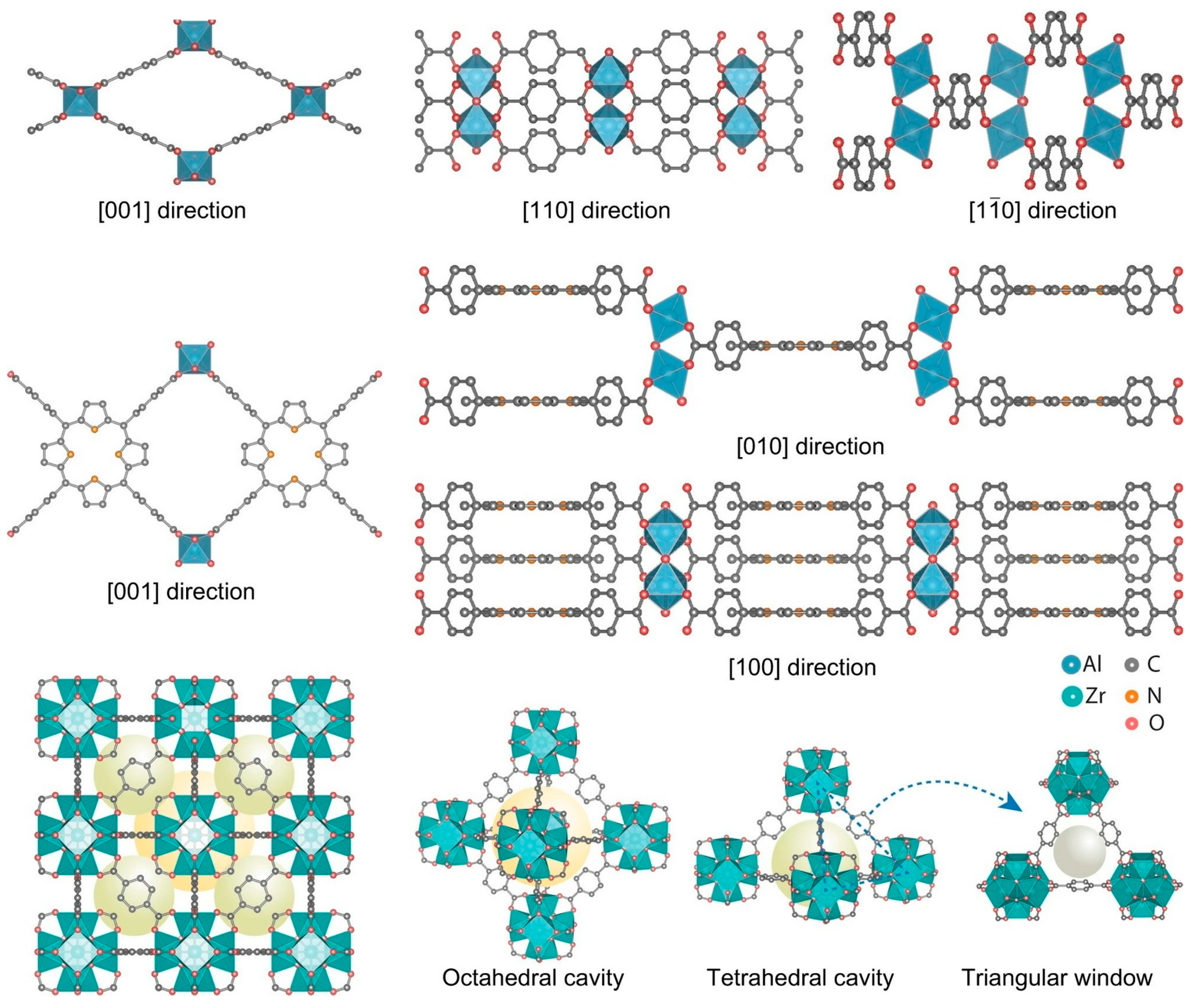
Two-dimensional conductive MOFs have attracted much attention for their usefulness in applications ranging from electrochemical energy storage to electronic devices. However, the stacked two-dimensional structure limits access to the internal pores. The full potential has not yet been realized. Park and co-workers reported a method for converting two-dimensionally conjugated MOFs into a three-dimensional framework by post-synthetic pillar ligand insertion (Figure 12) [292]. Such structural transformation improves ion accessibility to the internal pores. As a result, there can be up to a 2-fold increase in capacitance per weight. It is expected that such nanoarchitectonics methods can be used to functionalize a variety of two-dimensional conductive MOFs. Increased accessibility through the introduction of pillars increases the potential for sensing, electronics, and energy-related applications. The pillar portion can also be given a role other than that of a spacer. For example, providing a coordination site for the pillar ligand could lead to advanced applications including electrocatalysis and sensing. The restitution of conductive MOFs from two dimensional to three dimensional and the introduction of additional functionality is expected to facilitate the volatilization of possible materials in this new field.
Wee et al. reported the preparation of a new hierarchical MOF with Cu(II) centers linked by benzene tricarboxylates (BTC) (Figure 13) [293]. It is prepared by thermally induced solid-state transformation of a dense CuBTC precursor phase. It is found that ribbon-like one-dimensional constituent units transform into two-dimensional layers and finally into a three-dimensional network. The formed phases contain excess copper. The charge is compensated by hydroxyl groups, forming an open microporous framework with microporosity. This structure is useful for molecular separation. For example, it was superior to other hierarchical materials in the separation of 11-component mixtures of C1–C6 alkanes. Microscopic insights into structural host–guest interactions were obtained, confirming a significant entropic contribution to molecular separation.
Li and co-workers reported the synthesis of a two-dimension-on-three-dimension (2D-on-3D) hetero-MOF structure (Figure 14) [294]. They introduced a kinetic control method using polyvinylpyrrolidone to realize an anti-epitaxial growth pattern of foreign MOF nuclei on the (111) plane of the UiO-66-NH2 octahedron. This nanoarchitectonics methodology has led to the successful fabrication of 2D-on-3D MOFs with various heterostructures (Cu, Zn, Cd, Co, Ni). 2D-on-3D MOFs exhibit unique dimensional hybridization effects in the photocatalytic hydrogen evolution process in a photocatalytic hydrogen evolution process. The photoactivity was significantly enhanced compared to the usual dimensionally identical two-dimensional, three-dimensional, and 3D-on-3D MOF structures. This can be attributed to faster electron transfer rates and more efficient electron–hole separation.
Conversely, there are examples where dimensional reduction nanoarchitectonics of MOF structures from three dimensions to two dimensions is functionally superior. For example, a dimensional reduction approach has been proposed to improve the cryopreservation efficiency of red blood cells by MOFs. Such an approach takes the methodology of a stepwise reduction in three-dimensional MOF nanoparticles into two-dimensional ultrathin metal–organic layers (MOLs). Guo, Zhu, and co-workers synthesized a series of hafnium (Hf)-based two-dimensional metal–organic layers with different thicknesses (from single layer to stacked multilayers) and densities of hydrogen-bonding sites (Figure 15) [295]. Two-dimensional MOLs enhance the interaction of interfacial hydrogen-bonded water networks due to their high surface-to-volume ratio. This increases the utilization of internally ordered structures. The ability of the hydrogen donor group to recognize and match ice crystal planes can effectively inhibit ice growth and recrystallization for red blood cell cryopreservation. Thin-layered Hf-MOL was found to have significantly better ice recrystallization inhibitory activity and superior cell retrieval efficiency compared to three-dimensional MOL. Flexible two-dimensional MOLs have a higher density of hydrogen donor groups compared to three-dimensional MOF nanoparticles, which have a rigid structure and limited exposure of lattice planes. They also have smaller steric hindrance. These structural advantages may account for the marked efficiency of MOFs in ice suppression and erythrocyte recovery.
MOF and COF nanoarchitectonics approaches are excellent methods for rationally designing and building material structures from unit molecules and ions. Compared to conventional material synthesis methods, MOF and COF have a short history of development and leave plenty of room for various developments. The excellent dimensionality controlled nanoarchitectonics method is expected to contribute to the development of various functional systems.
4. 2D to 3D Functional Amplification: Cells Regulated by the Surface
Within living cells, various functional units function through cascade-like coordination. Thus, external stimuli and molecular inputs often lead to sophisticated functions. This can be a useful system for linking two-dimensional nanoarchitectonics to three-dimensional function. Thus, creating artificial structures in two dimensions and altering the function of the cells that come into contact with them can be very sophisticated transfer from two-dimensional to three-dimensional nanoarchitectonics function. This section presents examples of cellular control by two-dimensional liquid interfaces and nanoarchitectonized two-dimensional structures.
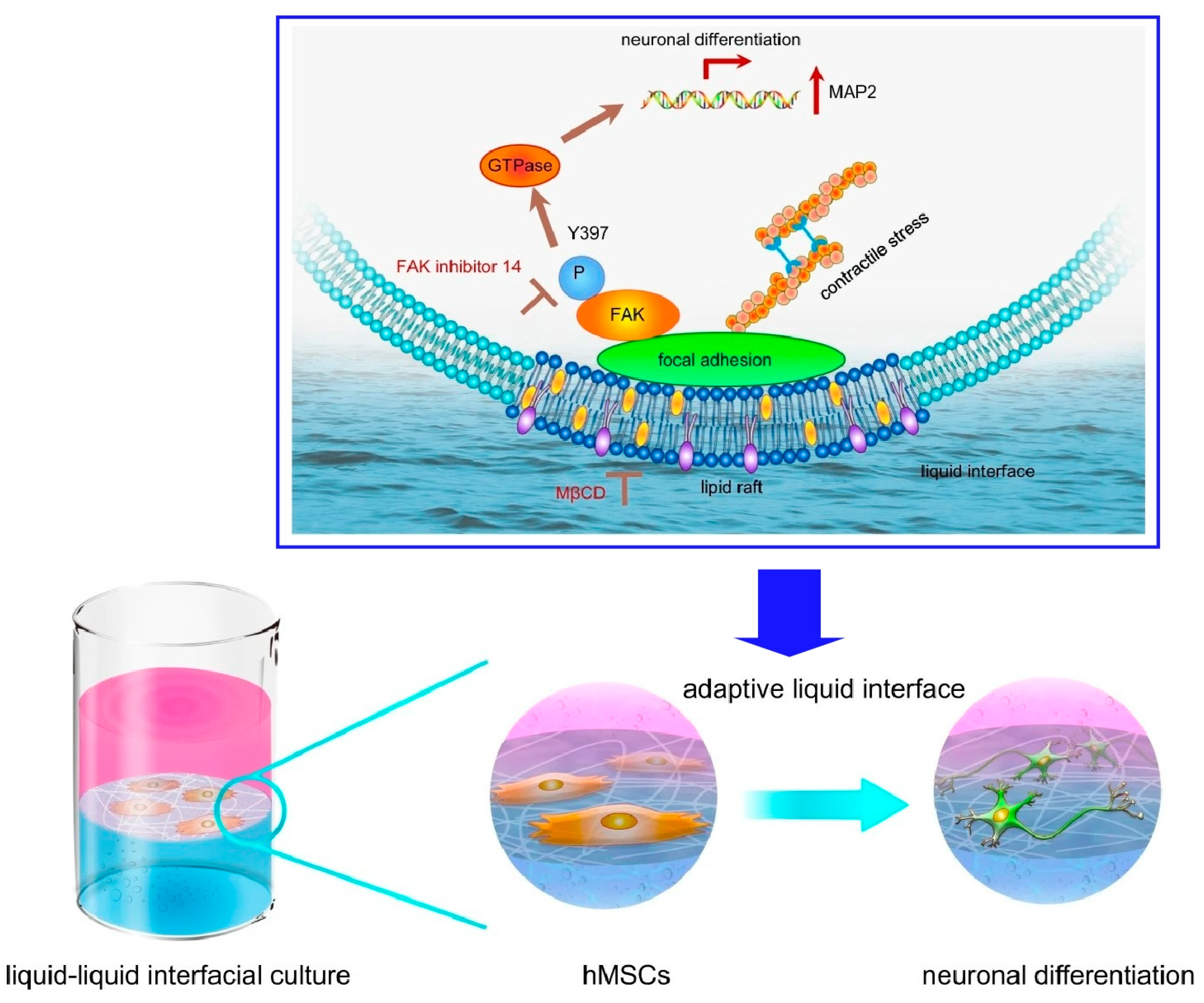
Cells are cultured on solid interfaces such as conventional culture media. In the culture process, cells are affected by the solid surface of the equipment, such as mechanical properties. On the other hand, cell control at the liquid interface, which is unaffected by materials such as containers, is a pioneer area. In fact, liquid interfaces are found in many places as adaptive systems in biological systems, from the tear film of the eye to the liquid lining of the lungs and stomach. At such interfaces, liquid interfaces are ubiquitous in natural adaptive systems. The fluidity and reconfigurability of liquids allow for unique response mechanisms. By anchoring living cells at the liquid interface, a system can be constructed that dynamically adapts to the forces generated by the cells. Such cell research at the soft liquid interface is attracting increasing attention. Jia et al. have successfully developed a technique that uses the interface between two immiscible liquids, aqueous cell culture solutions and perfluorocarbons, as a site for culturing and inducing differentiation of human mesenchymal stem cells (hMSCs) (Figure 16) [296]. At this interface, coexisting fibronectin proteins form organized nanosheets that exhibit transient mechanical effects with the cells. Early in culture, the traction force of the cells dynamically transformed the two-dimensional protein nanosheet into a hierarchical fiber structure. Elongated fibronectin aggregates modulate the size and elongation of the focal adhesions of the interacting network. Ultimately, these mechano-transduction signals determine the fate of stem cell differentiation. In this system, cultured hMSCs spontaneously differentiated into neurons without the addition of differentiation-inducing factors. The hMSCs adhered to the liquid interface and took an elongated adherent shape. Stress fibers of F-actin originating there were formed. The accumulation of downstream signaling protein phosphatase (FAK) activators in adhesion plaques and nuclear migration of Yes-associated protein (YAP) were also observed. hMSCs do not perceive protein nanosheets as soft, suggesting that some novel mechano-sensing mechanism is involved. Depending on the design of the two-dimensional nanoarchitectonics, it may be possible to induce differentiation into various types of cells, not only neural cells. This methodology is expected to lead to the development of new technologies for regenerative medicine.
Protein nanofibrils are polymeric β-sheet aggregates of proteins of several microns in length. This structure is also a promising material to mimic extracellular matrix (ECM) matrix fibers due to its biocompatibility for cell adhesion and high mechanical strength. Jia et al. created an adaptive biomaterial based on a two-dimensional network of protein nanofibrils at the liquid–liquid interface and cultured hMSCs [297]. Culture on a two-dimensional network of protein nanofibrils at the liquid–liquid interface promoted neural differentiation of hMSCs. Throughout the study, lipid raft microdomains were found to play a central regulatory role in both the initial cell adhesion and subsequent neural differentiation of hMSCs (Figure 17). They are receptive to biophysical stimuli involving the lipid raft/focal adhesion kinase (FAK) pathway. It seems to direct the neural differentiation of hMSCs. Lipid rafts internalize cell adhesion molecules. In addition, lipid rafts act as an enrichment platform. This induces the integration of large signaling complexes. Through these processes, cells can rapidly adapt to the constantly changing microenvironment. FAK is one of the key mechano-sensors at the adaptive liquid interface. Spatiotemporal regulation of FAK phosphorylation is a prerequisite for neural differentiation of hMSCs. Lipid raft formation and FAK phosphorylation at the adaptive liquid interface regulate hMSC differentiation. These findings also provide a better understanding of the fundamentals of cell-ECM dynamic interactions. Two-dimensional nanoarchitectonics can also incorporate bioactive proteins and responsive polymers. Two-dimensional nanoarchitectonics of liquid interfaces may provide opportunities to design adaptive biomaterials that were previously unimaginable.
Gautrot and co-workers examine the interfacial dynamics of bovine serum albumin (BSA) and β-lactoglobulin (BLG) aggregates at the fluorinated liquid-water interface (Figure 18) [298]. The design of protein nanosheets based on these two globular proteins biofunctionalized with RGDSP peptides that enable cell adhesion is mentioned. High cell proliferation can be achieved even on bioemulsions with protein nanosheet formation without surfactants. As a strategy for the rational design of scaffold proteins at liquid interfaces, the fabrication of interfaces with strong shear dynamics and elasticity, bioactivity, and cell adhesion was investigated. For example, in the case of BLG nanosheets, relatively high elasticity was observed even in the absence of the co-surfactant. Based on this as well, the research demonstrated adhesion and proliferation of mesenchymal stem cells and human embryonic kidney (HEK) cells on bioemulsions stabilized with RGD-functionalized protein nanosheets. Such protein nanosheets and bioemulsions are useful for the development of bioreactors for scale-up of cell production. Such studies could also lead to the control of biological activity with scaffold proteins commonly used in food processing and stem cell technology, without the use of surfactant molecules.
The use of liquid interfaces for cell culture has the potential to avoid the inconveniences of using solid substrates. In particular, it is advantageous in terms of scale-up and cell detachment. This methodology could be applied to other biotechnological platforms, such as microdroplet systems. For this purpose, analytical studies are needed. Gautrot and co-workers reconfirmed that cell spreading and growth on low-viscous liquid surfaces are enabled by the self-assembly of mechanically strong protein nanosheets at the interface [299]. Interfacial rheology and atomic force microscopy measurements revealed the mechanical properties of protein nanosheets and their associated liquid interfaces. The aggregation behavior of surfactant molecules with proteins and polymers associated at the liquid interface is directly related to the interfacial mechanics. Cells do not rely on surface tension to sustain diffusion, as in the case of amoebae, but primarily sense the in-plane mechanical properties of the interface (Figure 19). Based on these findings, it is essential to design bulk and nanoscale mechanical properties independently. Both scales can provide suitable structures and control cellular phenotypes. In other words, the design of biomaterials and implants requires nanoscale material design, where cell adhesion phenomena can be designed at the interface, independently of other bulk properties needed to provide flexibility and structure.
Gautrot and co-workers showed that mesenchymal and adherent stem cells can be cultured on liquid surfaces [300]. They also exemplified that this is mediated by the association of polymer nanosheets at the liquid–liquid interface. Despite the lack of bulk mechanical properties of the underlying liquid substrate, cell adhesion to the quasi-two-dimensional material is mediated by an integrin/actomyosin mechanism. Stem cell proliferation and differentiation are also controlled by the mechanical properties of the self-assembled protein nanosheets. Keratinocytes spread on rigid poly(l-lysine) nanosheets formed a structured actin cytoskeleton with distinct stress fibers. Focal adhesions were also formed. In contrast, cells adhering to the soft poly(l-lysine)-oil interface did not observe such structures. The use of solid substrates often requires potentially harmful enzymatic degradation for cell recovery. The use of liquid substrates avoids such problems.
Cells are controlled not only by two-dimensional nanoarchitectonics, such as protein nanosheets that spontaneously form at the liquid interface, but also by the two-dimensional structure of artificially placed nanomaterials. For example, the assembly of fullerenes, which are nanomaterials of carbon materials, is often used in two-dimensional nanoarchitectonics. Although fullerenes are zero-dimensional spherical structures, they exhibit a variety of self-assembled structures from one dimension to three dimensions, such as sheets, rods, pores, and whiskers [301,302,303,304,305,306,307,308,309]. For example, fullerene nanowhiskers can be prepared on a large scale using the liquid–liquid interface deposition method. Minami and co-workers have successfully induced muscle differentiation by arranging fullerene nanowhiskers while simultaneously controlling the direction of cell growth (Figure 20) [310]. The two-dimensional in-plane aligned structure of fullerene nanowhiskers can be applied as a cell scaffold, and the orientation of muscle fibers formed during muscle differentiation can be controlled by the orientation of fullerene nanowhiskers. Using the Langmuir–Blodgett (LB) method, highly aligned one-dimensional fullerene nanowhisker scaffolds were fabricated in the centimeter region. This scaffold can simultaneously control cell orientation and differentiation into muscle cells C2C12 myoblasts. Subsequently, myogenic differentiation and cell growth direction were analyzed by immunostaining for myosin heavy chain. A protein required for nucleus and myotube formation C2C12 myoblasts were found to fuse and form multinucleated myotubes. The fusion index increased from 12.3% on glass to 23.2% on aligned fullerene nanowhisker scaffolds. One-dimensional fullerene nanowhiskers stimulate myoblast fusion. The direction of myotube formation strongly coincided with the direction of aligned fullerene nanowhiskers. When the same experiment was performed on bare glass, myoblasts fused randomly.
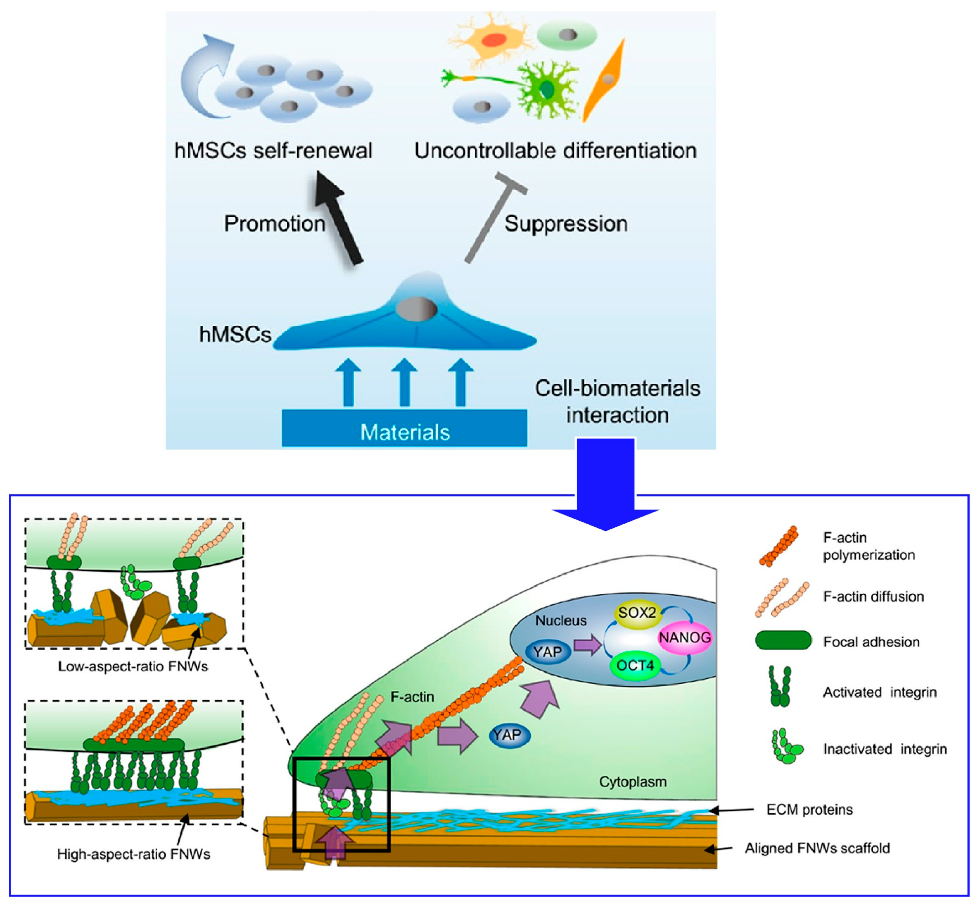
hMSCs are useful for cell-based tissue regeneration therapy because of their easy availability and potent immunosuppressive properties. However, the therapeutic efficacy based on hMSCs is limited by the small amount of human-derived cells isolated for clinical use. During in vitro growth, hMSCs undergo uncontrolled differentiation. In the process of in vitro proliferation, hMSCs undergo uncontrolled differentiation, thereby rapidly losing pluripotency and regenerative capacity. Therefore, new strategies to grow hMSCs in vitro while preserving their stem cell properties are strongly needed. This technique can produce large-area nanostructured surfaces with continuously adjustable alignment of the constituents. Nanopatterned surfaces fabricated with high-aspect-ratio fullerene nanowhiskers could promote long-term pluripotency retention and differentiation potential of hMSCs via appropriate cell contractility and nuclear localization of Yes-associated protein (YAP) as demonstrated by Song et al. (Figure 21) [311]. Mechanical signals are transmitted to the nucleus by YAP. As a transcriptional coactivator, YAP translocation to the nucleus positively regulates the activity of core regulators (OCT4, SOX2, NANOG). As a result, retention of pluripotency of hMSCs is promoted. High aspect ratio fullerene nanowhiskers as cell culture scaffolds create an intermediate situation where focal adhesion is effectively reduced but not eliminated. As a result, long-term proliferation of hMSCs that retain pluripotency is mediated by appropriate cell contractility and nuclear localization of YAP. The pseudo-LB method used here is a simple technique that can be manipulated manually. It can be easily used to fabricate centimeter-sized nanotopographic substrates for the mass proliferation of hMSCs in clinical practice. This study demonstrates the importance of two-dimensional nanoarchitectonics for improving hMSCs technology in regenerative therapies.
Cells have well-developed cascade-like information transmission systems, which are excellent systems for generating large functions from simple stimuli. Exposing cells to structure-controlled two-dimensional surfaces allows for the expression of sophisticated functions such as cell differentiation and proliferation. Various scaffolds such as protein nanosheets and very rigid fullerene nanowhisker arrays, which form in very soft fields such as liquid surfaces, produce diverse cellular changes. This is not only of basic scientific interest as two-dimensional nanoarchitectonics makes a breakthrough into three-dimensional function. This is an area that should also make a significant contribution to practical areas such as regenerative medicine.
5. Summary and Perspectives
Nanoarchitectonics is a post-nanotechnology concept that provides functional material systems from nano-units (atoms, molecules, and nanomaterials). Interfacial media contribute sufficiently to building functional materials. This is where two-dimensional (2D) materials as building units of three-dimensional (3D) functional nanoarchitectures are powerful. On top of that, materials nanoarchitectonics from two dimensions to three dimensions is necessary for the development of a wider range of functional materials. In this review, the control of liquid crystals from two dimensions, three-dimensional architecture of MOF/COF structures, and the control of cell arrangement and differentiation by two-dimensional contact are taken as examples.
Molecular films made of LB-type monolayers can control the orientation of liquid crystals as a command surface. The molecular-level phenomena of structural and orientation changes on the command surface are amplified into three-dimensional material properties such as liquid crystal orientation and phase transitions. This indicates that the combination of a soft oriented structure and a precise surface is useful. MOF and COF are methods that enable rational design and construction of material structures from unit molecules and ions. The structure produced is determined by the arrangement of interacting functional groups in the molecular unit and the coordination structure of the ions. This means that the two-dimensional and three-dimensional structures of MOFs and COFs are determined by the structural design of the molecular unit-ion unit in the zero dimension. Therefore, the conversion from two dimensional to three dimensional can be achieved by devising the combination of the two-dimensional and three-dimensional structures. For structures with fixed interaction points, the structural design of the units is shown to be useful. Organisms are complex complexes of functions. However, cells have well-developed cascade-like information transmission systems. By exposing cells to structure-controlled two-dimensional surfaces, sophisticated functions such as cell differentiation and proliferation are expressed. This is a rational system for generating large three-dimensional functions from simple stimuli from two dimensions. Utilizing the cascade stimulus transduction system of living organisms is also useful for generating three-dimensional functions from two-dimensional nanoarchitectonics. In addition to approaches and examples described in this review article, simulations/theoretical perspective of 2D materials nanoarchitectonics for 3D functionalities are crucial matters. For example, both DFT calculation [312] and ab initio molecular dynamics [313] are rapidly transforming the field. With the aids of experimental and theoretical considerations, representative differences between 2D and 3D materials will be more clearly elucidated. Such additional efforts would lead to more sophisticated transformation from 2D to 3D from viewviewpoints of applications.
The several examples presented here have extracted several keys to increasing two-dimensional nanoarchitectonics to three-dimensional functionality. These include the use of soft orientation systems, the use of structural architecture reflecting molecular unit structures, and the use of cascade-like function transfer systems in living organisms. These are typical examples, but there may be other ways to link two-dimensional nanoarchitectonics to three-dimensional functionality. The ultimate goal of nanoarchitectonics is to create bio-like functional materials whose functions are hierarchical and exhibit complex interactions. Therefore, we must consider systems that incorporate multiple and diverse multifunctional mechanisms in three-dimension as described above. There are an infinite number of choices of substances, actions, and functions, and their combinations are also diverse. Therefore, there may be a limit to the trial-and-error or few-theory-based approach. Fortunately, humankind has developed artificial intelligence technology. In the field of materials science, machine learning approaches [314,315,316] and the concept of materials informatics [317,318,319] are being used. The combination of nanoarchitectonics and materials informatics has also been proposed [320,321]. The fabrication of functional structures from a large number of options may be completed with the help of artificial intelligence under the concept of nanoarchitectonics. Finally, these developments are expected to become nanoarchitectonics as a method for everything in materials science.
Funding
This study was partially supported by JSPS KAKENHI, grant number JP20H00392 and JP23H05459.
Institutional Review Board Statement
Not applicable.
Informed Consent Statement
Not applicable.
Data Availability Statement
Data sharing not applicable.
Conflicts of Interest
The author declares no conflict of interest.
References
- Liu, J.; Zhang, J.-G.; Yang, Z.; Lemmon, J.P.; Imhoff, C.; Graff, G.L.; Li, L.; Hu, J.; Wang, C.; Xiao, J.; et al. Materials science and materials chemistry for large scale electrochemical energy storage: From transportation to electrical grid. Adv. Funct. Mater. 2013, 23, 929–946. [Google Scholar] [CrossRef]
- Kageyama, H.; Hayashi, K.; Maeda, K.; Attfield, J.P.; Hiroi, Z.; Rondinelli, J.M.; Poeppelmeier, K.R. Expanding frontiers in materials chemistry and physics with multiple anions. Nat. Commun. 2018, 9, 772. [Google Scholar] [CrossRef] [PubMed]
- Kudo, A.; Miseki, Y. Heterogeneous photocatalyst materials for water splitting. Chem. Soc. Rev. 2009, 38, 253–278. [Google Scholar] [CrossRef] [PubMed]
- Guo, D.; Shibuya, R.; Akiba, C.; Saji, S.; Kondo, T.; Nakamura, J. Active sites of nitrogen-doped carbon materials for oxygen reduction reaction clarified using model catalysts. Science 2016, 351, 361–365. [Google Scholar] [CrossRef] [PubMed]
- Nandihalli, N.; Liu, C.J.; Mori, T. Polymer based thermoelectric nanocomposite materials and devices: Fabrication and characteristics. Nano Energy 2020, 78, 105186. [Google Scholar] [CrossRef]
- Zhang, E.; Zhu, Q.; Huang, J.; Liu, J.; Tan, G.; Sun, C.; Li, T.; Liu, S.; Li, Y.; Wang, H.; et al. Visually resolving the direct Z-scheme heterojunction in CdS@ZnIn2S4 hollow cubes for photocatalytic evolution of H2 and H2O2 from pure water. Appl. Catal. B Environ. 2021, 293, 120213. [Google Scholar] [CrossRef]
- Qi, Y.; Zhang, J.; Kong, Y.; Zhao, Y.; Chen, S.; Li, D.; Liu, W.; Chen, Y.; Xie, T.; Cui, J.; et al. Unraveling of cocatalysts photodeposited selectively on facets of BiVO4 to boost solar water splitting. Nat. Commun. 2022, 13, 484. [Google Scholar] [CrossRef]
- Maeda, K.; Takeiri, F.; Kobayashi, G.; Matsuishi, S.; Ogino, H.; Ida, S.; Mori, T.; Uchimoto, Y.; Tanabe, S.; Hasegawa, T.; et al. Recent progress on mixed-anion materials for energy applications. Bull. Chem. Soc. Jpn. 2022, 95, 26–37. [Google Scholar] [CrossRef]
- Klyndyuk, A.I.; Chizhova, E.A.; Kharytonau, D.S.; Medvedev, D.A. Layered oxygen-deficient double perovskites as promising cathode materials for solid oxide fuel cells. Materials 2022, 15, 141. [Google Scholar] [CrossRef]
- Jakhar, M.; Kumar, A.; Ahluwalia, P.K.; Tankeshwar, K.; Pandey, R. Engineering 2D materials for photocatalytic water-splitting from a theoretical perspective. Materials 2022, 15, 2221. [Google Scholar] [CrossRef]
- Murugan, S.; Lee, E.-C. Recent Advances in the synthesis and application of vacancy-ordered halide double perovskite materials for solar cells: A promising alternative to lead-based perovskites. Materials 2023, 16, 5275. [Google Scholar] [CrossRef]
- Desoky, M.M.H.; Caldera, F.; Brunella, V.; Ferrero, R.; Hoti, G.; Trotta, F. Cyclodextrins for lithium batteries applications. Materials 2023, 16, 5540. [Google Scholar] [CrossRef] [PubMed]
- Pastuszak, J.; Węgierek, P. Photovoltaic cell generations and current research directions for their development. Materials 2022, 15, 5542. [Google Scholar] [CrossRef] [PubMed]
- Tsuchii, Y.; Menda, T.; Hwang, S.; Yasuda, T. Photovoltaic properties of p-conjugated polymers based on fused cyclic Imide and amide skeletons. Bull. Chem. Soc. Jpn. 2023, 96, 90–94. [Google Scholar] [CrossRef]
- Yano, J.; Suzuk, K.; Hashimoto, C.; Tsutsumi, C.; Hayase, N.; Kitani, A. Trial fabrication of NADH-dependent enzymatic ethanol biofuel cell providing H2 gas as well as Electricity. Bull. Chem. Soc. Jpn. 2023, 96, 331–338. [Google Scholar] [CrossRef]
- Imahori, H. Molecular photoinduced charge separation: Fundamentals and application. Bull. Chem. Soc. Jpn. 2023, 96, 339–352. [Google Scholar] [CrossRef]
- Chen, K.; Xiao, J.; Vequizo, J.J.M.; Hisatomi, T.; Ma, Y.; Nakabayashi, M.; Takata, T.; Yamakata, A.; Shibata, N.; Domen, K. Overall water splitting by a SrTaO2N-based photocatalyst decorated with an Ir-promoted Ru-based cocatalyst. J. Am. Chem. Soc. 2023, 145, 3839–3843. [Google Scholar] [CrossRef] [PubMed]
- Huang, G.; Hu, M.; Xu, X.; Alothman, A.A.; Mushab, M.S.S.; Ma, S.; Shen, P.K.; Zhu, J.; Yamauchi, Y. Optimizing heterointerface of Co2P–CoxOy Nanoparticles within a porous carbon network for deciphering superior water splitting. Small Struct. 2023, 4, 2200235. [Google Scholar] [CrossRef]
- Komaba, S.; Hasegawa, T.; Dahbi, M.; Kubota, K. Potassium intercalation into graphite to realize high-voltage/high-power potassium-ion batteries and potassium-ion capacitors. Electrochem. Commun. 2015, 60, 172–175. [Google Scholar] [CrossRef]
- Saidul Islam, M.S.; Shudo, Y.; Hayami, S. Energy conversion and storage in fuel cells and super-capacitors from chemical modifications of carbon allotropes: State-of-art and prospect. Bull. Chem. Soc. Jpn. 2022, 95, 1–25. [Google Scholar] [CrossRef]
- Yoshino, A. The lithium-ion battery: Two breakthroughs in development and two reasons for the Nobel Prize. Bull. Chem. Soc. Jpn. 2022, 95, 195–197. [Google Scholar] [CrossRef]
- Kumar, R.; Joanni, E.; Sahoo, S.; Shim, J.-J.; Tan, W.K.; Matsuda, A.; Singh, R.K. An overview of recent progress in nanostructured carbon-based supercapacitor electrodes: From zero to bi-dimensional materials. Carbon 2022, 193, 298–338. [Google Scholar] [CrossRef]
- Kumar, R.; Youssry, S.M.; Joanni, E.; Sahoo, S.; Kawamura, G.; Matsuda, A. Microwave-assisted synthesis of iron oxide homogeneously dispersed on reduced graphene oxide for high-performance supercapacitor electrodes. J. Energy Storage 2022, 56, 105896. [Google Scholar] [CrossRef]
- Das, H.T.; Dutta, S.; Balaji, T.E.; Das, N.; Das, P.; Dheer, N.; Kanojia, R.; Ahuja, P.; Ujjain, S.K. Recent rrends in carbon nanotube electrodes for flexible supercapacitors: A review of smart energy storage device assembly and performance. Chemosensors 2022, 10, 223. [Google Scholar] [CrossRef]
- Kim, E.J.; Kumar, P.R.; Gossage, Z.T.; Kubota, K.; Hosaka, T.; Tatara, R.; Komaba, S. Active material and interphase structures governing performance in sodium and potassium ion batteries. Chem. Sci. 2022, 13, 6121–6158. [Google Scholar] [CrossRef] [PubMed]
- Zhang, G.; Qiuhong Bai, Q.; Wang, X.; Li, C.; Uyama, H.; Shen, Y. Preparation and mechanism investigation of walnut shell-based hierarchical porous carbon for supercapacitors. Bull. Chem. Soc. Jpn. 2023, 96, 190–197. [Google Scholar] [CrossRef]
- Fu, M.; Chen, W.; Lei, Y.; Yu, H.; Lin, Y.; Terrones, M. Biomimetic construction of ferrite quantum dot/graphene heterostructure for enhancing ion/charge transfer in supercapacitors. Adv. Mater. 2023, 35, 2300940. [Google Scholar] [CrossRef] [PubMed]
- Ganesan, P.; Ishihara, A.; Staykov, A.; Nakashima, N. Recent advances in nanocarbon-based nonprecious metal catalysts for oxygen/hydrogen reduction/evolution reactions and Zn-air battery. Bull. Chem. Soc. Jpn. 2023, 96, 429–443. [Google Scholar] [CrossRef]
- Qiu, Y.-F.; Murayama, H.; Fujitomo, C.; Kawai, S.; Haruta, A.; Hiasa, T.; Mita, H.; Motohashi, K.; Yamamoto, E.; Tokunaga, M. Oxidative decomposition mechanism of ethylene carbonate on positive electrodes in lithium-ion batteries. Bull. Chem. Soc. Jpn. 2023, 96, 444–451. [Google Scholar] [CrossRef]
- Lakshmi, K.C.S.; Vedhanarayanan, B. High-performance supercapacitors: A comprehensive review on paradigm shift of conventional energy storage devices. Batteries 2023, 9, 202. [Google Scholar] [CrossRef]
- Honda, M.; Suzuki, N. Toxicities of polycyclic aromatic hydrocarbons for aquatic animals. Int. J. Environ. Res. Public Health 2020, 17, 1363. [Google Scholar] [CrossRef] [PubMed]
- Qaidi, S.; Najm, H.M.; Abed, S.M.; Özkılıç, Y.O.; Al Dughaishi, H.; Alosta, M.; Sabri, M.M.S.; Alkhatib, F.; Milad, A. Concrete containing waste glass as an environmentally friendly aggregate: A review on fresh and mechanical characteristics. Materials 2022, 15, 6222. [Google Scholar] [CrossRef] [PubMed]
- Li, S.L.; Chang, G.; Huang, Y.; Kinooka, K.; Chen, Y.; Fu, W.; Gong, G.; Yoshioka, T.; McKeown, N.B.; Hu, Y. Biphenol-based ultrathin microporous nanofilms for highly efficient molecular sieving separation. Angew. Chem. Int. Ed. 2022, 61, e202212816. [Google Scholar] [CrossRef]
- Kushida, W.; Gonzales, R.R.; Shintani, T.; Matsuoka, A.; Nakagawa, K.; Yoshioka, T.; Hideto Matsuyama, H. Organic solvent mixture separation using fluorine-incorporated thin film composite reverse osmosis membrane. J. Mater. Chem. A 2022, 10, 4146–4156. [Google Scholar] [CrossRef]
- Wang, X.; Liu, Z.; Lu, J.; Teng, H.; Fukuda, H.; Qin, W.; Wei, T.; Liu, Y. Highly selective membrane for efficient separation of environmental determinands: Enhanced molecular imprinting in polydopamine-embedded porous sleeve. Chem. Eng. J. 2022, 449, 137825. [Google Scholar] [CrossRef]
- Zhang, L.; Chong, H.L.H.; Moh, P.Y.; Albaqami, M.D.; Tighezza, A.M.; Qin, C.; Ni, X.; Cao, J.; Xu, X.; Yamauchi, Y. β-FeOOH nanospindles as chloride-capturing electrodes for electrochemical faradic deionization of saline water. Bull. Chem. Soc. Jpn. 2023, 96, 306–309. [Google Scholar] [CrossRef]
- Zhang, T.; Cain, A.K.; Semenec, L.; Liu, L.; Hosokawa, Y.; Inglis, D.W.; Yalikun, Y.; Li, M. Microfluidic separation and enrichment of Escherichia coli by size using viscoelastic flows. Anal. Chem. 2023, 95, 2561–2569. [Google Scholar] [CrossRef]
- Chowdhury, R.; Al Biruni, M.T.; Afia, A.; Hasan, M.; Islam, M.R.; Ahmed, T. Medical waste incineration fly ash as a mineral filler in dense bituminous course in flexible pavements. Materials 2023, 16, 5612. [Google Scholar] [CrossRef]
- Saleh, T.S.; Badawi, A.K.; Salama, R.S.; Mostafa, M.M.M. Design and development of novel composites containing nickel ferrites supported on activated carbon derived from agricultural wastes and its application in water remediation. Materials 2023, 16, 2170. [Google Scholar] [CrossRef]
- Yadav, A.A.; Hunge, Y.M.; Kang, S.-W.; Fujishima, A.; Terashima, C. Enhanced photocatalytic degradation activity using the V2O5/RGO Composite. Nanomaterials 2023, 13, 338. [Google Scholar] [CrossRef]
- Muradov, N.Z.; Veziroğlu, T.N. “Green” path from fossil-based to hydrogen economy: An overview of carbon-neutral technologies. Int. J. Hydrogen Energy 2008, 33, 6804–6839. [Google Scholar] [CrossRef]
- Chen, S.; Liu, J.; Zhang, Q.; Teng, F.; McLellan, B.C. A critical review on deployment planning and risk analysis of carbon capture, utilization, and storage (CCUS) toward carbon neutrality. Renew. Sust. Energ. Rev. 2022, 167, 112537. [Google Scholar] [CrossRef]
- Chapman, A.; Kubota, E.; Kubota, M.; Nagao, A.; Bertsch, K.; Macadre, A.; Tsuchiyama, T.; Masamura, T.; Takaki, S.; Komoda, R.; et al. Achieving a carbon neutral future through advanced functional materials and technologies. Bull. Chem. Soc. Jpn. 2022, 95, 73–103. [Google Scholar] [CrossRef]
- Li, Y.; Ma, G. A Study on the high-quality development path and implementation countermeasures of China’s construction industry toward the carbon peaking and carbon neutralization goals. Sustainability 2024, 16, 772. [Google Scholar] [CrossRef]
- Takeishi, K. Evolution of turbine cooled vanes and blades applied for large industrial gas turbines and its trend toward carbon neutrality. Energies 2022, 15, 8935. [Google Scholar] [CrossRef]
- Takase, S.; Aritsu, T.; Sakamoto, Y.; Sakuno, Y.; Shimizu, Y. Preparation of highly conductive phthalocyaninato-cobalt iodide at the interface between aqueous KI solution and organic solvent and catalytic properties for electrochemical reduction of CO2. Bull. Chem. Soc. Jpn. 2023, 96, 649–653. [Google Scholar] [CrossRef]
- Ishihara, S.; O’Kelly, C.J.; Takeshi Tanaka, T.; Kataura, H.; Labuta, J.; Shingaya, Y.; Nakayama, T.; Ohsawa, T.; Nakanishi, T.; Swager, T.M. Metallic versus semiconducting SWCNT chemiresistors: A case for separated SWCNTs wrapped by a metallosupramolecular polymer. ACS Appl. Mater. Interfaces 2017, 9, 38062–38067. [Google Scholar] [CrossRef]
- Sasaki, Y.; Kubota, R.; Minami, T. Molecular self-assembled chemosensors and their arrays. Coord. Chem. Rev. 2021, 429, 213607. [Google Scholar] [CrossRef]
- Masuda, Y. Recent advances in SnO2 nanostructure based gas sensors. Sens. Actuat. B Chem. 2022, 364, 131876. [Google Scholar] [CrossRef]
- Tatli, S.; Mirzaee-Ghaleh, E.; Rabbani, H.; Karami, H.; Wilson, A.D. Rapid Detection of Urea Fertilizer Effects on VOC Emissions from Cucumber Fruits Using a MOS E-Nose Sensor Array. Agronomy 2022, 12, 35. [Google Scholar] [CrossRef]
- Saito, S.; Haraga, T.; Marumo, K.; Sato, Y.; Nakano, Y.; Tasaki-Handa, Y.; Shibukawa, M. Americium(III)/curium(III) complete separation and sensitive fluorescence detection by capillary and gel electrophoresis using emissive hexadentate/octadentate polyaminocarboxylate ligands. Bull. Chem. Soc. Jpn. 2023, 96, 223–225. [Google Scholar] [CrossRef]
- Simonenko, N.P.; Glukhova, O.E.; Plugin, I.A.; Kolosov, D.A.; Nagornov, I.A.; Simonenko, T.L.; Varezhnikov, A.S.; Simonenko, E.P.; Sysoev, V.V.; Kuznetsov, N.T. The Ti0.2V1.8C MXene ink-prepared chemiresistor: From theory to tests with humidity versus VOCs. Chemosensors 2023, 11, 7. [Google Scholar] [CrossRef]
- Dong, L.; Wang, M.; Wu, J.; Zhu, C.; Shi, J.; Morikawa, H. Stretchable, adhesive, self-healable, and conductive hydrogel-based deformable triboelectric nanogenerator for energy harvesting and human motion sensing. ACS Appl. Mater. Interfaces 2022, 14, 9126–9137. [Google Scholar] [CrossRef]
- Anggraini, L.E.; Rahmawati, H.; Nasution, M.A.F.; Jiwanti, P.K.; Einaga, Y.; Ivandini, T.A. Development of an acrylamide biosensor using guanine and adenine as biomarkers at boron-doped diamond electrodes: Integrated molecular docking and experimental studies. Bull. Chem. Soc. Jpn. 2023, 96, 420–428. [Google Scholar] [CrossRef]
- Kalyana Sundaram, S.d.; Hossain, M.M.; Rezki, M.; Ariga, K.; Tsujimura, S. Enzyme cascade electrode reactions with nanomaterials and their applicability towards biosensor and biofuel cells. Biosensors 2023, 13, 1018. [Google Scholar] [CrossRef] [PubMed]
- Lostao, A.; Lim, K.S.; Pallarés, M.C.; Ptak, A.; Marcuello, C. Recent advances in sensing the inter-biomolecular interactions at the nanoscale—A comprehensive review of AFM-based force spectroscopy. Int. J. Biol. Macromol. 2023, 238, 124089. [Google Scholar] [CrossRef] [PubMed]
- Theyagarajan, K.; Kim, Y.-J. Recent Developments in the Design and Fabrication of Electrochemical Biosensors Using Functional Materials and Molecules. Biosensors 2023, 13, 424. [Google Scholar] [CrossRef]
- Bhaskar, S. Biosensing Technologies: A Focus Review on Recent Advancements in Surface Plasmon Coupled Emission. Micromachines 2023, 14, 574. [Google Scholar] [CrossRef]
- Cabral, H.; Miyata, K.; Osada, K.; Kataoka, K. Block copolymer micelles in nanomedicine applications. Chem. Rev. 2018, 118, 6844–6892. [Google Scholar] [CrossRef] [PubMed]
- Rabiee, N.; Ahmadi, S.; Akhavan, O.; Luque, R. Silver and gold nanoparticles for antimicrobial purposes against multi-drug resistance bacteria. Materials 2022, 15, 1799. [Google Scholar] [CrossRef]
- Naganthran, A.; Verasoundarapandian, G.; Khalid, F.E.; Masarudin, M.J.; Zulkharnain, A.; Nawawi, N.M.; Karim, M.; Che Abdullah, C.A.; Ahmad, S.A. Synthesis, characterization and biomedical application of silver nanoparticles. Materials 2022, 15, 427. [Google Scholar] [CrossRef]
- Bhattacharya, T.; Soares, G.A.B.e.; Chopra, H.; Rahman, M.M.; Hasan, Z.; Swain, S.S.; Cavalu, S. Applications of phyto-nanotechnology for the treatment of neurodegenerative disorders. Materials 2022, 15, 804. [Google Scholar] [CrossRef]
- Islam, F.; Shohag, S.; Uddin, M.J.; Islam, M.R.; Nafady, M.H.; Akter, A.; Mitra, S.; Roy, A.; Emran, T.B.; Cavalu, S. Exploring the journey of zinc oxide nanoparticles (ZnO-NPs) toward biomedical applications. Materials 2022, 15, 2160. [Google Scholar] [CrossRef]
- Komiyama, M. Molecular mechanisms of the medicines for COVID-19. Bull. Chem. Soc. Jpn. 2022, 95, 1308–1317. [Google Scholar] [CrossRef]
- Quader, S.; Kataoka, K.; Cabral, H. Nanomedicine for brain cancer. Adv. Drug Deliv. Rev. 2022, 182, 114115. [Google Scholar] [CrossRef]
- Yokoo, H.; Oba, M.; Uchida, S. Cell-penetrating peptides: Emerging tools for mRNA delivery. Pharmaceutics 2022, 14, 78. [Google Scholar] [CrossRef] [PubMed]
- Younis, M.A.; Sato, Y.; Elewa, Y.H.A.; Kon, Y.; Harashima, H. Self-homing nanocarriers for mRNA delivery to the activated hepatic stellate cells in liver fibrosis. J. Control Release 2023, 353, 685–698. [Google Scholar] [CrossRef] [PubMed]
- Rajan, R.; Kumar, N.; Zhao, D.; Dai, X.; Kawamoto, K.; Matsumura, K. Polyampholyte-based polymer hydrogels for the long-term storage, protection and delivery of therapeutic proteins. Adv. Healthcare Mater. 2023, 12, 2203253. [Google Scholar] [CrossRef] [PubMed]
- Khan, M.S.; Baskoy, S.A.; Yang, C.; Hong, J.; Chae, J.; Ha, H.; Lee, S.; Tanaka, M.; Choi, Y.; Choi, J. Lipid-based colloidal nanoparticles for applications in targeted vaccine delivery. Nanoscale Adv. 2023, 5, 1853–1869. [Google Scholar] [CrossRef] [PubMed]
- Minamiki, T.; Ichikawa, Y.; Kurita, R. The power of assemblies at interfaces: Nanosensor platforms based on synthetic receptor membranes. Sensors 2020, 20, 2228. [Google Scholar] [CrossRef] [PubMed]
- Saito, Y.; Sasabe, H.; Tsuneyama, H.; Abe, S.; Matsuya, M.; Kawano, T.; Kori, Y.; Hanayama, T.; Kido, J. Quinoline-modified phenanthroline electron-transporters as n-type exciplex partners for highly efficient and stable deep-red OLEDs. Bull. Chem. Soc. Jpn. 2023, 96, 24–28. [Google Scholar] [CrossRef]
- Matsuya, M.; Sasabe, H.; Sumikoshi, S.; Hoshi, K.; Nakao, K.; Kumada, K.; Sugiyama, R.; Sato, R.; Kido, J. Highly luminescent aluminum complex with β-diketone ligands exhibiting near-unity photoluminescence quantum yield, thermally activated delayed fluorescence, and rapid radiative decay rate properties in solution-processed organic light-emitting devices. Bull. Chem. Soc. Jpn. 2023, 96, 183–189. [Google Scholar] [CrossRef]
- Maeki, M.; Uno, S.; Niwa, A.; Okada, Y.; Tokeshi, M. Microfluidic technologies and devices for lipid nanoparticle-based RNA delivery. J. Control Release 2022, 344, 80–96. [Google Scholar] [CrossRef]
- Ishii, M.; Yamashita, Y.; Watanabe, S.; Ariga, K.; Takeya, J. Doping of molecular semiconductors through proton-coupled electron transfer. Nature 2023, 622, 285–291. [Google Scholar] [CrossRef]
- Hatakeyama-Sato, K.; Oyaizu, K. Redox: Organic robust radicals and their polymers for energy conversion/storage devices. Chem. Rev. 2023, 123, 11336–11391. [Google Scholar] [CrossRef]
- Jung, T.A.; Schlittler, R.R.; Gimzewski, J.K.; Tang, H.; Joachim, C. Controlled room-temperature positioning of individual molecules: Molecular flexure and motion. Science 1996, 271, 181–184. [Google Scholar] [CrossRef]
- Sugimoto, Y.; Pou, P.; Abe, M.; Jelinek, P.; Pérez, R.; Morita, S.; Custance, Ó. Chemical identification of individual surface atoms by atomic force microscopy. Nature 2007, 446, 64–67. [Google Scholar] [CrossRef] [PubMed]
- Kimura, K.; Miwa, K.; Imada, H.; Imai-Imada, M.; Kawahara, S.; Takeya, J.; Kawai, M.; Galperin, M.; Kim, Y. Selective triplet exciton formation in a single molecule. Nature 2019, 570, 210–213. [Google Scholar] [CrossRef] [PubMed]
- Yamashita, Y.; Tsurumi, J.; Ohno, M.; Fujimoto, R.; Kumagai, S.; Kurosawa, T.; Okamoto, T.; Takeya, J.; Watanabe, S. Efficient molecular doping of polymeric semiconductors driven by anion exchange. Nature 2019, 572, 634–638. [Google Scholar] [CrossRef] [PubMed]
- Soe, W.-H.; Srivastava, S.; Joachim, C. Train of single molecule-gears. J. Phys. Chem. Lett. 2019, 10, 6462–6467. [Google Scholar] [CrossRef] [PubMed]
- Kawai, S.; Krejčí, O.; Nishiuchi, T.; Sahara, K.; Kodama, T.; Pawlak, R.; Meyer, E.; Kubo, T.; Foster, A.S. Three-dimensional graphene nanoribbons as a framework for molecular assembly and local probe chemistry. Sci. Adv. 2020, 6, eaay8913. [Google Scholar] [CrossRef]
- Kawai, S.; Sang, H.; Kantorovich, L.; Takahashi, K.; Nozaki, K.; Ito, S. An endergonic synthesis of single Sondheimer–Wong diyne by local probe chemistry. Angew. Chem. Int. Ed. 2020, 59, 10842. [Google Scholar] [CrossRef]
- Wooten, B.L.; Iguchi, R.; Tang, P.; Kang, J.S.; Uchida, K.; Bauer, G.E.W.; Heremans, J.P. Electric field–dependent phonon spectrum and heat conduction in ferroelectrics. Sci. Adv. 2021, 9, eadd7194. [Google Scholar] [CrossRef]
- Liu, X.; Farahi, G.; Chiu, C.-L.; Papic, Z.; Watanabe, K.; Taniguchi, T.; Zaletel, M.P.; Yazdani, A. Visualizing broken symmetry and topological defects in a quantum Hall ferromagnet. Science 2022, 375, 321–326. [Google Scholar] [CrossRef]
- Xing, J.; Takeuchi, K.; Kamei, K.; Nakamuro, T.; Harano, K.; Nakamura, E. Atomic-number (Z)-correlated atomic sizes for deciphering electron microscopic molecular images. Proc. Natl. Acad. Sci. USA 2022, 119, e2114432119. [Google Scholar] [CrossRef]
- Matsuno, T.; Isobe, H. Trapped yet free inside the tube: Supramolecular chemistry of molecular peapods. Bull. Chem. Soc. Jpn. 2023, 96, 406–419. [Google Scholar] [CrossRef]
- Itoh, T.; Procházka, M.; Dong, Z.-C.; Ji, W.; Yamamoto, Y.S.; Zhang, Y.; Ozaki, Y. Toward a new era of SERS and TERS at the nanometer scale: From fundamentals to innovative applications. Chem. Rev. 2023, 123, 1552–1634. [Google Scholar] [CrossRef] [PubMed]
- Oyamada, N.; Minamimoto, H.; Fukushima, T.; Zhou, R.; Murakoshi, K. Beyond single-molecule chemistry for electrified interfaces using molecule polaritons. Bull. Chem. Soc. Jpn. 2024, 97, uoae007. [Google Scholar] [CrossRef]
- Fan, L.-B.; Shu, C.-C.; Dong, D.; He, J.; Henriksen, N.E.; Nori, F. Quantum coherent control of a single molecular-polariton rotation. Phys. Rev. Lett. 2023, 130, 043604. [Google Scholar] [CrossRef] [PubMed]
- Ariga, K. Nanoarchitectonics: What’s coming next after nanotechnology? Nanoscale Horiz. 2021, 6, 364–378. [Google Scholar] [CrossRef] [PubMed]
- Feynman, R.P. There’s plenty of room at the bottom. Calif. Inst. Technol. J. Eng. Sci. 1960, 4, 23–36. [Google Scholar]
- Roukes, M. Plenty of room, indeed. Sci. Am. 2001, 285, 48–57. [Google Scholar] [CrossRef]
- Ariga, K.; Minami, K.; Ebara, M.; Nakanishi, J. What are the emerging concepts and challenges in NANO? Nanoarchitectonics, hand-operating nanotechnology and mechanobiology. Polym. J. 2016, 48, 371–389. [Google Scholar] [CrossRef]
- Ariga, K. Chemistry of materials nanoarchitectonics for two-dimensional films: Langmuir–Blodgett, layer-by-layer assembly, and newcomers. Chem. Mater. 2023, 35, 5233–5254. [Google Scholar] [CrossRef]
- Ariga, K.; Nishikawa, M.; Mori, T.; Takeya, J.; Shrestha, L.K.; Hill, J.P. Self-assembly as a key player for materials nanoarchitectonics. Sci. Technol. Adv. Mater. 2019, 20, 51–95. [Google Scholar] [CrossRef]
- Cao, L.; Huang, Y.; Parakhonskiy, B.; Skirtach, A.G. Nanoarchitectonics beyond perfect order – not quite perfect but quite useful. Nanoscale 2022, 14, 15964–16002. [Google Scholar] [CrossRef]
- Ariga, K. Liquid–liquid interfacial nanoarchitectonics. Small 2023, 2305636. [Google Scholar] [CrossRef] [PubMed]
- Ariga, K.; Li, J.; Fei, J.; Ji, Q.; Hill, J.P. Nanoarchitectonics for dynamic functional materials from atomic-/molecular-level manipulation to macroscopic action. Adv. Mater. 2016, 28, 1251–1286. [Google Scholar] [CrossRef]
- Ariga, K.; Jia, X.; Song, J.; Hill, J.P.; Leong, D.T.; Jia, Y.; Li, J. Nanoarchitectonics beyond self-assembly: Challenges to create bio-like hierarchic organization. Angew. Chem. Int. Ed. 2020, 59, 15424–15446. [Google Scholar] [CrossRef] [PubMed]
- Aono, M.; Ariga, K. The way to nanoarchitectonics and the way of nanoarchitectonics. Adv. Mater. 2016, 28, 989–992. [Google Scholar] [CrossRef] [PubMed]
- Laughlin, R.B.; Pines, D. The theory of everything. Proc. Natl. Acad. Sci. USA 2000, 97, 28–31. [Google Scholar] [CrossRef]
- Ariga, K.; Fakhrullin, R. Materials nanoarchitectonics from atom to living cell: A method for everything. Bull. Chem. Soc. Jpn. 2022, 95, 774–795. [Google Scholar] [CrossRef]
- Ariga, K. Nanoarchitectonics: Method for everything in materials science. Bull. Chem. Soc. Jpn. 2024, 97, uoad001. [Google Scholar] [CrossRef]
- Oaki, Y.; Sato, K. Nanoarchitectonics for conductive polymers using solid and vapor phases. Nanoscale Adv. 2022, 4, 2773–2781. [Google Scholar] [CrossRef] [PubMed]
- Nguyen, N.T.; Lebastard, C.; Wilmet, M.; Dumait, N.; Renaud, A.; Cordier, S.; Ohashi, N.; Uchikoshi, T.; Grasset, F. A review on functional nanoarchitectonics nanocomposites based on octahedral metal atom clusters (Nb6, Mo6, Ta6, W6, Re6): Inorganic 0D and 2D powders and films. Sci. Technol. Adv. Mater. 2022, 23, 547–578. [Google Scholar] [CrossRef] [PubMed]
- Datta, K.K.R. Exploring the self-cleaning facets of fluorinated graphene nanoarchitectonics: Progress and perspectives. ChemNanoMat 2023, 9, e202300135. [Google Scholar] [CrossRef]
- Zhang, Z.-P.; Xia, H. Nanoarchitectonics and applications of gallium-based liquid metal micro- and nanoparticles. ChemNanoMat 2023, 9, e202300078. [Google Scholar] [CrossRef]
- Trifoi, A.R.; Matei, E.; Râpă, M.; Berbecaru, A.-C.; Panaitescu, C.; Banu, I.; Doukeh, R. Coprecipitation nanoarchitectonics for the synthesis of magnetite: A review of mechanism and characterization. React. Kinet. Mech. Catal. 2023, 136, 2835–2874. [Google Scholar] [CrossRef]
- Guan, X.; Li, Z.; Geng, X.; Lei, Z.; Karakoti, A.; Wu, T.; Kumar, P.; Yi, J.; Vinu, A. Emerging trends of carbon-based quantum dots: Nanoarchitectonics and applications. Small 2023, 19, 2207181. [Google Scholar] [CrossRef]
- Jiang, H.; Gao, J.; Zhang, X.; Guo, N. Composite micro-nanoarchitectonics of MMT-SiO2: Space charge characteristics under tensile state. Polymers 2021, 13, 4354. [Google Scholar] [CrossRef]
- Chen, Q.; Li, H. Nanoarchitectonics of carbon nanostructures: Sodium dodecyl sulfonate @ sodium chloride system. Nanomaterials 2022, 12, 1652. [Google Scholar] [CrossRef]
- Hakim, M.L.; Hanif, A.; Alam, T.; Islam, M.T.; Arshad, H.; Soliman, M.S.; Albadran, S.M.; Islam, M.S. Ultrawideband polarization-independent nanoarchitectonics: A prfect metamaterial absorber for visible and infrared optical window applications. Nanomaterials 2022, 12, 2849. [Google Scholar] [CrossRef]
- Ling, L.; Wu, C.; Xing, F.; Memon, S.A.; Sun, H. Recycling nanoarchitectonics of graphene oxide from carbon fiber reinforced polymer by the electrochemical method. Nanomaterials 2022, 12, 3657. [Google Scholar] [CrossRef]
- Tong, T.; Zhao, Y.; Wang, S.; Mo, D.; Deng, K.; Chao, P. Electropolymerization nanoarchitectonics of different bithiophene precursors for tuning optoelectronic performances of polythiophenes. Mater. Chem. Phys. 2024, 311, 128544. [Google Scholar] [CrossRef]
- Ariga, K.; Shionoya, M. Nanoarchitectonics for coordination asymmetry and related chemistry. Bull. Chem. Soc. Jpn. 2021, 94, 839–859. [Google Scholar] [CrossRef]
- Jia, J.; Wu, D.; Ren, Y.; Lin, J. Nanoarchitectonics of illite-based materials: Effect of metal oxides lntercalation on the mechanical properties. Nanomaterials 2022, 12, 997. [Google Scholar] [CrossRef]
- Lin, Y.-F.; Lai, Y.-R.; Sung, H.-L.; Chung, T.-W.; Lin, K.-Y.A. Design of amine-modified Zr–Mg mixed oxide aerogel nanoarchitectonics with dual lewis acidic and basic sites for CO2/propylene oxide cycloaddition reactions. Nanomaterials 2022, 12, 3442. [Google Scholar] [CrossRef] [PubMed]
- Zeng, L.; Liu, Z.; Huang, J.; Wang, X.; Guo, H.; Li, W.-H. Anti-fouling performance of hydrophobic hydrogels with unique surface hydrophobicity and nanoarchitectonics. Gels 2022, 8, 407. [Google Scholar] [CrossRef] [PubMed]
- Qiu, Z.; Jinschek, J.R.; Gouma, P.-I. Two-step solvothermal process for nanoarchitectonics of metastable hexagonal WO3 nanoplates. Crystals 2023, 13, 690. [Google Scholar] [CrossRef]
- Zhang, X.; Yang, P. Role of graphitic carbon in g-C3N4 nanoarchitectonics towards efficient photocatalytic reaction kinetics: A review. Carbon 2023, 216, 118584. [Google Scholar] [CrossRef]
- Haketa, Y.; Yamasumi, K.; Maeda, H. π-Electronic ion pairs: Building blocks for supramolecular nanoarchitectonics via ip-ip interactions. Chem. Soc. Rev. 2023, 52, 7170–7196. [Google Scholar] [CrossRef] [PubMed]
- Béres, K.A.; Homonnay, Z.; Bereczki, L.; Dürvanger, Z.; Petruševski, V.M.; Farkas, A.; Kótai, L. Crystal nanoarchitectonics and characterization of the octahedral iron(III)–nitrate complexes with isomer dimethylurea ligands. Crystals 2023, 13, 1019. [Google Scholar] [CrossRef]
- Lahmidi, S.; Anouar, E.H.; Ettahiri, W.; El Hafi, M.; Lazrak, F.; Alanazi, M.M.; Alanazi, A.S.; Hefnawy, M.; Essassi, E.M.; Mague, J.T. Nanoarchitectonics and molecular docking of 4-(dimethylamino)pyridin-1-ium 2-3 methyl-4-oxo-pyri-do [1,2-a]pyrimidine-3-carboxylate. Crystals 2023, 13, 1333. [Google Scholar] [CrossRef]
- Hecht, S. Welding, organizing, and planting organic molecules on substrate surfaces—Promising approaches towards nanoarchitectonics from the bottom up. Angew. Chem. Int. Ed. 2003, 42, 24–26. [Google Scholar] [CrossRef] [PubMed]
- Musa, A.; Hakim, M.L.; Alam, T.; Islam, M.T.; Alshammari, A.S.; Mat, K.; Almalki, S.H.; Islam, M.S. Polarization independent metamaterial absorber with anti-reflection coating nanoarchitectonics for visible and infrared window applications. Materials 2022, 15, 3733. [Google Scholar] [CrossRef] [PubMed]
- Ramanathan, M.; Shrestha, L.K.; Mori, T.; Ji, Q.; Hill, J.P.; Ariga, K. Amphiphile nanoarchitectonics: From basic physical chemistry to advanced applications. Phys. Chem. Chem. Phys. 2013, 15, 10580–10611. [Google Scholar] [CrossRef]
- Hikichi, R.; Tokura, Y.; Igarashi, Y.; Imai, H.; Oaki, Y. Fluorine-free substrate-independent superhydrophobic Coatings by nanoarchitectonics of polydispersed 2D materials. Bull. Chem. Soc. Jpn. 2023, 96, 766–774. [Google Scholar] [CrossRef]
- Xing, Z.; Zhang, C.; Xue, N.; Li, Z.; Li, F.; Wan, X.; Guo, S.; Hao, J. High-frequency surface insulation strength with nanoarchitectonics of disiloxane modified polyimide films. Polymers 2022, 14, 146. [Google Scholar] [CrossRef]
- Conti Nibali, V.; D’Angelo, G.; Arena, A.; Ciofi, C.; Scandurra, G.; Branca, C. TiO2 Nanoparticles dispersion in block-copolymer aqueous solutions: Nanoarchitectonics for self-assembly and aggregation. J. Funct. Biomater. 2022, 13, 39. [Google Scholar] [CrossRef]
- Kim, D.; Gu, M.; Park, M.; Kim, T.; Kim, B.-S. Layer-by-layer assembly for photoelectrochemical nanoarchitectonics. Mol. Syst. Des. Eng. 2019, 4, 65–77. [Google Scholar] [CrossRef]
- Zhang, L.; Wang, T.; Shen, Z.; Liu, M. Chiral nanoarchitectonics: Towards the design, self-assembly, and function of nanoscale chiral twists and helices. Adv. Mater. 2016, 28, 1044–1059. [Google Scholar] [CrossRef]
- Marin, E.; Tapeinos, C.; Sarasua, J.R.; Larrañaga, A. Exploiting the layer-by-layer nanoarchitectonics for the fabrication of polymer capsules: A toolbox to provide multifunctional properties to target complex pathologies. Adv. Colloid Interface Sci. 2022, 304, 102680. [Google Scholar] [CrossRef] [PubMed]
- Li, Z.; Zhao, C.; Lin, X.; Ouyang, G.; Liu, M. Stepwise solution-interfacial nanoarchitectonics for assembled film with full-color and white-light circularly polarized luminescence. ACS Appl. Mater. Interfaces 2023, 15, 31077–31086. [Google Scholar] [CrossRef]
- Parbat, D.; Jana, N.; Dhar, M.; Mann, U. Reactive multilayer coating as versatile nanoarchitectonics for customizing various bioinspired liquid wettabilities. ACS Appl. Mater. Interfaces 2022, 15, 25232–25247. [Google Scholar] [CrossRef]
- Huang, S.-Y.; Hsieh, P.-Y.; Chung, C.-J.; Chou, C.-M.; He, J.-L. Nanoarchitectonics for ultrathin gold films deposited on collagen fabric by high-power impulse magnetron sputtering. Nanomaterials 2022, 12, 1627. [Google Scholar] [CrossRef]
- Wang, Y.; Niu, D.; Ouyang, G.; Liu, M. Double helical π-aggregate nanoarchitectonics for amplified circularly polarized luminescence. Nat. Commun. 2022, 13, 1710. [Google Scholar] [CrossRef] [PubMed]
- Akamatsu, M. Inner and interfacial environmental nanoarchitectonics of supramolecular assemblies formed by amphiphiles: From emergence to application. J. Oleo Sci. 2023, 72, 105–116. [Google Scholar] [CrossRef] [PubMed]
- Pahal, S.; Boranna, R.; Tripathy, A.; Goudar, V.S.; Veetil, V.T.; Kurapati, R.; Prashanth, G.R.; Vemula, P.K. Nanoarchitectonics for free-standing polyelectrolyte multilayers films: Exploring the flipped surfaces. ChemNanoMat 2023, 9, e202200462. [Google Scholar] [CrossRef]
- Nayak, A.; Unayama, S.; Tai, S.; Tsuruoka, T.; Waser, R.; Aono, M.; Valov, I.; Hasegawa, T. Nanoarchitectonics for controlling the number of dopant atoms in solid electrolyte nanodots. Adv. Mater. 2018, 30, 1703261. [Google Scholar] [CrossRef]
- Eguchi, M.; Nugraha, A.S.; Rowan, A.E.; Shapter, J.; Yamauchi, Y. Adsorchromism: Molecular nanoarchitectonics at 2D nanosheets—Old chemistry for advanced chromism. Adv. Sci. 2021, 8, 2100539. [Google Scholar] [CrossRef]
- Yao, B.; Sun, H.; He, Y.; Wang, S.; Liu, X. Recent advances in the photoreactions triggered by porphyrin-based triplet–triplet annihilation upconversion systems: Molecular innovations and nanoarchitectonics. Int. J. Mol. Sci. 2022, 23, 8041. [Google Scholar] [CrossRef]
- Li, M.; Wu, Z.; Tian, Y.; Pan, F.; Gould, T.; Zhang, S. Nanoarchitectonics of two-dimensional electrochromic materials: Achievements and future challenges. Adv. Mater. Technol. 2023, 8, 2200917. [Google Scholar] [CrossRef]
- Ogawa, S. Aqueous sugar-based amphiphile systems: Recent advances in phase behavior and nanoarchitectonics. J. Oleo Sci. 2023, 72, 489–499. [Google Scholar] [CrossRef]
- Qiu, Y.; Zhou, X.; Tang, X.; Hao, Q.; Chen, M. Micro spectrometers based on materials nanoarchitectonics. Materials 2023, 16, 2253. [Google Scholar] [CrossRef] [PubMed]
- Zhang, X.; Yang, P. CsPbX3 (X = Cl, Br, and I) Nanocrystals in substrates toward stable photoluminescence: Nanoarchitectonics, properties, and applications. Langmuir 2023, 39, 11188–11212. [Google Scholar] [CrossRef] [PubMed]
- Lun-Fu, A.V.; Bubenchikov, A.M.; Bubenchikov, M.A.; Ovchinnikov, V.A. Numerical simulation of interaction between Kr+ ion and rotating C60 fullerene towards for nanoarchitectonics of fullerene materials. Crystals 2021, 11, 1204. [Google Scholar] [CrossRef]
- Li, X.; Weng, L.; Wang, H.; Wang, X. Nanoarchitectonics of BN/AgNWs/epoxy composites with high thermal conductivity and electrical insulation. Polymers 2021, 13, 4417. [Google Scholar] [CrossRef] [PubMed]
- Yang, K.; Qin, G.; Wang, L.; Zhao, M.; Lu, C. Theoretical nanoarchitectonics of GaN nanowires for ultraviolet irradiation-dependent electromechanical properties. Materials 2023, 16, 1080. [Google Scholar] [CrossRef]
- Tang, R.; Li, G.; Hu, X.; Gao, N.; Li, J.; Huang, K.; Kang, J.; Zhang, R. Micro-nanoarchitectonics of Ga2O3/GaN core-shell rod arrays for high-performance broadband ultraviolet photodetection. Crystals 2023, 13, 366. [Google Scholar] [CrossRef]
- Gao, K.; Wu, N.; Ji, B.; Liu, J. A film electrode upon nanoarchitectonics of bacterial cellulose and conductive fabric for forehead electroencephalogram measurement. Sensors 2023, 23, 7887. [Google Scholar] [CrossRef]
- Howorka, S. DNA Nanoarchitectonics: Assembled DNA at interfaces. Langmuir 2013, 29, 7344–7353. [Google Scholar] [CrossRef]
- Pandeeswar, M.; Senanayak, S.P.; Govindaraju, T. Nanoarchitectonics of small molecule and DNA for ultrasensitive detection of mercury. ACS Appl. Mater. Interfaces 2016, 8, 30362–30371. [Google Scholar] [CrossRef]
- Zou, Q.; Liu, K.; Abbas, M.; Yan, X. Peptide-modulated self-assembly of chromophores toward biomimetic light-harvesting nanoarchitectonics. Adv. Mater. 2016, 28, 1031–1043. [Google Scholar] [CrossRef] [PubMed]
- Komiyama, M.; Yoshimoto, K.; Sisido, M.; Ariga, K. Chemistry can make strict and fuzzy controls for bio-systems: DNA Nanoarchitectonics and cell-macromolecular nanoarchitectonics. Bull. Chem. Soc. Jpn. 2017, 90, 967–1004. [Google Scholar] [CrossRef]
- Stulz, E. Nanoarchitectonics with porphyrin functionalized DNA. Acc. Chem. Res. 2017, 50, 823–831. [Google Scholar] [CrossRef]
- Zhang, R.; Wang, Y.; Yang, G. DNA–lysozyme nanoarchitectonics: Quantitative investigation on charge inversion and compaction. Polymers 2022, 14, 1377. [Google Scholar] [CrossRef] [PubMed]
- Liu, Q.; Li, H.; Yu, B.; Meng, Z.; Zhang, X.; Li, J.; Zheng, L. DNA-based dissipative assembly toward nanoarchitectonics. Adv. Funct. Mater. 2022, 32, 2201196. [Google Scholar] [CrossRef]
- Shen, X.; Song, J.; Sevencan, C.; Leong, D.T.; Ariga, K. Bio-interactive nanoarchitectonics with two-dimensional materials and environments. Sci. Technol. Adv. Mater. 2022, 23, 199–224. [Google Scholar] [CrossRef] [PubMed]
- Jia, Y.; Yan, X.; Li, J. Schiff base mediated dipeptide assembly toward nanoarchitectonics. Angew. Chem. Int. Ed. 2022, 61, e202207752. [Google Scholar] [CrossRef] [PubMed]
- Czarnecka, E.; Nowaczyk, J.; Prochoń, M.; Masek, A. Nanoarchitectonics for biodegradable superabsorbent based on carboxymethyl starch and chitosan cross-linked with vanillin. Int. J. Mol. Sci. 2022, 23, 5386. [Google Scholar] [CrossRef]
- Kalinova, R.; Mladenova, K.; Petrova, S.; Doumanov, J.; Dimitrov, I. Nanoarchitectonics of spherical nucleic acids with biodegradable polymer cores: Synthesis and evaluation. Materials 2022, 15, 8917. [Google Scholar] [CrossRef]
- Chang, R.; Zhao, L.; Xing, R.; Li, J.; Yan, X. Functional chromopeptide nanoarchitectonics: Molecular design, self-assembly and biological applications. Chem. Soc. Rev. 2023, 52, 2688–2712. [Google Scholar] [CrossRef] [PubMed]
- Li, Z.; Yu, F.; Xu, X.; Wang, T.; Fei, J.; Hao, J.; Li, J. Photozyme-catalyzed ATP generation based on ATP synthase-reconstituted nanoarchitectonics. J. Am. Chem. Soc. 2023, 145, 20907–20912. [Google Scholar] [CrossRef] [PubMed]
- Wang, C.; Wang, H.; Na, J.; Yao, Y.; Azhar, A.; Yan, X.; Qi, J.; Yamauchi, Y.; Li, J. 0D–1D hybrid nanoarchitectonics: Tailored design of FeCo@N–C yolk–shell nanoreactors with dual sites for excellent Fenton-like catalysis. Chem. Sci. 2021, 12, 15418–15422. [Google Scholar] [CrossRef] [PubMed]
- Huang, C.; Qin, P.; Luo, Y.; Ruan, Q.; Liu, L.; Wu, Y.; Li, Q.; Xu, Y.; Liu, R.; Chu, P.K. Recent progress and perspective of co-balt-based catalysts for water splitting: Design and nanoarchitectonics. Mater. Today Energy 2022, 23, 100911. [Google Scholar] [CrossRef]
- Chen, G.; Singh, S.K.; Takeyasu, K.; Hill, J.P.; Nakamura, J.; Ariga, K. Versatile nanoarchitectonics of Pt with morphology control of oxygen reduction reaction catalysts. Sci. Technol. Adv. Mater. 2022, 23, 413–423. [Google Scholar] [CrossRef] [PubMed]
- Arulraj, A.; Murugesan, P.K.; Rajkumar, C.; Zamorano, A.T.; Mangalaraja, R.V. Nanoarchitectonics of layered metal chalcogenides-based ternary electrocatalyst for water splitting. Energies 2023, 16, 1669. [Google Scholar] [CrossRef]
- Kumar, A.; Choudhary, P.; Chhabra, T.; Kaur, H.; Kumar, A.; Qamar, M.; Krishnan, V. Frontier nanoarchitectonics of graphitic carbon nitride based plasmonic photocatalysts and photoelectrocatalysts for energy, environment and organic reactions. Mater. Chem. Front. 2023, 7, 1197–1247. [Google Scholar] [CrossRef]
- Jiang, B.; Guo, Y.; Sun, F.; Wang, S.; Kang, Y.; Xu, X.; Zhao, J.; You, J.; Eguchi, M.; Yamauch, Y.; et al. Nanoarchitectonics of metallene materials for electrocatalysis. ACS Nano 2023, 17, 13017–13043. [Google Scholar] [CrossRef]
- Lee, G.; Hossain, M.A.; Yoon, M.; Jhung, S.H. Nanoarchitectonics of metal–organic frameworks having hydroxy group for adsorption, catalysis, and sensing. J. Ind. Eng. Chem. 2023, 119, 181–192. [Google Scholar] [CrossRef]
- Sharma, D.; Choudhary, P.; Kumar, S.; Krishnan, V. Transition metal phosphide nanoarchitectonics for versatile organic catalysis. Small 2023, 19, 2207053. [Google Scholar] [CrossRef] [PubMed]
- Wang, Y.; Zhu, S.; He, S.; Lu, J.; Liu, J.; Lu, H.; Song, D.; Luo, Y. Nanoarchitectonics of Ni/CeO2 catalysts: The effect of pretreatment on the low-temperature steam reforming of glycerol. Nanomaterials 2022, 12, 816. [Google Scholar] [CrossRef] [PubMed]
- Cao, H.; Li, H.; Liu, L.; Xue, K.; Niu, X.; Hou, J.; Chen, L. Salt-templated nanoarchitectonics of CoSe2-NC nanosheets as an efficient bifunctional oxygen electrocatalyst for water splitting. Int. J. Mol. Sci. 2022, 23, 5239. [Google Scholar] [CrossRef] [PubMed]
- Liao, S.; Lin, L.; Huang, J.; Jing, X.; Chen, S.; Li, Q. Microorganism-templated nanoarchitectonics of hollow TiO2-SiO2 microspheres with enhanced photocatalytic activity for degradation of methyl orange. Nanomaterials 2022, 12, 1606. [Google Scholar] [CrossRef] [PubMed]
- Lv, Y.; Li, P.; Chen, X.; Wang, D.; Xiao, M.; Song, H.; Gao, J.; Shang, Y. Photo-induced surface oxygen vacancy in hierarchical nanoarchitectonics g-C3N4/BiOCOOH to boost photocatalytic CO2 reduction. J. Mol. Struct. 2024, 1297, 136961. [Google Scholar] [CrossRef]
- Kalidass, J.; Anandan, S.; Sivasankar, T. Sonoelectrochemical nanoarchitectonics of crystalline mesoporous magnetite @ manganese oxide nanocomposite as an alternate anode material for energy-storage applications. Crystals 2023, 13, 557. [Google Scholar] [CrossRef]
- Shin, M.; Awasthi, G.P.; Sharma, K.P.; Pandey, P.; Park, M.; Ojha, G.P.; Yu, C. Nanoarchitectonics of three-dimensional carbon nanofiber-supported hollow copper sulfide spheres for asymmetric supercapacitor applications. Int. J. Mol. Sci. 2023, 24, 9685. [Google Scholar] [CrossRef]
- Ali, N.; Funmilayo, O.R.; Khan, A.; Ali, F.; Bilal, M.; Yang, Y.; Akhter, M.S.; Zhou, C.; Wenjie, Y.; Iqbal, H.M.N. Nanoarchitectonics: Porous hydrogel as bio-sorbent for effective remediation of hazardous contaminants. J. Inorg. Organomet. Polym. 2022, 32, 3301–3320. [Google Scholar] [CrossRef]
- Ismail, K.S.I.K.; Tajudin, A.A.; Ikeno, S.; Hamzah, A.S.A. Heteroligand nanoarchitectonics of functionalized gold nanoparticle for Hg2+ detection. J. Nanopart. Res. 2022, 24, 253. [Google Scholar] [CrossRef]
- Salehipour, M.; Rezaei, S.; Asadi Khalili, H.F.; Motaharian, A.; Manzari, M.M. Nanoarchitectonics of enzyme/metal–organic framework composites for wastewater treatment. J. Inorg. Organomet. Polym. 2022, 32, 3321–3338. [Google Scholar] [CrossRef]
- Ren, Z.; Yang, X.; Ye, B.; Zhang, W.; Zhao, Z. Biomass-derived mesoporous nanoarchitectonics with magnetic MoS2 and activated carbon for enhanced adsorption of industrial cationic dye and tetracycline contaminants. Nano 2022, 17, 2250085. [Google Scholar] [CrossRef]
- Maimaitizi, H.; Abulizi, A.; Talifu, D.; Tursun, Y. Nanoarchitectonics of chlorophyll and Mg co-modified hierarchical BiOCl microsphere as an efficient photocatalyst for CO2 reduction and ciprofloxacin degradation. Adv. Powder Technol. 2022, 33, 103562. [Google Scholar] [CrossRef]
- Barreca, D.; Maccato, C. Nanoarchitectonics of metal oxide materials for sustainable technologies and environmental applications. CrystEngComm 2023, 25, 3968–3987. [Google Scholar] [CrossRef]
- Deng, G.; Xie, L.; Xu, S.; Kang, X.; Ma, J. Fiber nanoarchitectonics for pre-treatments in facile detection of short-chain fatty acids in waste water and faecal samples. Polymers 2021, 13, 3906. [Google Scholar] [CrossRef] [PubMed]
- Si, R.; Chen, Y.; Wang, D.; Yu, D.; Ding, Q.; Li, R.; Wu, C. Nanoarchitectonics for high adsorption capacity carboxymethyl cellulose nanofibrils-based adsorbents for efficient Cu2+ removal. Nanomaterials 2022, 12, 160. [Google Scholar] [CrossRef]
- Ashraf, I.; Li, R.; Chen, B.; Al-Ansari, N.; Aslam, M.R.; Altaf, A.R.; Elbeltagi, A. Nanoarchitectonics and kinetics insights into fluoride removal from drinking water using magnetic tea biochar. Int. J. Environ. Res. Public Health 2022, 19, 13092. [Google Scholar] [CrossRef]
- Cheng, Y.; He, J.; Yang, P. Construction of layered SnS2 and g-C3N4 nanoarchitectonics towards pollution degradation and H2 generation. Colloids Surf. A Physicochem. Eng. Asp. 2024, 680, 132678. [Google Scholar] [CrossRef]
- Ishihara, S.; Labuta, J.; Van Rossom, W.; Ishikawa, D.; Minami, K.; Hill, J.P.; Ariga, K. Porphyrin-based sensor nanoarchitectonics in diverse physical detection modes. Phys. Chem. Chem. Phys. 2014, 16, 9713–9746. [Google Scholar] [CrossRef]
- Komiyama, M.; Mori, T.; Ariga, K. Molecular imprinting: Materials nanoarchitectonics with molecular information. Bull. Chem. Soc. Jpn. 2018, 91, 1075–1111. [Google Scholar] [CrossRef]
- Jadhav, R.W.; Khobrekar, P.P.; Bugde, S.T.; Bhosale, S.V. Nanoarchitectonics of neomycin-derived fluorescent carbon dots for selective detection of Fe3+ ions. Anal. Methods 2022, 14, 3289–3298. [Google Scholar] [CrossRef]
- Ma, K.; Yang, L.; Liu, J.; Liu, J. Electrochemical sensor nanoarchitectonics for sensitive detection of uric acid in human whole blood based on screen-printed carbon electrode equipped with vertically-ordered mesoporous silica-nanochannel film. Nanomaterials 2022, 12, 1157. [Google Scholar] [CrossRef]
- Joshi, V.; Hussain, S.; Dua, S.; Arora, N.; Mir, S.H.; Rydzek, G.; Senthilkumar, T. Oligomer sensor nanoarchitectonics for “turn-on” fluorescence detection of cholesterol at the nanomolar level. Molecules 2022, 27, 2856. [Google Scholar] [CrossRef]
- Nishat, Z.S.; Hossain, T.; Islam, M.N.; Phan, H.-P.; Wahab, M.A.; Moni, M.A.; Salomon, C.; Amin, M.A.; Sina, A.A.I.; Hossain, M.S.A.; et al. Hydrogel nanoarchitectonics: An evolving paradigm for ultrasensitive biosensing. Small 2022, 18, 2107571. [Google Scholar] [CrossRef]
- Vaghasiya, J.V.; Mayorga-Martinez, C.C.; Pumera, M. Wearable sensors for telehealth based on emerging materials and nanoarchitectonics. npj Flex. Electron. 2023, 7, 26. [Google Scholar] [CrossRef]
- Kim, S.K.; Lee, J.U.; Jeon, M.J.; Kim, S.-K.; Hwang, S.-H.; Honge, M.E.; Sim, S.J. Bio-conjugated nanoarchitectonics with dual-labeled nanoparticles for a colorimetric and fluorescent dual-mode serological lateral flow immunoassay sensor in detection of SARS-CoV-2 in clinical samples. RSC Adv. 2023, 13, 27225–27232. [Google Scholar] [CrossRef]
- Singh, V.; Thamizhanban, A.; Lalitha, K.; Subbiah, D.K.; Rachamalla, A.K.; Rebaka, V.P.; Banoo, T.; Kumar, Y.; Sridharan, V.; Ahmad, A.; et al. Self-assembling nanoarchitectonics of twisted nanofibers of fluorescent amphiphiles as chemo-resistive sensor for methanol detection. Gels 2023, 9, 442. [Google Scholar] [CrossRef] [PubMed]
- Deng, H.-M.; Xiao, M.-J.; Yuan, Y.-L.; Yuan, R.; Chai, Y.-Q. Organic-inorganic cascade-sensitized nanoarchitectonics for photoelectrochemical detection of β2-MG protein. Sens. Actuat. B Chem. 2024, 398, 134715. [Google Scholar] [CrossRef]
- Ariga, K.; Ji, Q.; Mori, T.; Naito, M.; Yamauchi, Y.; Abe, H.; Hill, J.P. Enzyme nanoarchitectonics: Organization and device application. Chem. Soc. Rev. 2013, 42, 6322–6345. [Google Scholar] [CrossRef] [PubMed]
- Tsuchiya, T.; Nakayama, T.; Ariga, K. Nanoarchitectonics intelligence with atomic switch and neuromorphic network system. Appl. Phys. Express 2022, 15, 100101. [Google Scholar] [CrossRef]
- Vuk, D.; Radovanović-Perić, F.; Mandić, V.; Lovrinčević, V.; Rath, T.; Panžić, I.; Le-Cunff, J. Synthesis and nanoarchitectonics of novel squaraine derivatives for organic photovoltaic devices. Nanomaterials 2022, 12, 1206. [Google Scholar] [CrossRef]
- Baek, S.; Kim, S.; Han, S.A.; Kim, Y.H.; Kim, S.; Kim, J.H. Synthesis strategies and nanoarchitectonics for high-performance transition metal dichalcogenide thin film field-effect transistors. ChemNanoMat 2023, 9, e202300104. [Google Scholar] [CrossRef]
- Zhang, H.; Lin, D.-Q.; Wang, Y.-C.; Li, Z.-X.; Hu, S.; Huang, L.; Zhang, X.-W.; Jin, D.; Sheng, C.-X.; Xu, C.-X.; et al. Hierarchical nanoarchitectonics of ultrathin 2D organic nanosheets for aqueous processed electroluminescent devices. Small 2023, 19, 2208174. [Google Scholar] [CrossRef] [PubMed]
- Halder, S.; Chakraborty, C. Fe(II)–Pt(II) based metallo-supramolecular macrocycle nanoarchitectonics for high-performance gel-state electrochromic device. Dye. Pigment. 2023, 212, 111131. [Google Scholar] [CrossRef]
- Zhou, F.; Zhao, Y.; Fu, F.; Liu, L.; Luo, Z. Thickness nanoarchitectonics with edge-enhanced Raman, polarization Raman, optoelectronic properties of GaS nanosheets devices. Crystals 2023, 13, 1506. [Google Scholar] [CrossRef]
- Zhao, H.; Li, J.; Sun, W.; He, L.; Li, X.; Jia, X.; Qin, D. Dye-based nanoarchitectonics for the effective bandgap and stability of blue phosphorescent organic light-emitting diodes. Appl. Phys. A 2024, 130, 53. [Google Scholar] [CrossRef]
- Khan, A.H.; Ghosh, S.; Pradhan, B.; Dalui, A.; Shrestha, L.K.; Acharya, S.; Ariga, K. Two-dimensional (2D) nanomaterials towards electrochemical nanoarchitectonics in energy-related applications. Bull. Chem. Soc. Jpn. 2017, 90, 627–648. [Google Scholar] [CrossRef]
- Tang, Y.; Yang, C.; Xu, X.; Kang, Y.; Henzie, J.; Que, W.; Yamauchi, Y. MXene nanoarchitectonics: Defect-engineered 2D MXenes towards enhanced electrochemical water splitting. Adv. Energy Mater. 2022, 12, 2103867. [Google Scholar] [CrossRef]
- Liu, X.; Chen, T.; Xue, Y.; Fan, J.; Shen, S.; Hossain, M.S.A.; Amin, M.A.; Pan, L.; Xu, X.; Yamauchi, Y. Nanoarchitectonics of MXene/semiconductor heterojunctions toward artificial photosynthesis via photocatalytic CO2 reduction. Coord. Chem. Rev. 2022, 459, 214440. [Google Scholar] [CrossRef]
- Feng, J.-C.; Xia, H. Application of nanoarchitectonics in moist-electric generation. Beilstein J. Nanotechnol. 2022, 13, 1185–1200. [Google Scholar] [CrossRef]
- Deepak, D.; Soin, N.; Roy, S.S. Optimizing the efficiency of triboelectric nanogenerators by surface nanoarchitectonics of graphene-based electrodes: A review. Mater. Today Commun. 2023, 34, 105412. [Google Scholar] [CrossRef]
- Zhang, X.; Yang, P. g-C3N4 Nanosheet nanoarchitectonics: H2 Generation and CO2 reduction. ChemNanoMat 2023, 9, e202300041. [Google Scholar] [CrossRef]
- Ravipati, M.; Badhulika, S. Solvothermal synthesis of hybrid nanoarchitectonics nickel-metal organic framework modified nickel foam as a bifunctional electrocatalyst for direct urea and nitrate fuel cell. Adv. Powder Technol. 2023, 34, 104087. [Google Scholar] [CrossRef]
- Zhang, X.; Matras-Postolek, K.; Yang, P.; Jiang, S.P. Z-scheme WOx/Cu-g-C3N4 heterojunction nanoarchitectonics with promoted charge separation and transfer towards efficient full solar-spectrum photocatalysis. J. Colloid Interface Sci. 2023, 636, 646–656. [Google Scholar] [CrossRef]
- Kumar, A.V.N.; Yin, S.; Wang, Z.; Qian, X.; Yang, D.; Xu, Y.; Li, X.; Wang, H.; Wang, L. Direct fabrication of bimetallic AuPt nanobrick spherical nanoarchitectonics for the oxygen reduction reaction. New J. Chem. 2019, 43, 9628–9633. [Google Scholar] [CrossRef]
- Kim, J.; Kim, J.H.; Ariga, K. Redox-active polymers for energy storage nanoarchitectonics. Joule 2017, 1, 739–768. [Google Scholar] [CrossRef]
- Ezika, A.C.; Sadiku, E.R.; Ray, S.S.; Hamam, Y.; Folorunso, O.; Adekoya, C.J. Emerging advancements in polypyrrole MXene hybrid nanoarchitectonics for capacitive energy storage applications. J. Inorg. Organomet. Polym. 2022, 32, 1521–1540. [Google Scholar] [CrossRef]
- Yan, D.; Liu, L.; Wang, X.; Xu, K.; Zhong, J. Biomass-derived activated carbon nanoarchitectonics with hibiscus flowers for high-performance supercapacitor electrode applications. Chem. Eng. Technol. 2022, 45, 649–657. [Google Scholar] [CrossRef]
- Ramadass, K.; Sathish, C.; Singh, G.; Ruban, S.M.; Ruban, A.M.; Bahadur, R.; Kothandam, G.; Belperio, T.; Marsh, J.; Karakoti, A.; et al. Morphologically tunable nanoarchitectonics of mixed kaolin-halloysite derived nitrogen-doped activated nanoporous carbons for supercapacitor and CO2 capture applications. Carbon 2022, 192, 133–144. [Google Scholar] [CrossRef]
- Olivares, R.D.O.; Lobato-Peralta, D.R.; Arias, D.M.; Okolie, J.A.; Cuentas-Gallegos, A.K.; Sebastian, P.J.; Mayer, A.R.; Okoye, P.U. Production of nanoarchitectonics corncob activated carbon as electrode material for enhanced supercapacitor performance. J. Energy Storage 2022, 55, 105447. [Google Scholar] [CrossRef]
- Li, T.; Dong, H.; Shi, Z.; Yue, H.; Yin, Y.; Li, X.; Zhang, H.; Wu, X.; Li, B.; Yang, S. Composite nanoarchitectonics with CoS2 nanoparticles embedded in graphene sheets for an anode for lithium-ion batteries. Nanomaterials 2022, 12, 724. [Google Scholar] [CrossRef]
- Liu, S.; Wei, C.; Wang, H.; Yang, W.; Zhang, J.; Wang, Z.; Zhao, W.; Lee, P.S.; Ai, G. Processable nanoarchitectonics of two-dimensional metallo-supramolecular polymer for electrochromic energy storage devices with high coloration efficiency and stability. Nano Energy 2023, 110, 108337. [Google Scholar] [CrossRef]
- Zhang, S.; Tamura, A.; Yui, N. Supramolecular nanoarchitectonics of propionylated polyrotaxanes with bulky nitrobenzyl stoppers for light-triggered drug release. RSC Adv. 2024, 14, 3798–3806. [Google Scholar] [CrossRef]
- Piorecka, K.; Kurjata, J.; Stanczyk, W.A. Nanoarchitectonics: Complexes and conjugates of platinum drugs with silicon containing nanocarriers. An overview. Int. J. Mol. Sci. 2021, 22, 9264. [Google Scholar] [CrossRef]
- Ferhan, A.R.; Park, S.; Park, H.; Tae, H.; Jackman, J.A.; Cho, N.-J. Lipid nanoparticle technologies for nucleic acid delivery: A nanoarchitectonics perspective. Adv. Funct. Mater. 2022, 32, 2203669. [Google Scholar] [CrossRef]
- Komiyama, M. Cyclodextrins as eminent constituents in nanoarchitectonics for drug delivery systems. Beilstein J. Nanotechnol. 2023, 14, 218–232. [Google Scholar] [CrossRef] [PubMed]
- Aziz, T.; Nadeem, A.A.; Sarwar, A.; Perveen, I.; Hussain, N.; Khan, A.A.; Daudzai, Z.; Cui, H.; Lin, L. Particle nanoarchitectonics for nanomedicine and nanotherapeutic drugs with special emphasis on nasal drugs and aging. Biomedicines 2023, 11, 354. [Google Scholar] [CrossRef] [PubMed]
- Jayachandran, P.; Ilango, S.; Suseela, V.; Nirmaladevi, R.; Shaik, M.R.; Khan, M.; Khan, M.; Shaik, B. Green synthesized silver nanoparticle-loaded liposome-based nanoarchitectonics for cancer management: In Vitro drug release analysis. Biomedicines 2023, 11, 217. [Google Scholar] [CrossRef] [PubMed]
- Hu, W.; Shi, J.; Lv, W.; Jia, X.; Ariga, K. Regulation of stem cell fate and function by using bioactive materials with nanoarchitectonics for regenerative medicine. Sci. Technol. Adv. Mater. 2022, 23, 393–412. [Google Scholar] [CrossRef] [PubMed]
- Tian, B.; Liu, J.; Guo, S.; Li, A.; Wan, J.-B. Macromolecule-based hydrogels nanoarchitectonics with mesenchymal stem cells for regenerative medicine: A review. Int. J. Biol. Macromol. 2023, 243, 125161. [Google Scholar] [CrossRef]
- Jia, X.; Chen, J.; Lv, W.; Li, H.; Ariga, K. Engineering dynamic and interactive biomaterials using material nanoarchitectonics for modulation of cellular behaviors. Cell Rep. Phys. Sci. 2023, 4, 101251. [Google Scholar] [CrossRef]
- Yuan, Y.; Chen, L.; Shi, Z.; Chen, J. Micro/nanoarchitectonics of 3D printed scaffolds with excellent biocompatibility prepared using femtosecond laser two-photon polymerization for tissue engineering applications. Nanomaterials 2022, 12, 391. [Google Scholar] [CrossRef]
- Carrara, S.; Rouvier, F.; Auditto, S.; Brunel, F.; Jeanneau, C.; Camplo, M.; Sergent, M.; About, I.; Bolla, J.-M.; Raimundo, J.-M. Nanoarchitectonics of electrically activable phosphonium self-assembled monolayers to efficiently kill and tackle bacterial infections on demand. Int. J. Mol. Sci. 2022, 23, 2183. [Google Scholar] [CrossRef]
- Kulikov, O.A.; Zharkov, M.N.; Ageev, V.P.; Yakobson, D.E.; Shlyapkina, V.I.; Zaborovskiy, A.V.; Inchina, V.I.; Balykova, L.A.; Tishin, A.M.; Sukhorukov, G.B.; et al. Magnetic Hyperthermia Nanoarchitectonics via iron oxide nanoparticles stabilised by oleic acid: Anti-tumour efficiency and safety evaluation in animals with transplanted carcinoma. Int. J. Mol. Sci. 2022, 23, 4234. [Google Scholar] [CrossRef]
- Do, T.T.A.; Wicaksono, K.; Soendoro, A.; Imae, T.; Garcia-Celma, M.J.; Grijalvo, S. Complexation nanoarchitectonics of carbon dots with doxorubicin toward photodynamic anti-cancer therapy. J. Funct. Biomater. 2022, 13, 219. [Google Scholar] [CrossRef]
- Wu, M.; Liu, J.; Wang, X.; Zeng, H. Recent advances in antimicrobial surfaces via tunable molecular interactions: Nanoarchitectonics and bioengineering applications. Curr. Opin. Colloid Interface Sci. 2023, 66, 101707. [Google Scholar] [CrossRef]
- Osetrov, K.; Uspenskaya, M.; Zaripova, F.; Olekhnovich, R. Nanoarchitectonics of a skin-adhesive hydrogel based on the gelatin resuscitation fluid Gelatinol®. Gels 2023, 9, 330. [Google Scholar] [CrossRef]
- Sutrisno, L.; Ariga, K. Pore-engineered nanoarchitectonics for cancer therapy. NPG Asia Mater. 2023, 15, 21. [Google Scholar] [CrossRef]
- Kumbhar, P.; Kolekar, K.; Khot, C.; Dabhole, S.; Salawi, A.; Sabei, F.A.; Mohite, A.; Kole, K.; Mhatre, S.; Jha, N.K.; et al. Co-crystal nanoarchitectonics as an emerging strategy in attenuating cancer: Fundamentals and applications. J. Control Release 2023, 353, 1150–1170. [Google Scholar] [CrossRef] [PubMed]
- Li, B.; Huang, Y.; Zou, Q. Peptide-based nanoarchitectonics for the treatment of liver fibrosis. ChemBioChem 2023, 24, e202300002. [Google Scholar] [CrossRef] [PubMed]
- Kim, S.; Baek, S.; Sluyter, R.; Konstantinov, K.; Kim, J.H.; Kim, S.; Kim, Y.H. Wearable and implantable bioelectronics as eco-friendly and patient-friendly integrated nanoarchitectonics for next-generation smart healthcare technology. EcoMat 2023, 5, e12356. [Google Scholar] [CrossRef]
- Wang, Y.-M.; Xu, Y.; Zhang, X.; Cui, Y.; Liang, Q.; Liu, C.; Wang, X.; Wu, S.; Yang, R. Single Nano-sized metal–organic framework for bio-nanoarchitectonics with in vivo fluorescence imaging and chemo-photodynamic therapy. Nanomaterials 2022, 12, 287. [Google Scholar] [CrossRef]
- Papadopoulou-Fermeli, N.; Lagopati, N.; Pippa, N.; Sakellis, E.; Boukos, N.; Gorgoulis, V.G.; Gazouli, M.; Pavlatou, E.A. Composite nanoarchitectonics of photoactivated titania-based materials with anticancer properties. Pharmaceutics 2023, 15, 135. [Google Scholar] [CrossRef]
- Kaganer, V.M.; Möhwald, H.; Dutta, P. Structure and phase transitions in Langmuir monolayers. Rev. Mod. Phys. 1999, 71, 779–819. [Google Scholar] [CrossRef]
- Ariga, K.; Mori, T.; Hill, J.P. Mechanical control of nanomaterials and nanosystems. Adv. Mater. 2012, 24, 158–176. [Google Scholar] [CrossRef] [PubMed]
- Ariga, K.; Yamauchi, Y.; Mori, T.; Hill, J.P. 25th Anniversary article: What can be done with the Langmuir-Blodgett method? Recent developments and its critical role in materials science. Adv. Mater. 2013, 25, 6477–6512. [Google Scholar] [CrossRef]
- Ariga, K. Don’t forget Langmuir–Blodgett films 2020: Interfacial nanoarchitectonics with molecules, materials, and living objects. Langmuir 2020, 36, 7158–7180. [Google Scholar] [CrossRef] [PubMed]
- Makiura, R. Creation of metal–organic framework nanosheets by the Langmuir-Blodgett technique. Coord. Chem. Rev. 2022, 469, 214650. [Google Scholar] [CrossRef]
- Oliveira, O.N., Jr.; Caseli, L.; Ariga, K. The Past and the future of Langmuir and Langmuir–Blodgett films. Chem. Rev. 2022, 122, 6459–6513. [Google Scholar] [CrossRef]
- Negi, S.; Hamori, M.; Kubo, Y.; Kitagishi, H.; Kano, K. Monolayer formation and chiral recognition of binaphthyl amphiphiles at the air–water interface. Bull. Chem. Soc. Jpn. 2023, 96, 48–56. [Google Scholar] [CrossRef]
- Decher, G. Fuzzy nanoassemblies: Toward layered polymeric multicomposites. Science 1997, 277, 1232–1237. [Google Scholar] [CrossRef]
- Rydzek, G.; Ji, Q.; Li, M.; Schaaf, P.; Hill, J.P.; Boulmedais, F.; Ariga, K. Electrochemical nanoarchitectonics and layer-by-layer assembly: From basics to future. Nano Today 2015, 10, 138–167. [Google Scholar] [CrossRef]
- Richardson, J.J.; Björnmalm, M.; Caruso, F. Technology-driven layer-by-layer assembly of nanofilms. Science 2015, 348, aaa2491. [Google Scholar] [CrossRef] [PubMed]
- Akiba, U.; Minaki, D.; Anzai, J. Host-guest chemistry in layer-by-layer assemblies containing calix[n]arenes and cucurbit[n]urils: A Review. Polymers 2018, 10, 130. [Google Scholar] [CrossRef] [PubMed]
- Zhang, Z.; Zeng, J.; Groll, J.; Matsusaki, M. Layer-by-layer assembly methods and their biomedical applications. Biomater. Sci. 2022, 10, 4077–4094. [Google Scholar] [CrossRef] [PubMed]
- Ariga, K.; Lvov, Y.; Decher, G. There is still plenty of room for layer-by-layer assembly for constructing nanoarchitectonics-based materials and devices. Phys. Chem. Chem. Phys. 2022, 24, 4097–4115. [Google Scholar] [CrossRef] [PubMed]
- Wang, C.; Park, M.J.; Yu, H.; Matsuyama, H.; Drioli, E.; Shon, H.K. Recent advances of nanocomposite membranes using layer-by-layer assembly. J. Membr. Sci. 2022, 661, 120926. [Google Scholar] [CrossRef]
- Simons, K.; Toomre, D. Lipid rafts and signal transduction. Nat. Rev. Mol. Cell Biol. 2000, 1, 31–39. [Google Scholar] [CrossRef] [PubMed]
- Chang, L.; Karin, M. Mammalian MAP kinase signalling cascades. Nature 2001, 410, 37–40. [Google Scholar] [CrossRef]
- Chen, S.; Getter, T.; Salom, D.; Wu, D.; Quetschlich, D.; Chorev, D.S.; Palczewski, K.; Robinson, C.V. Capturing a rhodopsin receptor signalling cascade across a native membrane. Nature 2022, 604, 384–390. [Google Scholar] [CrossRef]
- Kobashi, J.; Yoshida, H.; Ozaki, M. Planar optics with patterned chiral liquid crystals. Nat. Photon. 2016, 10, 389–392. [Google Scholar] [CrossRef]
- Kato, T.; Uchida, J.; Ichikawa, T.; Sakamoto, T. Functional liquid crystals towards the next generation of materials. Angew. Chem. Int. Ed. 2018, 57, 4355–4371. [Google Scholar] [CrossRef]
- Uchida, J.; Soberats, B.; Gupta, M.; Kato, T. Advanced functional liquid crystals. Adv. Mater. 2022, 34, 2109063. [Google Scholar] [CrossRef]
- Ichimura, K. Photoalignment of liquid-crystal systems. Chem. Rev. 2000, 100, 1847–1874. [Google Scholar] [CrossRef]
- Ichimura, K.; Suzuki, Y.; Seki, T.; Hosoki, A.; Aoki, K. Reversible change in alignment mode of nematic liquid crystals regulated photochemically by command surfaces modified with an azobenzene monolayer. Langmuir 1988, 4, 1214–1216. [Google Scholar] [CrossRef]
- Nassrah, A.R.K.; Batkova, M.; Tomašovičová, N.; Tóth-Katona, T. Photoaligning polymeric command Surfaces: Bind, or mix? Polymers 2023, 15, 4271. [Google Scholar] [CrossRef] [PubMed]
- Seki, T.; Sakuragi, M.; Kawanishi, Y.; Tamaki, T.; Fukuda, R.; Ichimura, K.; Suzuki, Y. “Command surfaces” of Langmuir-Blodgett films. Photoregulations of liquid crystal alignment by molecularly tailored surface azobenzene layers. Langmuir 1993, 9, 211–218. [Google Scholar] [CrossRef]
- Kawakami, C.; Hara, M.; Nagano, S.; Seki, T. Induction of highly ordered liquid crystalline phase of an azobenzene side chain polymer by contact with 4’-pentyl-4-cyanobiphenyl: An in situ study. Langmuir 2023, 39, 619–626. [Google Scholar] [CrossRef] [PubMed]
- Thum, M.D.; Ratchford, D.C.; Casalini, R.; Kołacz, J.; Lundin, J.G. Photochemical phase and alignment control of a nematic liquid crystal in core-sheath nanofibers. J. Mater. Chem. C 2021, 9, 12859–12867. [Google Scholar] [CrossRef]
- Kitagawa, S.; Kitaura, R.; Noro, S. Functional porous coordination polymers. Angew. Chem. Int. Ed. 2004, 43, 2334–2375. [Google Scholar] [CrossRef] [PubMed]
- Kambe, T.; Sakamoto, R.; Kusamoto, T.; Pal, T.; Fukui, N.; Hoshiko, K.; Shimojima, T.; Wang, Z.; Hirahara, T.; Ishizaka, K.; et al. Redox control and high conductivity of nickel bis(dithiolene) complex π-nanosheet: A potential organic two-dimensional topological insulator. J. Am. Chem. Soc. 2014, 136, 14357–14360. [Google Scholar] [CrossRef] [PubMed]
- Makiura, R.; Motoyama, S.; Umemura, Y.; Yamanaka, H.; Sakata, O.; Kitagawa, H. Surface nano-architecture of a metal–organic framework. Nat. Mater. 2010, 9, 565–571. [Google Scholar] [CrossRef]
- Kitao, T.; Zhang, Y.; Kitagawa, S.; Wan, B.; Uemura, T. Hybridization of MOFs and polymers. Chem. Soc. Rev. 2017, 46, 3108–3133. [Google Scholar] [CrossRef]
- Bieniek, A.; Terzyk, A.P.; Wiśniewski, M.; Roszek, K.; Kowalczyk, P.; Sarkisov, L.; Keskin, S.; Kaneko, K. MOF materials as therapeutic agents, drug carriers, imaging agents and biosensors in cancer biomedicine: Recent advances and perspectives. Prog. Mater. Sci. 2021, 117, 100743. [Google Scholar] [CrossRef]
- Kim, M.; Xin, R.; Earnshaw, J.; Tang, J.; Hill, J.P.; Ashok, A.; Nanjundan, A.K.; Kim, J.; Young, C.; Sugahara, Y.; et al. MOF-derived nanoporous carbons with diverse tunable nanoarchitectures. Nat. Protoc. 2022, 17, 2990–3027. [Google Scholar] [CrossRef]
- Sánchez-González, E.; Tsang, M.Y.; Troyano, J.; Craig, G.A.; Furukawa, S. Assembling metal–organic cages as porous materials. Chem. Soc. Rev. 2022, 51, 4876–4889. [Google Scholar] [CrossRef] [PubMed]
- Sarango-Ramírez, M.K.; Donoshita, M.; Yoshida, Y.; Lim, D.-W.; Kitagawa, H. Cooperative proton and Li-ion conduction in a 2D-layered MOF via mechanical insertion of lithium halides. Angew. Chem. Int. Ed. 2023, 62, e202301284. [Google Scholar] [CrossRef]
- Fujiwara, A.; Wang, J.; Hiraide, S.; Götz, A.; Miyahara, M.T.; Hartmann, M.; Apeleo Zubiri, B.; Spiecker, E.; Vogel, N.; Watanabe, S. Fast gas-adsorption kinetics in supraparticle-based MOF packings with hierarchical porosity. Adv. Mater. 2023, 35, e2305980. [Google Scholar] [CrossRef] [PubMed]
- Hung, H.-L.; Iizuka, T.; Deng, X.; Lyu, Q.; Hsu, C.-H.; Oe, N.; Lin, L.-C.; Hosono, N.; Kang, D.-Y. Engineering gas separation property of metal–organic framework membranes via polymer insertion. Sep. Purif. Technol. 2023, 310, 123115. [Google Scholar] [CrossRef]
- Ohata, T.; Tachimoto, K.; Takeno, K.J.; Nomoto, A.; Watanabe, T.; Hirosawa, I.; Makiura, R. Influence of the solvent on the assembly of Ni3(hexaiminotriphenylene)2 metal–organic framework nanosheets at the air/liquid interface. Bull. Chem. Soc. Jpn. 2023, 96, 274–282. [Google Scholar] [CrossRef]
- Dey, K.; Pal, M.; Rou, K.C.; Kunjattu, H.S.; Das, A.; Mukherjee, R.; Kharul, U.K.; Banerjee, R. Selective molecular separation by interfacially crystallized covalent organic framework thin films. J. Am. Chem. Soc. 2017, 139, 13083–13091. [Google Scholar] [CrossRef]
- Sun, K.; Wang, C.; Dong, Y.; Guo, P.; Cheng, P.; Fu, Y.; Liu, D.; He, D.; Das, S.; Negishi, Y. Ion-selective covalent organic framework membranes as a catalytic polysulfide trap to arrest the redox shuttle effect in lithium–sulfur batteries. ACS Appl. Mater. Interfaces 2022, 14, 4079–4090. [Google Scholar] [CrossRef] [PubMed]
- Zhao, Y.; Das, S.; Sekine, T.; Mabuchi, H.; Irie, T.; Sakai, J.; Wen, D.; Zhu, W.; Ben, T.; Negishi, Y. Record ultralarge-pores, low density three-dimensional covalent organic Framework for controlled drug delivery. Angew. Chem. Int. Ed. 2023, 62, e202300172. [Google Scholar] [CrossRef]
- Huang, L.; Yang, J.; Asakura, Y.; Shuai, Q.; Yamauchi, Y. Nanoarchitectonics of hollow covalent organic frameworks: Synthesis and applications. ACS Nano 2023, 17, 8918–8934. [Google Scholar] [CrossRef]
- Du, J.; Sun, Q.; He, W.; Liu, L.; Song, Z.; Yao, A.; Ma, J.; Cao, D.; Hassan, S.U.; Guan, J.; et al. A 2D soft covalent organic framework membrane prepared via a molecular bridge. Adv. Mater. 2023, 35, 2300975. [Google Scholar] [CrossRef]
- Jin, F.; Wang, T.; Zheng, H.; Lin, E.; Zheng, Y.; Hao, L.; Wang, T.; Chen, Y.; Cheng, P.; Yu, K.; et al. Bottom-up synthesis of covalent organic Frameworks with quasi-three-dimensional integrated architecture via interlayer cross-linking. J. Am. Chem. Soc. 2023, 145, 6507–6515. [Google Scholar] [CrossRef]
- Xiao, Y.; Ling, Y.; Wang, K.; Ren, S.; Ma, Y.; Li, L. Constructing a 3D covalent organic framework from 2D hcb nets through inclined interpenetration. J. Am. Chem. Soc. 2023, 145, 13537–13541. [Google Scholar] [CrossRef]
- Cheng, Y.; Xin, J.; Xiao, L.; Wang, X.; Zhou, X.; Li, D.; Gui, B.; Sun, J.; Wang, C. A Fluorescent three-dimensional covalent organic framework formed by the entanglement of two-dimensional sheets. J. Am. Chem. Soc. 2023, 145, 18737–18741. [Google Scholar] [CrossRef] [PubMed]
- Parvin, N.; Mandal, T.K.; Joo, S.W. Development of pseudo 3D covalent organic framework nanosheets for sensitive and selective biomolecule detection of infectious disease. J. Mater. Chem. C 2023, 11, 16398–16410. [Google Scholar] [CrossRef]
- Gutiérrez-Tarriño, S.; Olloqui-Sariego, J.L.; Calvente, J.J.; Espallargas, G.M.; Rey, F.; Corma, A.; Oña-Burgos, P. Cobalt metal–organic framework based on layered double nanosheets for enhanced electrocatalytic water oxidation in neutral media. J. Am. Chem. Soc. 2020, 142, 19198–19208. [Google Scholar] [CrossRef]
- Zou, Y.; Liu, C.; Zhang, C.; Yuan, L.; Li, J.; Bao, T.; Wei, G.; Zou, J.; Yu, C. Epitaxial growth of metal-organic framework nanosheets into single-crystalline orthogonal arrays. Nat. Commun. 2023, 14, 5780. [Google Scholar] [CrossRef]
- Li, X.; Jiang, G.; Jian, M.; Zhao, C.; Hou, J.; Thornton, A.W.; Zhang, X.; Liu, J.Z.; Freeman, B.D.; Wang, H.; et al. Construction of angstrom-scale ion channels with versatile pore configurations and sizes by metal-organic frameworks. Nat. Commun. 2021, 14, 286. [Google Scholar] [CrossRef]
- Choi, J.Y.; Flood, J.; Stodolka, M.; Pham, H.T.B.; Park, J. From 2D to 3D: Postsynthetic pillar insertion in electrically conductive MOF. ACS Nano 2022, 16, 3145–3151. [Google Scholar] [CrossRef]
- Wee, L.H.; Meledina, M.; Turner, S.; Van Tendeloo, G.; Zhang, K.; Rodriguez-Albelo, L.M.; Masala, A.; Bordiga, S.; Jiang, J.; Navarro, J.A.R.; et al. 1D-2D-3D Transformation synthesis of hierarchical metal–organic framework adsorbent for multicomponent alkane separation. J. Am. Chem. Soc. 2017, 139, 819–828. [Google Scholar] [CrossRef]
- Wang, Y.; Zhang, Z.; Li, J.; Yuan, Y.; Yang, J.; Xu, W.; An, P.; Xi, S.; Guo, J.; Liu, B.; et al. Two-dimensional-on-three-dimensional metal-organic frameworks for photocatalytic H2 production. Angew. Chem. Int. Ed. 2022, 61, e202211031. [Google Scholar] [CrossRef]
- Lei, Q.; Sun, Y.; Huang, J.; Liu, W.; Zhan, X.; Yin, W.; Guo, S.; Sinelshchikova, A.; Brinker, C.J.; He, Z.; et al. Dimensional reduction of metal–organic frameworks for enhanced cryopreservation of red blood cells. Angew. Chem. Int. Ed. 2023, 62, e202217374. [Google Scholar] [CrossRef]
- Jia, X.; Minami, K.; Uto, K.; Chang, A.C.; Hill, J.P.; Nakanishi, J.; Ariga, K. Adaptive liquid interfacially assembled protein nanosheets for guiding mesenchymal stem cell fate. Adv. Mater. 2020, 32, 1905942. [Google Scholar] [CrossRef] [PubMed]
- Jia, X.; Song, J.; Lv, W.; Hill, J.P.; Nakanishi, J.; Ariga, K. Adaptive liquid interfaces induce neuronal differentiation of mesenchymal stem cells through lipid raft assembly. Nat. Commun. 2022, 13, 3110. [Google Scholar] [CrossRef] [PubMed]
- Chrysanthou, A.; Bosch-Fortea, M.; Gautrot, J.E. Co-surfactant-free bioactive protein nanosheets for the stabilization of bioemulsions enabling adherent cell expansion. Biomacromolecules 2023, 24, 4465–4477. [Google Scholar] [CrossRef] [PubMed]
- Kong, D.; Megone, W.; Nguyen, K.D.Q.; Cio, S.D.; Ramstedt, M.; Gautrot, J.E. Protein nanosheet mechanics controls cell adhesion and expansion on low-viscosity liquids. Nano Lett. 2018, 18, 1946–1951. [Google Scholar] [CrossRef] [PubMed]
- Kong, D.; Peng, L.; Cio, S.D.; Novak, P.; Gautrot, J.E. Stem cell expansion and fate decision on liquid substrates are regulated by self-assembled nanosheets. ACS Nano 2018, 12, 9206–9213. [Google Scholar] [CrossRef] [PubMed]
- Miyazawa, K.; Kuwasaki, Y.; Obayashi, A.; Kuwabara, M. C60 nanowhiskers formed by the liquid–liquid interfacial precipitation method. J. Mater. Res. 2002, 17, 83–88. [Google Scholar] [CrossRef]
- Sathish, M.; Miyazawa, K. Size-tunable hexagonal fullerene (C60) nanosheets at the liquid−liquid interface. J. Am. Chem. Soc. 2007, 129, 13816–13817. [Google Scholar] [CrossRef]
- Park, C.; Yoon, E.; Kawano, M.; Joo, T.; Choi, H.C. Self-crystallization of C70 cubes and remarkable enhancement of photoluminescence. Angew. Chem. Int. Ed. 2010, 49, 9670–9675. [Google Scholar] [CrossRef] [PubMed]
- Nakanishi, W.; Minami, K.; Shrestha, L.K.; Ji, Q.; Hill, J.P.; Ariga, K. Bioactive nanocarbon assemblies: Nanoarchitectonics and applications. Nano Today 2014, 9, 378–394. [Google Scholar] [CrossRef]
- Miyazawa, K. Synthesis of fullerene nanowhiskers using the liquid–liquid interfacial precipitation method and their mechanical, electrical and superconducting properties. Sci. Technol. Adv. Mater. 2015, 16, 013502. [Google Scholar] [CrossRef] [PubMed]
- Kim, J.; Park, C.; Choi, H.C. Selective growth of a C70 crystal in a mixed solvent system: From cube to tube. Chem. Mater. 2015, 27, 2408–2413. [Google Scholar] [CrossRef]
- Song, J.; Murata, T.; Tsai, K.-C.; Jia, X.; Sciortino, F.; Ma, R.; Yamauchi, Y.; Hill, J.P.; Shrestha, L.K.; Ariga, K. Fullerphene nanosheets: A bottom-Up 2D material for single-carbon-atom-level molecular discrimination. Adv. Mater. Interfaces 2022, 9, 2102241. [Google Scholar] [CrossRef]
- Koya, I.; Yokoyama, Y.; Sakka, T.; Nishi, N. Formation of Au nanofiber/fullerene nanowhisker 1D/1D composites via reductive deposition at the interface between an ionic liquid and water. Chem. Lett. 2022, 51, 643–645. [Google Scholar] [CrossRef]
- Chen, G.; Sciortino, F.; Takeyasu, K.; Nakamura, J.; Hill, J.P.; Shrestha, L.K.; Ariga, K. Hollow spherical fullerene obtained by kinetically controlled liquid-liquid interfacial precipitation. Chem. Asian J. 2022, 17, e20220075. [Google Scholar] [CrossRef]
- Minami, K.; Kasuya, Y.; Yamazaki, Y.; Ji, Q.; Nakanishi, W.; Hill, J.P.; Sakai, H.; Ariga, K. Highly ordered 1D fullerene crystals for concurrent control of macroscopic cellular orientation and differentiation toward large-scale tissue engineering. Adv. Mater. 2015, 27, 4020–4026. [Google Scholar] [CrossRef]
- Song, J.; Jia, X.; Minami, K.; Hill, J.P.; Nakanishi, J.; Shrestha, L.K.; Ariga, K. Large-area aligned fullerene nanocrystal scaffolds as culture substrates for enhancing mesenchymal stem cell self-renewal and multipotency. ACS Appl. Nano Mater. 2020, 3, 6497–6506. [Google Scholar] [CrossRef]
- dos Santos, R.B.; Rivelino, R.; Gueorguiev, G.K.; Kakanakova-Georgieva, A. Exploring 2D structures of indium oxide of different stoichiometry. CrystEngComm 2021, 23, 6661–6667. [Google Scholar] [CrossRef]
- Sangiovanni, D.G.; Faccio, R.; Gueorguiev, G.K.; Kakanakova-Georgieva, A. Discovering atomistic pathways for supply of metal atoms from methyl-based precursors to graphene surface. Phys. Chem. Chem. Phys. 2023, 25, 829–837. [Google Scholar] [CrossRef]
- Mahmood, A.; Sandali, Y.; Wang, J.-L. Easy and fast prediction of green solvents for small molecule donor-based organic solar cells through machine learning. Phys. Chem. Chem. Phys. 2023, 25, 10417–10426. [Google Scholar] [CrossRef]
- Yang, C.; Zhang, D.; Wang, D.; Luan, H.; Chen, X.; Yan, W. In Situ polymerized MXene/polypyrrole/hydroxyethyl cellulose-based flexible strain sensor enabled by machine learning for handwriting recognition. ACS Appl. Mater. Interfaces 2023, 15, 5811–5821. [Google Scholar] [CrossRef]
- Hangai, Y.; Ozawa, S.; Okada, K.; Tanaka, Y.; Amagai, K.; Suzuki, R. Machine learning estimation of plateau stress of aluminum foam using X-ray computed tomography images. Materials 2023, 16, 1894. [Google Scholar] [CrossRef]
- Agrawala, A.; Choudhary, A. Perspective: Materials informatics and big data: Realization of the “fourth paradigm” of science in materials science. APL Mater. 2016, 4, 053208. [Google Scholar] [CrossRef]
- Ramprasad, R.; Batra, R.; Pilania, G.; Mannodi-Kanakkithodi, A.; Kim, C. Machine learning in materials informatics: Recent applications and prospects. NPJ Comput. Mater. 2017, 3, 54. [Google Scholar] [CrossRef]
- Hatakeyama-Sato, K.; Umeki, M.; Adachi, H.; Kuwata, N.; Hasegawa, G.; Oyaizu, K. Exploration of organic superionic glassy conductors by process and materials informatics with lossless graph database. npj Comput. Mater. 2022, 8, 170. [Google Scholar] [CrossRef]
- Chaikittisilp, W.; Yamauchi, Y.; Ariga, K. Material evolution with nanotechnology, nanoarchitectonics, and materials informatics: What will be the next paradigm shift in nanoporous materials? Adv. Mater. 2022, 34, 2107212. [Google Scholar] [CrossRef] [PubMed]
- Oviedo, L.R.; Oviedo, V.R.; Martins, M.O.; Fagan, S.B.; da Silva, W.L. Nanoarchitectonics: The role of artificial intelligence in the design and application of nanoarchitectures. J. Nanopart. Res. 2022, 24, 157. [Google Scholar] [CrossRef]
Figure 1.
Outline of the nanoarchitectonics concept which establishes a methodology for building functional material systems from nano-units such as atoms, molecules, and nanomaterials.
Figure 1.
Outline of the nanoarchitectonics concept which establishes a methodology for building functional material systems from nano-units such as atoms, molecules, and nanomaterials.

Figure 2.
Three topics presented in this review article: (i) 2D to 3D dynamic structure control: liquid crystal commanded by the surface; (ii) 2D to 3D rational construction: MOF and COF; (iii) 2D to 3D functional amplification: cells regulated by the surface.
Figure 2.
Three topics presented in this review article: (i) 2D to 3D dynamic structure control: liquid crystal commanded by the surface; (ii) 2D to 3D rational construction: MOF and COF; (iii) 2D to 3D functional amplification: cells regulated by the surface.
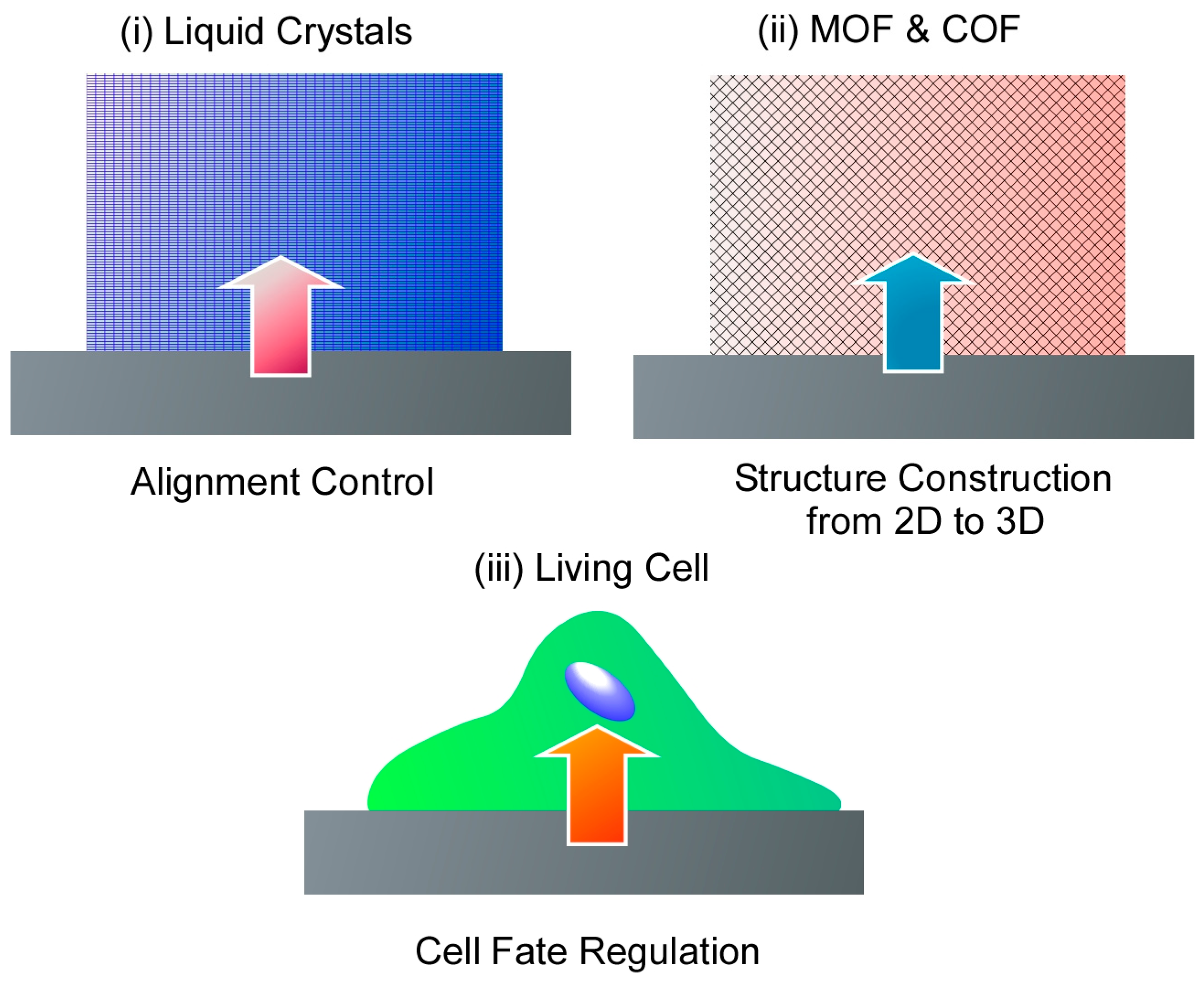
Figure 3.
Evaluation of the optical orientation behavior of side-chain liquid crystalline azobenzene polymer films with a thickness of approximately 400 nm and a 35 μm thick low-molecular-weight nematic liquid crystal, 4′-pentyl-4-cyanobiphenyl in situ at the interface. Reprinted with permission from [267]. Copyright 2023 American Chemical Society.

Figure 4.
Nematic-to-isotropic transition of liquid crystalline components doped with a photochromic azobenzene surfactant in core–sheath nanofibers. Reprinted with permission from [268]. Copyright 2021 Royal Society of Chemistry.
Figure 4.
Nematic-to-isotropic transition of liquid crystalline components doped with a photochromic azobenzene surfactant in core–sheath nanofibers. Reprinted with permission from [268]. Copyright 2021 Royal Society of Chemistry.

Figure 5.
A method to rationally design and synthesize crosslinked COFs in which the two-dimensional COF layers are covalently fixed and linked by poly(ethylene glycol) (PEG) three-dimensional or alkyl chains. Reprinted with permission from [285]. Copyright 2023 American Chemical Society.
Figure 5.
A method to rationally design and synthesize crosslinked COFs in which the two-dimensional COF layers are covalently fixed and linked by poly(ethylene glycol) (PEG) three-dimensional or alkyl chains. Reprinted with permission from [285]. Copyright 2023 American Chemical Society.

Figure 6.
Nanoarchitectonics of three-dimensional COFs based on a two-dimensional network with interpenetrations where the structures were formed by [3+2]imine condensation reactions using triangular knots and linear linkers. Reprinted with permission from [286]. Copyright 2023 American Chemical Society.

Figure 7.
A method to form three-dimensional COFs by introducing steric hindrance to molecular blocks that inhibit π–π stacking, as a method for two-dimensional COFs to intertwine with each other. Reprinted with permission from [287]. Copyright 2023 American Chemical Society.
Figure 7.
A method to form three-dimensional COFs by introducing steric hindrance to molecular blocks that inhibit π–π stacking, as a method for two-dimensional COFs to intertwine with each other. Reprinted with permission from [287]. Copyright 2023 American Chemical Society.

Figure 8.
Pseudo-three-dimensional COF nanosheets for detection of biomolecules, including COVID-19 virus where the pseudo-three-dimensional COF nanosheet functions as an adsorbent for biomolecular probes and an acceptor to quench the fluorescence of dye-labeled probes. Reprinted with permission from [288]. Copyright 2023 Royal Society of Chemistry.

Figure 9.
A cobalt MOF based on a well-defined layered double core strongly bound by intermolecular bonds in which its three-dimensional structure is maintained by π–π stacking interactions between the unstable pyridine ligands of the nanosheets. Reprinted with permission from [289]. Copyright 2020 American Chemical Society.

Figure 10.
Organometallic frameworks with orthogonal nanosheet arrays for cubic MOFs. Reproduced under terms of the CC-BY license [290]. Copyright 2023 Springer-Nature.
Figure 10.
Organometallic frameworks with orthogonal nanosheet arrays for cubic MOFs. Reproduced under terms of the CC-BY license [290]. Copyright 2023 Springer-Nature.
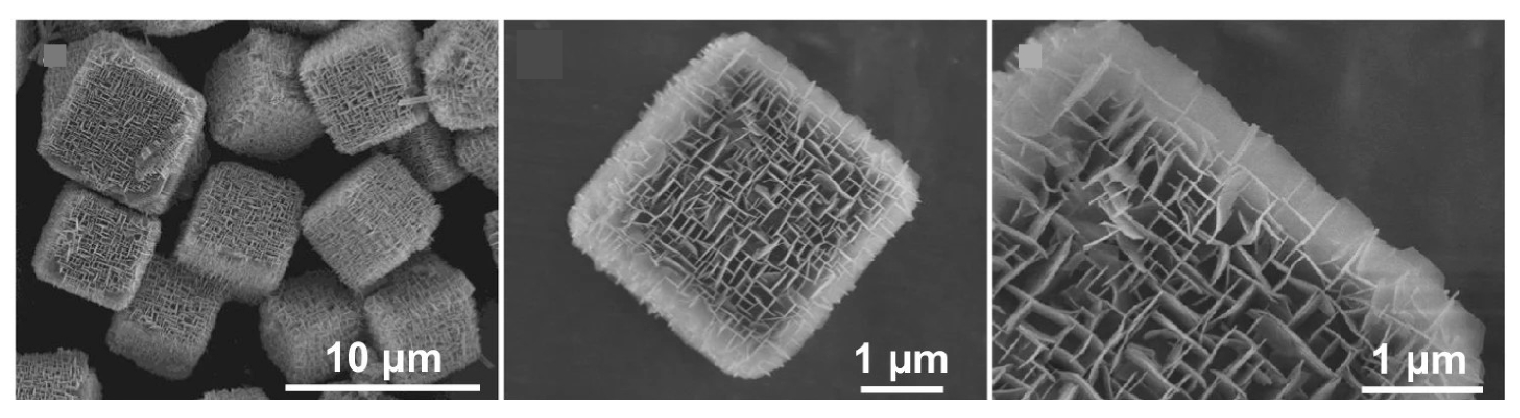
Figure 11.
A strategy to grow MOFs into nanochannels with angstrom-scale ion channels with one- to three-dimensional pore structures. Reproduced under terms of the CC-BY license [291]. Copyright 2023 Springer-Nature.
Figure 11.
A strategy to grow MOFs into nanochannels with angstrom-scale ion channels with one- to three-dimensional pore structures. Reproduced under terms of the CC-BY license [291]. Copyright 2023 Springer-Nature.

Figure 12.
A method for converting two-dimensionally conjugated MOFs into a three-dimensional framework by post-synthetic pillar ligand insertion. Reprinted with permission from [292]. Copyright 2022 American Chemical Society.
Figure 12.
A method for converting two-dimensionally conjugated MOFs into a three-dimensional framework by post-synthetic pillar ligand insertion. Reprinted with permission from [292]. Copyright 2022 American Chemical Society.
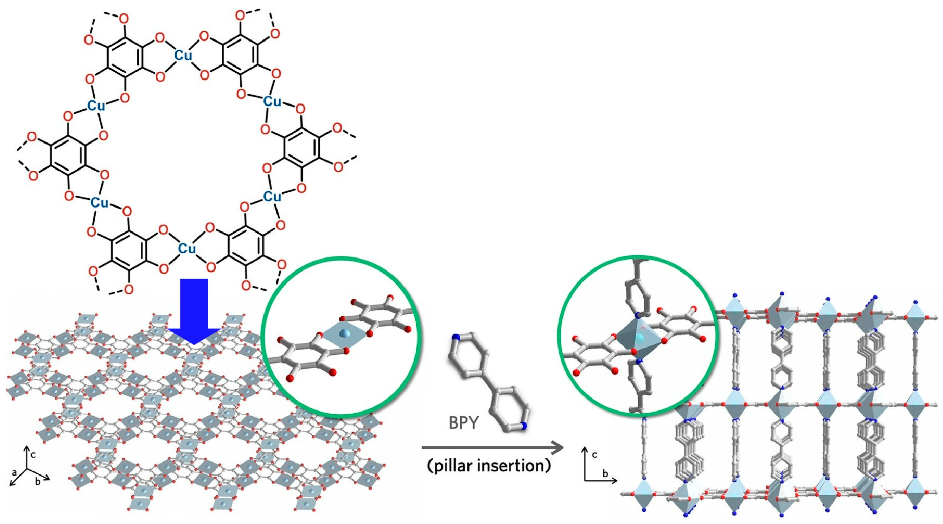
Figure 13.
Preparation of a new hierarchical MOF with Cu(II) centers linked by benzene tricarbox-ylates. Reprinted with permission from [293]. Copyright 2017 American Chemical Society.
Figure 13.
Preparation of a new hierarchical MOF with Cu(II) centers linked by benzene tricarbox-ylates. Reprinted with permission from [293]. Copyright 2017 American Chemical Society.
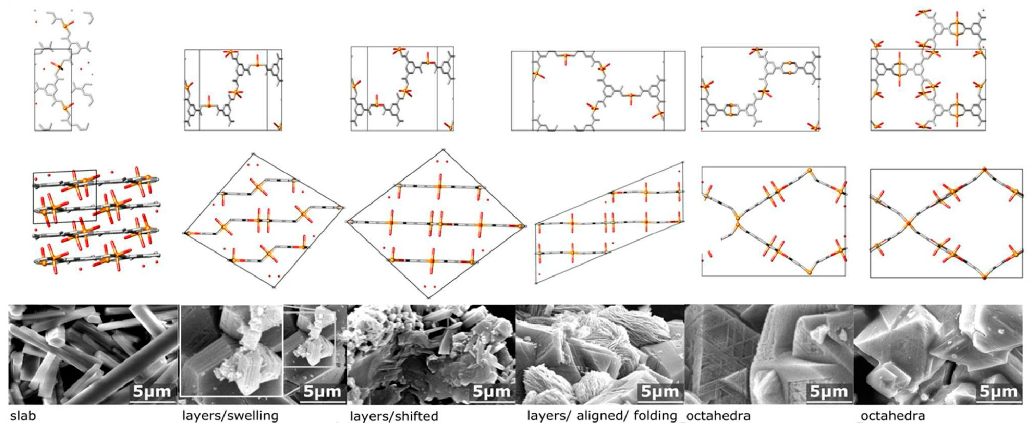
Figure 14.
Synthesis of a two-dimension-on-three-dimension (2D-on-3D) hetero-MOF structure. Reprinted with permission from [294]. Copyright 2022 Wiley VCH.
Figure 14.
Synthesis of a two-dimension-on-three-dimension (2D-on-3D) hetero-MOF structure. Reprinted with permission from [294]. Copyright 2022 Wiley VCH.

Figure 15.
Synthesis of a series of hafnium (Hf)-based two-dimensional metal–organic layers with different thicknesses (from single layer to stacked multilayers) and densities of hydrogen-bonding sites with inhibition capability of ice growth. Reprinted with permission from [295]. Copyright 2023 Wiley VCH.

Figure 16.
A technique that uses the interface between two immiscible liquids, aqueous cell culture solutions and perfluorocarbons, as a site for culturing and inducing differentiation of human mesenchymal stem cells (hMSCs). Reprinted with permission from [296]. Copyright 2019 Wiley VCH.
Figure 16.
A technique that uses the interface between two immiscible liquids, aqueous cell culture solutions and perfluorocarbons, as a site for culturing and inducing differentiation of human mesenchymal stem cells (hMSCs). Reprinted with permission from [296]. Copyright 2019 Wiley VCH.
Figure 17.
Culture on a two-dimensional network of protein nanofibrils at the liquid–liquid interface for neural differentiation of hMSCs where lipid raft microdomains were found to play a central regulatory role in both the initial cell adhesion and subsequent neural differentiation of hMSCs. Reproduced under terms of the CC-BY license [297]. Copyright 2022 Springer-Nature.

Figure 18.
Interfacial dynamics of bovine serum albumin (BSA) and β-lactoglobulin (BLG) aggregates at the fluorinated liquid-water interface where high cell proliferation can be achieved even on bioemulsions with protein nanosheet formation without surfactants. Reproduced under terms of the CC-BY license [298]. Copyright 2023 American Chemical Society.
Figure 19.
Cell spreading and growth on low-viscous liquid surfaces enabled by the self-assembly of mechanically strong protein nanosheets at the interface. Reproduced under terms of the CC-BY license [299]. Copyright 2018 American Chemical Society.
Figure 19.
Cell spreading and growth on low-viscous liquid surfaces enabled by the self-assembly of mechanically strong protein nanosheets at the interface. Reproduced under terms of the CC-BY license [299]. Copyright 2018 American Chemical Society.

Figure 20.
Muscle differentiation and simultaneously controlling the direction of cell growth on the two-dimensional in-plane aligned structure of fullerene nanowhiskers as a cell scaffold. Reprinted with permission from [310]. Copyright 2015 Wiley VCH.
Figure 20.
Muscle differentiation and simultaneously controlling the direction of cell growth on the two-dimensional in-plane aligned structure of fullerene nanowhiskers as a cell scaffold. Reprinted with permission from [310]. Copyright 2015 Wiley VCH.
Figure 21.
Nanopatterned surfaces fabricated with high-aspect-ratio fullerene nanowhiskers for long-term pluripotency retention and differentiation potential of hMSCs where mechanical signals are transmitted to the nucleus by YAP and YAP translocation to the nucleus positively regulates the activity of core regulators (OCT4, SOX2, NANOG). Reproduced under terms of the CC-BY license [311]. Copyright 2020 American Chemical Society.

Disclaimer/Publisher’s Note: The statements, opinions and data contained in all publications are solely those of the individual author(s) and contributor(s) and not of MDPI and/or the editor(s). MDPI and/or the editor(s) disclaim responsibility for any injury to people or property resulting from any ideas, methods, instructions or products referred to in the content. |
© 2024 by the author. Licensee MDPI, Basel, Switzerland. This article is an open access article distributed under the terms and conditions of the Creative Commons Attribution (CC BY) license (https://creativecommons.org/licenses/by/4.0/).
Share and Cite
MDPI and ACS Style
Ariga, K. 2D Materials Nanoarchitectonics for 3D Structures/Functions. Materials 2024, 17, 936. https://doi.org/10.3390/ma17040936
AMA Style
Ariga K. 2D Materials Nanoarchitectonics for 3D Structures/Functions. Materials. 2024; 17(4):936. https://doi.org/10.3390/ma17040936
Chicago/Turabian StyleAriga, Katsuhiko. 2024. "2D Materials Nanoarchitectonics for 3D Structures/Functions" Materials 17, no. 4: 936. https://doi.org/10.3390/ma17040936
Note that from the first issue of 2016, this journal uses article numbers instead of page numbers. See further details here.