
Carbon Nanotropes: A Contemporary Paradigm in Drug Delivery


Abstract
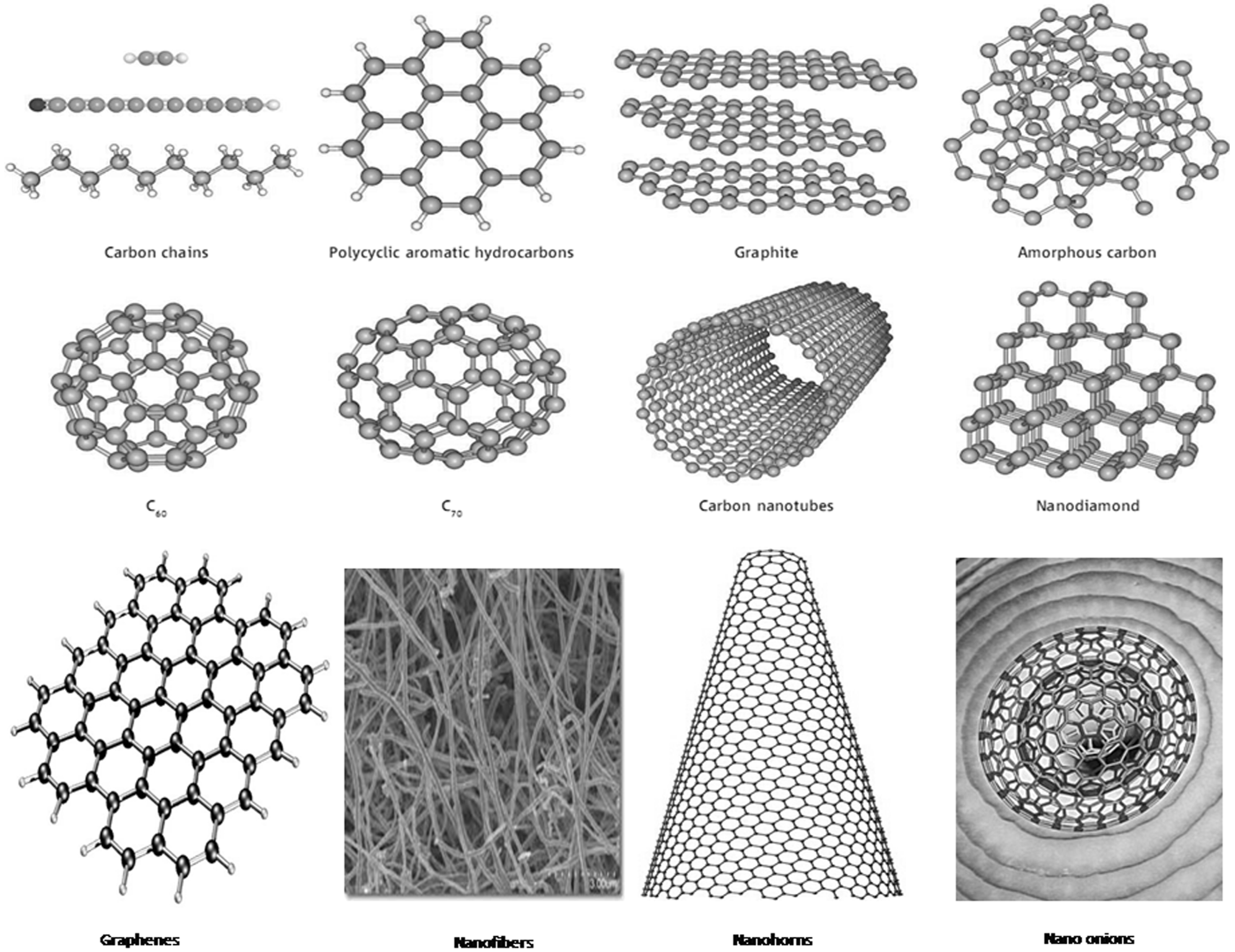
:1. Introduction
2. Structure and Properties of Carbon Nanotropes

3. Cellular Uptake and Biodistribution of Carbon Nanotropes
4. Toxicity of Carbon Nanotropes

| Nanotube | Biological System | Dosage | Toxicity |
|---|---|---|---|
| Plasmid DNA-SWCNT and Plasmid DNA-MWCNT | f-CNTs: HeLa cell lines in vitro | 10 mg/mL | 50% survival of HeLa cells |
| Fluorescein isothiocyanate-SWCNT and fluorescein isothiocyanate-MWCNT | f-SWCNT and f-MWCNT: HeLa cell lines in vitro | 5–10 mg/mL | 50% survival of HeLa cells |
| Pristine SWCNT | SWCNT: Mesothelioma cell line MSTO-211H in vitro | 7.5 μg/mL water | 10% decrease in cell proliferation and activity |
| Ammonium chloride-SWCNT, and poly(ethylene glycol)-SWCNT | Macrophages, B and T lymphocytes from BALB/c mice spleen and lymph nodes in vitro | 10 μg/mL water | 5% decrease in viability of B lymphocytes, but no adverse effects on T lymphocytes and macrophages |
| RNA-polymer SWCNT conjugate | MCF-7 breast cancer cells in vitro | 1 mg/mL | No significant cell damage |
| [111In] DTPA-SWCNT and [111In] DTPA-MWCNT | Intravenous injection, systemic, female BALB/c mice in vivo | 20 μg/μL PBS | No acute toxicity after single 200 μL dose |
| Pristine MWCNT | Human T lymphocytes in vitro | 40 μg/mL | Should have no toxicity on human T lymphocytes |
| Pristine SWCNT | Intravenous injection, systemic, rabbit in vivo | 7.5 mL of 20 μg/kg body mass | No toxicity |
| 125I-SWCNT (OH) | Intraperitoneal, intravenous, subcutaneous, in male KM mice in vivo | 1.5 μg/mouse | Accumulate in bone, but good biocompatibility |
| Glucosamine-MWCNT | Intraperitoneally into female Kunming mice in vivo | 300 μL single dose, suspension concentration unknown | Good biocompatibility |
| pEGFP-c1 plasmid DNA-SWCNT | Mouse B-cells and cortical neurons in vitro | 0.1 pM/10 mL serum-free medium | ~10% of cells were no longer viable |
| 6-Aminohexanoic acid–derivatized SWCNT | Human epidermal keratinocytes (HEK) in vitro | Multiple tests from 0.00000005 to 0.05 mg/mL | Highest concentration that can interact with HEKs without toxicity, 0.000005 mg/mL for 24 h |
| DNA-Cy3 (fluorescent label)-SWCNT | HeLa cell line in vitro | 2.5–5 mg/L water | No toxic effects, after six pulses of 10-s, 808-nm laser radiations at 1.4 W cm2 |
| Streptavidin-SWCNT | HL60 and Jurkat cells in vitro | 0.025 mg/mL | No adverse effects |
| SWCNTs dispersed in DMEM with 5% (vol/vol) fetal bovine serum | Human epithelial-like HeLa cells in vitro | 100 μg/mL | No effect on growth rate |
5. Carbon Nanotropes in Drug Delivery
5.1. Delivery of Anticancer Drugs
| Drug delivery system | Dosage and biological system employed | Application | Method of drug release | Remarks |
|---|---|---|---|---|
| HCPT-diamino-triethylene glycol-MWCNTs [64] | 5 mg kg−1 HCPT (Hepatic H22 tumor-bearing mice) | Gastric carcinoma | pH Triggered drug release | More efficient |
| C60-IONP-PEG/ Hematoporphyrin monomethyl ether (HMME) [65] | HMME, a new photodynamic anti-cancer drug, was conjugated to C60-IONP-PEG, forming a C60-IONP-PEG/HMME drug delivery system | Cancer theranostic | N.M. | Remarkably enhanced photodynamic cancer cell killing effect |
| Multi-functional C60-IONP-PEG-Folic acid (FA) [66] | Folic acid linked to C60-IONP-PEG in order to obtain an active tumor targeting effect to MCF-7 cells and malignant tumor in mice models | Cancer diagnosis, PDT, RF RTT and magnetic targeting | N.M. | Excellent physiological stability, neglegible toxicity, selectivity |
| Paclitaxel-ultrathin PLGA film-QD-MWCNT [67] | 100 ng mL−1 Paclitaxel-PLGA-CNT (Nu/nu nude mice; 6–8 weeks old, about 18 g) | Prostate carcinoma | N.M. | More efficient in tumor treating and low toxic to living mice |
| Doxorubicin-FA-CHI/ALG-SWCNT [68] | 50 μg mL−1 DOX-FA-CHI/ALG- SWCNT (HeLa cells) | Cervical carcinoma | FA–FA Receptor interaction, pH triggered drug release | More cytotoxic and selective |
| Doxorubicin-pluronic F127-MWCNT [69] | 10 μg mL−1 DOX:20 μg mL−1 CNT (MCF-7 cells) | Breast cancer | N.M. | More efficient |
| Doxorubicin-amphiphilic polymers-CNT [70] | 0.5 mg mL−1 DOX-CNT (B16F10 cells) | Melanoma | N.M. | More efficient |
| Doxorubicin/Folic acid-MWCNT@Fe [71] | 32 μg DOX per mg of FA-MWCNT@Fe (HeLa cells) | N.M. | FA–FA Receptor interaction (active) and magnetic force (passive) | Prolonged drug release |
| {Pt(IV)}-PL-PEG-SWCNT [72] | 65 Pint(IV) centers per nanotube (average), (NTera-2 cells) | Testicular cancer | pH Triggered drug release | Higher toxic to tumor cells |
| Electroactive polyimide-MWCNT [73] | 131.3–120.8 mg EPI per gram of c-MWCNT | N.M. | N.M. | Greater EPI release in acidic medium |
| Doxorubicin/PEGylated MWCNT [37] | N.M. (Hela, HepG2, K562 cells) | Liver cancer and leukemia | N.M. | Efficient anti-multi drug resistance effect |
| Cis-Diammine dichloroplatinum–SWCNT [74] | 100 μg mL−1 CDDP-CNT (DU145 and PC3 cells) | Prostate cancer | CDDP-Polynucleotide chain interaction | Similar effect on PC3 cells but less on DU145 cells |
| EGF-Cis-diammin dichloroplatinum–SWCNT [75] | 1.3 μM CDDP in EGF-CDDP-SWCNT (Female athymic (nu/nu) nude mice (4–6 weeks old, weighing 18–20 g) | Squamous carcinoma | EGF–EGF Receptor interaction | More efficient |
| Conducting Polymer-MWCNT [76] | 0.5 μg μL−1 CP-CNT (EJ28 cell line) | Bladder cancer | N.M. | N.M. |
| Biotin-SWCNT-cleavable disulfide linker-(taxoid-fluorescein) [77] | 13.9 μM Taxoid (L1210FR, L1210 and WI38 cell lines) | Leukemia | Biotin-biotin receptors mediated endocytosis | More efficient |
| C60-HMM-SWCNT/DWCNT [78] | N.M. | N.M. | CH2Cl2Triggered drugs removal | N.M. |
| Oxaliplatin/MMC-MWCNT [79] | (300 μM oxaliplatin + 100 μg CNT) per mL medium (RKO and HCT 116 cell lines) | Colorectal cancer | IR radiation stimulated, hyperthermic method | N.M. |
| Methotrexateand 1,3-dipolar cycloaddition on MWCNT [80] | 5 μg mL−1 Conjugate (Jurkat cells) | Jurkat cells | N.M. | N.M. |
| Methotrexate -Gly-Leu-Phe-Gly/6-hydroxy hexanoic ester on 1,3-dipolar cycloaddition f-MWCNT [81] | 10mM MTX–MWCNT (MCF-7 cells) | Human breast carcinoma | N.M. | Higher cytotoxicity in MTX–MWCNT using peptide linker |
5.2. Delivery of Neurological Drugs
5.3. Delivery of Anti-Tubercular Drugs
5.4. Delivery of Anti-Fungal Drugs
5.5 Delivery of Anti-Inflammatory Drugs
5.6. Delivery of Topical Agents
5.7. Delivery of Biomolecules, Gene Transfection and As Biosensors
| Delivery system | Biological system employed | Results |
|---|---|---|
| Antisense oligodeoxynucleotides (ASODNs)-PEI-MWCNT [106] | HeLa cells | ASODN interacted with positively charged amine groups on PEI-MWCNT |
| Plasmid DNA-carboxylic f-MWCNT with embedded Ni [26] | Bal17 B-lymphoma, ex vivo B cells and primary neurons | DNA-MWCNT entered in Bal17 B-lymphoma, ex- vivo B cells and primary neurons driven by magnetic field and remained highly viable even after transduction |
| Green fluorescent protein gene-Amino/carboxyl/hydroxyl/alkyl-MWCNT [107] | Human umbilical vein endothelial cells (HUVEC) | Only amino group functionalized MWCNT effectively delivered the pEGFPN1 plasmid into cells |
| Protective B cell epitope and 1,3-dipolar cycloaddition on SWCNT [108] | BHK 21 cells | B cell epitope was recognized by specific antibodies after being conjugated to SWCNT; mono-peptide-SWCNT led to higher virus neutralizing antibody titers than bis-peptide-SWCNT |
| Anti-HER2 IgY antibody-SWCNT-CONH2 [109] | SK-BR-3 and MCF-7 cells | CNT-antibody complex could detect and selectively kill SK-BR-3 (cancer cells expressing HER2) in- vitro in the presence of MCF-7 (non-HER2 expressing) cells |
| CpG-Oligodeoxynucleotidesand 1,3-dipolar cycloaddition on SWCNT [110] | N.M. | f-SWCNT enhanced immunostimulatory properties of ODN CpG; Concentration of IL-6 (stimulated by ODN CpG combined withf-SWCNT) in splenocyte cultures decreased more |
| NF-κB decoy-SWCNT [111] | HeLa cells | Covalent binding of NF-κB decoy on SWCNT greatly reduced the NF-κB dependent gene expression |
| Oligodeoxynucleotides-SWCNT with maleimide terminal group [112] | N.M. | Hybridization of complementary DNA was highly specific and reversible |
| DNA-PEI-MWCNT [113] | 293cells, COS7 and HepG2 cells | PEI served as anchor point for DNA immobilization; PEI-g-MWCNT exhibited good transfection efficiency for the delivery of DNA |
| EPO-PEG-8 caprylic/capric glycerides-CNT [114] | Male Wistar rats | Short CNT released twice the amount of EPO than long CNT in rat serum |
| GnRH-carboxylic-MWCNT [115] | DU 145 cells | GnRH–MWCNT killed Hela cells after internalization by GnRH receptor-positive cells |
| Single stranded DNA (ssDNA)-SWCNT dotted with Au nanocrystals (Au-SWCNT) [116] | N.M. | Target DNA hybridization to ssDNA probes, which were immobilized on Au-SWCNT |
| ssDNA-pristine SWCNT [117] | N.M. | ssDNA bound to SWCNT got released by desorption potential |
| Plasmid DNAand 1,3-dipolar cycloaddition on SWCNT/MWCNT [99] | HeLa cells | f-SWCNT complexed with plasmid DNA facilitated higher DNA uptake and gene expression in vitro |
| ssDNA-pristine SWCNT [117] | N.M. | ssDNA bound to SWCNT got released by desorption potential |
| Plasmid DNAand 1,3-dipolar cycloaddition on SWCNT/MWCNT [99] | HeLa cells | f-SWCNT complexed with plasmid DNA facilitated higher DNA uptake and gene expression in vitro |
| siRNA-PEI/pyridinium-f-MWCNT [118] | Human lung cancer cell line H1299 | Both types of f-MWCNTs showed 10%–30% silencing activity and 10%–60% cytotoxicity |
| siRNA-PDDA-HMDA-SWCNT [105] | Isolated rat heart cells | PDDA-HMDA-SWCNT bound negatively charged siRNA by electrostatic interactions |
| siRNA-PL-PEG-SWCNT [119] | Human T cells and primary cells | CNT were capable of siRNA delivery to human T cells and PBMCs, and caused RNAi of CXCR4 and CD4 receptors |
| siRNA/DNA-PL-PEG-SWCNT [120] | HeLa cells | Amine or maleimide terminal of PL-PEG-SWCNT could bind to various biomolecules |
| TERT siRNA-SWCNT–CONH–(CH2)6−NH3+Cl−[121] | HeLa cells | TERT siRNA specifically targeted TERT expression and led to growth arrest of tumor cells |
| Ferritin/SA/biotinyl-3,6-dioxaoctanediamine-1-Pyrenebutanoic acid, succinimidyl ester-SWCNT [122] | N.M. | Pyrenyl groups bound to CNT through strong π–π interaction, while succinimidyl ester groups worked as anchors for combining proteins |
| Bovine serum albumin (BSA)-SWCNT-CONH2 BSA-MWCNT-CONH2[102] | N.M. | 90% BSA retained activity after the formation of BSA-CNT conjugates |
| BSA/SA/Protein A/cytochromec(cyt-c)-carboxyl-SWCNT [123] | HL60, Jurkat, HeLa and NIH-3T3 cells | High level of cellular uptake of proteins (molecular weight <80 KDa); cyt-cSWCNT conjugate led to higher level of apoptosis in the presence of chloroquine |
| SA-Biotin-SWCNT [100] | HL60 and Jurkat cells | SA entered cells after binding to SWCNT-biotin transporter |
| Protein C1q/serum/plasma proteins-pristine SWCNT [124] | Red blood cells | CNT activated human complement through both classical and alternative pathways; C1q bound directly to CNT; fibrinogen and apolipoproteins (AI, AIV and CIII) bound selectively to DWCNT |
| GRGDSP peptide sequence/IKVAV peptide sequenceand 1,3-dipolar cycloaddition on MWCNT [125] | Jurkat cells, primary splenocytes and neurons | MWCNT exhibited biocompatibility with different cell types; they did not seem to change the neuronal morphology, viability, and basic functions |
| KGYYG sequence/ GSGVRGDFGSLAPRVARQL sequence and 1,3-dipolar cycloaddition on SWCNT [126] | N.M. | Bound peptides were recognized by monoclonal and polyclonal antibodies; peptide-SWCNT caused immune response |
| K(FITC)QRMHLRQYELLC sequenceand 1,3-dipolar cycloaddition on SWCNT [99] | 3T3 and 3T6 cells | CNT conjugate crossed the cell membrane; FITC-CNTs accumulated mainly in cytoplasm; Peptide-CNT accumulated in nucleus |
| BV2 microglia/GL261 glioma-pluronic F108-MWCNT [127] | BV2 microglia and GL261 glioma cells | CNT did not lead to proliferative or cytokine changes in vitro; they carried DNA and siRNA, and were internalized at higher levels in phagocytic cells than in tumor cells |
5.8. Delivery of Other Drugs
| Drug delivery system | Dosage and Biological system employed | Drug effect | Effect of drug-CNT conjugate |
|---|---|---|---|
| Theophylline-AL/CNT microsphere [130] | 20% (wt%) theophylline per drug-CNT complex | N.M. | More efficient |
| Dopsone-O-(7-azabenzotriazol-1-yl)-N,N,N′,N′-tetramethyluronium hexafluorophosphate/N,N-diisopropylethylamine-f-MWCNT [92] | 50 μg dopsone per mL off-MWCNT (rat peritoneal macrophages) | Anti-microbial and anti-inflammatory | More efficient |
| (d-α-Tocopheryl polyethylene glycol 1000 succinate–MWCNT [131] | 2.5 μM TPGS (N.M.) | Vitamin E delivery | N.M. |
| Polyethylene oxide-pentaerythritol triacrylate-[(S)-(+)-ketoprofen]-MWCNT [97] | N.M. (Mouse membrane) | Anti-inflammatory | More efficient |
| Dxamethasone-CHI–SWCNT [96] | 0.5 mg per mL CHI (N.M.) | Anti-inflammatory | More efficient |
| Amphotericin-B-fluorescein-MWCNT [94] | 40 μg mL−1 AmB-CNT (Human Jurkat lymphoma T cells) | Antibiotic | More efficient |
| Carvedilol–MWCNT [132] | 20%–60% (wt%) CAR per drug-CNT complex | Anti-hypertensive | More efficient |
| Acetyl choline-SWCNT [38] | 20-50 mg kg−1 Ach-CNT (Ach: 4–10 mg kg−1) | Alzheimer’s disease therapy agent | More efficient |
6. Challenges and Limitations with Carbon Nanomaterials
7. Recent Patents on Carbon Nanotropes in Drug Delivery
8. Conclusions
Acknowledgments
Author Contributions
Conflicts of Interest
References
- Hirsch, A. The era of carbon allotropes. Nat. Mater. 2010, 9, 868–871. [Google Scholar] [CrossRef] [PubMed]
- Iijima, S. Helical microtubules of graphitic carbon. Nature 1991, 354, 56–58. [Google Scholar] [CrossRef]
- Novoselov, K.S.; Geim, A.K.; Morozov, S.V.; Jiang, D.; Zhang, Y.; Dubonos, S.V.; Grigorieva, I.V.; Firsov, A.A. Electric field effect in atomically thin carbon films. Science 2004, 306, 666–669. [Google Scholar] [CrossRef] [PubMed]
- Avouris, P.; Chen, Z.; Perebeinos, V. Carbon-Based electronics. Nat. Nanotechnol. 2007, 2, 605–615. [Google Scholar] [CrossRef] [PubMed]
- Kuzmenko, A.B.; van Heumen, E.; Carbone, F.; van der Marel, D. Universal optical conductance of graphite. Phys. Rev. Lett. 2008, 100. [Google Scholar] [CrossRef]
- Iijima, S. Direct observation of the tetrahedral bonding in graphitized carbon black by high resolution electron microscopy. J. Cryst. Growth 1980, 50, 675–683. [Google Scholar] [CrossRef]
- Kroto, H.W.; Heath, J.R.; Obrien, S.C.; Curl, R.F.; Smalley, R.E. C60: Buckminsterfullerene. Nature 1985, 318, 162–163. [Google Scholar] [CrossRef]
- Xu, B. Prospects and research progress in nano onion-like fullerenes. New Carbon Mater. 2008, 23, 289–301. [Google Scholar] [CrossRef]
- Buseck, P.R.; Tsipursky, S.J.; Hettich, R. Fullerenes from the geological environment. Science 1992, 257, 215–217. [Google Scholar] [CrossRef] [PubMed]
- Ruoff, R.S.; Tse, D.S.; Malhotra, R.; Lorents, D.C. Solubility of fullerene (C60) in a variety of solvents. J. Phys. Chem. 1993, 97, 3379–3383. [Google Scholar] [CrossRef]
- Beck, M.T.; Mándi, G. Solubility of C60. Fuller. Nanotubes Carbon Nanostructur. 1997, 5, 291–310. [Google Scholar] [CrossRef]
- Yu, M.F.; Lourie, O.; Dyer, M.J.; Moloni, K.; Kelly, T.F.; Ruoff, R.S. Strength and breaking mechanism of multiwalled carbon nanotubes under tensile load. Science 2000, 287, 637–640. [Google Scholar] [CrossRef] [PubMed]
- Lu, X.; Chen, Z. Curved pi-conjugation, aromaticity, and the related chemistry of small fullerenes (<C60) and single-walled carbon nanotubes. Chem. Rev. 2005, 105, 3643–3696. [Google Scholar] [PubMed]
- Tang, Z.K.; Zhang, L.; Wang, N.; Zhang, X.X.; Wen, G.H.; Li, G.D.; Wang, J.N.; Chan, C.T.; Sheng, P. Superconductivity in 4 angstrom single-walled carbon nanotubes. Science 2001, 292, 2462–2465. [Google Scholar] [CrossRef] [PubMed]
- Thostenson, E.T.; Li, C.; Chou, T.W. Nanocomposites in context. Composites Sci. Technol. 2005, 65, 491–516. [Google Scholar] [CrossRef]
- Miessler, G.L.; Tarr, D.A. Inorganic Chemistry, 3rd ed.; Pearson Education Inc.: New York, NY, USA, 2004. [Google Scholar]
- Gonzalez Szwacki, N.; Sadrzadeh, A.; Yakobson, B.I. B80 fullerene: An ab initio prediction of geometry, stability, and electronic structure. Phys. Rev. Lett. 2007, 98. [Google Scholar] [CrossRef]
- Bezmel’nitsyn, V.N.; Eletskii, A.V.; Okun’, M.V. Fullerenes in solutions. Phys. Uspekh. 1998, 41, 1091–1114. [Google Scholar] [CrossRef]
- Su, Y.; Xu, J.Y.; Shen, P.; Li, J.; Wang, L.; Li, Q.; Li, W.; Xu, G.T.; Fan, C.; Huang, Q. Cellular uptake and cytotoxic evaluation of fullerenol in different cell lines. Toxicology 2010, 269, 155–159. [Google Scholar] [CrossRef] [PubMed]
- Dellinger, A.; Zhou, Z.; Norton, S.K.; Lenk, R.; Conrad, D.; Kepley, C.L. Uptake and distribution of fullerenes in human mast cells. Nanomedicine 2010, 6, 575–582. [Google Scholar] [CrossRef] [PubMed]
- Kraszewski, S.; Picaud, F.; Elhechmi, I.; Gharbi, T.; Ramseyer, C. How long a functionalized carbon nanotube can passively penetrate a lipid membrane. Carbon 2012, 50, 5301–5308. [Google Scholar] [CrossRef]
- Lacerda, L.; Russier, J.; Pastorin, G.; Herrero, M.A.; Venturelli, E.; Dumortier, H.; Al-Jamal, K.T.; Prato, M.; Kostarelos, K.; Bianco, A. Translocation mechanisms of chemically functionalised carbon nanotubes across plasma membranes. Biomaterials 2012, 33, 3334–3343. [Google Scholar] [CrossRef] [PubMed]
- Wick, P.; Manser, P.; Limbach, L.K.; Dettlaff-Weglikowska, U.; Krumeich, F.; Roth, S.; Stark, W.J.; Bruinink, A. The degree and kind of agglomeration affect carbon nanotube cytotoxicity. Toxicol. Lett. 2007, 168, 121–131. [Google Scholar] [CrossRef] [PubMed]
- Cherukuri, P.; Bachilo, S.M.; Litovsky, S.H.; Weisman, R.B. Near-infrared fluorescence microscopy of single-walled carbon nanotubes in phagocytic cells. J. Am. Chem. Soc. 2004, 126, 15638–15639. [Google Scholar] [CrossRef] [PubMed]
- Kam, N.W.; Liu, Z.; Dai, H. Carbon nanotubes as intracellular transporters for proteins and DNA: An investigation of the uptake mechanism and pathway. Angew. Chem. Int. Ed. Engl. 2006, 45, 577–581. [Google Scholar] [CrossRef] [PubMed]
- Cai, D.; Mataraza, J.M.; Qin, Z.H.; Huang, Z.; Huang, J.; Chiles, T.C.; Carnahan, D.; Kempa, K.; Ren, Z. Highly efficient molecular delivery into mammalian cells using carbon nanotube spearing. Nat. Methods 2005, 2, 449–454. [Google Scholar] [CrossRef] [PubMed]
- Mao, H.; Kawazoe, N.; Chen, G. Uptake and intracellular distribution of collagen-functionalized single-walled carbon nanotubes. Biomaterials 2013, 34, 2472–2479. [Google Scholar] [CrossRef] [PubMed]
- Arora, S.; Rajwade, J.M.; Paknikar, K.M. Nanotoxicology and in vitro studies: The need of the hour. Toxicol. Appl. Pharmacol. 2012, 258, 151–165. [Google Scholar] [CrossRef] [PubMed]
- Fenoglio, I.; Fubini, B.; Ghibaudi, E.M.; Turci, F. Multiple aspects of the interaction of biomacromolecules with inorganic surfaces. Adv. Drug Deliv. Rev. 2011, 63, 1186–1209. [Google Scholar] [CrossRef] [PubMed]
- Debouzy, J.C.; Crouzier, D.; Flahaut, E. Hydrophobic double walled carbon nanotubes interaction with phopholipidic model membranes: (1)h-, (2)h-, (31)p nmr and esr study. Environ. Toxicol. Pharmacol. 2010, 30, 147–152. [Google Scholar] [CrossRef] [PubMed] [Green Version]
- Poland, C.A.; Duffin, R.; Kinloch, I.; Maynard, A.; Wallace, W.A.; Seaton, A.; Stone, V.; Brown, S.; Macnee, W.; Donaldson, K. Carbon nanotubes introduced into the abdominal cavity of mice show asbestos-like pathogenicity in a pilot study. Nat. Nanotechnol. 2008, 3, 423–428. [Google Scholar] [CrossRef] [PubMed]
- Lacerda, L.; Bianco, A.; Prato, M.; Kostarelos, K. Carbon nanotubes as nanomedicines: From toxicology to pharmacology. Adv. Drug. Deliv. Rev. 2006, 58, 1460–1470. [Google Scholar] [CrossRef] [PubMed]
- Firme, C.P., 3rd; Bandaru, P.R. Toxicity issues in the application of carbon nanotubes to biological systems. Nanomedicine 2010, 6, 245–256. [Google Scholar] [CrossRef] [PubMed]
- Yang, S.T.; Wang, X.; Jia, G.; Gu, Y.; Wang, T.; Nie, H.; Ge, C.; Wang, H.; Liu, Y. Long-term accumulation and low toxicity of single-walled carbon nanotubes in intravenously exposed mice. Toxicol. Lett. 2008, 181, 182–189. [Google Scholar] [CrossRef] [PubMed]
- Wang, H.; Wang, J.; Deng, X.; Sun, H.; Shi, Z.; Gu, Z.; Liu, Y.; Zhao, Y. Biodistribution of carbon single-wall carbon nanotubes in mice. J. Nanosci. Nanotechnol. 2004, 4, 1019–1024. [Google Scholar] [CrossRef] [PubMed]
- Hirano, S.; Kanno, S.; Furuyama, A. Multi-walled carbon nanotubes injure the plasma membrane of macrophages. Toxicol. Appl. Pharmacol. 2008, 232, 244–251. [Google Scholar] [CrossRef] [PubMed]
- Cheng, J.; Meziani, M.J.; Sun, Y.P.; Cheng, S.H. Poly(ethylene glycol)-conjugated multi-walled carbon nanotubes as an efficient drug carrier for overcoming multidrug resistance. Toxicol. Appl. Pharmacol. 2011, 250, 184–193. [Google Scholar] [CrossRef] [PubMed]
- Yang, Z.; Zhang, Y.; Yang, Y.; Sun, L.; Han, D.; Li, H.; Wang, C. Pharmacological and toxicological target organelles and safe use of single-walled carbon nanotubes as drug carriers in treating alzheimer disease. Nanomed. Nanotechnol. Biol. Med. 2010, 6, 427–441. [Google Scholar] [CrossRef] [PubMed]
- Oberdorster, E. Manufactured nanomaterials (fullerenes, C60) induce oxidative stress in the brain of juvenile largemouth bass. Environ. Health. Perspect. 2004, 112, 1058–1062. [Google Scholar] [CrossRef] [PubMed]
- Pacurari, M.; Yin, X.J.; Zhao, J.; Ding, M.; Leonard, S.S.; Schwegler-Berry, D.; Ducatman, B.S.; Sbarra, D.; Hoover, M.D.; Castranova, V.; et al. Raw single-wall carbon nanotubes induce oxidative stress and activate mapks, ap-1, nf-kappab, and akt in normal and malignant human mesothelial cells. Environ. Health. Perspect. 2008, 116, 1211–1217. [Google Scholar] [CrossRef] [PubMed]
- Jacobsen, N.R.; Pojana, G.; White, P.; Moller, P.; Cohn, C.A.; Korsholm, K.S.; Vogel, U.; Marcomini, A.; Loft, S.; Wallin, H. Genotoxicity, cytotoxicity, and reactive oxygen species induced by single-walled carbon nanotubes and C(60) fullerenes in the fe1-mutatrade markmouse lung epithelial cells. Environ. Mol. Mutagen. 2008, 49, 476–487. [Google Scholar] [CrossRef] [PubMed]
- Uhrich, K.E.; Cannizzaro, S.M.; Langer, R.S.; Shakesheff, K.M. Polymeric systems for controlled drug release. Chem. Rev. 1999, 99, 3181–3198. [Google Scholar] [CrossRef] [PubMed]
- Aillon, K.L.; Xie, Y.; El-Gendy, N.; Berkland, C.J.; Forrest, M.L. Effects of nanomaterial physicochemical properties on in vivo toxicity. Adv. Drug Deliv. Rev. 2009, 61, 457–466. [Google Scholar] [CrossRef] [PubMed]
- Mudshinge, S.R.; Deore, A.B.; Patil, S.; Bhalgat, C.M. Nanoparticles: Emerging carriers for drug delivery. Saud. Pharmaceut. J. 2011, 19, 129–141. [Google Scholar] [CrossRef] [PubMed]
- Bolskar, R.D. Fullerenes for drug delivery. In Encyclopedia of Nanotechnology; Bhushan, B., Ed.; Springer: Amsterdam, The Neatherlands, 2012; pp. 898–911. [Google Scholar]
- Jain, K.K. The role of nanobiotechnology in drug discovery. Drug Discov. Today 2005, 10, 1435–1442. [Google Scholar] [CrossRef]
- Chawla, P.; Chawla, V.; Maheshwari, R.; Saraf, S.A.; Saraf, S.K. Fullerenes: From carbon to nanomedicine. Mini. Rev. Med. Chem. 2010, 10, 662–677. [Google Scholar] [CrossRef] [PubMed]
- Jensen, A.W.; Wilson, S.R.; Schuster, D.I. Biological applications of fullerenes. Bioorg. Med. Chem. 1996, 4, 767–779. [Google Scholar] [CrossRef]
- Bakry, R.; Vallant, R.M.; Najam-ul-Haq, M.; Rainer, M.; Szabo, Z.; Huck, C.W.; Bonn, G.K. Medicinal applications of fullerenes. Int. J. Nanomed. 2007, 2, 639–649. [Google Scholar]
- Partha, R.; Conyers, J.L. Biomedical applications of functionalized fullerene-based nanomaterials. Int. J. Nanomed. 2009, 4, 261–275. [Google Scholar]
- Isobe, H.; Nakanishi, W.; Tomita, N.; Jinno, S.; Okayama, H.; Nakamura, E. Nonviral gene delivery by tetraamino fullerene. Mol. Pharm. 2006, 3, 124–134. [Google Scholar] [CrossRef] [PubMed]
- Nakamura, E.; Isobe, H. Functionalized fullerenes in water. The first 10 years of their chemistry, biology, and nanoscience. Acc. Chem. Res. 2003, 36, 807–815. [Google Scholar] [CrossRef] [PubMed]
- Zakharian, T.Y.; Seryshev, A.; Sitharaman, B.; Gilbert, B.E.; Knight, V.; Wilson, L.J. A fullerene-paclitaxel chemotherapeutic: Synthesis, characterization, and study of biological activity in tissue culture. J. Am. Chem. Soc. 2005, 127, 12508–12509. [Google Scholar] [CrossRef] [PubMed]
- Ryman-Rasmussen, J.P.; Riviere, J.E.; Monteiro-Riviere, N.A. Penetration of intact skin by quantum dots with diverse physicochemical properties. Toxicol. Sci. 2006, 91, 159–165. [Google Scholar] [CrossRef] [PubMed]
- Rouse, J.G.; Yang, J.; Ryman-Rasmussen, J.P.; Barron, A.R.; Monteiro-Riviere, N.A. Effects of mechanical flexion on the penetration of fullerene amino acid-derivatized peptide nanoparticles through skin. Nano Lett. 2007, 7, 155–160. [Google Scholar] [CrossRef] [PubMed]
- Azzam, T.; Domb, A.J. Current developments in gene transfection agents. Curr. Drug Deliv. 2004, 1, 165–193. [Google Scholar] [CrossRef] [PubMed]
- Xu, Z.P.; Zeng, Q.H.; Lu, G.Q.; Yu, A.B. Inorganic nanoparticles as carriers for efficient cellular delivery. Chem. Eng. Sci. 2005, 61, 1027–1040. [Google Scholar] [CrossRef]
- Iannazzo, D.; Piperno, A.; Pistone, A.; Grassi, G.; Galvagno, S. Recent advances in carbon nanotubes as delivery systems for anticancer drugs. Curr. Med. Chem. 2013, 20, 1333–1354. [Google Scholar] [CrossRef] [PubMed]
- Lim, D.J.; Sim, M.; Oh, L.; Lim, K.; Park, H. Carbon-based drug delivery carriers for cancer therapy. Arch. Pharm. Res. 2014, 37, 43–52. [Google Scholar] [CrossRef] [PubMed]
- Fabbro, C.; Ali-Boucetta, H.; Da Ros, T.; Kostarelos, K.; Bianco, A.; Prato, M. Targeting carbon nanotubes against cancer. Chem. Commun. 2012, 48, 3911–3926. [Google Scholar] [CrossRef] [PubMed]
- Vittorio, O.; Raffa, V.; Cuschieri, A. Influence of purity and surface oxidation on cytotoxicity of multiwalled carbon nanotubes with human neuroblastoma cells. Nanomedicine 2009, 5, 424–431. [Google Scholar] [CrossRef] [PubMed]
- Lay, C.L.; Liu, J.; Liu, Y. Functionalized carbon nanotubes for anticancer drug delivery. Expert Rev. Med. Devices 2011, 8, 561–566. [Google Scholar] [CrossRef] [PubMed]
- Vashist, S.K.; Zheng, D.; Pastorind, G.; Al-Rubeaane, K.; Luong, J.H.T.; Sheu, F.S. Delivery of drugs and biomolecules using carbon nanotubes. Carbon 2011, 49, 4077–4097. [Google Scholar] [CrossRef]
- Wu, W.; Li, R.; Bian, X.; Zhu, Z.; Ding, D.; Li, X.; Jia, Z.; Jiang, X.; Hu, Y. Covalently combining carbon nanotubes with anticancer agent: Preparation and antitumor activity. ACS Nano 2009, 3, 2740–2750. [Google Scholar] [CrossRef] [PubMed]
- Shi, J.; Yu, X.; Wang, L.; Liu, Y.; Gao, J.; Zhang, J.; Ma, R.; Liu, R.; Zhang, Z. Pegylated fullerene/iron oxide nanocomposites for photodynamic therapy, targeted drug delivery and mr imaging. Biomaterials 2013, 34, 9666–9677. [Google Scholar] [CrossRef] [PubMed]
- Shi, J.; Wang, L.; Gao, J.; Liu, Y.; Zhang, J.; Ma, R.; Liu, R.; Zhang, Z. A fullerene-based multi-functional nanoplatform for cancer theranostic applications. Biomaterials 2014, 35, 5771–5784. [Google Scholar] [CrossRef] [PubMed]
- Guo, Y.; Shi, D.; Cho, H.; Dong, Z.; Kulkarni, A.; Pauletti, G.M. In vivo imaging and drug storage by quantum-dot-conjugated carbon nanotubes. Adv. Funct. Mater. 2008, 18, 2489–2497. [Google Scholar] [CrossRef]
- Zhang, X.; Meng, L.; Lu, Q.; Fei, Z.; Dyson, P.J. Targeted delivery and controlled release of doxorubicin to cancer cells using modified single wall carbon nanotubes. Biomaterials 2009, 30, 6041–6047. [Google Scholar] [CrossRef] [PubMed]
- Ali-Boucetta, H.; Al-Jamal, K.T.; McCarthy, D.; Prato, M.; Bianco, A.; Kostarelos, K. Multiwalled carbon nanotube-doxorubicin supramolecular complexes for cancer therapeutics. Chem. Commun. 2008, 8, 459–461. [Google Scholar] [CrossRef] [PubMed]
- Park, S.; Yang, H.S.; Kim, D.; Jo, K.; Jon, S. Rational design of amphiphilic polymers to make carbon nanotubes water-dispersible, anti-biofouling, and functionalizable. Chem. Commun. 2008, 25, 2876–2878. [Google Scholar] [CrossRef] [PubMed]
- Li, R.; Wu, R.; Zhao, L.; Hu, Z.; Guo, S.; Pan, X.; Zou, H. Folate and iron difunctionalized multiwall carbon nanotubes as dual-targeted drug nanocarrier to cancer cells. Carbon 2011, 49, 1797–1805. [Google Scholar] [CrossRef]
- Feazell, R.P.; Nakayama-Ratchford, N.; Dai, H.; Lippard, S.J. Soluble single-walled carbon nanotubes as longboat delivery systems for platinum(iv) anticancer drug design. J. Am. Chem. Soc. 2007, 129, 8438–8439. [Google Scholar] [CrossRef] [PubMed]
- Chen, Z.; Pierre, D.; He, H.; Tan, S.; Pham-Huy, C.; Hong, H.; Huang, J. Adsorption behavior of epirubicin hydrochloride on carboxylated carbon nanotubes. Int. J. Pharm. 2011, 405, 153–161. [Google Scholar] [CrossRef] [PubMed]
- Tripisciano, C.; Kraemer, K.; Taylor, A.; Borowiak-Palen, E. Single-wall carbon nanotubes based anticancer drug delivery system. Chem. Phys. Lett. 2009, 478, 200–205. [Google Scholar] [CrossRef]
- Bhirde, A.A.; Patel, V.; Gavard, J.; Zhang, G.; Sousa, A.A.; Masedunskas, A.; Leapman, R.D.; Weigert, R.; Gutkind, J.S.; Rusling, J.F. Targeted killing of cancer cells in vivo and in vitro with egf-directed carbon nanotube-based drug delivery. ACS Nano 2009, 3, 307–316. [Google Scholar] [CrossRef] [PubMed]
- Hampel, S.; Kunze, D.; Haase, D.; Kramer, K.; Rauschenbach, M.; Ritschel, M.; Leonhardt, A.; Thomas, J.; Oswald, S.; Hoffmann, V.; et al. Carbon nanotubes filled with a chemotherapeutic agent: A nanocarrier mediates inhibition of tumor cell growth. Nanomedicine 2008, 3, 175–182. [Google Scholar] [CrossRef] [PubMed]
- Chen, J.; Chen, S.; Zhao, X.; Kuznetsova, L.V.; Wong, S.S.; Ojima, I. Functionalized single-walled carbon nanotubes as rationally designed vehicles for tumor-targeted drug delivery. J. Am. Chem. Soc. 2008, 130, 16778–16785. [Google Scholar] [CrossRef] [PubMed]
- Ren, Y.; Pastorin, G. Incorporation of hexamethylmelamine inside capped carbon nanotubes. Adv. Mater. 2008, 20, 2031–2036. [Google Scholar] [CrossRef]
- Levi-Polyachenko, N.H.; Merkel, E.J.; Jones, B.T.; Carroll, D.L.; Stewart, J.H.T. Rapid photothermal intracellular drug delivery using multiwalled carbon nanotubes. Mol. Pharm. 2009, 6, 1092–1099. [Google Scholar] [CrossRef] [PubMed]
- Pastorin, G.; Wu, W.; Wieckowski, S.; Briand, J.P.; Kostarelos, K.; Prato, M.; Bianco, A. Double functionalization of carbon nanotubes for multimodal drug delivery. Chem. Commun. 2006, 1182–1184. [Google Scholar] [CrossRef] [PubMed]
- Samori, C.; Ali-Boucetta, H.; Sainz, R.; Guo, C.; Toma, F.M.; Fabbro, C.; da Ros, T.; Prato, M.; Kostarelos, K.; Bianco, A. Enhanced anticancer activity of multi-walled carbon nanotube-methotrexate conjugates using cleavable linkers. Chem. Commun. 2010, 46, 1494–1496. [Google Scholar] [CrossRef] [PubMed]
- Yang, K.; Feng, L.; Shi, X.; Liu, Z. Nano-graphene in biomedicine: Theranostic applications. Chem. Soc. Rev. 2013, 42, 530–547. [Google Scholar] [CrossRef] [PubMed]
- Shi, X.; Gong, H.; Li, Y.; Wang, C.; Cheng, L.; Liu, Z. Graphene-based magnetic plasmonic nanocomposite for dual bioimaging and photothermal therapy. Biomaterials 2013, 34, 4786–4793. [Google Scholar] [CrossRef] [PubMed]
- Brambilla, D.; Le Droumaguet, B.; Nicolas, J.; Hashemi, S.H.; Wu, L.P.; Moghimi, S.M.; Couvreur, P.; Andrieux, K. Nanotechnologies for alzheimer’s disease: Diagnosis, therapy, and safety issues. Nanomedicine 2011, 7, 521–540. [Google Scholar] [CrossRef] [PubMed]
- Nunes, A.; Al-Jamal, K.T.; Kostarelos, K. Therapeutics, imaging and toxicity of nanomaterials in the central nervous system. J. Control. Release 2012, 161, 290–306. [Google Scholar] [CrossRef] [PubMed]
- Nunes, A.; Al-Jamal, K.; Nakajima, T.; Hariz, M.; Kostarelos, K. Application of carbon nanotubes in neurology: Clinical perspectives and toxicological risks. Arch. Toxicol. 2012, 86, 1009–1020. [Google Scholar] [CrossRef] [PubMed]
- Zhao, D.; Alizadeh, D.; Zhang, L.; Liu, W.; Farrukh, O.; Manuel, E.; Diamond, D.J.; Badie, B. Carbon nanotubes enhance cpg uptake and potentiate antiglioma immunity. Clin. Cancer. Res. 2011, 17, 771–782. [Google Scholar] [CrossRef] [PubMed]
- Lee, H.J.; Park, J.; Yoon, O.J.; Kim, H.W.; Lee do, Y.; Kim do, H.; Lee, W.B.; Lee, N.E.; Bonventre, J.V.; Kim, S.S. Amine-modified single-walled carbon nanotubes protect neurons from injury in a rat stroke model. Nat. Nanotechnol. 2011, 6, 121–125. [Google Scholar] [CrossRef] [PubMed]
- VanHandel, M.; Alizadeh, D.; Zhang, L.; Kateb, B.; Bronikowski, M.; Manohara, H.; Badie, B. Selective uptake of multi-walled carbon nanotubes by tumor macrophages in a murine glioma model. J. Neuroimmunol. 2009, 208, 3–9. [Google Scholar] [CrossRef] [PubMed]
- Saikia, N.; Rajkhowa, S.; Deka, R.C. Density functional and molecular docking studies towards investigating the role of single-wall carbon nanotubes as nanocarrier for loading and delivery of pyrazinamide antitubercular drug onto pnca protein. J. Comput. Aided. Mol. Des. 2013, 27, 257–276. [Google Scholar] [CrossRef] [PubMed]
- Gallo, M.; Favila, A.; Glossman-Mitnik, D. Dft studies of functionalized carbon nanotubes and fullerenes as nanovectors for drug delivery of antitubercular compounds. Chem. Phys. Lett. 2007, 447, 105–109. [Google Scholar] [CrossRef]
- Vuković, G.D.; Tomić, S.Z.; Marinković, A.D.; Radmilović, V.; Uskoković, P.S.; Čolić, M. The response of peritoneal macrophages to dapsone covalently attached on the surface of carbon nanotubes. Carbon 2011, 48, 3066–3078. [Google Scholar] [CrossRef]
- Szlinder-Richert, J.; Cybulska, B.; Grzybowska, J.; Bolard, J.; Borowski, E. Interaction of amphotericin b and its low toxic derivative, n-methyl-n-d-fructosyl amphotericin b methyl ester, with fungal, mammalian and bacterial cells measured by the energy transfer method. Farmaco 2004, 59, 289–296. [Google Scholar] [CrossRef] [PubMed]
- Wu, W.; Wieckowski, S.; Pastorin, G.; Benincasa, M.; Klumpp, C.; Briand, J.P.; Gennaro, R.; Prato, M.; Bianco, A. Targeted delivery of amphotericin b to cells by using functionalized carbon nanotubes. Angew. Chem. Int. Ed. Engl. 2005, 44, 6358–6362. [Google Scholar] [CrossRef] [PubMed]
- Benincasa, M.; Pacor, S.; Wu, W.; Prato, M.; Bianco, A.; Gennaro, R. Antifungal activity of amphotericin b conjugated to carbon nanotubes. ACS Nano 2011, 5, 199–208. [Google Scholar] [CrossRef] [PubMed]
- Naficy, S.; Razal, J.M.; Spinks, G.M.; Wallace, G.G. Modulated release of dexamethasone from chitosan-carbon nanotube films. Sens. Actuators A 2009, 155, 120–124. [Google Scholar] [CrossRef]
- Im, J.S.; Bai, B.; Lee, Y.S. The effect of carbon nanotubes on drug delivery in an electro-sensitive transdermal drug delivery system. Biomaterials 2010, 31, 1414–1419. [Google Scholar] [CrossRef] [PubMed]
- Bogunia-Kubik, K.; Sugisaka, M. From molecular biology to nanotechnology and nanomedicine. Biosystems 2002, 65, 123–138. [Google Scholar] [CrossRef]
- Pantarotto, D.; Briand, J.P.; Prato, M.; Bianco, A. Translocation of bioactive peptides across cell membranes by carbon nanotubes. Chem. Commun. 2004, 16–17. [Google Scholar] [CrossRef] [PubMed]
- Shi Kam, N.W.; Jessop, T.C.; Wender, P.A.; Dai, H. Nanotube molecular transporters: Internalization of carbon nanotube-protein conjugates into mammalian cells. J. Am. Chem. Soc. 2004, 126, 6850–6851. [Google Scholar] [CrossRef] [PubMed]
- Monteiro-Riviere, N.A.; Nemanich, R.J.; Inman, A.O.; Wang, Y.Y.; Riviere, J.E. Multi-walled carbon nanotube interactions with human epidermal keratinocytes. Toxicol. Lett. 2005, 155, 377–384. [Google Scholar] [CrossRef] [PubMed]
- Huang, W.; Taylor, S.; Fu, K.; Lin, Y.; Zhang, D.; Hanks, T.W. Attaching proteins to carbon nanotubes via diimide-activated amidation. Nano. Lett. 2002, 2, 311–314. [Google Scholar] [CrossRef]
- Awasthi, K.; Singh, D.P.; Singh, S.; Dash, D.; Srivastava, O.N. Attachment of biomolecules (protein and DNA) to amino-functionalized carbon nanotubes. New Carbon Mater. 2009, 24, 301–306. [Google Scholar] [CrossRef]
- Ditto, A.J.; Shah, P.N.; Yun, Y.H. Non-viral gene delivery using nanoparticles. Expert. Opin. Drug Deliv. 2009, 6, 1149–1160. [Google Scholar] [CrossRef] [PubMed]
- Krajcik, R.; Jung, A.; Hirsch, A.; Neuhuber, W.; Zolk, O. Functionalization of carbon nanotubes enables non-covalent binding and intracellular delivery of small interfering RNA for efficient knock-down of genes. Biochem. Biophys. Res. Commun. 2008, 369, 595–602. [Google Scholar] [CrossRef] [PubMed]
- Jia, N.; Lian, Q.; Shen, H.; Wang, C.; Li, X.; Yang, Z. Intracellular delivery of quantum dots tagged antisense oligodeoxynucleotides by functionalized multiwalled carbon nanotubes. Nano Lett. 2007, 7, 2976–2980. [Google Scholar] [CrossRef] [PubMed]
- Gao, L.; Nie, L.; Wang, T.; Qin, Y.; Guo, Z.; Yang, D.; Yan, X. Carbon nanotube delivery of the gfp gene into mammalian cells. Chembiochem 2006, 7, 239–242. [Google Scholar] [CrossRef] [PubMed]
- Pantarotto, D.; Partidos, C.D.; Hoebeke, J.; Brown, F.; Kramer, E.; Briand, J.P.; Muller, S.; Prato, M.; Bianco, A. Immunization with peptide-functionalized carbon nanotubes enhances virus-specific neutralizing antibody responses. Chem. Biol. 2003, 10, 961–966. [Google Scholar] [CrossRef] [PubMed]
- Xiao, Y.; Gao, X.; Taratula, O.; Treado, S.; Urbas, A.; Holbrook, R.D.; Cavicchi, R.E.; Avedisian, C.T.; Mitra, S.; Savla, R.; et al. Anti-her2 igy antibody-functionalized single-walled carbon nanotubes for detection and selective destruction of breast cancer cells. BMC Cancer 2009, 9, 351. [Google Scholar] [CrossRef] [PubMed]
- Bianco, A.; Hoebeke, J.; Godefroy, S.; Chaloin, O.; Pantarotto, D.; Briand, J.P.; Muller, S.; Prato, M.; Partidos, C.D. Cationic carbon nanotubes bind to cpg oligodeoxynucleotides and enhance their immunostimulatory properties. J. Am. Chem. Soc. 2005, 127, 58–59. [Google Scholar] [CrossRef] [PubMed]
- Crinelli, R.; Carloni, E.; Menotta, M.; Giacomini, E.; Bianchi, M.; Ambrosi, G.; Giorgi, L.; Magnani, M. Oxidized ultrashort nanotubes as carbon scaffolds for the construction of cell-penetrating nf-kappab decoy molecules. ACS Nano 2010, 4, 2791–2803. [Google Scholar] [CrossRef] [PubMed]
- Baker, S.E.; Cai, W.; Lasseter, T.L.; Weidkamp, K.P.; Hamers, R.J. Covalently bonded adducts of deoxyribonucleic acid (DNA) oligonucleotides with single-wall carbon nanotubes: Synthesis and hybridization. Nano Lett. 2002, 2, 1413–1417. [Google Scholar] [CrossRef]
- Liu, Y.; Wu, D.C.; Zhang, W.D.; Jiang, X.; He, C.B.; Chung, T.S.; Goh, S.H.; Leong, K.W. Polyethylenimine-grafted multiwalled carbon nanotubes for secure noncovalent immobilization and efficient delivery of DNA. Angew. Chem. Int. Ed. Engl. 2005, 44, 4782–4785. [Google Scholar] [CrossRef] [PubMed]
- Ito, Y.; Venkatesan, N.; Hirako, N.; Sugioka, N.; Takada, K. Effect of fiber length of carbon nanotubes on the absorption of erythropoietin from rat small intestine. Int. J. Pharm. 2007, 337, 357–360. [Google Scholar] [CrossRef] [PubMed]
- Yu, B.; Yang, J.S.; Li, W.X. In vitro capability of multi-walled carbon nanotubes modified with gonadotrophin releasing hormone on killing cancer cells. Carbon 2007, 45, 1921–1927. [Google Scholar] [CrossRef]
- Jung, D.H.; Kim, B.H.; Lim, Y.T.; Kim, J.; Lee, S.Y.; Jung, H.T. Fabrication of single-walled carbon nanotubes dotted with au nanocrystals: Potential DNA delivery nanocarriers. Carbon 2010, 48, 1070–1107. [Google Scholar] [CrossRef]
- Zheng, D.; Li, X.; Ye, J. Adsorption and release behavior of bare and DNA-wrapped-carbon nanotubes on self-assembled monolayer surface. Bioelectrochemistry 2009, 74, 240–245. [Google Scholar] [CrossRef] [PubMed]
- Varkouhi, A.K.; Foillard, S.; Lammers, T.; Schiffelers, R.M.; Doris, E.; Hennink, W.E.; Storm, G. Sirna delivery with functionalized carbon nanotubes. Int. J. Pharm. 2011, 416, 419–425. [Google Scholar] [CrossRef] [PubMed]
- Liu, Z.; Winters, M.; Holodniy, M.; Dai, H. Sirna delivery into human t cells and primary cells with carbon-nanotube transporters. Angew. Chem. Int. Ed. Engl. 2007, 46, 2023–2027. [Google Scholar] [CrossRef] [PubMed]
- Kam, N.W.; Liu, Z.; Dai, H. Functionalization of carbon nanotubes via cleavable disulfide bonds for efficient intracellular delivery of sirna and potent gene silencing. J. Am. Chem. Soc. 2005, 127, 12492–12493. [Google Scholar] [CrossRef] [PubMed]
- Zhang, Z.; Yang, X.; Zhang, Y.; Zeng, B.; Wang, S.; Zhu, T.; Roden, R.B.; Chen, Y.; Yang, R. Delivery of telomerase reverse transcriptase small interfering rna in complex with positively charged single-walled carbon nanotubes suppresses tumor growth. Clin. Cancer Res. 2006, 12, 4933–4939. [Google Scholar] [CrossRef] [PubMed]
- Chen, R.J.; Zhang, Y.; Wang, D.; Dai, H. Noncovalent sidewall functionalization of single-walled carbon nanotubes for protein immobilization. J. Am. Chem. Soc. 2001, 123, 3838–3839. [Google Scholar] [CrossRef] [PubMed]
- Kam, N.W.; Dai, H. Carbon nanotubes as intracellular protein transporters: Generality and biological functionality. J. Am. Chem. Soc. 2005, 127, 6021–6026. [Google Scholar] [CrossRef] [PubMed]
- Salvador-Morales, C.; Flahaut, E.; Sim, E.; Sloan, J.; Green, M.L.; Sim, R.B. Complement activation and protein adsorption by carbon nanotubes. Mol. Immunol. 2006, 43, 193–201. [Google Scholar] [CrossRef] [PubMed] [Green Version]
- Gaillard, C.; Celiot, G.; Li, S.; Toma, F.M.; Dumortier, H.; Spaliuto, G. Carbon nanotubes carrying cell-adhesion peptides do not interfere with neuronal functionality. Adv. Mater. 2009, 21, 2903–2908. [Google Scholar] [CrossRef]
- Pantarotto, D.; Partidos, C.D.; Graff, R.; Hoebeke, J.; Briand, J.P.; Prato, M.; Bianco, A. Synthesis, structural characterization, and immunological properties of carbon nanotubes functionalized with peptides. J. Am. Chem. Soc. 2003, 125, 6160–6164. [Google Scholar] [CrossRef] [PubMed]
- Kateb, B.; Van Handel, M.; Zhang, L.; Bronikowski, M.J.; Manohara, H.; Badie, B. Internalization of mwcnts by microglia: Possible application in immunotherapy of brain tumors. Neuroimage 2007, 37, S9–S17. [Google Scholar] [CrossRef] [PubMed]
- Wu, X.C.; Zhang, W.J.; Sammynaiken, R.; Meng, Q.H.; Yang, Q.Q.; Zhan, E.; Liu, Q.; Yang, W.; Wang, R. Non-functionalized carbon nanotube binding with hemoglobin. Colloids Surfaces B Biointerfaces 2008, 65, 146–149. [Google Scholar] [CrossRef] [PubMed]
- Wong, B.S.; Yoong, S.L.; Jagusiak, A.; Panczyk, T.; Ho, H.K.; Ang, W.H.; Pastorin, G. Carbon nanotubes for delivery of small molecule drugs. Adv. Drug Deliv. Rev. 2013, 65, 1964–2015. [Google Scholar] [CrossRef] [PubMed]
- Zhang, X.; Hui, Z.; Wan, D.; Huang, H.; Huang, J.; Yuan, H.; Yu, J. Alginate microsphere filled with carbon nanotube as drug carrier. Int. J. Biol. Macromol. 2010, 47, 389–395. [Google Scholar] [CrossRef] [PubMed]
- Yan, A.; Von Dem Bussche, A.; Kane, A.B.; Hurt, R.H. Tocopheryl polyethylene glycol succinate as a safe, antioxidant surfactant for processing carbon nanotubes and fullerenes. Carbon N Y 2007, 45, 2463–2470. [Google Scholar] [CrossRef] [PubMed]
- Li, Y.; Wang, T.; Wang, J.; Jiang, T.; Cheng, G.; Wang, S. Functional and unmodified mwnts for delivery of the water-insoluble drug carvedilol — A drug-loading mechanism. Appl. Surf. Sci. 2011, 257, 5663–5670. [Google Scholar] [CrossRef]
- Kreuter, J. Nanoparticles. In Colloidal Drug Delivery Systems; Kreuter, J., Ed.; Marcel Dekker, Inc.: New York, NY, USA, 1994. [Google Scholar]
- Elamanchili, P.; Diwan, M.; Cao, M.; Samuel, J. Characterization of poly(d,l-lactic-co-glycolic acid) based nanoparticulate system for enhanced delivery of antigens to dendritic cells. Vaccine 2004, 22, 2406–2412. [Google Scholar] [CrossRef] [PubMed]
- Cui, Z.; Hsu, C.H.; Mumper, R.J. Physical characterization and macrophage cell uptake of mannan-coated nanoparticles. Drug. Dev. Ind. Pharm. 2003, 29, 689–700. [Google Scholar] [CrossRef] [PubMed]
- Brigger, I.; Dubernet, C.; Couvreur, P. Nanoparticles in cancer therapy and diagnosis. Adv. Drug Deliv. Rev. 2002, 54, 631–651. [Google Scholar] [CrossRef]
- Scheinberg, D.A.; McDevitt, M.; Antczak, C.; Chattopadhyay, D.; May, R.; Njardarson, J.; Philips, M.R. Single Wall Nanotube Constructs and Uses Therefor. U.S. Patent US8540965B2, 24 September 2013. [Google Scholar]
- Wilson, S.R.; Danville, V.A. Methods for Genetic Plant Transformation Using Water-Soluble Fullerene Derivatives. U.S. Patent US8614366B2, 24 December 2013. [Google Scholar]
- Sixty, C.I. Use of Carbon Nanotube for Drug Delivery. EP 2127680 A1, 2 December 2009. [Google Scholar]
- Mohapatra, S.S.; Kumar, A. Method of Drug Delivery by Carbon Nanotube-Chitosan Nanocomplexes. U.S. Patent US8536324B2, 17 September 2013. [Google Scholar]
- Prow, T.W.; Grice, J.E.; Lin, L.L.; Faye, R.; Butler, M.; Becker, W.; Wurm, E.M.; Yoong, C.; Robertson, T.A.; Soyer, H.P.; et al. Nanoparticles and microparticles for skin drug delivery. Adv. Drug Deliv. Rev. 2011, 63, 470–491. [Google Scholar] [CrossRef] [PubMed]
© 2015 by the authors; licensee MDPI, Basel, Switzerland. This article is an open access article distributed under the terms and conditions of the Creative Commons Attribution license (http://creativecommons.org/licenses/by/4.0/).
Share and Cite
Tripathi, A.C.; Saraf, S.A.; Saraf, S.K. Carbon Nanotropes: A Contemporary Paradigm in Drug Delivery. Materials 2015, 8, 3068-3100. https://doi.org/10.3390/ma8063068
Tripathi AC, Saraf SA, Saraf SK. Carbon Nanotropes: A Contemporary Paradigm in Drug Delivery. Materials. 2015; 8(6):3068-3100. https://doi.org/10.3390/ma8063068
Chicago/Turabian StyleTripathi, Avinash C., Shubhini A. Saraf, and Shailendra K. Saraf. 2015. "Carbon Nanotropes: A Contemporary Paradigm in Drug Delivery" Materials 8, no. 6: 3068-3100. https://doi.org/10.3390/ma8063068