Review

Neural Network Analysis and Evaluation of the Fetal Heart Rate

by
Yasuaki Noguchi
1,
Fujihiko Matsumoto
1,
Kazuo Maeda
2,* and
Takashi Nagasawa
3
1
Department of Applied Physics, National Defense Academy, 1-10-20 Hashirimizu, Yokosuka, Kanagawa 239-8686, Japan
2
Professor Emeritus, Department of Obstetrics and Gynecology, Tottori University. Home: 3-125, Nadamachi, Yonago, Tottori, 683-0835, Japan
3
Department of Information Technology, TOITU Ltd., 1-5-10, Ebisu-west, Shibuyaku, Tokyo, 150-0021, Japan
*
Author to whom correspondence should be addressed.
Algorithms 2009, 2(1), 19-30; https://doi.org/10.3390/a2010019
Submission received: 21 November 2008 / Revised: 4 January 2009 / Accepted: 8 January 2009 / Published: 16 January 2009
(This article belongs to the Special Issue Neural Networks and Sensors)

Abstract

:
The aim of the present study is to obtain a highly objective automatic fetal heart rate (FHR) diagnosis. The neural network software was composed of three layers with the back propagation, to which 8 FHR data, including sinusoidal FHR, were input and the system was educated by the data of 20 cases with a known outcome. The output was the probability of a normal, intermediate, or pathologic outcome. The neural index studied prolonged monitoring. The neonatal states and the FHR score strongly correlated with the outcome probability. The neural index diagnosis was correct. The completed software was transferred to other computers, where the system function was correct.

1. Introduction

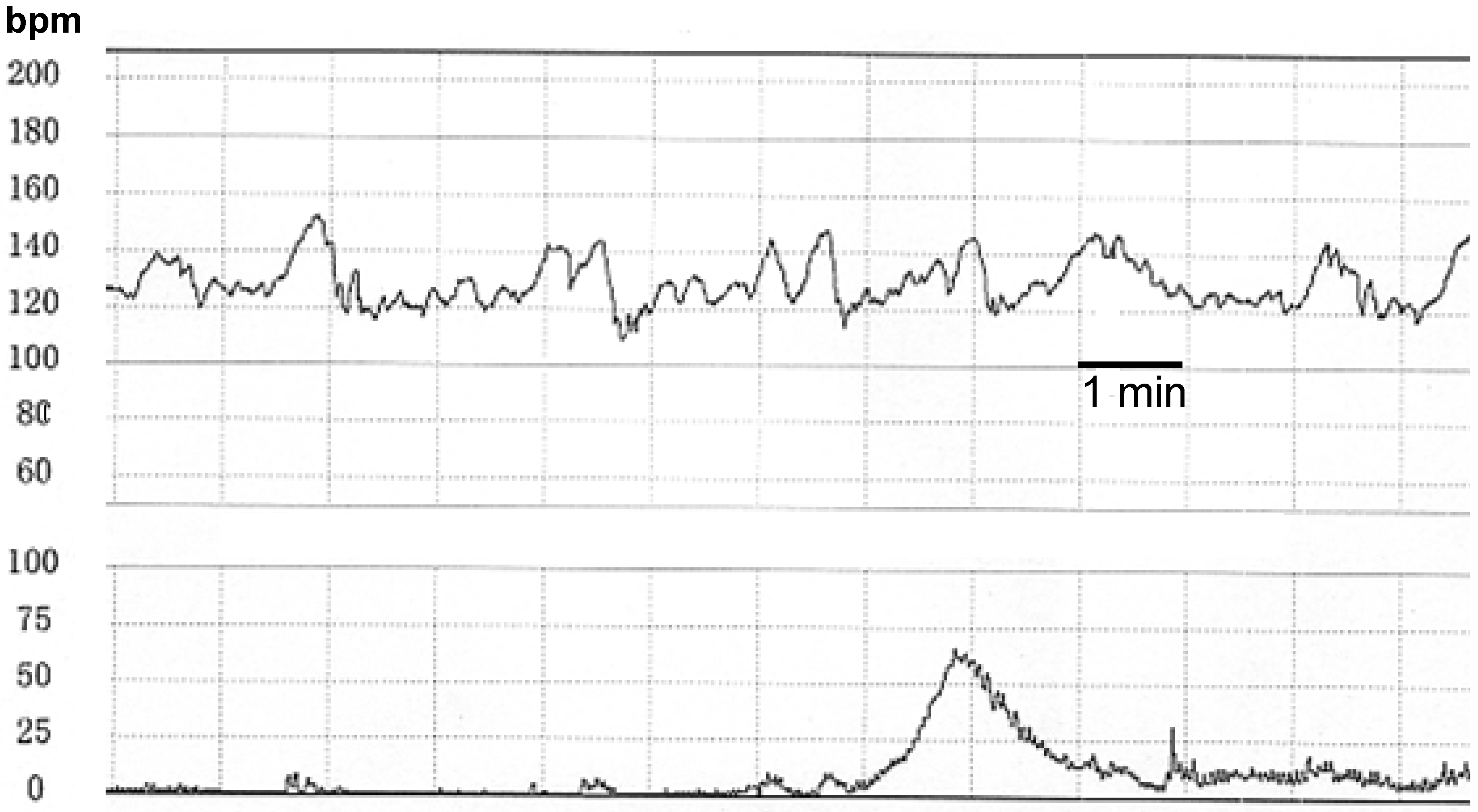
The traditional methods for fetal monitoring included listening to augmented fetal heart tones and recording the fetal phono- and electro-cardiograms (FPCG and FECG) [1,2]. More recently, emphasis was placed on the importance of fetal heart rate (FHR) analysis, where the fetal scalp ECG, fetal heart tones [3,4] and ultrasonic Doppler fetal heart signals [5,6] were introduced to trace FHR in the fetal monitoring devise to identify fetal abnormalities. The simultaneous recording of uterine contraction is the reference for FHR in the cardiotocogram (CTG). The autocorrelation technique [6] is commonly used for the FHR pattern diagnosis in the external CTG method. The CTG chart was continuously monitored and the non-reassuring fetal status (NRFS) was diagnosed by the observer during the course of labor (Figure 1). However, there was a marked difference in the inter-observer diagnoses and false positive FHR patterns, and the value of a common CTG diagnosis remains controversial.
Figure 1. The cardiotocogram (CTG) recorded during the labor. Fetal heart rate (FHR) is traced in the upper channel and uterine contraction in the lower one. The CTG is normal and reassuring because of normal baseline level, normal baseline variability, transient FHR accelerations but no transient deceleration. The time bar represents 1 minute. (Courtesy of Dr. RK Pooh).
Algorithms 02 00019 g001
Favorable results have been obtained by our CTG monitoring technique; the incidence neonatal asphyxia, perinatal mortality and the cerebral palsy (CP) have been reduced in an obstetrical hospital [7], and a large study of 160,000 deliveries revealed a reduction in the incidence of CP [8]. These favorable results were obtained by the focused effort of continuous monitoring for a small number of deliveries. However, fetal monitoring might be incomplete in hospitals that manage a large number of deliveries. Thus, the introduction of automated computer diagnosis was considered for monitoring of the whole course of all deliveries. The idea of global fetal monitoring with the aim of improving outcome was the inspiration for Maeda to create an experts’ knowledge system program for FHR analysis and diagnosis in 1980 [9]. Other computer systems including the System 8000, an antenatal automated fetal diagnosis created by Dawes [10], the Porto system [11] to analyze CTG signals and others [12,13,14,15] have been studied for fetal monitoring.
Although our computerized intrapartum fetal monitoring system utilizes the FHR score, which strongly correlates with the Apgar score [16], the system may still contain experts’ knowledge. The more objective diagnostic technique employed by the neural network (NNW) system, which requires no expert knowledge, provides an avenue to globally expand the use of automatic fetal monitoring.

2. Methods

2.1 Acquisition of the Fetal Heart Rate and Uterine Contraction Data

The ultrasonic Doppler autocorrelation FHR meter and guard-ring type strain-gauge tocodynamometer were used for fetal monitoring in this study. The electrical output of FHR and contraction signals from the monitor is introduced into the analogue-to-digital (AD) converter of the analyzing computer at a sampling rate of 4 Hz. The data are then averaged to a rate of 0.5 Hz. This sampling rate was chosen as the optimal rate to achieve minimal recorded FHR variation, compared with the original FHR tracing. In this way, a data set consisting of 150 recordings are processed for FHR and contraction signals. Analysis time was 5 minutes in order to detect FHR changes in the earliest stages of the monitoring.
The analyzing computer determines the FHR baseline in 5 minutes by averaging the FHR data in the step of the most frequent data among 20 FHR steps from 0 to 200 beats per minutes (bpm). The averaged data-to-data variation in FHR within this 5 minute period was added to the baseline to achieve a reference line for detection of transient FHR acceleration. Similarly, this data was subtracted from the baseline data to achieve a reference line for detection of transient FHR deceleration; FHR deceleration starts when the FHR data repeatedly falls below the reference line and ends when it reaches the reference line. Deceleration is defined as a FHR decrease of 20 bpm or more for 20 seconds or more. The last 5 minutes of an unfinished deceleration period is transferred to the first 5 minutes of the next period. A transient increase is defined as a FHR increase of 15 bpm or more for 15 or more seconds. The down-hill amplitude in FHR increases is defined as the long-term variability in the baseline, except for acceleration and deceleration. The uterine contraction is detected by using a similar algorithm. The area under the contraction curve is determined to assess uterine hyperactivity [9,16,17].
The FHR baseline, long-term variability, deceleration, acceleration, and uterine contraction are detected as described above and the data are analyzed by the computer system [17,23]. The 8 FHR parameters used for computer training and diagnosis are transformed into 16 ranks (0 to 15) and input into the trained NNW computer, where each of the 8 FHR parameters in the three succeeding 5 minutes periods are input into the input layers. Thus, the hybrid system of the NNW computer has the capacity to analyze a data set consisting of 24 FHR recording over 15 minutes instead of the entire FHR data set.

2.2 Detection of Sinusoidal FHR

Sine wave-like FHR baseline oscillation appears infrequently in cases of severe fetal anemia or hypoxia and should be accurately detected because it is a state of imminent fetal death. However, there is a physiological false positive sinusoidal FHR change followed by favorable outcome that is not caused by an underlying fetal disorder. The false positive sinusoidal FHR is evoked by the periodic fetal respiratory or mouthing movement and differentiated by the actocardiogram [18]. In the present study, the physiological sinusoidal FHR tracings were collected and compared to the pathological sinusoidal FHR characterized by fetal or neonatal death or severe disorders by using the frequency spectrum determined by FFT analysis. In FFT analysis the area under the low frequency spectrum and the density of the peak power spectrum are markedly smaller in false positive sinusoidal FHR than the sinusoidal FHR changes associated with fetal disorders [19]. The spectrum frequency parameters were also markedly smaller when loss of FHR baseline variability was detected compared with normal FHR. The positive sinusoidal FHR was input into the NNW computer.

2.3 The Neural Network Computer

Rumelhart, Hinton, and Williams [20,21,22] developed a powerful rule for learning networks with hidden (intermediate) layers of units, capable of learning an internal presentation adequate for performing the task at hand. It was called the generalized delta rule, or the back-propagation method, by which output unit errors propagate backward. A classic example is the exclusive-or (XOR) problem, which was completely linearly separated by using this method [21].
Learning has always been a key issue in the development of network models. Simple, homogeneous learning procedures enable the networks to self-modify and adapt to their environments. The error back-propagation learning procedure allows the study of internal representations that evolve to support processing for particular problems and, in that way, improves the understanding of problems under study as well as the nature of networks required to solve them. Moreover, these powerful learning procedures may be used to design systems capable of discovering new ways to solve difficult neural problems.
Learning and indeed knowledge is in the connections rather than in the units themselves. Thus, “education” is the key issue for developing a neural network computer able to “learn”.
The problems to solve are: (1) input signals, (2) input patterns, (3) teaching patterns, (4) output signals, (5) coding, and (6) numbers of layers and units.
First, FHR must be recognized directly and automatically. However, FHR patterns are complicated and too data-rich to analyze automatically. Thus, eight parameters were obtained from FHR, as detailed in Table 1.
Table 1. The 8 FHR Parameters Used in NNW [23].
1. Baseline FHR (beats/minute)
2. Baseline variability amplitude (beats/minute)
3. Presence of sinusoidal FHR pattern
4. Number of decelerations
5. Duration of decelerations (in seconds)
6. Bottom FHR of decelerations (beats/minute)
7. Lag time of decelerations (in seconds)
8. Recovery time of decelerations (in seconds)
Second, the reliability of the parameters depends on the length of data, although the timeliness depends on the shortness of data. Thus, with a lot of trial and error, each of the 8 FHR parameters was used in the three succeeding 5 minutes, that is, an 8 x 3 pattern served as an input.
Third, 20 cases with a known outcome were selected as teaching patterns for supervised learning: 3 normal cases, 3 intermediate cases, and 14 pathological cases. The number of teaching patterns was selected with the consideration that a misdiagnosis occurs as the worse case even if error occurs.
Fourth, the output signals designated to the probability of a normal, intermediate, and pathological outcome. In processing by the back-propagation algorithm, the neural network calculated an output value by a learning process. If the learning was correctly advanced, the output value of an output unit was designed to approach 1. Then, O1, O2, and O3 were taken as an output vector where O1 indicates normal, O2 intermediate, and O3 pathological. The output vector should be (1, 0, 0), (0, 1, 0), and (0, 0, 1), if the input pattern is normal, intermediate, and pathological, respectively. Thus, the deeper the learning advances, the closer the output value is to 1. The output value means certainty, and is very likely equivalent to probability, although not exactly probability itself because the sum of the output value is not 1. To obtain exact probability from the output value, the output values are divided by the sum of three output values, and then, by using the probability, the recognition rate is clear.
Fifth, the 8 parameters obtained from FHR were normalized to 16 steps for the pattern coding, as shown in Table 2.
Table 2. Normalization of 16-step Coding Data [23].
1. Baseline FHR (beats/minute)
  From 50 to 210, we divided 16 steps, and named from 0 to 15, respectively.
2. Baseline variability amplitude (beats/minute)
  From 0 to 63, we divided 16 steps, and named from 0 to 15, respectively.
3. Presence of sinusoidal FHR pattern
  Absent: 0, Present: 15
4. Number of decelerations
  No deceleration: 0, 1-2: 5, 3-4: 10, 5 and over: 15
5. Duration of decelerations (in seconds)
  From 0 to 320, we divided 16 steps, and named from 0 to 15, respectively.
6. Bottom FHR of decelerations (beats/minute)
  From 0 to 160, we divided 16 steps, and named from 0 to 15, respectively.
7. Lag time of decelerations (in seconds)
  From 0 to 240, we divided 16 steps, and named from 0 to 15, respectively.
8. Recovery time of decelerations (in seconds)
  From 0 to 240, we divided 16 steps, and named from 0 to 15, respectively.
Finally, we designed the neural network as 3 layers: 24 units for input layer, 30 units for hidden (intermediate) layer, and 3 units for output layer. The numbers of units for input layer and output layer were easily calculated from the input pattern number (8 x 3) and output signals (3), respectively. In contrast, the number of units for hidden (intermediate) layer was adjusted to 30 after repeated trial and error.

3. Results and Discussion

3.1 Performance of the Neural Network Computer

The recognition results obtained by our NNW with teaching input after training for 10,000 times, that is to say closed recognition, are shown in Table 3. All data carried the probability of 0.998 (or 99.8%) to 1.000 (or 100%). Therefore, this well-trained NNW can be used to recognize new patterns not included in the teaching patterns, that is to say “open” recognition.
Table 3. Recognition Probability.
Example No.  Probability
namenormalintermediatepathologic
1svd* 00.0010.999
2mvd** 0.0010.9990
3normal0.9980.0010.001
4normal0.9980.0010.001
5ssp*** 001
6bdc****0.00100.999
7svd* 00.0010.999
8ld*****001
9lv *6001
10normal0.9980.0010.001
11mvd** 0.0010.9990.001
12mvd** 0.0010.9980.001
13lv *6001
14lv *6001
15ld*****001
16ld*****001
17bdc****0.00100.999
18bdc****0.00100.999
19ssp*** 001
20svd* 00.0010.999
* svd: Severe variable deceleration; ** mvd: Mild variable deceleration; *** ssp: Sinusoidal patter; **** bdc: Bradycardia; ***** ld: Late deceleration; *6: lv: Loss of variability.

3.2 Comparison of Neural Computer Results to Clinical Data

3.2.1 Neural Computer Results and Neonatal State

The normal and pathological outcome probabilities obtained by the NNW computer were compared to the neonatal states evaluated by the Apgar score, clinical condition, and umbilical cord blood pH, where the analysis time was as long as 50 minutes and the neural computer training reference data was also obtained in 50 minutes. The neonatal states matched outcome probabilities in 25 out of 29 cases (86 %) [23].

3.2.2 The FHR Score of the Experts’ System Strongly Correlates with the Outcome Probability

The FHR score is the sum of evaluation values determined by the grade of the low Apgar score in 8 quantitative FHR parameters in 5 minutes of non-interventional deliveries in the 1960s. The recent statistical study on previous monitoring studies revealed that maximal FHR scores in the first stage of labor strongly correlated with the Apgar score, i.e., neonatal depression is quantitatively estimated by the FHR score in the early stage of labor [16]. The FHR scores also correlated with fetal scalp blood pH in the 2nd stage of labor [24]. Fetal acidosis is detected by the FD index, which was determined by the FHR score, the loss of FHR baseline variability, and late FHR deceleration [25].
The outcome probabilities obtained by the NNW analysis correlated with the FHR score; the FHR score was high when the pathological probability was large, the FHR score was moderate when the intermediate probability was positive, and the score was low when the outcome probability was normal [23]. Therefore, outcome probabilities of the NNW analysis are also capable to detect fetal hypoxic deterioration (Figure 2).
Figure 2. The relationship between outcome probability and FHR score [23]. The X-axis is the probability of normal (1), intermediate (2) and pathological outcome (3), and the Y-axis shows the FHR score. The probability of a pathological outcome (3) is distributed in the high FHR score area, the probability of an intermediate outcome (2) is distributed in the intermediate FHR score area, and the probability of a normal outcome (1) is distributed in the low FHR score area [14]. Thus, the NNW outcome probability strongly correlates with the FHR score revealing that the NNW diagnosis based on FHR monitoring is accurate.
Algorithms 02 00019 g002

3.3 The Neural Index

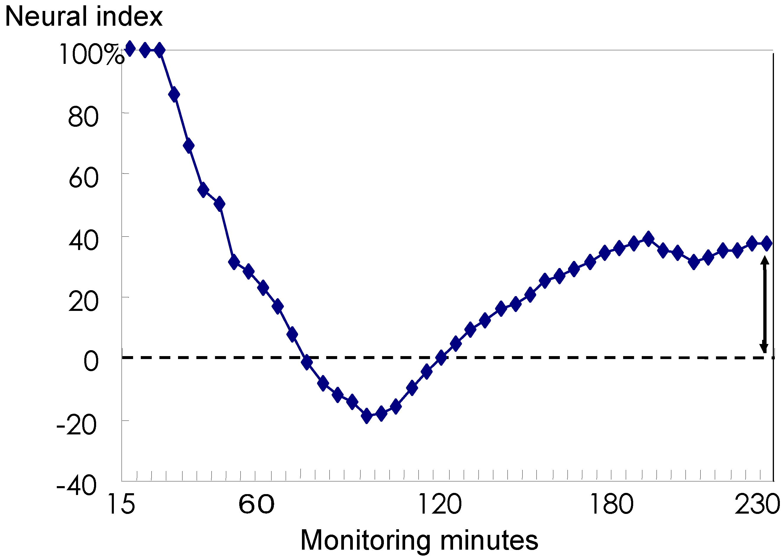
The evaluation of prolonged fetal monitoring for more than hours was difficult despite the introduction of the quantified FHR diagnosis. In the present study we analyzed prolonged fetal monitoring by averaging the outcome probability obtained by the NNW analysis, where the past probabilities for pathological outcome were averaged every 5 minutes and then subtracted from the averaged normal outcome probabilities. The difference in averaged outcome probabilities formed a curve, and the last value was positive in normal outcome cases of no asphyxia, whereas it was negative and less than zero in cases of neonatal depression (Figure 3) [26]. The final value calculated from fetal monitoring was referred to as the neural index, which was analyzed from the stored data from continuous monitoring. When the neural index is positive immediately before the birth the neonatal state is normal. In another hospital, umbilical cord blood pH was low for cases with a negative index. The neural index provides a useful measurement in clinical obstetrics.
Figure 3. The neural index [26]. Neural index values in every 5 minutes form a curve in a case of prolonged fetal monitoring for 230 minutes, where the final value of the curve was +37% and the neonatal status was normal. The neonates showed a depressed state when the neural index was negative immediately before birth, and no neonatal asphyxia was noted when the final neural index value was positive.
Algorithms 02 00019 g003

3.4 Discussion

NNW analysis has been used for objective, definitive or quick decision making in a wide range of fields such as traveling sales [27], business, and in the medical field for applications including electroencephalography [28], histology [29], brain function analysis [30], brain tumor detection [31], smear categorization [32], visual field studies [33], response to fault [34], coronary artery disease [35], HIV studies [36], prediction of the duration of the first stage of labor [37], fetal weight estimation [38], and fetal monitoring [12,13,14,15,23].
A back-propagation system trained by typical teacher data is common in the NNW system. A NNW computer is composed of the input layer, intermediate hidden layer and output layer. Each layer is composed of multiple active units and connected with each other. Our NNW computer is composed of an intermediate layer with 30 active units, because a high number of active units are essential for accurate function. The NNW software package is usually a back-propagation system that feeds error signals back to the network, effectively increasing the coupling weight of appropriate units, and thus minimizing decision errors after repeated training.
The use of complete FHR data in the NNW computer is time consuming for system training and diagnosis. Since automated FHR analysis is utilized in the centralized system for the multiple simultaneous monitoring with time sharing, the analysis time should be as short as possible. For this reason, the hybrid system with limited parameters is ideal and was the chosen method for analysis in the present study. Although sinusoidal FHR is rare in obstetrical practice, it should be automatically diagnosed from FHR changes within a short time frame. We identified the physiological false positive sinusoidal FHR by frequency analysis and cases with pathological sinusoidal FHR were identified by the NNW system. The 20 cases used for training the NNW computer were selected to include indispensable FHR changes, such as severe variable deceleration, late deceleration, the loss of baseline variability, bradycardia, and sinusoidal FHR.
Although the ratio of coincidence to clinical findings was 86% over 50 minutes of monitoring, the long interval between FHR analysis and birth may influence the accuracy rate. Shorter analysis times, such as 15 minutes of monitoring evaluated every 5 minutes, as in the present study, may improve the accuracy rate.
Although the trend gram was studied in previous report [23], the neural index curve may be a better indicator of fetal status in prolonged monitoring [26]. Since the A/B ratio of an actocardiogram clearly shows fetal status, computerized evaluations of actocardiograms may be introduced into automated monitoring in the future, although the present NNW diagnosis depends only on FHR changes and not on fetal movements.
The recent progress in the automated fetal evaluation is the application of wavelet analysis [40] and entropy system [39,41] in the computerized studies on the fetal heart rate. The fetal state is analyzed for the fetal well-being with the wavelet [40], and for the detection of fetal distress by the entropy system [39]. The fetal ECG masking maternal ECG signal is rejected on the abdominal wall in order to improve fetal heart signals for the precise beat-to-beat variability using neuro-fuzzy inference system [42]. Fetal study will be further progressed by the improved analysis of fetal heart rate by introducing the new techniques.

Conclusion

A hybrid NNW computer composed of a back propagation system achieved a high level of accuracy in outcome probabilities, which is characteristic of the objective nature of NNW analysis. Thus, this study highlights the ability of the NNW computer system to achieve the full monitoring of all parturient women on a global basis to improve the perinatal state of mothers and infants. The NNW system is also promising for data analysis in other areas of obstetrics and gynecology, particularly in central fetal monitoring and possibly in image analysis.

Acknowledgements

The authors express sincere gratitude to Dr M. Utsu of Seirei Mikatahara hospital and Dr T. Fuchiwaki of Urayasu-Ichikawa Municipal hospital for kindly agreeing to analyze FHR data in the study of neural index.

References

  1. Maeda, K. Fetal heart sound recorded by using slow down tape technique. In Proc. 8th Int. Conf. Med. Biol. Eng. 1969. Session 19-2. [Google Scholar]
  2. Symonds, E.M. Configuration of the fetal electrocardiogram in relation to the fetal acid-base balance and plasma electrolytes. J. Obstetet. Gynaecol. Brit. Commonw. 1971, 78, 957–970. [Google Scholar] [CrossRef]
  3. Gentner, O.; Hammacher, K. An improved method for the determination of the instantaneous fetal heart frequency from the fetal phonocardiogram. In Proc. 7th Int. Conf. Med. Biol. Eng. 1967. No. 140. [Google Scholar]
  4. Maeda, K.; Ezaki, I. Fetal cardiotachography recorded with fetal heart sound during pregnancy and labor. In Proc. 7th Int. Conf. Med. Biol. Eng. 1967. No. 144. [Google Scholar]
  5. Hammacher, K. Einführung in die Cardiotokographie. 5. Teil: Die Herzfrequenzmessung mit US = Ultraschall. Die Schweizer Hebamme 1976, 74, 17–21. (in German). [Google Scholar]
  6. Takeuchi, Y.; Hogaki, N. Auto correlation method for fetal heart rate measurement from ultrasonic Doppler fetal signal. In Ultrasound in Medicine; White, D., Brown, R.E., Eds.; Plenum Press: New York, 1977; Vol. 3B “Engineering Aspect”, pp. 1327–1332. [Google Scholar]
  7. Tsuzaki, T.; Sekijima, A.; Morishita, K.; Takeuchi, Y.; Mizuta, M.; Minagawa, Y.; Nakajima, K.; Maeda, K. Survey on the perinatal variables and the incidence of cerebral palsy for 12 years before and after the application of the fetal monitoring system. Nippon Sanka Fujinka Gakkai Zasshi 1990, 42, 99–105. [Google Scholar] [PubMed]
  8. Takeshita, K.; Ando, Y.; Ohtani, K.; Takashima, S. Cerebral palsy in Tottori, Japan. Benefits and risks of progress in perinatal medicine. Neuroepidemiology 1989, 4, 184–192. [Google Scholar] [CrossRef]
  9. Maeda, K.; Arima, T.; Tatsumura, M.; Nagasawa, T. Computer-aided fetal heart rate analysis and automatic fetal-distress diagnosis during labor and pregnancy utilizing external techniques in fetal monitoring. MEDINFO 1980, 80, 1214–1219. [Google Scholar]
  10. Dawes, G.S.; Moulden, M.; Redman, C.W. System 8000: computerized antenatal FHR analysis. J. Perinat. Med. 1991, 19, 47–51. [Google Scholar] [CrossRef] [PubMed]
  11. Bernardes, J.; Moura, C.; de Sa, J.P.; Leite, L.P. The Porto system for automated cardiotocogaphic signal analysis. J. Perinat. Med. 1991, 19, 61–65. [Google Scholar] [CrossRef] [PubMed]
  12. Keith, R.D.; Westgate, J.; Ifeachor, E.C.; Greene, K.R. Suitability of artificial neural networks for feature extraction from cardiotocogram during labor. Med. Biol. Eng. Comput. 1994, 32, S51–S57. [Google Scholar] [CrossRef] [PubMed]
  13. Ulbricht, C.; Dorfner, G.; Lee, A. Neural networks for recognizing patterns in cardiotocograms. Artif. Intell. Med. 1998, 12, 271–284. [Google Scholar] [CrossRef]
  14. Kupka, T.; Wrobel, J.; Jerewski, J.; Gacek, A.; Jerewski, M. Evalation of fetal heart rate baseline estimation method using testing signals based on a statistical model. Proc. IEEE Eng. Med. Biol. Soc. 2006, 1, 3728–3731. [Google Scholar]
  15. Jezewski, M.; Wrobel, J.; Labaj, P.; Leski, J.; Henzel, N.; Horoba, K.; Jezewski, J. Some practical remarks on neural networks approach to fetal cardiotocograms classification. Proc. IEEE Eng. Med. Biol. Soc. 2007, 5170–5173. [Google Scholar]
  16. Maeda, K.; Noguchi, Y.; Matsumoto, F.; Nagasawa, T. Quantitative fetal heart rate evaluation without pattern classification: FHR score and artificial neural network analysis. In Textbook of Perinatal Medicine, 2nd Ed.; Kurjak, A., Chervenak, F.A., Eds.; Informa: London, UK, 2006; Vol. 2, pp. 1487–1495. [Google Scholar]
  17. Maeda, K. Computerized automatic diagnosis of fetal distress with the use of external monitoring technique. In Computerdiagnostik in der Geburtsmedizin; Krause, W., Ed.; Friedrich-Schiller-Universität: Jena, Germany, 1981; pp. 28–49. [Google Scholar]
  18. Ito, T.; Maeda, K.; Takahashi, H.; Nagata, N.; Nakajima, K.; Terakawa, N. Differentiation between physiologic and pathologic sinusoidal FHR pattern by fetal actocardiogram. J. Perinat. Med. 1994, 221, 39–43. [Google Scholar] [CrossRef]
  19. Maeda, K.; Nagasawa, T. Automatic computerized diagnosis of fetal sinusoidal heart rate. Fetal Diag. Ther. 2006, 20, 328–334. [Google Scholar] [CrossRef] [PubMed]
  20. Rumelhart, D.E.; McClelland, J.L. Parallel Distributed Processing; The MIT Press: Cambridge, MA, 1986; Vol. 1 & 2. [Google Scholar]
  21. Rumelhart, D.E.; Hinton, G.E.; Williams, R.J. Learning Internal Representations by Error Propagation. In Parallel Distributed Processing; The MIT Press: Cambridge, MA, 1986; Vol. 1, pp. 318–362. [Google Scholar]
  22. Rumelhart, D.E.; Hinton, G.E.; Williams, R.J. Learning Representations by Back-Propagating Errors. Nature 1986, 323, 533–536. [Google Scholar] [CrossRef]
  23. Maeda, K.; Utsu, M.; Makio, A.; Serizawa, M.; Noguchi, Y.; Hamada, T.; Mariko, K,; Matsumoto, F. Neural network computer analysis of fetal heart rate. J. Matern. Fetal Invest. 1998, 8, 163–171. [Google Scholar]
  24. Maeda, K.; Kimura, S.; Fukui, Y.; Ozawa, S.; Kosaka, T.; Wang, C.F.M.; Tamura, M.; Takata, D.; Nakano, H.; Mitoma, M. Pathophysiology of Fetus; Fukuoka Printing: Fukuoka, Japan, 1969. [Google Scholar]
  25. Irie, T. Automated fetal heart rate analysis and its trend gram in relation to the gas analysis and acid-base balance of umbilical arterial blood. Nippon Sanka Fujinka Gakkai Zasshi 1986, 38, 1623–1631. [Google Scholar] [PubMed]
  26. Maeda, K.; Noguchi, Y.; Matsumoto, F. Evaluation of prolonged fetal monitoring with normal and pathologic outcome probabilities determined by artificial neural network. Fetal Diag. Ther. 2003, 18, 370–376. [Google Scholar] [CrossRef]
  27. Tokuda, I.; Nagashima, T.; Aihara, K. Global bifurcation structure of chaotic neural networks and its application to traveling salesman problems. Neural Networks 1997, 10, 1673–1690. [Google Scholar] [CrossRef]
  28. Weng, W.; Khorasani, K. An adaptive structure neural networks application to EEG automatic seizure detection. Neural Networks 1996, 9, 1223–1240. [Google Scholar] [CrossRef]
  29. Moul, J.W.; Snow, P.H.; Fernandez, E.B.; Maher, P.D.; Sesterhenn, I.A. Neural network analysis of quantitative histological factors to predict pathological stage in clinical stage I nonseminomatous testicular cancer. J. Urol. 1995, 153, 1674–1677. [Google Scholar] [CrossRef]
  30. Ruppin, E.; Reggia, J.A. A neural model of memory impairment in diffuse cerebral atrophy. Br. J. Psychiatry 1995, 166, 19–28. [Google Scholar] [CrossRef] [PubMed]
  31. Arle, J.E.; Moriss, C.; Wang, Z.J.; Zimmerman, R.A.; Philipps, P.G.; Sutton, L.N. Prediction of posterior fossa tumor type in children by means of magnetic resonance image properties, spectroscopy, and neural networks. J. Neurosurg. 1997, 86, 755–761. [Google Scholar] [CrossRef] [PubMed]
  32. Brickley, M.R.; Cowpe, J.G.; Shepherd, J.P. Performance of a computer-simulated neural network trained to categorize normal, premalignant and malignant oral smears. J. Oral. Pathol. Med. 1996, 25, 424–428. [Google Scholar] [CrossRef] [PubMed]
  33. Mutlukan, E.; Keating, D. Visual field interpretation with a personal computer-aided neural network. Eye 1994, 8, 321–323. [Google Scholar] [CrossRef] [PubMed]
  34. Westenskow, D.R.; Orr, J.A.; Simon, F.H.; Bender, H.J.; Frankenberger, H. Intelligent alarms reduce anesthesiologist’s response time to critical faults. Anesthesiology 1992, 77, 1074–1079. [Google Scholar] [CrossRef] [PubMed]
  35. Fujita, H.; Katafuchi, T.; Uehara, T.; Nishimura, T. Application of artificial neural network to computer-aided diagnosis of coronary artery disease in myocardial SPECT bull’s eye images. J. Nucl. Med. 1992, 33, 272–276. [Google Scholar] [PubMed]
  36. Andreassen, H.; Bohr, H.; Bohr, J.; Brunak, S.; Bugge, T. Analysis of the secondary structure of the human immunodeficiency virus (HIV) proteins p17, gp120 and gp41 by computer modeling based on neural network methods. J. Acquir. Immune Def. Syndr. 1990, 3, 615–622. [Google Scholar]
  37. De Voe, L.D.; Samuel, S.; Prescott, P.; Work, B.A. Predicting the duration of the first stage of spontaneous labor using a neural network. J. Mat. Fet. Med. 1996, 5, 256–261. [Google Scholar]
  38. Farmer, R.M.; Medearis, A.L.; Hirata, G.I.; Platt, L.D. The use of a neural network for the ultrasonographic estimation of fetal weight in the macrosomic fetus. Am. J. Obstet. Gynecol. 1992, 166, 1467–1472. [Google Scholar] [CrossRef]
  39. Ferrario, M.; Signorini, M.G.; Magenes, G. Comparison of entropy-bases regularity estimators: application to the fetal heart rate signal for the identification of fetal distress. IEEE Trans. Biomed. Eng. 2006, 53, 119–125. [Google Scholar] [CrossRef] [PubMed]
  40. Cattani, C.; Doubrovina, O.; Rogosin, S.; Voskresensky, S.L.; Zelianko, E. On the creation of a new diagnostic model for fetal well-being on the base of wavelet analysis of cardiotocograms. J. Med. Sys. 2006, 30, 489–494. [Google Scholar] [CrossRef]
  41. Al-Angari, H.M.; Sahakian, A.V. Use of sample entropy approach to study heart rate variability in obstructive sleep apnea syndrome. IEEE Trans. Biomed. Eng. 2007, 54, 1900–1904. [Google Scholar] [CrossRef] [PubMed]
  42. Assaleh, K. Extraction of fetal electrocardiogram using adaptive neuro-fuzzy inference systems. IEEE Trans. Biomed. Eng. 2007, 54, 59–68. [Google Scholar] [CrossRef] [PubMed]