Article

Characterization of Microbial Decay and Microbial Communities in Waterlogged Archaeological Rosewood (Dalbergia Species)

1
Department of Wood Science and Engineering, Chonnam National University, Gwangju 61186, Republic of Korea
2
Department of Animal Science, Chonnam National University, Gwangju 61186, Republic of Korea
3
Underwater Excavation & Conservation Division, National Research Institute of Maritime Cultural Heritage, Mokpo 530839, Republic of Korea
4
Center for Research Facilities, Chonnam National University, Gwangju 61186, Republic of Korea
5
Conservation & Collection Management Division, National Research Institute of Maritime Cultural Heritage, Mokpo 530839, Republic of Korea
*
Authors to whom correspondence should be addressed.
Present address: Preservation & Restoration Division, Presidential Archives, Sejong-si 30107, Republic of Korea.
Forests 2023, 14(10), 1992; https://doi.org/10.3390/f14101992
Submission received: 5 September 2023 / Revised: 24 September 2023 / Accepted: 28 September 2023 / Published: 3 October 2023
(This article belongs to the Special Issue Wood as Cultural Heritage Material)

Abstract

:
While numerous studies have examined microbial attacks on waterlogged archaeological wood, limited information is available regarding microbial attacks in waterlogged tropical hardwoods submerged in marine environments. In this context, we explored microbial attacks in waterlogged archaeological rosewood (Dalbergia species), a tropical hardwood species that was submerged in the Yellow Sea for approximately 700 years, using various microscopic techniques and next-generation sequencing (NGS) methods. Based on morphological features, Type-I soft rot decay was identified as the main decay type. Most fibers in waterlogged archaeological rosewood studied were gelatinous (G) fibers of tension wood and the mode of soft rot decay differed from fibers without the G-layer. Differences in decay resistance between vessel/axial parenchyma cells and fibers were not obvious. Vestured- and simple pit membranes showed higher decay resistance than vessel and axial parenchyma cell walls, respectively. Microbial community analysis by NGS revealed the dominance of Ascomycota and Basidiomycota in the fungal community. Various bacterial communities were also identified, although no prominent signs of bacterial decay were noted. The identified bacterial communities markedly differed from those reported previously in terms of their composition and abundance. Together, our results offer detailed insights into the microbial types and communities responsible for degrading waterlogged archaeological rosewood, contributing to a better understanding of microbial attacks in tropical hardwoods exposed to marine environments.

1. Introduction

Archaeological woods are closely associated with cultural history; however, they commonly undergo degradation by biotic and abiotic agents. Among biotic agents, Basidiomycota fungi are predominantly responsible for wood decay in terrestrial environments [1,2]. By contrast, waterlogged archaeological woods (WAWs) excavated from marine and wet terrestrial burial sites are mainly decomposed by bacteria [1,3,4,5] and soft rot fungi [5,6,7,8]. Many studies have identified various wood-inhabiting bacteria in WAWs even though their precise role in wood decay remains largely unknown [5,6,7]. Many aquatic fungi belonging to Ascomycetes and Fungi Imperfecti have also been identified to cause soft rot decay in WAWs [5,6,7,8]. However, little information is available regarding microbial attacks on tropical hardwoods, as most previous studies have focused on exploring wood species grown in temperate regions [9]. Observations on microbial decay in waterlogged archaeological tropical hardwood submerged in marine environments are particularly limited. High-density and extractive-rich tropical hardwood species generally exhibit strong biological resistance against microbial attacks [10,11,12]. Some tropical hardwood species also have unique multilayered cell wall structures of fibers, which affect soft rot decay [13,14]. These features indicate that the mode of microbial decay of waterlogged archaeological tropical hardwood species may differ from that of temperate species in marine environments. However, this issue is still not clarified. Understanding microbial decay in waterlogged archaeological tropical hardwoods is valuable not only for preserving historical and cultural artifacts but also for developing preservation methods for tropical hardwoods in marine environments.
In this study, we explored microbial attacks on waterlogged archaeological rosewood (Dalbergia species), a tropical hardwood species that was submerged in the Yellow Sea for a long period. Approximately 1017 pieces of waterlogged archaeological rosewood were excavated in 1984 from a point located 4 km away from both Jeungdo and Imjado islands (Yellow Sea), Shinan-gun, Jellanam-do, Republic of Korea (37°1.715.7″ N; 126°3.352.0″ E) [15]. These rosewoods were shipped in an international trade ship, known as the Shinan shipwreck, as trading goods. The Shinan shipwreck was a Chinese trade ship that departed in 1323 from a Chinese port and traveled across Korea to Japan [15]. The rosewoods were submerged at a depth of 20 m, with a tidal variation of 4 m and were never exposed to air until they were salvaged. The underwater temperature was around 10–15 °C [16]. Rosewoods were mostly buried in a mudflat [16]. Rosewoods exposed to water were damaged by marine borers [16]. After desalination, rosewoods were restored by controlled air drying at room temperature and are currently on display in the Mokpo National Maritime Museum as a national artifact (Mokpo, Jellanam-do, Republic of Korea). The origin of these waterlogged archaeological rosewoods was unknown. Dalbergia species are widely distributed in tropical countries, including Central and South America, Africa, and Asia, and are known as heavy varieties of wood with distinct colors and fragrances [17]. According to ICRAF’s (International Council for Research in Agroforestry) wood density database, the average density of 45 Dalbergia species is 0.82 g/cm3 [18]. In Asia, at least seven Dalbergia species are considered to have a high economic value [17].
This study assessed the microbial decay patterns and microbial communities in waterlogged archaeological rosewood. To identify which microbial types are responsible for degradation, the micromorphological characteristics of the waterlogged archaeological rosewood were investigated using various microscopic techniques, including light microscopy (LM), confocal laser scanning microscopy (CLSM), scanning electron microscopy (SEM), and transmission electron microscopy (TEM). Fungal and bacterial communities in waterlogged archaeological rosewood were confirmed using next-generation sequencing (NGS). NGS-based internal transcribed spacer (ITS) and 16S ribosomal RNA (rRNA) gene sequencing methods have been used to analyze microbial communities in WAWs [19,20,21,22,23,24].

2. Materials and Methods

2.1. Materials

The waterlogged archaeological rosewood (Dalbergia species) that is currently displayed at the National Maritime Museum (temperature 20 ± 4 °C, relative humidity 55 ± 5%), Mokpo, Jeonnam, Republic of Korea, was used in this study (Figure 1). The length and diameter of rosewood pieces varied greatly and were roughly classified into three groups according to their size: (1) Small size (7–10 cm in diameter, 30–40 cm in length), (2) middle size (10–30 cm in diameter, 50–150 cm in length), and (3) large size (40–70 cm in diameter, 150–200 cm in length) [25]. Based on the appearance of the rosewood surface, rosewood pieces were also divided into three groups: (1) Rosewoods with an apparently intact surface, (2) rosewoods with marine borer damage, and (3) rosewoods with signs of microbial decay. Considering the size, appearance, and availability of sample collection. A small-sized rosewood with signs of microbial decay was selected for experiments (Figure 1A). The rosewood selected was 8 years old (approximately 6.1 cm in diameter at the top end), suggesting that it would be juvenile wood. The moisture content and density of rosewood studied were 10.6% and 0.88 g/cm3, respectively. Whether the rosewood came from the stem or the branch was unclear. The surface of the waterlogged archaeological rosewood showed brownish coloration with many cracks (Figure 1A,B). On the transverse plane, ring and radial cracks were observed (white arrow in Figure 1C). These cracks seemed to have progressed a few millimeters inward from the surface (axial direction). A semicircular disk of approximately 2.5 cm thickness was cut from the top end of the rosewood for the experiment (asterisk in Figure 1C). The collection of more waterlogged archaeological rosewood samples was not allowed because they were designated as national artifacts.

2.2. Microscopy

Small pieces of wood were taken from the block at the surface and at a depth of 4 and 25 mm from the surface (axial direction). After fixation with a mixture of 2% v/v paraformaldehyde and 2% glutaraldehyde in 0.05 M sodium cacodylate buffer (pH 7.2) for 4 h at room temperature, the wood samples were embedded in LR White resin (London Resin Company Ltd., Basingstoke, UK) or Spurr resin (EMS, Hatfield, MA, USA). Using an ultramicrotome equipped with a diamond knife, transverse and radial semithin resin sections (approximately 1 µm thickness) were prepared, mounted on a slide, and stained with 1% w/v toluidine blue on a hot plate (approximately 60 °C). The sections were mounted with Canada balsam (Sigma-Aldrich, MA, USA) or distilled water and then examined under an Olympus BX53 light microscope equipped with an Olympus DP 73 digital camera (Tokyo, Japan). Some serial sections were also mounted with FluorSave reagent (Sigma-Aldrich, Burlington, MA, USA) and examined under a confocal laser scanning microscope (Leica TCS SP5 AOBS/Tandem, Wetzlar, Germany) with a diode laser (excitation and emission wavelengths of 488 nm and 504–554 nm, respectively). For TEM, ultrathin transverse sections (approximately 90 nm thickness) were stained with 1% w/v KMnO4 and examined by field emission TEM (JEM-2100F, Jeol Ltd., Tokyo, Japan) at an accelerating voltage of 200 kV. For focused ion beam–field emission SEM (FIB/FE-SEM), transverse sections (approximately 50 µm thickness) were collected from the wood block at a depth of approximately 2 and 25 mm from the surface (axial direction) using a Leica RM 2145 rotary microtome (Wetzlar, Germany). Some radial longitudinal sections covering the region from the surface to a depth of 10 mm in the axial direction were also prepared from the blocks. The sections were then coated with a layer of platinum (approximately 10 nm thickness) using a Cressington Sputter Coater 108 auto (Watford, England) and examined by SEM (FEI Helios G3 CX FIB/SEM, Hillsboro, OR, USA) at an accelerating voltage of 2 kV.

2.3. DNA Extraction, Amplification, and Sequencing

Metagenomic DNA was extracted from 250 mg of waterlogged archaeological rosewood samples frozen in liquid nitrogen using the repeated bead beating plus column (RBB + C) method [26] and stored at −20 °C before sequencing. The quality and quantity of the extracted DNA were analyzed by 1% agarose gel electrophoresis and using a NanoDrop ND-2000 spectrophotometer (Wilmington, DE, USA). For bacterial DNA library preparation, PCR amplification of the V3–V4 region of the 16S rRNA gene was performed using the 341F (5′-CCTACGGGNGGCWGCAG-3′) and 805R (5′-GACTACHVGGGTATCTAATCC-3′) primers. The fungal DNA library was generated using the ITS7F (5′-GTGARTCATCGAATCTTTG-3′) and ITS4R (5′-TCCTCCGCTTATTGATATGC-3′) primers. Moreover, 16S rRNA and ITS gene amplicon sequencing were performed using the MiSeq® Illumina sequencing platform (Illumina, San Diego, CA, USA) at Macrogen (Seoul, Republic of Korea). Paired reads from amplicon sequencing were merged using the FLASH program v1.2.11 [27]. Sequence processing and microbial community analysis were performed using Quantitative Insights Into Microbial Ecology (QIIME, version 1.9) [28]. Operational taxonomic units (OTUs) were analyzed using the CD-HIT-OTU method [29]. The subsequent high-quality sequences were classified into bacterial taxa using BLASTN v.2.9.0+ [30] against the National Center for Biotechnology Information (NCBI) Reference Sequence (RefSeq) database (http://www.ncbi.nlm.nih.gov/RefSeq/; accessed on 10 May 2023) and into fungal taxa using the UCLUST method [31] against the UNITE database v.8.2 (https://unite.ut.ee; accessed on 10 May 2023).

3. Results

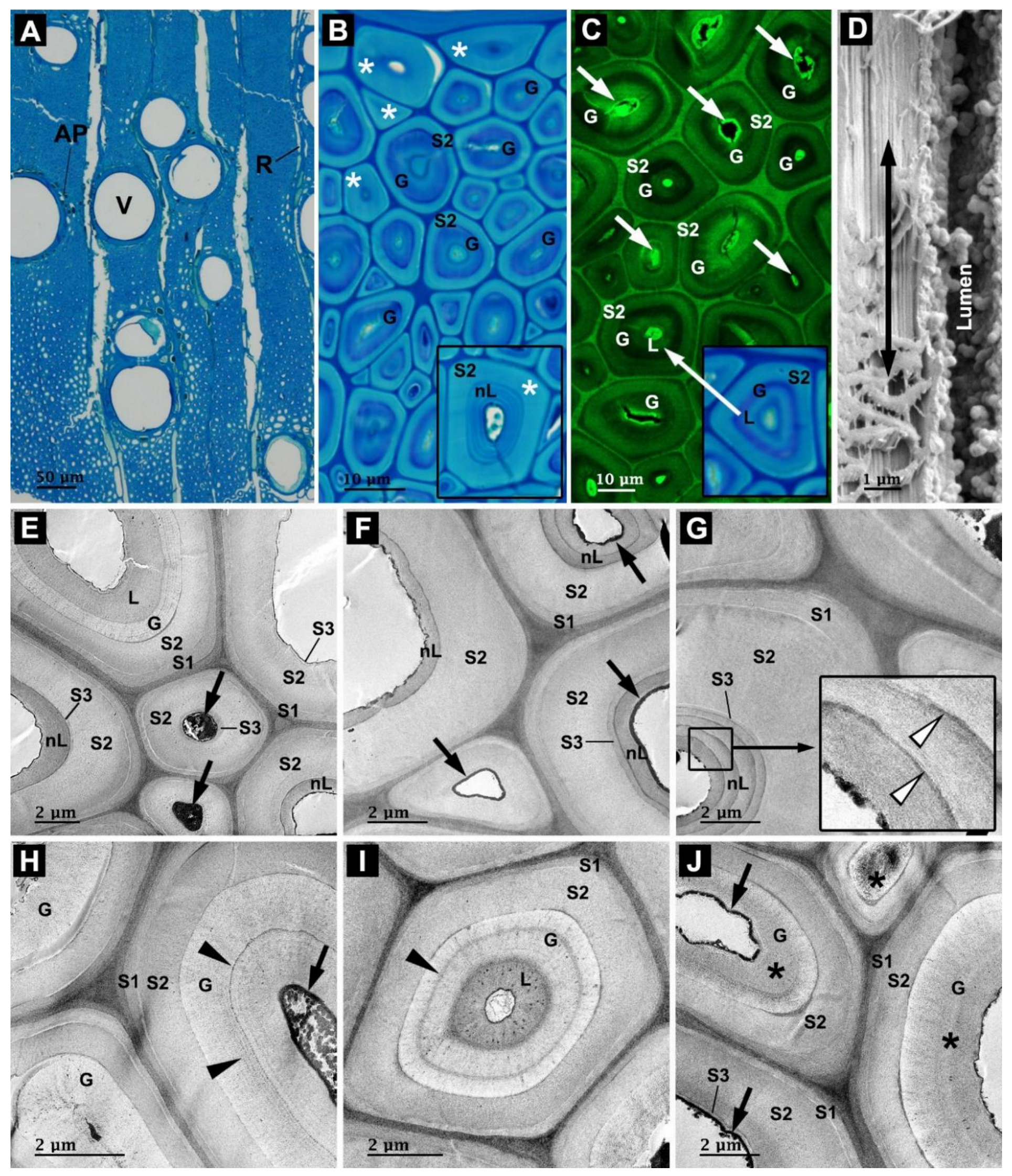
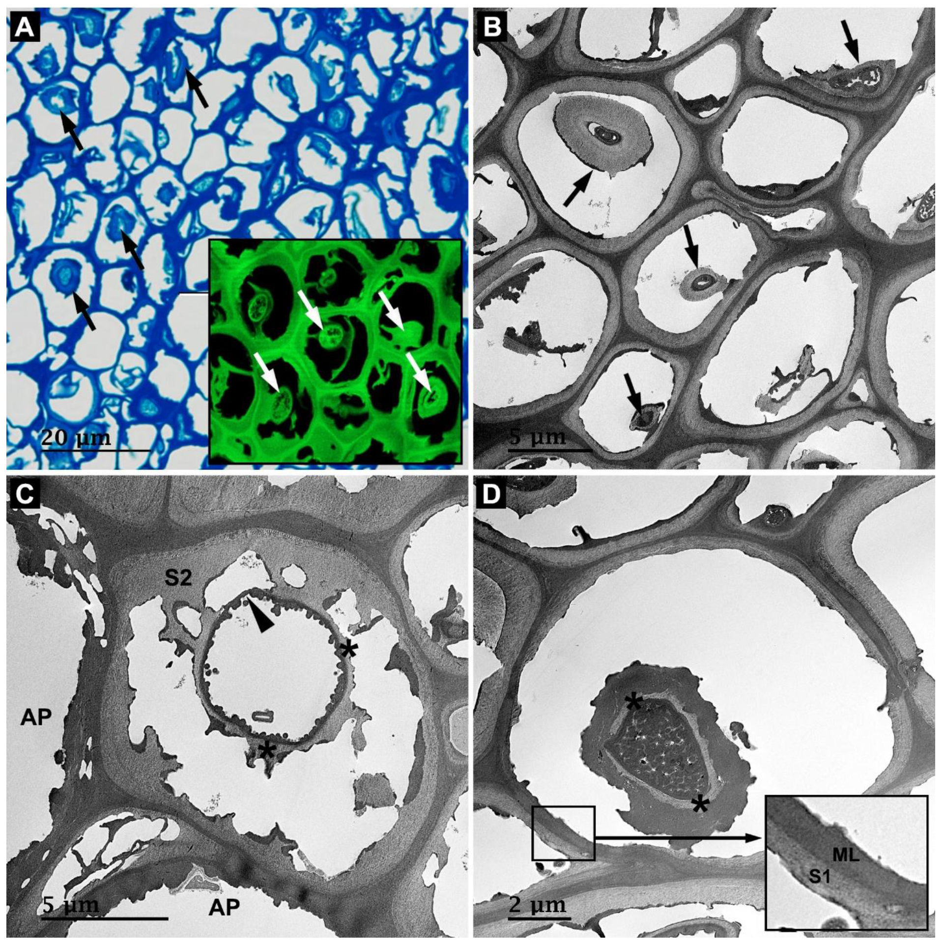
3.1. Anatomy of Rosewood

Figure 2 and Figure 8 show the anatomical structure of the studied waterlogged archaeological rosewood. The waterlogged archaeological rosewood was diffuse porous, in which vessels were solitary, radial multiples of 2–4, and clusters of 4 (Figure 2A and Figure 8B). The vessel lumen was occasionally impregnated (Figure 8B,D) and/or coated (arrowheads in Figure 8B,C) with certain deposits. Axial parenchyma cells were mostly arranged around the vessels (i.e., paratracheal) and filled with phenolic compounds (Figure 2A). Some axial parenchyma cells were highly multilayered (inset in Figure 8A). Rays were uniseriate or biseriate (Figure 2A).
Two distinct staining patterns were detected in wood fibers collected at a depth of 25 mm from the surface using LM combined with toluidine blue staining (Figure 2B). In the first pattern, strong blue coloration was noted in the inner cell wall layers. Most fibers in the studied waterlogged archaeological rosewood samples exhibited this staining pattern. In the analysis using CLSM, the inner cell wall layer of these fibers exhibited very weak lignin autofluorescence (Figure 2C) and had cellulose microfibrils oriented nearly parallel to the fiber axis (double-headed arrow in Figure 2D). These features resembled a gelatinous (G) layer of tension wood fibers (G-fibers). Therefore, in this study, this inner layer was defined as the G-layer. In several G-fibers, a layer with toluidine blue coloration different from that in the G-layer and with strong lignin autofluorescence was frequently observed inside the G-layer (inset in Figure 2C). This layer was defined as the lignified cell wall layer (L-layer) in our study (inset in Figure 2C). Strong autofluorescence derived from phenolic extractives in the cell lumen was also noted in many G-fibers (white arrows in Figure 2C). In the second pattern, light blue coloration was noted across the secondary cell wall (i.e., fibers without G-layers) (white asterisks in Figure 2B and inset). Only a few fibers showed this staining pattern. Some fibers with this staining pattern had a highly multilayered cell wall structure (inset in Figure 2B).
KMnO4 staining (lignin) and TEM revealed further details regarding the fiber cell wall structure. The fibers were mainly classified into two types depending on the presence or absence of the G-layer: (1) Fibers without the G-layer and (2) G-fibers. In fibers without the G-layer, two distinct cell wall structures were detected. The first involved normal fibers with a three-layered secondary cell wall (S1–S3) (Figure 2E,F,J), while the second involved multilayered fibers, in which different numbers of narrow lignified layers (nL-layers) were formed inside the S3 layer (Figure 2E–G). Thin concentric lines with strong KMnO4 staining intensity were detected between the nL-layers (white arrowheads in the inset of Figure 2G). However, whether this concentric line indicated an additional cell wall layer or a highly lignified cell wall region remained unclear. The cell wall organization of these fibers could be described as S1 + S2 + S3 + n (nL), in which “n” is 1–4. Heavy accumulation of extractives in the lumen or lumen wall of fibers was frequently detected (black arrows in Figure 2E,F). G-fibers also showed two distinct cell wall structures: (1) S1 + S2 + G and (2) S1 + S2 + G + L. Many lamellae were observed within the G-layer (arrowheads in Figure 2H, I). Moreover, several G-fibers showed higher electron density in the G-layer (asterisks in Figure 2J). Heavy extractives were detected in the lumen or lumen wall of the G-fibers (black arrows in Figure 2H,J).

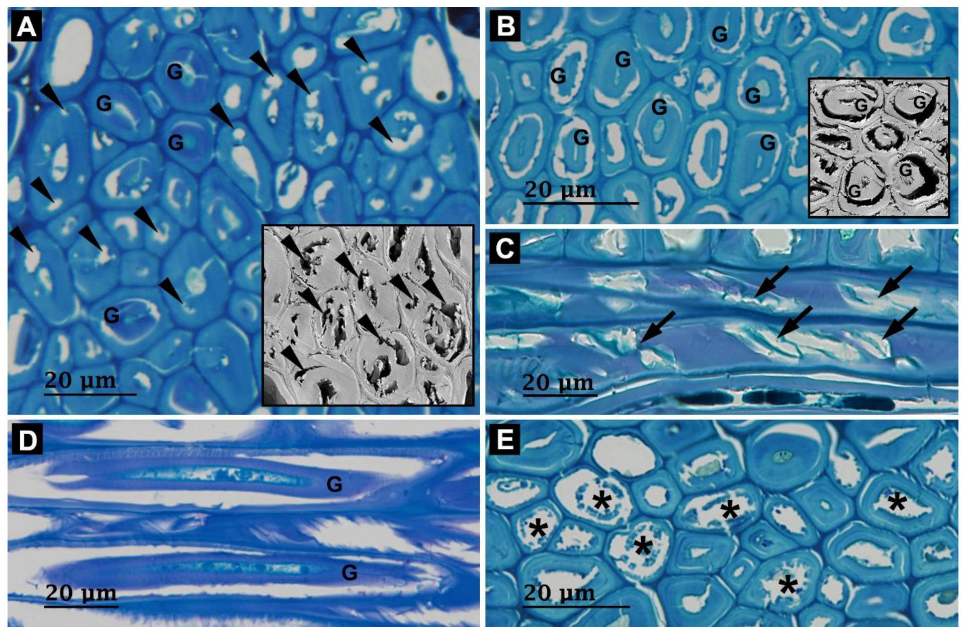
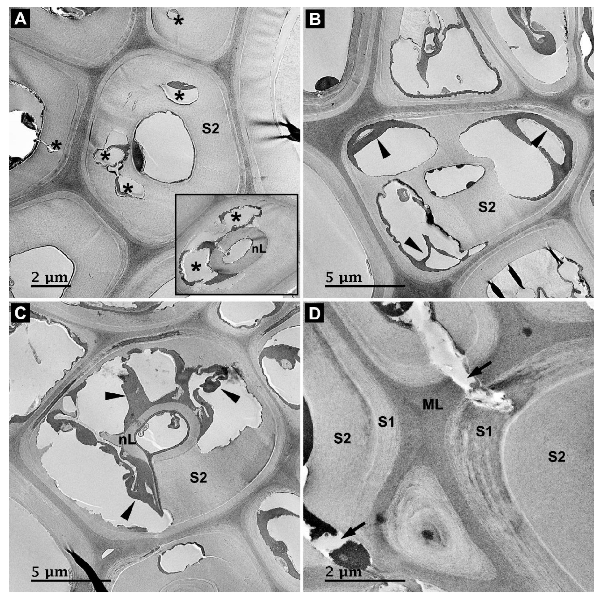
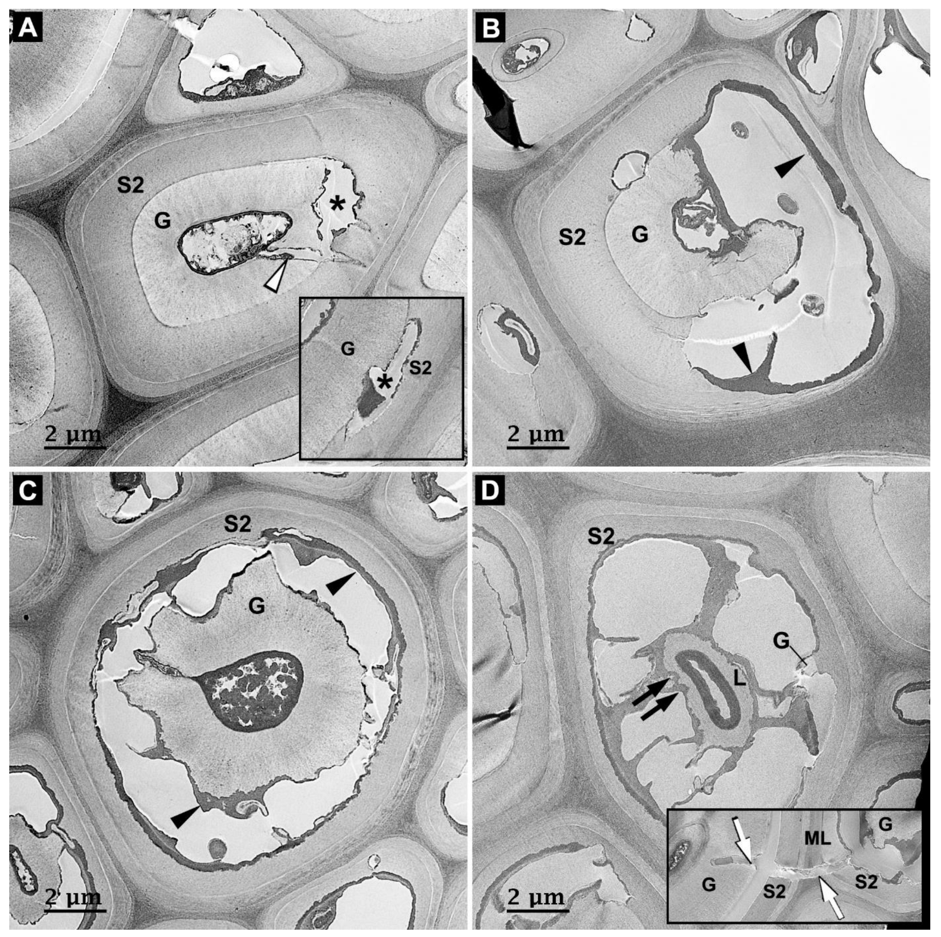
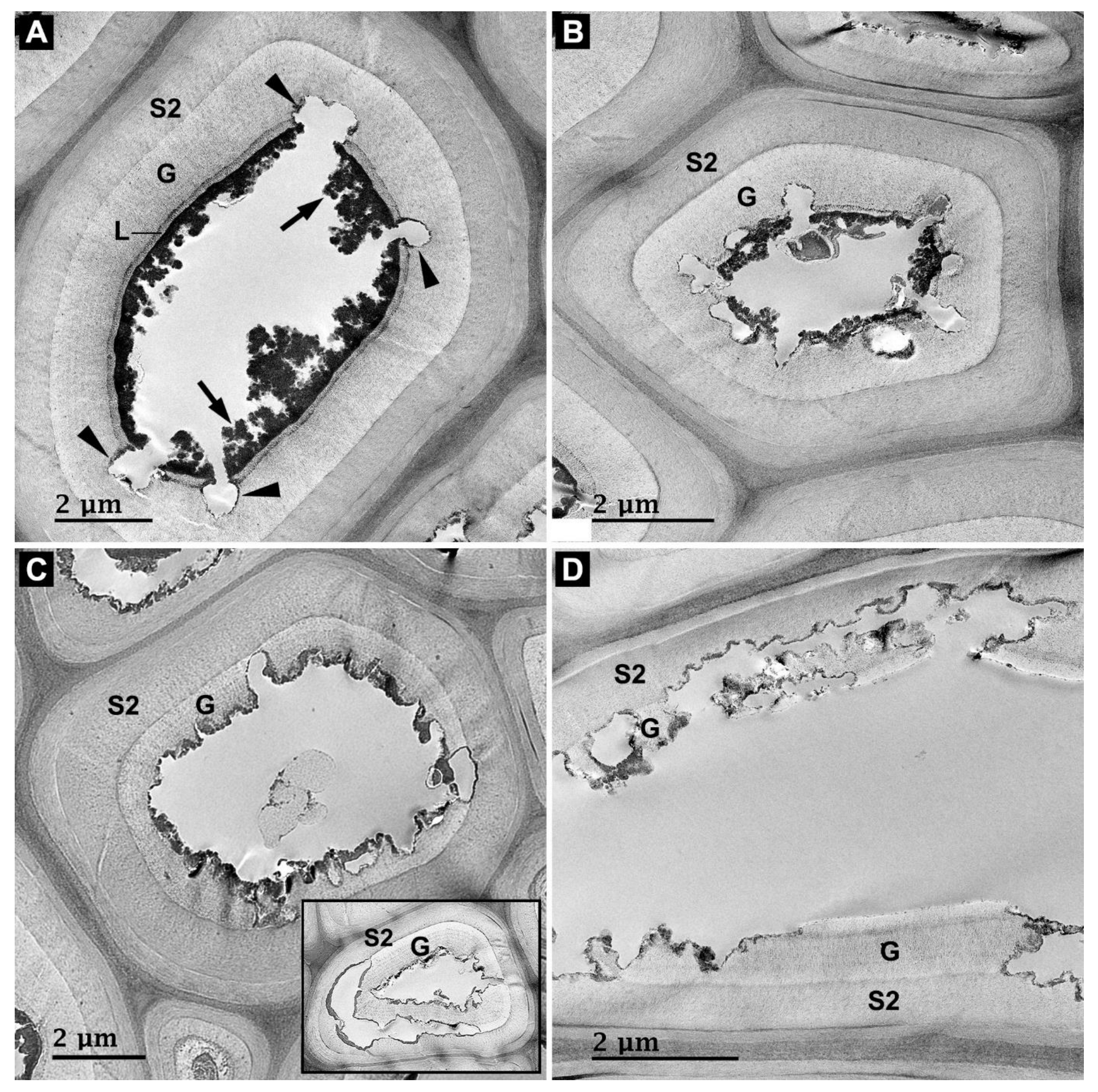
3.2. Microbial Degradation of Wood Fibers

Two distinct types of decay were mainly detected in fibers collected at a depth of 4 and 25 mm from the surface. The first type was Type-I soft rot decay (Figure 3A,B). Most fibers exhibited this type of decay, in which two distinct forms of decay were noted based on the presence or absence of the G-layer. Fibers without G-layers, i.e., S1 + S2 + S3 and S1 + S2 + S3 + n (nL) wall structures, showed the formation of soft rot cavities in the S2 layer (arrowheads in Figure 3A and inset) at the early stages of decay. By contrast, G-fibers with S1 + S2 + G and S1 + S2 + G + L wall structures exhibited the formation of soft rot cavities at the boundary of the S2 and G-layers (Figure 3B and inset). In the longitudinal section, soft rot cavities that appeared to be aligned along cellulose microfibrils were detected in the cell wall of fibers without G-layers (arrows in Figure 3C); however, they were not detected in the G-layers of G-fibers (Figure 3D). The second type of decay was the erosion type (asterisks in Figure 3E). This type of decay was only detected in a few G-fibers, with fibers showing a relatively wider cell lumen than the other G-fibers described above (Figure 3E).
Assessments using TEM revealed details regarding the decay. In Type-I soft rot decay, cavities in fibers without G-layers were detected in the S2 layer but not in the S1 and nL-layers at the early stages of decay (asterisks in Figure 4A and inset). As the decay progressed, cavities coalesced to form large voids in the secondary cell walls of fibers (Figure 4B,C) with remnants of the innermost cell wall layers, including the S3 and/or nL-layers (Figure 4B,C). Electron-dense materials were frequently detected around decayed cell wall regions in the cavities (arrowheads in Figure 4B,C). Cell wall penetration by fungal hyphae across the middle lamella (ML) was occasionally observed (arrows in Figure 4D). By contrast, G-fibers showed initial decay at the boundary of the S2 and G-layers with soft rot cavities (asterisks in Figure 5A and inset). Movement of fungal hyphae across G-layers was occasionally observed (white arrowhead in Figure 5A). As the decay progressed, degradation proceeded toward the outer S2 and inner G-layers (Figure 5B,C), with the accumulation of electron-dense materials noted around decayed cell wall regions in the cavities (arrowheads in Figure 5B,C). The formation of soft rot cavities was not detected in the middle of the G-layers (Figure 5B,C) and L-layers (Figure 5D) during decay. The L-layer was gradually degraded by erosion after the degradation of the G-layer (black arrows in Figure 5D). Cell wall penetration by fungal hyphae across the ML region of G-fibers was occasionally observed (white arrows in the inset of Figure 5D). In the erosion type of decay, at the early stages of decay, erosion trough-like localized depressions in the G-layers were detected in fiber cell walls (arrowheads in Figure 6A) across the extractive layer of the lumen wall (arrows in Figure 6A). As the decay progressed, the cell wall erosion processed toward the S2 layer (Figure 6B,C). The erosion type of decay was occasionally detected together with decay at the boundary of S2 and G-layers (i.e., the second type of decay; inset in Figure 6C). At the advanced stages of decay, fragmented S2 and/or G-layers were frequently detected in the decayed cell walls (Figure 6D).
In the surface sample, most fibers showed severe degradation of secondary cell walls with and without cell wall residues (arrows in Figure 7A). Using TEM, cell wall residues in the lumen were detected as non-degraded inner secondary cell walls (arrows in Figure 7B). These inner secondary cell walls showed similar KMnO4 staining intensity (Figure 7B) and lignin autofluorescence to those noted in the normal S2 layer (white arrows in the inset of Figure 7A), indicating that the observed inner secondary cell walls were likely to be L-layers and/or G-layers impregnated with electron-dense materials (Figure 2J). In some fibers without G-layers, stretches of S3 were observed even though the decay of the S2 layer was relatively advanced (asterisks in Figure 7C,D). Only a few fibers showed degradation of the S1 layer (Figure 7D and inset). Thick extractive layers coating the lumen wall were frequently detected even after the complete degradation of secondary cell walls (arrowhead in Figure 7C).

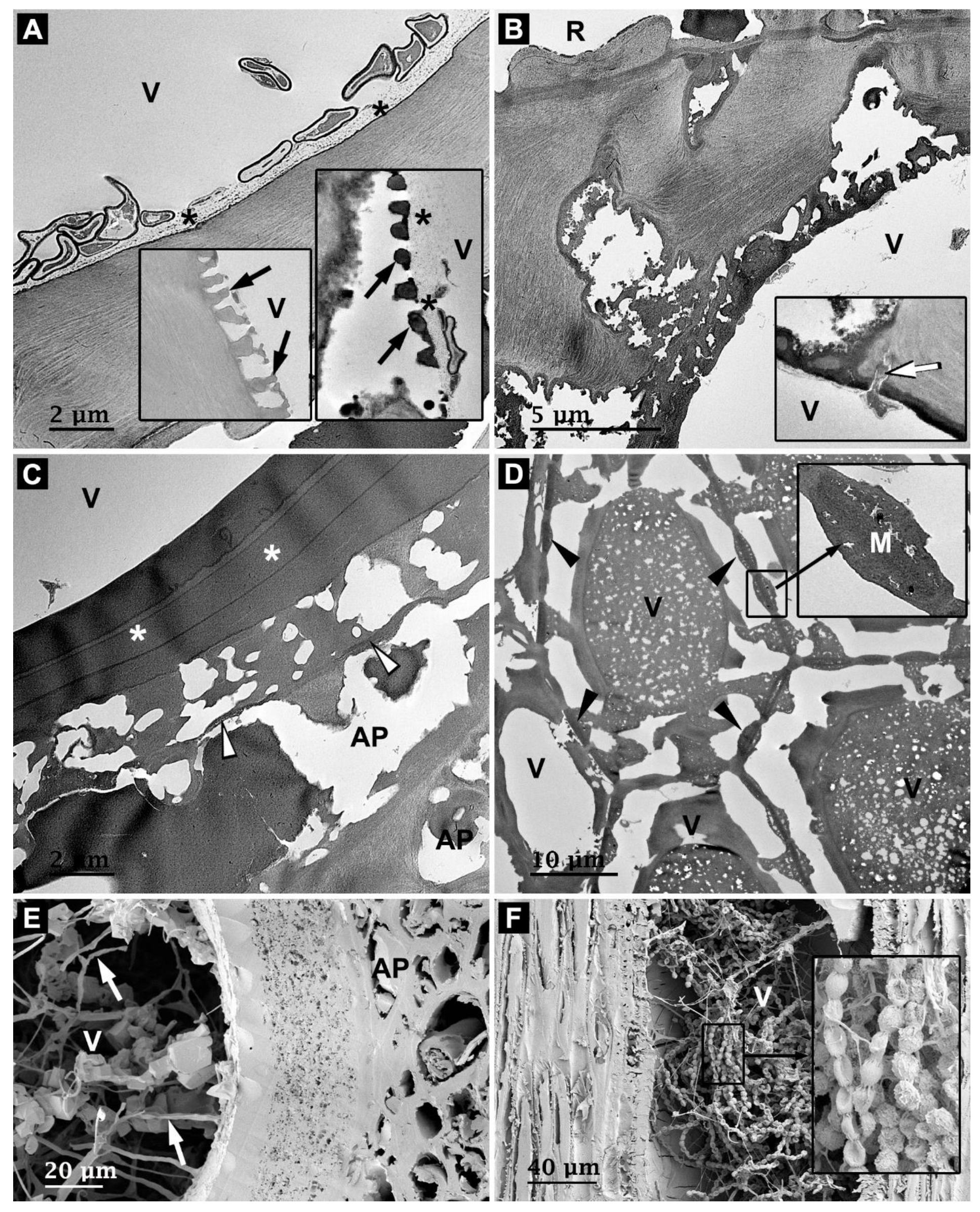
3.3. Microbial Degradation of Vessels

In assessments using LM, the degradation of vessels was not obvious in the samples collected at a depth of 4 and 25 mm from the surface (Figure 8A). By contrast, the surface sample exhibited severe vessel degradation (Figure 8B,D). Many irregular forms of cavities were frequently observed in the decayed vessel cell walls (asterisks in Figure 8C), and deposits remained (arrowheads in Figure 8B). The decay in the intervessel pit membranes was less severe than that in the adjacent vessel cell walls (arrows in Figure 8D).
Assessments using TEM and SEM provided further information on the decay patterns of vessels. The samples collected at a depth of 4 and 25 mm from the surface (i.e., the early stages of decay) showed frequent signs of degradation along the innermost cell wall layer (asterisks in Figure 9A and right inset). Vestures were detected in the vessel cell wall (arrows in the left inset of Figure 9A), vessel-ray pit (arrows in the right inset of Figure 9A), and intervessel pit (Figure 9D and inset). Vestures in the vessel cell wall exhibited less strong KMnO4 staining intensity than the other two vestures. Early degradation caused by crossing the extractive layer of the lumen wall was also detected occasionally in the vessels (white arrow in the inset of Figure 9B). At the early stages of decay, the formation of soft rot cavities was not obvious in the vessels. In the surface sample with advanced stages of decay, degradation of vessel cell walls through the pit chamber was frequently detected (Figure 9B). At more advanced stages of decay, many irregular forms of small cavities were observed in the decayed vessel cell wall (Figure 9C). Degradation of ML regions between vessel and axial parenchyma cells was also detected frequently in the surface sample (white arrowheads in Figure 9C). Degradation of tylosis-like layered materials (white asterisks in Figure 9C) and vestured intervessel pit membranes (black arrowheads in Figure 9D and inset) was not obvious even though the adjacent vessel cell wall was extensively decayed. SEM revealed many fungal hyphae in the vessel lumen together (white arrows in Figure 9E) with the formation of many small cavities in cell walls. In particular, many conidial chains were frequently detected in the lumen (Figure 9F). The conidia showed rough surface ornamentation and an overall globose shape (inset of Figure 9F).

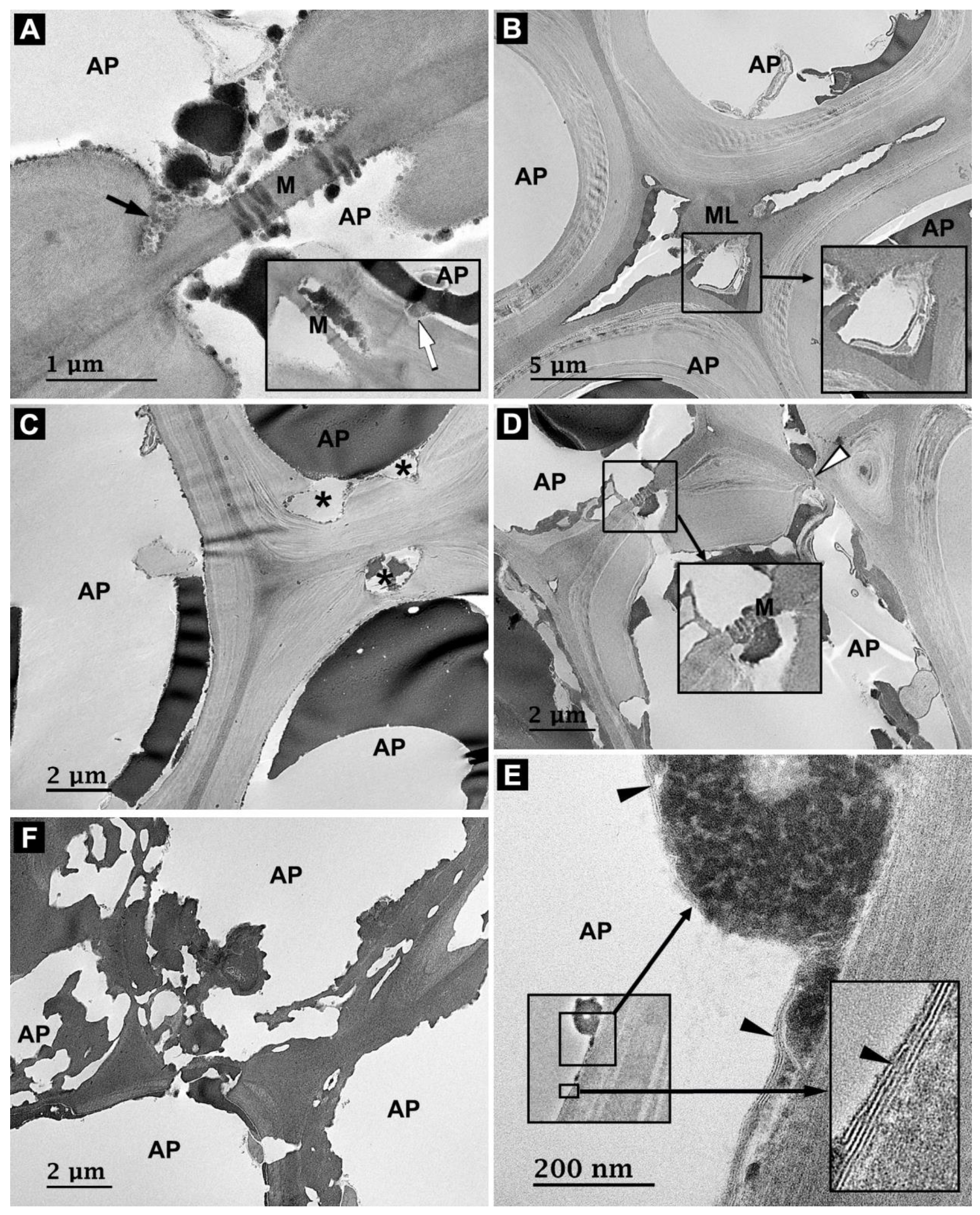
3.4. Microbial Degradation of Axial Parenchyma Cells

Similar to vessels, axial parenchyma cells showed earlier stages of decay in the samples collected at a depth of 4 and 25 mm than in the surface samples, as revealed using LM (Figure 8A versus Figure 8B). Phenolic compounds were often observed in the lumen, regardless of decay (Figure 8A–C). TEM revealed early degradation of parenchyma cells through the simple pit chamber (black arrow in Figure 10A). Early signs of decay were also detected in the innermost cell wall layer (white arrow in the inset of Figure 10A) and ML regions (Figure 10B and inset). At the advanced stages of decay, cavities were detected in cell walls (asterisks in Figure 10C). Similar to vestured intervessel pit membranes (Figure 9D), simple pit membranes (between axial parenchyma cells) showed an almost intact structure even though the adjacent parenchyma cell walls showed advanced stages of decay on the same cross-section plane (Figure 10D and inset). Cell wall penetration across the ML region was occasionally observed (white arrowhead in Figure 10D). At high magnification, a fibrillar structure along the innermost cell wall layer and/or around certain microbes was frequently observed in decayed parenchyma cells. The thickness of this structure was measured to be around 5–6 nm (black arrowheads in Figure 10E). The fibrillar structure was not detected in decayed fibers and vessels. In the surface sample, the axial parenchyma cell wall including ML regions was extensively degraded. Moreover, the cell wall showed many irregular forms of small cavities (Figure 10F). The overall degradation patterns of axial parenchyma cell walls were similar to those of vessel cell walls in the surface sample.

3.5. Microbial Community Analysis

Fungal and bacterial communities in waterlogged archaeological rosewood were analyzed through NGS using ITS (fungi) and 16S rRNA gene (bacteria) sequencing methods. A total of 56,198 and 19,167 sequencing reads were obtained for fungi and bacteria in waterlogged archaeological rosewood, respectively (Table 1).

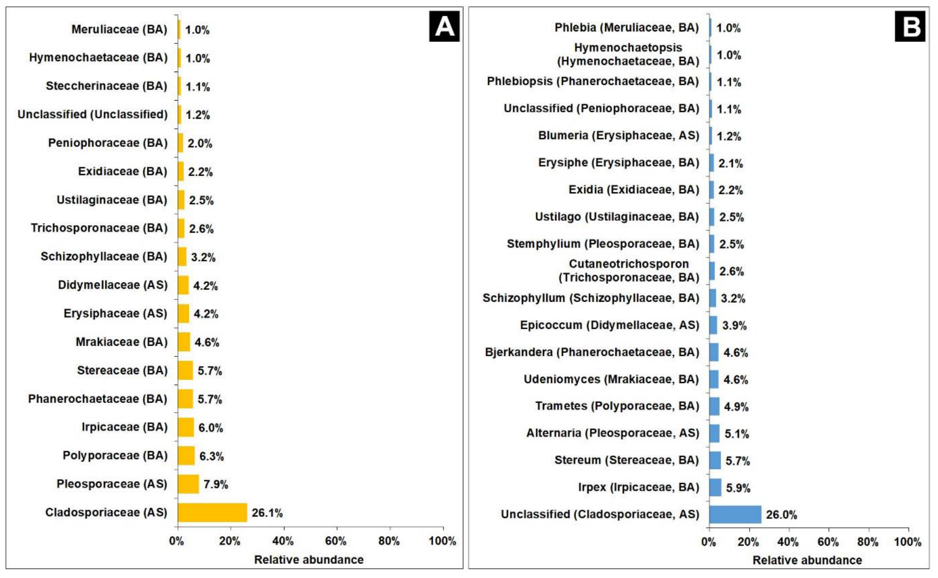
3.5.1. Fungal Community Analysis

Among fungal communities, the phyla Ascomycota (48%) and Basidiomycota (49.8%) were predominant (Table 1). At the family level, a total of 65 fungal communities were detected; of these, 11 families were unclassified. Figure 11A shows 18 fungal families with a relative abundance of >1%. Cladosporiaceae belonging to the phylum Ascomycota was the predominant family, accounting for 26.1% of the fungal community. Pleosporaceae (Ascomycota) was the second-largest family (7.9%) in the fungal community, followed by Polyporaceae (6.3%), Irpicaceae (6.0%), Phanerochaetaceae (5.7%), and Stereaceae (5.7%) belonging to the phylum Basidiomycota, in the given order. Two families belonging to Ascomycota (Erysiphaceae and Didymellaceae) and one family belonging to Basidiomycota (Schizophyllaceae) accounted for >3% of the fungal community.
At the genus level, a total of 78 fungal communities were detected; of these, 17 genera were unclassified. Nineteen genera, including two unclassified genera, had a relative abundance of >1% (Figure 11B). The largest genus group (26%) belonging to the family Cladosporiaceae (Ascomycota) was unclassified. The genera Irpex (5.9%, Basidiomycota–Irpicaceae), Stereum (5.7%, Basidiomycota–Stereaceae), and Alternaria (5.1%, Ascomycota–Pleosporaceae) had a relative abundance of >5%. Three genera belonging to Basidiomycota, including Trametes (Polyporaceae), Udeniomyces (Mrakiaceae), and Bjerkandera (Phanerochaetaceae), had a relative abundance of 4.9%, 4.6%, and 4.6%, respectively. Next, the genera Epicoccum (3.9%, Ascomycota–Didymellaceae) and Schizophyllum (3.2%, Basidiomycota–Schizophyllaceae) accounted for >3% of the fungal community.

3.5.2. Bacterial Community Analysis

Fourteen bacterial phyla were detected in waterlogged archaeological rosewood (Table 1). Cyanobacteria (37.7%) and Proteobacteria (36.9%) were the predominant phyla in the bacterial community (Table 1). Four phyla, namely Firmicutes (10.1%), Actinobacteria (6.3%), Bacteroidetes (5.6%), and Acidobacteria (2.0%), had a relative abundance of >2% (Table 1). The remaining eight phyla accounted for <1% of the fungal community (Table 1).
At the family level, a total of 109 bacterial communities were detected in waterlogged archaeological rosewood; of these, six families were unclassified. Fifteen families had a relative abundance of >1% (Figure 12A). Oscillatoriaceae (Cyanobacteria) and Comamonadaceae (Proteobacteria) were the predominant families, accounting for 27.6% and 20.6% of the bacterial community, respectively (Figure 12A). Next, the families Chroococcaceae (6.7%, Cyanobacteria), Sphingomonadaceae (3.7%, Proteobacteria), and Weeksellaceae (3.5%, Bacteroidetes) accounted for >3% of the bacterial community (Figure 12A).
At the genus level, a total of 155 bacterial communities were detected. Seventeen genera had a relative abundance of >1% at the genus level (Figure 12B). The genera Aerosakkonema (27.6%) and Diaphorobacter (20.5%) were predominant in the bacterial community (Figure 12B), followed by the genera Gloeocapsopsis (6.7%) and Cloacibacterium (3.5%) with a relative abundance of >3% (Figure 12B).

4. Discussion

The degradation of WAWs greatly differs depending on the type of wood, environment, and burial time. The microbial community involved in the decay of WAWs also varies depending on the local environmental conditions. WAWs are vulnerable to further microbial decay after excavation if not handled and restored properly. This study assessed the decay types and microbial communities in high-density rosewood (Dalbergia species) that was submerged in the Yellow Sea for approximately 700 years. The results revealed differences in cell wall degradation patterns depending on the cell wall structure and provided details regarding the microbial community during burial and after excavation. However, it is important to remember that this study focused only on a piece of rosewood out of more than 1000 pieces of waterlogged archaeological rosewood. Therefore, we cannot rule out the possibility of variations in microbial decay types and microbial communities among the rosewood pieces.
Microscopic observations indicated that soft rot decay was the main type of decay in wood fibers of waterlogged archaeological rosewood. In the early stages of decay, characteristics of Type-I soft rot, such as cavity formation in the secondary cell wall and accumulation of electron-dense materials around the cavities, were detected in fibers, regardless of the presence or absence of the G-layer. However, in a few G-fibers, cell wall erosion from the lumen was observed together with the formation of erosion trough-like localized depressions into secondary cell walls. This type of attack could be classified as Type-II soft rot and/or erosion bacterial decay because the degradation of ML regions of fibers was not distinct even in the surface sample (i.e., advanced stages of decay) [2,3,6,32]. Decay with cell wall erosion can also be caused by white rot fungi; however, white rot decay is generally accompanied by extensive degradation of ML regions [2]. Consequently, our results indicate that Type-I soft rot is the main decay type in fibers of waterlogged archaeological rosewood, whereas Type-II soft rot and/or erosion bacterial decay are occasionally present only in G-fibers with a wide lumen. However, how the lumen width affects the decay type remains unclear.
The decay patterns of waterlogged archaeological rosewood described above are overall similar to those observed in the hull of the Shinan shipwreck (composed of Pinus massoniana and Cunninghamia laneolata) loaded with the waterlogged archaeological rosewood assessed in our study. Kim [33] reported soft rot decay in the hull of the Shinan shipwreck as the major decay type together with some bacterial degradation. This result suggests that the type of microbial decay is affected by the burial environment rather than the wood species. Soft rot fungi are present in a wide range of terrestrial and aquatic environments and have lower oxygen requirements than brown and white rot fungi [3,5,6,7,34]. Soft rot fungi are frequently associated with wood exposed to excessive moisture under low oxygen conditions [3,5,35]. The waterlogged archaeological rosewood assessed in our study was totally submerged in the Yellow Sea for approximately 700 years. The average water depth at the evacuation site was 20 m, with a tidal variation of 4 m [17]. Fungal species causing soft rot decay in waterlogged archaeological rosewood may have a relatively high oxygen tolerance. As rosewood in the Shinan shipwreck was reportedly loaded for the trade, soft rot decay on the rosewood surface was not expected to occur before waterlogging. Several studies have also reported marine soft rot decay in wood [8,9,36], including WAWs [33,37]. Björdal and Dayton [36] particularly noted soft rot decay in wood (Douglas fir) recovered from the seafloor at a depth of approximately 20–30 m in the coastal water of the Antarctic.
A distinct relationship was noted between fiber cell wall structure and its degradation by soft rot fungi. Fibers without G-layers showed the formation of typical soft rot cavities in S2 layers at the early stages of decay, during which cavities did not form in the nL-layer. By contrast, fibers with G-layers (G-fibers) showed cavity formation at the boundary of S2 and G-layers at the early stages of decay. Cavity formation was not obvious in thick G-layers even after advanced soft rot decay. Similar findings were reported in ancient Egyptian woods [34,38], in which an elongated and unusual form of soft rot cavities was restricted to the S2 layer of G-fibers at the early stages of decay. Blanchette et al. [34] reported that the G-layer could resist decay even at the advanced stages of soft rot decay. However, in this study, the G-layer was eroded inward from the boundary of the S2 and G-layers (lumen side) as the decay progressed.
Several studies have suggested that lignin type and concentration influence soft rot decay in wood [2,32,39]. This effect is particularly apparent in lignin-rich ML regions and highly lignified S3 layers of softwood tracheids [2,32]. Based on the absence and/or rareness of cavity formation in thin S1 layers of normal tracheids and fibers and in extremely thin layers of multilayered fiber cell walls, Singh et al. [14,32] suggested physical constraints imposed by cell wall thickness on the formation of soft rot cavity. Our study also revealed the absence of cavity formation in the S1 layer and narrow nL-layers of fiber cell walls even at advanced stages of decay. Cavity formation was only detected in thick S2 layers of fibers. In G-fibers, cavity formation was not detected in the G-layer even though its cell wall was even thicker than that of the S2 layer. This indicates that the absence of cavity formation in G-layers may not be related to physical constraints (i.e., cell wall thickness). G-layers are predominately composed of cellulose together with small amounts of various non-cellulosic polysaccharides [38,39]. Although the presence of lignin in G-layers remains debatable, G-layers are generally considered to contain much less lignin than normal secondary cell walls [40,41]. Our study also revealed a much weaker intensity of KMnO4 staining and lignin autofluorescence in G-layers than in normal secondary cell walls. This result suggests that the absence of cavity formation in G-layers of waterlogged archaeological rosewood is not closely related to lignin. At present, it is hypothesized that cellulose microfibrils oriented nearly parallel to the fiber axis suppress the formation of typical soft rot cavities in G-layers. However, this does not mean that G-layers are resistant to soft rot decay. Instead, Type-I soft rot decay in G-layers may occur differently from that in normal secondary cell walls.
It is generally considered that vessels have a greater ratio of guaiacyl to syringyl units than fibers in hardwood and that guaiacyl lignin is more difficult to degrade by fungi than syringyl lignin [2,39]. Vessels and parenchyma cells also frequently contain tyloses and deposits of aromatic compounds/extractives in the cell lumina, respectively [42,43]. These features are considered to provide higher decay resistance in vessels/parenchyma cells than in fibers [44,45,46]. However, in our study, waterlogged archaeological rosewood showed the accumulation of extractives in the cell lumina of not only vessels and parenchyma cells but also fibers. It was also unclear whether vessels and parenchyma cells would exhibit higher decay resistance than fibers. In some cases, lower decay resistance was detected in vessels/parenchyma cells than in fibers in the surface sample (i.e., advanced stages of decay) (Figure 7C). Unlike fibers, degradation of the ML region between vessels and axial parenchyma cells was detected in some areas. However, whether this feature is related to soft rot decay remains unclear because the degradation of ML regions is not common in soft rot decay [2]. Interestingly, at the early stages of decay, axial parenchyma cells showed a fibrillar structure, which lined the innermost secondary cell wall. This structure was not detected in fibers and vessels. However, its exact chemical compositions and origin remain unclear.
Compared with secondary cell walls, pit membranes and vestures have exhibited higher resistance to microorganisms, including erosion bacteria and soft rot fungi [14,47,48,49]. Our results in waterlogged archaeological rosewood also revealed higher resistance to soft rot decay in vestured pit membranes (between vessels) and simple pit membranes (between axial parenchyma cells) than in secondary cell walls. Several studies have suggested that heavy accumulation of phenolic extractives in pit membranes of hardwood is related to high decay resistance to soft rot attack [14,45,46]. Our study also revealed a heavier deposition of phenolic compounds in vestured intervessel pit membranes than in secondary cell walls (stronger KMnO4 staining intensity, Figure 9D) and the infiltration of extractives in simple pit membranes of parenchyma cells (Figure 10C). These extractives may inhibit microbial growth and activity in vestured and simple pit membranes, providing higher decay resistance than that noted in vessel and axial parenchyma cell walls during soft rot decay of waterlogged archaeological rosewood.
Interestingly, Basidiomycota was the predominant phylum in waterlogged archaeological rosewood together with Ascomycota. Marine environments are known to suppress the activity of Basidiomycota [7,32]. Instead, bacteria and/or soft rot fungi (belonging to Ascomycetes and Fungi Imperfecti) play a major role in the degradation of wood in marine environments [3,4,7,36,38,49]. In the Basidiomycota strains detected, the genera Irpex, Stereum, Trametes, Bjerkandera, Schizophyllum, Exidia, Phlebiopsis, Hymenochaetopsis, and Phlebia, identified as white rot degraders, were predominant [2,50,51,52,53,54,55]. However, the signs of white rot decay were not obvious in waterlogged archaeological rosewood in our study. Therefore, the Basidiomycota strains present in waterlogged archaeological rosewood may not be involved in the degradation of rosewood in aquatic environments. The identified Basidiomycota strains may have mostly infected waterlogged archaeological rosewood during the conservation process after excavation. This result indicates that care should be taken during the conservation process to avoid secondary fungal infection.
With respect to soft rot decay, which is the major decay type noted in this study, several fungal groups belonging to Ascomycota were detected. The predominant fungal family was Cladosporiaceae. Although this family was not clearly classified at the genus level, several Cladosporium species belonging to Cladosporiaceae have been reported to cause soft rot decay in aquatic/marine environments [7,50]. Based on morphology [56] and abundance, conidial chains abundantly present in the vessel lumina are thought to be related to Cladosporium species. Some Alternaria and Epicoccum species, accounting for the second- and third-largest genera belonging to Ascomycota, respectively, are also known as soft rot degraders of wood [7,57].
In addition to fungi, many bacterial taxa were identified in waterlogged archaeological rosewood. However, bacterial decay patterns known as erosion and tunneling types were not obvious in the waterlogged archaeological rosewood samples. This suggests that the bacteria identified in this study are not deeply involved in the degradation of waterlogged archaeological rosewood. With the advancement of molecular biological techniques, several studies have identified various bacterial taxa in WAWs [21,22,23,24]. Compared with these previous studies, the bacterial community identified in waterlogged archaeological rosewood in our study markedly differed in terms of composition and abundance. Approximately half of the bacterial community was composed of the genera Aerosakkonema and Diaphorobacter in waterlogged archaeological rosewood. However, none of the previous studies reported these genera in WAWs. By analyzing 108 WAWs collected from 19 different European sites, Landy et al. [21] reported Cytophaga and Flavobacterium as the most frequent and abundant bacterial genera in WAW. Other studies also reported Marinomonas [23], Idiomarina, Aquiflexum, Gracilibacillus, Bacillus, Halomonas, Marinobacter, Alicyclobacillus and Azoarcus [24], and Alicyclobacillus and Acidiphilium [22] as the dominant bacterial genera in WAWs. In our study, Gloeocapsopsis, Cloacibacterium, and Prauserella were the third-, fourth-, and fifth-largest bacterial genera in waterlogged archaeological rosewood, respectively; these genera were also not reported previously in WAWs. Environmental conditions, such as temperature, salinity, and depth, are generally known to be important factors affecting bacterial growth and activity [7]. The difference in bacterial communities is considered to be attributed to differences in the environmental conditions in which waterlogged archaeological rosewood was submerged. Similar to previous studies, the precise role of bacteria detected in waterlogged archaeological rosewood in relation to wood decay remained largely unknown in our study. In particular, no prominent signs of bacterial decay were noted in waterlogged archaeological rosewood, making it more difficult to determine the role of the bacteria in relation to wood decay.
Many ligninolytic bacteria mainly belonging to the phyla Actinobacteria, Proteobacteria, and Firmicutes have been isolated from soil, sludge, coal, and the guts of termite and wood-boring beetles [58,59,60,61,62]. Most bacterial genera with a relative abundance of >1% (Figure 12), including Diaphorobacter, Prauserella, Massilia, Sphingomonas, Moraxella, Sphingobium, Methylobacterium, Alteribacillus, Granulicella, Syntrophaceticus, Hymenobacter, Cryptanaerobacter, and Pelotomaculum, belonged to these three phyla. However, except for Diaphorobacter [60], whether these detected bacteria have the potential to degrade lignin remains unclear. Similar to ligninolytic bacteria, many cellulolytic bacteria have been isolated from different sources, including pulp, soil, and various animals [63,64,65,66]. Compared with previous studies, no major bacterial genera detected in waterlogged archaeological rosewood could degrade cellulose in our study. The only exception was the genus Cloacibacterium. Cui et al. [64] reported that Cloacibacterium isolated from ancient paper-making bamboo pulp (APMP) may have the potential to degrade cellulose. Aerosakkonema and Gloeocapsopsis, which are major bacterial genera in waterlogged archaeological rosewood, belong to the phylum Cyanobacteria. Cyanobacteria species are widely distributed throughout aquatic environments, including freshwater and marine environments, and are major contributors to global carbon and nitrogen fluxes [67,68]. However, their role in the degradation of wood cell wall components has not been reported. Consequently, it is hypothesized that most bacteria identified in waterlogged archaeological rosewood do not have enzyme systems capable of efficiently breaking down lignin and cellulose, thereby inducing no prominent signs of bacterial decay in waterlogged archaeological rosewood.

5. Conclusions

Micromorphological studies indicated that most fibers in waterlogged archaeological rosewood (Dalbergia species) submerged in the Yellow Sea are tension woods with G-fibers and are mainly decayed by soft rot fungi (Type-I). Microscopic observations also indicated that the soft rot decay patterns differed between G-fibers and fibers without G-layers (i.e., normal and multilayered fibers), suggesting a close relationship between the fiber cell wall structure and its degradation. No clear difference was noted in decay resistance between vessels/axial parenchyma cells and fibers. Moreover, higher decay resistance was noted in the vestured intervessel and simple pit membranes than in the vessel and axial parenchyma cell walls. Microbial community analysis revealed various fungal and bacterial communities in waterlogged archaeological rosewood. Ascomycota and Basidiomycota were the predominant fungal phyla. Basidiomycota species were considered to have contaminated the rosewood after excavation. Bacterial communities in waterlogged archaeological rosewood markedly differed in their composition and abundance compared with those reported previously in WAWs.

Author Contributions

Conceptualization, J.S.K., M.K., M.Y.C. and Y.S.K.; methodology, J.S.K., M.K. and K.H.L.; validation, J.S.K., M.K. and J.W.L.; formal analysis, J.S.K., M.K. and J.W.L.; investigation, J.S.K., M.K. and J.W.L.; resources, M.Y.C. and Y.H.Y.; data curation, J.S.K., M.K. and J.W.L.; writing—original draft preparation, J.S.K. and M.K.; writing—review and editing, J.S.K., M.K., and Y.S.K.; visualization, J.S.K.; supervision, J.S.K. and M.K.; project administration, J.S.K. and M.K.; funding acquisition, J.S.K. All authors have read and agreed to the published version of the manuscript.

Funding

This study was financially supported by Chonnnam National University (Grant number: 2021-2478) and the National Research Foundation of Korea (NRF) grant funded by the Korea government (MSIT) (No. NRF-2021R1F1A1063024).

Data Availability Statement

All data generated or analyzed during this study are included in this published article.

Acknowledgments

The authors gratefully acknowledge the National Research Institute of Maritime Cultural Heritage in the Republic of Korea for providing the rosewood sample.

Conflicts of Interest

The authors declare no conflict of interest.

References

  1. Blanchette, R.A.; Nilsson, T.; Daniel, G. Biological degradation of wood. In Archaeological Wood: Properties, Chemistry and Preservation, Advances in Chemistry; Rowel, R.M., Barbour, J., Eds.; American Chemical Society: Washington, DC, USA, 1990; pp. 141–174. [Google Scholar]
  2. Daniel, G. Fungal degradation of wood cell walls. In Secondary Xylem; Kim, Y.S., Funada, R., Singh, A.P., Eds.; Academic Press: London, UK, 2016; pp. 131–167. [Google Scholar]
  3. Kim, Y.S.; Singh, A.P. Micromorphological characteristics of wood biodegradation in wet environments: A review. IAWA J. 2000, 21, 135–155. [Google Scholar] [CrossRef]
  4. Singh, A.P.; Kim, Y.S.; Singh, T. Bacterial degradation of wood. In Secondary Xylem; Kim, Y.S., Funada, R., Singh, A.P., Eds.; Academic Press: London, UK, 2016; pp. 169–190. [Google Scholar]
  5. Pournou, A. Wood deterioration by aquatic microorganisms. In Biodeterioration of Wooden Cultural Heritage, Organisms and Decay Mechanisms in Aquatic and Terrestrial Ecosystems; Pournou, A., Ed.; Springer: Cham, Switzerland, 2020; pp. 177–260. [Google Scholar]
  6. Daniel, G.; Nilsson, T. Developments in the study of soft rot and bacterial decay. In Forest Products Biotechnology; Bruce, A., Palfreyman, J.W., Eds.; Taylor & Francis: London, UK, 1998; pp. 37–62. [Google Scholar]
  7. Björdal, C.G. Evaluation of microbial degradation of shipwrecks in the Baltic sea. Int. Biodeterior. Biodegrad. 2012, 70, 126–140. [Google Scholar] [CrossRef]
  8. Mouzouras, R. Soft rot decay of wood by marine microfungi. J. Inst. Wood Sci. 1989, 11, 193–201. [Google Scholar]
  9. Santhakumaran, L.N.; Singh, A.P. Destruction of two tropical timbers by marine borers and microorganisms in Goa water (India). In Proceedings of the 23rd Annual Meeting of the International Research Group on Wood Preservation (Document No. IRG/WP/4176-92), Harrogate, UK, 10–15 May 1992. [Google Scholar]
  10. Takahashi, M.; Kishima, T. Decay resistance of sixty-five southeast Asian timber specimens in accelerated laboratory tests. Tonan Ajia Kenkyu 1973, 10, 525–541. [Google Scholar] [CrossRef]
  11. Tsunoda, K. The natural resistance of tropical woods against biodeterioration. Wood Res. 1990, 77, 18–27. Available online: http://hdl.handle.net/2433/53275 (accessed on 27 September 2023).
  12. Wong, A.H.H.; Kim, Y.S.; Singh, A.P.; Ling, C.W. Natural durability of tropical species with emphasis on Malaysian hardwoods—Variations and prospects. In Proceedings of the 35th Annual Meeting of the International Research Group on Wood Preservation, Bangalore, India, 24–28 April 2005. Document No. IRG/WP 05-10568. [Google Scholar]
  13. Daniel, G.; Nilsson, T. Polylaminate concentric cell wall layering in fibers of Homalium foetidum and its effects on degradation by soft rot fungi. In Recent Advances in Wood Anatomy; Donaldson, L.A., Singh, A.P., Butterfield, B.G., Whitehouse, L.J., Eds.; New Zealand Forest Research Institute: Rotorua, New Zealand, 1998; pp. 369–372. [Google Scholar]
  14. Singh, A.P.; Wong, A.H.H.; Kim, Y.S.; Wi, S.G. The relationship of fiber cell wall ultrastructure to soft rot decay in kempas (Koompassia malaccensis) heartwood. In Proceedings of the 36th Annual Meeting of the International Research Group on Wood Preservation, Ljubljana, Slovenia, 6–10 June 2004. Document No. IRG/WP 04-10541. [Google Scholar]
  15. National Research Institute of Maritime Cultural Heritage. Underwater Archaeology in Korea; Gongmyoung: Seoul, Republic of Korea, 2016; pp. 55–77. [Google Scholar]
  16. National Research Institute of Maritime Cultural Heritage. The Shinan Wreck; National Maritime Museum of Korea: Mokpo, Republic of Korea, 2016; pp. 3–4. [Google Scholar]
  17. Fonseka, D.L.C.K.; Wickramaarachchi, W.W.U.I. Rosewood (Dalbergia spp.): A luxury timber. SSRG–IJAES 2018, 5, 25–28. [Google Scholar] [CrossRef]
  18. ICRAF Database—Wood Density. Available online: http://db.worldagroforestry.org/wd (accessed on 18 August 2023).
  19. Antonelli, F.; Exposito, A.; Galotta, G.; Petriaggi, B.D.; Piazza, S.; Romagnoli, M.; Guerrieri, F. Microbiota in waterlogged archaeological wood: Use of next-generation sequencing to evaluate the risk of biodegradation. App. Sci. 2020, 10, 4636. [Google Scholar] [CrossRef]
  20. Jia, Y.; Yin, L.; Zhang, F.; Wang, M.; Sun, M.; Hu, C.; Liu, Z.; Chen, Y.; Liu, J.; Pan, J. Fungal community analysis and biodeterioration of waterlogged wooden lacquerware from the Nanhai no. 1 shipwreck. App. Sci. 2020, 10, 3797. [Google Scholar] [CrossRef]
  21. Landy, E.T.; Mitchell, J.I.; Hotchkiss, S.; Eaton, R.A. Bacterial diversity associated with archaeological waterlogged wood: Ribosomal RNA clone libraries and denaturing gradient gel electrophoresis (DGGE). Int. Biodeter. Biodegr. 2007, 61, 106–116. [Google Scholar] [CrossRef]
  22. Preston, J.; Watts, J.E.M.; Jones, M. Novel bacterial community associated with 500-year-old unpreserved archaeological wood from King Henry VIII’s Tudor warship the Mary Rose. AEM 2012, 78, 8822–8828. [Google Scholar] [CrossRef]
  23. Li, Q.; Cao, L.; Wang, W.; Tan, H.; Jin, T.; Wang, G.; Lin, G.; Xu, R. Analysis of the bacterial communities in the waterlogged wooden cultural relics of the Xiaobaijiao No. 1 shipwreck via high-throughput sequencing technology. Holzforschung 2018, 72, 609–619. [Google Scholar] [CrossRef]
  24. Liu, Z.; Fu, T.; Hu, C.; Shen, D.; Macchioni, N.; Sozzi, L.; Chen, Y.; Liu, J.; Tian, X.; Ge, Q.; et al. Microbial community analysis and biodeterioration of waterlogged archaeological wood from the Nanhai No. 14 shipwreck during storage. Sci. Rep. 2018, 8, 7170. [Google Scholar] [CrossRef] [PubMed]
  25. Kim, B.-K. Review of the red sandalwood cargo of the Shinan ship. Marit. Cult. Herit. 2013, 6, 37–70. (In Korean) [Google Scholar]
  26. Yu, Z.; Morrrison, M. Improved extraction of PCR-quality community DNA from digesta and fecal samples. Biotechniques 2004, 36, 808–812. [Google Scholar] [CrossRef]
  27. Magoc, T.; Salzberg, S.L. FLASH: Fast length adjustment of short reads to improve genome assemblies. Bioinformatics 2011, 27, 2957–2963. [Google Scholar] [CrossRef] [PubMed]
  28. Caporaso, J.G.; Kuczynski, J.; Stombaugh, J.; Bittinger, K.; Bushman, F.D.; Costello, E.K.; Fierer, N.; Pena, A.G.; Goodrich, J.K.; Gordon, J.I.; et al. QIIME allows analysis of high-throughput community sequencing data. Nat. Methods 2012, 7, 335–336. [Google Scholar] [CrossRef]
  29. Li, W.; Fu, L.; Niu, B.; Wu, S.; Wooley, J. Ultrafast clustering algorithms for metagenomic sequence analysis. Brief. Bioinform. 2012, 13, 656–668. [Google Scholar] [CrossRef]
  30. Zhang, Z.; Schwartz, S.; Wagner, L.; Miller, W. A greedy algorithm for aligning DNA sequences. J. Comput. Biol. 2000, 7, 203–214. [Google Scholar] [CrossRef]
  31. Edgar, R.C. Search and clustering orders of magnitude faster than BLAST. Bioinformatics 2010, 26, 2460–2461. [Google Scholar] [CrossRef]
  32. Singh, A.P.; Kim, Y.S.; Chavan, R.R. Advances in understanding microbial deterioration of buried and waterlogged archaeological woods: A review. Forests 2022, 13, 394. [Google Scholar] [CrossRef]
  33. Kim, Y.S. Micromorphology of degraded archaeological pine wood in waterlogged situations. Mater. Org. 1989, 24, 271–286. [Google Scholar]
  34. Blanchette, R.A.; Haight, J.E.; Koestler, R.J.; Hatchfield, P.B.; Arnold, D. Assessment of deterioration in archaeological wood from ancient Egypt. JAIC 1994, 33, 55–70. [Google Scholar] [CrossRef]
  35. Kim, Y.S.; Singh, A.P. Wood as cultural heritage material and its deterioration by biotic and abiotic agents. In Secondary Xylem; Kim, Y.S., Funada, R., Singh, A.P., Eds.; Academic Press: London, UK, 2016; pp. 169–190. [Google Scholar]
  36. Björdal, C.G.; Dayton, P.K. First evidence of microbial wood degradation in the coastal waters of the Antarctic. Sci. Rep. 2020, 10, 12774. [Google Scholar] [CrossRef] [PubMed]
  37. Björdal, C.G.; Daniel, G.; Nilsson, T. Microbial decay of waterlogged archaeological wood found in Sweden—Applicable to archaeology and conservation. Int. Biodeterior. Biodegrad. 1999, 43, 63–73. [Google Scholar] [CrossRef]
  38. Blanchette, R.A. A review of microbial deterioration found in archaeological wood from different environments. Int. Biodeter. Biodegr. 2000, 46, 189–204. [Google Scholar] [CrossRef]
  39. Daniel, G. Fungal and bacteria. In Deterioration and Protection of Sustainable Biomaterials; Schultz, T.P., Goodell, B., Nicholas, D.D., Eds.; American Chemical Society: Washington, DC, USA, 2014; pp. 23–58. [Google Scholar]
  40. Pilate, G.; Chabbert, B.; Cathala, B.; Yoshinaga, A.; Leplé, J.-C.; Laurans, F.; Lapierre, C.; Ruel, K. Lignification and tension wood. Comptes Rendus Biol. 2004, 327, 889–901. [Google Scholar] [CrossRef]
  41. Gorshkova, T.A.; Gurjanov, O.P.; Mikshina, P.V.; Ibragimova, N.N.; Mokshina, N.E.; Salnikov, V.V.; Ageeva, M.V.; Amenitskii, S.I.; Chernova, T.E.; Chemikosova, S.B. Specific type of secondary cell wall formed by plant fibers. Russ. J. Plant Physiol. 2010, 57, 328–341. [Google Scholar] [CrossRef]
  42. Schwarze, F.; Baum, S. Mechanisms of reaction zone penetration by decay fungi in wood of beech (Fagus sylvatica). New Phytol. 2000, 146, 129–140. [Google Scholar] [CrossRef]
  43. Sachs, I.; Kuntz, J.; Ward, J.; Nair, G.; Schultz, N. Tyloses structure. Wood Fiber Sci. 1970, 2, 259–268. [Google Scholar]
  44. Nilsson, T.; Singh, A.; Daniel, G. Ultrastructure of the attack of Eusideroxylon zwageri wood by tunneling bacteria. Holzforschung 1992, 46, 361–368. [Google Scholar] [CrossRef]
  45. Singh, A.P.; Wong, A.H.H.; Kim, Y.S.; Wi, S.G. Soft rot decay of cengal (Neobalanocarpus heimii) heartwood in ground contact in relation to extractive microdistribution. In Proceedings of the 34th Annual Meeting of the International Research Group on Wood Preservation, Brisbane, Australia, 18–23 May 2003. Document No. IRG/WP 03-10501. [Google Scholar]
  46. Kim, Y.S.; Singh, A.P.; Wong, A.H.H.; Eom, T.J.; Lee, K.H. Micromorphological and chemical characteristics of cengal (Neobalanocarpus heimii) heartwood decayed by soft rot fungi. J. Kor. Wood Sci. Technol. 2006, 34, 68–77. [Google Scholar]
  47. Singh, A.P.; Nilsson, T.; Daniel, G.F. Alstonia scholaris vestures are resistant to degradation by tunnelling bacteria. IAWA J. 1993, 14, 119–126. [Google Scholar] [CrossRef]
  48. Singh, A.P.; Kim, Y.S.; Wi, S.G. Inhomogeneity in the composition of vesture walls in an archaeological wood. IAWA J. 2002, 23, 77–81. [Google Scholar] [CrossRef]
  49. Singh, A.P.; Kim, Y.S.; Chavan, R.R. Relationship of wood cell wall ultrastructure to bacterial degradation of wood. IAWA J. 2019, 40, 1–26. [Google Scholar] [CrossRef]
  50. Worrall, J.J.; Anagnost, S.E.; Zabel, R.A. Comparison of wood decay among diverse lignicolous fungi. Mycologia 1997, 89, 199–219. [Google Scholar] [CrossRef]
  51. Xiaobin, C.; Rong, J.; Pingsheng, L.; Shiqian, T.; Qin, Z.; Wenzhong, T.; Xudong, L. Purification of a new manganese peroxidase of the white-rot fungus Schizophyllum sp. F17, and decolorization of azo dyes by the enzyme. Enzyme Microb. Technol. 2007, 41, 258–264. [Google Scholar] [CrossRef]
  52. Gao, D.; Du, L.; Yang, J.; Wu, W.-M.; Liang, H. A critical review of the application of white rot fungus to environmental pollution control. Crit. Rev. Biotechnol. 2010, 30, 70–77. [Google Scholar] [CrossRef]
  53. Kamei, I.; Yoshida, T.; Enami, D.; Meguro, S. Coexisting Curtobacterium bacterium promotes growth of white-rot fungus Stereum sp. Curr. Microbiol. 2012, 64, 173–178. [Google Scholar] [CrossRef]
  54. Tovar-Herrera, O.E.; Batista-García, R.A.; Sánchez-Carbente, M.d.R.; Iracheta-Cárdenas, M.M.; Arévalo-Niño, K.; Folch-Mallol, J.L. A novel expansion protein from the white-rot fungus Schizophyllum commune. PLoS ONE 2015, 10, e0122296. [Google Scholar] [CrossRef]
  55. Corfixen, P.; Parmasto, E. Hymenochaete and Hymenochaetopsis (Basidiomycota) in Europe. Karstenia 2017, 57, 49–80. [Google Scholar] [CrossRef]
  56. Bensch, K.; Braun, U.; Groenewald, J.Z.; Crous, P.W. The genus Cladosporium. Stud. Micol. 2012, 72, 1–401. [Google Scholar] [CrossRef] [PubMed]
  57. Zabel, R.A.; Wang, C.J.K.; Anagnost, S.E. Soft-rot capabilities of the major microfungi, isolated from Douglas-fir poles in the northeast. Wood Fiber Sci. 1991, 23, 220–237. [Google Scholar]
  58. Klankeo, P.; Nopcharoenkul, W.; Pinyakong, O. Two novel pyrene-degrading Diaphorobacter sp. and Pseudoxanthomonas sp. isolated from soil. J. Biosci. Bioeng. 2009, 108, 488–495. [Google Scholar] [CrossRef] [PubMed]
  59. Bugg, T.D.H.; Ahmad, M.; Hardiman, E.M.; Singh, R. The emerging role for bacteria in lignin degradation and bio-product formation. Curr. Opin. Biotechnol. 2011, 22, 394–400. [Google Scholar] [CrossRef]
  60. Wang, L.; Nie, Y.; Tang, Y.-Q.; Song, X.-M.; Cao, K.; Sun, L.-Z.; Wang, Z.-J.; Wu, X.-L. Diverse bacteria with lignin degrading potentials isolated from two ranks of coal. Front. Microbiol. 2016, 7, 1428. [Google Scholar] [CrossRef] [PubMed]
  61. Xiong, Y.; Zhao, Y.; Ni, K.; Shi, Y.; Xu, Q. Characterization of ligninolytic bacteria and analysis of alkali-lignin biodegradation products. Poi. J. Microbiol. 2020, 69, 339–347. [Google Scholar] [CrossRef]
  62. Grgas, D.; Rukavina, M.; Bešlo, D.; Štefanac, T.; Crnek, V.; Šikić, T.; Habuda-Stanić, M.; Dragićević, T.L. The bacterial degradation of lignin-a review. Water 2023, 15, 1272. [Google Scholar] [CrossRef]
  63. Ali, H.R.K.; Hemeda, N.F.; Abdelaliem, Y.F. Symbiotic cellulolytic bacteria from the gut of the subterranean termite Psammotermes hypostoma Desneux and their role in cellulose digestion. AMB Expr. 2019, 9, 111. [Google Scholar] [CrossRef]
  64. Cui, J.; Mai, G.; Want, Z.; Liu, Q.; Zhou, Y.; Ma, T.; Liu, C. Metagenomic insights into a cellulose-rich niche reveal microbial cooperation in cellulose degradation. Front. Microbiol. 2016, 10, 618. [Google Scholar] [CrossRef]
  65. Batubara, U.M.; Mardalisa, M.; Suparjo, S.; Maritsa, H.U.; Pujianto, E.; Herlini, M. Isolation and characterization of cellulolytic bacteria diversity in peatland ecosystem and their cellulolytic activities. IOP Conf. Ser. Earth Environ. Sci. 2021, 934, 12028. [Google Scholar] [CrossRef]
  66. Froidurot, A.; Julliand, V. Cellulolytic bacteria in the large intestine of mammals. Gut Microbes 2022, 14, e2031694. [Google Scholar] [CrossRef] [PubMed]
  67. Thu, N.K.; Tanabe, Y.; Yoshida, M.; Matsuura, H.; Watanabe, M.M. Aerosakkonema funiforme gen. et sp. nov. (Oscillatoriales), a new gas-vacuolated oscillatorioid cyanobacterium isolated from a mesotrophic reservoir. Phycologia 2012, 51, 672–683. [Google Scholar] [CrossRef]
  68. Aguilera, A.; Klemenčič, M.; Sueldo, D.J.; Rzymski, P.; Giannuzzi, L.; Martin, M.V. Cell death in Cyanobacteria: Current understanding and recommendations for a consensus on its nomenclature. Front. Microbiol. 2021, 12, 631654. [Google Scholar] [CrossRef] [PubMed]
Figure 1. Studied waterlogged archaeological rosewood. (A) Black arrow indicates the sampling site. (B) Surface of waterlogged archaeological rosewood showing brownish coloration with brash fractures. (C) Transverse plane of waterlogged archaeological rosewood showing the formation of cracks along with growth rings (white arrow). Note the semicircular disk cut from the top end of waterlogged archaeological rosewood for the experiment (asterisk).
Forests 14 01992 g001
Figure 2. Anatomy of fiber cell walls at a depth of 25 mm in waterlogged archaeological rosewood stained with toluidine blue (A,B) and KMnO4 (EJ). (A) Anatomy of waterlogged archaeological rosewood showing diffuse porous type of vessel (V) porosity, axial parenchyma cells (AP) arranged around vessels (paratracheal), and uniseriate/biseriate rays (R). (BD) Fibers with and without (white asterisks in (B)) gelatinous (G) layers. G-layers showed strong blue coloration (B), weak lignin autofluorescence (B), and cellulose microfibrils oriented nearly parallel to the fiber axis (double arrowheads in (D)). Several G-fibers showed the formation of a lignified (L) layer between the S2 and G-layers (inset in (C)). Note the multilayered narrow lignified (nL) layers in the inner secondary cell walls of fibers without the G-layer (inset in (B)) and strong autofluorescence in the lumen and/or lumen wall (white arrows in (C)). (EJ) Fibers with various cell wall structures: S1–S3 (E,F,J), S1 + S2 + S3 + n (nL) (EG), S1 + S2 + G (H,I), and S1 + S2 + G + L (E,I). Note the thin lamellae developed within the G-layer (arrowheads in (G,H)), uneven patterns of KMnO4 staining intensity across the G-layer (asterisks in (J)), thin concentric lines with strong KMnO4 staining intensity between the nL-layers (white arrowheads in the inset of (G)), and the accumulation of phenolic extractives in the lumen and/or lumen wall (black arrows in (E,F,H,J)).
Forests 14 01992 g002
Figure 3. Degradation of fibers at a depth of 4 and 25 mm in waterlogged archaeological rosewood stained with toluidine blue. (A) Fibers without gelatinous (G) layers showing the formation of soft rot cavities in secondary cell walls (arrowheads). Inset indicates the SEM micrograph of corresponding fibers. (B) Fibers with G-layers showing degradation at the boundary of the S2 and G-layers. The inset reveals the SEM micrograph of corresponding G-fibers. (C,D) Longitudinal sections showing the formation of cavities aligned along cellulose microfibrils in fibers without G-layers (arrows in (C)). Note the absence of cavity formation in G-layers (D). (E) Fibers with a relatively wide lumen showing the erosion type of decay (asterisks).
Forests 14 01992 g003
Figure 4. Degradation of fibers without gelatinous (G) layers at a depth of 4 and 25 mm in waterlogged archaeological rosewood stained with KMnO4. (A) Formation of soft rot cavities (asterisks) in S2 layers at the early stages of decay. Note the absence of cavity formation in the narrow lignified (nL) layer (inset). (B,C) Coalescence between adjoining cavities with the progression of decay. Note the nL-layer with an almost intact structure (C) and electron-dense materials detected around decayed cell wall regions in cavities (arrowheads). (D) Cell wall penetration by fungal hyphae across the middle lamella (ML, arrows).
Forests 14 01992 g004
Figure 5. Degradation of gelatinous (G) fibers at a depth of 4 and 25 mm in waterlogged archaeological rosewood stained with KMnO4. (A) Formation of soft rot cavities at the boundary of S2 and G-layers at the early stages of decay (asterisks). The cavity was more biased toward the S2 layers than the G-layers (inset). Note cell wall penetration across the G-layer (white arrowhead). (B,C) Degradation of S2 and G-layers with the progression of decay. Note the presence of electron-dense materials around decayed cell wall regions in cavities (black arrowheads). (D) Advanced stages of decay showing an almost completely decayed G-layer, with a remaining highly lignified (L) layer. Note the absence of cavity formation and decay (black arrows) in the lignified (L) layer. The inset indicates cell wall penetration by fungal hyphae across the G-layer, normal secondary wall, and middle lamella (white arrows in the inset).
Forests 14 01992 g005
Figure 6. Erosion type of decay in gelatinous (G) fibers at a depth of 4 and 25 mm in waterlogged archaeological rosewood stained with KMnO4. (A) Erosion trough-like localized depressions in the G-layer (arrowheads) across the extractive layer (arrows). Note the relatively wider cell lumen than that of G-fibers described in Figure 5. (B,C) Progression of cell wall erosion outward from the G-layer with the progression of decay. Cell wall erosion was occasionally observed together with decay at the boundary of S2 and G-layers described in Figure 5 (inset in (C)). (D) Degradation of the S2 layer beyond the G-layer at the advanced stages of decay.
Forests 14 01992 g006
Figure 7. Degradation of fibers on the surface of waterlogged archaeological rosewood stained with toluidine blue (A) and KMnO4 (BD). (A,B) Fibers showing heavy decay with and without cell wall residues (black arrows in A,B). Cell wall residues showed fluorescence (assessed using CLSM; white arrows in inset) and KMnO4 staining intensity similar to those noted in normal S2 layers. (C) Fibers without a G-layer showing heavy degradation of the secondary cell wall, with the innermost layer remaining (asterisks). Note the un-decayed extractive layer even after complete degradation of the secondary cell wall (arrowhead) and more advanced stages of decay in axial parenchyma cells (AP) than in fibers. (D) Fiber showing degradation of the S1 layer (inset), with the innermost layer (asterisks) and extractives in the lumen remaining. ML, middle lamella.
Forests 14 01992 g007
Figure 8. Degradation of vessels and axial parenchyma cells in waterlogged archaeological rosewood stained with toluidine blue. (A) A sample with 25 mm axial depth showing vessels (V) in 2 multiples and axial parenchyma cells (AP) with phenolic extractives in the lumen. Note the multilayered axial parenchyma cell wall with deposits in the lumen (inset). (BD) A surface sample showing severe degradation of vessel and parenchyma cell walls. Many irregular forms of cavities were observed in vessel cell walls (asterisks in (C)). Note the accumulation of materials attached to the vessel lumen (arrowheads in (B)). Intervessel pit membranes remained even though adjacent vessel cell walls were heavily degraded (arrows in (D)).
Forests 14 01992 g008
Figure 9. Degradation of vessels (V) in waterlogged archaeological rosewood stained with KMnO4 (AD). (A,B) Vessels at the early stages of decay showing the degradation of certain deposits coating the innermost cell wall layer (black asterisks in (A) and right inset of (A)), cell wall degradation through pit chambers (B), and invasion of fungal hyphae into the cell wall across the extractive layer of the lumen (white arrow in the inset of (B)). Note the presence of vestures in the vessel cell wall (black arrows in the left inset of (A)) and vessel-ray pit (black arrows in the right inset of (A)) with a difference in KMnO4 staining intensity. (C) A surface sample showing severe degradation of vessel cell walls with many irregular forms of cavities. Note un-decayed layered materials in the lumen (white asterisks) and degradation of the middle lamella between vessel and axial parenchyma cells (AP) (white arrowheads). (D) Vestured intervessel pit membranes (M) at the advanced stages of decay showing an almost intact structure (black arrowheads) even though the adjacent vessel cell walls were extensively degraded. (E) A sample with 1–2 mm axial depth showing fungal hyphae in the vessel lumen (white arrows) together with the formation of many small cavities in the cell wall. (F) Fungal conidia in the lumen showing rough surface ornamentation and an overall globose shape (inset). R, ray cell.
Forests 14 01992 g009
Figure 10. Degradation of axial parenchyma cells (AP) in waterlogged archaeological rosewood stained with KMnO4. (A,B) Axial parenchyma cells at the early stages of decay showing cell wall degradation through simple pit chambers (black arrow in (A)) and across the extractive layer of the lumen (white arrow in the inset of (A)), in addition to the degradation of middle lamella (ML) regions ((B) and inset). Note the intact structure of the simple pit membrane (M) ((A) and inset). (C) Axial parenchyma cells with the formation of soft rot cavities in secondary cell walls (asterisks). (D) Simple pit membrane (M) at the advanced stages of decay showing an almost intact structure even though adjacent axial parenchyma cell walls were extensively degraded. Note cell wall penetration by fungal hyphae (white arrowhead). (E) Fibrillar structure covering the decayed axial parenchyma cell wall at the early stages of decay (black arrowheads). (F) A surface sample showing extensive degradation of axial parenchyma cell walls with many irregular forms of cavities.
Forests 14 01992 g010
Figure 11. Fungal families (A) and genera (B) with a relative abundance of more than 1% in waterlogged archaeological rosewood. Parenthesis indicates fungal phylum (A) and family/phylum (B) names. AS, Ascomycota; BA, Basidiomycota.
Forests 14 01992 g011
Figure 12. Bacterial families (A) and genera (B) with a relative abundance of more than 1% in waterlogged archaeological rosewood. Parenthesis indicates bacterial phylum (A) and family (B) names.
Forests 14 01992 g012
Table 1. Taxonomic profile of fungi and bacteria at phylum level in waterlogged archaeological rosewood.
KingdomPhylumRelative Abundance (%)Number of Reads
FungiAscomycota48.026,971
Basidiomycota49.827,973
Unclassified2.21254
BacteriaCyanobacteria37.77229
Proteobacteria36.97065
Firmicutes10.11944
Actinobacteria6.31212
Bacteroidetes5.61074
Acidobacteria2.0376
Deinococcus–Thermus0.364
Gemmatimonadetes0.364
Chloroflexi0.356
Planctomycetes0.235
Verrucomicrobia0.119
Ignavibacteriae0.119
Chlamydiae0.07
Fusobacteria0.03
Disclaimer/Publisher’s Note: The statements, opinions and data contained in all publications are solely those of the individual author(s) and contributor(s) and not of MDPI and/or the editor(s). MDPI and/or the editor(s) disclaim responsibility for any injury to people or property resulting from any ideas, methods, instructions or products referred to in the content.

