Article

Genome-Wide Identification and Analysis of the DGAT Gene Family in Lindera glauca and Expression Analysis during Fruit Development Stages

1
College of Tea Science, Guizhou University, Guiyang 550025, China
2
Department of Botany, College of Science, University of British Columbia, Vancouver, BC V6T 1Z4, Canada
*
Author to whom correspondence should be addressed.
Forests 2023, 14(8), 1633; https://doi.org/10.3390/f14081633
Submission received: 13 June 2023 / Revised: 8 August 2023 / Accepted: 11 August 2023 / Published: 13 August 2023

Abstract

:
Diacylglycerol acyltransferase (DGAT) is a vital and sole rate-limiting enzyme involved in triacylglycerol synthesis. Identifying DGAT genes in Lindera glauca is essential for studying lipid metabolism pathways and developing novel oil crops with enhanced value. In the study reported in this paper, 15 LgDGAT family genes were first obtained from the L. glauca genome via bioinformatics analysis. We comprehensively analyzed their chromosome distribution, gene structure, subcellular localization, promoter prediction, phylogenetic relationships, tissue-specific expression, and expression patterns during different stages of fruit development. Our findings revealed that LgDGATs can be classified into DGAT1, DGAT2, DGAT3, and WSD (wax ester synthase/acyl-CoA: diacylglycerol acyltransferase) subfamilies distributed across chromosome 3, 5, 6, 8 and 11. LgDGATs’ promoter region showed abundant elements linked to the light response and plant hormone response. Forms of LgDGAT1, LgDGAT2, and LgDGAT3 were primarily expressed in the early and late phases of fruit development, indicating their potential function in the growth and development of L. glauca, particularly in oil accumulation. Conversely, LgWSDs exhibited predominant expression in stems and leaves. This paper elucidates the gene structure and expression patterns of LgDGATs, providing a theoretical foundation for understanding the functionality of DGAT genes in Lindera species.

1. Introduction

Triacylglycerol (TAG) serves as plants’ primary lipid storage form and is valuable for human consumption and biofuel production [1]. Among the various pathways involved in TAG biosynthesis in different plant organs and tissues, the Kennedy pathway is one of the most critical [2,3]. Diacylglycerol acyltransferase (DGAT) is the only rate-limiting enzyme involved in this pathway’s ultimate conversion of diacylglycerol (DAG) to TAG, and as such, it is crucial in controlling the amount of TAG present [4].
DGAT enzymes are categorized into four types based on their cell location and structural variations: DGAT1, DGAT2, DGAT3 and WS/DGAT (wax ester synthase/acyl-CoA: diacylglycerol acyltransferase) [5,6]. As genetically engineered targets for enhancing the yield of plant storage lipids, DGAT1s are crucial for the cultivation of oilseed crops [7,8]. They have also been associated with plant growth [9,10]. Arabidopsis seedlings mutant for DGAT1 exhibit abnormal growth, shrunk seeds, reduced lipids content, and delay seed maturation [11,12]. Overexpression of Arabidopsis AtDGAT1 in tobacco significantly increases TAG content in transgenic tobacco seeds [13]. DGAT2 selectively accumulates unsaturated fatty acids in TAG. Overexpression of PfDGAT2 in Perilla frutescens [14] and JcDGAT2 in Jatropha curcas [15] increases their unsaturated fatty acid content, respectively. Additionally, DGAT2 is co-expressed with transcription factor ethylene response factors in tobacco to promote the flow of carbon sources toward fat and fatty acid biosynthesis [16]. DGAT3 is a plant cytoplasmic soluble metalloenzyme [17]. The accumulation of unsaturated fatty acids was dramatically increased when camelina CaDGAT3-3 was expressed specifically in tobacco [18]. WS/DAGT is an enzyme that possesses dual functionality as both a TAG synthetase and a wax ester synthase (WS), with its WS activity surpassing that of TAG synthetase [6]. Arabidopsis AtWSD1 exhibits high level of WS activity, while DGAT activity is approximately tenfold lower than its WS activity [19]. Moreover, AtWSD1 is crucial in producing epidermal wax, which is essential for plant moisture retention and salinity tolerance [20].
Lindera glauca (Sieb. et Zucc.) Blume is a deciduous shrub or small fruit tree found in lowland woodland forest margins in China, Japan, and Korea [21,22,23], where it undergoes non-fusion (seed asexual reproduction) [24] and sexual reproduction [25,26]. L. glauca is a secondary forest species in low and middle altitude area, and considered as a significant ecological and economic tree species due to its abundant resources, high adaptability, and ecological advantages in China [27]. The fatty acids and aromatic oils found in L. glauca fruits, as well as the terpenoids, flavonoids, and alkaloids that they contain, are rich in traditional medicinal uses [28]. The primary fatty acids in L. glauca fruits and seeds are capric acid, oleic acid, palmitic acid, and linoleic acid [29]. Fruits and essential oils produced in China annually amounts to 120,000 metric tons and 1000 kiloliters, respectively [30,31]. L. glauca fruit or seed oil is frequently utilized in food oils, biodiesel, or daily-use chemical goods, including soaps, surfactants, and lubricants [32,33,34].
However, there were limited studies on the biosynthesis and accumulation of oil in L. glauca, hindering the discovery of related genes and the improvement in oil content. Therefore, we gathered fruits, 60, 90, and 150 days after flowering to examine the expression of the DGAT family of essential genes for oil synthesis. Additionally, to gain comprehensive insights into the physicochemical properties, chromosome localization, conserved motifs, gene structure, evolutionary relationship and cis-acting elements of LgDGATs, we performed bioinformatics analysis of all LgDGATs in the whole genome. The findings will serve as a basis for further investigation into oil biosynthesis and accumulation in L. glauca fruits and the breeding of new varieties of in L. glauca fruits with high oil.

2. Materials and Methods

2.1. Identification and Characterization of DGAT Gene Family in L. glauca

The potential LgDGAT family members were identified and retrieved from the L. glauca genome sequence by downloading the Hidden Markov Model profiles of DGAT genes (PF03982) from the Pfam database (http://pfam.xfam.org/) (accessed on 20 July 2022) [35] using HMMER 3.0 (http://hmmer.janelia.org/) (accessed on 20 July 2022) software (E-value ≤ 1 × 10⁻⁵) [36]. The L. glauca genome was searched using the Arabidopsis DGAT genes as a probe using the Blastp tool, and the candidate sequence with an E-value 1 × 10⁻¹⁰ was chosen after eliminating duplicates. The candidate sequences lacking the specific DGAT family protein domains were excluded using the CD-search program [37] (https://www.ncbi.nlm.nih.gov/Structure/bwrpsb/bwrpsb.cgi) (accessed on 20 July 2022), leaving the LgDGATs.
Utilizing the ProtParam tool (https://web.expasy.org/Protparam/) (accessed on 13 April 2023), each LgDGAT protein’s physicochemical characteristics were predicted [38]. In addition, the Cell-PLoc 2.0 (http://www.csbio.sjtu.edu.cn/bioinf/Cell–PLoc–2/) (accessed on 13 April 2023) and TMHMM Server v. 2.0 (https://services.healthtech.dtu.dk/services/TMHMM–2.0/) (accessed on 13 April 2023) online tools were employed, respectively, for the prediction of subcellular locations and transmembrane domains [39].

2.2. Phylogenetic Analysis of LgDGATs

The rice (http://rice.plantbiology.msu.edu/) (accessed on 4 August 2022) and maize (https://maizegdb.org/) (accessed on 4 August 2022) databases, which contain the DGAT protein sequences of Oryza sativa and Zea mays, respectively, were used to download the data used in this study. The UniProt website (https://www.uniprot.org/), which contains the sequences for Glycine max and Brassica napus, were used to download the data (accessed on 4 August 2022). ClustalW (http://www.clustal.org/clustal2/) (accessed on 19 April 2023) was used to align the protein sequences from L. glauca, A. thaliana, O. sativa, Z. mays, G. max, and B. napus in order to examine the evolutionary relationship between LgDGATs and other species DGATs. The aligned sequences were subsequently used to create a phylogenetic tree based on the maximum likelihood technique using MEGA-X (https://www.megasoftware.net/dload_win_gui) (accessed on 19 April 2023) with the default parameters and a bootstrap value set to 1000. The online tool iTOL (https://itol.embl.de/) (accessed on 19 April 2023) was used to enrich and show the resulting phylogenetic tree [40].

2.3. Motifs and Gene Structure Analysis

Using the MEME website (https://meme–suite.org/meme/tools/meme) (accessed on 26 April 2023) and the motif number was set to 20 to predict the conserved motif of the LgDGAT protein [41]. Next, the motif and gene structure of the LgDGATs were visualized using TBtools v1.120 software, combining the conserved motif data file and the genome database GFF3 file [42].

2.4. Cis-Acting Elements Analysis for LgDGAT Gene Promoters

The PlantCARE online website (http://bioinformatics.psb.ugent.be/webtools/plantcare/html/) (accessed on 27 July 2022) [43] was used to predict cis-acting elements from the sequences of LgDGATs that were 2000 bp upstream of the start codon. TBtools was used to display the results.

2.5. Chromosomal Distribution, Gene Duplication and Synteny Analysis of LgDGATs

Using the annotation file for the genome of L. glauca, the MapChart program was used to visualize the chromosomal position of LgDGATs [44]. Gene duplication events of DGATs and the collinearity relationships between intraspecies and interspecies were analyzed using MCScanX-2019 software [45]. The results were visualized using Circos 0.69 [46] and TBtools v1.120 software.

2.6. Calculation of the Ka/Ks Values

The non-synonymous replacement rate (Ka) and synonymous replacement rate (Ks) were calculated using the Simple Ka/Ks Calculator in TBtools software. The Ka/Ks ratio was explored to investigate the selection pressure on genes in the evolutionary process. Using the formula T = Ks/(2 × 3.02 × 10−9) × 10−6 million years (Mya), the divergence time (T) was calculated [47].

2.7. Expression of the LgDGAT Genes During Fruit Development

Transcriptome sequencing (RNA–Seq) experiments were conducted in three stages during fruit development of L. glauca: 60 (early fruiting stage), 90 (rapid fruit growth), and 150 (fruit ripening) days after flowering (DAF). The fruit samples were collected from the nursery (31.8° N, 114.1° E) of Jigongshan National Nature Reserve (Henan, China). Three replicates of each sample were frozen in liquid nitrogen and kept at ultra-low temperatures at –80 °C after being collected in the field. RNAprep Pure Plant Kit (TIANGEN, Germany) was used to extract total RNA from the obtained samples. Separate cDNAs were generated for each of the nine replicate RNA samples using the KAPA Stranded mRNA-seq Kit. Firstly, mRNA enrichment of total RNA was performed using RNA cleanXP purified magnetic beads with oligo(dT) produced by Beckman Coulter (USA). The mRNA is then broken into fragments by heating, and the first strand of cDNA is synthesized with random primers based on this template. RNase H is added to create a gap, and the RNA will continue to extend in the gap to generate a second strand of cDNA. The double-stranded cDNA is then purified, end-repaired, and the 3’ end is tailed. Finally, the obtained library DNA was amplified and purified using PCR, and fragments of 250~500 bp size were selected via agarose gel electrophoresis for recovery. The nine separate libraries were then barcoded, and the barcoded libraries were normalized to ensure equal representation in the library pool prior to sequencing on the Illumina NovaSeq 6000 platform. The ID of each library can be found at NCBI (accession number: PRJNA977679). The extraction of total RNA, cDNA library construction and sequencing were carried out by Biomarker Technologies Ltd (Beijing, China). The sequencing results were submitted to NCBI (accession number: PRJNA977679). We employed the TPM method for expression quantification. Based on the differential expression of the DGATs in transcriptome annotation data, one-way analysis of variance (ANOVA) was performed using the IBM SPSS Statistic 26 [48], and the results were represented in a histogram using Pheatmap (https://cran.r–project.org/web/packages/pheatmap/) (accessed on 17 May 2023) package in R–4.0.0 software (https://www.r–project.org/) (accessed on 17 May 2023).

2.8. Real-Time PCR Analysis of LgDGATs’ Gene Expression

After the samples were ground into powder with liquid nitrogen, total RNA was extracted using RNAprep Pure Kit (DP441, TIANGEN, Beijing, China). The NanoPhotometer N50 (Implen, Munich, Gemany) was used to detect RNA purity and concentration. Then, total RNA reverse transcription was carried out using PrimeScriptTM RT Master Mix (TaKaRa, Tokyo). The reaction procedure was as follows: 37 °C for 15 min and 85 °C for 5 s. After the reaction was terminated, the samples were quickly placed on ice and stored in a refrigerator at –20 °C. Specific primers of 9 LgDGATs and internal controls were designed at NCBI (Table 1) (https://www.ncbi.nlm.nih.gov/tools/primer-blast/index.cgi?LINK_LOC=BlastHome) (accessed on 23 July 2023). RPL32e (large subunit ribosomal protein L32e) and UBC (ubiquitin-conjugating enzyme) were used as internal controls. The primers were all synthesized by TSINGKE Biotechnology Co., Ltd. (Beijing, China). The gene expression of 9 LgDGATs was analyzed with qRT-PCR, which was performed using CFX96 real-time PCR system (Bio-RAD, Laboratories, Hercules, CA, USA). The reaction system (25 μL) contained TB Green® Premix Ex Taq™ II 12.5 μL, upstream primers 1 μL, downstream primers 1 μL, cDNA 2 μL, and sterile distilled water 8.5 μL. The preparation process was completed on ice. Reaction conditions were as follows: 95 °C for 30 s, followed by 39 cycles at 95 °C for 5 s and 60 °C for 30 s, with a melting curve analysis. The operation was repeated three times for each sample. The 2−ΔΔCT method was then used to calculate the levels of gene expression.

2.9. Tissue-Specific Expression of LgDGATs

The NCBI database (https://www.ncbi.nlm.nih.gov/sra/) (accessed on 9 May 2023) provided the transcriptome information for L. glauca in five different tissues, including the sarcocarps, roots, leaves, stems, and seeds (accession number: SRX591256). The expression of heat maps was generated using the HeatMap program in TBtools software based on the transcriptome data.

3. Results

3.1. Identification of DGAT Members in L. glauca

Fifteen DGAT candidates (Table 2) were identified in L. glauca through Blastp and hmmer searches and domain analysis. Among them, one belonged to the DGAT1 subfamily, and eight belonged to the DGAT2 subfamily, while each subfamily of the WSD and DGAT3 consisted of three members. Tables S1 and S2, respectively, contains the protein and gene sequences.
Analysis of the LgDGATs’ characteristics (Table 2) revealed that their length was between 90 and 1428 amino acids, their molecular weights were between 9.05 and 160.14 kDa, and their aliphatic indices were between 72.56 and 101.79. The theoretical isoelectric of these LgDGATs ranged from 5.23 to 9.33, and most were basic proteins. The grand average of hydropathicity was −0.557~0.293, of which LgDGAT1, LgDGAT2.1, LgDGAT2.2, LgDGAT2.3, LgDGAT2.4, LgDGAT2.6, and LgWSD1 were classified as hydrophobic proteins. Subcellular localization analysis indicated that eight genes (LgDGAT1, LgDGAT2.1, LgDGAT2.2, LgDGAT2.3, LgDGAT2.4, LgDGAT2.5, LgDGAT2.7, and LgDGAT2.8) were located in the endoplasmic reticulum, three genes (LgDGAT3.2, LgDGAT3.3, and LgWSD1) were located in the nucleus, and two genes (LgWSD2 and LgWSD3) were located in the chloroplast. LgDGAT2.6 and LgDGAT3.1 were found in cell membranes and cell walls, respectively. Furthermore, the analysis of transmembrane domains analysis showed that LgDGAT1, LgDGAT2.2, LgDGAT2.3, LgDGAT2.4, and LgDGAT2.6 possessed transmembrane regions, while the remaining LgDGAT proteins had no transmembrane structures detected.

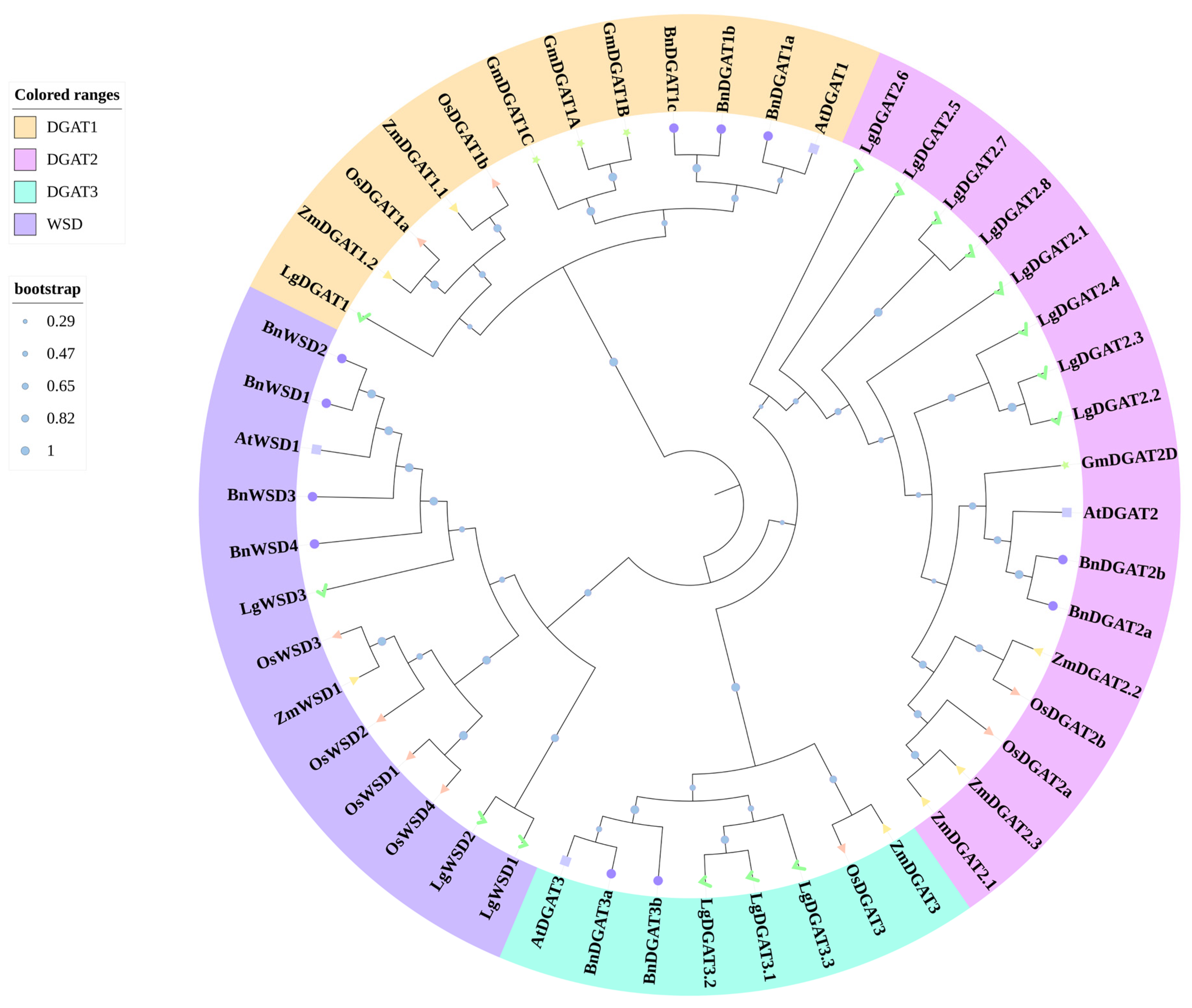
3.2. Phylogenetic Analysis of LgDGATs

The phylogenetic tree (Figure 1) constructed by the 50 DGATs from L. glauca, A. thaliana, O. sativa, Z. mays, G. max, and B. napus displayed that these proteins were clustered into four clades, DGAT1, DGAT2, DGAT3, and WSD. Further analysis indicated a close relationship between DGAT2 and DGAT3. Within the WSD clade, LgWSD1 and LgWSD2 were clustered together, suggesting that they possess similar and distinct functions. In the DGAT3 clade, LgDGAT3.1, LgDGAT3.2, and LgDGAT3.3 were hypothesized to have functions similar to DGAT3 in A. thaliana and B. napus. Similarly, LgDGAT1 was proposed to have more similar functions as DGAT1 in O. sativa and Z. mays. In the DGAT2 clade, eight LgDGATs (2.1 to 2.8) were clustered together, implying the presence of numerous redundant genes in DGAT2 due to gene replication during evolution.

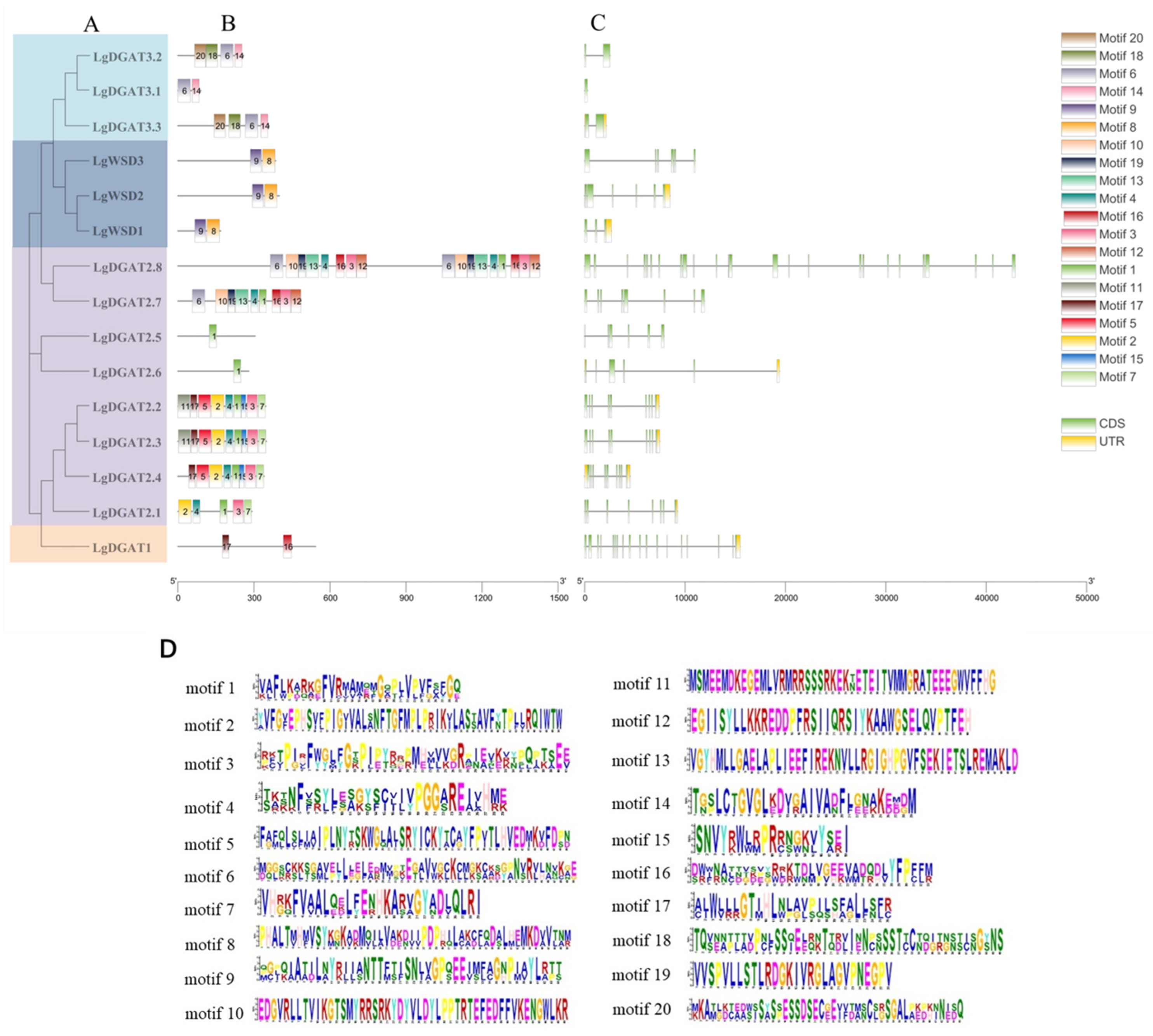
3.3. Analysis of Conserved Motifs and Gene Structure of LgDGATs

The LgDGAT sequences were subjected to conservative motif prediction using the MEME online tool. The results indicated significant differences among different subfamilies (Figure 2B). Motif 1 was present in all members of the LgDGAT2 subfamily, while motifs 6 and 14 were present in all LgDGAT3 subfamily members. Motifs 8 and 9 were unique to the LgWSD subfamily and absent in other subfamilies. The LgDGAT1 contained motifs 16 and 17, also found in the DGAT2 subfamily. Members within the same subfamily exhibited same or identical motif distributions, distinguishing them from other subfamilies. This finding suggested that genes in the same subfamily probably have similar roles, but genes in separate subfamilies have different roles. Moreover, the motif characteristics of each subfamily suggested that the LgDGAT2 subfamily may have more complex functions than the other three subfamilies.
Four subfamilies of LgDGATs indicated distinct gene structures (Figure 2C). The LgDGAT1 subfamily had 18 exons, while the LgWSD subfamily mainly consisted of three to six exons. The LgDGAT3 subfamily had 1 to 2 exons, and the LgDGAT2 subfamily exhibited a range of 6 to 9 exons, except the LgDGAT2.8 had more than 20 exons. Notably, LgDGAT2.8 possessed over 1000 amino acids, making it longer than other genes. This finding suggested that the length of this subfamily’s genes and the number of exons may have increased due to gene evolution.

3.4. Cis-Acting Elements in LgDGAT Promoters

The potential regulatory mechanisms of LgDGATs were analyzed using PlantCARE database with 2000 bp upstream sequences (Figure 3). Four cis-acting elements were present in the promoter regions: plant hormone responsiveness, abiotic and biological stress, plant growth and development, and light response.
Among all LgDGATs, LgDGAT2.3, LgDGAT2.7, LgDGAT2.8, LgDGAT3.2, and LgDGAT3.3 contained the most amounts of cis-acting elements (10 elements), while LgDGAT2.5 contained the fewest (5 elements). All LgDGAT promoters contained light-responsive elements. Regarding composition type specificity, most LgDGAT promoters contained MeJA-responsiveness, anaerobic induction, zein metabolism regulation, abscisic acid responsiveness, and MYB binding site elements. Approximately half of the LgDGAT promoters contained gibberellin-responsive element, auxin-responsive element, low-temperature responsiveness, and circadian control elements. Defense and stress-responsive elements were present in the promoters of LgDGAT1, LgDGAT2.1, LgDGAT2.7, LgDGAT3.3, and LgWSD3. Meristem expression elements were found in LgDGAT2.2, LgDGAT2.3, LgDGAT2.4, LgDGAT2.7, and LgDGAT2.8. Seed-specific regulation elements were present in LgDGAT2.2 and LgDGAT2.3, while salicylic acid responsiveness elements were found in LgDGAT2.4, LgDGAT3.2, and LgDGAT3.3. Endosperm expression elements were detected in LgDGAT2.8, LgDGAT3.1, LgDGAT3.2, and LgWSD2.

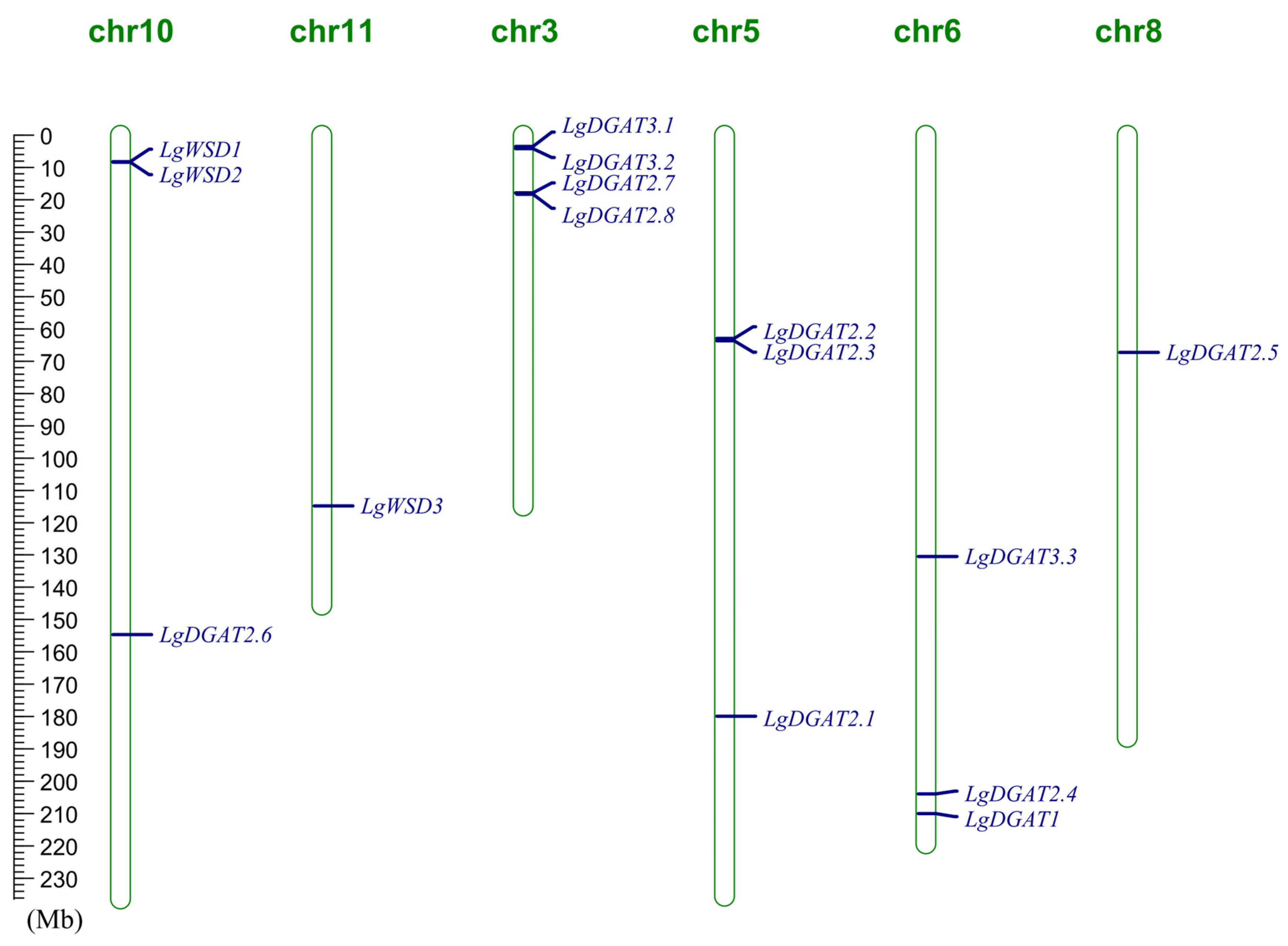
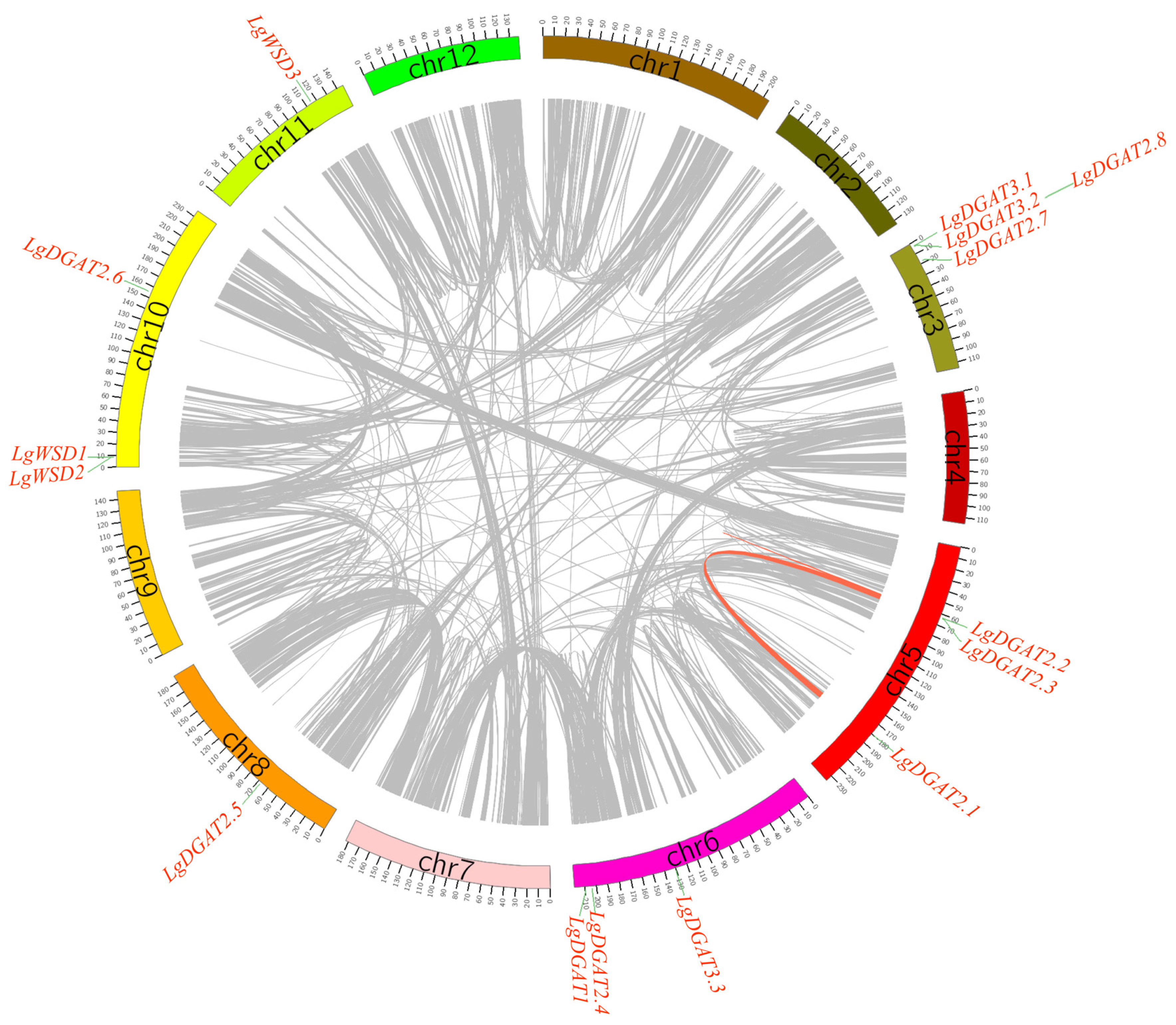
3.5. Chromosomal Localization, Gene Duplication, and Genome Synteny of LgDGATs

Based on genomic annotation data, distribution maps of 15 LgDGATs in the chromosomes were created (Figure 4), illustrating the positions of each gene on various chromosomes. The distribution of these genes was as follows: one gene each on chromosomes 8 and 11, three genes each on chromosomes 5, 6, and 10, and four on chromosome 3. Two gene duplication events were uncovered, LgDGAT2.1/LgDGAT2.2 and LgDGAT2.2/LgDGAT2.3 (Figure 5, Table 2). Both duplicate gene pairs resulted from segmental duplication events, with no tandem duplication events observed among LgDGATs.
The Ka/Ks ratios of the two pair of paralogous genes were all less than 1.0, indicating the occurrence of purifying selection. This finding suggested that LgDGATs have undergone evolutionary conservation, contributing to maintenance of functional stability. In terms of divergence time, the LgDGAT2.1/LgDGAT2.2 pair exhibited an earlier divergence time of 109.81 Mya, whereas divergence time of the LgDGAT2.2/LgDGAT2.3 pair was only 0.67 Mya (Table 3). Furthermore, a collinearity analysis of DGATs (Figure 6) from L. glauca, A. thaliana, and O. sativa indicated that LgWSD3 exhibited synteny with one AtDGAT (AT5G53380.1); LgDGAT3.1 and LgDGAT3.2 both exhibited synteny with one OsDGAT (OsKitaake05g027500.1), suggesting that the DGATs of L. glauca, A. thaliana, and O. sativa had undergone dramatic evolutionary changes.

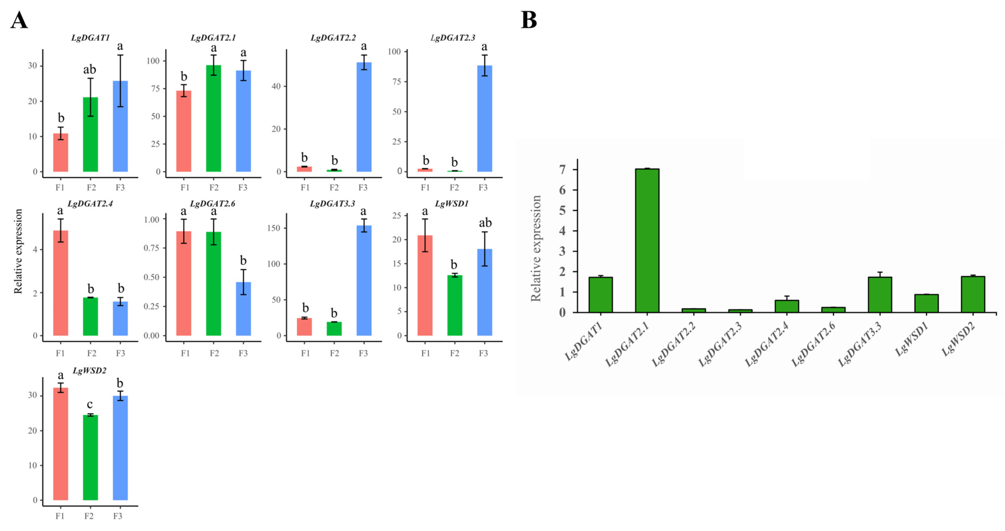
3.6. Differential Expression Levels of LgDGAT Genes in Developing Fruit

L. glauca fruits (LGF) have emerged as a novel resource in China, possessing industrial and medicinal value due to their rich content of terpenoids and oil [29]. The biosynthesis and accumulation of oil in LGF may be regulated by certain genes, thereby influencing fruit growth and development. Hence, we systematically studied the expression of LgDGATs during fruit development stages (fruits at 60, 90, and 150 DAF) and identified specific genes that may regulate oil biosynthesis and accumulation in fruits (Figure 7A).
Expression analysis was performed for nine LgDGATs detected at three developmental stages: 60, 90, and 150 DAF. All nine genes exhibited expression throughout the entire fruit development process. During fruit development, LgDGAT2.2, LgDGAT2.3, and LgDGAT3.3 showed similar expression patterns, with low expression levels at 60 and 90 DAF, reaching the lowest point at 90 DAF, followed by a significantly increase at 150 DAF. The expression level of LgDGAT1 displayed an increasing trend, while LgDGAT2.1 exhibited relatively stable and high expression levels, suggesting their potential involvement in TAG biosynthesis in fruits. LgWSD1 and LgWSD2 exhibited similar expression patterns, with a decrease followed by an increase in expression, peaking at 60 DAF. Conversely, LgDGAT2.4 and LgDGAT2.6 showed lower expression levels during fruit development, indicating their limited role in TAG accumulation in LGF. The qRT-PCR analysis was performed on randomly selected fruits at 90 DAF. The expression of nine genes was roughly consistent with the transcriptome data, which proved the accuracy of the transcriptome data.

3.7. Analysis of Expression Patterns of LgDGATs in Different Plant Tissues

Nine genes were found to have an expression when LgDGATs were examined for expression across various tissues, according to data retrieved from the NCBI database, while the other genes were not.
Their relative expression levels in seeds and stems were generally low (Figure 8). LgDGAT1, LgDGAT2.2, LgDGAT2.3, and LgDGAT2.1 exhibited peak expression in the sarcocarps and low expression levels in leaves. LgDGAT2.4, LgWSD1, and LgWSD2 showed peak levels in leaves and low sarcocarps expression. LgDGAT2.6 and LgDGAT3.3 exhibited peak expression in roots. These results indicate distinct main expression sites for each LgDGAT, highlighting their functional specialization and close coordination. Similar expression patterns among these genes within the same subfamily were observed, such as LgDGAT2.2 and LgDGAT2.3, and LgWSD1 and LgWSD2.

4. Discussion

L. glauca is known for its high oil content, surpassing that of conventional oil plants [49]. Apart from its edible applications, L. glauca oil derived from fruits or seeds has diverse industrial applications, such as in soaps, surfactants, and lubricants [32,33,34]. Mature L. glauca seeds have an oil content ranging from between 42.0% and 53.0% [50,51]. The biosynthesis of TAG, the primary oil component in plants, occurs in the endoplasmic reticulum through the catalysis action of multiple enzymes [52,53], with DGAT being the only rate-limiting enzyme for TAG biosynthesis [4]. The role of this enzyme in oil biosynthesis has been extensively studied in various plants [54,55,56,57,58,59], and overexpression of DGATs has been shown to enhance vegetable oils yield and quality [18]. However, no relevant report regarding the LgDGATs family exists. Using a bioinformatics method, we thoroughly analyzed LgDGATs in this study, encompassing phylogenetic evolution, gene structure, and gene expression, thus establishing a foundation for future investigations on improving L. glauca oil content.
We retrieved 15 LgDGATs from the L. glauca genome via aligning homologous sequences. Their number exceeds that of soybean (10), Arabidopsis (14), rice (5), and maize (5) [60,61]. The expansion of LgDGAT counts may reflect the evolution of L. glauca to adapt to its environment. The identified LgDGATs were categorized into four subgroups: LgDGAT1 (one gene), LgDGAT2 (eight genes), LgDGAT3 (three genes), and LgWSD (three genes). Exon–intron numbers and conserved motifs were similar among genes belonging to the same subfamily. However, gene structure and conserved motifs varied significantly among different subfamilies, indicating diverse biological functions within the same subfamily. Even though intron insertion or deletion aids in genome evolution [62], our results showed that not all LgDGATs lost introns equally, especially LgDGAT2.8. Recent studies have associated intron loss with genome reduction, suggesting that the number of introns primarily reflects the rate of evolution, with slower-evolving genes retaining more ancestral introns [63,64]. This diversity in LgDGATs’ evolution was observed. Evolutionary analysis indicated that the members of four LgDGAT subfamilies were distributed across multiple monocotyledonous and dicotyledonous plants, forming distinct clusters. This finding suggests that the differentiation of these subfamilies predates species divergence or, akin to DGAT1 and DGAT2, has an independent origin [65], consistent with previous research findings [66].
Previous research indicated that DGAT1 and DGAT2 were transmembrane proteins in different endoplasmic reticulum subdomains [67]. In this study, all LgDGAT1 and LgDGAT2s, except LgDGAT2.6, were localized in the endoplasmic reticulum. Based on hydrophobicity index, subcellular localization, and transmembrane prediction, LgDGAT1, LgDGAT2.2, LgDGAT2.3, and LgDGAT2.4 were identified as endoplasmic reticulum membrane proteins. Homologous genes derived from repetitive events often exhibit similar expression patterns [68]. Two pairs of fragment duplicates were found in LgDGATs, in which LgDGAT2.2 and LgDGAT2.3 had similar gene structures and conserved motifs and had similar expression patterns in different tissues and fruit development.
Further investigation indicated that these duplicated genes had a Ka/Ks ratio of less than 1.0, indicating purifying selection. Our findings support earlier evidence that the DGAT family may participate in abiotic stress responses, such as plant stress and low temperature, and contribute to TAG biosynthesis [69]. Plant adversity induces changes in membrane fluidity, and fatty acids were associated with membrane fluidity [70]. Most LgDGATs promoter contained MYB binding sites, suggesting the potential regulation of LgDGATs by MYB transcription factors. Additionally, we identified the cis-acting elements responsive to various phytohormones, including MeJA, GA, IAA, and ABA. ABA is widely recognized for its significant role in plants responses to biotic and abiotic stressors [71]. The ABA-responsive element was present in almost all LgDGATs, implying a potential association with the response to abiotic stress. The photoresponsive element was present in all LgDGATs, and thus we speculated a potential association between LgDGATs and photosynthesis.
While DGATs primarily participate in oil accumulation, they are also involved in lipid metabolism during biological processes such as seed germination, seedling development and leaf senescence [72]. Consequently, DGATs are expected to be expressed in various tissues, including seeds, flowers, and leaves, albeit with tissue-specific expression levels. For instance, DGAT1 in Tropaeolum majus is exclusively expressed in developing seeds [59], and DGAT1 expression in rosette increases with leaf senescence [73]. Tung tree DGAT1 shows minimal expression differences among organs, whereas DGAT2 is highly expressed in developing seeds [56]. AtDGAT1 exhibits high expression in developing Arabidopsis seeds, correlating with TAG accumulation [74]. In this study, nine expressed LgDGATs were identified via the transcriptome data in different L. glauca tissues obtained from the NCBI database and fruits during three developmental stages sequenced by us. These genes include LgDGAT1, LgDGAT2.1, LgDGAT2.2, LgDGAT2.3, LgDGAT2.4, LgDGAT2.6, LgDGAT3.3, LgWSD1, and LgWSD2. The remaining six LgDGATs were not detected in either transcriptome data, suggesting that they may be pseudogenes or expressed at shallow levels during these periods. In this study, LgDGAT2.1 was significantly expressed during fruit development, and fruits at 150 DAF had high LgDGAT2.2, LgDGAT2.3, and LgDGAT3.3 expression. These genes may play pivotal roles in oil accumulation in developing fruits. However, the LgWSD subfamilies were mainly expressed in stems, leaves, and other organs, consistent with the study on sunflower [75]. We hypothesized that the LgWSD subfamilies may involve lipid accumulation and cuticle wax formation in these tissues. Plant cuticle prevents excessive water loss, resists ultraviolet radiation, and protects against diseases and pests [76]. AtWSD1 has been implicated in epidermal wax synthesis in stems and is vital for Arabidopsis’ adaptation to drought stress [19,20]. AtWSD11 (FOP1) exhibits high expression in Arabidopsis flowers, and its encoded product may act as a lubricant to enable uninhibited growth of petals as they extend between sepals and anthers [77]. The distinct tissue expression patterns of the four LgDGAT subfamilies indicate functional divergence among LgDGATs, suggesting a finely regulated process of oil accumulation in L. glauca.

5. Conclusions

In this study, 15 LgDGATs were identified in the L. glauca genome through bioinformatics analysis. The LgDGATs were comprehensively analyzed for physicochemical properties, chromosome localization, conserved motifs, gene structure, evolutionary relationship and cis-acting elements. Based on the similarities between these genes’ structures and functions, they were divided into four groups. The cis-acting components in their promoter region were linked to plant hormone signaling, plant growth and development, abiotic and biological stress responses, and light responsiveness. According to transcriptome data, nine LgDGATs were among the identified genes and showed measurable expression levels in three stages of fruit development and different tissues. Expression pattern analysis indicated the significant involvement of LgDGAT2.1 in TAC accumulation in the LFG. Our results provide valuable insights and data for future investigations on the functional characterization of LgDGATs and offer novel candidate genes for utilizing L. glauca resources.

Supplementary Materials

The following supporting information can be downloaded at https://www.mdpi.com/article/10.3390/f14081633/s1, Table S1: The protein sequences of LgDGATs; Table S2: The gene sequences of LgDGATs.

Author Contributions

Conceptualization, B.X.; methodology, X.B.; software, X.B.; validation, Y.Y.; resources, B.X.; data curation, L.X.; writing—original draft preparation, X.B.; writing—review and editing, B.X.; visualization, Q.L.; supervision, B.X.; project administration, X.B.; funding acquisition, B.X. All authors have read and agreed to the published version of the manuscript.

Funding

This research was funded by the National Natural Science Foundation of China (31900272; 32260086), National Guidance of Local Science and Technology Development Fund of China ([2023]009), Guizhou Science and Technology Plan Project (Qiankehe Basics–ZK [2021] General 151 and Qiankehe Support [2022] General 057), Young Talents Program of Guizhou Provincial Department of Education (Qianjiaohe Basics–KY [2021]), Cultivation Project of Guizhou University (Gzu. 2020 No. 65), and Guizhou Provincial Postgraduate Research Fund (YJSKYJJ [2021] 005).

Data Availability Statement

The data that support the findings of this study are openly available in the NCBI database (https://www.ncbi.nlm.nih.gov/sra/) (accessed on 1 June 2023). The accession number is SRX591256 and PRJNA977679.

Conflicts of Interest

The authors declare no conflict of interest.

References

  1. Lung, S.C.; Weselake, R.J. Diacylglycerol acyltransferase: A key mediator of plant triacylglycerol synthesis. Lipids 2006, 41, 1073–1088. [Google Scholar] [CrossRef]
  2. Liu, Q.; Siloto, R.M.P.; Lehner, R.; Stone, S.J.; Weselake, R.J. Acy1–CoA: Diacylglycerol acyltransferase: Molecular biology, biochemistry and biotechnology. Prog. Lipid Res. 2012, 51, 350–377. [Google Scholar] [CrossRef] [PubMed]
  3. Kennedy, E.P.; Weiss, S.B. Function of cytidine coenzymes in the biosynthesis of phospholipides. J. Biol. Chem. 1956, 222, 193–213. [Google Scholar] [CrossRef] [PubMed]
  4. Chapman, K.D.; Ohlrogge, J.B. Compartmentation of triacylglycerol accumulation in plants. J. Biol. Chem. 2012, 287, 2288–2294. [Google Scholar] [CrossRef] [PubMed] [Green Version]
  5. Hernández, M.L.; Whitehead, L.; He, Z.; Gazda, V.; Gilday, A.; Kozhevnikova, E.; Vaistij, F.; Larson, T.R.; Graham, I.A. A cytosolic acyltransferase contributes to triacylglycerol synthesis in sucrose–rescued Arabidopsis seed oil catabolism mutants. Plant Physiol. 2012, 160, 215–225. [Google Scholar] [CrossRef] [PubMed] [Green Version]
  6. Kalscheuer, R.; Steinbüchel, A. A novel bifunctional wax ester synthase/acyl–CoA: Diacylglycerol acyltransferase mediates wax ester and triacylglycerol biosynthesis in Acinetobacter calcoaceticus ADP1. J. Biol. Chem. 2003, 278, 8075–8082. [Google Scholar] [CrossRef] [PubMed] [Green Version]
  7. Nykiforuk, C.L.; Furukawa-Stoffer, T.L.; Huff, P.W.; Sarna, M.; Laroche, A.; Moloney, M.M.; Weselake, R.J. Characterization of cDNAs encoding diacylglycerol acyltransferase from cultures of Brassica napus and sucrose–mediated induction of enzyme biosynthesis. Biochim. Biophys. Acta 2002, 1580, 95–109. [Google Scholar] [CrossRef]
  8. Zhang, F.Y.; Yang, M.F.; Xu, Y.N. Silencing of DGAT1 intobacco causes a reduction in seed oil content. Plant Sci. 2005, 169, 689–694. [Google Scholar] [CrossRef]
  9. Lock, Y.Y.; Snyder, C.L.; Zhu, W.; Siloto, R.M.; Weselake, R.J.; Shah, S. Antisense suppression of type 1 diacylglycerol acyltransferase adversely affects plant development in Brassica napus. Physiol. Plant 2009, 137, 61–71. [Google Scholar] [CrossRef]
  10. Zhang, M.; Fan, J.; Taylor, D.C.; Ohlrogge, J.B. DGAT1 and PDAT1 acyltransferases have overlapping functions in Arabidopsis triacylglycerol biosynthesis and are essential for normal pollen and seed development. Plant Cell. 2009, 21, 3885–3901. [Google Scholar] [CrossRef] [Green Version]
  11. Zou, J.; Wei, Y.; Jako, C.; Kumar, A.; Selvaraj, G.; Taylor, D.C. The Arabidopsis thaliana TAG1 mutant has a mutation in a diacylglycerol acyltransferase gene. Plant J. Cell Mol. Biol. 1999, 19, 645–653. [Google Scholar] [CrossRef] [PubMed] [Green Version]
  12. Poirier, Y.; Ventre, G.; Caldelari, D. Increased flow of fatty acids toward beta– oxidation in developing seeds of Arabidopsis deficient in diacylglycerol acyltransferase activity or synthesizing medium–chain–length fatty acids. Plant Physiol. 1999, 121, 1359–1366. [Google Scholar] [CrossRef] [PubMed] [Green Version]
  13. Andrianov, V.; Borisjuk, N.; Pogrebnyak, N.; Brinker, A.; Dixon, J.; Spitsin, S.; Flynn, J.; Matyszczuk, P.; Andryszak, K.; Laurelli, M.; et al. Tobacco as a production platform for biofuel: Overexpression of Arabidopsis DGAT and LEC2 genes increases accumulation and shifts the composition of lipids in green biomass. Plant Biotechnol. J. 2010, 8, 277–287. [Google Scholar] [CrossRef]
  14. Lu, G.; Li, D.; Zhang, T.; Tang, X.; Lu, J.X.; Hu, Q.M.; Hu, T.M. Cloning and function analysis of a type 2 diacylglycerol acyltransferase (DGAT2) from Perilla frutescens. Acta Agron. Sin. 2020, 46, 1283–1290. [Google Scholar] [CrossRef]
  15. Zhang, T.; He, H.; Xu, C.; Fu, Q.; Tao, Y.; Xu, R.; Xu, Z. Overexpression of Type 1 and 2 Diacylglycerol Acyltransferase Genes (JcDGAT1 and JcDGAT2) Enhances Oil Production in the Woody Perennial Biofuel Plant Jatropha curcas. Plants 2021, 10, 699. [Google Scholar] [CrossRef] [PubMed]
  16. Gao, Y.; Sun, Y.; Gao, H.L.; Xue, J.A.; Jia, X.Y.; Li, R.Z. Functional analysis of CEDGAT2–2 gene in Cyperus esculentus L. and cultivation of oil fuel–type tobacco. In Proceedings of the 19th Annual Conference of Crop Science Society of China, Wuhan, China, 8–9 November 2020; Crop Science Society of China: Wuhan, China, 2020; Volume 1. [Google Scholar]
  17. Ayme, L.; Arragain, S.; Canonge, M.; Baud, S.; Touati, N.; Bimai, O.; Jagic, F.; Louis-Mondesir, C.; Briozzo, P.; Fontecave, M.; et al. Arabidopsis thaliana DGAT3 is a [2Fe–2S] protein involved in TAG biosynthesis. Sci. Rep. 2018, 8, 17254. [Google Scholar] [CrossRef] [Green Version]
  18. Gao, H.L.; Gao, Y.; Zhang, F.; Liu, B.L.; Ji, C.L.; Xue, J.A.; Yuan, L.X.; Li, R.Z.; Sativa, C. Functional characterization of an novel acyl–CoA: Diacylglycerol acyltransferase 3–3 (CsDGAT3–3) gene from Camelina sativa. Plant Sci. 2021, 303, 110752. [Google Scholar] [CrossRef]
  19. Li, F.; Wu, X.; Lam, P.; Bird, D.; Zheng, H.; Samuels, L.; Jetter, R.; Kunst, L. Identification of the wax ester synthase/acyl–coenzyme A: Diacylglycerol acyltransferase WSD1 required for stem wax ester biosynthesis in Arabidopsis. Plant Physiol. 2008, 148, 97–107. [Google Scholar] [CrossRef] [Green Version]
  20. Patwari, P.; Salewski, V.; Gutbrod, K.; Kreszies, T.; Dresen-Scholz, B.; Peisker, H.; Steiner, U.; Meyer, A.J.; Schreiber, L.; Dormann, P. Surface wax esters contribute to drought tolerance in Arabidopsis. Plant J. 2019, 98, 727–744. [Google Scholar] [CrossRef]
  21. Wang, Y.L.; Gao, X.M.; Yu, X.P.; Cheng, S.L.; Kong, L.H. Study on the resource and its utilizations of Lindera glauca in China. Henan Sci. 1994, 12, 331–334. [Google Scholar]
  22. Seki, K.; Sasaki, T.; Haga, K.; Kaneko, R. Two methoxybutanolides from Lindera glauca. Phytochemistry 1994, 36, 949–951. [Google Scholar] [CrossRef]
  23. Huh, G.W.; Park, J.H.; Kang, J.H.; Jeong, T.S.; Kang, H.C.; Baek, N.I. Flavonoids from Lindera glauca Blume as low–density lipoprotein oxidation inhibitors. Nat. Prod. Res. 2014, 28, 831–834. [Google Scholar] [CrossRef] [PubMed]
  24. Dupont, Y.L. Evolution of apomixis as a strategy of colonization in the dioecious species Lindera glauca (Lauraceae). Popul. Ecol. 2002, 44, 293–297. [Google Scholar] [CrossRef]
  25. Tsui, H.P.; Xia, Z.D.; Li, J.L. Lauraceae and Hernandiaceae; Li, H.W., Ed.; Flora of China Science Press: Beijing, China, 1982; Volume 31, pp. 393–394. [Google Scholar]
  26. Tsui, H.P.; Werff, H. Lauraceae and Hernandiaceae; Li, H.W., Ed.; Flora of China Science Press: Beijing, China; Missouri Botanical Garden Press: St. Louis, MO, USA, 2008; Volume 7, pp. 146–147. [Google Scholar]
  27. Huh, G.W.; Park, J.H.; Shrestha, S.; Lee, Y.H.; Ahn, E.M.; Kang, H.C.; Baek, N.I. Sterols from Lindera glauca Blume stem wood. J. Appl. Biol. Chem. 2011, 54, 309–312. [Google Scholar] [CrossRef]
  28. Xiong, B.; Zhang, L.; Dong, S.; Zhang, Z. Population genetic structure and variability in Lindera glauca (Lauraceae) indicates low levels of genetic diversity and skewed sex ratios in natural populations in mainland China. PeerJ 2020, 8, 8304. [Google Scholar] [CrossRef] [Green Version]
  29. Qi, J.; Xiong, B.; Ju, Y.X.; Hao, Q.; Zhang, Z.X. Study on fruit growth regularity and lipid accumulation of Lindera glauca. Chinese Agri Sci Bull. 2015, 31, 29–33. [Google Scholar]
  30. Lin, Z.; An, J.; Wang, J.; Niu, J.; Ma, C.; Wang, L.; Yuan, G.; Shi, L.; Liu, L.; Zhang, J.; et al. Integrated analysis of 454 and Illumina transcriptomic sequencing characterizes carbon flux and energy source for fatty acid synthesis in developing Lindera glauca fruits for woody biodiesel. Biotechnol. Biofuels 2017, 10, 134. [Google Scholar] [CrossRef] [Green Version]
  31. Zhu, B.; Hou, X.; Niu, J.; Li, P.; Fang, C.; Qiu, L.; Ha, D.; Zhang, Z.; Sun, J.; Li, Y.; et al. Volatile constituents from the fruits of Lindera glauca (Sieb. et Zucc.) with different maturities. J. Essent. Oil Bear. Plants 2016, 19, 926–935. [Google Scholar] [CrossRef]
  32. Sun, H.L.; Wang, J.X.; Gu, X.Z.; Kang, W.Y. Analysis of volatile compounds from leaves and fruits of Lindera glauca. Chin. J. Exp. Tradit. Med. 2011, 7, 033. [Google Scholar]
  33. Kim, K.H.; Moon, E.; Ha, K.; Suh, W.S.; Kim, H.H.; Kim, S.Y.; Choi, S.U.; Lee, K.R. Bioactive lignan constituents from the twigs of Lindera glauca. Chem. Pharm. Bull. 2014, 62, 1136–1140. [Google Scholar] [CrossRef] [Green Version]
  34. Suh, W.S.; Kim, K.H.; Kim, H.K.; Choi, S.U.; Lee, K.R. Three new lignan derivatives from Lindera glauca (Siebold et Zucc.) Blume. Helv. Chim. Acta 2015, 98, 1087–1094. [Google Scholar] [CrossRef]
  35. El-Gebali, S.; Mistry, J.; Bateman, A.; Eddy, S.R.; Luciani, A.; Potter, S.C.; Qureshi, M.; Richardson, L.J.; Salazar, G.A.; Smart, A.; et al. The Pfam protein families database in 2019. Nucleic Acids Res. 2019, 47, D427–D432. [Google Scholar] [CrossRef]
  36. Johnson, L.S.; Eddy, S.R.; Portugaly, E. Hidden Markov model speed heuristic and iterative HMM search procedure. BMC Bioinform. 2010, 11, 431. [Google Scholar] [CrossRef] [PubMed] [Green Version]
  37. Marchler-Bauer, A.; Bryant, S.H. CD–Search: Protein domain annotations on the fly. Nucleic Acids Res. 2004, 32 (Suppl. S2), W327–W331. [Google Scholar] [CrossRef]
  38. Garg, V.K.; Avashthi, H.; Tiwari, A.; Jain, P.A.; Ramkete, P.W.; Kayastha, A.M.; Singh, V.K. MFPPI–multi FASTA ProtParam interface. Bio. Inf. 2016, 12, 74–77. [Google Scholar] [CrossRef]
  39. Krogh, A.; Larsson, B.; von Heijne, G.; Sonnhammer, E.L. Predicting transmembrane protein topology with a hidden Markov model: Application to complete genomes. J. Mol. Biol. 2001, 305, 567–580. [Google Scholar] [CrossRef] [PubMed] [Green Version]
  40. Letunic, I.; Bork, P. Interactive tree of life (ITOL) v5: An online tool for phylogenetic tree displays and annotation. Nucleic Acids Res. 2021, 49, W293–W296. [Google Scholar] [CrossRef] [PubMed]
  41. Bailey, T.L.; Boden, M.; Buske, F.A.; Frith, M.; Grant, C.E.; Clementi, L.; Ren, J.; Li, W.W.; Noble, W.S. MEME SUITE: Tools for motif discovery and searching. Nucleic Acids Res. 2009, 37, W202–W208. [Google Scholar] [CrossRef]
  42. Chen, C.; Chen, H.; Zhang, Y.; Thomas, H.R.; Frank, M.H.; He, Y.; Xia, R. TBtools: An integrative toolkit developed for interactive analyses of big biological data. Mol. Plant 2020, 13, 1194–1202. [Google Scholar] [CrossRef]
  43. Lescot, M.; Dehais, P.; Thijs, G.; Marchal, K.; Moreau, Y.; Van de Peer, Y.; Rouze, P.; Rombauts, S. PlantCARE, a database of plant cis-acting regulatory elements and a portal to tools for in silico analysis of promoter sequences. Nucleic Acids Res. 2002, 30, 325–327. [Google Scholar] [CrossRef]
  44. Voorrips, R.E. MapChart: Software for the graphical presentation of linkage maps and QTLs. J. Hered. 2002, 93, 77–78. [Google Scholar] [CrossRef] [Green Version]
  45. Wang, Y.; Tang, H.; Debarry, J.D.; Tan, X.; Li, J.; Wang, X.; Lee, T.H.; Jin, H.; Marler, B.; Guo, H.; et al. MCScanX: A toolkit for detection and evolutionary analysis of gene synteny and collinearity. Nucleic Acids Res. 2012, 40, e49. [Google Scholar] [CrossRef] [PubMed] [Green Version]
  46. Krzywinski, M.; Schein, J.; Birol, I.; Connors, J.; Gascoyne, R.; Horsman, D.; Jones, S.J.; Marra, M.A. Circos: An information aesthetic for comparative genomics. Genome Res. 2009, 19, 1639–1645. [Google Scholar] [CrossRef] [PubMed] [Green Version]
  47. Cui, L.; Wall, P.K.; Leebens-Mack, J.H.; Lindsay, B.G.; Soltis, D.E.; Doyle, J.J.; Soltis, P.S.; Carlson, J.E.; Arumuganathan, K.; Barakat, A.; et al. Widespread genome duplications throughout the history of flowering plants. Genome Res. 2006, 16, 738–749. [Google Scholar] [CrossRef] [PubMed] [Green Version]
  48. Field, A. Discovering Statistics Using IBM SPSS Statistics; Sage Publications Ltd.: Thousand Oaks, CA, USA, 2013. [Google Scholar]
  49. Wang, R.; Hanna, M.A.; Zhou, W.W.; Bhadury, P.S.; Chen, Q.; Song, B.A.; Yang, S. Production and selected fuel properties of biodiesel from promising non–edible oils: Euphorbia lathyris L., Sapium sebiferum L. and Jatropha curcas L. Bioresour. Technol. 2011, 102, 1194–1199. [Google Scholar] [CrossRef] [PubMed]
  50. Qian, X.S.; Xiao, Z.C. Study and utilization of Lindera glauca in China. Wild Plant Resour. 1985, 2, 2–6. [Google Scholar]
  51. Wang, J.P.; Meng, S.J.; Li, J.M. Fatty acid of oils of Lauraceae. Acta Bot. Sin. 1984, 27, 175–185. [Google Scholar]
  52. Baud, S.; Lepiniec, L. Physiological and developmental regulation of seed oil production. Prog. Lipid Res. 2010, 49, 235–249. [Google Scholar] [CrossRef]
  53. Barthole, G.; Lepiniec, L.; Rogowsky, P.M.; Baud, S. Controlling lipid accumulation in cereal grains. Plant Sci. 2012, 185, 33–39. [Google Scholar] [CrossRef]
  54. Chen, B.; Wang, J.; Zhang, G.; Liu, J.; Manan, S.; Hu, H.; Zhao, J. Two types of soybean diacylglycerol acyltransferases are differentially involved in triacylglycerol biosynthesis and response to environmental stresses and hormones. Sci. Rep. 2016, 6, 28541. [Google Scholar] [CrossRef] [Green Version]
  55. Li, R.; Hatanaka, T.; Yu, K.; Wu, Y.; Fukushige, H.; Hildebrand, D. Soybean oil biosynthesis: Role of diacylglycerol acyltransferases. Funct. Integr. Genom. 2013, 13, 99–113. [Google Scholar] [CrossRef] [PubMed]
  56. Shockey, J.M.; Gidda, S.K.; Chapital, D.C.; Kuan, J.C.; Dhanoa, P.K.; Bland, J.M.; Rothstein, S.J.; Mullen, R.T.; Dyer, J.M. Tung tree DGAT1 and DGAT2 have nonredundant functions in triacylglycerol biosynthesis and are localized to different subdomains of the endoplasmic reticulum. Plant Cell 2006, 18, 2294–2313. [Google Scholar] [CrossRef] [Green Version]
  57. Wang, H.; Zhang, J.; Gai, J.; Chen, S. Cloning and comparative analysis of the gene encoding diacylglycerol acyltransferase from wild type and cultivated soybean. Theor. Appl. Genet. 2006, 112, 1086–1097. [Google Scholar] [CrossRef] [PubMed]
  58. Zheng, P.; Allen, W.B.; Roesler, K.; Williams, M.E.; Zhang, S.; Li, J.; Glassman, K.; Ranch, J.; Nubel, D.; Solawetz, W.; et al. A phenylalanine in DGAT is a key determinant of oil content and composition in maize. Nat. Genet. 2008, 40, 367–372. [Google Scholar] [CrossRef]
  59. Xu, J.; Francis, T.; Mietkiewska, E.; Giblin, E.M.; Barton, D.L.; Zhang, Y.; Zhang, M.; Taylor, D.C. Cloning and characterization of an acyl–CoA–dependent diacylglycerol acyltransferase 1 (DGAT1) gene from Tropaeolum majus, and a study of the functional motifs of the DGAT protein using site–directed mutagenesis to modify enzyme activity and oil content. Plant Biotechnol. J. 2008, 6, 799–818. [Google Scholar] [CrossRef]
  60. Zhao, J.; Bi, R.; Li, S.; Zhou, D.; Bai, Y.; Jing, G.; Zhang, K.; Zhang, W. Genome–wide analysis and functional characterization of Acyl–CoA: Diacylglycerol acyltransferase from soybean identify GmDGAT1A and 1B roles in oil synthesis in Arabidopsis seeds. J. Plant Physiol. 2019, 242, 153019. [Google Scholar] [CrossRef]
  61. Yan, B.; Xu, X.; Gu, Y.; Zhao, Y.; Zhao, X.; He, L.; Zhao, C.; Li, Z.; Xu, J. Genome–wide characterization and expression profiling ofdiacylglycerol acyltransferase genes from maize. Genome 2018, 61, 735–743. [Google Scholar] [CrossRef]
  62. Sharpton, T.J.; Neafsey, D.E.; Galagan, J.E.; Taylor, J.W. Mechanisms of intron gain and loss in Cryptococcus. Genome Biol. 2008, 9, R24. [Google Scholar] [CrossRef] [Green Version]
  63. Lynch, M.; Richardson, A.O. The evolution of spliceosomal introns. Curr. Opin. Genet. Dev. 2002, 12, 701–710. [Google Scholar] [CrossRef] [PubMed]
  64. Roy, S.W.; Gilbert, W. The evolution of spliceosomal introns: Patterns, puzzles and progress. Nat. Rev. Genet. 2006, 7, 211–221. [Google Scholar]
  65. Turchetto-Zolet, A.C.; Maraschin, F.S.; de Morais, G.L.; Cagliari, A.; Andrade, C.M.; Margis-Pinheiro, M.; Margis, R. Evolutionaryview of acyl–CoA diacylglycerol acyltransferase (DGAT), a key enzyme in neutral lipid biosynthesis. BMC Evol. Biol. 2011, 11, 263. [Google Scholar] [CrossRef] [Green Version]
  66. Turchetto-Zolet, A.C.; Christoff, A.P.; Kulcheski, F.R.; Loss-Morais, G.; Margis, R.; Margis-Pinheiro, M. Diversity and evolution of plant diacylglycerol acyltransferase (DGATs) unveiled by phylogenetic, gene structure and expression analyses. Genet. Mol. Biol. 2016, 39, 524–538. [Google Scholar] [CrossRef] [Green Version]
  67. Bhatt-Wessel, B.; Jordan, T.W.; Miller, J.H.; Peng, L. Role of DGAT enzymes in triacylglycerol metabolism. Arch. Biochem. Biophys. 2018, 655, 1–11. [Google Scholar] [CrossRef] [PubMed]
  68. Ganko, E.W.; Meyers, B.C.; Vision, T.J. Divergence in expression between duplicated genes in Arabidopsis. Mol. Biol. Evol. 2007, 24, 2298–2309. [Google Scholar] [CrossRef] [PubMed] [Green Version]
  69. Singer, S.D.; Zou, J.; Weselake, R.J. Abiotic factors influence plant storage lipid accumulation and composition. Plant Sci. 2016, 243, 1–9. [Google Scholar] [CrossRef] [PubMed]
  70. Young, D.Y.; Shachar-Hill, Y. Large fluxes of fatty acids from membranes to triacylglycerol and back during N–deprivation and recovery in Chlamydomonas. Plant Physiol. 2020, 185, 796–814. [Google Scholar] [CrossRef]
  71. Chen, K.; Li, G.J.; Bressan, R.A.; Song, C.P.; Zhu, J.K.; Zhao, Y. Abscisic acid dynamics, signaling, and functions in plants. J. Integr.Plant Biol. 2020, 62, 25–54. [Google Scholar] [CrossRef] [Green Version]
  72. Liu, D.; Ji, H.; Yang, Z. Functional characterization of three novel genes encoding Diacylglycerol Acyltransferase (DGAT) from oil–rich tubers of Cyperus esculentus. Plant Cell Physiol. 2020, 61, 118–129. [Google Scholar] [CrossRef]
  73. Kaup, M.T.; Froese, C.D.; Thompson, J.E. A role for diacylglycerol acyltransferase during leaf senescence. Plant Physiol. 2002, 129, 1616–1626. [Google Scholar] [CrossRef] [Green Version]
  74. Lu, C.L.; de Noyer, S.B.; Hobbs, D.H.; Kang, J.; Wen, Y.; Krachtus, D.; Hills, M.J. Expression pattern of diacylglycerol acyltransferase–1, an enzyme involved in triacylglycerol biosynthesis, in Arabidopsis thaliana. Plant Mol. Biol. 2003, 52, 31–41. [Google Scholar] [CrossRef]
  75. Zhang, C.; Zhang, Z.; Yang, J.B.; Meng, W.Q.; Zeng, L.L.; Sun, L. Genome–wide identification and relative expression analysis of DGATs gene family in sunflower. Acta Agron. Sin. 2023, 49, 73–85. [Google Scholar]
  76. Zhao, Y.P.; Wu, N.; Li, W.J.; Shen, J.L.; Chen, C.; Li, F.G.; Hou, Y.X. Evolution and Characterization of Acetyl Coenzyme A: Diacylglycerol Acyltransferase Genes in Cotton Identify the Roles of GhDGAT3D in Oil Biosynthesis and Fatty Acid Composition. Genes 2021, 12, 1045. [Google Scholar] [CrossRef] [PubMed]
  77. Takeda, S.; Iwasaki, A.; Matsumoto, N.; Uemura, T.; Tatematsu, K.; Okada, K. Physical interaction of floral organs controls petal morphogenesis in Arabidopsis. Plant Physiol. 2013, 161, 1242–1250. [Google Scholar] [CrossRef] [PubMed] [Green Version]
Figure 1. Phylogenetic tree of proteins from the DGAT family. Fifty DGATs of Lindera glauca (Lg), Arabidopsis thaliana (At), Oryza sativa (Os), Zea mays (Zm), Glycine max (Gm), and Brassica napus (Bn) are included in this tree. Distinct colors are used to designate distinct groupings of DGAT proteins. Each species of proteins is labeled with specific symbols.
Forests 14 01633 g001
Figure 2. Phylogenetic relationships, gene structures and conserved motifs of the LgDGAT family. (A) Phylogenetic tree of LgDGATs. Different subfamily groupings are in different colors. (B) Conserved motifs of LgDGATs. Different motifs are represented by specific colors. (C) Gene structure of LgDGATs. Untranslated regions, introns, and exons are each denoted by a yellow box, black line, and green box, respectively. (D) Conserved motif logos of LgDGATs.
Forests 14 01633 g002
Figure 3. Distribution and number of cis-acting elements in the promoter region of LgDGATs. Different promoter elements are represented by different colored boxes.
Forests 14 01633 g003
Figure 4. Chromosomal locations of DGAT genes in L. glauca.
Forests 14 01633 g004
Figure 5. Collinearity analysis of LgDGATs in L. glauca genome. All of the genome’s collinear blocks are shown by gray lines, while gene pairs with duplication events are represented by red lines.
Forests 14 01633 g005
Figure 6. Collinearity analysis of DGATs among L. glauca, O. sativa, and A. thaliana. LgDGATs, OsDGATs, and AtDGATs are represented by orange, blue, and green, respectively. The gray lines in the background represent the collinear blocks identified in the genomes of O. sativa, L. glauca, and A. thaliana. Orange lines indicate the DGAT gene pairs.
Forests 14 01633 g006
Figure 7. LgDGATs’ expression patterns in developing fruit. (A) Expression analysis of nine LgDGAT genes during fruit development. F1, F2, and F3 indicate fruits at 60, 90, and 150 DAF, respectively. Data are indicated as means ± standard deviation (SD) from three samples. With the Duncan approach, lowercase letters above the bars demonstrate a significant difference (p < 0.05). (B) Gene expression data of nine LgDGATs in fruits at 90 DAF obtained via qRT-PCR analysis. Both RPL32e and UBC genes were used as the internal controls.
Forests 14 01633 g007
Figure 8. Relative patterns of LgDGATs’ expression in the various tissues of L. glauca.
Forests 14 01633 g008
Table 1. Primers for qRT-PCR.
Gene IDForward PrimerReverse Primer
LgDGAT1CGACTCCTCCTCCAAGACCTGACCGACGGATTCCTCTGTTCTC
LgDGAT2.1TCAGTGAGGTTATCTGTTGCGACCATAGCAGAAAACAGGA
LgDGAT2.2CACACCACTACTAAGGCAAAATATGAACAATCTCCCGAGC
LgDGAT2.3GAGGTCATCCTCCAGAAAAGAAGTTGAGATGAATGGTCCC
LgDGAT2.4GTGTTTGGGATGCTGTTATGACGTGAAGTGTAATGGGAAA
LgDGAT2.6CTCTGTCAACGCAACCATACTCACTGTGGACTGTGGTGTGGATGG
LgDGAT3.3ATAGACCAACCACAACCCATTCAGAAGAGAAGCAAGGAACAGCAGTAG
LgWSD1TCCCAAGCCAGTCCAGTGTCTTGAGATTGTGAAGGTTGTGTTAGC
LgWSD2AAGTTTCGACATTCAGGACAGTAGCCCTTCTAATTCTCGG
UBCCTGGGATACCATCCAGAACATCCTCAAGTGTCCTTCCAGCATAG
RPL32CCGCCACCTCTCTCTTTATTTGCGCTTCTTGACAATCTTCTTG
Table 2. Information on LgDGATs in L. glauca.
Gene IDAccession NumberAAMolecular WeightPIAliphatic IndexGRAVYPredicted Location(s)Transmembrane
Domain
LgDGAT1Lg06G698954361,266.017.1491.840.129Endoplasmic reticulumYES
LgDGAT2.1Lg05G613229232,678.249.2895.720.189Endoplasmic reticulumNO
LgDGAT2.2Lg05G312634739,417.389.3396.890.165Endoplasmic reticulumYES
LgDGAT2.3Lg05G316334939,622.659.3396.330.175Endoplasmic reticulumYES
LgDGAT2.4Lg06G663634038,598.369.26101.790.293Endoplasmic reticulumYES
LgDGAT2.5Lg08G289730433,914.545.2385.89−0.221Endoplasmic reticulumNO
LgDGAT2.6Lg10G426227930,661.568.8682.150.146Cell membraneYES
LgDGAT2.7Lg03G107348655,057.858.6498.27−0.134Endoplasmic reticulumNO
LgDGAT2.8Lg03G10891428160,137.897.0693.77−0.132Endoplasmic reticulumNO
LgDGAT3.1Lg03G163909049.46.2172.56−0.11Cell wallNO
LgDGAT3.2Lg03G19825927,851.628.5873.13−0.557NucleusNO
LgDGAT3.3Lg06G330136038,915.395.6866.89−0.399NucleusNO
LgWSD1Lg10G47916818,727.968.7296.370.083NucleusNO
LgWSD2Lg10G48939944,459.45.86100.13−0.026ChloroplastNO
LgWSD3Lg11G266638543,035.948.4794.21−0.023ChloroplastNO
Note: AA, PI, and GRAVY, respectively, indicate number of amino acids, theoretical isoelectric point, and average hydrophilicity of the protein.
Table 3. Gene duplication events and divergence time between paralogous pairs of LgDGATs.
Duplicated Gene 1Duplicated Gene 2KaKsKa/KsDuplicated TypeSelective TypeDivergence Time (Mya)
LgDGAT2.2LgDGAT2.10.2235080.6632430.336993WGD or SegmentalPurifying109.808444
LgDGAT2.2LgDGAT2.30.0012610.004060.310504WGD or SegmentalPurifying0.672185
Disclaimer/Publisher’s Note: The statements, opinions and data contained in all publications are solely those of the individual author(s) and contributor(s) and not of MDPI and/or the editor(s). MDPI and/or the editor(s) disclaim responsibility for any injury to people or property resulting from any ideas, methods, instructions or products referred to in the content.

