Evaluation of the Deterioration State of Historical Palm Leaf Manuscripts from Burma

1
China Academy of Cultural Heritage, Beijing 100029, China
2
Research Institute of Wood Industry, Chinese Academy of Forestry, Beijing 100091, China
*
Author to whom correspondence should be addressed.
Forests 2023, 14(9), 1775; https://doi.org/10.3390/f14091775
Submission received: 24 July 2023
/
Revised: 21 August 2023
/
Accepted: 29 August 2023
/
Published: 31 August 2023
(This article belongs to the Section Wood Science and Forest Products)
Abstract
:Palm leaf manuscripts were a prevalent literary medium from South Asia and Southeast Asia prior to the widespread use of paper. This study focuses on the analysis of historical palm leaf manuscripts from South and Southeast Asia. Sample palm leaf manuscripts from Burma were used as a case study; simulated palm leaf manuscripts were also created as a reference for comparison. The anatomy, chemical composition, and mechanical properties of the manuscripts were analyzed to find various forms of deterioration, including damage, fractures, pollution, acidification, and microbial deterioration. Specifically, the S1–S3 layers of the cell walls exhibited complete cracking, and the S2 layer showed numerous circular or nearly circular cavities caused by microbial erosion, while the middle lamella remained intact. The severe degradation of polysaccharides and pectin, accompanied by an increase in the relative content of lignin, caused the historical manuscripts to become more brittle. Additionally, the tensile strengths of historical palm leaf manuscripts were markedly reduced; their longitudinal tensile strength was significantly greater than their transverse tensile strength. This study can contribute to a better understanding of the deterioration process of historical palm leaf manuscripts and provide valuable insights for their restoration and preservation.
1. Introduction
Palm leaf manuscripts were a popular literary medium from South Asia and Southeast Asia, which were widely used before the advent of paper [1]. They were made from the leaves of Corypha umbraculifera, a particularly tall and sturdy arborescent plant with long and wide leaves that have a dense texture [2,3,4,5]. Palm leaf manuscripts were produced in a stepwise process that includes collection, cutting, steaming, polishing, flattening, air-drying, writing, inking, and binding. Palm leaf manuscripts contained knowledge spanning various domains such as history, mathematics, literature, astronomy, art, and medicine. Valued for their historical, cultural, and relics significance, palm leaf manuscripts are considered precious world cultural heritage [6,7].
Currently, research work on historical palm leaf manuscripts mainly focuses on collection and preservation, cataloging, reproduction, and heritage preservation [8,9]. However, due to the small number and preciousness of the palm leaf manuscripts, they have only been studied in a relatively simple manner and not in depth. Some scholars have conducted preliminary deterioration assessments and explored measures for the protection of palm leaf manuscripts [10]. Zhang et al. [6,7], for example, investigated the types of damage, surface damage, and pH of palm leaf manuscripts at the Potala Palace in Tibet, as well as the deterioration of palm leaf manuscripts in different preservation environments. Singh et al. [11] analyzed the composition of pigments in palm leaf manuscripts, improving our understanding of pigment usage in such manuscripts and providing fundamental data for preventive protection measures. Agrawal and Sah [12,13] systematically summarized the production methods, materials and techniques, deterioration, and protection of palm leaf manuscripts. In summary, previous studies on the deterioration of palm leaf manuscripts have focused on the macroscale, including surface diseases, pH, moisture content, etc., ignoring their complex structure and chemical composition, and without in-depth studies on the deterioration process.
The state of deterioration of the palm leaf manuscripts is closely related to the environment in which they are preserved. Most palm leaf manuscripts are preserved in museums and libraries across the globe; certain countries, such as India, have hundreds of these manuscripts in private collections [6]. Traditionally, palm leaf manuscripts were stored by wrapping them in cotton or silk cloth for protection against insect damage. More valuable manuscripts were wrapped and then stored in sandalwood boxes for additional safeguarding [7]. However, certain palm leaf manuscripts are susceptible to various forms of deterioration beyond insect damage due to their inherent characteristics and environmental factors [6,7]. Nevertheless, current research on the deterioration of historical palm leaf manuscripts focuses only on epiphenomena. It is crucial to assess their deterioration state and analyze their deterioration characteristics for appropriate repair and conservation measures.
Therefore, the structural, chemical composition, and tensile properties of historical palm leaf manuscripts from Burma were analyzed in this study, while utilizing simulated palm leaf manuscripts as a control group. A comprehensive range of techniques including optical microscopy, atomic force microscopy (AFM), scanning electron microscopy–energy-dispersive X-ray (SEM–EDX), attenuated total reflectance–Fourier transform infrared spectrometry (ATR–FTIR), X-ray diffraction (XRD), and mechanical testing machines were applied to investigate these aspects. This study makes an important contribution to unravelling the mechanisms of palm leaf manuscript deterioration through an exploration of the multiscale characteristics exhibited by historical palm leaf manuscripts. In addition, it provides a valuable theoretical basis for the conservation and preservation of these historically important artifacts.
2. Materials and Methods
2.1. Experimental Materials
The experimental samples in this study consisted of historical palm leaf manuscripts and control samples. The historical palm leaf manuscripts, sourced from Burma, were stored in a library. Four representative historical palm leaf manuscripts and a control sample were selected for deterioration analysis. Figure 1 provides details regarding their identification numbers, appearance, and observed deterioration. To gain a better understanding of the anatomical structural characteristics of the palm leaf, fresh leaves of Corypha umbraculifera were collected from Xishuangbanna Dai Autonomous Prefecture in Yunnan Province. Additionally, simulated palm leaf manuscripts, denoted as BP-R, were prepared as control samples. It was prepared as follows: Fresh leaves of Corypha umbraculifera were subjected to steaming in boiling water at 100 °C, supplemented with sour angle (an acidic agent), aimed at extracting the green sap, starch, and impurities. Subsequently, the steamed leaves underwent thorough scrubbing to ensure the complete removal of the acidic components. Following this, the leaves were meticulously cut into refined rectangles and exposed to gentle sunlight for the purpose of drying.
2.2. Optical Microscopy
Each mature leaf was carefully treated using a surgical blade to remove any unwanted tissues, leaving only the upper and lower epidermises. Transverse sections of the leaf approximately 20–30 μm in thickness were obtained using a slide-away slicer (SM2010R, Leica, Weztlar, Germany). To facilitate microscopic analysis, the upper epidermis, lower epidermis, and cross section of leaves were subjected to a series of procedures including staining, dehydration, transparency enhancement, and mounting. The prepared segments were observed under an optical microscope (CX31, Olympus, Tokyo, Japan) to analyze their microstructural features.
2.3. Physical Characterization
The surface pH values of the palm leaf manuscripts were measured on a portable pH meter according to GB/T 13528-2015: Paper and board—Determination of surface pH. A drop of distilled water was placed on the surface of the sample, and a flat-tipped electrode was immersed in the drop of water so that the pressure of the electrode on the specimen was kept constant, and the test value at 5 min of immersion was read. Each sample was tested five times, and the average of the five measurements was taken as the result.
The moisture content of the manuscripts was determined in accordance with GB/T 462-2008: Paper, board, and pulp—Determination of moisture content of analytical sample. The mass of the sample before drying was weighed, then the sample was dried to constant weight and weighed again. The ratio of the difference between the mass of the sample before and after drying to the mass before drying is the moisture content of the sample. Each sample was repeated three times, and the experimental results were the average of the three measurements.
The surface color of historical palm leaf manuscript samples and control samples was measured by a colorimeter (CR-400, Minolta Holdings Ltd., Tokyo, Japan) equipped with a D65 light source. The automatically recorded CIELAB color system consists of L* (lightness parameter), a* (red–green coordinates), and b* (yellow–blue coordinates). The test result of each sample was the average of 20 test data.
Any surface diseases or abnormalities on the historical palm leaf manuscripts were observed using a 3D ultra-depth-of-field microscope (VHX-7000, KEYENCE, Osaka, Japan).
2.4. SEM–EDX
The samples were cut into pieces with a size of 5 mm × 5 mm and dried completely in an oven, used for surface morphology observation. The manuscripts were embedded into small squares of 5 mm × 5 mm × 5 mm using hot-melt adhesive, and the cross sections of the manuscripts were obtained using a slide-away slicer. The samples were then mounted onto a conductive carrier and made conductive by sputter-coating them with platinum. A scanning electron microscope (S-3400, Hitachi, Tokyo, Japan) and EDX analyzer (7021-H, Horiba, Kyoto, Japan) were used to investigate the microscopic morphology and elemental distribution on the surface of the palm leaf manuscripts. The acceleration voltage during the analysis was set to 10 kV.
2.5. AFM
Atomic force microscopy (AFM) was conducted to clearly observe fiber cells in the cross section of the palm leaf manuscripts. In order to obtain a smooth surface and avoid sample damage during sample preparation, the samples were cut into strips approximately 1 mm × 1 mm × 5 mm in size and embedded using a resin mixture consisting of SPI-PON812 epoxy resin (16.2 mL), dodecenyl succinic anhydride (10.0 mL), nadic methyl anhydride (8.9 mL), and tris (dimethyl amino methyl) phenol (0.7 mL). Smooth cross sections of the embedded samples were obtained with an ultramicrotome (Ultracut UCT, Leica, Germany) equipped with a diamond knife. The morphological characteristics of fiber cells in the samples were then examined by AFM (Dimension Edge, Veeco, New York, NY, USA) with standard tapping mode probes (RTESP-300, Veeco, New York, USA) at a cantilever length of 125 μm, and the resonance frequency was 300 kHz.
2.6. ATR–FTIR
The palm leaf manuscript samples were sliced into thin pieces measuring 5 mm × 5 mm and placed in an infrared spectrometer equipped with diamond attenuated total reflectance (ATR) crystal accessories for detection. The scanning parameters included 32 scanning times with a resolution of 4 cm−1. The absorption range of 400–4000 cm−1 was recorded during the analysis.
2.7. XRD
The palm leaf manuscript samples were prepared as 10 mm × 30 mm slices, then dried to a constant weight of 60 °C. The dried samples were affixed to the sample table using double-sided tape. The cellulose crystal structure of the samples was characterized using an X-ray diffractometer (D8 Advance, Bruker, Karlsruhe, Germany) with a voltage of 40 kV, a current of 40 mA, a scan rate of 0.1°/s, a scan step of 0.05°, and a scan range of 5° < 2θ < 45°. Three rounds of testing were performed for each group of samples, and the mean spectra were derived. The cellulose relative crystallinity of samples was determined according to the Segal method (Equation (1)) [14]:
where CrI denotes the relative crystallinity of cellulose, I200 is the maximum intensity of the lattice diffraction angle of (200) near 2θ = 22.4°, which signifies both the crystalline and noncrystalline regions, and Iam is the minimum intensity near the 2θ angle of 18°, indicating the noncrystalline region.
2.8. Tensile Strength
With reference to GB/T 1938-2009, GB/T 14017-2009, and GB/T 12914-2018 of the China National Standard, rectangular samples of the palm leaf manuscripts were prepared, measuring 15 mm × 40 mm in both the parallelogram and transverse orientations. The tensile strength of the manuscript samples was measured using a universal mechanical testing machine at a speed of 5 mm/min. The tensile test was repeated three times for each sample, and the result was the average of the three measurements.
3. Results and Discussion
3.1. Anatomy of Palm Leaf
Figure 2 shows the anatomical structure of a palm leaf, including its strong network skeleton which is responsible for its high mechanical stability. The leaf has not only parallel longitudinal veins but also numerous transverse veins. The transverse veins are highly sinuous and irregular; the veins are short and connected to the longitudinal veins. Although well-developed transverse veins can serve as stringers connecting the ribs of the leaf, providing resistance against lateral loading forces, they are not as strong as the longitudinal tensile properties of palm leaves from a biomechanical standpoint. This network is comprised of not only veins but also nonvascular fibers that typically aggregate in bundles located adjacent to the surface layers (Figure 2c). Similar to plywood [15], the cross-laminated surface layers exhibited equalized in-plane strength and enhanced load-bearing capacity. The lower epidermis of leaf has more stomas than the upper epidermis. Interestingly, the cells of the upper epidermis are predominantly elongated, rectangular cells with sinuous walls (Figure 2a), which enables adjacent cells to interlock like pieces in a jigsaw puzzle. This characteristic serves to augment the mechanical strength of the epidermal layer. The cells of the lower epidermis, conversely, are predominantly rectangular and hexagonal in shape (Figure 2b). Overall, these histological characteristics can offer substantial mechanical support and are the primary cause of mechanical differences in different directions of palm leaves [16], which will be elaborated on in Section 3.5 Mechanical Properties and Analysis.
3.2. Basic Physical Parameters
Table 1 lists the basic physical parameters of the historical palm leaf manuscripts and their corresponding control samples. Color changes are often utilized as an initial assessment of deterioration in cultural relics, such as archaeological wood and paper [17]. Colorimetric analysis of the palm leaf manuscripts was conducted in the L* (luminance), a* (red–green index), and b* (yellow–blue index) chromaticity spaces. The L* of BP-R was 70.52, indicating a larger luminance value. In comparison, the historical palm leaf manuscripts exhibited lower luminosity values due to aging caused by factors such as light exposure, temperature, humidity, oxygen, and biological processes. The a* values of the historical palm leaf manuscripts differed significantly with a more red-oriented color, while the b* values indicated a more yellow-oriented color compared to the control samples.
It is worth noting that all palm manuscript samples showed signs of acidification compared to the control samples. The pH of BP-3 was only 4.85, much lower than that of the control sample. This can be attributed to the oxidative hydrolysis of lignin, leading to the formation of acids and chromogenic groups [18,19,20]. Additionally, substances added during the production process, such as tamarinds, lemon, and sour rice water, may have contributed to the acidification. Acidified palm leaf manuscripts are prone to fiber hydrolysis, resulting in a reduction in the degree of polymerization and a decrease in mechanical strength. Therefore, deacidification of historical palm leaf manuscripts is necessary during the conservation process.
3.3. Morphology Analysis
The surface condition of the historical palm leaf manuscripts was examined using a nondestructive ultra-deep field microscope, as depicted in Figure 3. The palm leaf manuscript BP-1 appeared to be well preserved but exhibited fragility and brittleness (Figure 3a). BP-2 had a significant amount of dust deposited on its surface (Figure 3b). Although the dust contaminated the manuscript, it also acted as somewhat of a protective “coating”. BP-3 displayed a substantial amount of white powder on its surface (Figure 3c), which will be further discussed below. BP-4 showed superior preservation, possibly due to the presence of a “film-like substance” on its surface (Figure 3d).
Figure 4 shows SEM images of the surfaces of the palm leaf manuscripts. BP-1 exhibited cracks primarily extending from the carved words, along the longitudinal direction of the manuscript’s body, as shown in Figure 4a. Ink in the carved areas appeared as irregularly granular material (Figure 4b), mainly composed of vegetable carbon. A considerable amount of dust particles adhered to the surface of BP-2, obstructing the stomata on the palm leaf (Figure 4c). However, wiping the surface with 75% alcohol effectively removed most of the dust (Figure 4f). The surface of BP-3 showed exfoliation, exposing internal mesophyll cells and fibers (Figure 4d,e). The palm leaf consists of three longitudinally and horizontally interlaced layers, which can be easily separated and flake off from one other, significantly compromising the integrity and structural stability of the artifact. Figure 4g,h show the microscopic morphology of the white powdery substance on the BP-3 surface, which is a uniformly spherical material with a diameter of approximately 2.5 μm, primarily composed of carbon, oxygen, and calcium.
The cross-sectional micromorphology of the palm leaf manuscripts with different degrees of deterioration was observed, as shown in Figure 5. BP-R displayed an intact fiber cell wall structure (Figure 5a,d). Partial collapse of the cell wall and fiber cell wall dehiscence was observed in BP-1 (Figure 5b,e). BP-3 exhibited severe degradation, with most of its cell walls broken and numerous circular holes in the S2 layer, attributed to microbial erosion (Figure 5c,f) [23,24]. In addition, cracks appeared in the secondary wall, and the direction of crack extension extended from layer S3 to S1 (Figure 5f). However, the middle lamella was intact due to the relatively high lignin content [25].
3.4. Chemical Component Analysis
Figure 6a shows the spectra of the palm leaf manuscripts obtained by ATR–FTIR between 4000 cm−1 and 500 cm−1. Similar to wood, palm leaves consist mainly of cellulose, hemicellulose, and lignin, along with other substances such as fats and proteins [26,27]. The bands at 2927 cm−1 and 2855 cm−1 correspond to asymmetric and symmetric stretching vibrations of CH2 groups, respectively, and are attributable to the methylene groups in fats and waxes [28,29]. The peak absorption intensity of BP-1 and BP-3 was lower than that of BP-R due to the effect of degradation. The presence of dust on the surface of BP-2 resulted in a slower degradation rate; the film-like substance on the surface of BP-4 had a similar effect. The band observed at 1735 cm−1 can be attributed to substances such as hemicellulose and pectin, whereas the peak at 1168 cm−1 is representative of ester compounds [30]. The absence of these peaks in the historical palm leaf manuscripts, except for BP-4, suggests the degradation of hemicellulose and pectin to some extent when compared with BP-R. A membranous substance was found on the BP-4 surface, resulting in an increase in the peak intensity at 1735 cm−1. Figure 6b shows the ATR–FTIR spectra of the film-like substance on the surface of BP-4. The presence of C-O stretching vibrations at 1086 cm−1 and 1237 cm−1 suggests the presence of triglycerides, which may be the plant oil used to preserve the toughness of the historical palm leaf manuscripts. Additionally, the 1650 cm−1 peak is associated with the conjugated carbonyl group of lignin [31]. The increased peak intensity in all historical palm leaf manuscripts, compared to BP-R, suggests an increase in the relative amount of lignin due to the degradation of hemicellulose and cellulose [32].
Figure 7a presents the XRD patterns of historical palm leaf manuscripts and control samples. Noncellulose diffraction peaks, primarily attributed to the crystalline substances of Ca and Si, were observed in the historical palm leaf manuscripts. In the control curves, the overlapping diffraction bands located in the 2θ range of 14°–17° are mainly composed of a combination of (1–10) crystallographic plane and (110) crystallographic plane near 14.5° and 16.5°. The diffraction peak with the highest intensity appearing near 2θ = 22.2° corresponds to the (200) crystallographic plane. In addition, the weaker diffraction peak appearing near 2θ = 34.5° may be jointly composed of multiple diffractions including the (040) crystallographic plane [32]. The application of Gaussian profiles in the deconvolution of XRD diffractograms proves beneficial for discerning potential alterations within the cellulose crystal structure. Four main peaks, including (10–1), (110), (002), and (040) at around 14.5°, 16.5°, 22.2°, and 34.5°, respectively, were selected for deconvolution (R2 > 0.98). The position of cellulose diffraction peaks in the historical manuscripts was similar to that in the control samples, indicating the presence of a type Iβ cellulose crystalline structure [33]. However, variations were observed in the intensity of the (200) diffraction peak, indicating changes in the relative crystallinity of cellulose. The relative crystallinity of the samples is illustrated in Figure 7b. BP-3 exhibited a significant decrease in cellulose relative crystallinity (37.4%) compared to the control samples (45.6%) due to degradation of the cellulose crystalline region [34]. However, the degradation of hemicellulose and amorphous cellulose also led to a significant increase in the relative crystallinity of cellulose in BP-1, BP-2, and BP-4 [35,36]. An increase in the relative crystallinity of cellulose may also be attributed to the recrystallization of cell wall degradation products or the formation of new chemical bonds [37]. In summary, hemicellulose degradation occurred preferentially under aging effects, followed by the degradation of amorphous cellulose and crystalline cellulose.
3.5. Mechanical Properties Analysis
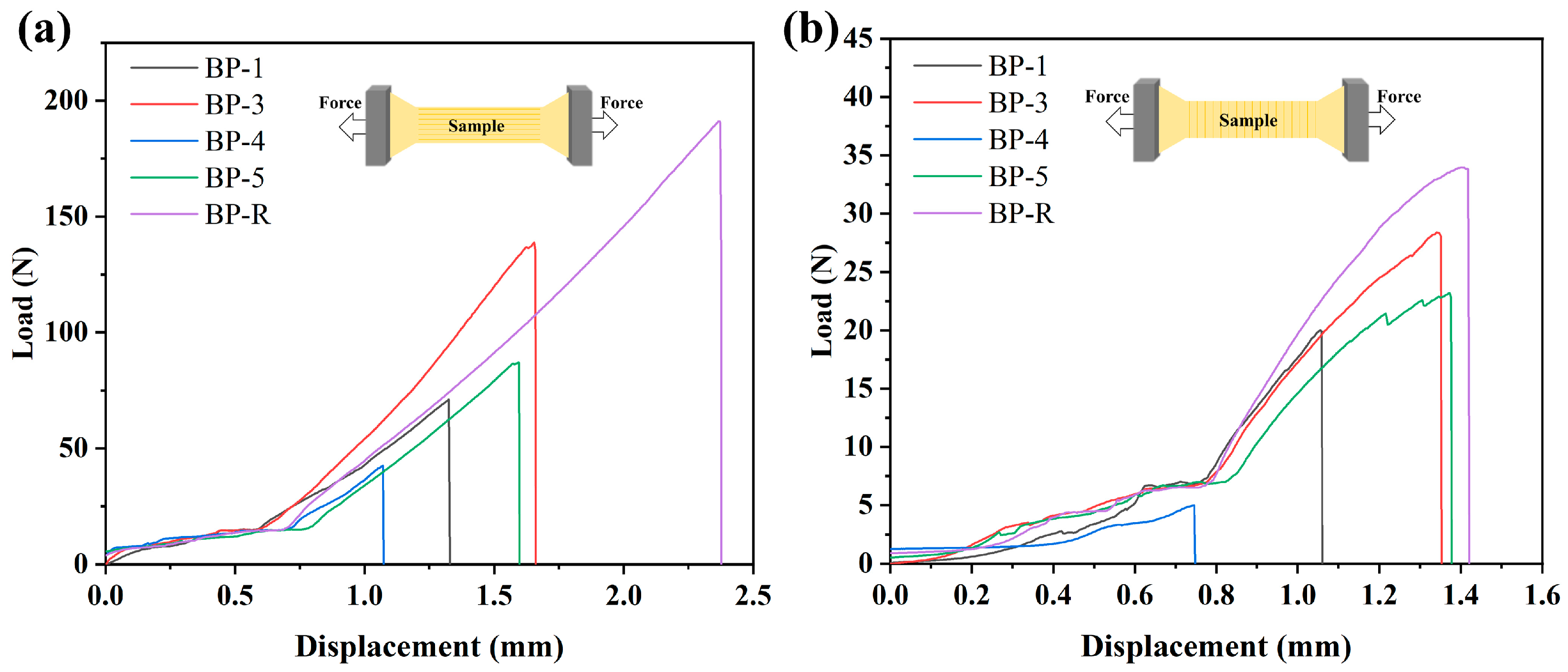
Leaves retain their original structure in a manner unlike paper; as a result, the fibers in the palm leaf manuscripts were not crushed, resulting in varying tensile strengths in different directions. Figure 8 shows the load–displacement curves of longitudinal and transverse tensile tests on palm leaf manuscripts. The tensile strength of the manuscripts is dependent on both fiber length and the bonding force between fibers [38]. In longitudinal tensile tests, BP-R samples exhibited a simulated displacement of 2.37 mm and a force of approximately 190 N (Figure 8a), whereas transverse tensile fracture resulted in a displacement of 1.42 mm and a force of approximately 34 N (Figure 8b). Historical palm leaf manuscripts exhibited lower longitudinal and transverse tensile strengths compared to BP-R, and the mechanical strength decreased as the degradation intensified. Compared with simulated palm leaf manuscripts, the longitudinal tensile strength of the historical palm leaf manuscript BP-4 decreased by 78%, while its transverse tensile strength decreased by 85%. The palm leaf manuscripts experienced an increase in relative lignin content and a reduction in the degree of cellulose polymerization as they degraded, which may be a crucial factor in their decreasing in tensile strength [39]. Longitudinal tensile strength was considerably higher than transverse tensile strength in the historical manuscripts, which can be attributed to the leaf’s anatomical structure. Longitudinal strength exceeding the transverse strength also led to the cracking and expansion of the historical palm manuscripts along the longitudinal direction.
4. Conclusions
The analysis of Burmese historical palm leaf manuscripts in this study may provide insights into the structure and performance of such manuscripts during prolonged aging. Samples of historical palm leaf manuscripts exhibit various problems such as brittleness, incompleteness, fracturing, contamination, and erosion by microorganisms. In highly degraded palm leaf manuscripts, cracking occurred in the secondary cell wall layer, and the direction of crack extension extended from the S3 layer to the S1 layer, while the composite middle layer was unaffected. Microbial erosion caused round or nearly round holes to appear in the S2 layer of the cell wall in the samples observed in this study. Degradation of the hemicellulose and amorphous cellulose in most degraded palm leaf manuscript samples led to an increase in cellulose relative crystallinity. Even the crystalline cellulose region also degraded, resulting in a decrease in cellulose relative crystallinity. Polysaccharide degradation increased the relative content of lignin, contributing to the brittleness of palm leaf manuscripts. The unique anatomical structure of the leaf provides greater tensile strength along the grain than across the grain. However, both longitudinal and transverse tensile strengths of historical palm leaf manuscripts were markedly reduced when compared with simulated palm leaf manuscript samples. Severely degraded historical palm leaf manuscripts exhibited a 78% decrease in longitudinal tensile strength and an 85% decrease in transverse tensile strength compared to their simulated counterparts. With an understanding of the multiscale structure and mechanical properties of historical palm leaf manuscripts, it is possible to determine the preservation and conservation of palm leaf manuscripts.
Author Contributions
Conceptualization, S.C., L.L. and X.T.; methodology, S.C.; software, S.C.; validation, L.L. and X.T.; formal analysis, X.T.; investigation, S.C.; resources, X.T.; data curation, S.C.; writing—original draft preparation, S.C.; writing—review and editing, L.L. and X.T.; visualization, S.C.; supervision, L.L.; project administration, X.T.; funding acquisition, X.T. All authors have read and agreed to the published version of the manuscript.
Funding
This research was funded by the National Key Research and Development Program of China, grant number 2022YFF0903903.
Data Availability Statement
The data presented in this study are available on request from the corresponding author.
Acknowledgments
The authors express their gratitude to Liusan Li, Li Li, Xiangdong Li, and Bingfeng Zhang for their assistance with this study.
Conflicts of Interest
The authors declare no conflict of interest.
References
- Vijaya Lakshmi, T.R.; Sastry, P.N.; Rajinikanth, T.V. Feature selection to recognize text from palm leaf manuscripts. Signal Image Video Process. 2018, 12, 223–229. [Google Scholar] [CrossRef]
- Wang, K.; Xia, W.; Ren, J.; Yu, W.; Feng, H.; Hu, S. Wind energy harvesting inspired by Palm leaf flutter: Observation, mechanism and experiment. Energy Convers. Manag. 2023, 284, 116971. [Google Scholar] [CrossRef]
- Junio, R.F.P.; de Mendonça Neuba, L.; Souza, A.T.; Pereira, A.C.; Nascimento, L.F.C.; Monteiro, S.N. Thermochemical and structural characterization of promising carnauba novel leaf fiber (Copernicia prunifera). J. Mater. Res. Technol. 2022, 18, 4714–4723. [Google Scholar] [CrossRef]
- Abdel-Azim, M.S. Development of prototype structure for low-cost and energy-efficient house by utilizing palm tree fronds. Build. Environ. 1997, 32, 375–380. [Google Scholar] [CrossRef]
- Wiland, J.; Brown, R.; Fuller, L.; Havelock, L.; Johnson, J.; Kenn, D.; Kralka, P.; Muzart, M.; Pollard, J.; Snowdon, J. A literature review of palm leaf manuscript conservation—Part 1: A historic overview, leaf preparation, materials and media, palm leaf manuscripts at the British Library and the common types of damage. J. Inst. Conserv. 2022, 45, 236–259. [Google Scholar] [CrossRef]
- Zhang, M.; Song, X.; Wang, Y. Two Different storage environments for palm leaf manuscripts: Comparison of deterioration phenomena. Restaurator 2021, 42, 147–168. [Google Scholar] [CrossRef]
- Zhang, M.; Song, X.; Wang, J.; Lyu, X. Preservation characteristics and restoration core technology of palm leaf manuscripts in Potala Palace. Arch. Sci. 2022, 22, 501–519. [Google Scholar] [CrossRef]
- Alexander, T.J.; Kumar, S.S. A novel binarization technique based on Whale Optimization Algorithm for better restoration of palm leaf manuscript. J. Ambient. Intell. Humaniz. Comput. 2020, 1–8. [Google Scholar] [CrossRef]
- Subramani, K.; Subramaniam, M. Creation of original Tamil character dataset through segregation of ancient palm leaf manuscripts in medicine. Expert Syst. 2021, 38, e12538. [Google Scholar] [CrossRef]
- Wiland, J.; Brown, R.; Fuller, L.; Havelock, L.; Johnson, J.; Kenn, D.; Kralka, P.; Muzart, M.; Pollard, J.; Snowdon, J. A literature review of palm leaf manuscript conservation—Part 2: Historic and current conservation treatments, boxing and storage, religious and ethical issues, recommendations for best practice. J. Inst. Conserv. 2023, 46, 64–91. [Google Scholar] [CrossRef]
- Singh, M.R.; Sharma, D. Investigation of pigments on an Indian palm leaf manuscript (18th–19th century) by SEM-EDX and other techniques. Restaurator 2020, 41, 49–65. [Google Scholar] [CrossRef]
- Agrawal, O.P. Conservation of Manuscripts and Paintings of South-East Asia; Butterworths: Boston, MA, USA, 1984. [Google Scholar]
- Sah, A. Palm leaf manuscripts of the world: Material, technology and conservation. Stud. Conserv. 2002, 47, 15–24. [Google Scholar] [CrossRef]
- Segal, L.; Creely, J.J.; Martin, A.E.; Conrad, C.M. An empirical method for estimating the degree of crystallinity of native cellulose using the X-Ray diffractometer. Text. Res. J. 1959, 29, 786–794. [Google Scholar] [CrossRef]
- Giraud-Guille, M.M. Plywood structures in nature. Curr. Opin. Solid State Mater. Sci. 1998, 3, 221–227. [Google Scholar] [CrossRef]
- Horn, J.W.; Fisher, J.B.; Tomlinson, P.B.; Lewis, C.E.; Laubengayer, K. Evolution of lamina anatomy in the palm family (Arecaceae). Am. J. Bot. 2009, 96, 1462–1486. [Google Scholar] [CrossRef] [PubMed]
- Schnabel, T.; Zimmer, B.; Petutschnigg, A.J. On the modelling of colour changes of wood surfaces. Eur. J. Wood Wood Prod. 2009, 67, 141–149. [Google Scholar] [CrossRef]
- Shahani, C.J.; Harrison, G. Spontaneous formation of acids in the natural aging of paper. Stud. Conserv. 2002, 47, 189–192. [Google Scholar] [CrossRef]
- Sugano, J.; Linnakoski, R.; Huhtinen, S.; Pappinen, A.; Niemelä, P.; Asiegbu, F.O. Cellulolytic activity of brown-rot Antrodia sinuosa at the initial stage of cellulose degradation. Holzforschung 2019, 73, 673–680. [Google Scholar] [CrossRef]
- Tanabe, J.; Ishiguri, F.; Ohshima, J.; Iizuka, K.; Yokota, S. Changes in lignocellulolytic enzyme activity during the degradation of Picea jezoensis wood by the white-rot fungus Porodaedalea pini. Int. Biodeterior. Biodegrad. 2016, 110, 108–112. [Google Scholar] [CrossRef]
- Mo, W.; Li, B.; Chen, K. Low-temperature thermal drying-induced pore expansion effects of cellulosic fibers. Cellulose 2023, 30, 3441–3453. [Google Scholar] [CrossRef]
- Mvondo, R.R.N.; Meukam, P.; Jeong, J.; Meneses, D.D.S.; Nkeng, E.G. Influence of water content on the mechanical and chemical properties of tropical wood species. Results Phys. 2017, 7, 2096–2103. [Google Scholar] [CrossRef]
- Björdal, C.G. Microbial degradation of waterlogged archaeological wood. J. Cult. Herit. 2012, 13, S118–S122. [Google Scholar] [CrossRef]
- Björdal, C.; Nilsson, T.; Bardage, S. Three-dimensional visualisation of bacterial decay in individual tracheids of Pinus sylvestris. Holzforschung 2005, 59, 178–182. [Google Scholar] [CrossRef]
- Guo, J.; Chen, J.; Meng, Q.; Ploszczanski, L.; Liu, J.A.; Luo, R.; Jin, T.; Siedlaczek, P.; Lichtenegger, H.C.; Yin, Y.; et al. Molecular and crystal structures of cellulose in severely deteriorated archaeological wood. Cellulose 2022, 29, 9549–9568. [Google Scholar] [CrossRef]
- Tang, Z.; Yang, M.; Qiang, M.; Li, X.; Morrell, J.J.; Yao, Y. Preparation of cellulose nanoparticles from foliage by bio-enzyme methods. Materials 2021, 14, 4557. [Google Scholar] [CrossRef]
- Fang, S.; Lyu, X.; Tong, T.; Lim, A.I.; Li, T.; Bao, J.; Hu, Y.H. Turning dead leaves into an active multifunctional material as evaporator, photocatalyst, and bioplastic. Nat. Commun. 2023, 14, 1203. [Google Scholar] [CrossRef] [PubMed]
- Mahdi, A.S.; Abd, A.M.; Awad, K.M. The role of nano-selenium in alleviating the effects of salt stress in date palm trees (Phoenix dactylifera L.): A fourier transform infrared (FTIR) spectroscopy study. BioNanoScience 2023, 13, 74–80. [Google Scholar] [CrossRef]
- Kayabas, A.; Yildirim, E. Biochemical fingerprints of some endemic plants growing in gypsum soils: Attenuated total reflection-Fourier transform infrared (ATR-FTIR) spectroscopic study. Eur. J. Biol. 2021, 80, 97–106. [Google Scholar] [CrossRef]
- Gorgulu, S.T.; Dogan, M.; Severcan, F. The characterization and differentiation of higher plants by Fourier transform infrared spectroscopy. Appl. Spectrosc. 2007, 61, 300–308. [Google Scholar] [CrossRef]
- Ivanova, D.G.; Singh, B.R. Nondestructive FTIR monitoring of leaf senescence and elicitin-induced changes in plant leaves. Biopolym. Orig. Res. Biomol. 2003, 72, 79–85. [Google Scholar] [CrossRef]
- Wang, Z.; Xu, E.; Fu, F.; Lin, L.; Yi, S. Characterization of wood cell walls treated by high-intensity microwaves: Effects on physicochemical structures and micromechanical properties. Ind. Crops Prod. 2022, 187, 115341. [Google Scholar] [CrossRef]
- Fernandes, A.N.; Thomas, L.H.; Altaner, C.M.; Callow, P.; Forsyth, V.T.; Apperley, D.C.; Kennedy, C.J.; Jarvis, M.C. Nanostructure of cellulose microfibrils in spruce wood. Proc. Natl. Acad. Sci. USA 2011, 108, E1195–E1203. [Google Scholar] [CrossRef] [PubMed]
- Zhu, Y.; Li, W.; Meng, D.; Li, X.; Goodell, B. Non-enzymatic modification of the crystalline structure and chemistry of Masson pine in brown-rot decay. Carbohydr. Polym. 2022, 286, 119242. [Google Scholar] [CrossRef] [PubMed]
- Lin, Q.; Huang, Y.; Yu, W. An in-depth study of molecular and supramolecular structures of bamboo cellulose upon heat treatment. Carbohydr. Polym. 2020, 241, 116412. [Google Scholar] [CrossRef] [PubMed]
- Zhang, J.; Li, Y.; Ke, D.; Wang, C.; Pan, H.; Chen, K.; Zhang, H. Modified lignin nanoparticles as potential conservation materials for waterlogged archaeological wood. ACS Appl. Nano Mater. 2023, 6, 12351–12363. [Google Scholar] [CrossRef]
- Guo, J.; Xiao, L.; Han, L.; Wu, H.; Yang, T.; Wu, S.; Yin, Y. Deterioration of the cell wall in waterlogged wooden archeological artifacts, 2400 years old. IAWA J. 2019, 40, 820–844. [Google Scholar] [CrossRef]
- Volkmer, T.; Arietano, L.; Plummer, C.; Strautmann, J.; Noël, M. Loss of tensile strength in cellulose tissue on the surface of spruce (Picea abies) caused by natural photodegradation and delignification. Polym. Degrad. Stab. 2013, 98, 1118–1125. [Google Scholar] [CrossRef]
- Zhai, S.; Horikawa, Y.; Imai, T.; Sugiyama, J. Anatomical and mechanical characteristics of leaf-sheath fibrovascular bundles in palms. IAWA J. 2013, 34, 285–300. [Google Scholar] [CrossRef]
Figure 1.
Historical palm leaf manuscripts and simulated palm leaf manuscripts.

Figure 2.
Optical microscopy of (a) upper, (b) lower epidermises and (c) transverse section of palm leaf.
Figure 2.
Optical microscopy of (a) upper, (b) lower epidermises and (c) transverse section of palm leaf.
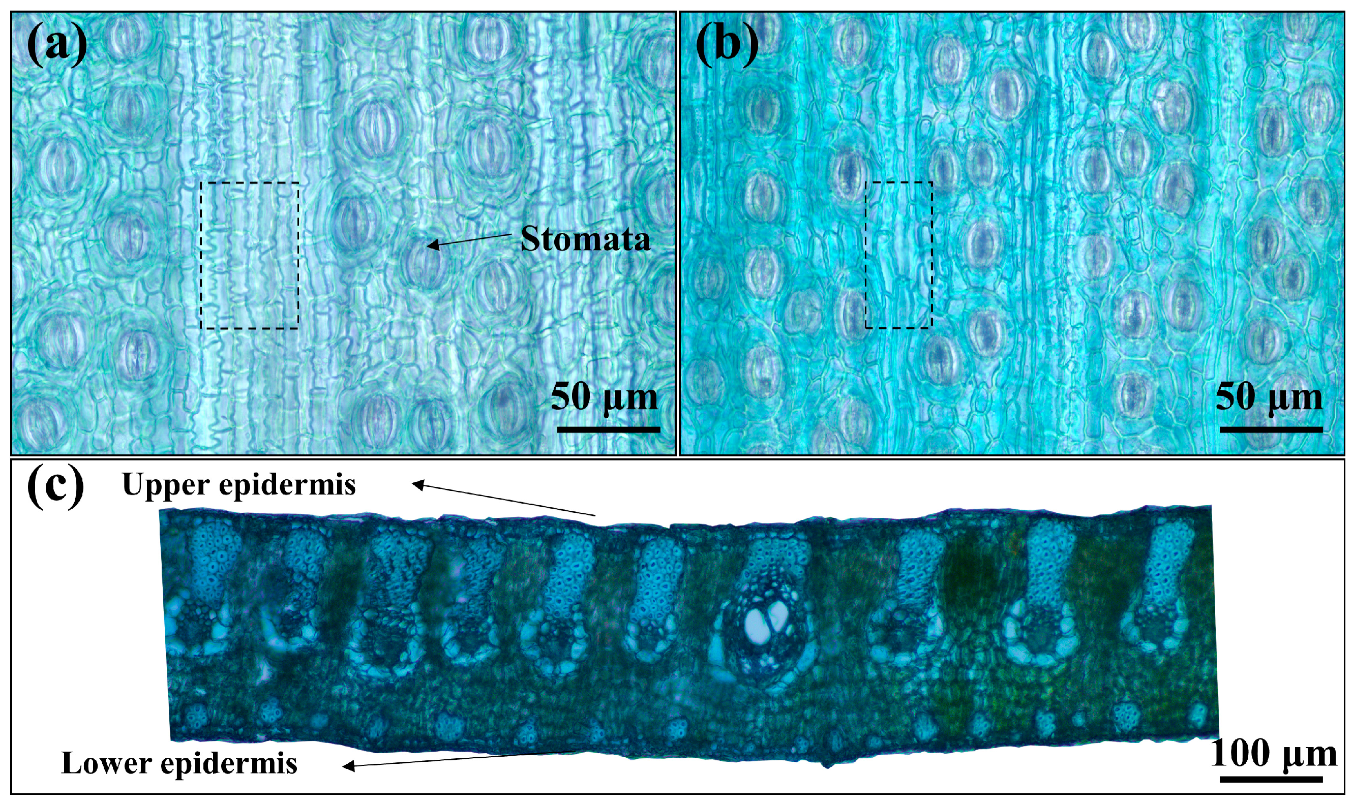
Figure 3.
Diseases on surfaces of historical palm leaf manuscripts: (a) BP-1, (b) BP-2, (c) BP-3, (d) BP-4.
Figure 3.
Diseases on surfaces of historical palm leaf manuscripts: (a) BP-1, (b) BP-2, (c) BP-3, (d) BP-4.
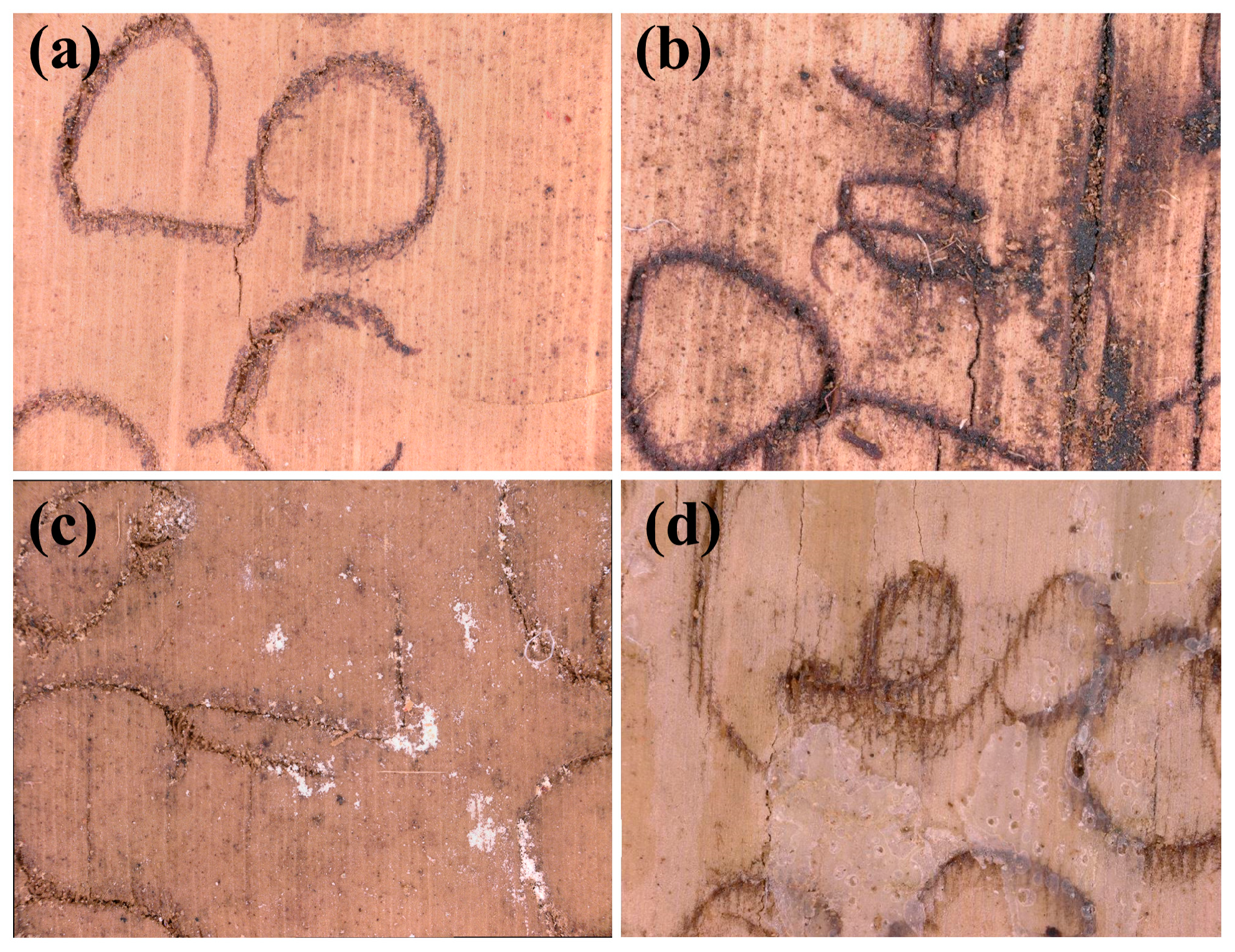
Figure 4.
SEM images of historical palm leaf manuscripts: (a) cracks in BP-1 manuscripts, (b) ink in BP-1 manuscripts, (c,f) surface of BP-2 manuscripts before and after alcohol wiping, (d,e) exfoliation of BP-3 manuscripts surface, (g,h) white powdery substance in BP-3 manuscripts, (i) EDX images of white powdery substance.
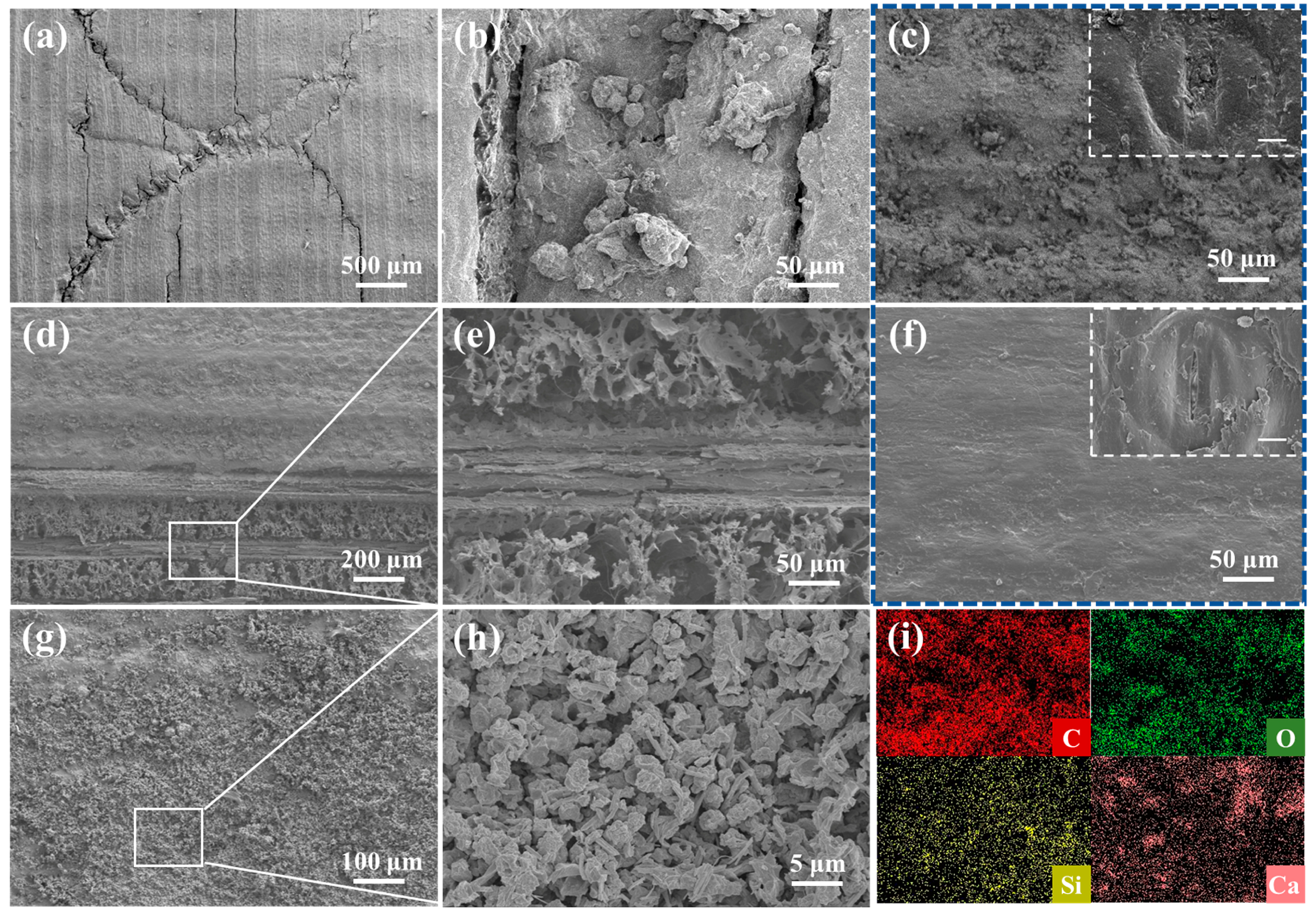
Figure 5.
Cross-sectional micromorphology of palm leaf manuscripts: (a,d) BP-R, (b,e) BP-1, (c,f) BP-3.
Figure 5.
Cross-sectional micromorphology of palm leaf manuscripts: (a,d) BP-R, (b,e) BP-1, (c,f) BP-3.
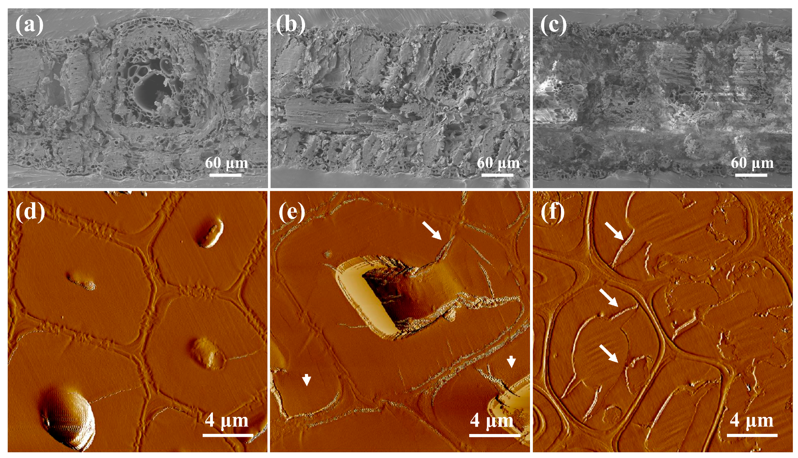
Figure 6.
ATR–FTIR spectra between 4000 cm-1 and 500 cm-1 of (a) palm leaf manuscripts and (b) membranous substance.
Figure 6.
ATR–FTIR spectra between 4000 cm-1 and 500 cm-1 of (a) palm leaf manuscripts and (b) membranous substance.
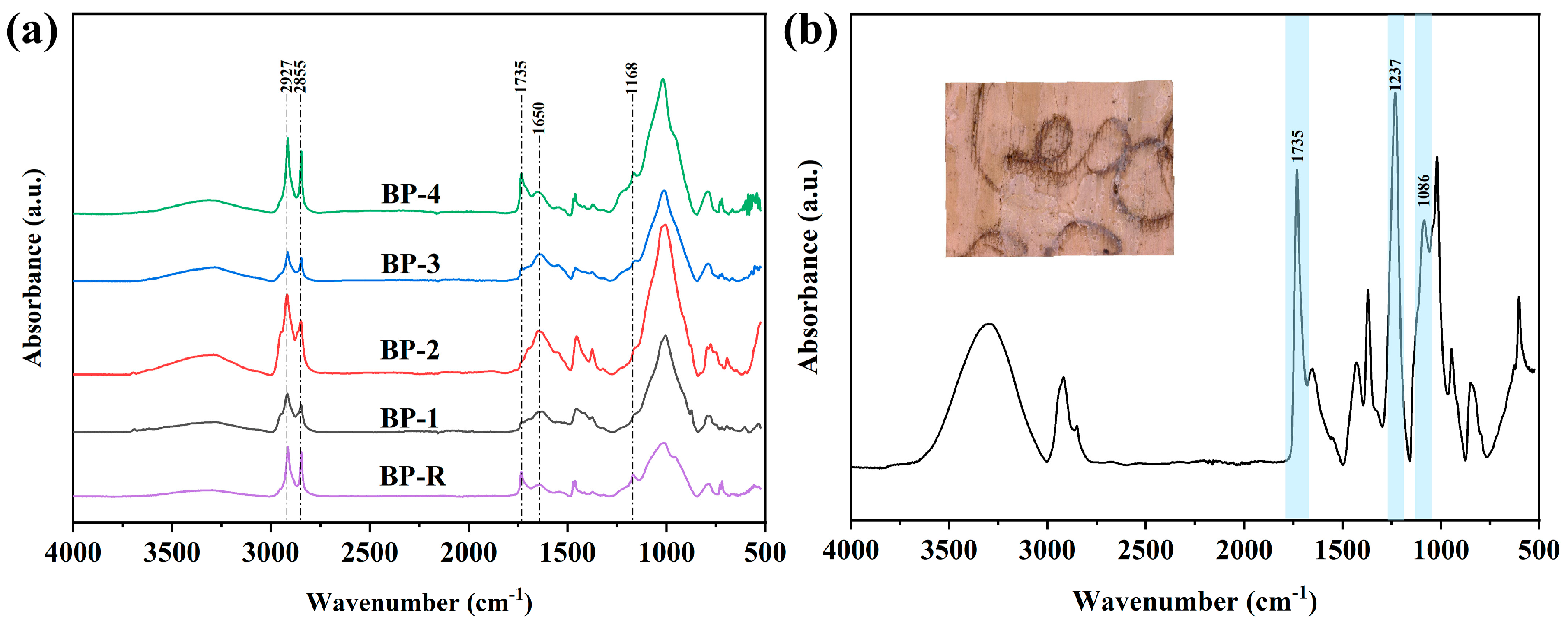
Figure 7.
Cellulose crystalline structures of historical palm leaf manuscripts and control samples: (a) X-ray diffractograms, (b) relative crystallinities.
Figure 7.
Cellulose crystalline structures of historical palm leaf manuscripts and control samples: (a) X-ray diffractograms, (b) relative crystallinities.
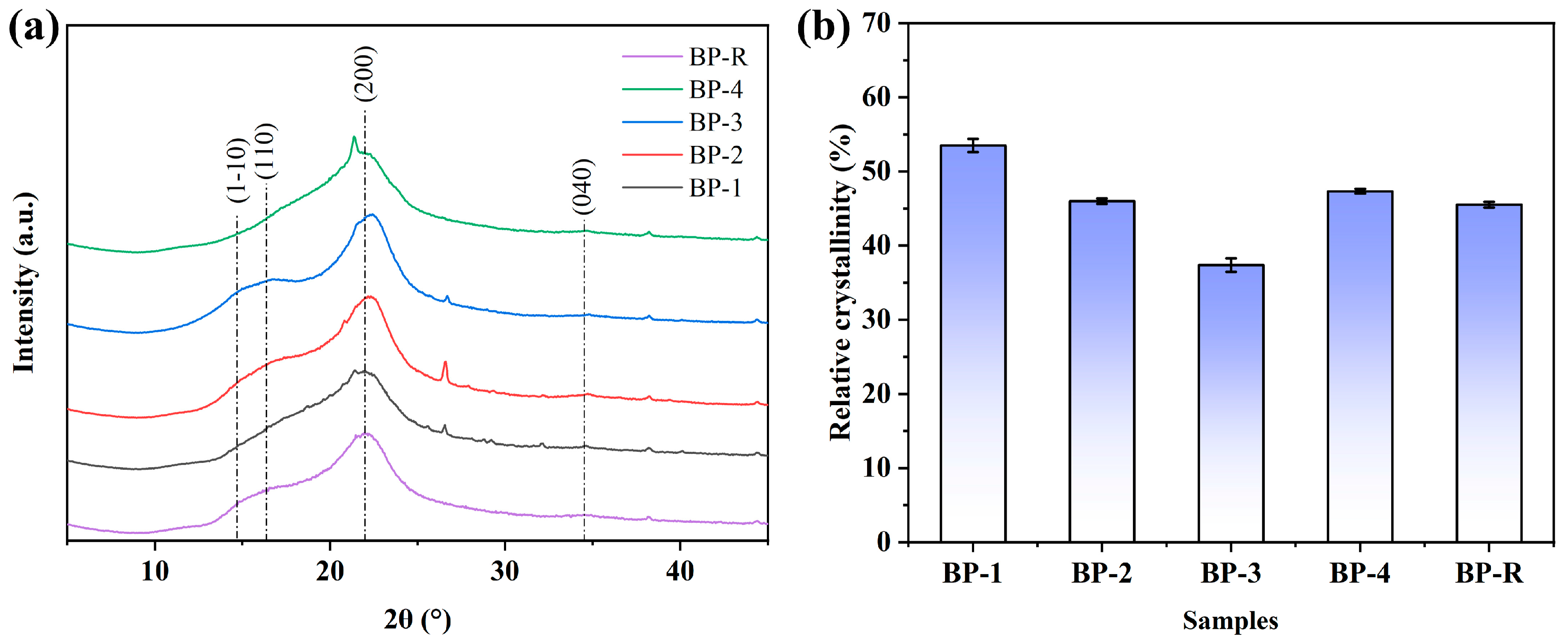
Figure 8.
The load–displacement curves of the longitudinal (a) and transverse (b) tensile tests of palm leaf manuscripts.
Figure 8.
The load–displacement curves of the longitudinal (a) and transverse (b) tensile tests of palm leaf manuscripts.

Table 1.
Basic physical parameters of historical palm leaf manuscripts and control samples.
| No. | Color Parameters | pH | Moisture Content (%) | ||
|---|---|---|---|---|---|
| L* | a* | b* | |||
| BP-1 | 44.46 (±1.30) | 10.10 (±0.64) | 24.12 (±0.87) | 4.95 (±0.21) | 8.13 (±0.30) |
| BP-2 | 38.59 (±2.67) | 8.23 (±0.84) | 18.36 (±2.33) | 5.96 (±0.28) | 10.20 (±0.43) |
| BP-3 | 43.91 (±0.86) | 9.63 (±0.17) | 24.03 (±0.75) | 4.85 (±0.25) | 7.13 (±0.25) |
| BP-4 | 56.86 (±2.25) | 5.58 (±0.74) | 24.39 (±1.25) | 5.81 (±0.33) | 9.00 (±0.16) |
| BP-R | 70.52 (±0.55) | 0.91 (±0.31) | 17.96 (±0.50) | 6.36 (±0.04) | 8.87 (±0.21) |
Disclaimer/Publisher’s Note: The statements, opinions and data contained in all publications are solely those of the individual author(s) and contributor(s) and not of MDPI and/or the editor(s). MDPI and/or the editor(s) disclaim responsibility for any injury to people or property resulting from any ideas, methods, instructions or products referred to in the content. |
© 2023 by the authors. Licensee MDPI, Basel, Switzerland. This article is an open access article distributed under the terms and conditions of the Creative Commons Attribution (CC BY) license (https://creativecommons.org/licenses/by/4.0/).
Share and Cite
MDPI and ACS Style
Chu, S.; Lin, L.; Tian, X. Evaluation of the Deterioration State of Historical Palm Leaf Manuscripts from Burma. Forests 2023, 14, 1775. https://doi.org/10.3390/f14091775
AMA Style
Chu S, Lin L, Tian X. Evaluation of the Deterioration State of Historical Palm Leaf Manuscripts from Burma. Forests. 2023; 14(9):1775. https://doi.org/10.3390/f14091775
Chicago/Turabian StyleChu, Shimin, Lanying Lin, and Xingling Tian. 2023. "Evaluation of the Deterioration State of Historical Palm Leaf Manuscripts from Burma" Forests 14, no. 9: 1775. https://doi.org/10.3390/f14091775
Note that from the first issue of 2016, this journal uses article numbers instead of page numbers. See further details here.