Short Communication

Molecular Phylogeny of Hantaviruses Harbored by Insectivorous Bats in Côte d’Ivoire and Vietnam

1
Pacific Center for Emerging Infectious Diseases Research, John A. Burns School of Medicine, University of Hawaii at Manoa, Honolulu, HI 96813, USA
2
Department of Natural History, Royal Ontario Museum, Toronto, ON M5S 2C6, Canada
3
Department of Biology, Université de Cocody, Abidjan 22, Côte d'Ivoire
4
Infectious Disease Surveillance Center, National Institute of Infectious Diseases, Tokyo 162-8640, Japan
5
Department of Microbiology, College of Medicine, Korea University, Seoul 136-705, Korea
6
Departement Systematique et Evolution, UMR CNRS 7205, Muséum National d'Histoire Naturelle, Paris 75005, France
7
Department of Biology, Museum of Southwestern Biology, University of New Mexico, Albuquerque, NM 87131, USA
8
Department of Pediatrics, School of Medicine, University of Colorado, Aurora, CO 80045, USA
9
National Center for Disease Control and Public Health, Tbilisi 0177, Georgia
10
Institute of Chemical Biology, Ilia State University, Tbilisi 0162, Georgia
11
Global Alliance for Rabies Control, Manhattan, KS 66502, USA
12
Division of Vector Borne Diseases, Centers for Disease Control and Prevention, Fort Collins, CO 80521, USA
*
Author to whom correspondence should be addressed.
Viruses 2014, 6(5), 1897-1910; https://doi.org/10.3390/v6051897
Submission received: 24 February 2014 / Revised: 4 April 2014 / Accepted: 8 April 2014 / Published: 29 April 2014
(This article belongs to the Special Issue Viruses and Bats)

Abstract

:
The recent discovery of genetically distinct hantaviruses in multiple species of shrews and moles prompted a further exploration of their host diversification by analyzing frozen, ethanol-fixed and RNAlater®-preserved archival tissues and fecal samples from 533 bats (representing seven families, 28 genera and 53 species in the order Chiroptera), captured in Asia, Africa and the Americas in 1981–2012, using RT-PCR. Hantavirus RNA was detected in Pomona roundleaf bats (Hipposideros pomona) (family Hipposideridae), captured in Vietnam in 1997 and 1999, and in banana pipistrelles (Neoromicia nanus) (family Vespertilionidae), captured in Côte d’Ivoire in 2011. Phylogenetic analysis, based on the full-length S- and partial M- and L-segment sequences using maximum likelihood and Bayesian methods, demonstrated that the newfound hantaviruses formed highly divergent lineages, comprising other recently recognized bat-borne hantaviruses in Sierra Leone and China. The detection of bat-associated hantaviruses opens a new era in hantavirology and provides insights into their evolutionary origins.

1. Introduction

Hantaviruses (genus Hantavirus, family Bunyaviridae) possess a negative-sense, single-stranded, tripartite segmented RNA genome, consisting of large (L), medium (M) and small (S) segments, encoding an RNA-dependent RNA polymerase (RdRp), envelope glycoproteins (Gn and Gc) and a nucleocapsid (N) protein, respectively [1]. To date, 23 hantaviruses, hosted by reservoir rodent species, have been recognized as distinct species by the International Committee on Taxonomy of Viruses [2]. Several of these rodent-borne hantaviruses cause acute, febrile diseases of varying clinical severity and lethality in humans, known as hemorrhagic fever with renal syndrome and hantavirus cardiopulmonary syndrome [3]. Though once believed to be restricted to rodents (order Rodentia, family Muridae and Cricetidae), the reservoir host range of hantaviruses is far more expansive, as evidenced by the detection of divergent lineages of hantaviruses in multiple species of shrews and moles (order Soricomorpha, family Soricidae and Talpidae) throughout Asia, Europe, Africa and North America [4,5,6,7,8,9,10,11,12,13,14,15,16,17,18,19].
Despite their phylogenetic relatedness to the European mole (Talpa europaea) within the Laurasiatheria [20,21], as well as their rich genetic diversity, vast geographic range and ability to host many disease-causing viruses [22,23,24], bats (order Chiroptera) have not been extensively studied as potential reservoirs of hantaviruses. Although serological evidence of hantavirus infection was reported in the common serotine (Eptesicus serotinus) and greater horseshoe bat (Rhinolophus ferrumequinum) captured in Korea [25], genetic analysis of hantavirus isolates from these bat species suggested laboratory contamination [26].
The genetic diversity of newfound hantaviruses recently detected in insectivorous bats preclude any possibility of contamination: Mouyassué virus (MOYV) in the banana pipistrelle (Neoromicia nanus) from Côte d’Ivoire [27]; Magboi virus (MGBV) in the hairy slit-faced bat (Nycteris hispida) from Sierra Leone [28]; Xuan Son virus (XSV) in the Pomona roundleaf bat (Hipposideros pomona) from Vietnam [29]; Huangpi virus (HUPV) in the Japanese house bat (Pipistrellus abramus) and Longquan virus (LQUV) in the Chinese horseshoe bat (Rhinolophus sinicus), Formosan lesser horseshoe bat (Rhinolophus monoceros) and intermediate horseshoe bat (Rhinolophus affinis) from China [30]. The primary goal of this multi-national collaborative study was to extend the search for hantaviruses in bats and to obtain more of the MOYV and XSV genomes. Our data indicate that bat-borne hantaviruses and Nova virus, a hantavirus hosted by the European mole, comprise a highly divergent phylogenetic lineage, suggesting that ancestral bats and/or soricomorphs, rather than rodents, may have served as the early reservoir hosts of primordial hantaviruses.

2. Results and Discussion

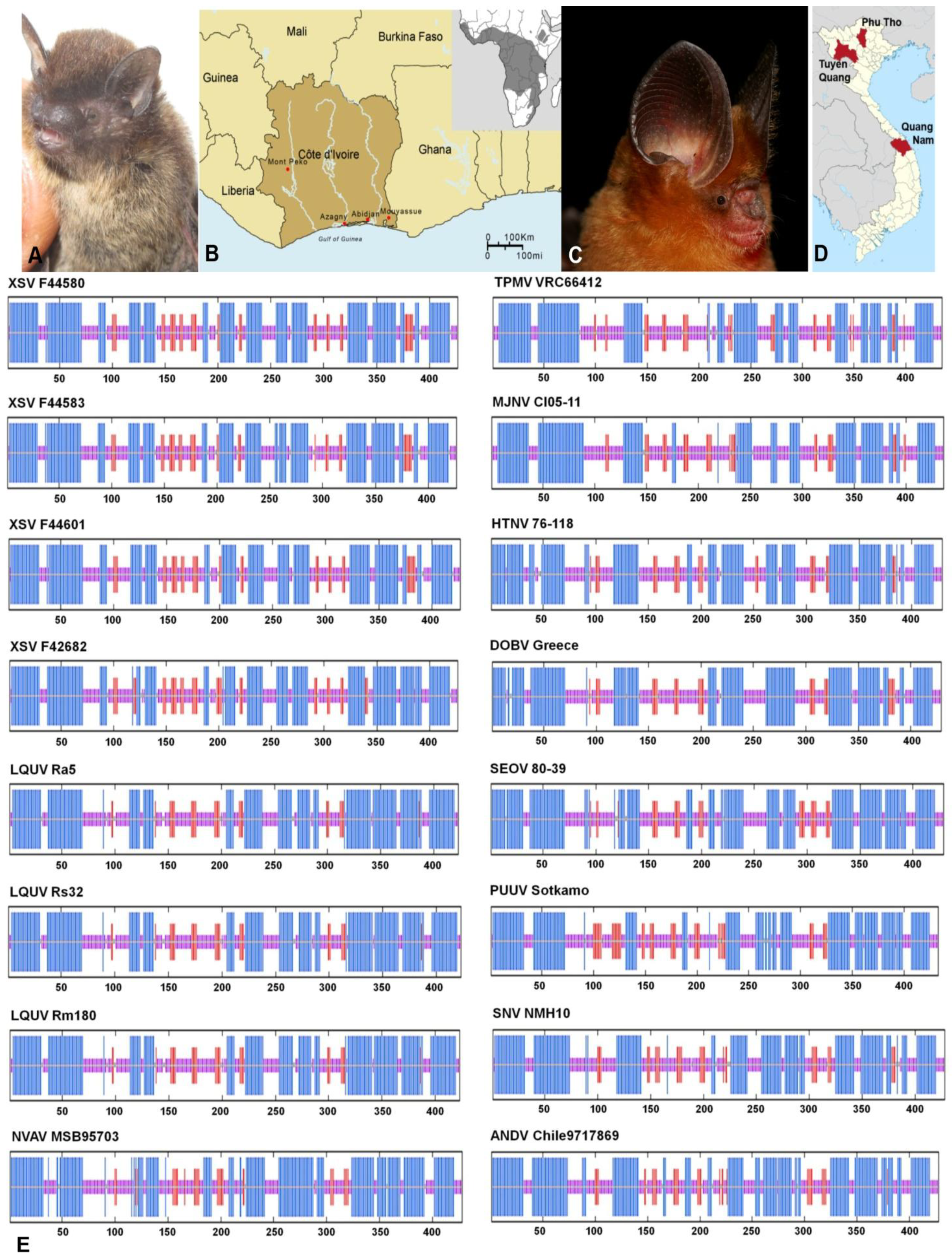
2.1. Hantavirus Detection and Sequence Analysis

Exhaustive attempts to detect hantaviruses were unsuccessful in nearly all of the 454 bat tissue samples (Table 1 and Figure 1), despite employing oligonucleotide primers and PCR cycling conditions used to find MOYV [27] and XSV [29]. In addition, hantavirus RNA was not detected in any of the 79 rectal swab and fecal samples. Because LQUV was previously found in four species of horseshoe bats in China [30], we expected to find the same or a similar hantavirus in the greater horseshoe bat, captured on Jeju Island in Korea. However, this was not the case, in spite of using LQUV-specific primers. Nevertheless, we did manage to obtain more of the MOYV and XSV genomes. That is, the original report of MOYV in the banana pipistrelle (Figure 2A,B) was based on a 423-nucleotide region of the L segment [27]. Through repeated trial-and-error efforts, suitable primers were designed to obtain an additional 1268 nucleotides of the L segment (Table 2).
In addition, Arai and colleagues previously reported a novel hantavirus, designated XSV, in one of five Pomona roundleaf bats, captured during July 2012 in Xuan Son National Park in Phu Tho province in northern Vietnam [29]. In analyzing archival kidney tissues from 44 Pomona roundleaf bats trapped in Tuyên Quang and Quang Nam provinces, hantavirus L-segment sequences were detected in five animals (Figure 2C,D). Although a 15.7%–19.2% difference was found at the nucleotide level with prototype XSV, the high amino acid sequence similarity was consistent with these sequences representing genetic variants of XSV. Pair-wise alignment and comparison of the full-length S segment of XSV, amplified and sequenced from four bats (Table 2), indicated sequence similarity of 58.9%–60.3% at the amino acid level with LQUV, the only other bat-borne hantavirus for which the entire S segment has been sequenced. And sequence analysis of a 663-nucleotide (221 amino acid) region of the Gc envelope glycoprotein-encoding M segment showed that XSV differed by >45% from representative hantaviruses harbored by rodents and most soricomorphs. Collectively, the high level of sequence divergence in the N protein and Gc glycoprotein between XSV and other hantaviruses suggests that it might represent a new hantavirus species, using the guidelines proposed by Maes and co-workers [31]. However, the definitive taxonomic classification of XSV and other bat-borne hantaviruses must await their isolation in cell culture.
Table 1. Specimen types analyzed for hantavirus RNA.
Bat FamilyFrozenRNAlater®Ethanol-fixedTotal
LungLiverKidneyLungIntercostal MuscleIntestineRectal Swab or FecesLiver
Hipposideridae 507 57
Molossidae1 35 6 42
Nycteridae 11 2
Pteropodidae 4218 60
Phyllostomidae2 2
Rhinolophidae150 12 162
Vespertilionidae1117149 457312208
Total164175114619457912533
Table 2. Xuan Son virus and Mouyassué virus in insectivorous bats.
VirusStrainBat SpeciesCountryProvinceSML
XSVVN1982Hipposideros pomonaVietnamPhu Tho499 bp 4582 bp
KC688335JX912953
F42640Tuyên Quang516 bp 567 bp
KF704708 KF704713
F42682 1752 bp663 bp1160 bp
KF704709KJ000538KF704714
F44580Quang Nam1728 bp 804 bp
KF704710KF704715
F44583 1728 bp 1160 bp
KF704711 KF704716
F44601 1728 bp663 bp1160 bp
KF704712KJ000539KF704717
MOYVKB576Neoromicia nanusCôte d'IvoireMouyassué 1691 bp
JQ287716
KB577 372 bp
KJ000540
Figure 1. Geographic origin of 533 specimens from bats, belonging to seven families, were analyzed for hantavirus RNA, using RT-PCR. The number of samples and genera and species of bats are shown for each country.
Viruses 06 01897 g001
Figure 2. (A) Banana pipistrelle (Neoromicia nanus); (B) Map of Cote d’Ivoire, showing site where Mouyassué virus-infected banana pipistrelles were captured during June 2011; inset shows geographic distribution of banana pipistrelle; (C) Pomona roundleaf bat (Hipposideros pomona); (D) Map of Vietnam, showing Phu Tho, where Xuan Son virus (XSV) was first discovered, and Tuyên Quang and Quang Nam, where Pomona roundleaf bats were captured in May 1997 and March 1999, respectively; (E) Comparison of the consensus secondary structures of the nucleocapsid protein of XSV, Longquan virus (LQUV), Nova virus (NVAV), Thottapalayam virus (TPMV), Imjin virus (MJNV), Hantaan virus (HTNV), Dobrava virus (DOBV), Seoul virus (SEOV), Puumala virus (PUUV), Sin Nombre virus (SNV) and Andes virus (ANDV), as predicted using methods available on the NPS@ structure server [32]. Alpha helices are represented by blue bars, beta strands by red bars, and random coils and unclassified structures by magenta and gray bars, respectively.
Viruses 06 01897 g002

2.2. Nucleocapsid Secondary Structure

In employing software available on the @NPS structure server [32], the overall predicted secondary structures of the N proteins were similar. That is, despite the relatively low amino acid sequence similarity among the rodent-, shrew-, mole- and bat-borne hantaviruses, the N protein comprised two major α-helical domains packed against a central β-pleated sheet (Figure 2E). However, the central β-pleated sheet motif of XSV, including the RNA-binding region (amino acid positions 175 to 217), was unlike that of other hantaviruses, even that of LQUV, which more closely resembled murid rodent-borne hantaviruses, such as Hantaan virus (HTNV 76-118), Dobrava virus (DOBV Greece) and Seoul virus (SEOV 80-39) (Figure 2E). The distinctive α-helix motif between two β-strands of the RNA-binding region, observed in the prototype mole-borne hantavirus, Nova virus (NVAV MSB95703), as well as HTNV and SEOV, but not in LQUV, may have a significant effect on binding specificity.

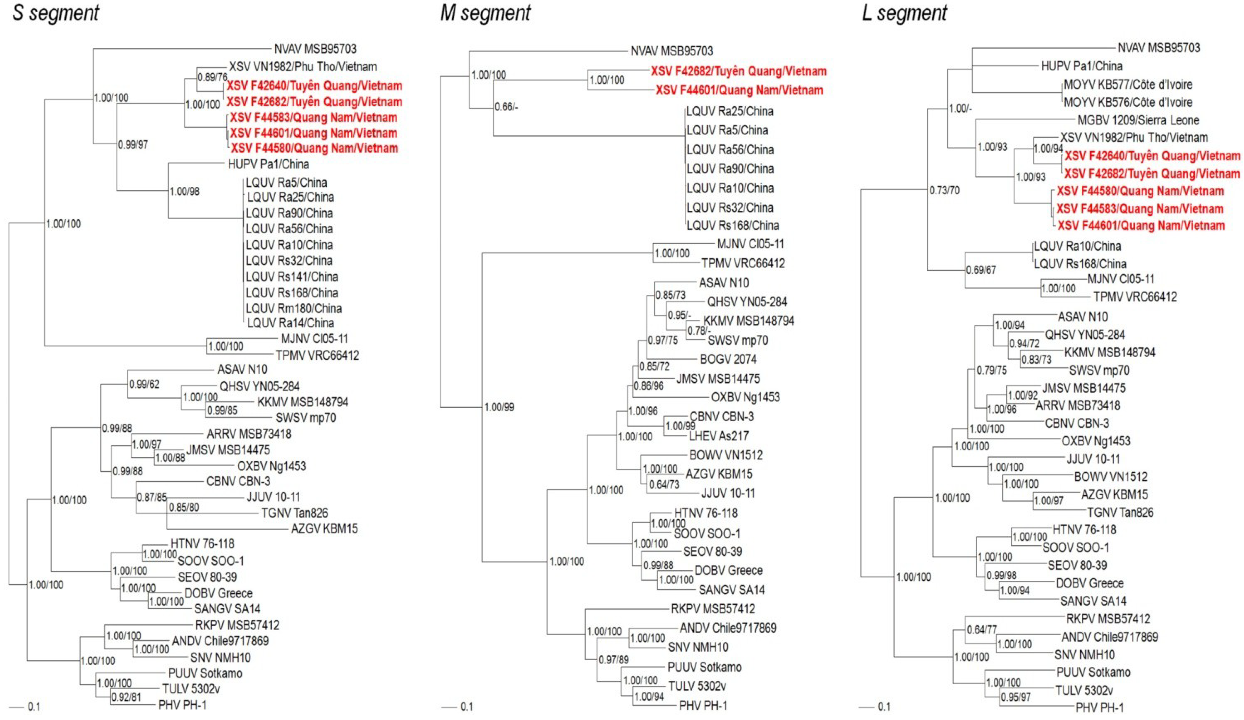
2.3. Phylogenetic Analysis

Phylogenetic analyses, based on S-, M- and L-genomic sequences, indicated that XSV and MOYV shared a common ancestry with other bat-borne hantaviruses (Figure 3). In all analyses, NVAV from the European mole segregated with the bat-associated hantaviruses, which was reminiscent of trees based on the complete mitochondrial genomes of the European mole and bats [20,21]. The basal position of chiropteran-borne hantaviruses and selected soricomorph-borne hantaviruses, such as Nova virus in the European mole, Thottapalayam virus in the Asian house shrew and Imjin virus in the Ussuri white-toothed shrew, in phylogenetic trees based on the S- and L-genomic sequences suggests that soricomorphs and/or chiropterans, rather than rodents, may have been the primordial mammalian hosts of ancestral hantaviruses (Figure 3). Geographic-specific clustering was evidenced by the close phylogenetic relationship between prototype XSV VN1982 from Phu Tho province and XSV F42640 and XSV F42682 from neighboring Tuyên Quang province in northern Vietnam. On the other hand, XSV F44583, XSV 44601 and XSV 44580 from Quang Nam province in central Vietnam clustered together. Although limited differences were present in phylogenetic trees based on each segment, tree topologies were generally congruent and supported by significant bootstrap values (>70%) and posterior node probabilities (>0.70).
Figure 3. Phylogenetic trees were generated by maximum-likelihood and Bayesian methods, using the GTR+I+Γ model of evolution, based on the S-, M- and L-genomic sequences of hantavirus strains. Because tree topologies were nearly identical using RAxML and MrBayes programs, the trees generated by MrBayes were displayed. The evolutionary relationships between Xuan Son virus (XSV), Mouyassué virus (MOYV) and other bat-borne hantaviruses, including Magboi virus (MGBV), Longquan virus (LQUV)and Huangpi virus (HUPV), are shown, as are representative soricomorph-borne hantaviruses, including Nova virus (NVAV MSB95703, S: FJ539168; M: HQ840957; L: FJ593498), Thottapalayam virus (TPMV VRC66412, S: AY526097; M: EU001329; L: EU001330), Imjin virus (MJNV Cl05-11, S: EF641804; M: EF641798; L: EF641806), Seewis virus (SWSV mp70, S: EF636024; M: EF636025; L: EF636026), Kenkeme virus (KKMV MSB148794, S: GQ306148, M: GQ306149; L: GQ306150), Lianghe virus (LHEV As217, M: JX465406), Boginia virus (BOGV 2074, M: JX990966), Cao Bang virus (CBNV CBN-3, S: EF543524; M: EF543526; L: EF543525), Ash River virus (ARRV MSB 73418, S: EF650086; L: EF619961), Jemez Springs virus (JMSV MSB144475, S: FJ593499; M: FJ593500; L: FJ593501), Qian Hu Shan virus (QHSV YN05-284, S: GU566023; M: GU566022; L: GU566021), Tanganya virus (TGNV Tan826, S: EF050455; L: EF050454), Azagny virus (AZGV KBM15, S: JF276226; M: JF276227; L: JF276228), Jeju virus (JJUV 10-11, S: HQ834695; M: HQ834696; L: HQ834697), Bowé virus (BOWV VN1512, M: KC631783; L: KC631784), Asama virus (ASAV N10, S: EU929072; M: EU929075; L: EU929078), Oxbow virus (OXBV Ng1453, S: FJ5339166; M: FJ539167; L: FJ593497) and Rockport virus (RKPV MSB57412, S: HM015223; M: HM015219; L: HM015221). Also shown are the phylogenetic positions of representative rodent-borne hantaviruses, including Hantaan virus (HTNV 76-118, S: NC_005218; M: Y00386; L: NC_005222), Soochong virus (SOOV SOO-1, S: AY675349; M: AY675353; L: DQ056292), Dobrava virus (DOBV Greece, S: NC_005233; M: NC_005234L: NC_005235), Seoul virus (SEOV 80-39, S: NC_005236; M: NC_005237; L: NC_005238), Sangassou virus (SANG SA14, S: JQ082300; M: JQ082301; L: JQ082302),Tula virus (TULV M5302v, S: NC_005227; M: NC_005228; L: NC_005226), Puumala virus (PUUV Sotkamo, S: NC_005224; M: NC_005223; L: NC_005225), Prospect Hill virus (PHV PH-1, S: Z49098; M: X55129; L: EF646763), Sin Nombre virus (SNV NMH10, S: NC_005216; M: NC_005215; L: NC_005217) and Andes virus (ANDV Chile9717869, S: NC_003466; M: NC_003467; L: NC_003468). The numbers at each node are posterior node probabilities (left) based on 150,000 trees and bootstrap values (right) based 1000 replicates executed on the RAxML BlackBox web server, respectively. The scale bars indicate nucleotide substitutions per site.
Viruses 06 01897 g003

2.4. Bats as Hosts of Hantaviruses

The phylogeny of bats is not fully resolved [21]. The order Chiroptera was traditionally divided in two suborders, Megachiroptera and Microchiroptera. However, due to the paraphyly of the Microchiroptera, a new taxonomic nomenclature, comprising the suborder Yinpterochiroptera (megabats or fruit bats in the Pteropodidae family in Megachiroptera and a few Microchiroptera families) and Yangochiroptera (the remaining Microchiroptera families), has been proposed [33]. In the former classification, bat species hosting hantaviruses belong only to the Microchiroptera suborder, but in the Yinpterochiroptera-Yangochiroptera classification, they belong to both suborders, suggesting that primordial hantaviruses may have emerged in an early common ancestor of bats.
Within the Microchiroptera, hantaviruses are found in bats belonging to four phylogenetically distant families, namely Hipposideridae (Old World leaf-nosed bats) and Rhinolophidae (horseshoe bats) in the suborder Yinpterochiroptera, and Nycteridae (hollow-faced bats) and Vespertilionidae (vesper bats) in the suborder Yangochiroptera. The families Hipposideridae and Vespertilionidae are among the most speciose insectivorous bats, with member species distributed across Africa, Europe, Asia, the Americas and Australia. Their vast geographic distribution provides unlimited opportunities to search for related bat-associated hantaviruses.
Compared to the multitude of hantaviruses reported from approximately 50% of soricomorph species tested [34,35], the cumulative number of newly recognized bat-borne hantaviruses is exceedingly low, if one considers the 533 bat samples tested in the present study, along with the nearly 1200 bat specimens analyzed in four other studies [27,28,29,30]. The modest proportion of hantavirus RNA detection in bat tissues may be attributed to the highly divergent nature of their genomes, as well as the very focal or localized nature of hantavirus infection, small sample sizes of bat species, primer mismatches, suboptimal PCR cycling conditions, and variable tissue preservation with degraded RNA [27,29]. Alternatively, bats may be less susceptible to hantavirus infection or may have developed immune mechanisms to curtail viral replication and/or persistence. For answers to such questions, and myriad others, reagents need to be developed and multidisciplinary collaborative studies must be designed to collect optimal specimens to isolate and characterize these newfound bat-borne hantaviruses. Only then will a better understanding be gained about their evolutionary origins and phylogeography, co-evolution history, transmission dynamics and pathogenic potential.

3. Experimental Section
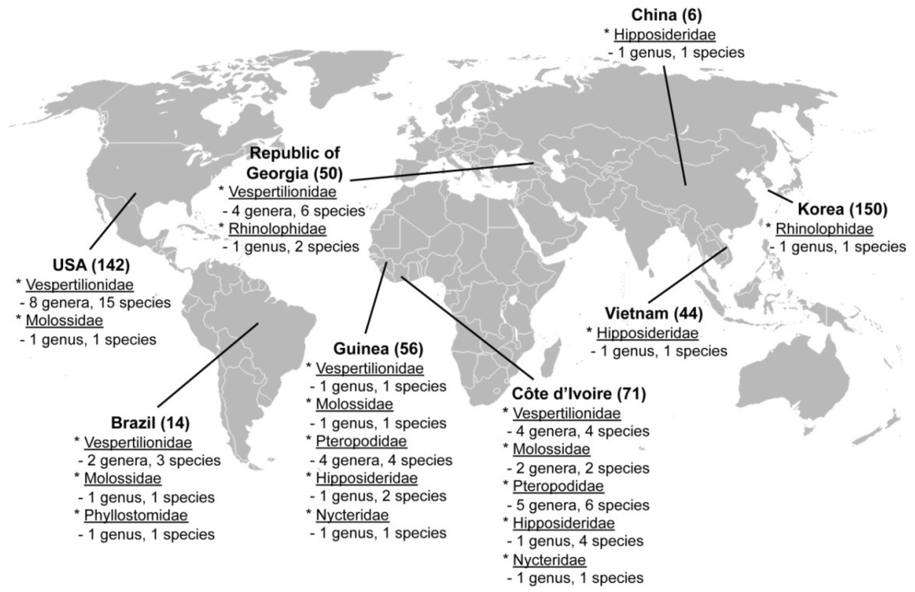
3.1. Samples

Archival frozen, ethanol-fixed and RNAlater®-preserved tissues from bats, captured during 1981–2012 in Brazil, China, Cote d’Ivoire, Guinea, Korea, Republic of Georgia, Vietnam and the United States (Figure 1 and Table 1), were tested for hantavirus RNA by RT-PCR, using newly designed and previously employed oligonucleotide primers [12,18,27,29]. Of the 533 samples tested, the majority consisted of lung (310) and kidney (51) tissues (Table 1). RNA extracted from rectal swabs and feces (79) were also tested. Bats were from seven families (Hipposideridae, Molossidae, Nycteridae, Pteropodidae, Phyllostomidae, Rhinolophidae and Vespertilionidae), 28 genera and 53 species (Figure 1). The University of Hawaii Institutional Animal Care and Use Committee approved the use of archival tissues as being exempt from protocol review.

3.2. Genome Detection and Sequencing

Total RNA extraction from tissues, using the PureLink Micro-to-Midi total RNA purification kit (Invitrogen, San Diego, CA, USA), and cDNA synthesis, using the SuperScript III First-Strand Synthesis Systems (Invitrogen) with random hexamers, were performed as described previously [9,12,18]. Oligonucleotide primers used to amplify S-, M- and L-genomic segments of bat-borne hantaviruses are listed on Table 3. First- and second-round PCR were performed in 20-μL reaction mixtures, containing 250 μMdNTP, 2.5 mM MgCl2, 1 U of Takara LA Taq polymerase (Takara, Shiga, Japan) and 0.25 μM of each primer [16]. Initial denaturation at 94 °C for 2 min was followed by two cycles each of denaturation at 94 °C for 30 s, two-degree step-down annealing from 46 °C to 38 °C for 40 s, and elongation at 72 °C for 1 min, then 30 cycles of denaturation at 94 °C for 30 s, annealing at 42 °C for 40 s, and elongation at 72 °C for 1 min, in a GeneAmp PCR 9700 thermal cycler (Perkin-Elmer, Waltham, MA, USA) [6,9,11,12,16]. PCR products, separated using MobiSpin S-400 spin columns (MoBiTec, Goettingen, Germany), were sequenced directly using an ABI Prism 3130 Genetic Analyzer (Applied Biosystems, Foster City, CA, USA) [9,16].
Table 3. Oligonucleotide primers used to amplify Xuan Son virus and Mouyassue virus from insectivorous bat tissues.
PrimerSequence (5’-3’)SegmentPolarity
Han-5’end-EcoRICTC GAA TTC TAG TAG TAG ACS+
Shrew-S777RAAN CCD ATN ACN CCC ATS-
Shrew-S764RCCA TNA CWG GRC TNA TCAS-
XSV-S627FAGA AGA ATT GAC ACC TGG GCG ATS+
XSV-S1040FCAT TCT TTT CAC TGT TGC AGG AS+
XSV-S1235RGTT CTT CTG AGA TAT GAC TGA TAS-
Bat-3’endRTAG TAG TAK RCT CCC TS-
G2F1TGG GCT GCA AGT GCM+
Han-M2957RGAR CCC CAN GCN CCN TCW ATM-
Han-M2631RCAT NAY RTC NCC RGG RTC NCCM-
Han-L1880FCAR AAR ATG AAR NTN TGT GCL+
Bat-L1929FATG AAR NTN TGT GCA YTG TTT GAL+
Han-L2520FATN WGH YTD AAR GGN ATG TCN GGL+
Bat-L2810FGAR GAY TAY TAT GAT GL+
Han-L3000RGCN GAR TTR TCN CCN GGN GAC CAL-
Han-L2970RCCN GGN GAC CAY TTN GTD GCA TCL-
MOYV-L2683RGCT GGA TAA CAG TCG GGT TTA ATCL-
MOYV-L2612RTAA GTG CCC ATC TTC TTG TAL-
Bat-L3442RACC ART CWG AMC CAT CAT CL-
Bat-L3613RGTA GAG AGA AAC TCT GCA TTT GTL-

3.3. Phylogenetic Analysis

Maximum likelihood and Bayesian methods, implemented in RAxML Blackbox webserver [36] and MrBayes 3.1 [37], under the best-fit GTR+I+Γ model of evolution [38] and jModelTest version 0.1 [39], were used to generate phylogenetic trees. Two replicate Bayesian Metropolis–Hastings Markov Chain Monte Carlo runs, each consisting of six chains of 10 million generations sampled every 100 generations with a burn-in of 25,000 (25%), resulted in 150,000 trees overall. The S, M and L segments were treated separately in phylogenetic analyses. Topologies were evaluated by bootstrap analysis of 1000 iterations, and posterior node probabilities were based on 2 million generations and estimated sample sizes over 100 (implemented in MrBayes) [18].

4. Conclusions

Mammalian reservoirs of zoonotic viruses typically do not display host restrictions within a given taxonomic order. Also, infection is usually chronic, persistent and subclinical. For example, rodents of multiple genera and species, belonging to four subfamilies in the order Rodentia, serve as reservoirs of hantaviruses in Eurasia, Africa and the Americas and do not exhibit clinical disease or survival disadvantage. In addition, recently, hantaviruses exhibiting far greater genetic diversity have been detected in healthy-appearing shrews and moles representing many genera in six subfamilies within the order Soricomorpha in Eurasia, Africa and North America. Similarly, as mentioned earlier, bat species belonging to both suborders of Chiroptera host hantaviruses without evidence of apparent disease. However, some might contend that the low prevalence of hantavirus RNA in a few bat species, and the absence of hantavirus infection in the majority of bat species analyzed to date, would argue against a long-standing hantavirus-reservoir host relationship, and instead support spillover or host switching. That is, the gleaning feeding behavior of some bats, such as Nycteris, presents the possibility of acquired infection from excreta of well-established terrestrial reservoirs of hantaviruses. However, this seems highly improbable because bat-borne hantaviruses are among the most genetically diverse described to date.
With the discovery of divergent hantavirus lineages in three taxonomic orders of placental mammals, there is renewed interest in investigating their genetic diversity, geographic distributions, and evolutionary dynamics [34,35]. Newfound knowledge that insectivorous bats harbor a distinctly divergent lineage of hantaviruses emphasizes the truly complex evolutionary origins and phylogeography of a group of viruses once thought to be restricted to rodents. At this point, it would not be surprising if hantaviruses are found in small mammals belonging to other taxonomic orders, such as Erinaceomorpha (hedgehogs) and even Afrosoricida (tenrecs). Such anticipated discoveries may provide additional insights into the dynamics of hantavirus transmission, potential reassortment of genomes, and molecular determinants of hantavirus pathogenicity. As importantly, a sizable expansion of the hantavirus sequence database would provide valuable tools for refining diagnostic tests and enhancing preparedness for future outbreaks caused by emerging hantaviruses.

Acknowledgments

This work was supported by U.S. Public Health Service grants R01AI075057 and P20GM103516 from the National Institutes of Health, grant 24405045 from the Japan Society for the Promotion of Science, grant H25-Shinko-Ippan-008 for Research on Emerging and Re-emerging Infectious Diseases, and grant UE134020ID from the Agency for Defense Development of Korea. The services provided by the Genomics Core Facility, funded partially by the Centers of Biomedical Research Excellence program (P30GM103341), are gratefully acknowledged.

Author Contributions

S.H.G., S.A. and J.A.K. performed primer design, RNA extraction, RT-PCR and DNA sequencing reactions and phylogenetic analysis. B.K.L., B.K., V.N., A.L., C.D., J.A.C., S.R.D., K.V.H., L.U., K.S., D.P., I.V.K. and M.Y.K. provided bat tissues. R.Y. conceived the project, and R.Y. and J.W.S. provided overall scientific oversight. All authors contributed to the preparation of the final manuscript.

Conflicts of Interest

The authors declare no conflict of interest.

References

  1. Plyusnin, A.; Vapalahti, O.; Vaheri, A. Hantaviruses: Genome structure, expression and evolution. J. Gen. Virol. 1996, 77, 2677–2687. [Google Scholar] [CrossRef]
  2. Plyusnin, A.; Beatty, B.J.; Elliott, R.M.; Goldbach, R.; Kormelink, R.; Lundkvist, A.; Schmaljohn, C.S.; Tesh, R.B. Bunyaviridae. In Virus Taxonomy: Classification and Nomenclature of Viruses; King, A.M.Q., Lefkowitz, E.J., Adams, M.J., Carstens, E.B., Eds.; Elsevier Academic Press: San Diego, CA, USA, 2012; pp. 725–741. [Google Scholar]
  3. Jonsson, C.B.; Figueiredo, L.T.; Vapalahti, O. A global perspective on hantavirus ecology, epidemiology, and disease. Clin. Microbiol. Rev. 2010, 23, 412–441. [Google Scholar] [CrossRef]
  4. Klempa, B.; Fichet-Calvet, E.; Lecompte, E.; Auste, B.; Aniskin, V.; Meisel, H.; Barriere, P.; Koivogui, L.; terMeulen, J.; Krüger, D.H. Novel hantavirus sequences in shrew, Guinea. Emerg. Infect. Dis. 2007, 13, 520–522. [Google Scholar] [CrossRef]
  5. Arai, S.; Song, J.W.; Sumibcay, L.; Bennett, S.N.; Nerurkar, V.R.; Parmenter, C.; Cook, J.A.; Yates, T.L.; Yanagihara, R. Hantavirus in northern short-tailed shrew, United States. Emerg. Infect. Dis. 2007, 13, 1420–1423. [Google Scholar] [CrossRef]
  6. Song, J.-W.; Kang, H.J.; Song, K.J.; Truong, T.T.; Bennett, S.N.; Arai, S.; Truong, N.U.; Yanagihara, R. Newfound hantavirus in Chinese mole hantavirus in Chinese mole shrew, Vietnam. Emerg. Infect. Dis. 2007, 13, 1784–1787. [Google Scholar] [CrossRef]
  7. Song, J.-W.; Gu, S.H.; Bennett, S.N.; Arai, S.; Puorger, M.; Hilbe, M.; Yanagihara, R. Seewis virus, a genetically distinct hantavirus in the Eurasian common shrew (Sorex araneus). Virol. J. 2007, 4, 114. [Google Scholar] [CrossRef] [Green Version]
  8. Arai, S.; Bennett, S.N.; Sumibcay, L.; Cook, J.A.; Song, J.-W.; Hope, A.; Parmenter, C.; Nerurkar, V.R.; Yates, T.L.; Yanagihara, R. Phylogenetically distinct hantaviruses in the masked hrew (Sorex cinereus) and dusky shrew (Sorex monticolus) in the United States. Am. J. Trop. Med. Hyg. 2008, 78, 348–351. [Google Scholar]
  9. Arai, S.; Ohdachi, S.D.; Asakawa, M.; Kang, H.J.; Mocz, G.; Arikawa, J.; Okabe, N.; Yanagihara, R. Molecular phylogeny of a newfound hantavirus in the Japanese shrew mole (Urotrichus talpoides). Proc. Natl. Acad. Sci. USA 2008, 105, 16296–16301. [Google Scholar]
  10. Song, J.-W.; Kang, H.J.; Gu, S.H.; Moon, S.S.; Bennett, S.N.; Song, K.-J.; Baek, L.J.; Kim, H.C.; O’Guinn, M.L.; Chong, S.T.; et al. Characterization of Imjin virus, a newly isolated hantavirus from the Ussuri white-toothed shrew (Crocidura lasiura). J. Virol. 2009, 83, 6184–6191. [Google Scholar] [CrossRef]
  11. Kang, H.J.; Bennett, S.N.; Dizney, L.; Sumibcay, L.; Arai, S.; Ruedas, L.A.; Song, J.-W.; Yanagihara, R. Host switch during evolution of a genetically distinct hantavirus in the American shrew mole (Neurotrichus gibbsii). Virology 2009, 388, 8–14. [Google Scholar] [CrossRef]
  12. Kang, H.J.; Bennett, S.N.; Sumibcay, L.; Arai, S.; Hope, A.G.; Mocz, G.; Song, J.-W.; Cook, J.A.; Yanagihara, R. Evolutionary insights from a genetically divergent hantavirus harbored by the European common mole (Talpa europaea). PLoS One 2009, 4, e6149. [Google Scholar] [CrossRef]
  13. Kang, H.J.; Arai, S.; Hope, A.G.; Cook, J.A.; Yanagihara, R. Novel hantavirus in the flat-skulled shrew (Sorex roboratus). Vector Borne Zoonot. Dis. 2010, 10, 593–597. [Google Scholar] [CrossRef]
  14. Kang, H.J.; Bennett, S.N.; Hope, A.G.; Cook, J.A.; Yanagihara, R. Shared ancestry between a mole-borne hantavirus and hantaviruses harbored by cricetid rodents. J. Virol. 2011, 85, 7496–7503. [Google Scholar] [CrossRef]
  15. Kang, H.J.; Kadjo, B.; Dubey, S.; Jacquet, F.; Yanagihara, R. Molecular evolution of Azagny virus, a newfound hantavirus harbored by the West African pygmy shrew (Crocidura obscurior) in Côte d’Ivoire. Virol. J. 2011, 8, 373. [Google Scholar] [CrossRef]
  16. Arai, S.; Gu, S.H.; Baek, L.J.; Tabara, K.; Bennett, S.N.; Oh, H.S.; Takada, N.; Kang, H.J.; Tanaka-Taya, K.; Morikawa, S.; et al. Divergent ancestral lineages of newfound hantaviruses harbored by phylogenetically related crocidurine shrew species in Korea. Virology 2012, 424, 99–105. [Google Scholar] [CrossRef]
  17. Gu, S.H.; Markowski, J.; Kang, H.J.; Hejduk, J.; Sikorska, B.; Liberski, P.P.; Yanagihara, R. Boginia virus, a newfound hantavirus harbored by the Eurasian water shrew (Neomys fodiens) in Poland. Virol. J. 2013, 10, 160. [Google Scholar] [CrossRef]
  18. Gu, S.H.; Nicolas, V.; Lalis, A.; Sathirapongsasuti, N.; Yanagihara, R. Complete genome sequence analysis and molecular phylogeny of a newfound hantavirus harbored by the Doucet’s musk shrew (Crocidura douceti) in Guinea. Infect. Genet. Evol. 2013, 20, 118–123. [Google Scholar] [CrossRef]
  19. Radosa, L.; Schlegel, M.; Gebauer, P.; Ansorge, H.; Heroldová, M.; Jánová, E.; Stanko, M.; Mošanský, L.; Fričová, J.; Pejčoch, M.; et al. Detection of shrew-borne hantavirus in Eurasian pygmy shrew (Sorex minutus) in Central Europe. Infect. Genet. Evol. 2013. [Google Scholar] [CrossRef]
  20. Mouchaty, S.K.; Gullberg, A.; Janke, A.; Arnason, U. The phylogenetic position of the Talpidae within Eutheria based on analysis of complete mitochondrial sequences. Mol. Biol. Evol. 2000, 17, 60–67. [Google Scholar] [CrossRef]
  21. Lin, Y.-H.; Penny, D. Implications for bat evolution from two new complete mitochondrial genomes. Mol. Biol. Evol. 2001, 18, 684–688. [Google Scholar] [CrossRef]
  22. Calisher, C.H.; Childs, J.E.; Field, H.E.; Holmes, K.V.; Schountz, T. Bats: Important reservoir hosts of emerging viruses. Clin. Microbiol. Rev. 2006, 19, 531–545. [Google Scholar] [CrossRef]
  23. Johara, M.; Field, H.; Rashdi, A.; Morrissy, C.; vanderHeide, B.; Rota, P.; Azri, A.; White, J.; Daniels, P.; Jamaluddin, A.; et al. Serological evidence of infection with Nipah virus in bats (order Chiroptera) in Peninsular Malaysia. Emerg. Infect. Dis. 2001, 7, 439–441. [Google Scholar] [CrossRef]
  24. Li, W.; Shi, Z.; Yu, M.; Ren, W.; Smith, C.; Epstein, J.H.; Wang, H.; Crameri, G.; Hu, Z.; Zhang, H.; et al. Bats are natural reservoirs of SARS-like coronaviruses. Science 2005, 310, 676–679. [Google Scholar] [CrossRef]
  25. Kim, G.R.; Lee, Y.T.; Park, C.H. A new natural reservoir of hantavirus: Isolation of hantaviruses from lung tissues of bats. Arch. Virol. 1994, 134, 85–95. [Google Scholar] [CrossRef]
  26. Jung, Y.T.; Kim, G.R. Genomic characterization of M and S RNA segments of hantaviruses isolated from bats. Acta Virol. 1995, 39, 231–233. [Google Scholar]
  27. Sumibcay, L.; Kadjo, B.; Gu, S.H.; Kang, H.J.; Lim, B.K.; Cook, J.A.; Song, J.-W.; Yanagihara, R. Divergent lineage of a novel hantavirus in the banana pipistrelle (Neoromicia nanus) in Côte d’Ivoire. Virol. J. 2012, 9, 34. [Google Scholar] [CrossRef]
  28. Weiss, S.; Witkowski, P.T.; Auste, B.; Nowak, K.; Weber, N.; Fahr, J.; Mombouli, J.V.; Wolfe, N.D.; Drexler, J.F.; Drosten, C.; et al. Hantavirus in bat, Sierra Leone. Emerg. Infect. Dis. 2012, 18, 159–161. [Google Scholar] [CrossRef]
  29. Arai, S.; Nguyen, S.T.; Boldgiv, B.; Fukui, D.; Araki, K.; Dang, C.N.; Ohdachi, S.D.; Nguyen, N.X.; Pham, T.D.; Boldbaatar, B.; et al. Novel bat-borne hantavirus, Vietnam. Emerg. Infect. Dis. 2013, 19, 1159–1161. [Google Scholar] [CrossRef]
  30. Guo, W.-P.; Lin, X.-D.; Wang, W.; Tian, J.-H.; Cong, M.-L.; Zhang, H.-L.; Wang, M.-R.; Zhou, R.-H.; Wang, J.-B.; Li, M.-H.; et al. Phylogeny and origins of hantaviruses harbored by bats, insectivores and rodents. PLoS Pathog. 2013, 9, e1003159. [Google Scholar] [CrossRef]
  31. Maes, P.; Klempa, B.; Clement, J.; Matthijnssens, J.; Gajdusek, D.C.; Krüger, D.H.; van Ranst, M. A proposal for new criteria for the classification of hantaviruses, based on S and M segment protein sequences. Infect. Genet. Evol. 2009, 9, 813–820. [Google Scholar] [CrossRef]
  32. Combet, C.; Blanchet, C.; Geourjon, C.; Deléage, G. NPS@: Network protein sequence analysis. Trends Biochem. Sci. 2000, 25, 147–150. [Google Scholar] [CrossRef]
  33. Teeling, E.C. Bats (Chiroptera). In The Timetere of Life; Hedges, S.B., Kumar, S., Eds.; Oxford University Press: Oxford, UK, 2009; pp. 499–503. [Google Scholar]
  34. Yanagihara, R.; Gu, S.H.; Arai, S.; Kang, H.J.; Song, J.-W. Hantaviruses: Rediscovery and new beginnings. Virus Res. 2014. [Google Scholar] [CrossRef]
  35. Bennett, S.N.; Gu, S.H.; Kang, H.J.; Arai, S.; Yanagihara, R. Reconstructing the evolutionary origins and phylogeography of hantaviruses. Trends Microbiol. 2014, in press. [Google Scholar]
  36. Stamatakis, A.; Hoover, P.; Rougemont, J. A rapid bootstrap algorithm for the RAxML web-servers. Syst. Biol. 2008, 75, 758–771. [Google Scholar] [CrossRef]
  37. Ronquist, F.; Huelsenbeck, J.P. MrBayes 3: Bayesian phylogenetic inference under mixed models. Bioinformatics 2003, 19, 1572–1574. [Google Scholar] [CrossRef]
  38. Posada, D.; Crandall, K.A. MODELTEST: Testing the model of DNA substitution. Bioinformatics 1998, 14, 817–818. [Google Scholar] [CrossRef]
  39. Posada, D. jModelTest: Phylogenetic model averaging. Mol. Biol. Evol. 2008, 25, 1253–1256. [Google Scholar] [CrossRef]

