Review

Unveiling the Potential of Extracellular Vesicles as Biomarkers and Therapeutic Nanotools for Gastrointestinal Diseases

by
Valentina Arrè
1,†,
Rita Mastrogiacomo
2,3,4,†,
Francesco Balestra
1,
Grazia Serino
1,
Federica Viti
5,
Federica Rizzi
3,4,
Maria Lucia Curri
2,3,4,
Gianluigi Giannelli
1,
Nicoletta Depalo
3,4,*,‡ and
Maria Principia Scavo
1,*,‡
1
National Institute of Gastroenterology, IRCCS de Bellis, Via Turi 27, 70013 Castellana Grotte, Italy
2
Department of Chemistry, University of Bari, Via Orabona 4, 70125 Bari, Italy
3
Institute for Chemical-Physical Processes (IPCF)-CNR SS, Via Orabona 4, 70125 Bari, Italy
4
National Interuniversity Consortium of Materials Science and Technology (INSTM), Bari Research Unit, 70126 Bari, Italy
5
Institute of Biophysics—National Research Council (IBF-CNR), Via De Marini 6, 16149 Genova, Italy
*
Authors to whom correspondence should be addressed.
These authors contributed equally to this work.
Pharmaceutics 2024, 16(4), 567; https://doi.org/10.3390/pharmaceutics16040567
Submission received: 11 March 2024 / Revised: 17 April 2024 / Accepted: 19 April 2024 / Published: 21 April 2024
(This article belongs to the Special Issue Recent Advances in Exosomes as Drug Carriers)

Abstract

:
Extracellular vesicles (EVs), acting as inherent nanocarriers adept at transporting a range of different biological molecules such as proteins, lipids, and genetic material, exhibit diverse functions within the gastroenteric tract. In states of normal health, they participate in the upkeep of systemic and organ homeostasis. Conversely, in pathological conditions, they significantly contribute to the pathogenesis of gastrointestinal diseases (GIDs). Isolating EVs from patients’ biofluids facilitates the discovery of new biomarkers that have the potential to offer a rapid, cost-effective, and non-invasive method for diagnosing and prognosing specific GIDs. Furthermore, EVs demonstrate considerable therapeutic potential as naturally targeted physiological carriers for the intercellular delivery of therapeutic cargo molecules or as nanoscale tools engineered specifically to regulate physio-pathological conditions or disease progression. Their attributes including safety, high permeability, stability, biocompatibility, low immunogenicity, and homing/tropism capabilities contribute to their promising clinical therapeutic applications. This review will delve into various examples of EVs serving as biomarkers or nanocarriers for therapeutic cargo in the context of GIDs, highlighting their clinical potential for both functional and structural gastrointestinal conditions. The versatile and advantageous properties of EVs position them as promising candidates for innovative therapeutic strategies in advancing personalized medicine approaches tailored to the gastroenteric tract, addressing both functional and structural GIDs.

1. Introduction

Gastrointestinal diseases (GIDs) encompass a broad spectrum of both benign and malignant pathologies affecting the digestive tract, including the accessory organs of digestion such as the liver, biliary tract, and pancreas [1]. The GIDs can be classified into two main groups, functional and structural, characterized by common symptoms such as diarrhea, gastrointestinal bleeding, abdominal pain, nausea, heartburn, vomiting, constipation, and bloating [1].
Functional GIDs: The functional GIDs are represented by irritable bowel syndrome (IBS), associated with nausea, bloating, constipation, diarrhea, or uncomplicated gastroesophageal reflux disease (GERD).
Structural GIDs: The structural GIDs consist in various conditions, such as viral and autoimmune hepatitis, acute and chronic pancreatitis, peptic ulcer disease in gastric and duodenal regions, cholelithiasis, complicated GERD, such as diverticular disease, and inflammatory bowel diseases (IBD), including Crohn’s disease (CD) and ulcerative colitis (UC). Additionally, both benign and malignant neoplasms are considered within the category of structural GIDs [2].
In severe cases, GIDs can lead to debilitating symptoms, significantly impacting the quality of life for affected individuals. Depending on the diagnosis, current treatment options include lifestyle modifications, dietary interventions, gut microbiota manipulation, and the use of specific drugs [3]. However, the therapeutic response is often suboptimal, leading to the need for GID treatment optimization. As a result, there is a growing interest in investigating new therapeutic approaches.
In this context, extracellular vesicles (EVs) are emerging as a promising area of research. EVs are lipid bilayer-enclosed vesicles secreted by most cells, originating from a diverse array of sources. In mammals, EVs have been identified across a wide spectrum of biological samples, including tissues, bodily fluids (such as blood, urine, saliva, milk, amniotic fluid, cerebrospinal fluid, etc.), and cell cultures derived from both human and non-human origins. Moreover, EVs have been effectively also isolated from plants (e.g., fruits, leaves, and roots), as well as from other organisms, including bacteria, fungi, and parasites [4,5,6]. EVs originating from diverse cellular sources exhibit unique characteristics and functions, necessitating a comprehensive classification scheme. This following classification delineates EVs derived from eukaryotic cells (including humans, other mammals, and plants) and prokaryotic cells (including archaeal bacteria).

1.1. Prokaryotic Cell-Derived EVs

Bacterial EVs (BEVs): Both Gram-negative and Gram-positive bacteria secrete EVs, commonly referred to as bacteria-derived EVs (BEVs). BEVs produced by Gram-negative bacteria can be categorized into three types: outer membrane vesicles (OMVs), outer-inner membrane vesicles (O-IMVs), and explosive outer membrane vesicles (E-OMVs). In contrast, Gram-positive bacteria possess a single-cell membrane encased by a robust cell wall, and their EVs consist of cytoplasmic membrane vesicles. BEVs play diverse roles including virulence, biofilm formation, and antibiotic resistance, carrying a cargo comprising proteins, lipids, nucleic acids, and virulence factors. They are integral components of the microbiota, influencing microbial communication, community dynamics, and host-microbe interactions [7].
Archaeal EVs: Archaeal EVs (AEVs) exhibit differences in biogenesis and the taxa of origin, with crenarchaeotal AEVs (C-AEVs) formed via the archaeal ESCRT machinery and euryarchaeotal AEVs (E-AEVs) produced through cell membrane blebbing. Similar to bacterial OMVs, these EVs function in intercellular communication, stress response, and survival strategies within archaeal cells [8].

1.2. Eukaryotic Cell-Derived EVs

Mammalian Cells

The EVs derived from mammalian cells are canonically classified on the basis of their dimension, and recently, also by the origin and characteristic proteins used as specific EV markers (Table 1).
i. Exosomes: Exosomes are small EVs (30–150 nm) originating from the endosomal system. They encapsulate diverse cargo, including proteins (notably the tetraspanin family and heat shock proteins), lipids, and nucleic acids, facilitating intercellular communication and modulating various physiological processes.
ii. Microvesicles or Ectosomes (including small ectosomes): These vesicles bud directly from the plasma membrane and exhibit a broad size range (50–1000 nm). Small ectosomes, a subset of microvesicles, typically range from approximately 50 to 200 nm in diameter. They share many similarities with larger microvesicles but are characterized by their smaller size. Microvesicles carry a cargo reflective of the parental cell membrane and participate in cell signaling, immune response modulation, and tissue repair.
iii. Apoptotic Bodies: Generated during apoptosis, apoptotic bodies are larger vesicles (>1000 nm) containing fragmented DNA and cellular organelles. They contribute to the clearance of apoptotic material and immune regulation.
Figure 1 illustrates a schematic size-based classification of EVs secreted by mammalian cells, while Table 1 provides the classification of EVs released from mammalian cells, categorized according to their definition, dimensions, origin, and molecular expression markers.

1.3. Plant-Derived EVs

The lack of definitive understanding regarding the biogenetic pathways of plant-derived extracellular vesicles typically precludes their classification using mammalian vesicle classification methods. Additionally, a lack of uniform and standardized nomenclature exists in the relevant literature. Throughout this review, the term “plant-derived extracellular vesicles” (PDEVs) will be consistently employed. Released by plants, PDEVs constitute heterogeneous populations of vesicles harboring diverse functions, primarily originating from multivesicular bodies (MVBs), autophagosomes, vacuoles, and exocyst-positive organelles (EXPOs). The size range of these nanovesicles typically falls between 50 and 1000 nm, with variations observed depending on the specific plant source [16]. PDEV biogenesis occurs in several phases of blebbing, budding, and shedding of plasma membranes or the exocytosis of multivesicular bodies [17].

1.4. EVs for Therapeutic or Diagnostic Purposes

Each category of EVs exhibits unique characteristics, cargo composition, and functional roles. EVs serve as carriers for various active biomolecules, both as internal cargo and on their surfaces, facilitating their transfer from donor cells to recipient cells. Consequently, they play crucial roles in numerous intercellular signaling pathways, modulating diverse physiological and pathological cellular processes [6].
Due to their capacity to transport various bioactive molecules, such as proteins, lipids, genetic material, and metabolites, EVs present a significant potential role in the diagnosis, monitoring, and prognostic estimation of diseases. They can serve as tools for detecting clinically relevant molecular markers from biofluids through liquid biopsy, which entails the non-invasive analysis of biofluids (e.g., blood, urine) to acquire disease-related information [18,19,20].
Given their ability to transport bioactive molecules and traverse biological barriers within the body, including the mucous barrier in the gastrointestinal tract, EVs present promising therapeutic avenues. These innovative perspectives extend to the treatment of GIDs, capitalizing on EVs capability to cross physiological barriers and deliver therapeutic payloads to the target tissues. In contrast to synthetic nanoparticles, which often provoke adverse immunological reactions such as cytokine release syndrome, cross-reactivity with endogenous proteins, anaphylaxis, the neutralization of biological activity, and non-acute immune responses, EVs derived from both human and non-human cells are distinguished by their high biocompatibility, stability, very low immunogenicity, and overall safety profile [21,22,23]. These findings support the idea of utilizing EVs as drug carriers, emphasizing their potential safety and biocompatibility.
For therapeutic applications, it is worth noting that both i. naturally occurring EVs carrying endogenous cargos known for their beneficial properties and ii. artificially engineered EVs loaded with therapeutic exogenous cargo can be explored.
For instance, intact PDEVs retaining their natural structural integrity and endogenous bioactive cargo, following simple isolation from plants, hold the potential to alleviate the pathological conditions in various species, including humans. They offer diverse therapeutic alternatives such as anti-infection, anti-inflammation, anti-tumor, anti-aging, and more [24]. Furthermore, Zheng et al. demonstrated the therapeutic potential of BEVs derived from Akkermansia muciniphila in reducing mucosal damage, increasing MUC2 expression, and subsequently improving mucus integrity, leading to a reduction in the intestinal permeability in mice with colitis [25]. Similarly, BEVs derived from Bifidobacterium acidifaciens were shown to induce the repair of mucosal damage and achieve complete restoration of the mucus and gut microbiota balance in mice with dextran sulfate sodium (DSS)-induced colitis [26].
In an alternative approach, numerous studies have showcased the feasibility of skillfully engineering EVs for their utilization as natural delivery nanosystems for exogenous therapeutic agents. By genetically modifying EV-producing cells or manipulating purified EVs, it becomes feasible to encapsulate therapeutic molecules within EVs. For instance, in a study involving the intravenous administration of EVs purified from bovine milk to mice and loaded with chemotherapeutic/chemo preventive agents, no adverse events were induced, and only moderate cytokine release was observed [27].
Furthermore, the surface membrane of EVs can be engineered for tissue-specific peptides or ligands, enabling them to be directed to specific cells by displaying targeting moieties on their surface [28]. Interestingly, this manipulation enables enhanced organ or cell-specific targeting, thereby bolstering the therapeutic effectiveness of EVs in treating various specific pathologies [29,30,31].
EVs originating from different tissues have been shown to exhibit specific homing capabilities. Central nervous system-derived EVs possess the ability to traverse the blood-brain barrier (BBB) and function as a distinctive drug delivery system targeting specific neuron populations. Additionally, EVs derived from microglial cells exhibit the capability to target conditions such as multiple sclerosis and chronic inflammatory diseases of the central nervous system [32]. Notably, EVs derived from cancer cells demonstrate a selective tropism for the tumor tissue from which the vesicles originated [33]. This offers an opportunity to leverage a naturally occurring biological process for therapeutic applications. Indeed, while unmodified EVs may contribute to the formation of metastases in healthy tissues, it is evident that cancer cell-derived EVs engineered for the delivery of exogenous therapeutic compound possess the capability to target their parental tumor cells and exert therapeutic activity when properly modified. This suggests a potential avenue for delivering anti-cancer drugs to specific organs [34].
Interestingly, the utilization of EVs in an autologous manner involves transferring patient cells into culture medium, isolating vesicles, loading them with therapeutic agents, and subsequently re-administering them to the patient. This approach has been proposed as a strategy for the development of new personalized treatments for diseases [35].
Hence, the targeted therapeutic potential of EVs can be realized either through the innate targetability of EVs, achieved by selecting specific EV donors, or through the acquired targetability of EVs, achieved via bioengineering of the EV surface.
The aim of this review is to i. underscore the potential of EVs delivering their endogenous cargo as biomarkers in GIDs, and ii. explore the fundamentals of the emerging concept of EVs as drug delivery nanotools, with a specific emphasis on their application in gastroenteric disease (Figure 2).
The utilization of EV-derived biomarkers introduces a novel dimension to the field, providing non-invasive and potentially more accessible diagnostic alternatives compared to conventional techniques (such as endoscopy, CT, and ultrasound), which are considered the gold standard in the clinical diagnosis for GIDs.
The EV-based therapeutic approach shows significant promise in advancing personalized medicine strategies tailored to the gastroenteric tract, addressing both functional and structural GIDs.

2. EVs as Mediators in Gastroenteric Pathogenesis and Biomarkers of GIDs

EVs play an endogen crucial role in both the pathogenesis of diseases, as well as in the maintenance of system and organ homeostasis. Indeed, physiological, and pathological EVs act as intracellular messengers, transporting cargo, including pathologically active biomolecules, to both local and distant tissues. The multifaceted roles of EVs in the gastrointestinal tract involve the active participation of various cell types, including intestinal epithelial cells, endothelial cells, and immune cells. Microbiota-derived EVs have to be also considered as important negative or positive players in regulating the intestinal microenvironment [36]. Indeed, various gut-related issues encompassing intestinal infections, IBD, metabolic dysregulation, and even cancers, can arise due to an imbalance in the production of microbiota-derived EVs [37]. In health conditions, EVs play a crucial role in regulating the homeostasis of anti-inflammatory conditions, influencing gut microbiota composition, the functionality of vascular and epithelial barriers, and stimulating immune cells [38]. Their active beneficial involvement contributes significantly to the preservation of intestinal homeostasis, mitigation of intestinal inflammation, and modulation of systemic metabolism. In pathological conditions, EVs play a significant role in the spread of diseases, including within the gastroenteric apparatus. They not only have the capability to alter gene expression in target cells through their mRNA or miR cargo [39] but also crucially contribute to modulating the microenvironment of GIDs. For example, they are involved in modifying the tumor microenvironment, triggering processes such as angiogenesis [40] and metastasis, and establishing hypoxia [31,41].

2.1. Aim of the Review

In this section, various and recent studies that emphasize the substantial involvement of EVs in the progression of different pathologies in the gastroenteric tract will be explored. These studies shed light on the mechanisms that actively involve EVs and are related to carcinogenesis, inflammation processes, or chronic abdominal pains, underscoring the significance of EVs as excellent biomarkers for several GIDs. Indeed, given that EVs act as nanocarriers of pathological bioactive molecules, their isolation from patients’ biofluids enables the characterization of their cargo, thus identifying biomarkers that potentially may provide fast, low-cost, and non-invasive approaches to diagnose and prognose specific GIDs.

2.2. EVs in Liver Pathogenesis

In liver-related GIDs, specific proteins from the frizzled (FZD) family, namely FZD1 and FZD7, associated with the Wnt pathway, have been implicated in contributing to the heightened hepatic fibrosis and inflammation in metabolic dysfunction-associated steatotic liver disease (MASLD). These proteins have been identified in the exosomes isolated from the plasma of MASLD-affected subjects [42,43]. In the context of MASLD, recent findings have highlighted an imbalance of specific lipids, specifically oleic and palmitic acid, delivered within circulating exosomes. The relative levels of palmitic acid to oleic acid within these exosomes play a crucial role in MASLD degeneration. When the exosomes carry a higher concentration of palmitic acid compared to oleic acid, the hepatic cells that endocytose these vesicles activate the necroptosis pathway, leading to cell death. Conversely, restoring the balance of these two acids in exosomes from MASLD patients prevents cell death [44]. Therefore, the use of a lipidomic analysis of plasma-derived exosomes could be a good diagnostic and prognostic approach in patients with MASLD.
Recently, changes in lipid membranes have been observed through the exosomal fraction of EVs derived from patients affected by MASLD before the occurrence of hepatic fibrosis detectable by performing a fibroscan. These modifications in exosomal membranes influence the membranes of stellate cells (LX2) stimulated by the exosomes from these patients. This suggests a potential role in the early detection of disease progression, which could be discerned through the lipidomic analysis of exosomal membranes [44,45]. In the realm of liver pathologies, the analysis of circulating EVs has proven effective in distinguishing patients with metabolic dysfunction-associated steatohepatitis (MASH) from those with chronic hepatitis compared to healthy controls. Notably, in various studies, two miRs found in serum EVs, namely miR-122 (a major hepatic miR) and miR-192, have been demonstrated to increase with the progression of activity and the development of fibrosis [46]. Thietart et al. conducted research on the utility of EVs as biomarkers for alcoholic hepatitis. They found that Cytokeratin 18 contained within the EVs served as a diagnostic marker with higher specificity (81%) and sensitivity (76%) [47]. As the pathology progressed to cirrhosis, other studies indicated, in large EVs, an increase of platelet-derived growth factor B (PDGF-b), which could be associated with an instrumental fibrosis diagnosis [48]. The prediction of a poor prognosis is crucial for ensuring the correct therapy with higher impact efficacy. A significant study focused on hepatocyte-derived EVs demonstrated their ability to predict 6-month mortality by evaluating the variation of CD144+ and CD62E+ molecules carried by EVs. This predictive capability was observed independently of the Child-Pugh (the score for cirrhosis mortality estimates cirrhosis severity) and MELD scores (Model for End-Stage Liver Disease is validated as a predictor of survival in patients with cirrhosis, alcoholic hepatitis, acute liver failure, and in patients with acute hepatitis) [49].
EVs play a relevant role in the evolution of hepatocellular carcinoma (HCC) [50]; indeed, in EVs derived from the serum of patients with HCC, the presence of miR-21 is elevated compared to healthy subjects, thereby identifying miR-21 as a diagnostic biomarker for HCC [51,52]. Another miR-12-92 cluster originating from the EVs of tumor-associated macrophages significantly has been found to contribute to the dysregulation of the TGF-β1/BMP-7 pathways in HCC cells. This leads to the promotion of invasion and metastasis in HCC through the inhibition of the TGFBR2/Smad ubiquitylation regulatory factor 1 (Smurf1)/activin A receptor type 1 (ACVR1) signaling pathway [53].

2.3. EVs in Pancreas Pathogenesis

In pancreas-related GIDs, various studies have highlighted the involvement of EVs in conditions such as autoimmune pancreatitis, chronic pancreatitis, and acute pancreatitis. These studies suggest that EVs play a role in the development and progression of pancreatic diseases by utilizing their biological capability to fuse with the plasma membrane of recipient cells and deliver their contents, which include transcription factors, oncogenes, microRNAs (miRNAs), and messenger RNAs (mRNAs), into recipient cells [54].
During acute pancreatitis, studies have demonstrated that circulating exosomes released by the pancreas into the pancreatitis-associated ascitic fluid (PAAF) can migrate to alveolar compartments and activate macrophages. Particularly noteworthy is the identification of two distinct populations of exosomes during acute pancreatitis, characterized by significant differences in cell distribution, protein content, and miRNA composition. These differences lead to varied physiological effects and implications associated with each exosome population. During pancreatitis, plasma-derived exosomes exhibit high levels of the inflammatory miRNA miR-155, while showing low levels of miR-21 and miR-122. In contrast, proteomic analysis revealed that PAAF exosomes contain higher concentrations of histones and ribosomal proteins compared to plasma exosomes [55]. Recently, a prospective study aimed at reviewing the diagnostic performance of EV biomarkers for pancreatic cancer (PC) has demonstrated that combining EV RNAs with EV proteins enables highly efficient diagnosis [56].
The early detection of pancreatic ductal adenocarcinoma (PDAC) poses a challenge due to the late onset of symptoms and limited visibility of sub-centimeter cancers upon imaging. EVs derived from pancreatic juice (PJ) and loaded with miRNAs have emerged as excellent biomarkers. Nesteruk et al. highlighted the utility of certain EV-associated miRNAs, such as EV-miR-21, EV-miR-25, and EV-miR-16, which show increased levels in PDAC cases compared to controls in PJ. Additionally, EV-miR-210 was found to be increased only in serum. The combined use of both PJ EV-associated miRNAs and serum EV-associated miRNAs, along with the traditional biomarker, CA 19-9, yielded a specificity of 84.2% and a sensitivity of 81.5% [57]. A notable study conducted by Flammang et al. demonstrated the prediction of PDAC with a diagnostic accuracy comparable to CA 19-9 using the expression of EV miR-192-5p. In a recent study by Yang et al., a wide panel of 22 EV biomarkers, including MUC1, MUC2, MUC4, MUC5AC, MUC6, Das-1, STMN1, TSP1, TSP2, EGFR, EpCAM, GPC1, WNT-2, EphA2, S100A4, PSCA, MUC13, ZEB1, PLEC1, HOOK1, PTPN6, and FBN1, was employed to detect the development of pancreatic cancer in a plasma-based discovery [58,59,60,61,62].

2.4. EVs in Gastric Pathogenesis

In gastric pathologies, similar to liver and pancreatic pathologies, EVs have been implicated in the modulation of both oncological and chronic conditions. This involvement has facilitated the identification of molecules contained in EVs that can be useful for monitoring diseases. In GERD, the expression of serum exosomal miR-29a-3p was found to significantly increase in rats with chronic GERD, but not in rats with gastric ulcer or colitis. This suggests that serum exosomal miR-29a-3p may serve as a specific marker for chronic reflux esophagitis (RE) [63].
Helicobacter pylori (HP) gastritis is an induced disease that can progress to chronicity if HP is not eradicated. The chronicization process is attributed not only to the bacterium’s ability to replicate and survive in the gastric environment, but also, notably, to the capability of gastric mucosa cells to transfer bacterial toxins into recipient cells via endogen EVs. This transfer leads to the induction of IL-8 production and subsequent morphological cellular changes [64], potentially contributing to the induction of malignancy. As a result, it has been reported that the resident cells and their EVs serve as mediators in relation to the induced pathology [65].
Chronic atrophic gastritis (CAG) is considered a precancerous stage of intestinal-type gastric cancer (GC), and its diagnosis and follow-up traditionally rely on endoscopic and histopathological evaluation [66]. However, in recent years, a new non-invasive diagnostic method has emerged, complementing existing approaches. This method involves the use of small RNA sequencing (sRNA-Seq) to analyse miRNA profiles in the serum exosomes of CAG patients. Liu et al. identified a significant upregulation of exosomal miRNA, namely hsa-miR-122-5p, in CAG patients. This finding suggests that hsa-miR-122-5p holds promise as a biomarker for CAG [67].
Recently, several EV proteins, miRNAs, and long non-coding RNAs (Lnc-RNAs) have shown great potential for the non-invasive diagnosis and prognosis of GC [68]. Among these biomarkers, one protein identified as a potential marker and found in exosomes derived from gastric juice is BarH-Like2 homeobox protein (BARHL2). BARHL2 exhibits a high level of methylation, demonstrating a sensitivity of 90% and specificity of 100% in distinguishing between GC patients and healthy subjects [69]. Additionally, two other promising markers for the early diagnosis of GC are LncUEGC1 and LncRNA HOTTIP, which were found to be transported by plasma exosomes in GC patients. An upregulation of LncRNA HOTTIP was observed in 126 GC patients compared to 120 healthy subjects [70,71]. Furthermore, similar to observations in the liver, an increase in FZD10, expressed on the exosomal membrane, has been demonstrated to be implicated in the growth and spread of GC. The presence of FZD10 on exosomes correlates with its presence in cancer tissue and is directly associated, via phospho-ERK1/2, with tissue Ki-67 levels [18].

2.5. EVs in Intestinal Pathogenesis

In chronic diseases, such as those affecting the gastroenteric tract, like IBD, EVs play a crucial role. A non-invasive analysis could be facilitated by the use of salivary exosomes. Recent findings indicate that salivary exosomes containing proteasome alpha subunit type 7 (PSMA7) showed a remarkable increase in patients with active IBD, making it a promising biomarker to be integrated into diagnostic tools in clinical settings [72]. Additionally, nuclear paraspeckle assembly transcript 1 (NEAT1), a protein involved in macrophage polarization, and NEAT1 lncRNA transported by EVs have been found at high levels in the sera of active IBD patients. NEAT1 lncRNA is implicated in modulating the inflammatory response and regulating intestinal barrier function [73]. Another lncRNA, named lncRNA H19, has been identified as a potential diagnostic biomarker for IBD due to its overexpression and conveyance in EVs [74]. Similarly, high levels of annexin 1 were found in the EVs derived from the sera of IBD patients, suggesting its potential as a biomarker for intestinal mucosal inflammation in IBD [75].
Furthermore, miRs encapsulated in EVs and involved in modulating IBD, could serve as novel biomarkers to monitor disease progression [76]. In acute colitis models, EVs-miR-200b-3p, a member of the miR-200 family known to regulate epithelial-mesenchymal transition (EMT), was significantly increased, suggesting its potential as a biomarker for active disease [77].
As observed in GC, the FZD10 protein was also detected in plasma-derived exosomes from patients affected by colorectal cancer (CRC), indicating its potential as a CRC biomarker [18,20,78]. In addition, the Wnt protein, a ligand of FZD10, has been demonstrated to be transported by exosomes and to be implicated in drug resistance in differentiated CRC cells, potentially correlating with shortened patient survival [79].
Another protein evaluated using ELISA directly on the plasma exosomes of CRC patients is Copine III (CPNE3). Combined with a carcinoembryonic antigen (CEA), this approach achieved 84.8% sensitivity and 81.2% specificity, thereby procuring an optimal diagnostic and prognostic tool [80]. Novel biomarkers for the non-invasive diagnosis and prognosis of CRC have been found in feces-derived EVs, including CD147 and A33, which offer a clinical sensitivity of 89% compared to the commonly used serum biomarker for CRC diagnosis, a carcinoembryonic antigen (CEA) [81]. Furthermore, in EVs from metastatic CRC patients, myristoylated alanine-rich protein kinase C substrate-like protein 1 (MARCKSL1) has been identified as a potential biomarker, showing higher levels in patients with metastatic CRC compared to non-metastatic CRC and healthy individuals [82].
A summary of the biomarkers involved in various gastroenteric pathologies is provided in Table 2.

3. Functional Gastrointestinal Diseases and EV-Based Therapy

Functional gastrointestinal disorders, such as irritable bowel syndrome and functional dyspepsia, represent prevalent conditions associated with a markedly diminished quality of life and extensive healthcare utilization. Excluding germline genetic causes, these non-neoplastic disorders arise from dysregulations in gastrointestinal functioning, encompassing alterations in gut sensitivity, motility, microbiota composition, immune responses, central nervous system processing, chronic stress, and some medications. Persistent symptoms throughout the gastrointestinal tract, including pain, dyspepsia, and irregular bowel habits, characterize these disorders. Maladaptive patient behaviors, stress, and psychological comorbidity exacerbate these chronic symptoms [85]. GERD commonly exhibits symptoms such as heartburn and regurgitation; however, it can also manifest in atypical or extraesophageal symptoms. These atypical symptoms may include asthma, chronic cough, laryngitis, hoarseness, chronic sore throat, dental erosions, and noncardiac chest pain.
As EVs play a crucial role in mediating inflammation, immune responses, the maintenance of gut barrier integrity, and overall intestinal homeostasis, there has been a growing exploration of the application of EVs in the treatment of functional GIDs (Table 3).

3.1. EVs in Liver Disease Therapy

EVs derived from erythrocytes possess a natural propensity to accumulate in the liver, contributing to a favorable safety profile [86]. Specifically, the presence of integrin αvβ5 is associated with the liver tropism of EVs [87], while the presence of EpCAM defines their localization in intestinal epithelial cells, maintaining immune balance [88]. In the context of inflammatory GIDs, EVs derived from intestinal microbiota hold promise for reducing adipose dysfunction and reversing obesity and inflammation [89].
EVs have emerged as effective targeted drug delivery systems for treating hepatitis, a condition characterized by liver inflammation and damage. Notably, EVs released by mesenchymal stem cells (MSCs) have been used for liver disease treatment, leading to regeneration in several cases [90]. For instance, EVs derived from human umbilical cord mesenchymal stem cells (Huc-MSCs) have been employed to treat liver fibrosis by reducing transforming growth factor-beta 1 (TGF-β1) expression and inhibiting liver EMT in vivo. The effective dosage of EVs required to restore liver status was determined to be 250 μg, administered through direct injection into the liver lobes [91]. The protective mechanism of Huc-MSC in liver failure involves the enrichment of miR-455-3p in EVs, which plays a crucial role in inhibiting the activation of cytokines produced by infiltrated macrophages in local liver damage [92]. Furthermore, MSC-derived EVs, in combination with the drug, Nilotinib, have shown efficacy in reducing steatosis in the treatment of liver fibrosis [93].
Studies have also demonstrated the inhibition of the NLRP3 pathway during liver inflammation by EVs derived from Huc-MSC, pre-treated with TNF-α, thereby promoting liver repair processes [94] as drug delivery systems for treating other hepatic diseases, such as fulminant hepatitis and virus infections. A preventive approach against hepatitis C has been developed, involving the testing of an EV-based vaccine on mice. In particular, the DNA plasmids were used to generate modified retrovirus-like EVs, aiming to confer immune protection against HCV [95].
MASLD stands out as a multifactorial and chronic liver ailment, affecting over 30% of the global population, and lacking a well-defined therapy as of yet [96]. Lifestyle modifications and weight loss are recognized for their potential to ameliorate the syndrome [97]. Recent findings indicate a connection between the progression of MASLD and an imbalanced diet, specifically the disproportionate intake of palmitic acid delivered by exosomes over oleic acid. This dietary imbalance hastens MASLD development into cirrhosis [98]. Scavo et al. demonstrated that rebalancing the fatty acid composition in liver cells, using exosomes derived from MASLD patients loaded with oleic acid, can reverse MASLD degeneration. This approach inhibits the enzymatic cascade involved in disease progression and reduces the fibrotic state [44].

3.2. EVs in Gastritis and Esophagitis Therapy

EVs have gained attention as potential therapeutic tools also in gastric functional disorders, such as gastritis, ulcers, and oesophageal reflux. Though the research in this area is still unexplored, therapies based on EVs hold promise owing to their capability to deliver bioactive molecules and modulate cellular processes. Investigations into the therapeutic potential of Bone Marrow Stromal Cell (BMSC)-derived EVs reveal beneficial effects in conditions such as 2,4,6-trinitrobenzene sulfonic acid (TNBS)-induced colitis. This is particularly significant as colitis is often implicated in gastric functional disorders like functional dyspepsia, irritable bowel syndrome (IBS), and esophagitis reflux. The observed therapeutic benefits include the regulation of inflammation, suppression of oxidative stress, and abatement of apoptosis [99]. EVs can convey anti-inflammatory cytokines, miRs, and si-RNA which can modulate the immune response and reduce inflammation. As previously reported, several studies evaluated modified exosomes derived from vaccine milk (mExo) as a novel oral delivery system of si-RNA by combining the intrinsic casein chelation with ultracentrifugation and chromatography methods, to obtain high-yield and purified exosomes. This method has been further developed by fabricating PEG-coated mExo to improve their stability, to avoid their degradation in the stomach, and significantly enhance their permeability through intestinal mucin [100].
In recent years, it has been found that PDEVs can potentially help overcoming the limit of oral drug administration. Indeed, PDEVs present physical features that can enhance drug delivery, especially through oral administration [101]. The utilization of EVs derived from medicinal plants, exemplified by Kaempferia parviflora (KP), has shown intriguing potential in the treatment of gastric inflammation. This was demonstrated in a mouse model of ulcers induced by H. pylori, where EVs derived from KP contributed to the enhancement of mucosal regeneration [102].
Other diseases of the gastrointestinal tract involve the oesophagus, whose physiology limits the delivery of specific drugs. Hence, a new generation of EV application has been developed as biological pro-regenerative nanotherapeutic agents in esophagitis. Recently, the autologous EVs from adipose-tissue-derived stromal cells, have been administrated in a porcine fistula model by applying thermo-responsive gel directly into the entire fistula. This approach has been found to significantly reduce fibrosis and inflammation, and increase angiogenesis, although it is a minimally invasive strategy in the therapeutic fistula model [103].
The research on the use of EVs for the treatment of GERD is in its early stages, and specific therapies based on EVs are not yet established for clinical use. However, the potential application of EVs in managing conditions like GERD can be explored. EVs anti-inflammatory capabilities represent one of the crucial aspects in the regulation of gastroesophageal reflux, with a potential action on the modulation of the inflammatory responses associated with GERD. Several studies have demonstrated that miRs contained within human umbilical cord mesenchymal stem cell (MSC)-derived exosomes can inhibit the expression of Enamelin. Enamelin is strongly expressed in GERD and in the cells associated with gastroesophageal cancer [104]. In addition, the role of exosomes in GERD has been reported in other recent studies conducted on rats. Additionally, the regenerative properties of EVs, including exosomes, are being explored in various contexts, including tissue repair. In the context of GERD, where chronic exposure to stomach acid can damage the tissues in the oesophagus, the regenerative properties of EVs may be relevant. EVs have the potential to promote tissue repair and protect the oesophageal lining, providing a potential avenue for therapeutic intervention. This area of research holds significant promise for the development of novel approaches to manage and treat GERD and its associated complications.
In the light of such evidence, the EV-based therapies for functional gastric and oesophageal disorders have great potential, although several challenges need to be addressed to finally achieve standard clinical treatments for gastric functional disorders.

3.3. EVs in Colitis Therapy

Colitis, also belonging to the functional GIDs, refers to the inflammation of the colon or large intestine, and it can cause a range of symptoms, including abdominal pain, diarrhoea, and rectal bleeding. In this context, recent research has explored the potential of using EVs as drug delivery vehicles. The gut microbiota, which constitutes a diverse community of microorganisms residing in the digestive tract, plays a pivotal role in the development and progression of colitis. Imbalances in the composition of gut bacteria have been consistently linked to the occurrence of these inflammatory conditions [105]. In particular, several studies report the PDEVs use, namely ginger-derived EVs containing miRs, that target several genes in Lactobacillus rhamnosus [106]. It has been reported that the miRs in ginger-based EVs target a specific gene or enzyme, called monooxygenase ycnE, suggesting their capability to modulate the expression of these genes, with an increase in indole-3-carboxaldehyde, a ligand of the arylhydrocarbon receptor (AhR) [107]. The AhR is a transcription factor that participates in various cellular processes, notably immune responses. Its involvement includes the upregulation of IL-22, a cytokine that has been associated with the induction of colitis in mouse models [108].
EVs have the potential to influence the communication between the gut and the central nervous system, which is central in the pathophysiology of functional gastrointestinal disorders. This crucial interaction is referred to as the gut-brain axis [109]. EVs may transport signalling molecules that contribute to regulating this interaction and managing the associated symptoms. In addition to PDEVs, there are now chemically modified EVs that can be utilized in oral administration. A proposed mechanism for EV absorption in intestinal epithelial cells is transendocytosis [110] or paracellular translocation via the epithelial barrier [111]. A study demonstrated an improvement of pharmacokinetic parameters of orally administered of cow’s-milk-derived EVs modified with paclitaxel (PAC) to increase its stability in circulation in nude mice [112]. The authors proved that orally administered PAC-loaded EVs had lower systemic and immunological toxicities compared to intravenously administered PAC at the same dose [113]. Furthermore, another group modified the EVs derived from murine RAW 264.7 macrophages using PAC, showing a more than 50-fold increase in cytotoxicity in drug-resistant MDCKMDR1 (Pgp+) cells in vitro [114]. These last examples underline the predisposition of the colon to absorb EVs derived from different sources (Figure 3).

3.4. EVs in Pancreatitis Therapy

Pancreatitis, being an inflammatory condition, can give rise to diverse complications, included cardiac injury [115]. In the context of acute pancreatitis, this inflammatory state can lead to systemic complications such as hyperglycemia, hypoglycemia, and occasional ketoacidosis. MSCs can play a crucial role in mitigating these effects through their paracrine secretion of bioactive molecules [116,117]. These bioactive molecules are mostly delivered by MSC-EVs transporting growth factors, cytokines, chemokines, mRNA, miR, lncRNA, etc. [118]. Recent evidence has demonstrated the recovery of pancreas inflammation by inhibiting acinar cell apoptosis and controlling systemic inflammatory responses. This approach facilitates the repair of pancreatic tissue and contributes to the resolution of inflammation in the pancreas [119]. Li, S. et al. have demonstrated the enhancement of MSC-EVs in cell viability, mitigation of inflammation, and reduction in the expression of pyroptosis-related proteins in caerulein-stimulated pancreatic acinar cells, with a restoring effect on cell functions. The same study has proved the reduction of pancreatic acinar cell pyroptosis, in vivo, with a consequent decrease in the inflammatory response and oxidative stress in chronic pancreatitis, via the intraperitoneal and intravenous administration of MSC-EVs [120]. Furthermore, Wang et al. reported that the overexpression of the Klotho protein in MSC-derived exosomes has been effective in attenuating the severity of pancreatic inflammation in caerulein-stimulated AR42J cells [121]. The Klotho protein, naturally expressed in the pancreas, plays a crucial role in the digestive enzyme secretion from pancreatic acinar cells [122]. Other studies have corroborated these findings by demonstrating a decrease in the expression of IL-6 and TNF-α in acinar pancreatic cells treated with MSC-EVs overexpressing Klotho compared to control groups [123].
Table 3. Overview of different bio-technological EV-based preparations used in several functional GIDs. ↑ increase expression ↓ decrease expression.
OrganPathologyBiotechnology EV-Based PreparationTargets and/or
Pathways
Bibliography
LiverFatty liver and
inflammation
EVs derived from intestinal microbiota: reduction in adipose dysfunction and inflammationTLR2 ↑
TLR4 ↓
[89]
MASLD/
MASH
EVs derived from Huc-MSCs (combined with Nilotinib):
Reduction in liver fibrosis, liver steatosis
and inflammation; liver reparation and regeneration
FTO ↓
TGF-β1 ↓
ERK1/2 and Bcl-2 ↑
IKKB/NFkB/casp-9/-3 ↓
NLRP3 ↓
[90,91,94]
MASLDEnrichment of oleic acid in exosomes: reduction in liver fibrosis in MASLDelongase-6/RIP-1 ↑[44]
StomachEsophagitis refluxEVs released by BMSC:
-
reduction in functional dyspepsia, IBS, and esophagitis reflux
and
-
modulation of the immune response; inflammation reduction
H3K27me3[99]
GastritisExosomes derived from vaccine milk functionalized by PEG: stable new oral delivery system of
si-RNA
ONCOGENES ↓
PD1/PD-L1 inhibitory axis ↓
[100,102]
Esophagitis
fistula
Allogenic EVs from adipose tissue-derived stromal cells, co-administered with a thermo-responsive gel: new strategy in the therapy of esophagitis fistulaEpigenetic
regulation
[103]
ColonColitisEVs released by ginger: improvement of colitis in a mice modelIL-22 ↑
IL-18 ↑
IL-10 ↑
[106,107,108]
EVs derived from cow’s milk enriched with paclitaxel: improvement of pharmacokinetics and reduction in drug toxicityMAPK ↑[112]
PancreasPancreatitisEVs released by MSCs:
-
repair of pancreatic tissue
-
reduction in pancreatic inflammation severity
VEGF ↑
IL-6 ↓
TNF-α ↓
BAX ↓
BCL-2 ↑
NF-kB in
Nucleoprotein ↓
NF-kB in plasmaprotein ↑
NLRP3 ↓
[116,117,120,122,123]

4. Structural Gastroenteric Disease and EV-Based Therapy

Structural GIDs manifest from abnormalities in the gastrointestinal tract, resulting in improper functioning of an organ or internal structure. These disorders may exhibit symptoms that overlap with functional GIDs but tend to be more severe, including significant and prolonged changes in bowel habits, obstructed bowel movements, and rectal bleeding. Common structural GIDs encompass conditions such as gastroenteric cancers, Crohn’s disease, and IBD. Unlike functional GIDs, the diagnosis and treatment of structural GIDs often necessitate more extensive procedures, frequently requiring surgical intervention for rectification and treatment. Neglecting serious structural GIDs may exacerbate symptoms and give rise to additional complications. This discussion focuses on several examples that showcase the use of EVs as drug delivery nanocarriers for treating structural GIDs. In gastroenteric cancers, properly modified EVs function as a Trojan horse, proficient in inhibiting tumor proliferation, metastasis, and, moreover, eliciting an anti-tumor immune response. The reported examples underscore the effective therapeutic potential of EVs, offering a valid alternative to conventional disease management approaches of structural GIDs.

4.1. Inflammatory Bowel Diseases

IBD, such as Crohn’s disease (CD) and ulcerative colitis (UC), are chronic conditions characterized by an inflammatory state of the gastrointestinal tract affecting more than 3.5 million people, whose incidence is increasing worldwide [124]. The current therapies do not guarantee the complete remission from the pathology with the consequent development of side effects due to the dose increase changes in therapy [125]. The EV treatments for colitis can be classified into two categories based on EVs’ provenience: microbiota, immune cells, stem cells, and ingesta. In this last decade, several anti-inflammatory miR-146 have been loaded into MSC-EVs for the treatment of colitis in mice models, considering their important role in the NF-kB activation of the TNF-receptor gene transcription associated with the factor 6 and IL-1 receptor in colon tissue [126]. Depletion of the microbiota is currently considered one of the possible causes of the evolution of IBD [77]. Both host derived EVs and BEVs, specifically OMVs, play a role in intestinal homeostasis. Several studies have demonstrated the role of miR-200-3p and miR-181b-5p delivered by EVs in regulating microbiota composition. EVs derived from normal feces have been shown to reverse intestinal dysbiosis in IBD subjects and restore the intestinal barrier, offering a new treatment for colitis. This effect is mediated by the enhancement of M2 polarization, inhibition of inflammation via PRKCD suppression, and activation of p-AKT [127]. BEVs also play a significant role in IBD, particularly in evaluating immune responses and modulating the intestinal barrier [128]. Additionally, BMVs isolated from normal intestinal microbiota have been shown to reverse the dysbiosis observed in colitis by inhibiting pathogenic populations. Although fecal microbiota transplantation is discussed as an advanced therapy to repair the microbiota in IBD patients, it can sometimes increase relapse rates. In contrast, membrane vesicle transplantation appears to be a more controllable, less risky, and readily available alternative for these patients [38].

4.2. Gastrointestinal Cancers

4.2.1. EVs in HCC therapy

HCC is the most common type of primary liver cancer, and can occur in subjects with chronic liver disease, such as cirrhosis due to MASLD, hepatitis B (HBV), or hepatitis C (HCV) infection. Recently, several studies have shown that EVs secreted by HCC cells play a crucial role in HCC progression and metastasis [129]. Dwivedi et al. show the correlation of CD38 expression and response to anti-PD-1/PD-L1. CD38 is a multifunctional trans-membrane protein and is abnormally overexpressed in various tumors, which is associated with cancer progression [130]. The expression of the CD38 gene has been identified as a prognostic marker in HCC, particularly in relation to the pro-inflammatory state [131]. CD38 in the tumor microenvironment serves as an indicator of the response to anti-PD-1/PD-L1 immunotherapy in HCC [132]. Anti-PD-1/PD-L1 therapies have demonstrated efficacy in preventing immune evasion by tumor cells, making them a potent approach for various cancers, including HCC. However, despite the success of anti-PD-1/PD-L1 therapies, a significant number of patients become refractory to these treatments over time. This refractoriness limits the long-term effectiveness of anti-PD-1/PD-L1 therapies [133]. J. Deng et al. [134] have investigated the use of bone marrow MSC-EVs as a delivery system to convey CD38 siRNA (siCD38) (EVs/siCD38) to HCC cells. Their results have demonstrated that CD38 is upregulated in HCC cell-bearing mice with resistance to PD-1/PD-L1 inhibitor, and EVs/siCD38 reversed the resistance to PD-1/PD-L1 inhibitor in mice. This therapeutic approach holds the potential to not only limit the growth of HCC but also impede its metastasis. In a similar vein, C. He et al. [135] have developed EVs [136] as delivery vehicles for CRISPR-Cas9, aiming to reverse the therapy resistance of sorafenib by targeting key genes and cancer stem cells. Sorafenib is currently a highly recommended drug for HCC treatment; however, resistance to sorafenib is increasingly being reported [137,138]. Cancer stem cells (CSCs), representing a rare subpopulation within the tumor, are considered pivotal as they confer resistance to conventional cancer therapies [139,140] and are challenging to precisely eradicate due to their heterogeneity and plasticity. In this context, the surface of epithelial cell-derived EVs (HLC9-EVs) has been engineered using the human antibody HN3 to enhance the specific homing of these nanovectors to GPC3+, a protein overexpressed on the surface of liver cancer cells Huh-7. This approach has been reported in several chimeric antigen receptor T cell (CAR-T) studies [141,142]. The obtained HLC9-EVs have been loaded with sgIF to silence IQGAP1, a protein responsible for reactivating Akt/PI3K signaling in sorafenib resistance, and FOXM1, a self-renewal transcription factor in CSCs attributed to sorafenib resistance. This combination has been used in conjunction with sorafenib, demonstrating an effective synergistic anticancer effect in both in vitro and in vivo conditions.

4.2.2. EVs in Pancreatic Cancer Therapy

The pancreatic gland serves a dual role in the body. Primarily, it is responsible for aiding in digestion by secreting digestive enzymes into the small intestine. Additionally, the pancreas plays a crucial role in regulating glucose levels in the bloodstream. It accomplishes this by producing the hormones, insulin and glucagon, which work together to maintain glucose homeostasis in the body. Insulin helps lower blood sugar levels by facilitating the uptake of glucose into cells, while glucagon acts to raise blood sugar levels by stimulating the release of stored glucose from the liver. Overall, the pancreas plays a vital role in both the digestive and endocrine systems, ensuring proper digestion and regulation of blood glucose levels. Pancreatic cancer (PC) ranks as the seventh most common cause of death among oncological diseases [143]. Treatment typically involves a combination of approaches, including surgery (such as Whipple procedure, total pancreatectomy, or distal pancreatectomy), chemotherapy (often combined with targeted therapy), and chemoradiation [144]. In recent years, the engineering of EVs has advanced, leading to the development of new nanosystems with applications in both diagnostics and therapeutics, such as the imaging-guided photothermal therapy (PTT) for cancers. A novel delivery system utilizing cronocaine dye (CR)-modified EVs for PTT has demonstrated significant photothermal activity. CR exhibits the potential utility for near infrared (NIR) imaging and theranostic applications. The combination of CR and EVs exhibits strong NIR absorption, excellent photothermal activity, good biological compatibility, and strong active tumor-targeting capability [145].
Similar to other types of cancers, miRNAs have been found to possess suppressive effects on the proliferation of, and invasion by, PC cells. One vehicle used as a drug delivery system in PC is EVs derived from human umbilical cord mesenchymal stromal cells (HUCMSCs). In particular, Ding et al. explored HUCMSC-derived EVs as a vehicles for miR-145-5p. They demonstrated that the delivery of miR-145-5p promoted apoptosis in vitro. Moreover, in an in vivo, xenograft tumor model, overexpression of miR-145-5p resulted in a reduction in tumor growth [146]. Furthermore, using exosomes derived from the same source but loaded with hsa-miR-128-3p was shown to suppress the proliferation, invasion, and migration of PANC-1 cells in vitro by targeting Galectin-3 [147]. Additionally, HUCMSC-derived exosomes were loaded with KRASG12D targeting siRNA via electroporation. This combination resulted in the reduced proliferation, migration, and viability of the KRASG12D-PANC-1 cell line [148]. In the same study, Draguet et al. further investigated the endogenous loading of HUCMSC-EVs with doxorubicin (DOXO). They discovered that the rapid uptake of these loaded EVs in PANC-1 cells induced apoptotic cell death more efficiently than free DOXO. Their findings suggest that both methods could serve as effective therapeutic vehicles for delivering DOXO to PC cells [148]. S. Araujo-Abad et al. utilized small EVs derived from the RWP-1 cell line and loaded them with two chemotherapeutic drugs, temozolomide or EPZ015666, aiming to enhance the effectiveness of chemotherapy for treating PDAC, the most common aggressive form of PC. They observed a greater antiproliferative effect of temozolomide-loaded EVs compared to EPZ015666-loaded EVs when used in a PC cell line [149]. In a comparative investigation of various methods for loading EVs with chemotherapeutic agents, namely electroporation, sonication, and incubation with paclitaxel (PTX) and gemcitabine (GEM), it was demonstrated that sonication achieved superior efficiency in drug loading compared to incubation and electroporation. Additionally, EVs derived from the hTERT-HPNE cell line and incubated with PTX (HI-PTX) exhibited significantly increased cytotoxicity towards PDAC cells, employing clathrin-mediated endocytosis [150].

4.2.3. EVs in GC Therapy

GC ranks as the third leading cause of cancer-related mortality globally. Like many other types of cancer, GC exhibits the upregulation of several genes, although only a handful are regarded as viable biomarkers. Among these, Cadherin 17 (CDH17) stands out as one of the most significantly upregulated genes in GC, with documented roles in promoting tumor progression, invasion, and metastasis in affected patients. Prior investigations have underscored CDH17 as a highly sensitive and specific marker for GC, with approximately 70% of GC cases expressing CDH17 at varying levels [151]. Indeed, CDH17 represents a promising target for GC therapy. P. Xia et al. [152] conducted a study where they isolated EVs from human embryonic kidney (HEK-293) cells and employed genetic engineering techniques to incorporate nanobodies into EVs, aiming to enhance their tumor-targeting capabilities against CDH17-positive cancers. Nanobodies are single-domain antibody fragments derived from heavy-chain-only antibodies found in camelids (e.g., camels and llamas). These nanobodies exhibit notable characteristics such as high target specificity, strong target affinity, effective tissue penetration, low inherent toxicity, and greater ease of genetic engineering compared to traditional full-length antibodies [153]. The engineered EVs were further loaded with Indocyanine green (ICG) dye and/or the anti-cancer drug, dinitroazetidine derivative RRx-001 (RRx-001), resulting in ICG/EVs and RRx-001/EVs, respectively, or a combination of both (ICG-RRx-001/EVs), serving as a blocker for the CD47/Signal Regulatory Protein Alpha (SIRPα) axis. While ICG is FDA approved, its clinical utility is constrained by poor water stability and off-target effects. To address these challenges, the authors encapsulated ICG into nanobody-engineered EVs for PTT applications [154]. RRx-001 was chosen in combination with PTT due to its outstanding preclinical performance. The study findings demonstrate that ICG/EVs enable rapid tumor imaging in a CDH17-positive GC model and induce significant antitumor PTT effects upon irradiation. RRx-001/EVs exhibit notable inhibition of GC tumor growth. Remarkably, a single administration of the complete nanoformulation almost entirely eradicated GC tumor growth in both cancer cell and patient-derived GC models.
Dysregulation of m6A modulators is recognized to contribute to tumor initiation, progression, metastasis, and resistance to anticancer therapies. YTH N6-methyladenosine RNA binding protein 1 (YTHDF1) is a critical m6A reader protein implicated in GC development. Various small-molecule inhibitors and RNA interference (RNAi) systems have been developed to target dysregulated m6A modulators, demonstrating significant efficacy in suppressing tumor progression and restoring antitumor immunity. Q. You et al. [155] engineered small EVs with high CD47 expression and cyclic arginine–glycine–aspartic acid (c(RGDyC)) modification to deliver short RNAi against YTHDF1 for GC treatment via epigenetic and immune regulation. The authors observed that this delivery nanosystem effectively reduced YTHDF1 expression, resulting in the inhibition of GC progression and metastasis. This effect was achieved by impeding the translation of FZD7 and deactivating the Wnt/β-catenin pathway in an m6A-dependent manner. Furthermore, the engineered EVs expressing CD47 competitively bind with signal regulatory protein α, thereby enhancing the phagocytosis of tumor cells by tumor-associated macrophages.

4.2.4. EVs in CRC Therapy

CRC is a common type of cancer worldwide affecting the colon or rectum. The study conducted by S. Wu et al. introduces a promising therapeutic approach for CRC using folic acid-modified exosome–liposome hybrid nanoparticles loaded with ALKBH5 mRNA. The research demonstrates the effectiveness of these hybrid nanoparticles in significantly inhibiting the progression of CRC in preclinical tumor models. The underlying mechanism involves the modulation of the ALKBH5/JMJD8/PKM2 axis and the inhibition of glycolysis. Specifically, ALKBH5, an enzyme responsible for regulating m6A RNA methylation, is targeted in this novel therapeutic strategy. By applying folic acid-modified exosome–liposome hybrid nanoparticles loaded with ALKBH5 mRNA, the study presents a unique approach for CRC treatment. The modulation of the ALKBH5 pathway plays a crucial role in regulating RNA methylation patterns, ultimately influencing the progression of CRC [156].
The work by Phuong et al. delves into the utilization of exosomes derived from breast and CRC cells to enhance the uptake of nano-amorphous aspirin through both clathrin-dependent and independent endocytosis pathways. This investigation demonstrates the efficient delivery of aspirin to tumor sites in vivo, suggesting the potential of exosomes as carriers for therapeutic agents [157]. Similarly, Van et al. isolated EVs from CT26 CRC cells and 4T1 murine breast cancer cells and loaded them with doxorubicin (DOX) through electroporation. The results highlight the ability of these EVs to target CRC, with remarkable differences in uptake and penetration between CT26-derived EVs and 4T1 cell-derived EVs [158].
In the realm of CRC gene therapy, M. Hosseini et al. explored the use of CRC-derived EVs (CEXs) from CT26 loaded with miR-34a mimics. The CEX-miR-34a exhibits a reduction in invasion, angiogenesis, and immune evasion-related gene expressions in CRC. This intervention leads to prolonged survival in mice with colon cancer, demonstrating the potential of EVs as carriers for therapeutic miR cargo in the context of CRC gene therapy [159].
In a notable study focusing on novel oral delivery systems, EVs derived from natural sources have been employed. Grapefruit-derived EVs, loaded with methotrexate (MTX), were designed to target intestinal macrophages [160]. This approach resulted in a significant reduction in MTX toxicity and improved therapeutic effects in mouse colitis models. The use of natural sources, such as grapefruit-derived EVs, showcases the potential of EVs in the creation of innovative and targeted drug delivery systems. Similarly, milk-derived EVs (MEVs) have been explored for their advantageous properties. MEVs, characterized by a bilayer lipid membrane, facilitate easy crossing of the blood–brain barrier and cell membranes, ensuring the targeted delivery of cargo. Oral administration of MEVs was found to be stable in the gastrointestinal tract and demonstrated effectiveness in reducing the primary tumor burden in mouse models of CRC and breast cancer [160,161,162].
Another study reports on HEK293T cell transfection with a combined Her2-mCherry plasmid. These genetically engineered cells secrete exosomes expressing the Her2-LAMP2 fusion protein. The exosomes were then loaded with both 5-fluorouracil (5-FU) and a miR-21 inhibitor (miR-21i), creating a co-delivery system. This innovative system demonstrated efficacy in reversing drug resistance and significantly enhancing the cytotoxicity of 5-FU-resistant colon cancer cells. The use of exosomes for the co-delivery of therapeutic agents represents a promising strategy to overcome drug resistance and improve treatment outcomes in CRC [163]. Carboxylic acid-modified MUC1 aptamer (5TR1) has been covalently attached to the surface of MSC-derived exosomes, resulting in modified exosomes with higher tumor accumulation. These modified exosomes inhibited CRC growth in a mouse model [164]. Exosome-based hybrid nanostructures (EHNs) have been created by combining cancer cell-derived exosomes with magnetic nanoparticles and folic acid. These EHNs were loaded with the anticancer drug DOX. The application of alternating magnetic fields was found to enhance the CRC cell-killing effects and DOX cytotoxicity [165]. The afore mentioned studies concerning the treatment of structural GIDs wit EVs based nanoformulations have been summarized in Table 4.

5. Clinical Trials

To conclude, an overview of clinical trials registered at ClinicalTrials.gov pertaining to the investigation of EVs for diagnostic/prognostic and/or therapeutic purposes in gastroenteric tract diseases/conditions is reported in Table 5. A total of 18 registered clinical trials (2 withdrawn trials and not reported) were identified through a search using the keywords “extracellular vesicles” and “gastrointestinal diseases”. The majority of the identified trials were focused on exploring EVs for the identification of biomarkers, primarily through liquid biopsy methods. Among them, only four focused on the therapeutic use of EVs in gastroenteric tract diseases. These trials primarily utilized EVs derived from MSCs from various tissues, carrying their endogenous therapeutic cargo. Notably, none involved the use of engineered or modified EVs with exogenous components for the therapeutic purposes of GIDs. This suggests that the development of modified physiological EVs for clinical use is still in its early stages.

6. Conclusions and Perspective

The review showcases the considerable potential of EVs as biomarkers and nanocarriers for therapeutic agents in treating GIDs. These studies highlight the versatility of EVs in delivering treatments for various GIDs, promising better treatment outcomes. Despite the challenges, the review emphasizes the potential of EVs in revolutionizing gastroenterological therapeutics. Their role as delivery vehicles offers therapeutic hope in both functional and structural GIDs. From the medical point of view, it is evident that further clinical investigations are crucial to fully assess the safety and efficacy of these approaches in human patients. Challenges such as ethical considerations, large-scale production issues, isolation and purification complexities, drug loading efficiency, storage concerns, and the inherent heterogeneity of EVs pose hurdles in their clinical translation. Continued research and advancements in EV technology are deemed crucial for developing innovative therapeutic strategies in gastroenterology. Further clinical investigations will be essential to address the mentioned challenges for the successful translation of EV-based therapies into the clinical practice of GIDs.

Author Contributions

Conceptualization, M.P.S. and N.D.; methodology, M.P.S., F.B. and R.M.; software, V.A., F.V., G.S. and R.M.; validation, M.P.S., N.D. and F.R.; investigation, V.A., G.S., F.V., F.R. and R.M.; resources, F.R., F.V., R.M. and V.A.; data curation, R.M.,V.A., M.P.S. and N.D.; writing—original draft preparation, V.A., R.M., F.B., G.S., F.R., M.P.S. and N.D.; writing—review and editing, F.B., R.M., M.L.C. and G.G.; visualization, supervision, N.D. and M.P.S.; project administration, M.L.C. and G.G.; funding acquisition, M.L.C. and G.G. All authors have read and agreed to the published version of the manuscript.

Funding

This research was funded by the Italian Ministry of Health, RC 2023.

Informed Consent Statement

Not applicable.

Conflicts of Interest

The authors declare no conflicts of interest.

References

  1. Lee, K.S.; Kim, E.S. Explainable Artificial Intelligence in the Early Diagnosis of Gastrointestinal Disease. Diagnostics 2022, 12, 2740. [Google Scholar] [CrossRef] [PubMed]
  2. Milivojevic, V.; Milosavljevic, T. Burden of Gastroduodenal Diseases from the Global Perspective. Curr. Treat Options Gastroenterol. 2020, 18, 148–157. [Google Scholar] [CrossRef] [PubMed]
  3. Mignini, I.; Piccirilli, G.; Termite, F.; Paratore, M.; Esposto, G.; Laterza, L.; Scaldaferri, F.; Ainora, M.E.; Gasbarrini, A.; Zocco, M.A. Extracellular Vesicles: Novel Potential Therapeutic Agents in Inflammatory Bowel Diseases. Cells 2024, 13, 90. [Google Scholar] [CrossRef]
  4. Zhang, Y.; Bi, J.; Huang, J.; Tang, Y.; Du, S.; Li, P. Exosome: A Review of Its Classification, Isolation Techniques, Storage, Diagnostic and Targeted Therapy Applications. Int. J. Nanomed. 2020, 15, 6917–6934. [Google Scholar] [CrossRef]
  5. Liao, W.; Du, Y.; Zhang, C.; Pan, F.; Yao, Y.; Zhang, T.; Peng, Q. Exosomes: The next Generation of Endogenous Nanomaterials for Advanced Drug Delivery and Therapy. Acta Biomater 2019, 86, 1–14. [Google Scholar] [CrossRef] [PubMed]
  6. Lian, M.Q.; Chng, W.H.; Liang, J.; Yeo, H.Q.; Lee, C.K.; Belaid, M.; Tollemeto, M.; Wacker, M.G.; Czarny, B.; Pastorin, G. Plant-Derived Extracellular Vesicles: Recent Advancements and Current Challenges on Their Use for Biomedical Applications. J. Extracell. Vesicles 2022, 11, e12283. [Google Scholar] [CrossRef]
  7. Diaz-Garrido, N.; Cordero, C.; Olivo-Martinez, Y.; Badia, J.; Baldomà, L. Cell-to-Cell Communication by Host-Released Extracellular Vesicles in the Gut: Implications in Health and Disease. Int. J. Mol. Sci. 2021, 22, 2213. [Google Scholar] [CrossRef]
  8. Fang, Y.; Wang, Z.; Liu, X.; Tyler, B.M. Biogenesis and Biological Functions of Extracellular Vesicles in Cellular and Organismal Communication with Microbes. Front. Microbiol. 2022, 13, 817844. [Google Scholar] [CrossRef] [PubMed]
  9. Baietti, M.F.; Zhang, Z.; Mortier, E.; Melchior, A.; Degeest, G.; Geeraerts, A.; Ivarsson, Y.; Depoortere, F.; Coomans, C.; Vermeiren, E.; et al. Syndecan-Syntenin-ALIX Regulates the Biogenesis of Exosomes. Nat. Cell Biol. 2012, 14, 677–685. [Google Scholar] [CrossRef]
  10. Jeppesen, D.K.; Fenix, A.M.; Franklin, J.L.; Higginbotham, J.N.; Zhang, Q.; Zimmerman, L.J.; Liebler, D.C.; Ping, J.; Liu, Q.; Evans, R.; et al. Reassessment of Exosome Composition. Cell 2019, 177, 428–445.e18. [Google Scholar] [CrossRef]
  11. Wang, Q.; Yu, J.; Kadungure, T.; Beyene, J.; Zhang, H.; Lu, Q. ARMMs as a Versatile Platform for Intracellular Delivery of Macromolecules. Nat. Commun. 2018, 9, 960. [Google Scholar] [CrossRef]
  12. Fordjour, F.K.; Guo, C.; Ai, Y.; Daaboul, G.G.; Gould, S.J. A Shared, Stochastic Pathway Mediates Exosome Protein Budding along Plasma and Endosome Membranes. J. Biol. Chem. 2022, 298, 102394. [Google Scholar] [CrossRef] [PubMed]
  13. Mathieu, M.; Névo, N.; Jouve, M.; Valenzuela, J.I.; Maurin, M.; Verweij, F.J.; Palmulli, R.; Lankar, D.; Dingli, F.; Loew, D.; et al. Specificities of Exosome versus Small Ectosome Secretion Revealed by Live Intracellular Tracking of CD63 and CD9. Nat. Commun. 2021, 12, 4389. [Google Scholar] [CrossRef]
  14. Matsui, T.; Osaki, F.; Hiragi, S.; Sakamaki, Y.; Fukuda, M. ALIX and Ceramide Differentially Control Polarized Small Extracellular Vesicle Release from Epithelial Cells. EMBO Rep. 2021, 22, e51475. [Google Scholar] [CrossRef]
  15. Jeppesen, D.K.; Zhang, Q.; Franklin, J.L.; Coffey, R.J. Extracellular Vesicles and Nanoparticles: Emerging Complexities. Trends Cell Biol. 2023, 33, 667–681. [Google Scholar] [CrossRef] [PubMed]
  16. Zheng, S.; Bawazir, M.; Dhall, A.; Kim, H.E.; He, L.; Heo, J.; Hwang, G. Implication of Surface Properties, Bacterial Motility, and Hydrodynamic Conditions on Bacterial Surface Sensing and Their Initial Adhesion. Front. Bioeng. Biotechnol. 2021, 9, 643722. [Google Scholar] [CrossRef] [PubMed]
  17. De la Canal, L.; Pinedo, M. Extracellular Vesicles: A Missing Component in Plant Cell Wall Remodeling. J. Exp. Bot. 2018, 69, 4655–4658. [Google Scholar] [CrossRef]
  18. Scavo, M.P.; Rizzi, F.; Depalo, N.; Armentano, R.; Coletta, S.; Serino, G.; Fanizza, E.; Pesole, P.L.; Cervellera, A.; Carella, N.; et al. Exosome Released FZD10 Increases Ki-67 Expression via Phospho-ERK1/2 in Colorectal and Gastric Cancer. Front. Oncol. 2021, 11, 730093. [Google Scholar] [CrossRef]
  19. Scavo, M.P.; Rizzi, F.; Depalo, N.; Fanizza, E.; Ingrosso, C.; Curri, M.L.; Giannelli, G. A Possible Role of Fzd10 Delivering Exosomes Derived from Colon Cancers Cell Lines in Inducing Activation of Epithelial–Mesenchymal Transition in Normal Colon Epithelial Cell Line. Int. J. Mol. Sci. 2020, 21, 6705. [Google Scholar] [CrossRef]
  20. Scavo, M.P.; Cigliano, A.; Depalo, N.; Fanizza, E.; Bianco, M.G.; Denora, N.; Laquintana, V.; Curri, M.L.; Lorusso, D.; Lotesoriere, C.; et al. Erratum: Frizzled-10 Extracellular Vesicles Plasma Concentration Is Associated with Tumoral Progression in Patients with Colorectal and Gastric Cancer. J Oncol 2020, 2020, 2715968. [Google Scholar] [CrossRef]
  21. Zeng, Y.; Qiu, Y.; Jiang, W.; Shen, J.; Yao, X.; He, X.; Li, L.; Fu, B.; Liu, X. Biological Features of Extracellular Vesicles and Challenges. Front. Cell Dev. Biol. 2022, 10, 816698. [Google Scholar] [CrossRef] [PubMed]
  22. Aljabali, A.A.; Obeid, M.A.; Bashatwah, R.M.; Serrano-Aroca, Á.; Mishra, V.; Mishra, Y.; El-Tanani, M.; Hromić-Jahjefendić, A.; Kapoor, D.N.; Goyal, R.; et al. Nanomaterials and Their Impact on the Immune System. Int. J. Mol. Sci. 2023, 24, 2008. [Google Scholar] [CrossRef] [PubMed]
  23. Du, S.; Guan, Y.; Xie, A.; Yan, Z.; Gao, S.; Li, W.; Rao, L.; Chen, X.; Chen, T. Extracellular Vesicles: A Rising Star for Therapeutics and Drug Delivery. J. Nanobiotechnol. 2023, 21, 1–51. [Google Scholar] [CrossRef] [PubMed]
  24. Shao, M.; Jin, X.; Chen, S.; Yang, N.; Feng, G. Plant-Derived Extracellular Vesicles—A Novel Clinical Anti-Inflammatory Drug Carrier Worthy of Investigation. Biomed. Pharmacother. 2023, 169, 115904. [Google Scholar] [CrossRef] [PubMed]
  25. Zheng, T.; Hao, H.; Liu, Q.; Li, J.; Yao, Y.; Liu, Y.; Zhang, T.; Zhang, Z.; Yi, H. Effect of Extracelluar Vesicles Derived from Akkermansia Muciniphila on Intestinal Barrier in Colitis Mice. Nutrients 2023, 15, 4722. [Google Scholar] [CrossRef] [PubMed]
  26. Zheng, C.; Zhong, Y.; Xie, J.; Wang, Z.; Zhang, W.; Pi, Y.; Zhang, W.; Liu, L.; Luo, J.; Xu, W. Bacteroides Acidifaciens and Its Derived Extracellular Vesicles Improve DSS-Induced Colitis. Front. Microbiol. 2023, 14, 1304232. [Google Scholar] [CrossRef] [PubMed]
  27. Munagala, R.; Aqil, F.; Jeyabalan, J.; Gupta, R.C. Bovine Milk-Derived Exosomes for Drug Delivery. Cancer Lett. 2016, 371, 48–61. [Google Scholar] [CrossRef] [PubMed]
  28. Wang, L.; Wang, D.; Ye, Z.; Xu, J. Engineering Extracellular Vesicles as Delivery Systems in Therapeutic Applications. Adv. Sci. 2023, 10, e2300552. [Google Scholar] [CrossRef] [PubMed]
  29. Zhao, X.; Wu, D.; Ma, X.; Wang, J.; Hou, W.; Zhang, W. Exosomes as Drug Carriers for Cancer Therapy and Challenges Regarding Exosome Uptake. Biomed. Pharmacother. 2020, 128, 110237. [Google Scholar] [CrossRef]
  30. Zheng, Y.; Hasan, A.; Nejadi Babadaei, M.M.; Behzadi, E.; Nouri, M.; Sharifi, M.; Falahati, M. Exosomes: Multiple-Targeted Multifunctional Biological Nanoparticles in the Diagnosis, Drug Delivery, and Imaging of Cancer Cells. Biomed. Pharmacother. 2020, 129, 110442. [Google Scholar] [CrossRef]
  31. Longatti, A.; Schindler, C.; Collinson, A.; Jenkinson, L.; Matthews, C.; Fitzpatrick, L.; Blundy, M.; Minter, R.; Vaughan, T.; Shaw, M.; et al. High Affinity Single-Chain Variable Fragments Are Specific and Versatile Targeting Motifs for Extracellular Vesicles. Nanoscale 2018, 10, 14230–14244. [Google Scholar] [CrossRef]
  32. Yang, C.; Zhang, M.; Sung, J.; Wang, L.; Jung, Y.; Merlin, D. Autologous Exosome Transfer: A New Personalised Treatment Concept to Prevent Colitis in a Murine Model. J. Crohns Colitis 2020, 14, 841–855. [Google Scholar] [CrossRef] [PubMed]
  33. Garofalo, M.; Villa, A.; Crescenti, D.; Marzagalli, M.; Kuryk, L.; Limonta, P.; Mazzaferro, V.; Ciana, P. Heterologous and Cross-Species Tropism of Cancer-Derived Extracellular Vesicles. Theranostics 2019, 9, 5681–5693. [Google Scholar] [CrossRef] [PubMed]
  34. Kang, M.; Jordan, V.; Blenkiron, C.; Chamley, L.W. Biodistribution of Extracellular Vesicles Following Administration into Animals: A Systematic Review. J. Extracell. Vesicles 2021, 10, e12085. [Google Scholar] [CrossRef] [PubMed]
  35. Herrmann, I.K.; Wood, M.J.A.; Fuhrmann, G. Extracellular Vesicles as a Next-Generation Drug Delivery Platform. Nat. Nanotechnol. 2021, 16, 748–759. [Google Scholar] [CrossRef] [PubMed]
  36. Liang, X.; Dai, N.; Sheng, K.; Lu, H.; Wang, J.; Chen, L.; Wang, Y. Gut Bacterial Extracellular Vesicles: Important Players in Regulating Intestinal Microenvironment. Gut Microbes 2022, 14, 2134689. [Google Scholar] [CrossRef] [PubMed]
  37. Sun, D.; Chen, P.; Xi, Y.; Sheng, J. From Trash to Treasure: The Role of Bacterial Extracellular Vesicles in Gut Health and Disease. Front. Immunol. 2023, 14, 1274295. [Google Scholar] [CrossRef] [PubMed]
  38. Shen, Q.; Huang, Z.; Yao, J.; Jin, Y. Extracellular Vesicles-Mediated Interaction within Intestinal Microenvironment in Inflammatory Bowel Disease. J. Adv. Res. 2022, 37, 221–233. [Google Scholar] [CrossRef] [PubMed]
  39. Corrado, C.; Barreca, M.M.; Zichittella, C.; Alessandro, R.; Conigliaro, A. Molecular Mediators of Rna Loading into Extracellular Vesicles. Cells 2021, 10, 3355. [Google Scholar] [CrossRef]
  40. Schirizzi, A.; Contino, M.; Carrieri, L.; Riganti, C.; De Leonardis, G.; Scavo, M.P.; Perrone, M.G.; Miciaccia, M.; Kopecka, J.; Refolo, M.G.; et al. The Multiple Combination of Paclitaxel, Ramucirumab and Elacridar Reverses the Paclitaxel-Mediated Resistance in Gastric Cancer Cell Lines. Front. Oncol. 2023, 13, 1129832. [Google Scholar] [CrossRef]
  41. Wu, L.; Xue, M.; Lai, S.; Chen, J.; Lin, Y.; Ding, N.; Zhong, J.; Chen, S.; Wang, L. Hypoxia Derived Exosomes Promote the Proliferation and Metastasis of Colorectal Cancer through the Regulation of HIF-1α/MiR-4299/ZBTB4. Life Sci. 2023, 329, 121872. [Google Scholar] [CrossRef] [PubMed]
  42. Scavo, M.P.; Depalo, N.; Rizzi, F.; Carrieri, L.; Serino, G.; Franco, I.; Bonfiglio, C.; Pesole, P.L.; Cozzolongo, R.; Gianuzzi, V.; et al. Exosomal FZD-7 Expression Is Modulated by Different Lifestyle Interventions in Patients with NAFLD. Nutrients 2022, 14, 1133. [Google Scholar] [CrossRef] [PubMed]
  43. Negro, R.; Mastrogiacomo, R.; Carrieri, L.; Rizzi, F.; Arrè, V.; Minervini, G.; Fanizza, E.; Bianco, G.; Panniello, A.; Striccoli, M.; et al. Encapsulation of MCC950 in Liposomes Decorated with Anti-Frizzled 1 Improves Drug Bioavailability and Effectiveness in Fatty Liver Disease. ACS Appl. Mater. Interfaces 2023, 15, 33322–33334. [Google Scholar] [CrossRef] [PubMed]
  44. Scavo, M.P.; Negro, R.; Arrè, V.; Depalo, N.; Carrieri, L.; Rizzi, F.; Mastrogiacomo, R.; Serino, G.; Notarnicola, M.; De Nunzio, V.; et al. The Oleic/Palmitic Acid Imbalance in Exosomes Isolated from NAFLD Patients Induces Necroptosis of Liver Cells via the Elongase-6/RIP-1 Pathway. Cell Death Dis. 2023, 14, 635. [Google Scholar] [CrossRef] [PubMed]
  45. Scavo, M.P.; Lisco, G.; Depalo, N.; Rizzi, F.; Volpe, S.; Arrè, V.; Carrieri, L.; Notarnicola, M.; De Nunzio, V.; Curri, M.L.; et al. Semaglutide Modulates Extracellular Matrix Production of LX-2 Cells via Exosomes and Improves Metabolic Dysfunction-Associated Steatotic Liver Disease (MASLD). Int. J. Mol. Sci. 2024, 25, 1493. [Google Scholar] [CrossRef] [PubMed]
  46. Akuta, N.; Kawamura, Y.; Watanabe, C.; Nishimura, A.; Okubo, M.; Mori, Y.; Fujiyama, S.; Sezaki, H.; Hosaka, T.; Kobayashi, M.; et al. Impact of Sodium Glucose Cotransporter 2 Inhibitor on Histological Features and Glucose Metabolism of Non-Alcoholic Fatty Liver Disease Complicated by Diabetes Mellitus. Hepatol. Res. 2019, 49, 531–539. [Google Scholar] [CrossRef]
  47. Thietart, S.; Rautou, P.E. Extracellular Vesicles as Biomarkers in Liver Diseases: A Clinician’s Point of View. J. Hepatol. 2020, 73, 1507–1525. [Google Scholar] [CrossRef] [PubMed]
  48. Lambrecht, J.; Verhulst, S.; Mannaerts, I.; Sowa, J.P.; Best, J.; Canbay, A.; Reynaert, H.; van Grunsven, L.A. A PDGFRβ-Based Score Predicts Significant Liver Fibrosis in Patients with Chronic Alcohol Abuse, NAFLD and Viral Liver Disease. EBioMedicine 2019, 43, 501–512. [Google Scholar] [CrossRef] [PubMed]
  49. Payancé, A.; Silva-Junior, G.; Bissonnette, J.; Tanguy, M.; Pasquet, B.; Levi, C.; Roux, O.; Nekachtali, O.; Baiges, A.; Hernández-Gea, V.; et al. Hepatocyte Microvesicle Levels Improve Prediction of Mortality in Patients with Cirrhosis. Hepatology 2018, 68, 1508–1518. [Google Scholar] [CrossRef]
  50. Nimitrungtawee, N.; Inmutto, N.; Chattipakorn, S.C.; Chattipakorn, N. Extracellular Vesicles as a New Hope for Diagnosis and Therapeutic Intervention for Hepatocellular Carcinoma. Cancer Med. 2021, 10, 8253–8271. [Google Scholar] [CrossRef]
  51. Pu, C.; Huang, H.; Wang, Z.; Zou, W.; Lv, Y.; Zhou, Z.; Zhang, Q.; Qiao, L.; Wu, F.; Shao, S. Extracellular Vesicle-Associated Mir-21 and Mir-144 Are Markedly Elevated in Serum of Patients with Hepatocellular Carcinoma. Front. Physiol. 2018, 9, 930. [Google Scholar] [CrossRef] [PubMed]
  52. Qu, J.; Yang, J.; Chen, M.; Cui, L.; Wang, T.; Gao, W.; Tian, J.; Wei, R. MicroRNA-21 as a Diagnostic Marker for Hepatocellular Carcinoma: A Systematic Review and Meta-Analysis. Pak. J. Med. Sci. 2019, 35, 1466–1471. [Google Scholar] [CrossRef] [PubMed]
  53. Ning, J.; Ye, Y.; Bu, D.; Zhao, G.; Song, T.; Liu, P.; Yu, W.; Wang, H.; Li, H.; Ren, X.; et al. Imbalance of TGF-Β1/BMP-7 Pathways Induced by M2-Polarized Macrophages Promotes Hepatocellular Carcinoma Aggressiveness. Mol. Ther. 2021, 29, 2067–2087. [Google Scholar] [CrossRef] [PubMed]
  54. LeBleu, V.S.; Kalluri, R. Exosomes as a Multicomponent Biomarker Platform in Cancer. Trends Cancer 2020, 6, 767–774. [Google Scholar] [CrossRef] [PubMed]
  55. Jiménez-Alesanco, A.; Marcuello, M.; Pastor-Jiménez, M.; López-Puerto, L.; Bonjoch, L.; Gironella, M.; Carrascal, M.; Abian, J.; de-Madaria, E.; Closa, D. Acute Pancreatitis Promotes the Generation of Two Different Exosome Populations. Sci. Rep. 2019, 9, 19887. [Google Scholar] [CrossRef] [PubMed]
  56. Liang, K.; Liu, F.; Fan, J.; Sun, D.; Liu, C.; Lyon, C.J.; Bernard, D.W.; Li, Y.; Yokoi, K.; Katz, M.H.; et al. Nanoplasmonic Quantification of Tumour-Derived Extracellular Vesicles in Plasma Microsamples for Diagnosis and Treatment Monitoring. Nat. Biomed. Eng. 2017, 1, 0021. [Google Scholar] [CrossRef] [PubMed]
  57. Nesteruk, K.; Levink, I.J.M.; de Vries, E.; Visser, I.J.; Peppelenbosch, M.P.; Cahen, D.L.; Fuhler, G.M.; Bruno, M.J. Extracellular Vesicle-Derived MicroRNAs in Pancreatic Juice as Biomarkers for Detection of Pancreatic Ductal Adenocarcinoma. Pancreatology 2022, 22, 626–635. [Google Scholar] [CrossRef] [PubMed]
  58. Yang, K.S.; Ciprani, D.; O’Shea, A.; Liss, A.S.; Yang, R.; Fletcher-Mercaldo, S.; Mino-Kenudson, M.; Fernández-del Castillo, C.; Weissleder, R. Extracellular Vesicle Analysis Allows for Identification of Invasive IPMN. Gastroenterology 2021, 160, 1345–1358.e11. [Google Scholar] [CrossRef] [PubMed]
  59. Papadakos, S.P.; Dedes, N.; Gkolemi, N.; Machairas, N.; Theocharis, S. The EPH/Ephrin System in Pancreatic Ductal Adenocarcinoma (PDAC): From Pathogenesis to Treatment. Int. J. Mol. Sci. 2023, 24, 3015. [Google Scholar] [CrossRef]
  60. Manne, A.; Esnakula, A.; Abushahin, L.; Tsung, A. Understanding the Clinical Impact of Muc5ac Expression on Pancreatic Ductal Adenocarcinoma. Cancers 2021, 13, 3059. [Google Scholar] [CrossRef]
  61. Manne, A.; Kasi, A.; Esnakula, A.K.; Paluri, R.K. Predictive Value of MUC5AC Signature in Pancreatic Ductal Adenocarcinoma: A Hypothesis Based on Preclinical Evidence. Int. J. Mol. Sci. 2023, 24, 8087. [Google Scholar] [CrossRef] [PubMed]
  62. Petrik, J.; Lauks, S.; Garlisi, B.; Lawler, J. Thrombospondins in the Tumor Microenvironment. Semin. Cell Dev. Biol. 2024, 155, 3–11. [Google Scholar] [CrossRef]
  63. Mo, W.Y.; Cao, S.Q. MiR-29a-3p: A Potential Biomarker and Therapeutic Target in Colorectal Cancer. Clin. Transl. Oncol. 2023, 25, 563–577. [Google Scholar] [CrossRef]
  64. Saberi, S.; Esmaeili, M.; Saghiri, R.; Shekari, F.; Mohammadi, M. Assessment of the Mixed Origin of the Gastric Epithelial Extracellular Vesicles in Acellular Transfer of Helicobacter Pylori Toxins and a Systematic Review. Microb. Pathog. 2023, 177, 106024. [Google Scholar] [CrossRef] [PubMed]
  65. González, M.F.; Burgos-Ravanal, R.; Shao, B.; Heinecke, J.; Valenzuela-Valderrama, M.; Corvalán, A.H.; Quest, A.F.G. Extracellular Vesicles from Gastric Epithelial GES-1 Cells Infected with Helicobacter Pylori Promote Changes in Recipient Cells Associated with Malignancy. Front. Oncol. 2022, 12, 962920. [Google Scholar] [CrossRef]
  66. Sugano, K. Screening of Gastric Cancer in Asia. Best Pract. Res. Clin. Gastroenterol. 2015, 29, 895–905. [Google Scholar] [CrossRef] [PubMed]
  67. Liu, H.; Li, P.W.; Yang, W.Q.; Mi, H.; Pan, J.L.; Huang, Y.C.; Hou, Z.K.; Hou, Q.K.; Luo, Q.; Liu, F. Bin Identification of Non-Invasive Biomarkers for Chronic Atrophic Gastritis from Serum Exosomal MicroRNAs. BMC Cancer 2019, 19, 129. [Google Scholar] [CrossRef]
  68. Jiang, C.; Zhang, J.; Wang, W.; Shan, Z.; Sun, F.; Tan, Y.; Tong, Y.; Qiu, Y. Extracellular Vesicles in Gastric Cancer: Role of Exosomal LncRNA and MicroRNA as Diagnostic and Therapeutic Targets. Front. Physiol. 2023, 14, 1158839. [Google Scholar] [CrossRef]
  69. Hur, J.Y.; Lee, K.Y. Characteristics and Clinical Application of Extracellular Vesicle-derived DNA. Cancers 2021, 13, 3827. [Google Scholar] [CrossRef]
  70. Lin, L.Y.; Yang, L.; Zeng, Q.; Wang, L.; Chen, M.L.; Zhao, Z.H.; Ye, G.D.; Luo, Q.C.; Lv, P.Y.; Guo, Q.W.; et al. Tumor-Originated Exosomal LncUEGC1 as a Circulating Biomarker for Early-Stage Gastric Cancer. Mol. Cancer 2018, 17, 84. [Google Scholar] [CrossRef]
  71. Zhao, R.; Zhang, Y.; Zhang, X.; Yang, Y.; Zheng, X.; Li, X.; Liu, Y.; Zhang, Y. Exosomal Long Noncoding RNA HOTTIP as Potential Novel Diagnostic and Prognostic Biomarker Test for Gastric Cancer. Mol. Cancer 2018, 17, 68. [Google Scholar] [CrossRef]
  72. Wang, X.; Zhou, G.; Zhou, W.; Wang, X.; Wang, X.; Miao, C. Exosomes as a New Delivery Vehicle in Inflammatory Bowel Disease. Pharmaceutics 2021, 13, 1644. [Google Scholar] [CrossRef]
  73. Liu, R.; Tang, A.; Wang, X.; Chen, X.; Zhao, L.; Xiao, Z.; Shen, S. Inhibition of LncRNA NEAT1 Suppresses the Inflammatory Response in IBD by Modulating the Intestinal Epithelial Barrier and by Exosome-Mediated Polarization of Macrophages. Int. J. Mol. Med. 2018, 42, 2903–2913. [Google Scholar] [CrossRef] [PubMed]
  74. Heydari, R.; Fayazzadeh, S.; Shahrokh, S.; Shekari, F.; Farsad, F.; Meyfour, A. Plasma Extracellular Vesicle LncRNA H19 as a Potential Diagnostic Biomarker for Inflammatory Bowel Diseases. Inflamm. Bowel Dis. 2023, izad219. [Google Scholar] [CrossRef] [PubMed]
  75. Hou, J.J.; Li, W.W.; Wang, X.L.; Ma, A.H.; Qin, Y.H. Efficacy of Extracellular Vesicles as a Cell-Free Therapy in Colitis: A Systematic Review and Meta-Analysis of Animal Studies. Front. Pharmacol. 2023, 14, 1260134. [Google Scholar] [CrossRef] [PubMed]
  76. Masi, L.; Capobianco, I.; Magrì, C.; Marafini, I.; Petito, V.; Scaldaferri, F. MicroRNAs as Innovative Biomarkers for Inflammatory Bowel Disease and Prediction of Colorectal Cancer. Int. J. Mol. Sci. 2022, 23, 7991. [Google Scholar] [CrossRef]
  77. Shen, Q.; Huang, Z.; Ma, L.; Yao, J.; Luo, T.; Zhao, Y.; Xiao, Y.; Jin, Y. Extracellular Vesicle MiRNAs Promote the Intestinal Microenvironment by Interacting with Microbes in Colitis. Gut Microbes 2022, 14, 2128604. [Google Scholar] [CrossRef]
  78. Scavo, M.P.; Depalo, N.; Tutino, V.; De Nunzio, V.; Ingrosso, C.; Rizzi, F.; Notarnicola, M.; Curri, M.L.; Giannelli, G. Exosomes for Diagnosis and Therapy in Gastrointestinal Cancers. Int. J. Mol. Sci. 2020, 21, 367. [Google Scholar] [CrossRef] [PubMed]
  79. Hu, Y.B.; Yan, C.; Mu, L.; Mi, Y.L.; Zhao, H.; Hu, H.; Li, X.L.; Tao, D.D.; Wu, Y.Q.; Gong, J.P.; et al. Exosomal Wnt-Induced Dedifferentiation of Colorectal Cancer Cells Contributes to Chemotherapy Resistance. Oncogene 2019, 38, 1951–1965. [Google Scholar] [CrossRef]
  80. Sun, B.; Li, Y.; Zhou, Y.; Ng, T.K.; Zhao, C.; Gan, Q.; Gu, X.; Xiang, J. Circulating Exosomal CPNE3 as a Diagnostic and Prognostic Biomarker for Colorectal Cancer. J. Cell Physiol. 2019, 234, 1416–1425. [Google Scholar] [CrossRef]
  81. Zhang, Z.; Liu, X.; Yang, X.; Jiang, Y.; Li, A.; Cong, J.; Li, Y.; Xie, Q.; Xu, C.; Liu, D. Identification of Faecal Extracellular Vesicles as Novel Biomarkers for the Non-Invasive Diagnosis and Prognosis of Colorectal Cancer. J. Extracell. Vesicles 2023, 12, e12300. [Google Scholar] [CrossRef]
  82. Rong, W.; Shao, S.; Pu, Y.; Ji, Q.; Zhu, H. Circulating Extracellular Vesicle-Derived MARCKSL1 Is a Potential Diagnostic Non-Invasive Biomarker in Metastatic Colorectal Cancer Patients. Sci. Rep. 2023, 13, 9957. [Google Scholar] [CrossRef] [PubMed]
  83. Sato, Y.; Suzuki, R.; Takagi, T.; Sugimoto, M.; Ohira, H. Circulating Extracellular Vesicle-encapsulated MicroRNA as Screening Biomarkers for Intraductal Papillary Mucinous Neoplasm. Oncol. Lett. 2020, 20, 315. [Google Scholar] [CrossRef]
  84. Leoni, G.; Neumann, P.A.; Kamaly, N.; Quiros, M.; Nishio, H.; Jones, H.R.; Sumagin, R.; Hilgarth, R.S.; Alam, A.; Fredman, G.; et al. Annexin A1’containing Extracellular Vesicles and Polymeric Nanoparticles Promote Epithelial Wound Repair. J. Clin. Investig. 2015, 125, 1215–1227. [Google Scholar] [CrossRef] [PubMed]
  85. Fikree, A.; Byrne, P. Management of Functional Gastrointestinal Disorders. Clin. Med. J. R. Coll. Physicians Lond. 2021, 21, 44–52. [Google Scholar] [CrossRef] [PubMed]
  86. Zhang, G.; Huang, X.; Xiu, H.; Sun, Y.; Chen, J.; Cheng, G.; Song, Z.; Peng, Y.; Shen, Y.; Wang, J.; et al. Extracellular Vesicles: Natural Liver-Accumulating Drug Delivery Vehicles for the Treatment of Liver Diseases. J. Extracell. Vesicles 2020, 10, e12030. [Google Scholar] [CrossRef]
  87. Grigoryeva, E.S.; Tashireva, L.A.; Savelieva, O.E.; Zavyalova, M.V.; Popova, N.O.; Kuznetsov, G.A.; Andryuhova, E.S.; Perelmuter, V.M. The Association of Integrins Β3, Β4, and AVβ5 on Exosomes, CTCs and Tumor Cells with Localization of Distant Metastasis in Breast Cancer Patients. Int. J. Mol. Sci. 2023, 24, 2929. [Google Scholar] [CrossRef]
  88. Liu, Y.; Wang, Y.; Sun, S.; Chen, Z.; Xiang, S.; Ding, Z.; Huang, Z.; Zhang, B. Understanding the Versatile Roles and Applications of EpCAM in Cancers: From Bench to Bedside. Exp. Hematol. Oncol. 2022, 11, 97. [Google Scholar] [CrossRef]
  89. Ashrafian, F.; Shahriary, A.; Behrouzi, A.; Moradi, H.R.; Keshavarz Azizi Raftar, S.; Lari, A.; Hadifar, S.; Yaghoubfar, R.; Ahmadi Badi, S.; Khatami, S.; et al. Akkermansia Muciniphila-Derived Extracellular Vesicles as a Mucosal Delivery Vector for Amelioration of Obesity in Mice. Front. Microbiol. 2019, 10, 2155. [Google Scholar] [CrossRef]
  90. Zhu, L.; Wang, Q.; Guo, M.; Fang, H.; Li, T.; Zhu, Y.; Jiang, H.; Xiao, P.; Hu, M. Mesenchymal Stem Cell–Derived Exosomes in Various Chronic Liver Diseases: Hype or Hope? J. Inflamm. Res. 2024, 17, 171–189. [Google Scholar] [CrossRef]
  91. Azparren-Angulo, M.; Royo, F.; Gonzalez, E.; Liebana, M.; Brotons, B.; Berganza, J.; Goñi-de-Cerio, F.; Manicardi, N.; Abad-Jordà, L.; Gracia-Sancho, J.; et al. Extracellular Vesicles in Hepatology: Physiological Role, Involvement in Pathogenesis, and Therapeutic Opportunities. Pharmacol. Ther. 2021, 218, 107683. [Google Scholar] [CrossRef]
  92. Shao, M.; Xu, Q.; Wu, Z.; Chen, Y.; Shu, Y.; Cao, X.; Chen, M.; Zhang, B.; Zhou, Y.; Yao, R.; et al. Exosomes Derived from Human Umbilical Cord Mesenchymal Stem Cells Ameliorate IL-6-Induced Acute Liver Injury through MiR-455-3p. Stem. Cell Res. Ther. 2020, 11, 37. [Google Scholar] [CrossRef]
  93. Shiha, G.; Nabil, A.; Lotfy, A.; Soliman, R.; Hassan, A.A.; Ali, I.S.; Gad, D.F.; Zahran, F. Antifibrotic Effect of Combination of Nilotinib and Stem Cell-Conditioned Media on CCl4-Induced Liver Fibrosis. Stem. Cells Int. 2020, 2020, 6574010. [Google Scholar] [CrossRef]
  94. Zhang, S.; Jiang, L.; Hu, H.; Wang, H.; Wang, X.; Jiang, J.; Ma, Y.; Yang, J.; Hou, Y.; Xie, D.; et al. Pretreatment of Exosomes Derived from HUCMSCs with TNF-α Ameliorates Acute Liver Failure by Inhibiting the Activation of NLRP3 in Macrophage. Life Sci. 2020, 246, 117401. [Google Scholar] [CrossRef]
  95. Moulin, C.; Crupi, M.J.F.; Ilkow, C.S.; Bell, J.C.; Boulton, S. Extracellular Vesicles and Viruses: Two Intertwined Entities. Int. J. Mol. Sci. 2023, 24, 1036. [Google Scholar] [CrossRef]
  96. De, A.; Bhagat, N.; Mehta, M.; Taneja, S.; Duseja, A. Metabolic Dysfunction-Associated Steatotic Liver Disease (MASLD) Definition Is Better than MAFLD Criteria for Lean Patients with NAFLD. J. Hepatol. 2023, 80, e61–e62. [Google Scholar] [CrossRef]
  97. Long, M.T.; Noureddin, M.; Lim, J.K. AGA Clinical Practice Update: Diagnosis and Management of Nonalcoholic Fatty Liver Disease in Lean Individuals: Expert Review. Gastroenterology 2022, 163, 764–774.e1. [Google Scholar] [CrossRef]
  98. Zeng, X.; Zhu, M.; Liu, X.; Chen, X.; Yuan, Y.; Li, L.; Liu, J.; Lu, Y.; Cheng, J.; Chen, Y. Oleic Acid Ameliorates Palmitic Acid Induced Hepatocellular Lipotoxicity by Inhibition of ER Stress and Pyroptosis. Nutr. Metab. 2020, 17, 11. [Google Scholar] [CrossRef]
  99. Chen, Q.; Duan, X.; Xu, M.; Fan, H.; Dong, Y.; Wu, H.; Zhang, M.; Liu, Y.; Nan, Z.; Deng, S.; et al. BMSC-EVs Regulate Th17 Cell Differentiation in UC via H3K27me3. Mol. Immunol. 2020, 118, 191–200. [Google Scholar] [CrossRef]
  100. Warren, M.R.; Zhang, C.; Vedadghavami, A.; Bokvist, K.; Dhal, P.K.; Bajpayee, A.G. Milk Exosomes with Enhanced Mucus Penetrability for Oral Delivery of SiRNA. Biomater. Sci. 2021, 9, 4260–4277. [Google Scholar] [CrossRef] [PubMed]
  101. Kameli, N.; Dragojlovic-kerkache, A.; Savelkoul, P.; Stassen, F.R. Plant-derived Extracellular Vesicles: Current Findings, Challenges, and Future Applications. Membranes 2021, 11, 411. [Google Scholar] [CrossRef] [PubMed]
  102. Wang, C.; Li, W.; Shao, L.; Zhou, A.; Zhao, M.; Li, P.; Zhang, Z.; Wu, J. Both Extracellular Vesicles from Helicobacter Pylori-Infected Cells and Helicobacter Pylori Outer Membrane Vesicles Are Involved in Gastric/Extragastric Diseases. Eur. J. Med. Res. 2023, 28. [Google Scholar] [CrossRef] [PubMed]
  103. Silva, A.K.A.; Perretta, S.; Perrod, G.; Pidial, L.; Lindner, V.; Carn, F.; Lemieux, S.; Alloyeau, D.; Boucenna, I.; Menasché, P.; et al. Thermoresponsive Gel Embedded with Adipose Stem-Cell-Derived Extracellular Vesicles Promotes Esophageal Fistula Healing in a Thermo-Actuated Delivery Strategy. ACS Nano 2018, 12, 9800–9814. [Google Scholar] [CrossRef]
  104. He, Z.; Li, W.; Zheng, T.; Liu, D.; Zhao, S. Human Umbilical Cord Mesenchymal Stem Cells-Derived Exosomes Deliver MicroRNA-375 to Downregulate ENAH and Thus Retard Esophageal Squamous Cell Carcinoma Progression. J. Exp. Clin. Cancer Res. 2020, 39, 140. [Google Scholar] [CrossRef]
  105. Vijay, A.; Valdes, A.M. Role of the Gut Microbiome in Chronic Diseases: A Narrative Review. Eur. J. Clin. Nutr. 2022, 76, 489–501. [Google Scholar] [CrossRef] [PubMed]
  106. Teng, Y.; Ren, Y.; Sayed, M.; Hu, X.; Lei, C.; Kumar, A.; Hutchins, E.; Mu, J.; Deng, Z.; Luo, C.; et al. Plant-Derived Exosomal MicroRNAs Shape the Gut Microbiota. Cell Host Microbe 2018, 24, 637–652.e8. [Google Scholar] [CrossRef] [PubMed]
  107. Borghi, M.; Pariano, M.; Solito, V.; Puccetti, M.; Bellet, M.M.; Stincardini, C.; Renga, G.; Vacca, C.; Sellitto, F.; Mosci, P.; et al. Targeting the Aryl Hydrocarbon Receptor with Indole-3-Aldehyde Protects From Vulvovaginal Candidiasis via the IL-22-IL-18 Cross-Talk. Front. Immunol. 2019, 10, 2364. [Google Scholar] [CrossRef] [PubMed]
  108. Powell, D.N.; Swimm, A.; Sonowal, R.; Bretin, A.; Gewirtz, A.T.; Jones, R.M.; Kalman, D. Indoles from the Commensal Microbiota Act via the AHR and IL-10 to Tune the Cellular Composition of the Colonic Epithelium during Aging. Proc. Natl. Acad. Sci. USA 2020, 117, 21519–21526. [Google Scholar] [CrossRef]
  109. Sun, B.; Sawant, H.; Borthakur, A.; Bihl, J.C. Emerging Therapeutic Role of Gut Microbial Extracellular Vesicles in Neurological Disorders. Front. Neurosci. 2023, 17, 1241418. [Google Scholar] [CrossRef]
  110. Donoso-Meneses, D.; Figueroa-Valdés, A.I.; Khoury, M.; Alcayaga-Miranda, F. Oral Administration as a Potential Alternative for the Delivery of Small Extracellular Vesicles. Pharmaceutics 2023, 15, 716. [Google Scholar] [CrossRef]
  111. Tulkens, J.; Vergauwen, G.; Van Deun, J.; Geeurickx, E.; Dhondt, B.; Lippens, L.; De Scheerder, M.A.; Miinalainen, I.; Rappu, P.; De Geest, B.G.; et al. Increased Levels of Systemic LPS-Positive Bacterial Extracellular Vesicles in Patients with Intestinal Barrier Dysfunction. Gut 2020, 69, 191–193. [Google Scholar] [CrossRef] [PubMed]
  112. Mondal, J.; Pillarisetti, S.; Junnuthula, V.; Surwase, S.S.; Hwang, S.R.; Park, I.K.; Lee, Y.K. Extracellular Vesicles and Exosome-like Nanovesicles as Pioneering Oral Drug Delivery Systems. Front. Bioeng Biotechnol. 2023, 11, 1307878. [Google Scholar] [CrossRef]
  113. Kandimalla, R.; Aqil, F.; Alhakeem, S.S.; Jeyabalan, J.; Tyagi, N.; Agrawal, A.; Yan, J.; Spencer, W.; Bondada, S.; Gupta, R.C. Targeted Oral Delivery of Paclitaxel Using Colostrum-Derived Exosomes. Cancers 2021, 13, 3700. [Google Scholar] [CrossRef] [PubMed]
  114. Barone, A.; D’avanzo, N.; Cristiano, M.C.; Paolino, D.; Fresta, M. Macrophage-Derived Extracellular Vesicles: A Promising Tool for Personalized Cancer Therapy. Biomedicines 2022, 10, 1252. [Google Scholar] [CrossRef]
  115. Oz, T.; Kaushik, A.; Kujawska, M. Neural Stem Cells for Parkinson’s Disease Management: Challenges, Nanobased Support, and Prospects. World J. Stem. Cells 2023, 15, 687–700. [Google Scholar] [CrossRef] [PubMed]
  116. Maacha, S.; Sidahmed, H.; Jacob, S.; Gentilcore, G.; Calzone, R.; Grivel, J.C.; Cugno, C. Paracrine Mechanisms of Mesenchymal Stromal Cells in Angiogenesis. Stem. Cells Int. 2020, 2020, 4356359. [Google Scholar] [CrossRef] [PubMed]
  117. Ferreira, J.R.; Teixeira, G.Q.; Santos, S.G.; Barbosa, M.A.; Almeida-Porada, G.; Gonçalves, R.M. Mesenchymal Stromal Cell Secretome: Influencing Therapeutic Potential by Cellular Pre-Conditioning. Front. Immunol. 2018, 9, 2837. [Google Scholar] [CrossRef]
  118. Harrell, C.R.; Jankovic, M.G.; Fellabaum, C.; Volarevic, A.; Djonov, V.; Arsenijevic, A.; Volarevic, V. Molecular Mechanisms Responsible for Anti-Inflammatory and Immunosuppressive Effects of Mesenchymal Stem Cell-Derived Factors. In Advances in Experimental Medicine and Biology; Springer International Publishing: Berlin/Heidelberg, Germany, 2019; Volume 1084. [Google Scholar]
  119. Han, L.; Zhao, Z.; Chen, X.; Yang, K.; Tan, Z.; Huang, Z.; Zhou, L.; Dai, R. Human Umbilical Cord Mesenchymal Stem Cells-Derived Exosomes for Treating Traumatic Pancreatitis in Rats. Stem. Cell Res. Ther. 2022, 13, 221. [Google Scholar] [CrossRef] [PubMed]
  120. Li, S.; Li, H.; Zhangdi, H.; Xu, R.; Zhang, X.; Liu, J.; Hu, Y.; Ning, D.; Jin, S. Hair Follicle-MSC-Derived Small Extracellular Vesicles as a Novel Remedy for Acute Pancreatitis. J. Control. Release 2022, 352, 1104–1115. [Google Scholar] [CrossRef]
  121. Wang, N.; Ma, J.; Ren, Y.; Xiang, S.H.; Jia, R.R. Secreted Klotho from Exosomes Alleviates Inflammatioand Apoptosis in Acute Pancreatitis. Am. J. Transl. Res. 2019, 11, 3375–3383. [Google Scholar]
  122. Jia, Y.C.; Ding, Y.X.; Mei, W.T.; Wang, Y.T.; Zheng, Z.; Qu, Y.X.; Liang, K.; Li, J.; Cao, F.; Li, F. Extracellular Vesicles and Pancreatitis: Mechanisms, Status and Perspectives. Int. J. Biol. Sci. 2021, 17, 549–561. [Google Scholar] [CrossRef] [PubMed]
  123. Pang, K.; Kong, F.; Wu, D. Prospect of Mesenchymal Stem-Cell-Conditioned Medium in the Treatment of Acute Pancreatitis: A Systematic Review. Biomedicines 2023, 11, 2343. [Google Scholar] [CrossRef] [PubMed]
  124. Kaplan, G.G. The Global Burden of IBD: From 2015 to 2025. Nat. Rev. Gastroenterol. Hepatol. 2015, 12, 720–727. [Google Scholar] [CrossRef] [PubMed]
  125. Tang, T.T.; Wang, B.; Lv, L.L.; Liu, B.C. Extracellular Vesicle-Based Nanotherapeutics: Emerging Frontiers in Anti-Inflammatory Therapy. Theranostics 2020, 10. [Google Scholar] [CrossRef] [PubMed]
  126. Wu, H.; Fan, H.; Shou, Z.; Xu, M.; Chen, Q.; Ai, C.; Dong, Y.; Liu, Y.; Nan, Z.; Wang, Y.; et al. Extracellular Vesicles Containing MiR-146a Attenuate Experimental Colitis by Targeting TRAF6 and IRAK1. Int. Immunopharmacol. 2019, 68, 204–212. [Google Scholar] [CrossRef] [PubMed]
  127. Liu, W.; Yu, M.; Chen, F.; Wang, L.; Ye, C.; Chen, Q.; Zhu, Q.; Xie, D.; Shao, M.; Yang, L. A Novel Delivery Nanobiotechnology: Engineered MiR-181b Exosomes Improved Osteointegration by Regulating Macrophage Polarization. J. Nanobiotechnol. 2021, 19, 269. [Google Scholar] [CrossRef]
  128. Ma, L.; Shen, Q.; Lyu, W.; Lv, L.; Wang, W.; Yu, M.; Yang, H.; Tao, S.; Xiao, Y. Clostridium Butyricum and Its Derived Extracellular Vesicles Modulate Gut Homeostasis and Ameliorate Acute Experimental Colitis. Microbiol. Spectr. 2022, 10, e0136822. [Google Scholar] [CrossRef]
  129. Seay, T.W.; Suo, Z. Roles of Extracellular Vesicles on the Progression and Metastasis of Hepatocellular Carcinoma. Cells 2023, 12, 1879. [Google Scholar] [CrossRef] [PubMed]
  130. Dwivedi, S.; Rendón-Huerta, E.P.; Ortiz-Navarrete, V.; Montaño, L.F. CD38 and Regulation of the Immune Response Cells in Cancer. J. Oncol. 2021, 2021, 6630295. [Google Scholar] [CrossRef]
  131. Lam, J.H.; Ng, H.H.M.; Lim, C.J.; Sim, X.N.; Malavasi, F.; Li, H.; Loh, J.J.H.; Sabai, K.; Kim, J.K.; Ong, C.C.H.; et al. Expression of CD38 on Macrophages Predicts Improved Prognosis in Hepatocellular Carcinoma. Front. Immunol. 2019, 10, 2093. [Google Scholar] [CrossRef]
  132. Joey Lee, J.Y.; Yeong, J.; Nadia Lee, L.W.J.; Loo, L.-H.; Dong, J. 627 ImmunoAtlas: An Online Public Portal for Sharing, Visualizing, and Referencing Multiplex Immunohistochemistry/Immunofluorescence (MIHC/IF) Images and Results for Immuno-Oncology. J. Immunother. Cancer 2021, 9, A657. [Google Scholar] [CrossRef]
  133. Ke, M.; Zhang, Z.; Xu, B.; Zhao, S.; Ding, Y.; Wu, X.; Wu, R.; Lv, Y.; Dong, J. Baicalein and Baicalin Promote Antitumor Immunity by Suppressing PD-L1 Expression in Hepatocellular Carcinoma Cells. Int. Immunopharmacol. 2019, 75, 105824. [Google Scholar] [CrossRef] [PubMed]
  134. Deng, J.; Ke, H. Overcoming the Resistance of Hepatocellular Carcinoma to PD-1/PD-L1 Inhibitor and the Resultant Immunosuppression by CD38 SiRNA-Loaded Extracellular Vesicles. Oncoimmunology 2023, 12, 2152635. [Google Scholar] [CrossRef] [PubMed]
  135. He, C.; Jaffar Ali, D.; Qi, Y.; Li, Y.; Sun, B.; Liu, R.; Sun, B.; Xiao, Z. Engineered Extracellular Vesicles Mediated CRISPR-Induced Deficiency of IQGAP1/FOXM1 Reverses Sorafenib Resistance in HCC by Suppressing Cancer Stem Cells. J. Nanobiotechnol. 2023, 21, 154. [Google Scholar] [CrossRef] [PubMed]
  136. Théry, C.; Witwer, K.W.; Aikawa, E.; Alcaraz, M.J.; Anderson, J.D.; Andriantsitohaina, R.; Antoniou, A.; Arab, T.; Archer, F.; Atkin-Smith, G.K.; et al. Minimal Information for Studies of Extracellular Vesicles 2018 (MISEV2018): A Position Statement of the International Society for Extracellular Vesicles and Update of the MISEV2014 Guidelines. J. Extracell. Vesicles 2018, 7, 1535750. [Google Scholar] [CrossRef] [PubMed]
  137. Zhu, Y.J.; Zheng, B.; Wang, H.Y.; Chen, L. New Knowledge of the Mechanisms of Sorafenib Resistance in Liver Cancer. Acta Pharmacol. Sin. 2017, 38, 614–622. [Google Scholar] [CrossRef] [PubMed]
  138. Chen, J.; Duda, D.G. Overcoming Sorafenib Treatment-Resistance in Hepatocellular Carcinoma: A Future Perspective at a Time of Rapidly Changing Treatment Paradigms. EBioMedicine 2020, 52, 102644. [Google Scholar] [CrossRef] [PubMed]
  139. Zou, G.-M. Liver Cancer Stem Cells as an Important Target in Liver Cancer Therapies. Anticancer. Agents. Med. Chem. 2012, 10, 172–175. [Google Scholar] [CrossRef] [PubMed]
  140. Nio, K.; Yamashita, T.; Kaneko, S. The Evolving Concept of Liver Cancer Stem Cells. Mol. Cancer 2017, 16, 4. [Google Scholar] [CrossRef]
  141. Sun, Y.; Dong, Y.; Sun, R.; Liu, Y.; Wang, Y.; Luo, H.; Shi, B.; Jiang, H.; Li, Z. Chimeric Anti-GPC3 SFv-CD3ε Receptor-Modified T Cells with IL7 Co-Expression for the Treatment of Solid Tumors. Mol. Ther. Oncolytics 2022, 25, 160–173. [Google Scholar] [CrossRef]
  142. Shih, T.C.; Wang, L.; Wang, H.C.; Wan, Y.J.Y. Glypican-3: A Molecular Marker for the Detection and Treatment of Hepatocellular Carcinoma. Liver Res. 2020, 4, 168–172. [Google Scholar] [CrossRef]
  143. Partyka, O.; Pajewska, M.; Kwaśniewska, D.; Czerw, A.; Deptała, A.; Budzik, M.; Cipora, E.; Gąska, I.; Gazdowicz, L.; Mielnik, A.; et al. Overview of Pancreatic Cancer Epidemiology in Europe and Recommendations for Screening in High-Risk Populations. Cancers 2023, 15, 3634. [Google Scholar] [CrossRef] [PubMed]
  144. Neoptolemos, J.P.; Kleeff, J.; Michl, P.; Costello, E.; Greenhalf, W.; Palmer, D.H. Therapeutic Developments in Pancreatic Cancer: Current and Future Perspectives. Nat. Rev. Gastroenterol. Hepatol. 2018, 15, 333–348. [Google Scholar] [CrossRef] [PubMed]
  145. Dong, Y.; Xia, P.; Xu, X.; Shen, J.; Ding, Y.; Jiang, Y.; Wang, H.; Xie, X.; Zhang, X.; Li, W.; et al. Targeted Delivery of Organic Small-Molecule Photothermal Materials with Engineered Extracellular Vesicles for Imaging-Guided Tumor Photothermal Therapy. J. Nanobiotechnol. 2023, 21, 442. [Google Scholar] [CrossRef]
  146. Ding, Y.; Cao, F.; Sun, H.; Wang, Y.; Liu, S.; Wu, Y.; Cui, Q.; Mei, W.T.; Li, F. Exosomes Derived from Human Umbilical Cord Mesenchymal Stromal Cells Deliver Exogenous MiR-145-5p to Inhibit Pancreatic Ductal Adenocarcinoma Progression. Cancer Lett. 2019, 442, 351–361. [Google Scholar] [CrossRef] [PubMed]
  147. Xie, X.; Ji, J.; Chen, X.; Xu, W.; Chen, H.; Zhu, S.; Wu, J.; Wu, Y.; Sun, Y.; Sai, W.; et al. Human Umbilical Cord Mesenchymal Stem Cell-Derived Exosomes Carrying Hsa-MiRNA-128-3p Suppress Pancreatic Ductal Cell Carcinoma by Inhibiting Galectin-3. Clin. Transl. Oncol. 2022, 24, 517–531. [Google Scholar] [CrossRef]
  148. Draguet, F.; Dubois, N.; Bouland, C.; Pieters, K.; Bron, D.; Meuleman, N.; Stamatopoulos, B.; Lagneaux, L. Extracellular Vesicles Derived from Human Umbilical Cord Mesenchymal Stromal Cells as an Efficient Nanocarrier to Deliver SiRNA or Drug to Pancreatic Cancer Cells. Cancers 2023, 15, 2901. [Google Scholar] [CrossRef]
  149. Araujo-Abad, S.; Manresa-Manresa, A.; Rodríguez-Cañas, E.; Fuentes-Baile, M.; García-Morales, P.; Mallavia, R.; Saceda, M.; de Juan Romero, C. New Therapy for Pancreatic Cancer Based on Extracellular Vesicles. Biomed. Pharmacother. 2023, 162, 114657. [Google Scholar] [CrossRef]
  150. Sun, H.; Bhandari, K.; Burrola, S.; Wu, J.; Ding, W.Q. Pancreatic Ductal Cell-Derived Extracellular Vesicles Are Effective Drug Carriers to Enhance Paclitaxel’s Efficacy in Pancreatic Cancer Cells through Clathrin-Mediated Endocytosis. Int. J. Mol. Sci. 2022, 2, 4773. [Google Scholar] [CrossRef]
  151. Marshall, J.F. Targeting CDH17 in Cancer: When Blocking the Ligand Beats Blocking the Receptor? Clin. Cancer Res. 2018, 24, 253–255. [Google Scholar] [CrossRef]
  152. Xia, P.; Yuan, H.; Tian, M.; Zhong, T.; Hou, R.; Xu, X.; Ma, J.; Wang, H.; Li, Z.; Huang, D.; et al. Surface-Engineered Extracellular Vesicles with CDH17 Nanobodies to Efficiently Deliver Imaging Probes and Chemo-Photothermal Drugs for Gastric Cancer Theragnostic. Adv. Funct. Mater. 2023, 33, 2209393. [Google Scholar] [CrossRef]
  153. Liu, M.; Li, L.; Jin, D.; Liu, Y. Nanobody—A Versatile Tool for Cancer Diagnosis and Therapeutics. Wiley Interdiscip. Rev. Nanomed. Nanobiotechnol. 2021, 13, e1697. [Google Scholar] [CrossRef]
  154. Li, D.H.; Smith, B.D. Deuterated Indocyanine Green (ICG) with Extended Aqueous Storage Shelf-Life: Chemical and Clinical Implications. Chem. Eur. J. 2021, 27, 14535–14542. [Google Scholar] [CrossRef] [PubMed]
  155. You, Q.; Wang, F.; Du, R.; Pi, J.; Wang, H.; Huo, Y.; Liu, J.; Wang, C.; Yu, J.; Yang, Y.; et al. M6A Reader YTHDF1-Targeting Engineered Small Extracellular Vesicles for Gastric Cancer Therapy via Epigenetic and Immune Regulation. Adv. Mater. 2023, 35, e2204910. [Google Scholar] [CrossRef] [PubMed]
  156. Wu, S.; Yun, J.; Tang, W.; Familiari, G.; Relucenti, M.; Wu, J.; Li, X.; Chen, H.; Chen, R. Therapeutic M6A Eraser ALKBH5 MRNA-Loaded Exosome-Liposome Hybrid Nanoparticles Inhibit Progression of Colorectal Cancer in Preclinical Tumor Models. ACS Nano 2023, 17, 11838–11854. [Google Scholar] [CrossRef] [PubMed]
  157. Tran, P.H.L.; Wang, T.; Yin, W.; Tran, T.T.D.; Nguyen, T.N.G.; Lee, B.J.; Duan, W. Aspirin-Loaded Nanoexosomes as Cancer Therapeutics. Int. J. Pharm. 2019, 572, 118786. [Google Scholar] [CrossRef] [PubMed]
  158. Nguyen, V.D.; Kim, H.Y.; Choi, Y.H.; Park, J.O.; Choi, E. Tumor-Derived Extracellular Vesicles for the Active Targeting and Effective Treatment of Colorectal Tumors in Vivo. Drug Deliv. 2022, 29, 2105444. [Google Scholar] [CrossRef] [PubMed]
  159. Hosseini, M.; Baghaei, K.; Hajivalili, M.; Zali, M.R.; Ebtekar, M.; Amani, D. The Anti-Tumor Effects of CT-26 Derived Exosomes Enriched by MicroRNA-34a on Murine Model of Colorectal Cancer. Life Sci. 2022, 290, 120234. [Google Scholar] [CrossRef]
  160. Di Gioia, S.; Hossain, M.N.; Conese, M. Biological Properties and Therapeutic Effects of Plant-Derived Nanovesicles. Open Med. 2020, 15, 1096–1122. [Google Scholar] [CrossRef]
  161. Babaker, M.A.; Aljoud, F.A.; Alkhilaiwi, F.; Algarni, A.; Ahmed, A.; Khan, M.I.; Saadeldin, I.M.; Alzahrani, F.A. The Therapeutic Potential of Milk Extracellular Vesicles on Colorectal Cancer. Int. J. Mol. Sci. 2022, 23, 6812. [Google Scholar] [CrossRef]
  162. Xiong, L.; Wei, Y.; Jia, Q.; Chen, J.; Chen, T.; Yuan, J.; Pi, C.; Liu, H.; Tang, J.; Yin, S.; et al. The Application of Extracellular Vesicles in Colorectal Cancer Metastasis and Drug Resistance: Recent Advances and Trends. J. Nanobiotechnol. 2023, 21, 143. [Google Scholar] [CrossRef]
  163. Liang, G.; Zhu, Y.; Ali, D.J.; Tian, T.; Xu, H.; Si, K.; Sun, B.; Chen, B.; Xiao, Z. Engineered Exosomes for Targeted Co-Delivery of MiR-21 Inhibitor and Chemotherapeutics to Reverse Drug Resistance in Colon Cancer. J. Nanobiotechnol. 2020, 18, 10. [Google Scholar] [CrossRef]
  164. Bagheri, E.; Abnous, K.; Farzad, S.A.; Taghdisi, S.M.; Ramezani, M.; Alibolandi, M. Targeted Doxorubicin-Loaded Mesenchymal Stem Cells-Derived Exosomes as a Versatile Platform for Fighting against Colorectal Cancer. Life Sci. 2020, 261, 118369. [Google Scholar] [CrossRef]
  165. Kwon, S.H.; Al Faruque, H.; Kee, H.; Kim, E.; Park, S. Exosome-Based Hybrid Nanostructures for Enhanced Tumor Targeting and Hyperthermia Therapy. Colloids Surf. B Biointerfaces 2021, 205, 111915. [Google Scholar] [CrossRef]
  166. Su, R.; Dong, L.; Li, C.; Nachtergaele, S.; Wunderlich, M.; Qing, Y.; Deng, X.; Wang, Y.; Weng, X.; Hu, C.; et al. R-2HG Exhibits Anti-Tumor Activity by Targeting FTO/M6A/MYC/CEBPA Signaling. Cell 2018, 172, 90–105.e23. [Google Scholar] [CrossRef]
  167. Fiorucci, S.; Baldoni, M.; Ricci, P.; Zampella, A.; Distrutti, E.; Biagioli, M. Bile Acid-Activated Receptors and the Regulation of Macrophages Function in Metabolic Disorders. Curr. Opin. Pharmacol. 2020, 53, 45–54. [Google Scholar] [CrossRef]
  168. Zhang, H.; Wang, L.; Li, C.; Yu, Y.; Yi, Y.; Wang, J.; Chen, D. Exosome-Induced Regulation in Inflammatory Bowel Disease. Front. Immunol. 2019, 10, 1464. [Google Scholar] [CrossRef] [PubMed]
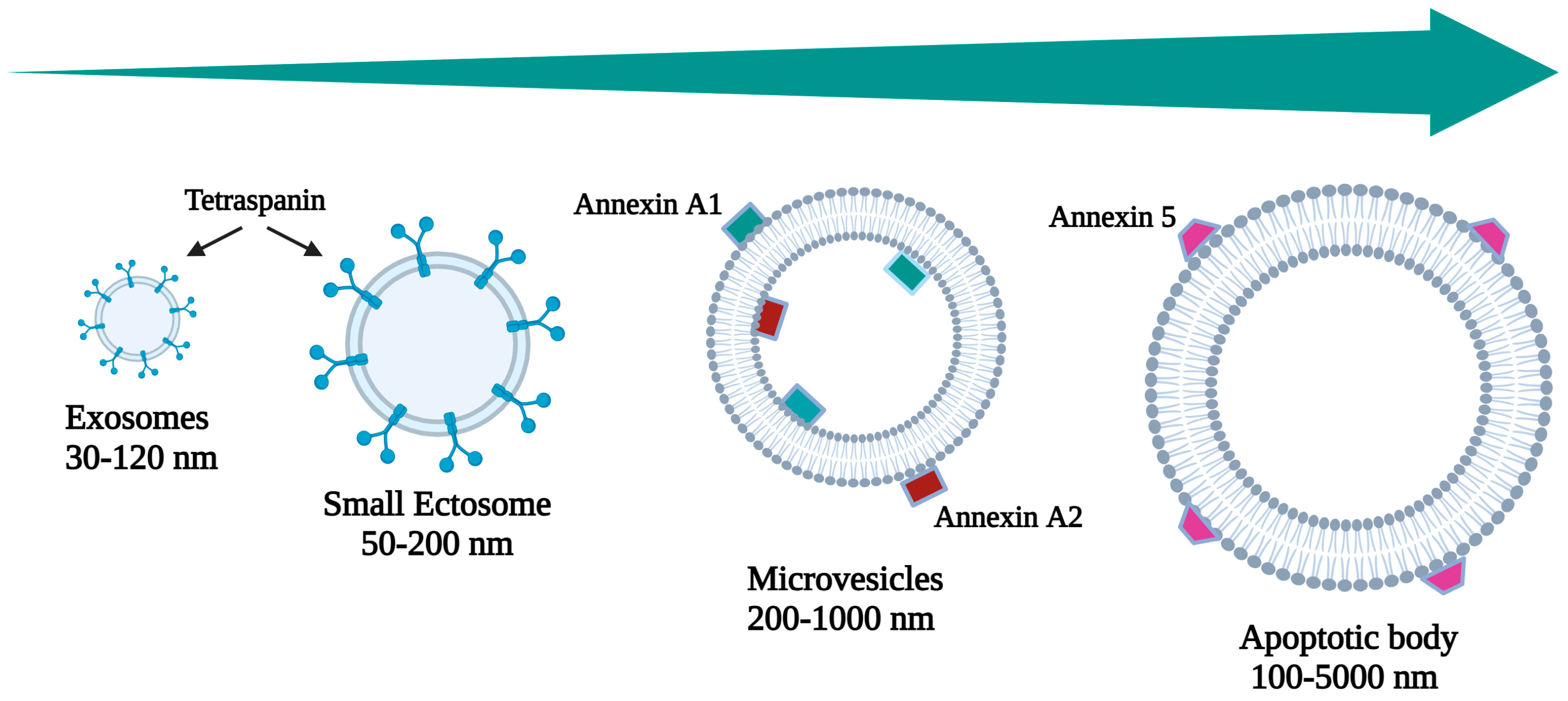
Figure 1. Schematic size-based classification of EVs secreted by mammalian cells.
Pharmaceutics 16 00567 g001
Figure 2. Schematic representation of EVs and their use for diagnostic and therapeutic purposes in GIDs.
Pharmaceutics 16 00567 g002
Figure 3. Source of EVs resistant to digestion and absorption in gastrointestinal tract.
Pharmaceutics 16 00567 g003
Table 1. Classification of EVs secreted by mammalian cells on the basis of their definition, size, genesis, and expression markers.
NameCategorySize (nm)OriginMarkersBibliography
ExosomesSmall EVs30–120Multivesicular endosomes and amphisomesCD63 Syntenin 1 CD81[9,10]
Small ectosomesSmall EVs50–200EctosomeTSG101 CD9 ARDC1
CD 147
[11,12,13]
MicrovesiclesSmall to large200–1000EctosomeAnnexin A1 Annexin A2 α-Actin 4[14]
Apoptotic BodySmall to large100–5000ApoptosisAnnexin V[15]
Table 2. Different EV biomarkers reported for organs of the gastroenteric apparatus and related pathologies.
OrganPathologyEVs MarkersTargets and/or PathwayBibliography
PancreasPancreatitismiR-155SOCS1 regulation[55,83]
PDACmiR-192-5pEpigenetic regulation[57]
miR-21MMP2, MMP9, and VEGF regulation
miR-25Cell proliferation promotion
miR-16Post-transcriptional expression of Bcl-2 regulation
PCMUC2, MUC4, MUC5AC, MUC6, MUC13Elevated pathway regulation determining the severity of PC[58,59,60,61,62]
EGFR, GPC1, WNT-2, EpCAM, MUC1Aberrant protein
Das-13′-sulfated Lewis A/C recognition
STMN1Cell proliferation inhibition
TSP1Participation in the differentiation of Th17 cells
TSP2Osteoblasts
regulation
ZEB1EMT induction
EphA2Cell migration
S100A4Src and FAK activation
PSCAThymic lymphocyte differentiation, maturation, and activation
HOOK1Re-arrangement of the cytoskeleton
PTPN6SP1/MAPK signaling pathway inhibition
FBN1Immune cell infiltration in tumors increases
Stomach
and
Esofagous
GERD
and
Gastritis
miR-29a-3pEMT, ZEB 1 and ZEB2 regulation[63]
Atrofic GastritismiR-122-5pTargeting CTDNEP1/LPIN1[67]
GCBARHL2Cell differentiation[69]
LncUEGC1Unknown[70]
LncRNA HOTTIPProliferation promotion and apoptosis inhibition[71]
FZD10Increasing of Ki-67 expression via Phospho-ERK1/2[20]
ColonIBDAnnexin A1Inflammation reduction[75,84]
miR-200b-3p
NEAT lncRNA
Protein expression regulation[76,77]
PSMA7Role in proteosomal activity[73]
lncRNA H19Unknown[72]
CRCFZD10Increasing of Ki-67 expression via Phos-pho-ERK1/2[20]
CD147Glycolipid metabolism reprogrammation[18]
A33Cell surface targeting for antibody-based therapy[81]
MARCKSL1Role in the Immune System[82]
WntRole in via β-catenin/SOX2[79]
CPNE3Promotion of cell proliferation via the PI3K/AKT pathway[80]
Table 4. Overview of the modifications of EVs and their possible application in structural GIDs.
OrganPathologyEV-Based Preparations and ApplicationsReferences
liverHCCEVs released by BM-MSCs as a delivery system to convey siCD38 to HCC cells: potential limitation of HCC growth and spread[134]
Engineered EVs mediated CRISPR-Cas9 to reverse the therapy resistance of sorafenib: the suppression of HCC cancer stem cells
HLC9-EVs engineered with HN3: increasing in
specific homing
[135,141,142]
pancreasPDAC
PC
Delivery system based on CR-modified EVs for PTT[145]
EVs derived from HUCMSCs loaded with miR-145-5p: apoptosis and cancer growth inhibition[146]
EVs derived from HUCMSCs loaded with hsa-miRNA-128–3p: apoptosis and cancer growth inhibition[147]
EVs derived from HUCMSCs loaded with KRAS G12D targeting siRNA: a reduction in the proliferation, migration, and viability in PANC-1 cells[148]
HUCMSC-EVs with DOXO: the rapid uptake in PANC-1 cells induced apoptotic cell death more efficiently than free DOXO
RWP-1-derived EVs loaded with temozolomide and EPZ015666: a greater antiproliferative action of EVs encapsulating on the PC cell line
EVs derived from the hTERT-HPNE cell line and incubated with PTX: PDAC cell death via clathrin-mediated endocytosis
[149,150]
stomachGCEVs from HEK-293 cells targeting CDH17-positive GC cancers by the engineered nanobodies into EVs with genetic engineering techniques[151,152,153,154]
Engineered small EVs with high CD47 expression and c(RGDyC) modification to deliver short RNAi against YTHDF1: a novel therapeutic strategy for GC via epigenetic and immune regulation[155,166]
colonIBDEVs released by MSCs and loaded with miR-146: a new strategy in the treatment of IBD in mouse models[126]
EVs and bacteria-derived membrane vesicles (OMVs) derived from normal feces: restore the intestinal barrier in IBD subjects[38,127,128,167]
Exo containing PSMA7 derived from saliva: an important protein biomarker for IBD[168]
CRCFolic acid-modified exo–liposome hybrid nanoparticles loaded with ALKBH5 mRNA: a novel therapeutic strategy for CRC[156]
EVs derived from CT26 and loaded with miR-34a mimics: a new strategy for CRC gene therapy in mouse model[159]
EVs derived from milk: a reduction in primary tumor burden in mouse models of CRC and breast cancer[161,162]
Exosomes secreted from HEK293T transfected with Her2-mCherry plasmid and loaded with both 5-FU and miR-21i: reversing the drug resistance in CRC cells[164,165]
Exosomes secreted from MSCs modified with 5TR1 or with magnetic nanoparticles, folic acid, and DOXO: the inhibition of CRC growth in mouse models[151,152,153,154]
Table 5. Registered clinical trials (clinicaltrials.gov, accessed date 4 March 2024) related to diagnosis, follow-up, and therapy by using extracellular vesicles as biomarkers or natural carriers of therapeutic cargo.
Study TitleCondition or DiseaseEVs for Biomarker DetectionEVs for TherapyClinicalTrials.gov Identifier
Portal Hypertension in Non-alcoholic Fatty Liver Disease: Association with Cardiovascular Risk and Identification of non-invasive biomarkersNAFLDIdentification of non-invasive biomarkers, including EVs, and of the presence and severity of portal hypertension by liquid biopsy NCT04191044
Liver Health and Metabolic Func-tion in People with ObesityNAFLDAmount, content, and function of EVs from adipose, liver, and blood tissue collected before and after ~20% weight loss and at the time of surgery NCT03701828
Role of Immune System in Obesity-related Inflammation and Cardiometabolic RiskNAFLD
MASLD
Investigation of the signaling between cells and organs examined by isolating the exosomes
from the blood, subcutaneous and omental adipose tissue of patients scheduled for gallbladder, inguinal hernia, hysterectomy, or myomectomy surgery
NCT01104220
Muscles in Liver DiseasesLiver DiseasesIdentification of circulating mediators, including EVs, that could be responsible for the complications of liver disease:
EVs released by the muscle and acting on different organs
NCT04758793
Safety of Injection of Placental MSC-Derived Exosomes for Treatment of Resistant Perianal Fistula in Crohn’s PatientsPerianal Fistula Crohn’s DiseaseIdentification of inflammatory markers in exosomes through laboratory workup, including CRP, IL-6, TNF-a, calprotectinEvaluation of the safety and clinical efficacy of injected exosomesNCT05499156
Study of ExoFlo for the Treatment of Perianal FistulasPerianal Fistula Crohn’s Disease Ex Vivo Culture-expanded Adult Allogeneic Bone Marrow MSC-derived EVs for the Treatment of Perianal Fistulizing Crohn’s Disease
Local injection of normal saline
NCT05836883
Study of ExoFlo for the Treatment of Medically refractory Crohn’s DiseaseCrohn’s Disease Intravenous ex vivo culture-expanded adult allogeneic bone marrow MSC-EVs in subjects with medically refractory Crohn’s disease who do not respond to monoclonal antibody-based therapy: evaluation of feasibility and efficacyNCT05130983
Study to Evaluate the Epidemiology and the Characteristics “Omics” in Patients Recently Diagnosed of Inflammatory Bowel Disease in Spain (IBDomics)Inflammatory Bowel DiseasesCharacterization of the composition of serum EVs in newly diagnosed IBD patients, aiming to identify the molecular and cellular pathways involved in IBD development and pathogenesis
correlation of the serum proteomic profile and the density and composition of serum EVs with an IBD phenotype
NCT03689257
Study of ExoFlo for the Treatment of Medically Refractory Ulcerative ColitisUlcerative Colitis
Inflammatory Bowel Diseases
Intravenous ex vivo culture-expanded adult allogeneic bone marrow MSC-EVs in subjects with medically refractory Crohn’s disease who do not respond to monoclonal antibody-based therapy: evaluation of feasibility, safety, and efficacyNCT05176366
Pancreatic Cancer Initial Detection Via Liquid Biopsy (PANCAID)Pancreatic Cancer
Chronic
Detection of pancreatic cancer in bio-banked samples, including EVs, of patients with histologically confirmed pancreatic lesions via liquid biopsy NCT06283576
Impact of Graft Reconditioning with Hypothermic Machine Perfusion on HCC Recurrence After Liver TransplantationHepatocellular CarcinomaIdentification of microRNAs from liquid-biopsy-derived EVs, as tools of prognostic information on IRI favoring hepatocellular carcinoma recurrence NCT06236568
Metronomic Capecitabine, Oxaliplatin and UGT1A1 Genotype-directed Irinotecan in Metastatic Pancreatic Cancer PatientsMetastatic Pancreatic CancerIdentification of exosomal proteins secreted by EVs from plasma using mass spectrometry, at different times: at pre-dose, the end of oxaliplatin infusion, and the end of irinotecan infusion NCT05929885
A Prospective Feasibility Study Evaluating EVs Obtained by Liquid Biopsy for Neoadjuvant Treatment Response Assessment in Rectal CancerRectal CancerTumor-EV detection and quantification for the response assessment and follow-up of patients with adenocarcinoma of the rectum NCT04852653
Contents of Circulating Extracellular Vesicles: Biomarkers in Colorectal Cancer PatientsColorectal CancerExploitation via liquid biopsy of circulating tumor-exosomes containing markers, namely specific miRNAs, useful as biomarkers of the early prognosis of patients with colon cancer NCT04523389
Exosome-based Liquid Biopsies for Upper Gastrointestinal Cancers DiagnosisGastric Cancer Esophagus CancerIdentification and quantification via proteomic analysis of specific proteins as biomarkers for upper gastrointestinal tumors
Multicentre and retrospective study
NCT06278064
Prospectively Predict the Efficacy of Treatment of Gastrointestinal Tumors Based on Peripheral Multi-omics Liquid BiopsyAdvanced Gastric AdenocarcinomaValidation of 4 plasma EV-derived proteins and their combination as a signature score for robustly predicting immunotherapeutic outcomes
Prospective study
NCT04993378
Disclaimer/Publisher’s Note: The statements, opinions and data contained in all publications are solely those of the individual author(s) and contributor(s) and not of MDPI and/or the editor(s). MDPI and/or the editor(s) disclaim responsibility for any injury to people or property resulting from any ideas, methods, instructions or products referred to in the content.

