Article

Fabrication of Au-Nanoparticle-Decorated Cu Mesh/Cu(OH)2@HKUST-1 Nanorod Arrays and Their Applications in Surface-Enhanced Raman Scattering

1
College of Light-Textile Engineering and Art, Anhui Agricultural University, Hefei 230036, China
2
College of Physics and Electronic Information, Luoyang Normal University, Luoyang 471934, China
*
Authors to whom correspondence should be addressed.
These authors contributed equally to this work.
Sustainability 2022, 14(1), 228; https://doi.org/10.3390/su14010228
Submission received: 8 November 2021 / Revised: 12 December 2021 / Accepted: 20 December 2021 / Published: 27 December 2021
(This article belongs to the Special Issue Advanced Semiconductor Materials for Energy, Electronics and Sensors)

Abstract

:
Here we report a simple fabrication method for large-scale hybrid surface-enhanced Raman scattering (SERS) active substrates composed of Au-nanoparticle-decorated three-dimensional (3D) Cu(OH)2@HKUST-1 (Cu3(btc)2, H3btc = 1,3,5-benzenetricarboxylic acid) nanorod arrays on a woven Cu mesh (Cu mesh/Cu(OH)2@HKUST-1@Au). Cu(OH)2 nanorods were first obtained from a simple in situ chemical engraving Cu mesh and then utilized as self-sacrificing templates to achieve HKUST-1 nanocube-assembled nanorods; finally, Au nanoparticles (Au NPs) were sputtered onto the Cu(OH)2@HKUST-1 nanorods. Due to the large surface area, the three-dimensional Cu mesh/Cu(OH)2@HKUST-1 nanorods could load high-density Au NPs and capture target detection molecules, which is beneficial to the formation of a strong electromagnetic field coupling between Au NPs, and provides abundant “hot spots” for a sensitive and uniform SERS effect. Using the Cu mesh/Cu(OH)2@HKUST-1@Au nanorod arrays as the SERS substrate, 10−9 M Rhodamine 6G and 10−8 M 4-aminothiophenolcan were identified. To verify their practical application, the fabricated arrays were employed as SERS substrates for the detection of thiram, and 10−8 M thiram could be recognized. The hybrid SERS substrates show potential applications in the field of environmental pollutant detection and this is of great significance to the sustainable development of the environment.

1. Introduction

Since the British scientist Fleischman discovered surface-enhanced Raman scattering (SERS) in 1974 [1,2], SERS as a spectral analysis method has gained widespread interest owing to its fingerprint peak recognition [3], high sensitivity [4] and rapid response, and it is non-destructive to samples [5,6]; it is widely used in various trace molecular identification and material structure characterization procedures [7]. It is generally recognized that SERS enhancement is attributed to both the electromagnetic enhancement and chemical enhancement, where the electromagnetic enhancement plays a predominant role in the SERS effect [8,9,10]. It has been proved that the electromagnetic enhancement is mainly confined to sharp metallic nanotips and nanogaps, called hotspots [11]. In order to maximize the SERS effect, the construction of SERS substrates has attracted many researchers to explore the generation of more hot spots to generate strong local electromagnetic fields [11].
So far, a variety of methods have been used to fabricate a large number of high-performance SERS substrates, including noble metal nanoparticle aggregation [12,13,14], photolithography or nanoimprint lithography [15] and template-based methods [16]. Among them, the agglomeration phenomenon of metal nanoparticles in a suspension could significantly enhance the SERS effect [17] and achieve single-molecule detection [18,19]. However, the difficulty in achieving a precise control of the number and density of noble metal aggregates in the solution hinders the assurance of the consistency and reproducibility of the SERS signal. Although photolithography or nanoimprint lithography and template-based methods [20,21,22,23] improve the consistency and accuracy of the arrangement of noble metal nanoparticles [24,25,26,27], these processes are generally time-consuming and require additional expensive instruments [28,29,30]. Therefore, it is still a challenge to realize a large-area substrate with a high SERS activity and signal uniformity through a simple and low-cost synthesis method. To meet this challenge, assembling Au or Ag nanoparticles onto the surface of one-dimensional or two-dimensional semiconductor nanostructure arrays to achieve three-dimensional (3D) SERS substrates is highly desirable. The electromagnetic field enhancement based on noble metal nanostructures and the chemical enhancement of semiconductor nanostructures can simultaneously increase the Raman signal and improve the detection sensitivity [31]. Furthermore, the semiconductor nanostructure arrays can load more noble metal nanoparticles and ensure the reproducibility and uniformity of the Raman signal [32,33,34].
Here, we demonstrate a low-cost and reliable method to obtain a large-area 3D uniform SERS substrate (denoted as Cu mesh/Cu(OH)2@HKUST-1@Au) composed of Au-nanoparticle decorated Cu(OH)2@HKUST-1 (Cu3(btc)2, H3btc = 1,3,5-benzenetricarboxylic acid) nanorods on a commercial woven Cu mesh, as shown in Figure 1. The large-area and high-density HKUST-1 nanocube-assembled nanorods with a 3D hierarchical structure can be used as a platform to load high-density Au nanoparticles (NPs); thus, abundant nanogaps between coupled Au NPs decorated on the same and different HKUST-1 nanorods can be realized. Therefore, this method can realize hot spots with a high density on a large area, which is very useful for obtaining high SERS signals from molecules. In addition, as a typical metal-organic framework (MOF) material [35,36,37,38], HKUST-1 has a porous structure that is favorable for the capture of target detection molecules, so it can further improve the signals of target detection molecules. The results show that the Cu mesh/Cu(OH)2@HKUST-1@Au nanorod arrays exhibit a high SERS activity toward Rhodamine 6G (R6G), 4-aminothiophenol (4-ATP) and thiram molecules. The molecules 10−9 MR6G, 108 M 4-ATP and 10−8 M thiram have been detected, respectively. This work presents a strategy for the feasible assembly of Au NPs on the well-aligned MOF arrays, which could serve as a promising platform for environmental pollutant detection.

2. Materials and Methods

2.1. Chemicals

Sodium hydroxide (NaOH), ammonium persulfate ((NH4)2S2O8), 1,3,5-trimesic acid, polyvinylpyrrolidone (PVP) and N,N-dimethylformamide (DMF) were purchased from Sinopharm Chemical Reagent Co. Ltd.; 4-aminothiophenol (4-ATP) was purchased from Saen Chemical Technology (Shanghai) Co. Ltd. (Shanghai, China); Rhodamine 6G (R6G) and thiram were purchased from Shanghai Macklin Biochemical Technology Co. Ltd. (Shanghai, China); commercial woven Cu mesh with mesh number of 400 was purchased from Shanghai Xinhu screen shop; all reagents are analytic reagents without special treatment before use. The golden target used for ion sputtering (Au, 99.99%) was purchased from Zhongnuo Advanced Materials (Beijing) Technology Co. Ltd. (Beijing, China).

2.2. Pretreatment of Cu Mesh

The commercial Cu mesh was first cut into small pieces of 1 × 1 cm. Then, these small pieces of Cu mesh were ultrasonically rinsed in ethanol, acetone and deionized water for 15 min, respectively.

2.3. Preparation of Cu(OH)2 Nanorods

NaOH weighing 1 g and 0.228 g of (NH4)2S2O8 were completely dissolved in 10 mL of deionized water and kept in water bath at 20 °C. Then, a small piece of Cu mesh was immersed into the solution and kept at 20 °C for a certain period of time (10–30 min). Finally, the resultant samples were collected and rinsed with deionized water and dried in the oven at 20 °C for 12 h.

2.4. Preparation of Cu(OH)2@HKUST-1 Nanorods

A certain amount (0.005 g, 0.01 g, 0.02 g) of 1,3,5-trimellitic acid and a certain amount (0.05 g, 0.1 g, 0.2 g) of PVP were dissolved in 2.5 mL deionized water and 2.5 mL DMF. Then, the Cu mesh with Cu(OH)2 nanorods was placed into the above reaction solution for 10–30 min. The Cu(OH)2@HKUST-1 nanorods on the woven Cu mesh were collected and rinsed with ethanol several times and then dried in the oven at 85 °C for 12 h.

2.5. Preparation of CuO Nanorods

The as-prepared Cu(OH)2 nanorods on the Cu mesh were dried in the oven at 180 °C for 2 h to obtain CuO nanorods on the Cu mesh.

2.6. Sputtering Au NPs onto the Different Arrays on the Cu Mesh

The different arrays on the Cu mesh were placed below the gold target with a distance of about 2 cm in the ion sputter (Quorum K550X, UK), while the sputtering current was set to 40 mA/cm2, and the sputtering duration was controlled from 4 to 16 min (the interval is 4 min).

2.7. Characterizations

Scanning electron microscopy (SEM, S-4800, Hitachi, Japan) and transmission electron microscopy (TEM, HT-7700, Hitachi, Japan) was carried out to observe the morphology of samples. Energy dispersive X-ray spectroscopy (EDS) was conducted using an Oxford X-MaxN 80T IE250. X-ray diffraction (XRD) (Beijing Puxitong Instrument Co., Ltd., Beijing, China) was used to determine the crystal structure of the synthesized products. The UV–vis spectra were taken on a Shimadzu MPC-3100 equipped with an integrated sphere accessory.

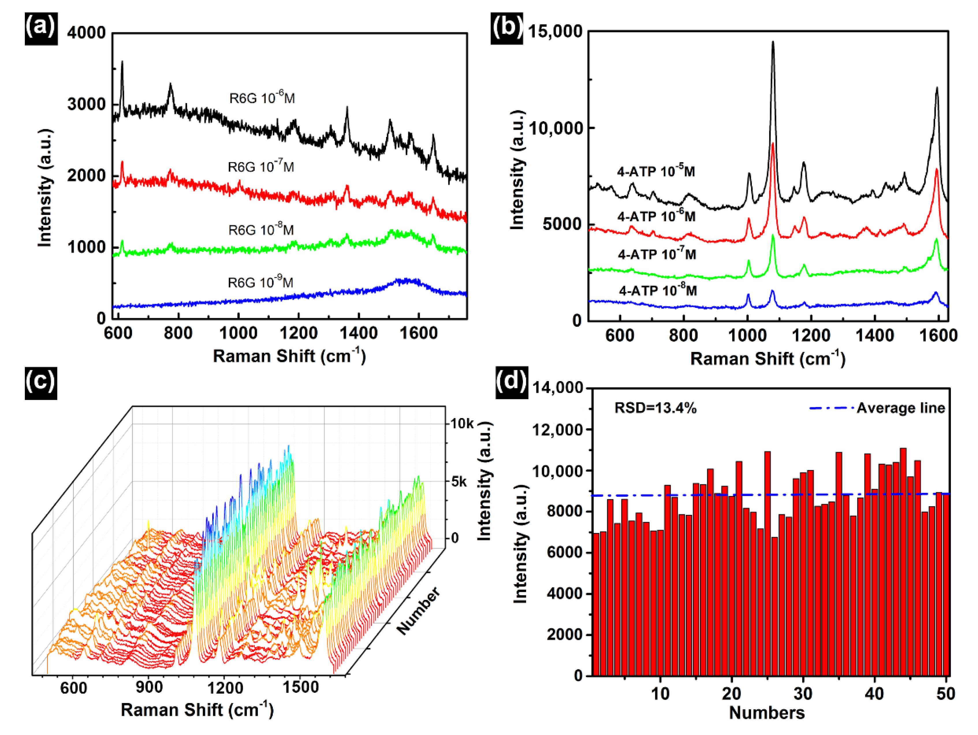
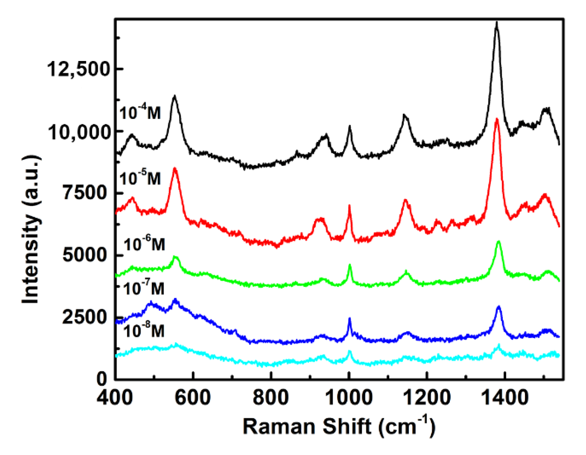
2.8. SERS Measurements

The SERS measurements were carried out with a confocal microscopic Raman spectrometer (Renishaw, inVia 50× objective). Before the SERS measurements, different samples were soaked in R6G solution for 10 h, taken out and rinsed with deionized water and then dried in the air. In order to detect thiram, thiram was dissolved in acetone at different concentrations (from 10−4 M to 10−8 M), and the substrates were immersed into thiram acetone solution for 10 h, rinsed with deionized water and then dried in the air. For 4-ATP detection, the substrates were immersed into 4-ATP ethanol solution for 10 h, taken out and rinsed with deionized water and then dried in the air. The SERS spectra of R6G were acquired with an excitation wavelength of 532 nm. The SERS spectra of 4-ATP and thiram were acquired with an excitation wavelength of 785 nm.

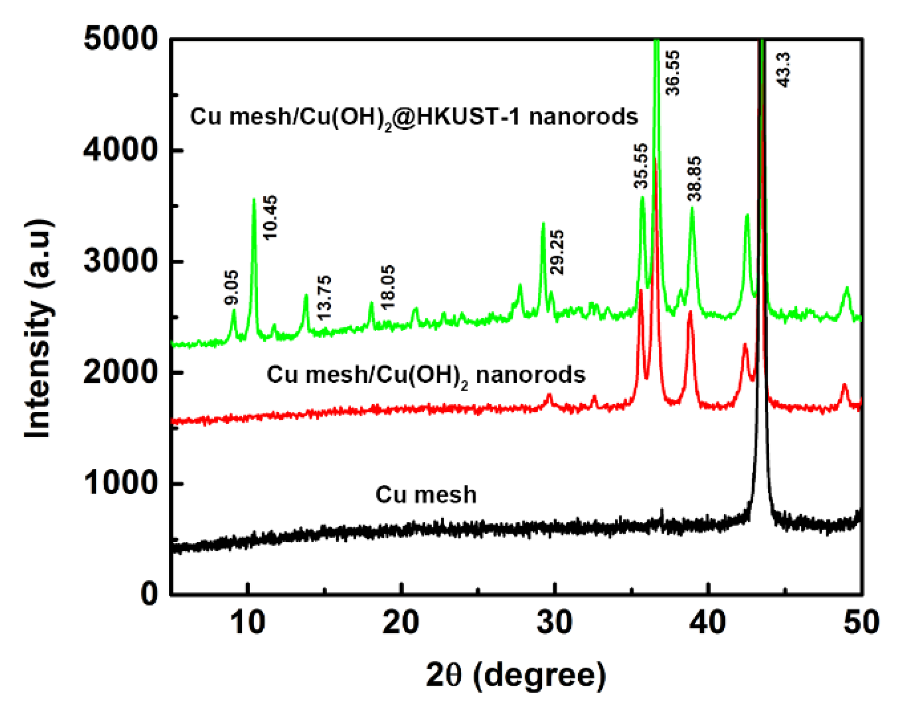
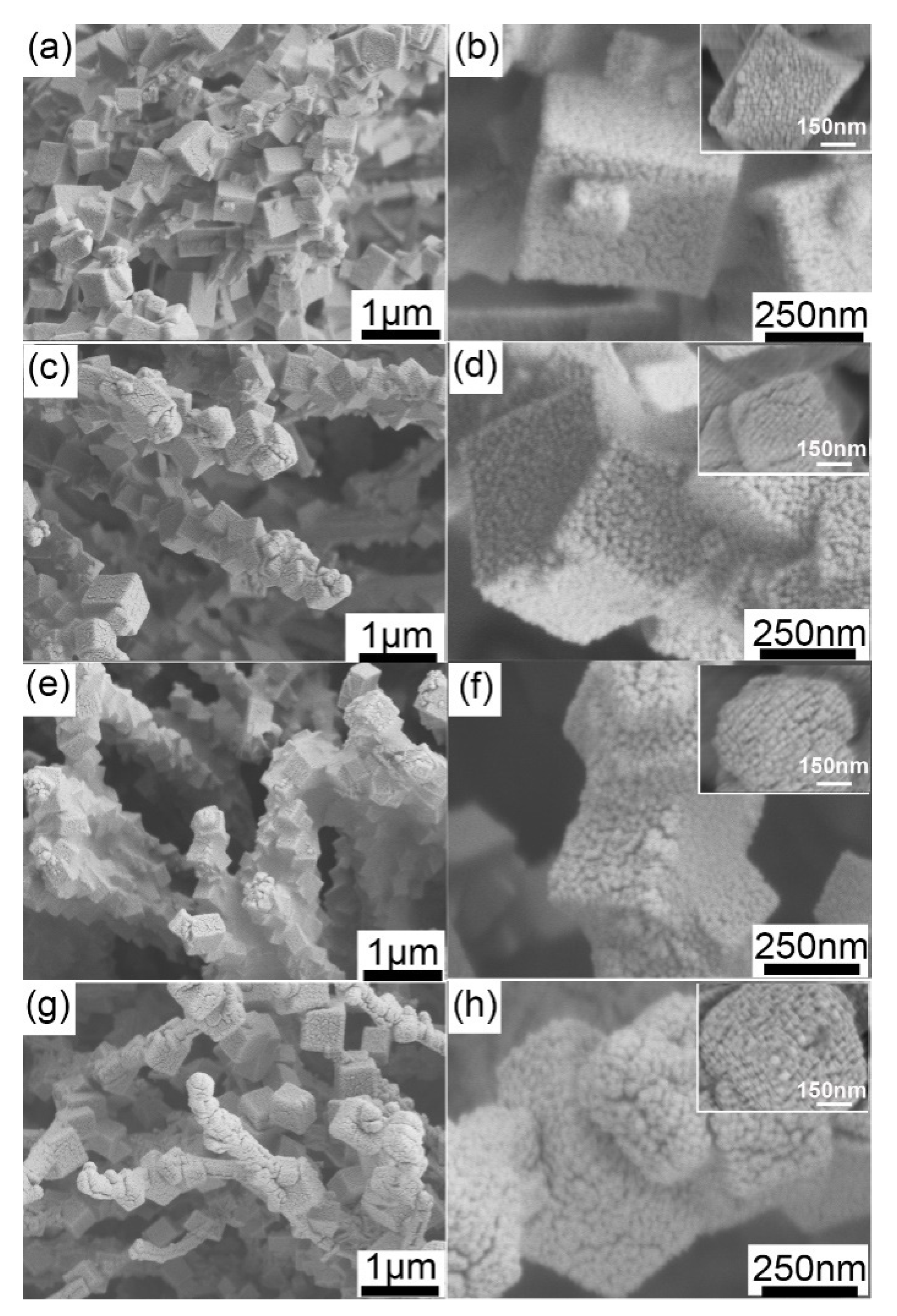
3. Results and Discussion

Figure 2a,b and Figure S1 show the SEM images of Cu(OH)2 nanorods grown on a Cu mesh. It can be seen that the Cu mesh was uniformly covered with dense Cu(OH)2 nanorods. The SEM image of the Cu(OH)2 nanorods on a Cu mesh under high magnification (Figure 2b) demonstrates that high-density upright Cu(OH)2 nanorods with an average length of about 5 μm on the Cu mesh formed a 3D or quasi-3D connected stereoscopic structure. In addition, Figure 2b inset shows a close-up SEM image of Cu(OH)2 nanorods with a polygonal shape and an average diameter of about 250 nm. After immersion in 1,3,5-trimellitic acid and PVP solution for 20 min, large-area dense HKUST-1 nanostructures on the Cu mesh were achieved, as shown in Figure 2c,d. A close-up SEM observation (Figure 2d) reveals that the dense HKUST-1 nanocubes were assembled on the surface of the Cu(OH)2 nanorods, forming vertically aligned HKUST-1 nanocube-assembled nanorods (Figure 2d inset). TEM observation on the broken HKUST-1 nanocubes (Figure 2e) and a corresponding line scan at high magnification (Figure S2) further demonstrate the HKUST-1 nanocube-assembled nanorods’ geometrical features—they form a core–shell structure. The average length of the HKUST-1 nanocube assembled nanorods was about 5 μm and the average diameter was about 1μm. Figure 2f is the EDS of the Cu(OH)2@HKUST-1 nanorods on a Cu mesh, which reveals that the composite structure was mainly composed of Cu and a small amount of carbon and oxygen, which further confirms that the Cu(OH)2@HKUST-1 nanorods on the Cu mesh were synthesized.
XRD can be used to determine the crystal structure of the synthesized products. Figure 3 presents the corresponding XRD patterns of Cu mesh, Cu mesh/Cu(OH)2 nanorods and Cu mesh/Cu(OH)2@HKUST-1 nanorods. It is clear that all the characteristic diffraction peaks of crystal products match well with the peaks of Cu, Cu(OH)2 and HKUST-1. The diffraction pattern shows peaks at 43.30°, corresponding to (111) of Cu (JCPDS) [39]. The diffraction pattern shows peaks at 35.55°, 36.55° and 38.85°, corresponding to (002), (111) and (130) of Cu(OH)2 (JCPDS) [40]. It also shows the relative peak intensities of 9.05°, 13.75° and 18.05° of cubic HKUST-1, corresponding to the (220), (400) and (333) lattices [41], respectively.
The deposition duration and the concentrations of 1,3,5-trimellitic acid and PVP play critical roles in the construction of Cu mesh/Cu(OH)2 nanorods. In order to investigate the evolution process of HKUST-1 morphologies, a series of comparative experiments were carried out (Figures S3–S5). The optimal parameters to fabricate the Cu(OH)2@HKUST-1 nanorods were: a deposition duration of 20 min, a concentration of PVP of 20 g/L and a concentration of trimellitic acid of 2 g/L. After the Au NPs were sputtered onto the Cu(OH)2@HKUST-1 nanorods, the 3D hybrid hierarchical Cu mesh/Cu(OH)2@HKUST-1@Au nanorod arrays were achieved. Figure 4 and Figure S6 shows the SEM images and UV–vis absorption spectra of the Cu(OH)2@HKUST-1@Au samples with different Au-NP sputtering durations. Compared with the Cu(OH)2@HKUST-1 nanorods before Au-sputtering, it can be observed that the number and size of Au NPs on the HKUST-1 nanocubes gradually increased with the increase of the sputtering duration. The UV–vis absorption spectra of the samples show a gradual red shift of the absorption peak from 450 nm to 580 nm with the sputtering duration increases (Figure S6). It is beneficial to obtain SERS signals under the excitation of 532, 633 and 785 nm lasers [42].
It can be noted that there are sub-10 nm gaps (insets of Figure 4) between the neighboring Au NPs located on the same Cu(OH)2@HKUST-1 nanorods, which can serve as “hot spots” to ensure a high SERS-activity [43]. The SERS activity of the Cu mesh/Cu(OH)2@HKUST-1@Au depending on the different Au-sputtering durations was analyzed. As shown in Figure 4a,b, when the Au-sputtering durations was 4 min, Au NPs with a diameter of about 5 nm were attached to the surfaces of Cu(OH)2@HKUST-1 nanorods with parts of the surfaces uncovered. With short Au-sputtering durations, weak Raman signal appeared at 611 cm−1 [44] indicating a low SERS activity (Figure 5a). When the Au-sputtering duration was 8 min, the number and size of Au NPs gradually increased, while the diameter of Au NPs could be up to 10 nm (Figure 4c,d). The EDS also confirms the composition of the hybrid particles. A line scan of a single Cu(OH)2@HKUST-1@Au nanorod and overlay of Au, Cu and C EDS maps (shown in Figures S7 and S8) indicate a clear separation of the three elements and confirm the core–shell structure. The surface coverage of Au NPs on the HKUST-1 nanocubes increased, but no obvious aggregations were found. Therefore, the gaps between adjacent Au NPs was reduced to sub-10 nm, resulting in an increase in the density of the “hot spots” and enhanced SERS activity (Figure 5a). When the sputtering duration increased to 12 min, the number of Au NPs on the HKUST-1 nanocubes greatly increased with the diameter of about 15 nm (Figure 4e,f). At this stage, the gap between neighboring Au NPs increased and the SERS intensity of the R6G was improved (Figure 5a). By extending the sputtering duration to 16 min, we could obverse that the Au NPs were stacked on the surfaces of the HKUST-1 nanocubes and obscured the nanocubes’ shapes (Figure 4g,h). Au NPs could accumulate on the surface of HKUST-1 to form an Au film, leading to a lower density of nanogaps and decreased “hot spots” between the adjacent Au NPs. Therefore, corresponding to the Raman spectra, we could observe that the SERS activity decreased.
Figure 5b shows the SERS spectra of R6G acquired from the Au NP-decorated Cu(OH)2 nanorods and Cu(OH)2@HKUST-1 nanorods obtained by immersing Cu(OH)2 nanorods in 1,3,5-trimellitic acid and PVP solution for different durations. With the immersion duration increasing, the SERS signal from the Cu mesh/Cu(OH)2@HKUST-1@Au nanorod arrays gradually increased. When the immersion duration was 10 min, the Raman signal from the Cu mesh/Cu(OH)2@HKUST-1@Au nanorod arrays was higher than that of the Cu mesh/Cu(OH)2@Au. When the immersion duration was increased to 20 min, the signal from the Cu mesh/Cu(OH)2@HKUST-1@Au nanorod arrays was significantly higher than that of the Cu mesh/Cu(OH)2@Au. When the immersion time increased, the higher surface area of the porous HKUST-1 nanocubes could load a higher density of Au NPs, inducing a higher SERS activity. Figure 5c,d shows the SERS spectra of 10−6 M R6G and 10−6 M 4-ATP acquired from the Cu mesh/CuO@Au, Cu mesh/Cu(OH)2@Au and Cu mesh/Cu(OH)2@HKUST-1@Au nanorod arrays’ substrates, respectively. It can be seen that the Raman signal from the Cu mesh/Cu(OH)2@HKUST-1@Au nanorod arrays was the strongest and was significantly higher than those of the Cu mesh/CuO@Au and Cu mesh/Cu(OH)2@Au. Therefore, HKUST-1 nanocubes were better for use as platforms to construct hybrid SERS substrates than Cu mesh/CuO nanorods and Cu mesh/Cu(OH)2 nanorods. As a result, Cu mesh/Cu(OH)2@HKUST-1@Au nanorod arrays with 12 min Au-sputtering durations exhibited the highest SERS activity and were chosen as the optimal SERS substrate.
Figure 6a,b show the Raman spectra of R6G and 4-ATP at different concentrations. It can be seen that the characteristic peaks of R6G clearly appear at 611 cm−1 (in the C–C–C in-plane bending mode), 772 cm−1 (in the C–H out-of-plane bending mode) and 1362 cm−1 (in the aromatic C–C pull extending vibration mode) [44]. As the concentration of R6G decreased, the corresponding SERS signal also decreased. Similarly, as the concentration of 4-ATP decreased, the SERS signal from the optimal SERS substrate became weaker. When the concentrations of R6G and 4-ATP were further diluted to 10−9 M and 10−8 M, respectively, the characteristic peaks of R6G and 4-ATP could still be identified, indicating that the optimal substrate had a good sensitivity. The enhancement factor (EF) was estimated to be about 1.09 × 10⁶ (Part S2 of the Supplementary Materials). The reproducibility of the SERS signal was also investigated by taking the SERS spectra of 4-ATP from different locations on the Cu mesh/Cu(OH)2@HKUST-1@Au substrate (Figure 6c). The relative standard deviation of the characteristic peak intensity at 1078 cm−1 was 13.4% (Figure 6d), indicating that the optimal SERS substrate had a good signal uniformity.
To verify the potential applications of the as-prepared optimal SERS substrate, Cu mesh/Cu(OH)2@Au hierarchical arrays were used as substrates to detect thiram (a commonly used dithiocarbamate fungicide). Figure 7 shows the SERS spectra of thiram with concentrations ranging from 10−4 M to 10−8 M acquired on the optimal SERS substrate. The characteristic peaks at 443 cm−1 (in the CH3–N–C deformation and C–S stretching modes), 565 cm−1 (in the S–S stretching mode), 934 cm−1 (in the stretching CH3-N and C–S modes), 1146 cm−1 (in the C–N stretching mode and rocking CH3), 1382 cm−1 (in the C–N stretching mode and symmetric CH3 deformation mode) and 1508 cm−1 (in the rocking CH3 mode and C–N stretching mode) can be clearly seen for the thiram molecule [45,46,47]. When the concentration was reduced to 10−8 M, there was still a strong signal intensity at the characteristic peak, indicating that the fabricated Cu mesh/Cu(OH)2@HKUST-1@Au nanorod arrays substrate have promising applications in the practical pesticide residue detection.

4. Conclusions

In summary, Cu mesh/Cu(OH)2@HKUST-1@Au hierarchical arrays have been achieved via sputtering Au NPs onto hybrid 3D Cu mesh/Cu(OH)2 nanorod arrays, which can support high-density Au NPs and form abundant SERS “hot spots” beneficial to SERS applications. The Cu mesh/Cu(OH)2@HKUST-1@Au hierarchical arrays used as SERS substrate showed a high SERS activity. Not only 10−9 M R6G and 10−8 M 4-ATP, but also 10−8 M thiram, were identified, which shows a great potential in the detection of trace organic pollutants in the environment. Therefore, the 3D hybrid substrates have an important practical significance for sustainable development in the detection of pesticides and other environmental pollutants.

Supplementary Materials

The following are available online at https://www.mdpi.com/article/10.3390/su14010228/s1. Figure S1: SEM images of Cu(OH)2 nanorods prepared under different reaction durations. Figure S2: Line scan results of a single Cu(OH)2@HKUST-1 nanorod. Figure S3: SEM images of Cu mesh/Cu(OH)2 nanorods prepared under different reaction durations. Figure S4: SEM images of Cu mesh/Cu(OH)2@HKUST-1 nanorods prepared with different concentrations of 1,3,5 trimesic acid. Figure S5: SEM images of Cu mesh/Cu(OH)2@HKUST-1 nanorods prepared with different concentrations of PVP. Figure S6: UV–vis absorption spectra of the Cu mesh/Cu(OH)2 nanorod arrays, Cu mesh/Cu(OH)2@HKUST-1 nanorod arrays and Cu mesh/Cu(OH)2@HKUST-1@Au nanorod arrays with different Au-sputtering durations. Figure S7: Lines scan results of a single Cu(OH)2@HKUST-1@Au nanorod. Figure S8: EDS map from a single Cu(OH)2@HKUST-1@Au nanorod. Part S1:Figures S1 to S8. Part S2: Estimation of the enhancement factor.

Author Contributions

Conceptualization, Z.L., X.H. and L.C.; methodology, X.H., K.S., T.F. and L.Y.; validation, X.H., Z.L. and L.Z.; formal analysis, L.C. and Z.L.; investigation, X.H.; resources, X.H. and Z.L.; data curation, X.H.; writing—original draft preparation, X.H. and L.C.; writing—review and editing, L.Z. and Z.L.; funding acquisition, L.Z., K.S. and Z.L. All authors have read and agreed to the published version of the manuscript.

Funding

This research was funded by the National Natural Science Foundation of China (NSFC) (grant nos. 51501183 and 51801093) and Natural Science Foundation of Anhui Province, China (grant no. 1908085QB89).

Data Availability Statement

The data used to support the findings of this study are included within the article.

Conflicts of Interest

The authors declare no conflict of interest.

References

  1. Fleischmann, M.; Hendra, P.J.; McQuillan, A.J. Raman spectra of pyridine adsorbed at a silver electrode. Chem. Phys. Lett. 1974, 26, 163–166. [Google Scholar] [CrossRef]
  2. Cao, W.Y.C.; Jin, R.; Mirkin, C.A. Nanoparticles with Raman Spectroscopic Fingerprints for DNA and RNA Detection. Science 2002, 297, 1536. [Google Scholar] [CrossRef] [PubMed] [Green Version]
  3. Zheng, P.; Shi, X.; Curtin, K.; Yang, F.; Wu, N. Detection of mercury(II) with a surface-enhanced Raman scattering sensor based on functionalized gold nanoparticles. Mater. Res. Express. 2017, 4, 055017. [Google Scholar] [CrossRef]
  4. Tezcan, T.; Hsu, C.-H. High-sensitivity SERS based sensing on the labeling side of glass slides using low branched gold nanoparticles prepared with surfactant-free synthesis. RSC Adv. 2020, 10, 34290–34298. [Google Scholar] [CrossRef]
  5. Neng, J.; Zhang, Q.; Sun, P. Application of surface-enhanced Raman spectroscopy in fast detection of toxic and harmful substances in food. Biosens. Bioelectron. 2020, 167, 112480. [Google Scholar] [CrossRef] [PubMed]
  6. Qu, L.L.; Ying, Y.L.; Yu, R.J.; Long, Y.T. In situ food-borne pathogen sensors in a nanoconfined space by surface enhanced Raman scattering. Microchim. Acta 2021, 188, 201. [Google Scholar] [CrossRef]
  7. Liu, Y.; Li, X.; Cheng, J.; Zhou, N.; Zhang, L.; Mao, H.; Huang, C. SERS devices with “hedgehog-like” nanosphere arrays for detection of trace pesticides. J. Innov. Opt. Health Sci. 2021, 14, 2141005. [Google Scholar] [CrossRef]
  8. Chen, L.; Liu, W.; Shen, D.; Liu, Y.; Zhou, Z.; Liang, X.; Wan, W. All-optical tunable plasmonic nano-aggregations for surface-enhanced Raman scattering. Nanoscale 2019, 11, 13558–13566. [Google Scholar] [CrossRef] [PubMed]
  9. Peng, Y.; Lin, C.; Long, L.; Masaki, T.; Tang, M.; Yang, L.; Liu, J.; Huang, Z.; Li, Z.; Luo, X.; et al. Charge-Transfer Resonance and Electromagnetic Enhancement Synergistically Enabling MXenes with Excellent SERS Sensitivity for SARS-CoV-2 S Protein Detection. Nano-Micro Lett. 2021, 13, 52. [Google Scholar] [CrossRef]
  10. Xiao, T.-H.; Cheng, Z.; Luo, Z.; Isozaki, A.; Hiramatsu, K.; Itoh, T.; Nomura, M.; Iwamoto, S.; Goda, K. All-dielectric chiral-field-enhanced Raman optical activity. Nat. Commun. 2021, 12, 3062. [Google Scholar] [CrossRef]
  11. Kim, N.H.; Hwang, W.; Baek, K.; Rohman, M.R.; Kim, J.; Kim, H.W.; Mun, J.; Lee, S.Y.; Yun, G.; Murray, J.; et al. Smart SERS Hot Spots: Single Molecules Can Be Positioned in a Plasmonic Nanojunction Using Host-Guest Chemistry. J. Am. Chem. Soc. 2018, 140, 4705–4711. [Google Scholar] [CrossRef]
  12. Markina, N.E.; Ustinov, S.N.; Zakharevich, A.M.; Markin, A.V. Copper nanoparticles for SERS-based determination of some cephalosporin antibiotics in spiked human urine. Anal. Chim. Acta 2020, 1138, 9–17. [Google Scholar] [CrossRef] [PubMed]
  13. Liu, Y.; Lei, L.; Wu, Y.; Chen, Y.; Yan, J.; Zhu, W.; Tan, X.; Wang, Q. Fabrication of sea urchin-like Au@SiO2 nanoparticles SERS substrate for the determination of malachite green in tilapia. Vib. Spectrosc. 2021, 118, 103319. [Google Scholar] [CrossRef]
  14. Xie, B.; Wang, Z.-P.; Zhang, R.; Zhang, Z.; He, Y. A SERS aptasensor based on porous Au-NC nanoballoons for Staphylococcus aureus detection. Anal. Chim. Acta 2022, 1190, 339175. [Google Scholar] [CrossRef] [PubMed]
  15. Alvarez-Puebla, R.; Cui, B.; Bravo-Vasquez, J.-P.; Veres, T.; Fenniri, H. Nanoimprinted SERS-Active Substrates with Tunable Surface Plasmon Resonances. J. Phys. Chem. C 2007, 111, 6720–6723. [Google Scholar] [CrossRef]
  16. Endo, T.; Yamada, H.; Yamada, K. Template Stripping Method-Based Au Nanoarray for Surface-Enhanced Raman Scattering Detection of Antiepileptic Drug. Micromachines 2020, 11, 936. [Google Scholar] [CrossRef]
  17. Chen, X.; Liang, M.-M.; Xu, J.; Sun, H.-L.; Wang, C.; Wei, J.; Zhang, H.; Yang, W.-M.; Yang, Z.-L.; Sun, J.-J.; et al. Unveiling the size effect of Pt-on-Au nanostructures on CO and methanol electrooxidation by in situ electrochemical SERS. Nanoscale 2020, 12, 5341–5346. [Google Scholar] [CrossRef]
  18. Kneipp, K.; Wang, Y.; Kneipp, H.; Perelman, L.T.; Itzkan, I.; Dasari, R.R.; Feld, M.S. Single Molecule Detection Using Surface-Enhanced Raman Scattering (SERS). Phys. Rev. Lett. 1997, 78, 1667–1670. [Google Scholar] [CrossRef] [Green Version]
  19. Lee, H.K.; Lee, Y.H.; Koh, C.S.L.; Phan-Quang, G.C.; Han, X.; Lay, C.L.; Sim, H.Y.F.; Kao, Y.C.; An, Q.; Ling, X.Y. Designing surface-enhanced Raman scattering (SERS) platforms beyond hotspot engineering: Emerging opportunities in analyte manipulations and hybrid materials. Chem. Soc. Rev. 2019, 48, 731–756. [Google Scholar] [CrossRef]
  20. Zhao, F.; Wang, X.; Zhang, Y.; Lu, X.; Xie, H.; Xu, B.; Ye, W.; Ni, W. In situ monitoring of silver adsorption on assembled gold nanorods by surface-enhanced Raman scattering. Nanotechnology 2020, 31, 295601. [Google Scholar] [CrossRef]
  21. Zhang, H.; Wang, J.; Li, G.; Chen, L.; Wang, H.; Tian, X. Fabrication of Ag-nanosheet-assembled hollow tubular array and their SERS effect. J. Less-Common Met. 2019, 772, 663–668. [Google Scholar] [CrossRef]
  22. Hu, B.; Sun, D.-W.; Pu, H.; Wei, Q. A dynamically optical and highly stable pNIPAM@Au NRs nanohybrid substrate for sensitive SERS detection of malachite green in fish fillet. Talanta 2020, 218, 121188. [Google Scholar] [CrossRef]
  23. Hu, W.; Xia, L.; Hu, Y.; Li, G. Recent progress on three-dimensional substrates for surface-enhanced Raman spectroscopic analysis. Microchem. J. 2022, 172, 106908. [Google Scholar] [CrossRef]
  24. Ostovar, B.; Cai, Y.Y.; Tauzin, L.J.; Lee, S.A.; Ahmadivand, A.; Zhang, R.; Nordlander, P.; Link, S. Increased Intraband Transitions in Smaller Gold Nanorods Enhance Light Emission. ACS Nano 2020, 14, 15757–15765. [Google Scholar] [CrossRef]
  25. Zhang, L.; Weng, Y.-j.; Liu, X.; Gu, W.; Zhang, X.; Han, L. Fe(III) Mixed IP6@Au NPs with Enhanced SERS Activity for Detection of 4-ATP. Sci. Rep. 2020, 10, 5752. [Google Scholar] [CrossRef] [PubMed] [Green Version]
  26. Yilmaz, M.; Erkartal, M.; Ozdemir, M.; Sen, U.; Usta, H.; Demirel, G. Three-Dimensional Au-Coated Electrosprayed Nanostructured BODIPY Films on Aluminum Foil as Surface-Enhanced Raman Scattering Platforms and Their Catalytic Applications. ACS Appl. Mater. Interfaces 2017, 9, 18199–18206. [Google Scholar] [CrossRef]
  27. Guo, L.; Cao, H.; Cao, L.; Li, N.; Zhang, A.; Shang, Z.; Jiao, T.; Liu, H.L.; Wang, M. Improve optical properties by modifying Ag nanoparticles on a razor clam SERS substrate. Opt. Express 2021, 29, 5152–5165. [Google Scholar] [CrossRef]
  28. Ma, X.; Wang, W.; Sun, C.; Li, H.; Sun, J.; Liu, X. Adsorption performance and kinetic study of hierarchical porous Fe-based MOFs for toluene removal. Sci. Total Environ. 2021, 793, 148622. [Google Scholar] [CrossRef]
  29. Huang, Z.; Meng, G.; Hu, X.; Pan, Q.; Huo, D.; Zhou, H.; Ke, Y.; Wu, N. Plasmon-tunable Au@Ag core-shell spiky nanoparticles for surface-enhanced Raman scattering. Nano Res. 2019, 12, 449–455. [Google Scholar] [CrossRef]
  30. Si, S.; Liang, W.; Sun, Y.; Huang, J.; Ma, W.; Liang, Z.; Bao, Q.; Jiang, L. Facile Fabrication of High-Density Sub-1-nm Gaps from Au Nanoparticle Monolayers as Reproducible SERS Substrates. Adv. Funct. Mater. 2016, 26, 8137–8145. [Google Scholar] [CrossRef]
  31. Maity, A.; Samanta, S.; Biswas, D.; Chakravorty, D. Studies on nanoconfinement effect of NiO-SiO2 spin glass within mesoporous Al2O3 template. J. Alloys Compd. 2021, 887, 161447. [Google Scholar] [CrossRef]
  32. Li, Y.; Yang, S.; Lu, X.; Duan, W.; Moriga, T. Synthesis and evaluation of the SERS effect of Fe3O4–Ag Janus composite materials for separable, highly sensitive substrates. RSC Adv. 2019, 9, 2877–2884. [Google Scholar] [CrossRef] [Green Version]
  33. Wang, X.; Shi, W.; Wang, S.; Zhao, H.; Lin, J.; Yang, Z.; Chen, M.; Guo, L. Two-Dimensional Amorphous TiO2 Nanosheets Enabling High-Efficiency Photoinduced Charge Transfer for Excellent SERS Activity. J. Am. Chem. Soc. 2019, 141, 5856–5862. [Google Scholar] [CrossRef]
  34. Wang, X.; Zhu, J.; Wu, Y.; Xu, Y.; Su, Y.; Zhang, L.; Qi, Y.; Wen, X.; Yang, H. Hybrid surface plasmon effect and SERS characterization in a heterogeneous composite structure of Au nano-array and Ag film. Results Phys. 2020, 17, 103175. [Google Scholar] [CrossRef]
  35. Pal, S.; Bhunia, A.; Jana, P.P.; Dey, S.; Möllmer, J.; Janiak, C.; Nayek, H.P. Microporous La–Metal–Organic Framework (MOF) with Large Surface Area. Chem.-Eur. J. 2015, 21, 2789–2792. [Google Scholar] [CrossRef]
  36. Li, X.; Mao, Y.; Leng, K.; Ye, G.; Sun, Y.; Xu, W. Synthesis of amino-functionalized MIL-101(Cr) with large surface area. Mater. Lett. 2017, 197, 192–195. [Google Scholar] [CrossRef]
  37. Phan-Quang, G.C.; Yang, N.; Lee, H.K.; Sim, H.Y.F.; Koh, C.S.L.; Kao, Y.-C.; Wong, Z.C.; Tan, E.K.M.; Miao, Y.-E.; Fan, W.; et al. Tracking Airborne Molecules from Afar: Three-Dimensional Metal–Organic Framework-Surface-Enhanced Raman Scattering Platform for Stand-Off and Real-Time Atmospheric Monitoring. ACS Nano 2019, 13, 12090–12099. [Google Scholar] [CrossRef]
  38. Lafuente, M.; De Marchi, S.; Urbiztondo, M.; Pastoriza-Santos, I.; Pérez-Juste, I.; Santamaría, J.; Mallada, R.; Pina, M. Plasmonic MOF Thin Films with Raman Internal Standard for Fast and Ultrasensitive SERS Detection of Chemical Warfare Agents in Ambient Air. ACS Sens. 2021, 6, 2241–2251. [Google Scholar] [CrossRef] [PubMed]
  39. Chandra, S.; Kumar, A.; Tomar, P.K. Synthesis and characterization of copper nanoparticles by reducing agent. J. Saudi Chem. Soc. 2014, 18, 149–153. [Google Scholar] [CrossRef] [Green Version]
  40. Peng, C.-Y.; Hou, C.-C.; Chen, Q.-Q.; Wang, C.-J.; Lv, X.-J.; Zhong, J.; Fu, W.-F.; Che, C.-M.; Chen, Y. Cu(OH)2 supported on Fe(OH)3 as a synergistic and highly efficient system for the dehydrogenation of ammonia-borane. Sci. Bull. 2018, 63, 1583–1590. [Google Scholar] [CrossRef] [Green Version]
  41. Lin, K.-S.; Adhikari, A.K.; Ku, C.-N.; Chiang, C.-L.; Kuo, H. Synthesis and characterization of porous HKUST-1 metal organic frameworks for hydrogen storage. Int. J. Hydrog. Energy 2012, 37, 13865–13871. [Google Scholar] [CrossRef]
  42. Liu, Y.; Huang, Z.; Zhou, F.; Lei, X.; Yao, B.; Meng, G.; Mao, Q. Highly sensitive fibre surface-enhanced Raman scattering probes fabricated using laser-induced self-assembly in a meniscus. Nanoscale 2016, 8, 10607–10614. [Google Scholar] [CrossRef] [PubMed]
  43. Zhu, Q.; Li, Y.; Wang, W.; Sun, G.; Yan, K.; Wang, D. High performance HKUST-1@PVA-co-PE/PVA hybrid hydrogel with enhanced selective adsorption. Compos. Commun. 2018, 10, 36–40. [Google Scholar] [CrossRef]
  44. Wu, C.; Chen, E.; Wei, J. Surface enhanced Raman spectroscopy of Rhodamine 6G on agglomerates of different-sized silver truncated nanotriangles. Colloids Surf. A 2016, 506, 450–456. [Google Scholar] [CrossRef] [Green Version]
  45. Yuan, C.; Liu, R.; Wang, S.; Han, G.; Han, M.-Y.; Jiang, C.; Zhang, Z. Single clusters of self-assembled silver nanoparticles for surface-enhanced Raman scattering sensing of a dithiocarbamate fungicide. J. Mater. Chem. 2011, 21, 16264–16270. [Google Scholar] [CrossRef]
  46. Saute, B.; Narayanan, R. Solution-based direct readout surface enhanced Raman spectroscopic (SERS) detection of ultra-low levels of thiram with dogbone shaped gold nanoparticles. Analyst 2011, 136, 527–532. [Google Scholar] [CrossRef] [PubMed]
  47. Chen, M.; Liu, D.; Du, X.; Lo, K.H.; Wang, S.; Zhou, B.; Pan, H. 2D materials: Excellent substrates for surface-enhanced Raman scattering (SERS) in chemical sensing and biosensing. TrAC Trends Anal. Chem. 2020, 130, 115983. [Google Scholar] [CrossRef]
Figure 1. Scheme showing the fabrication of the Cu mesh/Cu(OH)2@HKUST-1@Au nanorod arrays hierarchical SERS substrate.
Sustainability 14 00228 g001
Figure 2. (a,b) SEM images of Cu mesh/Cu(OH)2 nanorods; (c,d) SEM images of Cu mesh/Cu(OH)2@HKUST-1 nanorods; (e) TEM image of HKUST-1 nanorods broken from Cu mesh/Cu(OH)2@HKUST-1 nanorods; (f) EDS of the Cu mesh/Cu(OH)2@HKUST-1 nanorods.
Sustainability 14 00228 g002
Figure 3. XRD patterns of Cu mesh and Cu mesh/Cu(OH)2 nanorods, Cu mesh/Cu(OH)2@HKUST-1 nanorods.
Sustainability 14 00228 g003
Figure 4. SEM images of Cu mesh/Cu(OH)2@HKUST-1@Au with different Au-sputtering durations ranging from 4 to 16 min: (a,b) 4 min, (c,d) 8 min, (e,f) 12 min, (g,h) 16 min.
Sustainability 14 00228 g004
Figure 5. (a) SERS spectra of 10−6 M R6G collected on the Cu mesh/Cu(OH)2@HKUST-1@Au substrates with different Au-sputtering durations. The exposure time was 10 s. (b) SERS spectra of 10−6 M R6G collected on the Au NPs decorated Cu(OH)2 nanorods and Cu(OH)2@HKUST-1 nanorods that were obtained by immersing Cu(OH)2 nanorods in 1,3,5-trimellitic acid and PVP solution for different durations. The exposure time was 2 s. (c) SERS spectra of 106 M R6G collected on Cu mesh/CuO @Au, Cu mesh/Cu(OH)2 @Au and Cu mesh/Cu(OH)2@HKUST-1@Au, respectively. The exposure time was 5 s. (d) SERS spectra of 10−6 M 4-ATP collected on Cu mesh/CuO @Au, Cu mesh/Cu(OH)2@Au and Cu mesh/Cu(OH)2@HKUST-1@Au, respectively.
Sustainability 14 00228 g005
Figure 6. (a) SERS spectra collected on the Cu mesh/Cu(OH)2@HKUST-1@Au nanorod arrays exposed to different concentrations of R6G aqueous solution. (b) SERS spectra collected on the Cu mesh/Cu(OH)2@HKUST-1@Au nanorod arrays exposed to different concentrations of 4-ATP aqueous solution. (c) Raman spectra of 4-ATP obtained from 50 random points on the substrate. (d) Intensities of the peaks at 1078 cm−1 from 50 random spots on the substrate.
Sustainability 14 00228 g006
Figure 7. SERS spectra of 10−4, 10−5, 10−6, 10−7 and 10−8 M thiram probed using the optimal substrate at the excitation wavelength of a 785 nm laser.
Sustainability 14 00228 g007
Publisher’s Note: MDPI stays neutral with regard to jurisdictional claims in published maps and institutional affiliations.

