Article

Green Synthesis of Silver and Titanium Oxide Nanoparticles Using Tea and Eggshell Wastes, Their Characterization, and Biocompatibility Evaluation

by
Jamila S. Al Malki
,
Nahed Ahmed Hussien
*,
Lamia M. Akkad
,
Shatha O. Al Thurmani
and
Anhal E. Al Motiri
Department of Biology, College of Science, Taif University, P.O. Box 11099, Taif 21944, Saudi Arabia
*
Author to whom correspondence should be addressed.
Sustainability 2023, 15(15), 11858; https://doi.org/10.3390/su151511858
Submission received: 15 May 2023 / Revised: 22 June 2023 / Accepted: 21 July 2023 / Published: 1 August 2023

Abstract

:
Using biodegradable wastes represents a viable alternative to creating a sustainable economy that benefits all humans. The present study aimed to use daily used waste products, tea (TE) and eggshell (ES) wastes, to synthesize silver (AgNPs) and titanium oxide (TiO2NPs) nanoparticles, respectively. Firstly, the green-synthesized nanoparticles were characterized using an ultraviolet-visible spectrophotometer (UV-VIS), Scanning (SEM), transmission electron microscope (TEM), Dynamic light scattering (DLS), zeta potential analysis, X-ray diffraction (XRD), and Fourier transform infrared (FTIR) spectroscopy. Then, followed by their cytotoxic assessment against normal human skin fibroblast (HSF) cells using sulforhodamine B (SRB) assay, AgNPs_TE (300 and 470 nm) and TiO2NPs_ESE (320 nm) formation was confirmed using UV-vis spectra. SEM and XRD showed their crystalline shape. TEM images determined the nano-size of AgNPs_TE (25 nm) and TiO2NPs_ESE (120 nm), which appeared smaller in comparison with DLS analysis (299.8 and 742.9 nm), with zeta potentials of −20.5 mV and −12.6 mV, respectively. There was a great difference in both NPs’ sizes using TEM and DLS measurements because DLS is known to be more sensitive to larger particles due to their light scattering. FTIR detected the functional groups found in TE and ESE that were responsible for the synthesis, capping, and stabilization of the synthesized AgNPs and TiO2NPs. The SRB assay reveals the safety of TiO2NPs on normal HSF cells with an IC50 > 100, while AgNPs have a high cytotoxic effect with an IC50 = 54.99 μg/mL.

1. Introduction

One of the most significant advancements in nanotechnology is nanoparticle (NP) synthesis, which has various applications. NPs have tiny sizes that range from 1 to 100 nm, with a high surface area. There are three major methods that are used for NPs synthesis: chemical, physical, and biological methods [1,2]. The chemical method represents the most common one yet due to its plentiful product and versatility. Its low cost and its ability to produce very small NPs with definite sizes, shapes, and dimensions makes the chemical process the most popular one. In this method, different chemicals are used as metal precursors, reducing agents, and stabilizing agents in NP formation [3]. The solvents and reducing agents used in chemically synthesized NPs make them toxic to human health and the environment [4]. Physical methods represent a good alternative to NP synthesis with a definite size and shape, because no chemicals that could be toxic to humans and the environment are used. Evaporation–condensation and laser ablation represent the most common processes used in physical NP synthesis. The main disadvantage of these processes is the consumption of enormous amounts of energy, which increases the cost of production [5].
Therefore, the biological method (green synthesis) has emerged as a new trend in NP synthesis due to its safety, repeatability, and ease of scaling up. Various plants, bacteria, fungi, and yeast are used in metal NPs’ green synthesis. This eco-friendly method prevents the generation of undesirable or toxic by-products in the environment [6]. In addition, it provides biogenic NPs that have specific characteristics due to their tiny size and are also accompanied by characteristics of the biomolecules present on their surfaces due to capping agents found in the naturally used extracts. Agri-food wastes are largely considered useless and are discarded, which mainly contributes to environmental pollution. The use of agri-food waste extracts for nanoparticle (NP) synthesis represents denominated sustainable green synthesis. Moreover, food waste adds great value to biosynthesized NPs [7].
Tea (Camellia sinensis) is the most popular beverage, consumed by 2/3 of the world’s population. In 2020, the worldwide tea consumption was ~6.3 million metric tons, and this is expected to rise to 7.4 million metric tons by 2025, leaving huge amounts of waste in the environment [8]. There are three major types of tea based on their processing and harvested leaf development, namely black, green, and oolong [9]. Various studies have reported different beneficial effects in reducing the risk of developing chronic diseases such as cancer, diabetes, arthritis, and cardiovascular diseases [10,11,12,13,14]. Tea leaves consist of different chemical compositions, including catechins, flavonols, oxy-aromatic acids, theaflavins, thearubigins, pigments, teagallins, alkaloids, amino acids, sugars, vitamins, dibasic acids, cations, lignans, metals, and triterpenoid saponins [15]. Most of those components act as reducing and capping agents and seem to be responsible for nanoparticle formation and stabilization [16]. Previous studies succeeded in gold (AuNPs) and silver (AgNPs) nanoparticle synthesis with a nanoscale range (AuNPs ~10 nm and AgNPs ~30 nm) using green and black tea waste extracts [17].
Worldwide, the egg represents a primary source of protein. According to the FAO [18], there has been an increase in egg production by more than 150%. Higher egg production will generate great quantities of eggshell waste that will end up in the environment. Eggshell (ES) is a solid waste produced by households and industries. It is usually dumped in landfills with high management costs because it is unconsumed and has a low economic value [19]. Therefore, the abundant existence of ESs has the potential to be an environmental pollutant. ES consists mainly of CaCO3 (90–95%) and the rest contains proteins, glycoproteins, and proteoglycans [20]. ESs can be used as a fertilizer, or as a filter for polypropylene composites, lime, and calcium supplements. They are used as a purification material for soil and water containing different heavy metals. In addition to their use in the medical field, they are used to treat osteoarthritis, as well as for their anti-inflammatory properties; they are used in the treatment of gastrointestinal disorders, hypertension treatment, and in cosmetic products [21]. Moreover, ESs are used as an alternative source of calcium in food diets [22]. ES extract was used for calcium oxide (CaONPs) synthesis, acting as a good adsorbent for the removal of lead ions from aqueous solutions [23]. In addition, ES extract was the precursor natural extract used in titanium dioxide nanoparticle (TiO2NP) synthesis with antimicrobial, anticancer, and photocatalytic applications [24]. A real problem concerning eggshell utilization is the risk of being contaminated with Salmonella. Worldwide, eggs and eggshell contamination with Salmonella has been identified as a public health concern [25]. Therefore, there must be careful handling of eggshell waste materials and careful following of safety protocols during preparation to avoid any contamination risk.
Therefore, the synthesis of NPs from biodegradable wastes (such as TE and ES) is a means to reduce and reuse the waste that pollutes our ecosystem and obtain a product with good value that could be used in various applications [26]. Therefore, the aim of the present study is the use of agri-food waste (tea waste (TE) and eggshell (ES)) extracts as an alternative for the sustainable green/biological synthesis of silver (AgNPs_TE) and titanium dioxide (TiO2NPs_ESE) nanoparticles, respectively. A study of the characteristics of green AgNPs_TE and TiO2NPs_ESE was conducted using different techniques: UV-VIS, SEM, TEM, DLS, Zeta potential, XRD, and FTIR. In addition, the cytocompatibility of green-synthesized AgNPs_TE and TiO2NPs_ESE was assessed against human skin fibroblasts (HSF). The present study contributes to an eco-friendly, simple, and sustainable method of NPs synthesis that could be applied on a large scale and could help in reducing environmental pollution.

2. Materials and Methods

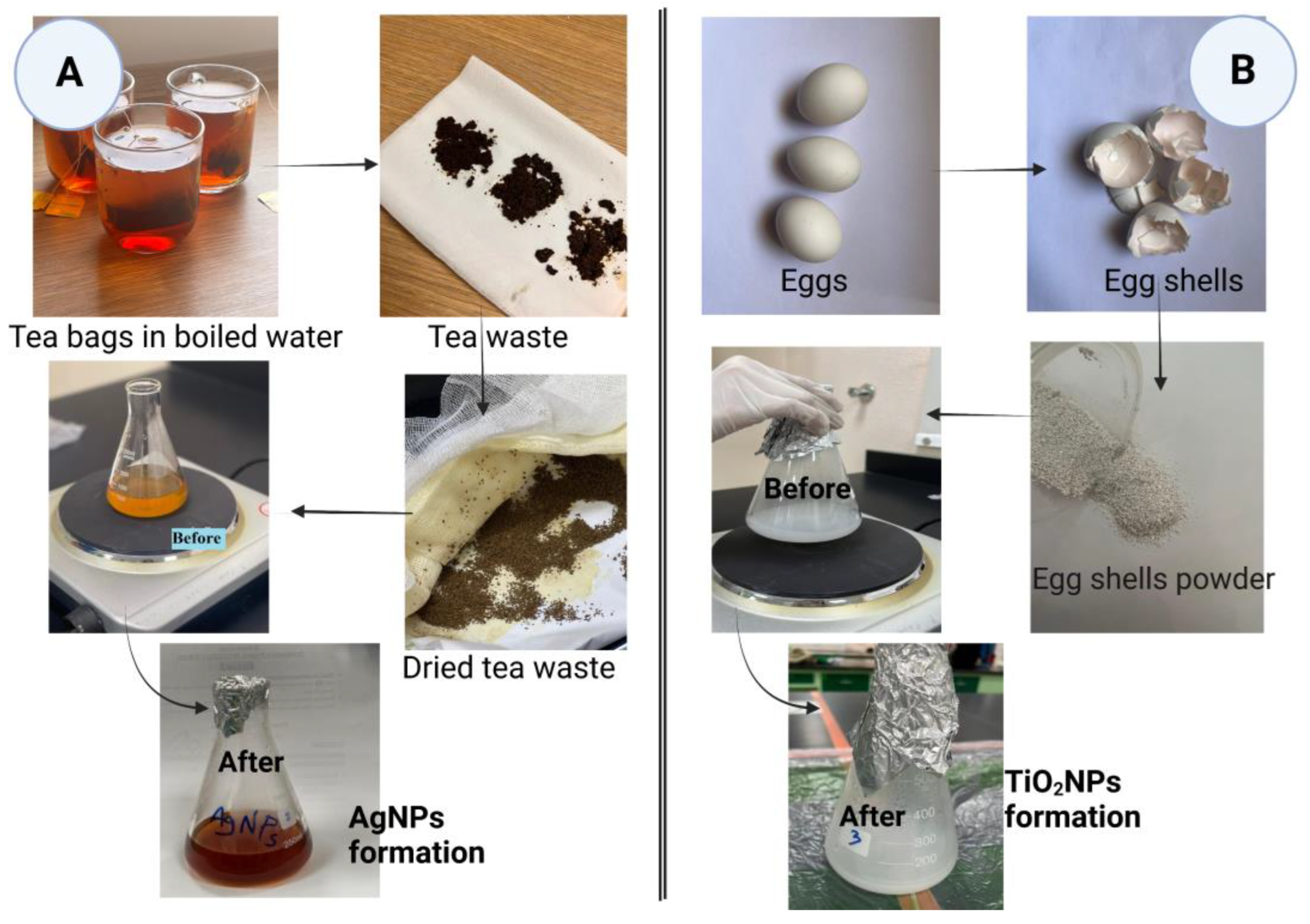
2.1. Preparation of Tea and Eggshell Waste Extracts

Black tea bags and eggs were purchased from a local shop in Taif governorate, Saudi Arabia (KSA). In the present study, we have used the most common wastes found in the kitchen: tea waste and eggshell. Collected white eggshell was washed well with distilled water before use. Tea waste was collected from its bags and spread on a clean tissue. Both tea waste and eggshell were left covered away from the sun until fully dry (for about 48 h). After that, eggshells were ground into fine powder using a domestic blender. For each waste product powder separately, tea waste (TE) or eggshell (ES) was mixed with autoclaved distilled water (1:10 w/v), boiled (10 min), filtered, and then cooled down at room temperature for further preparation. Finally, we obtained tea waste (TE), and eggshell (ESE) extracts that would be used later in silver (AgNPs) and titanium dioxide (TiO2NPs) nanoparticles green synthesis, respectively (Figure 1).

2.2. Green Synthesis of Nanoparticles Using Waste Products

For AgNPs biosynthesis: tea waste extract (TE) was mixed with an aqueous solution of silver nitrate (1:9 v/v, AgNO3, 10−3 M, Alfa Aesar, Kandel, Germany) and heated at 80 °C with continuous stirring until the color of the extract changed from dark yellow to dark brown (Figure 1) [27]. For TiO2NPs biosynthesis: eggshell extract (ESE) was mixed with aqueous titanium dioxide solution (10−3 M TiO2, Acros Organics, Geel, Belgium) at a ratio of 1:9 v/v and heated at 60 °C with continuous stirring until the extract changed to a milky color (Figure 1) [28,29]. Part of the green-synthesized NPs (AgNPs and TiO2NPs) was kept in its aqueous form for ultraviolet-visible (UV–VIS) spectrophotometric analysis. The rest of the biosynthesized NPs (AgNPs and TiO2NPs) were independently left overnight in glass Petri dishes in an oven (60 °C) until completely dry, and then the NPs were scraped out for further evaluation [30,31].

2.3. Characterization of Green AgNPs and TiO2NPs

Different techniques were used to characterize the green-synthesized AgNPs and TiO2NPs. First, the aqueous form of the biologically synthesized NPs was analyzed using an ultraviolet-visible (UV–VIS-NIR) spectrophotometer (UV-1601, Shimadzu, Kyoto, Japan) at a wavelength range of 200–800 nm. On the other hand, the powder form of AgNPs and TiO2NPs was used to measure their size using a Transmission electron microscope (TEM, JEOL–JSM-1400 PLUS, Tokyo, Japan). TEM images were analyzed using ImageJ software (1.53t) for accurate size analysis.
The shape of NPs was determined using the Scanning electron microscope (SEM) and X-ray diffractometer (XRD). For SEM analysis, NPs were coated with carbon (Cressington Sputter Coater, 108auto, thickness controller MTM-10, Watford, UK) for 10 min and then scanned at 20 kV using SEM (JEOL JSM-639OLA, Analytical Scanning Electron Microscope, at Electron microscope unit of Taif University) with various magnifications from ×500 (scale bars = 50 μm) to ×6000 (scale bars= 2 μm). XRD spectra were recorded using CuKα radiation (at 30 kV, and 100 mA) with a wavelength of 1.5406 Å in the 2θ (from the range of 20°–80°) and the temperature of data collection was 293.00 K. The XRD patterns of the synthesized AgNPs and TiO2NPs were analyzed according to standard values of the Joint Committee on Powder Diffraction Standards (JCPDS) card no. 04-0783 and 21-1272, respectively.
In addition, Dynamic light scattering (DLS, Zetasizer Nano ZN, Malvern Panalytical Ltd., Worcester, UK) was used for size analysis. Moreover, the NPs surface charge was analyzed by measuring their zeta potential distribution using the DLS equipment. Finally, Fourier Transforms Infrared Spectroscopy (FTIR, Agilent Technologies, Santa Clara, CA, USA, at wavelength ranges 450–4000 cm−1) was used to determine the possible functional groups found in TE and ESE that are responsible for the synthesis, capping, and stabilization of green AgNPs and TiO2NPs.

2.4. Cytotoxicity Assessment of NPs Using SRB

Human Skin Fibroblasts (HSF) were obtained from Nawah Scientific Inc. (Mokatam, Cairo, Egypt). The cells were maintained in Dulbecco’s Modified Eagle Medium (DMEM) media supplemented with 100 mg/mL streptomycin, 100 units/mL penicillin, and 10% heat-inactivated fetal bovine serum in a humidified, 5% (v/v) CO2 atmosphere at 37 °C. Cell viability was assessed using sulforhodamine B (SRB) assay. Aliquots of cell suspension (100 μL, 5 × 10³ cells) were added to 96-well plates and then incubated in DMEM media for 24 h. Cells were treated with another aliquot of 100 μL of media containing green NPs (AgNPs and TiO2NPs, separately) at various concentrations. After 72 h of NPs exposure, the cells were fixed by replacing the media with 150 μL of 10% trichloroacetic acid (TCA) and incubated at 4 °C for 1 h. The TCA solution was removed and the cells were washed with distilled water (5×). Aliquots of 70 μL of SRB solution (0.4% w/v) were added and incubated in the dark at room temperature (10 min). The plates were washed with acetic acid (1%, 3×) and air-dried overnight. Finally, 150 μL of TRIS (10 mM) was added to dissolve the protein-bound SRB stain, and absorbance was measured at 540 nm (BMGLABTECH®®-FLUOstar Omega microplate reader, Ortenberg, Germany) [32,33].
The effect of AgNPs and TiO2NPs was calculated as the percentage of control absorbance of the reduced dye at 570 nm according to the following equation of cell viability % (percentage):
Cell viability (%) = mean OD of treated cells/mean OD of control (untreated cells) × 100.
The data were plotted using the OriginLab program and IC50 values (50% inhibition concentration) for different prepared AgNPs and TiO2NPs at different sampling times were determined.

3. Results and Discussion

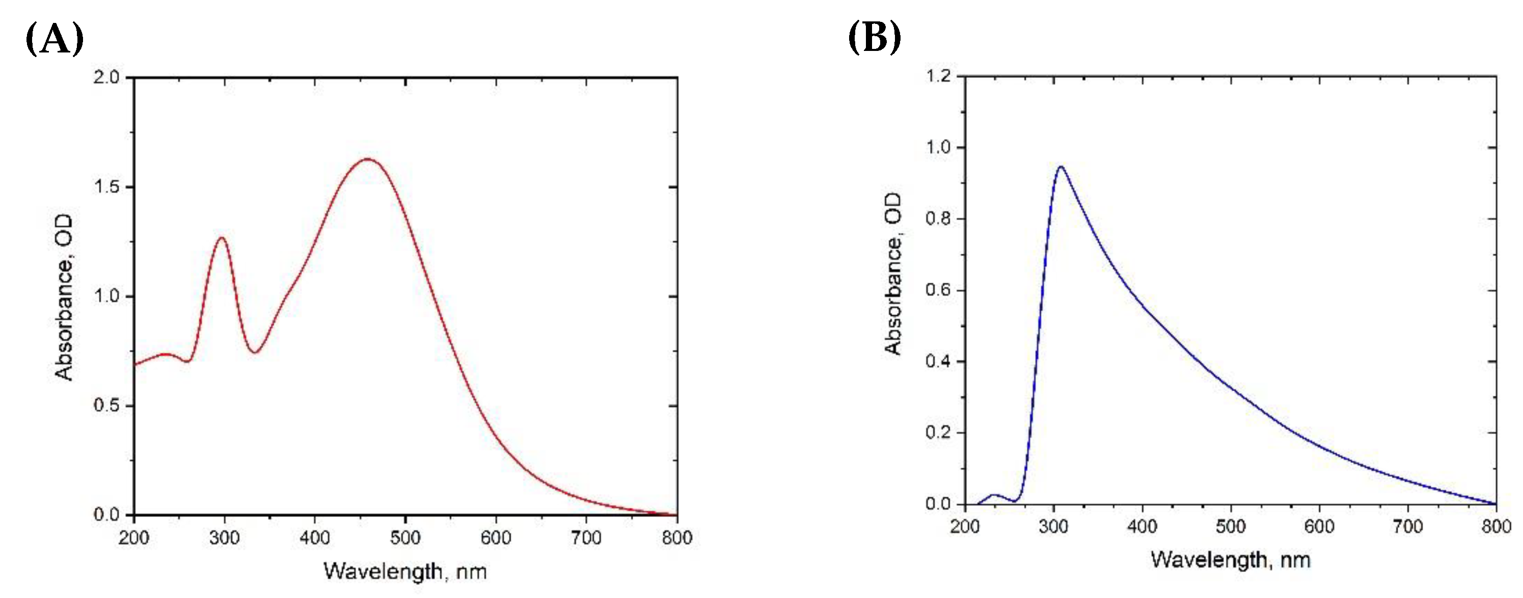
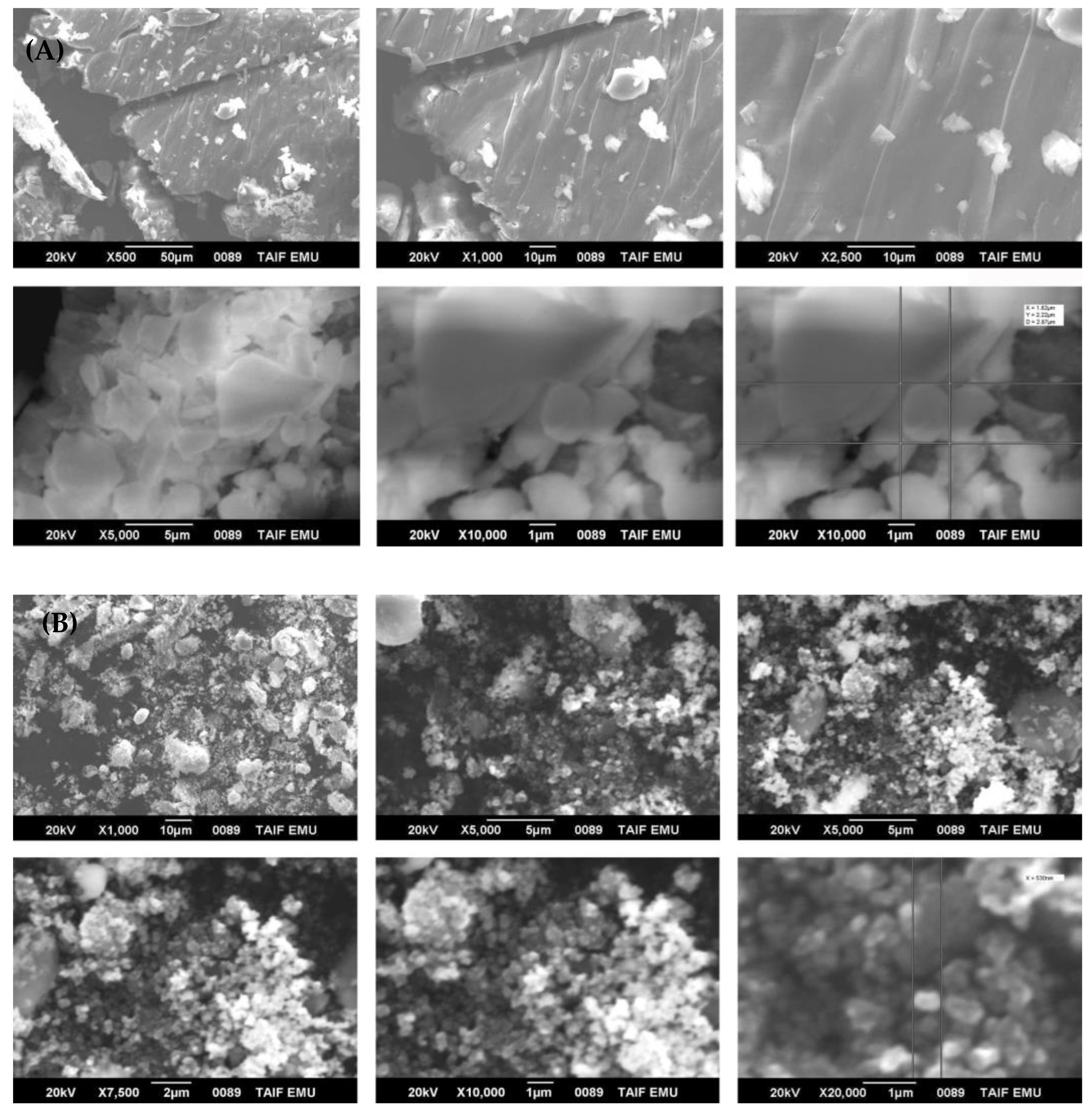
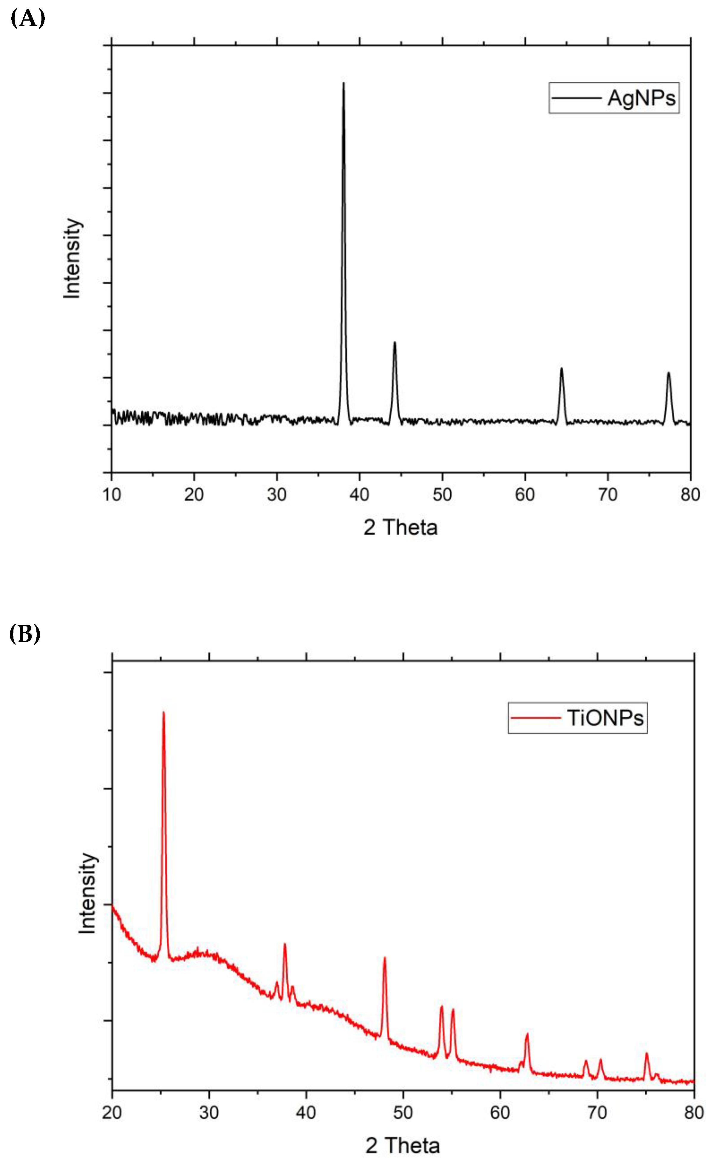
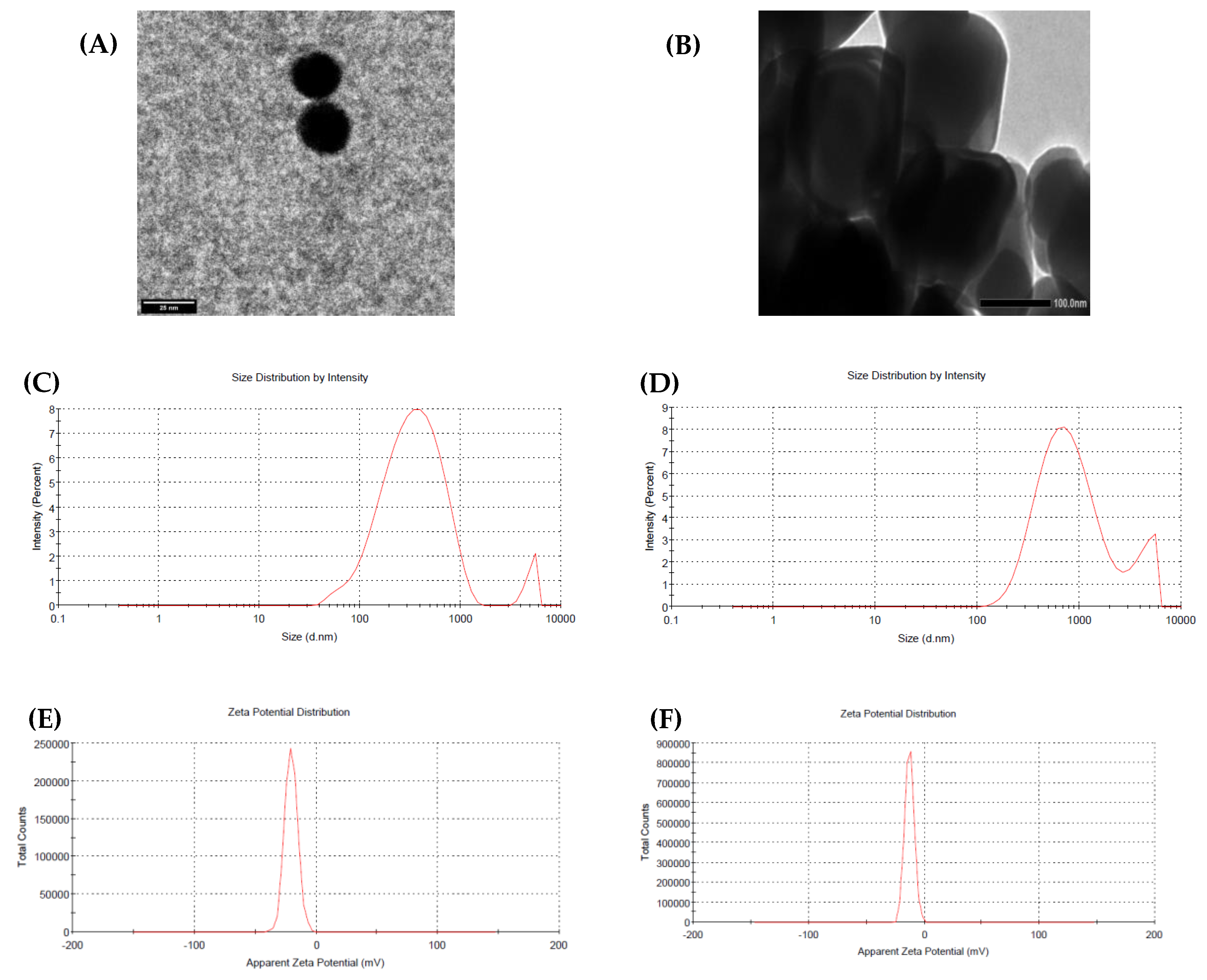
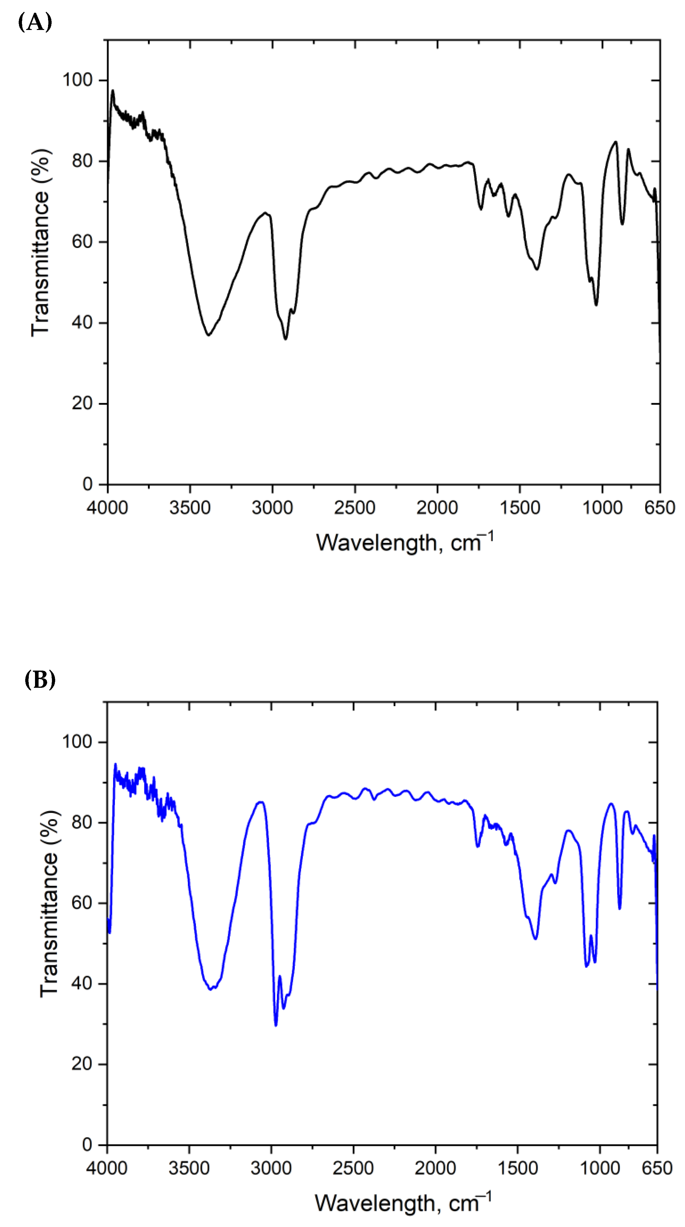
3.1. Characterization of Nanoparticles (NPs)

In the present study, we have used the most common waste product found in kitchens, worldwide, to biosynthesize AgNPs and TiO2NPs. Since eggs represent the primary source of protein, there is a large amount of egg consumption worldwide that makes eggshell wastes abundant and easy to obtain. On the other hand, tea (especially black tea) is the second most widely consumed beverage globally, after freshwater [34,35]. Therefore, the use of these wastes in NPs synthesis could help with saving raw materials, waste management, reducing environmental pollution, and help with the synthesis of safe NPs to be used on a large scale.
Silver (AgNPs_TE) and titanium dioxide (TiO2NPs_ESE) nanoparticles were successfully biosynthesized using tea waste (TE) and eggshell (ESE) extracts, respectively. The change in the color of TE extracts (from dark yellow to dark brown) and ESE (changed to a white milky color) was considered the primary indication of NPs formation, as shown in Figure 1. This was confirmed by UV–Visible spectrophotometer analysis, which represents the simplest way to confirm NPs formation due to their high surface plasmon resonances (SPR) [36]. As shown in Figure 2, AgNPs_TE UV-vis spectra show two sharp peaks at 300 and 470 nm (A), while TiO2NPs_ESE UV shows one sharp peak at 320 nm (B) that confirms their formation. The present result was consistent with previous studies that recorded AgNP formation due to Ag+ reduction to Ag0 using different waste extracts such as pomegranate fruit peel, coffee ground waste, and tea waste. This was proven by the change of extracts’ color and UV-vis spectra at a range of 390–440 nm [27,37]. On the other hand, TiO2NPs_ESE shows a maximum absorbance near the range of 320–350 nm, which proves titanium ions reduction to colloidal titanium with anatase phase. The present results agree with other reports that have revealed different absorption spectra of green-synthesized TiO2NPs using different plant extracts (range of 200–400 nm) such as Echinacea purpurea herba, Hibiscus flower, and Aloe vera [38,39]. SPR peak shifting of green synthesized AgNPs towards a higher wavelength is accompanied by a decrease in their size in comparison to TiO2NPs [40]. In addition, it was determined that the obtained SPR of synthesized NPs is affected by particle size, shape, surface charge, and agglomeration [41].
Scanning Electron Microscope (SEM) and XRD analyses were used to determine the shape of the formed NPs. Figure 3A shows the crystalline shape of biologically synthesized AgNPs using SEM at different magnifications. On the other hand, SEM images of TiO2NPs show random agglomeration of NPs with irregular sizes and shapes (Figure 3B). The crystalline shape of the nanoparticles was confirmed using XRD evaluation. AgNPs_TE shows four main standard peaks at 2θ = 38.3° (Ag plane = 111), 44.49° (200), 64.6° (220), and 77.5° (311) (Figure 4A). Those referenced peaks indicate the face-centered cubic (FCC) crystalline shape of AgNPs according to standard values of JCPDS card no. 04-0783. The FCC crystalline shape of AgNPs was previously detected using various plants and bio-waste extracts [26,41]. Al Sufyani et al. [42] and Abdelmigid et al. [27] have succeeded in the synthesis of AgNPs with FCC crystalline shape using Olea chrysophylla and Lavandula dentata leaf extracts, pomegranate peel, and coffee ground waste extracts, respectively.
The XRD pattern of the synthesized TiO2NPs revealed six main peaks at 2θ = 25.28° (plane = 101), 37.93°(004), 48.37° (200), 53.89° (105), 55.29° (211), and 62.73° (204) according to standard values of JCPDS no. 21-1272 (Figure 4B). Those main peaks refer to the crystalline shape of TiO2NPs with an anatase phase [43]. The present results agree with previous studies that synthesize TiO2NPs using agro-wastes (Punica granatum L. and coffee waste) with crystalline anatase phase [28]. In the TiO2NPs XRD pattern, the presence of other indefinite peaks could be attributed to organic or amorphous traces found in the preparation. This could be avoided later via calcination at temperatures higher than 400 °C. Ba-Abbad et al. [44] reported that during TiO2NPs synthesis, calcination up to 400 °C gives an amorphous phase of TiO2 due to defects acting as recombination centers for the photo-formed electrons [45]. On the other hand, TiO2 calcination at 500 °C or 600 °C gives a good crystalline shape with anatase or rutile phases with less degradation.
TEM and DLS techniques were used to determine the size of green synthesized AgNPs and TiO2NPs. AgNPs, formed using TE extract, show a very small size using TEM = 25 nm (Figure 5A), while their average size distribution using DLS is larger = 299.8 nm (Figure 5C). On the other hand, TiO2NPs have larger sizes using TEM = 120 nm and DLS = 742.9 nm, as shown in Figure 5B,D. Differences in their size were attributed to differences in the average zeta potential distribution of AgNPs = −20.5 mV and TiO2NPs = −12.6 mV (Figure 5E and Figure 5F, respectively). The present results agree with those of previous studies, in which AgNPs biologically synthesized using different biological extracts, such as coffee grounds (273.7 nm), pomegranate peel (591.9 nm), Olea chrysophylla leaf (328.6 nm), and Lavandula dentata leaf (284.5 nm), have larger sizes according to DLS [27,42].
It is known that an increase in zeta potential negativity, in turn, increases the repulsion between NPs and makes them smaller in size and more stable, as found in AgNPs. On the contrary, TiO2NPs have a lower negative charge on their surfaces that leads to their aggregation, as shown in SEM images, resulting in larger-sized NPs [46]. The size of the biosynthesized AgNPs and TiO2NPs was controlled by different factors during synthesis, including precursor concentration (AgNO3 and TiO2), the used extract (TE and ESE), pH, time, and temperature [47]. It was clear that DLS measurements of NPs size for both (AgNPs = 299.8 nm and TiO2NPs = 742.9 nm) were larger in size than their TEM analysis (AgNPs = 25 nm and TiO2NPs = 120 nm). Biologically prepared NPs have a mixture of different sizes, and DLS measurement is known to be more sensitive to larger particles (due to their light scattering) but may not be accurate for polydisperse samples [48,49].
Fourier transforms infrared (FTIR) spectroscopy was used to detect the functional groups that were found on the surfaces of green synthesized NPs. These functional groups are present in the used extracts (TE and ESE) that are responsible for the synthesis, capping, and stabilization of AgNPs and TiO2NPs. Figure 6A shows the functional groups present on the surfaces of green-synthesized AgNPs using TE. A wide band was observed at 3401 cm−1, which was attributed to the O–H and N–H stretching modes of polyphenols [50,51]. It was reported that 2917 and 2848 cm−1 peaks were attributed to the C–H and O–H stretch in alkane and carboxylic acid [51]. The peak at 1628 cm−1 was linked to the C=O and C=C bond stretching in polyphenols and aromatic rings, respectively [51,52]. The present results are consistent with those of previous studies that demonstrated the presence of polyphenols in different tea extracts (black, oolong, and green) due to O–H/N–H, C=C, C–O–C stretches at 3388 cm−1, 1636 cm−1, and 1039 cm−1 [51,52,53,54,55]. Tea extracts contain polyphenols, flavonoids, carboxylic acid, proteins, and caffeine that interact with silver cations (Ag+) to form AgNPs [56,57]. FTIR analysis of TiO2NPs using ESE showed sharp peaks at 720, 870, 1047/1060, and 1410/1418 cm−1, which can be attributed to the vibrations of carbonate CO3−2 anions (Figure 6B) [58,59]. This confirms the presence of CaCO3 in the eggshells. In addition, ESE contains organic compounds (proteins, glycoproteins, and proteoglycans [20]) that are present as weak peaks at 2359 and 2514 cm−1 [60]. Eggshell, as an organic waste, contains calcium carbonate that shares TiO2NPs’ special characteristics that can be used in different applications [61].

3.2. Biocompatibility of Green Synthesized AgNPs and TiO2NPs

Cell viability of the HSF cell line after treatment with serial dilutions of both NPs (AgNPs and TiO2NPs), separately at 72 h was evaluated using the SRB assay. This method monitors cell density based on cellular protein content determination. We chose to use human skin fibroblast cell lines in the present study because NPs are extensively used in cosmetics and skin care products, including AgNPs and TiO2NPs [62]. Therefore, it is important to assess the green-synthesized NPs on skin cells in an applicable way for their safety. Figure 7A shows a high cytotoxic effect of the green-synthesized AgNPs IC50 = 54.99 μg/mL, but TiO2NPs appear to be safer on the same cell line with IC50 > 100 (Figure 7B).
The present results show that green-synthesized TiO2NPs are safe for use in normal human skin fibroblast cell lines. It is known that TiO2NPs are widely used in a variety of cosmetic products, such as sunscreens, day creams, foundations, and lip balms [63]. This makes the biosynthesized TiO2NPs safer for use in contact with the skin than chemically synthesized ones. In addition, CaCO3 from eggshells exists on the surface of TiO2NPs, as proven by FTIR, playing a vital role in improving skin health and delaying aging [64]. Moreover, Chellappa et al. [65] determined the cytocompatibility of TiO2NPs on osteoblast-like MG63 cell lines at different concentrations of 1, 10, 25, 50, and 100 µg/mL at 24 and 48 h. They have reported that TiO2NPs improve cell viability/proliferation and have not shown any toxicity at various concentrations. Therefore, they have concluded that TiO2NPs are safe to be used in the biomedical field.
On the contrary, green-synthesized AgNPs using TE have a higher cytotoxic effect that could be attributed to their smaller size. But AgNPs have other different applications, including anticancer, antimicrobial, catalytic, larvicidal, and wound healing activities, due to their unique properties [66]. Previous in vitro studies have reported the toxic effects of AgNPs on different mammalian cells that are derived from the skin, brain, lung, liver, vascular system, and reproductive organs [67]. Al Sufyani et al. [68] have reported a higher toxic effect of AgNPs that are synthesized by Lavandula dentata L. leaves (IC50 = 44.95 μg/mL) on normal human gingival fibroblast GF01 cells than Olea chrysophylla (IC50 = 186.51 μg/mL) synthesized AgNPs. Therefore, many factors affect AgNP toxicity such as NPs size, surface charge, shape, capping agent, dosage, oxidation state, and aggregation [69,70]. The major toxicological mechanism of AgNPs has not been clear until now; however, different suggestions are present according to previous studies. Ahamed et al. [71] reported that AgNPs (50 and 100 µg/mL) have a role in the activation of apoptotic markers, caspase 3 and caspase 9 that cause apoptosis in mouse embryonic stem cells. In addition, AgNPs increase membrane leakage in mammalian germline stem cells, reduce mitochondrial function and antioxidant GSH content, and increase the generation of the reactive oxygen species (ROS) [72]. In turn, ROS production (peroxide, superoxide, and hydroxyl radicals) causes DNA damage such as single and double-strand DNA breaks, a multitude of oxidized base lesions, and abasic sites; these can be cytotoxic and/or mutagenic [73,74,75]. Excessive ROS production induces apoptosis and cell death in different cell culture models [76,77]. p53 accumulates in the cell in the presence of DNA damage and triggers cell cycle arrest to provide time for DNA repair. But if the damage is too extensive to be repaired, p53 triggers self-mediated apoptosis or death [78,79].

4. Conclusions

Biological waste products (TE and ESE) were used to synthesize silver and titanium dioxide nanoparticles using a green method. The synthesized nanoparticles (AgNPs and TiO2NPs) were evaluated using UV-VIS, SEM, TEM, DLS, Zeta potential, XRD, and FTIR. The biocompatibility of the green synthesized AgNPs and TiO2NPs was evaluated against HSF cell lines. Green synthesized TiO2NPs are safe for normal human skin fibroblasts because of their shape, large size, and other physicochemical properties. We suggest that according to our findings, TiO2NPs could not enter the cell membrane or damage DNA, making them safer than chemically synthesized NPs ones for use in cosmetic products. However, further in vivo studies are required to assess their toxicity. In addition, further future advancements in the possible antimicrobial and anticancer effects of green AgNPs and their mechanisms are strongly recommended.

Author Contributions

Conceptualization, N.A.H.; data curation, N.A.H.; funding acquisition, J.S.A.M.; investigation, N.A.H., S.O.A.T. and A.E.A.M.; methodology, J.S.A.M., N.A.H., L.M.A., S.O.A.T. and A.E.A.M.; project administration, J.S.A.M.; resources, J.S.A.M.; software, N.A.H.; validation, J.S.A.M. and N.A.H.; visualization, N.A.H.; writing—original draft, N.A.H. and L.M.A.; writing—review and editing, J.S.A.M., N.A.H., S.O.A.T. and A.E.A.M. All authors have read and agreed to the published version of the manuscript.

Funding

The researchers would like to acknowledge Deanship of Scientific Research, Taif University for funding this work.

Institutional Review Board Statement

Not applicable.

Informed Consent Statement

Not applicable.

Data Availability Statement

All data generated or analyzed during this study are included in this published article.

Acknowledgments

The researchers would like to acknowledge Deanship of Scientific Research, Taif University for funding this work.

Conflicts of Interest

The authors declare no conflict of interest.

References

  1. El-Nour, K.M.; Eftaiha, A.; Al-Wartgan, A.; Ammar, R.A. Synthesis and applications of silver nanoparticles. Arab. J. Chem. 2010, 3, 135–140. [Google Scholar] [CrossRef] [Green Version]
  2. Irvani, S.; Korbekandi, H.; Mirmohammadi, S.; Zolfaghari, B. Synthesis of silver nanoparticles: Chemical, physical and biological methods. Res. Pharm. Sci. 2014, 9, 385–406. [Google Scholar]
  3. Vishwanath, R.; Negi, B. Conventional and green methods of synthesis of silver nanoparticles and their antimicrobial properties. Curr. Opin. Green Sustain. Chem. 2021, 4, 100205. [Google Scholar] [CrossRef]
  4. Ovais, M.; Khalil, A.T.; Ayaz, M.; Ahmad, I.; Nethi, S.K.; Mukherjee, S. Biosynthesis of metal nanoparticles via microbial enzymes: A mechanistic approach. Int. J. Mol. Sci. 2018, 19, 4100. [Google Scholar] [CrossRef] [Green Version]
  5. Sportelli, M.C.; Izzi, M.; Annalisa, V.; Clemente, M.; Picca, R.A.; Ancona, A.; Cioffi, N. The pros and cons of the use of laser ablation synthesis for the production of silver nano-antimicrobials. Antibiotics 2018, 7, 67. [Google Scholar] [CrossRef] [Green Version]
  6. Chopra, H.; Bibi, S.; Singh, I.; Hasan, M.M.; Khan, M.S.; Yousafi, Q.; Baig, A.A.; Rahman, M.M.; Islam, F.; Emran, T.B.; et al. Green Metallic Nanoparticles: Biosynthesis to Applications. Front. Bioeng. Biotechnol. 2022, 10, 874742. [Google Scholar] [CrossRef]
  7. Rodríguez-Félix, F.; Graciano-Verdugo, A.Z.; Moreno-Vásquez, M.J.; Lagarda-Díaz, I.; Barreras-Urbina, C.G.; Armenta-Villegas, L.; Olguín-Moreno, A.; Tapia-Hernández, J.A. Trends in Sustainable Green Synthesis of Silver Nanoparticles Using Agri-Food Waste Extracts and Their Applications in Health. J. Nanomater. 2022, 2022, 8874003. [Google Scholar] [CrossRef]
  8. Debnath, B.; Haldar, D.; Purkait, M.K. Potential and sustainable utilization of tea waste: A review on present status and future trends. Environ. Chem. Eng. 2021, 9, 106179. [Google Scholar] [CrossRef]
  9. Khan, N.; Mukhtar, H. Tea and health: Studies in humans. Curr. Pharm. Des. 2013, 19, 6141–6147. [Google Scholar] [CrossRef] [Green Version]
  10. Su, L.J.; Arab, L. Tea consumption and the reduced risk of colon cancer—Results from a national prospective cohort study. Public Health Nutr. 2002, 5, 419–425. [Google Scholar] [CrossRef]
  11. Wang, J.; Zhang, W.; Sun, L.; Yu, H.; Ni, Q.X.; Risch, H.A.; Gao, Y.T. Green tea drinking and risk of pancreatic cancer: A large-scale, population-based case-control study in urban Shanghai. Cancer Epidemiol. 2012, 36, e354–e358. [Google Scholar] [CrossRef] [PubMed] [Green Version]
  12. Negishi, H.; Xu, J.W.; Ikeda, K.; Njelekela, M.; Nara, Y.; Yamori, Y. Black and green tea polyphenols attenuate blood pressure increases in stroke-prone spontaneously hypertensive rats. J. Nutr. 2004, 134, 38–42. [Google Scholar] [PubMed] [Green Version]
  13. Mikuls, T.R.; Cerhan, J.R.; Criswell, L.A.; Merlino, L.; Mudano, A.S.; Burma, M.; Folsom, A.R.; Saag, K.G. Coffee, tea, and caffeine consumption and risk of rheumatoid arthritis: Results from the Iowa Women’s Health Study. Arthritis Rheum. 2002, 46, 83–91. [Google Scholar] [CrossRef] [PubMed]
  14. Iso, H.; Date, C.; Wakai, K.; Fukui, M.; Tamakoshi, A. The relationship between green tea and total caffeine intake and risk for self-reported type 2 diabetes among Japanese adults. Ann. Intern. Med. 2006, 144, 554–562. [Google Scholar] [CrossRef] [PubMed]
  15. Yashin, A.Y.; Nemzer, B.V.; Combet, E.; Yashin, Y.I. Determination of the Chemical Composition of Tea by Chromatographic Methods: A Review. J. Food Res. 2015, 4, 56. [Google Scholar] [CrossRef] [Green Version]
  16. Rolim, W.R.; Pelegrino, M.T.; de Araújo Lima, B.; Ferraz, L.S.; Costa, F.N.; Bernardes, J.S.; Rodigues, T.; Brocchi, M.; Seabra, A.B. Green tea extract mediated biogenic synthesis of silver nanoparticles: Characterization, cytotoxicity evaluation and antibacterial activity. Appl. Surf. Sci. 2019, 463, 66–74. [Google Scholar] [CrossRef]
  17. Onitsuka, S.; Hamada, T.; Okamura, H. Preparation of antimicrobial gold and silver nanoparticles from tea leaf extracts. Colloids Surf. B. 2019, 173, 242–248. [Google Scholar] [CrossRef]
  18. Food and Agriculture Organization of the United Nations (FAO). FAOSTAT: Livestock Primary. Available online: https://www.fao.org/statistics/en/ (accessed on 2 September 2020).
  19. Faridi, H.; Arabhosseini, A. Application of eggshell wastes as valuable and utilizable products: A review. Res. Agric. Eng. 2018, 64, 104–114. [Google Scholar] [CrossRef] [Green Version]
  20. Hincke, M.T.; Nys, Y.; Gautron, J.; Mann, K.; Rodriguez-Navarro, A.B.; McKee, M.D. The eggshell: Structure, composition and mineralization. Front. Biosci. 2012, 17, 1266–1280. [Google Scholar] [CrossRef] [Green Version]
  21. Cordeiro, M.M.C.; Hincke, T.M. Recent Patents on Eggshell: Shell and Membrane Applications. Recent Pat. Food Nutr. Agric. 2012, 3, 1–8. [Google Scholar] [CrossRef]
  22. Chakraborty, S.; De, S.D. Eggshell: An alternative, cheap, bioavailable source of calcium in human diet. Res. Rev. J. Dairy Sci. Technol. 2019, 8, 25–33. [Google Scholar]
  23. Jalu, R.G.; Chamada, T.A.; Kasirajan, R. Calcium oxide nanoparticles synthesis from hen eggshells for removal of lead (Pb (II)) from aqueous solution. Environ. Chall. 2021, 4, 100193. [Google Scholar] [CrossRef]
  24. Ramya, S.; Vijayakumar, S.; Vidhya, E.; Bukhari, N.A.; Hatamleh, A.A.; Nilavukkarasi, M.; Pham, T.H. TiO2 nanoparticles derived from egg shell waste: Eco synthesis, characterization, biological and photocatalytic applications. Environ. Res. 2022, 214, 113829. [Google Scholar] [CrossRef] [PubMed]
  25. Whiley, H.; Ross, K. Salmonella and eggs: From production to plate. Int. J. Environ. Res. Public Health 2015, 12, 2543–2556. [Google Scholar] [CrossRef] [PubMed] [Green Version]
  26. Aswathi, V.P.; Meera, S.; Maria, C.A.; Nidhin, M. Green synthesis of nanoparticles from biodegradable waste extracts and their applications: A critical review. Nanotechnol. Environ. Eng. 2022, 8, 377–397. [Google Scholar] [CrossRef]
  27. Abdelmigid, H.M.; Morsi, M.M.; Hussien, N.A.; Alyamani, A.A.; Al Sufyani, N.M. Comparative Analysis of nanosilver Particles synthesized by different approaches and their antimicrobial efficacy. J. Nanomater. 2021, 2021, 2204776. [Google Scholar] [CrossRef]
  28. Abdelmigid, H.M.; Alyamani, A.A.; Hussien, N.A.; Morsi, M.M.; Alhumaidi, A. Integrated Approaches for Adsorption and Incorporation Testing of Green-Synthesized TiO2NPs Mediated by Seed-Priming Technology in Punica granatum L. Agronomy 2022, 12, 1601. [Google Scholar] [CrossRef]
  29. Swathi, N.; Sandhiya, D.; Rajeshkumar, S.; Lakshmi, T. Green synthesis of titanium dioxide nanoparticles using Cassia fistula and its antibacterial activity. Int. J. Res. Pharm. Sci. 2019, 10, 856–860. [Google Scholar] [CrossRef]
  30. Ahmad, W.; Jaiswal, K.K.; Soni, S. Green synthesis of titanium dioxide (TiO2) nanoparticles by using Mentha arvensis leaves extract and its antimicrobial properties. Inorg. Nano Met. Chem. 2020, 50, 1032–1038. [Google Scholar] [CrossRef]
  31. Raja, S.; Ramesh, V.; Thivaharan, V. Green biosynthesis of silver nanoparticles using Calliandra haematocephala leaf extract, their antibacterial activity and hydrogen peroxide sensing capability. Arab. J. Chem. 2017, 10, 253–261. [Google Scholar] [CrossRef] [Green Version]
  32. Skehan, P.; Storeng, R.; Scudiero, D.; Monks, A.; McMahon, J.; Vistica, D.; Warren, J.T.; Bokesch, H.; Kenney, S.; Boyd, M.R. New colorimetric cytotoxicity assay for anticancer-drug screening. J. Natl. Cancer Inst. 1990, 82, 1107–1112. [Google Scholar] [CrossRef] [PubMed]
  33. Allam, R.M.; Al-Abd, A.M.; Khedr, A.; Sharaf, O.A.; Nofal, S.M.; Khalifa, A.E.; Mosli, H.A.; Abdel-Naim, A.B. Fingolimod interrupts the cross talk between estrogen metabolism and sphingolipid metabolism within prostate cancer cells. Toxicol. Lett. 2018, 291, 77–85. [Google Scholar] [CrossRef] [PubMed]
  34. Katha, P.S.; Ahmed, Z.; Alam, R.; Saha, B.; Acharjee, A.; Rahman, M.S. Efficiency analysis of eggshell and tea waste as Low cost adsorbents for Cr removal from wastewater sample. S. Afr. J. Chem. Eng. 2021, 37, 186–195. [Google Scholar] [CrossRef]
  35. Valavanidis, A. Tea, the Most Popular Beverage Worldwide, is Beneficial to Human Health. Stud. Antioxid. Polyphenolic Const. Epidemiol. Evid. Dis. Prev. 2019, 1, 1–35. Available online: https://www.chem-tox-ecotox.org/ScientificReview (accessed on 14 May 2023).
  36. Mulvaney, P. Surface plasmon spectroscopy of nanosized metal particles. Langmuir 1996, 12, 788–800. [Google Scholar] [CrossRef]
  37. Rajput, D.; Paul, S.; Gupta, A. Green Synthesis of Silver Nanoparticles Using Waste Tea Leaves. Adv. Nan. Res 2020, 3, 1–14. [Google Scholar] [CrossRef]
  38. Bekele, E.T.; Gonfa, B.A.; Sabir, F.K. Use of Different Natural Products to Control Growth of Titanium Oxide Nanoparticles in Green Solvent Emulsion, Characterization, and Their Photocatalytic Application. Bioinorg. Chem. Appl. 2021, 2021, 6626313. [Google Scholar] [CrossRef]
  39. Waleczek, M.; Dendooven, J.; Dyachenko, P.; Petrov, A.Y.; Eich, M.; Blick, R.H.; Detavernier, C.; Nielsch, K.; Furlan, K.P.; Zierold, R. Influence of Alumina Addition on the Optical Properties and the Thermal Stability of Titania Thin Films and Inverse Opals Produced by Atomic Layer Deposition. Nanomaterials 2021, 11, 1053. [Google Scholar] [CrossRef]
  40. Lakkappa, B.A.; Jasmith, S.C.; Prabhuodeyara, M.G. Effect of concentration and pH on the size of silver nanoparticles synthesized by green chemistry. Int. J. Med. Chem. 2017, 3, 5444. [Google Scholar]
  41. Saeb, A.T.; Alshammari, A.S.; Al-Brahim, H.; Al-Rubeaan, K.A. Production of silver nanoparticles with strong and stable antimicrobial activity against highly pathogenic and multidrug resistant bacteria. Sci. World J. 2014, 2014, 704708. [Google Scholar] [CrossRef] [Green Version]
  42. Al Sufyani, N.M.; Hussien, N.A.; Hawsawi, Y.M. Characterization and anticancer potential of silver nanoparticles biosynthesized from Olea chrysophylla and Lavandula dentata leaf extracts on HCT116 colon cancer cells. J. Nanomater. 2019, 2019, 7361695. [Google Scholar] [CrossRef] [Green Version]
  43. Prabhu, C.A.; Silambarasan, D.; Sarika, R.; Selvam, V. Synthesis and characterization of TiO2. Mater. Today Proc. 2022, 64, 1793–1797. [Google Scholar] [CrossRef]
  44. Ba-Abbad, M.M.; Kadhum, A.A.H.; Mohamad, A.B.; Takriff, M.S.; Sopian, K. Synthesis and catalytic activity of TiO2 nanoparticles for photochemical oxidation of concentrated chlorophenols under direct solar radiation. Int. J. Electrochem. Sci. 2012, 7, 4871–4888. [Google Scholar] [CrossRef]
  45. Sakthivel, S.; Hidalgo, M.C.; Bahnemann, D.W.; Geissen, S.U.; Murugesan, V.; Vogelpohl, A. A fine route to tune the photocatalytic activity of TiO2. Appl. Catal. B 2006, 63, 31–40. [Google Scholar] [CrossRef]
  46. Sundrarajan, M.; Bama, K.; Bhavani, M.; Jegatheeswaran, S.; Ambika, S.; Sangili, A.; Nithya, P.; Sumathi, R. Obtaining titanium dioxide nanoparticles with spherical shape and antimicrobial properties using M. citrifolia leaves extract by hydrothermal method. J. Photochem. Photobiol. 2017, 171, 117–124. [Google Scholar]
  47. Amin, G.; Asif, M.H.; Zainelabdin, A.; Zaman, S.; Nur, O.; Willander, M. Influence of pH, Precursor Concentration, Growth Time, and Temperature on the Morphology of ZnO Nanostructures Grown by the Hydrothermal Method. J. Nanomater. 2011, 2011, 269692. [Google Scholar] [CrossRef] [Green Version]
  48. Jang, M.H.; Lee, S.; Hwang, Y.S. Characterization of Silver Nanoparticles under Environmentally Relevant Conditions Using Asymmetrical Flow Field-Flow Fractionation (AF4). PLoS ONE 2015, 10, e0143149. [Google Scholar] [CrossRef]
  49. Domingos, R.F.; Baalousha, M.A.; Ju-Nam, Y.; Reid, N.; Tufenkji, N.; Lead, J.R.; Leppard, G.G.; Wilkinson, K.J. Characterizing manufactured nanoparticles in the Environment: Multimethod determination of particle sizes. Environ. Sci. Technol. 2009, 43, 7277–7284. [Google Scholar] [CrossRef]
  50. Loo, Y.Y.; Chieng, B.W.; Nishibuchi, M.; Radu, S. Synthesis of silver nanoparticles by using tea leaf extract from Camellia sinensis. Int. J. Nanomed. 2012, 7, 4263–4267. [Google Scholar]
  51. Senthilkumar, S.R.; Sivakumar, T. Green tea (Camellia sinensis) mediated synthesis of zinc oxide (ZnO) nanoparticles and studies on their antimicrobial activities. Int. J. Pharm. Pharm. Sci. 2014, 6, 461–465. [Google Scholar]
  52. Dubey, S.P.; Sillanpaa, M.; Varma, R.S. Reduction of hexavalent chromium using Sorbaria sorbifolia aqueous leaf extract. Appl. Sci. 2017, 7, 715. [Google Scholar] [CrossRef] [Green Version]
  53. Cai, J.; Wang, Y.; Xi, X.; Li, H.; Wei, X. Using FTIR spectra and pattern recognition for discrimination of tea varieties. Int. J. Biol. Macromol. 2015, 78, 439–446. [Google Scholar] [CrossRef] [Green Version]
  54. Huang, L.; Weng, X.; Chen, Z.; Megharaj, M.; Naidu, R. Synthesis of iron-based nanoparticles using oolong tea extract for the degradation of malachite green. Spectrochim. Acta A Mol. Biomol. Spectrosc. 2014, 117, 801–804. [Google Scholar] [CrossRef] [PubMed]
  55. Weng, X.; Huang, L.; Chen, Z.; Megharaj, M.; Naidu, R. Synthesis of iron-based nanoparticles by green tea extract and their degradation of malachite. Ind. Crops Prod. 2013, 51, 342–347. [Google Scholar] [CrossRef]
  56. Wu, D.; Bird, M.R. The interaction of protein and polyphenol species in ready to drink black tea liquor production. J. Food Process Eng. 2010, 33, 481–505. [Google Scholar] [CrossRef]
  57. Islam, M.A.; Nikoloutsou, Z.; Sakkas, V.; Papatheodorou, M.; Albanis, T. Statistical optimisation by combination of response surface methodology and desirability function for removal of azo dye from aqueous solution. J. Environ. Anal. Chem. 2010, 90, 497–509. [Google Scholar] [CrossRef]
  58. Engin, B.; Demirtas, H.; Eken, M. Temperature effects on egg shells investigated by XRD, IR and ESR techniques. Radiat. Phys. Chem. 2006, 75, 268–277. [Google Scholar] [CrossRef]
  59. Eswararao, Y.; Niju, S.; Begum, K.M.M.S.; Anantharaman, N.; Raj, S.M. Transesterification of jatropha oil using a mixture of natural shells as solid catalyst. Biofuels 2016, 7, 345–351. [Google Scholar] [CrossRef]
  60. Nakanot, T.; Ikawani, N.I.; Ozimek, L. Chemical composition of chicken eggshell and shell membranes. Poultry Sci. 2003, 82, 510–514. [Google Scholar] [CrossRef]
  61. Nikhil, P.; Sudheer, P.; Joy, P.; Sruthi, T.; Vangalpati, M. Synthesis and characterization of nanoparticles using egg shell. Mater. Today Proc. 2020, 26, 3246–3249. [Google Scholar] [CrossRef]
  62. Gupta, V.; Mohapatra, S.; Mishra, H.; Farooq, U.; Kumar, K.; Ansari, M.J.; Aldawsari, M.F.; Alalaiwe, A.S.; Mirza, M.A.; Iqbal, Z. Nanotechnology in Cosmetics and Cosmeceuticals—A Review of Latest Advancements. Gels 2022, 8, 173. [Google Scholar] [CrossRef] [PubMed]
  63. Dréno, B.; Alexis, A.; Chuberre, B.; Marinovich, M. Safety of titanium dioxide nanoparticles in cosmetics. J. Eur. Acad. Dermatol. Venereol. 2019, 33 (Suppl. S7), 34–46. [Google Scholar] [CrossRef] [PubMed] [Green Version]
  64. Herman, H.; Minhajat, R.; Muis, M.; Umar, B.T.; Cangara, M.H.; Zainuddin, A.A. Review of the Benefits of Eggshell Content in Body Tissue Structure Repair. Mal. J. Med. Health Sci. 2023, 19, 278–285. [Google Scholar] [CrossRef]
  65. Chellappa, M.; Anjaneyulu, U.; Manivasagam, G.; Vijayalakshmi, U. Preparation and evaluation of the cytotoxic nature of TiO2 nanoparticles by direct contact method. Int. J. Nanomed. 2015, 10 (Suppl. S2), 31–41. [Google Scholar] [CrossRef] [Green Version]
  66. Jannathul Firdhouse, M.; Lalitha, P. Biosynthesis of Silver Nanoparticles and Its Applications. J. Nanotechnol. 2015, 2015, 829526. [Google Scholar] [CrossRef] [Green Version]
  67. Liao, C.; Li, Y.; Tjong, S.C. Bactericidal and Cytotoxic Properties of Silver Nanoparticles. Int. J. Mol. Sci. 2019, 20, 449. [Google Scholar] [CrossRef] [Green Version]
  68. Al Sufyani, N.M.; Hussien, N.A.; Hawsawi, Y.M. Cytotoxic effect of synthesized silver nanoparticles on normal human gingival fibroblast GF01 cells. In Proceedings of the 2021 International Conference of Women in Data Science at Taif University (WiDSTaif), Taif, Saudi Arabia, 30–31 March 2021; pp. 1–6. [Google Scholar] [CrossRef]
  69. Butler, K.S.; Peeler, D.J.; Casey, B.J.; Dair, B.J.; Elespuru, R.K. Silver nanoparticles: Correlating nanoparticle size and cellular uptake with genotoxicity. Mutagenesis 2015, 30, 577–591. [Google Scholar] [CrossRef]
  70. Abdal Dayem, A.; Hossain, M.; Lee, S.; Kim, K.; Saha, S.; Yang, G.-M.; Choi, H.; Cho, S.-G. The role of reactive oxygen species (ROS) in the biological activities of metallic nanoparticles. Int. J. Mol. Sci. 2017, 18, 120. [Google Scholar] [CrossRef] [Green Version]
  71. Ahamed, M.; Karns, M.; Goodson, M.; Rowe, J.; Hussain, S.M.; Schlager, J.J.; Hong, Y. DNA damage response to different surface chemistry of silver nanoparticles in mammalian cells. Toxicol. Appl. Pharmacol. 2008, 233, 404–410. [Google Scholar] [CrossRef]
  72. Braydich-Stolle, L.; Hussain, S.M.; Schlager, J.J.; Hoffman, M.C. In vitro cytotoxicty of nanoparticles in mammalian germline stem cells. Toxicol. Sci. 2005, 88, 412–419. [Google Scholar] [CrossRef] [Green Version]
  73. Bhakat, K.K.; Mokkapati, S.K.; Boldogh, I.; Hazra, T.K.; Mitra, S. Acetylation of human 8-oxoguanine-DNA glycosylase by p300 and its role in 8-oxoguanine repair in vivo. Mol. Cell Biol. 2006, 26, 1654–1665. [Google Scholar] [CrossRef] [PubMed] [Green Version]
  74. Diakowska, D.; Lewandowski, A.; Kopec, W.; Diakowski, W.; Chrzanowska, T. Oxidative DNA damage and total antioxidant status in serum of patients with esophageal squamous cell carcinoma. Hepatogastroenterology 2007, 78, 1701–1704. [Google Scholar]
  75. Paz-Elizur, T.; Sevilya, Z.; Leitner-Dagan, Y.; Elinger, D.; Roisman, L.C.; Livneh, Z. DNA repair of oxidative DNA damage in human carcinogenesis: Potential application for cancer risk assessment and prevention. Cancer Lett. 2008, 266, 60–72. [Google Scholar] [CrossRef] [Green Version]
  76. Hussain, S.M.; Frazier, J.M. Involvement of apoptosis in hydrazine induced toxicity in rat primary hepatocytes. Toxicol. In Vitro 2003, 17, 343–355. [Google Scholar] [CrossRef] [PubMed]
  77. Li, G.Y.; Osborne, N.N. Oxidative-induced apoptosis to an immortalized ganglion cell line is caspase independent but involves the activation of poly (ADP-ribose) polymerase and apoptosis-inducing factor. Brain Res. 2008, 188, 35–43. [Google Scholar] [CrossRef]
  78. Liang, S.H.; Clarke, M.F. Regulation of p53 location. Eur. J. Biochem. 2001, 289, 2779–2783. [Google Scholar] [CrossRef]
  79. Sherr, C.J. Principles of tumor suppression. Cell 2004, 11, 235–246. [Google Scholar] [CrossRef] [Green Version]
Figure 1. Schematic illustration of AgNPs (A) and TiO2NPs (B) preparation using tea waste extract (TE), and eggshell extract (ESE). Created in BioRender.com, with agreement number: WZ25FWOG19, accessed on 3 June 2023.
Sustainability 15 11858 g001
Figure 2. UV-Visible spectral analysis of green synthesized AgNPs (A) and TiO2NPs (B).
Sustainability 15 11858 g002
Figure 3. Scanning electron microscope (SEM) images showing the crystalline shape of AgNPs (A) and TiO2NPs (B) with different magnifications.
Sustainability 15 11858 g003
Figure 4. XRD patterns of green synthesized AgNPs (A) and TiO2NPs (B).
Sustainability 15 11858 g004
Figure 5. Average size of green synthesized AgNPs (25 nm (A), 299.8 nm (C)) and TiO2NPs (120 nm (B), 742.9 nm (D)) using TEM and DLS, respectively. The average zeta potential distribution of AgNPs = −20.5 mV (E) and TiO2NPs = −12.6 mV (F).
Sustainability 15 11858 g005
Figure 6. FTIR spectra of green synthesized AgNPs (A) and TiO2NPs (B).
Sustainability 15 11858 g006
Figure 7. Percentage of cell viability of HSF according to serial dilutions treatment of AgNPs (A) and TiO2NPs (B) after 72 h.
Sustainability 15 11858 g007
Disclaimer/Publisher’s Note: The statements, opinions and data contained in all publications are solely those of the individual author(s) and contributor(s) and not of MDPI and/or the editor(s). MDPI and/or the editor(s) disclaim responsibility for any injury to people or property resulting from any ideas, methods, instructions or products referred to in the content.

