
The Relationship between Post-Traumatic Stress Disorder Due to Brain Injury and Glutamate Intake: A Systematic Review
 , and
, and 
Abstract
1. Introduction
2. Materials and Methods
2.1. Protocol and Registration
2.2. Literature Search
2.3. Inclusion Criteria
2.4. Exclusion Criteria
2.5. Study Screening and Selection
2.6. Statistical Analysis
3. Results
3.1. Search Results
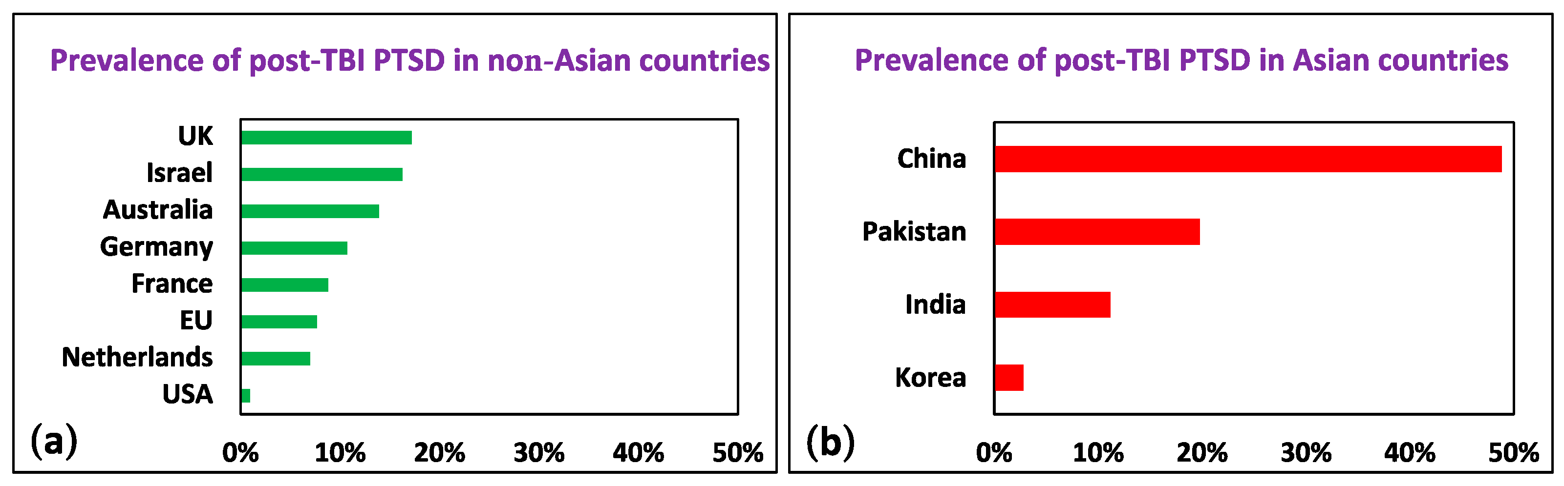
3.2. Comparison of Study Groups
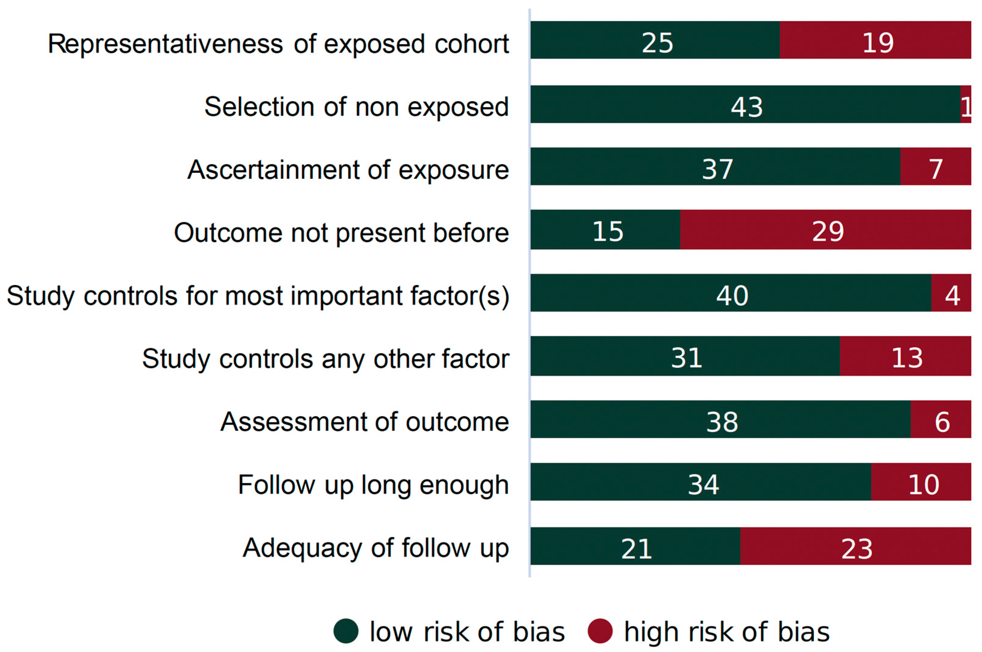
3.3. Article Quality
4. Discussion
4.1. The Relationship between PTSD and TBI
4.2. The Relationship between BBB and PTSD
4.3. TBI and the Disruption of the BBB
4.3.1. BBB Regulation in CNS Disorders
4.3.2. Mechanisms of the BBB Dysfunction in TBI
4.4. The Relationship between Neurodegeneration and the BBB
4.5. The Relationship between Neurodegeneration and PTSD
4.6. The Relationship between Neurodegeneration and Glutamate Neurotoxicity
4.7. Impaired BBB Permeability Disturbance in the Balance of Glutamate Concentration between the Blood and Brain Compartments
4.8. The Role of Diet on Blood Glutamate Concentration: The Involvement of Glutamate in Neurocognitive Processes
4.9. Current and Potential Therapeutic Strategies for Post-TBI PTSD
4.10. Limitations
5. Conclusions
Supplementary Materials
Author Contributions
Funding
Data Availability Statement
Acknowledgments
Conflicts of Interest
References
- Maas, A.I.R.; Menon, D.K.; Adelson, P.D.; Andelic, N.; Bell, M.J.; Belli, A.; Bragge, P.; Brazinova, A.; Büki, A.; Chesnut, R.M. Traumatic brain injury: Integrated approaches to improve prevention, clinical care, and research. Lancet Neurol. 2017, 16, 987–1048. [Google Scholar] [CrossRef]
- Peterson, C.; Miller, G.F.; Barnett, S.B.L.; Florence, C. Economic cost of injury—United States, 2019. Morb. Mortal. Wkly. Rep. 2021, 70, 1655. [Google Scholar] [CrossRef]
- Ahmed, S.; Venigalla, H.; Mekala, H.M.; Dar, S.; Hassan, M.; Ayub, S. Traumatic brain injury and neuropsychiatric complications. Indian J. Psychol. Med. 2017, 39, 114–121. [Google Scholar] [CrossRef]
- Torregrossa, W.; Raciti, L.; Rifici, C.; Rizzo, G.; Raciti, G.; Casella, C.; Naro, A.; Calabrò, R.S. Behavioral and Psychiatric Symptoms in Patients with Severe Traumatic Brain Injury: A Comprehensive Overview. Biomedicines 2023, 11, 1449. [Google Scholar] [CrossRef] [PubMed]
- Frank, D.; Gruenbaum, B.F.; Shelef, I.; Severynovska, O.; Gal, R.; Dubilet, M.; Zlotnik, A.; Kofman, O.; Boyko, M. Blood glutamate scavenging with pyruvate as a novel preventative and therapeutic approach for depressive-like behavior following traumatic brain injury in a rat model. Front. Neurosci. 2022, 16, 832478. [Google Scholar] [CrossRef] [PubMed]
- Frank, D.; Gruenbaum, B.F.; Shelef, I.; Zvenigorodsky, V.; Severynovska, O.; Fleidervish, I.; Knyazer, B.; Frenkel, A.; Zlotnik, A.; Kofman, O. Blood glutamate scavenging as a novel glutamate-based therapeutic approach for post-traumatic brain injury anxiety and social impairment. Transl. Psychiatry 2023, 13, 41. [Google Scholar] [CrossRef] [PubMed]
- Boyko, M.; Gruenbaum, B.F.; Shelef, I.; Zvenigorodsky, V.; Severynovska, O.; Binyamin, Y.; Knyazer, B.; Frenkel, A.; Frank, D.; Zlotnik, A. Traumatic brain injury-induced submissive behavior in rats: Link to depression and anxiety. Transl. Psychiatry 2022, 12, 239. [Google Scholar] [CrossRef] [PubMed]
- King, N.S. PTSD and traumatic brain injury: Folklore and fact? Brain Inj. 2008, 22, 1–5. [Google Scholar] [CrossRef]
- Sbordone, R.J.; Liter, J.C. Mild traumatic brain injury does not produce post-traumatic stress disorder. Brain Inj. 1995, 9, 405–412. [Google Scholar] [CrossRef] [PubMed]
- Sbordone, R.J.; Ruff, R.M. Re-examination of the controversial coexistence of traumatic brain injury and posttraumatic stress disorder: Misdiagnosis and self-report measures. Psychol. Inj. Law 2010, 3, 63–76. [Google Scholar] [CrossRef]
- Bryant, R. Post-traumatic stress disorder vs traumatic brain injury. Dialogues Clin. Neurosci. 2011, 13, 251–262. [Google Scholar] [CrossRef]
- Villalobos, D.; Bivona, U. Post-traumatic stress disorder after severe traumatic brain injury: A systematic review. Arch. Clin. Neuropsychol. 2022, 37, 583–594. [Google Scholar] [CrossRef]
- Iljazi, A.; Ashina, H.; Al-Khazali, H.M.; Ashina, M.; Winther Schytz, H.; Ashina, S. Post-traumatic stress disorder attributed to traumatic brain injury in children–a systematic review. Brain Inj. 2020, 34, 857–863. [Google Scholar] [CrossRef]
- Iljazi, A.; Ashina, H.; Al-Khazali, H.M.; Lipton, R.B.; Ashina, M.; Schytz, H.W.; Ashina, S. Post-traumatic stress disorder after traumatic brain injury—A systematic review and meta-analysis. Neurol. Sci. 2020, 41, 2737–2746. [Google Scholar] [CrossRef]
- Esagoff, A.I.; Stevens, D.A.; Kosyakova, N.; Woodard, K.; Jung, D.; Richey, L.N.; Daneshvari, N.O.; Luna, L.P.; Bray, M.J.C.; Bryant, B.R. Neuroimaging Correlates of Post-Traumatic Stress Disorder in Traumatic Brain Injury: A Systematic Review of the Literature. J. Neurotrauma 2023, 40, 1029–1044. [Google Scholar] [CrossRef]
- Uiterwijk, D.; Stargatt, R.; Humphrey, S.; Crowe, S.F. The relationship between cognitive functioning and symptoms of depression, anxiety, and post-traumatic stress disorder in adults with a traumatic brain injury: A meta-analysis. Neuropsychol. Rev. 2022, 32, 758–806. [Google Scholar] [CrossRef]
- Van Praag, D.L.G.; Cnossen, M.C.; Polinder, S.; Wilson, L.; Maas, A.I.R. Post-traumatic stress disorder after civilian traumatic brain injury: A systematic review and meta-analysis of prevalence rates. J. Neurotrauma 2019, 36, 3220–3232. [Google Scholar] [CrossRef]
- Stein, M.B.; Jain, S.; Giacino, J.T.; Levin, H.; Dikmen, S.; Nelson, L.D.; Vassar, M.J.; Okonkwo, D.O.; Diaz-Arrastia, R.; Robertson, C.S. Risk of posttraumatic stress disorder and major depression in civilian patients after mild traumatic brain injury: A TRACK-TBI study. JAMA Psychiatry 2019, 76, 249–258. [Google Scholar] [CrossRef] [PubMed]
- Bryant, R.A.; Marosszeky, J.E.; Crooks, J.; Gurka, J.A. Posttraumatic stress disorder after severe traumatic brain injury. Am. J. Psychiatry 2000, 157, 629–631. [Google Scholar] [CrossRef] [PubMed]
- Heim, C.; Schultebraucks, K.; Marmar, C.R.; Nemeroff, C.B. Neurobiological pathways involved in fear, stress, and PTSD. In Post-Traumatic Stress Disorder; Oxford University Press: Oxford, UK, 2018; pp. 331–352. [Google Scholar]
- Vasterling, J.J.; Jacob, S.N.; Rasmusson, A. Traumatic brain injury and posttraumatic stress disorder: Conceptual, diagnostic, and therapeutic considerations in the context of co-occurrence. J. Neuropsychiatry Clin. Neurosci. 2018, 30, 91–100. [Google Scholar] [CrossRef] [PubMed]
- Sherin, J.E.; Nemeroff, C.B. Post-traumatic stress disorder: The neurobiological impact of psychological trauma. Dialogues Clin. Neurosci. 2011, 13, 263–278. [Google Scholar] [CrossRef] [PubMed]
- Risbrough, V.B.; Vaughn, M.N.; Friend, S.F. Role of inflammation in traumatic brain injury–associated risk for neuropsychiatric disorders: State of the evidence and where do we go from here. Biol. Psychiatry 2022, 91, 438–448. [Google Scholar] [CrossRef] [PubMed]
- Rajkumar, R.P. Biomarkers of Neurodegeneration in Post-Traumatic Stress Disorder: An Integrative Review. Biomedicines 2023, 11, 1465. [Google Scholar] [CrossRef] [PubMed]
- Malejko, K.; Abler, B.; Plener, P.L.; Straub, J. Neural correlates of psychotherapeutic treatment of post-traumatic stress disorder: A systematic literature review. Front. Psychiatry 2017, 8, 85. [Google Scholar] [CrossRef] [PubMed]
- Brady, K.T.; Killeen, T.K.; Brewerton, T.; Lucerini, S. Comorbidity of psychiatric disorders and posttraumatic stress disorder. J. Clin. Psychiatry 2000, 61, 22–32. [Google Scholar] [PubMed]
- Qassem, T.; Aly-ElGabry, D.; Alzarouni, A.; Abdel-Aziz, K.; Arnone, D. Psychiatric co-morbidities in post-traumatic stress disorder: Detailed findings from the adult psychiatric morbidity survey in the English population. Psychiatr. Q. 2021, 92, 321–330. [Google Scholar] [CrossRef]
- Hoskins, M.D.; Bridges, J.; Sinnerton, R.; Nakamura, A.; Underwood, J.F.G.; Slater, A.; Lee, M.R.D.; Clarke, L.; Lewis, C.; Roberts, N.P. Pharmacological therapy for post-traumatic stress disorder: A systematic review and meta-analysis of monotherapy, augmentation and head-to-head approaches. Eur. J. Psychotraumatol. 2021, 12, 1802920. [Google Scholar] [CrossRef]
- Williamson, J.B.; Heilman, K.M.; Porges, E.C.; Lamb, D.G.; Porges, S.W. A possible mechanism for PTSD symptoms in patients with traumatic brain injury: Central autonomic network disruption. Front. Neuroeng. 2013, 6, 13. [Google Scholar] [CrossRef]
- Monsour, M.; Ebedes, D.; Borlongan, C.V. A review of the pathology and treatment of TBI and PTSD. Exp. Neurol. 2022, 351, 114009. [Google Scholar] [CrossRef]
- Gruenbaum, B.F.; Zlotnik, A.; Frenkel, A.; Fleidervish, I.; Boyko, M. Glutamate Efflux across the Blood–Brain Barrier: New Perspectives on the Relationship between Depression and the Glutamatergic System. Metabolites 2022, 12, 459. [Google Scholar] [CrossRef]
- Gruenbaum, B.F.; Zlotnik, A.; Fleidervish, I.; Frenkel, A.; Boyko, M. Glutamate Neurotoxicity and Destruction of the Blood–Brain Barrier: Key Pathways for the Development of Neuropsychiatric Consequences of TBI and Their Potential Treatment Strategies. Int. J. Mol. Sci. 2022, 23, 9628. [Google Scholar] [CrossRef]
- Boyko, M.; Gruenbaum, B.F.; Oleshko, A.; Merzlikin, I.; Zlotnik, A. Diet’s Impact on Post-Traumatic Brain Injury Depression: Exploring Neurodegeneration, Chronic Blood–Brain Barrier Destruction, and Glutamate Neurotoxicity Mechanisms. Nutrients 2023, 15, 4681. [Google Scholar] [CrossRef]
- Page, M.J.; McKenzie, J.E.; Bossuyt, P.M.; Boutron, I.; Hoffmann, T.C.; Mulrow, C.D.; Shamseer, L.; Tetzlaff, J.M.; Akl, E.A.; Brennan, S.E. The PRISMA 2020 statement: An updated guideline for reporting systematic reviews. Int. J. Surg. 2021, 88, 105906. [Google Scholar] [CrossRef]
- Lamberty, G.J.; Nelson, N.W.; Yamada, T. Effects and outcomes in civilian and military traumatic brain injury: Similarities, differences, and forensic implications. Behav. Sci. Law 2013, 31, 814–832. [Google Scholar] [CrossRef]
- Denning, J.H.; Shura, R.D. Cost of malingering mild traumatic brain injury-related cognitive deficits during compensation and pension evaluations in the veterans benefits administration. Appl. Neuropsychol. Adult 2019, 26, 1–16. [Google Scholar] [CrossRef]
- Wells, G.; Shea, B.; O’Connell, D.; Peterson, J.; Welch, V.; Losos, M.; Tugwell, P. Newcastle-Ottawa Quality Assessment Scale Cohort Studies; University of Ottawa: Ottawa, ON, USA, 2014. [Google Scholar]
- Stolze, T.; Franke, S.; Haybaeck, J.; Moehler, M.; Grimminger, P.P.; Lang, H.; Roth, W.; Gockel, I.; Kreuser, N.; Bläker, H. Mismatch repair deficiency, chemotherapy and survival for resectable gastric cancer: An observational study from the German staR cohort and a meta-analysis. J. Cancer Res. Clin. Oncol. 2023, 149, 1007–1017. [Google Scholar] [CrossRef]
- Petousis, S.; Christidis, P.; Margioula-Siarkou, C.; Liberis, A.; Vavoulidis, E.; Margioula-Siarkou, G.; Vatopoulou, A.; Papanikolaou, A.; Mavromatidis, G.; Dinas, K. Axillary lymph node dissection vs. sentinel node biopsy for early-stage clinically node-negative breast cancer: A systematic review and meta-analysis. Arch. Gynecol. Obstet. 2022, 306, 1221–1234. [Google Scholar] [CrossRef] [PubMed]
- Bhutta, M.S.; Shechter, O.; Gallo, E.S.; Martin, S.D.; Jones, E.; Doncel, G.F.; Borenstein, R. Ginkgolic acid inhibits herpes simplex virus type 1 skin infection and prevents zosteriform spread in mice. Viruses 2021, 13, 86. [Google Scholar] [CrossRef]
- Bryant, R.A.; Marosszeky, J.E.; Crooks, J.; Baguley, I.; Gurka, J. Coping style and post-traumatic stress disorder following severe traumatic brain injury. Brain Inj. 2000, 14, 175–180. [Google Scholar] [PubMed]
- Bryant, R.A.; Marosszeky, J.E.; Crooks, J.; Baguley, I.J.; Gurka, J.A. Posttraumatic stress disorder and psychosocial functioning after severe traumatic brain injury. J. Nerv. Ment. Dis. 2001, 189, 109–113. [Google Scholar] [CrossRef] [PubMed]
- Bryant, R.A.; Harvey, A.G. Relationship between acute stress disorder and posttraumatic stress disorder following mild traumatic brain injury. Am. J. Psychiatry 1998, 155, 625–629. [Google Scholar] [CrossRef] [PubMed]
- Bryant, R.A.; Harvey, A.G. The influence of traumatic brain injury on acute stress disorder and post-traumatic stress disorder following motor vehicle accidents. Brain Inj. 1999, 13, 15–22. [Google Scholar] [CrossRef] [PubMed]
- Xu, C.; Li, Q.; Gao, Y.; Huo, H.; Zhang, W. Changes and influencing factors of stress disorder in patients with mild traumatic brain injury stress disorder. BioMed Res. Int. 2022, 2022, 9082946. [Google Scholar] [CrossRef] [PubMed]
- Dieter, J.N.I.; Engel, S.D. Traumatic brain injury and posttraumatic stress disorder: Comorbid consequences of war. Neurosci. Insights 2019, 14, 1179069519892933. [Google Scholar] [CrossRef] [PubMed]
- Ashman, T.A.; Spielman, L.A.; Hibbard, M.R.; Silver, J.M.; Chandna, T.; Gordon, W.A. Psychiatric challenges in the first 6 years after traumatic brain injury: Cross-sequential analyses of Axis I disorders. Arch. Phys. Med. Rehabil. 2004, 85, 36–42. [Google Scholar] [CrossRef] [PubMed]
- Hendrickson, R.C.; Schindler, A.G.; Pagulayan, K.F. Untangling PTSD and TBI: Challenges and strategies in clinical care and research. Curr. Neurol. Neurosci. Rep. 2018, 18, 106. [Google Scholar] [CrossRef] [PubMed]
- Sumpter, R.E.; McMillan, T.M. Errors in self-report of post-traumatic stress disorder after severe traumatic brain injury. Brain Inj. 2006, 20, 93–99. [Google Scholar] [CrossRef] [PubMed]
- Glaesser, J.; Neuner, F.; Lütgehetmann, R.; Schmidt, R.; Elbert, T. Posttraumatic stress disorder in patients with traumatic brain injury. BMC Psychiatry 2004, 4, 5. [Google Scholar] [CrossRef]
- Sfera, A.; Osorio, C.; Rahman, L.; Zapata-Martín del Campo, C.M.; Maldonado, J.C.; Jafri, N.; Cummings, M.A.; Maurer, S.; Kozlakidis, Z. PTSD as an endothelial disease: Insights from COVID-19. Front. Cell. Neurosci. 2021, 15, 770387. [Google Scholar] [CrossRef]
- Ni, K.; Zhu, J.; Xu, X.; Liu, Y.; Yang, S.; Huang, Y.; Xu, R.; Jiang, L.; Zhang, J.; Zhang, W. Hippocampal Activated Microglia May Contribute to Blood–Brain Barrier Impairment and Cognitive Dysfunction in Post-Traumatic Stress Disorder-Like Rats. J. Mol. Neurosci. 2022, 72, 975–982. [Google Scholar] [CrossRef]
- Taghadosi, Z.; Zarifkar, A.; Razban, V.; Owjfard, M.; Aligholi, H. Effect of chronically electric foot shock stress on spatial memory and hippocampal blood brain barrier permeability. Behav. Brain Res. 2021, 410, 113364. [Google Scholar] [CrossRef]
- Welcome, M.O.; Mastorakis, N.E. Stress-induced blood brain barrier disruption: Molecular mechanisms and signaling pathways. Pharmacol. Res. 2020, 157, 104769. [Google Scholar] [CrossRef]
- Wu, Y.; Wu, H.; Guo, X.; Pluimer, B.; Zhao, Z. Blood–brain barrier dysfunction in mild traumatic brain injury: Evidence from preclinical murine models. Front. Physiol. 2020, 11, 1030. [Google Scholar] [CrossRef]
- Price, L.; Wilson, C.; Grant, G. Blood–brain barrier pathophysiology following traumatic brain injury. In Translational Research in Traumatic Brain Injury; CRC Press/Taylor and Francis Group: Boca Raton, FL, USA, 2016. [Google Scholar]
- Archie, S.R.; Al Shoyaib, A.; Cucullo, L. Blood-Brain Barrier Dysfunction in CNS Disorders and Putative Therapeutic Targets: An Overview. Pharmaceutics 2021, 13, 1779. [Google Scholar] [CrossRef] [PubMed]
- Kealy, J.; Greene, C.; Campbell, M. Blood-brain barrier regulation in psychiatric disorders. Neurosci. Lett. 2020, 726, 133664. [Google Scholar] [CrossRef] [PubMed]
- Xiao, M.; Xiao, Z.J.; Yang, B.; Lan, Z.; Fang, F. Blood-brain barrier: More contributor to disruption of central nervous system homeostasis than victim in neurological disorders. Front. Neurosci. 2020, 14, 764. [Google Scholar] [CrossRef] [PubMed]
- Hu, Q.; Manaenko, A. Blood-brain Barrier Dysfunction in Cerebrovascular Diseases. Curr. Neuropharmacol. 2020, 18, 1166. [Google Scholar] [CrossRef] [PubMed]
- Segarra, M.; Aburto, M.R.; Acker-Palmer, A. Blood–brain barrier dynamics to maintain brain homeostasis. Trends Neurosci. 2021, 44, 393–405. [Google Scholar] [CrossRef] [PubMed]
- Chodobski, A.; Zink, B.J.; Szmydynger-Chodobska, J. Blood–brain barrier pathophysiology in traumatic brain injury. Transl. Stroke Res. 2011, 2, 492–516. [Google Scholar] [CrossRef]
- Toyota, S.; Graf, R.; Valentino, M.; Yoshimine, T.; Heiss, W.-D. Malignant Infarction in Cats After Prolonged Middle Cerebral Artery Occlusion: Glutamate Elevation Related to Decrease of Cerebral Perfusion Pressure. Stroke 2002, 33, 1383–1391. [Google Scholar] [CrossRef][Green Version]
- Hone, E.A.; Hu, H.; Sprowls, S.A.; Farooqi, I.; Grasmick, K.; Lockman, P.R.; Simpkins, J.W.; Ren, X. Biphasic blood-brain barrier openings after stroke. Neurol. Disord. Stroke Int. 2018, 1, 1011. [Google Scholar]
- Başkaya, M.K.; Rao, A.M.; Doğan, A.; Donaldson, D.; Dempsey, R.J. The biphasic opening of the blood–brain barrier in the cortex and hippocampus after traumatic brain injury in rats. Neurosci. Lett. 1997, 226, 33–36. [Google Scholar] [CrossRef] [PubMed]
- Chamoun, R.; Suki, D.; Gopinath, S.P.; Goodman, J.C.; Robertson, C. Role of extracellular glutamate measured by cerebral microdialysis in severe traumatic brain injury. J. Neurosurg. 2010, 113, 564–570. [Google Scholar] [CrossRef] [PubMed]
- Bullock, R.; Zauner, A.; Woodward, J.J.; Myseros, J.; Choi, S.C.; Ward, J.D.; Marmarou, A.; Young, H.F. Factors affecting excitatory amino acid release following severe human head injury. J. Neurosurg. 1998, 89, 507–518. [Google Scholar] [CrossRef]
- Vespa, P.; Prins, M.; Ronne-Engstrom, E.; Caron, M.; Shalmon, E.; Hovda, D.A.; Martin, N.A.; Becker, D.P. Increase in extracellular glutamate caused by reduced cerebral perfusion pressure and seizures after human traumatic brain injury: A microdialysis study. J. Neurosurg. 1998, 89, 971–982. [Google Scholar] [CrossRef]
- Yu, M.; Wang, M.; Yang, D.; Wei, X.; Li, W. Dynamics of blood brain barrier permeability and tissue microstructure following controlled cortical impact injury in rat: A dynamic contrast-enhanced magnetic resonance imaging and diffusion kurtosis imaging study. Magn. Reson. Imaging 2019, 62, 1–9. [Google Scholar] [CrossRef]
- van Vliet, E.A.; Ndode-Ekane, X.E.; Lehto, L.J.; Gorter, J.A.; Andrade, P.; Aronica, E.; Gröhn, O.; Pitkänen, A. Long-lasting blood-brain barrier dysfunction and neuroinflammation after traumatic brain injury. Neurobiol. Dis. 2020, 145, 105080. [Google Scholar] [CrossRef]
- Hay, J.; Johnson, V.; Young, A.; Smith, D.; Stewart, W. Blood-Brain Barrier Disruption Is an Early Event That May Persist for Many Years After Traumatic Brain Injury in Humans. J. Neuropathol. Exp. Neurol. 2015, 74, 1147–1157. [Google Scholar] [CrossRef] [PubMed]
- Cash, A.; Theus, M.H. Mechanisms of blood–brain barrier dysfunction in traumatic brain injury. Int. J. Mol. Sci. 2020, 21, 3344. [Google Scholar] [CrossRef]
- Bai, W.; Zhou, Y.-G. Homeostasis of the Intraparenchymal-Blood Glutamate Concentration Gradient: Maintenance, Imbalance, and Regulation. Front. Mol. Neurosci. 2017, 10, 400. [Google Scholar] [CrossRef]
- Wu, S.; Yin, Y.; Du, L. Blood–brain barrier dysfunction in the pathogenesis of major depressive disorder. Cell. Mol. Neurobiol. 2022, 42, 2571–2591. [Google Scholar] [CrossRef]
- Knox, E.G.; Aburto, M.R.; Clarke, G.; Cryan, J.F.; O’Driscoll, C.M. The blood-brain barrier in aging and neurodegeneration. Mol. Psychiatry 2022, 27, 2659–2673. [Google Scholar] [CrossRef]
- Neylan, T.C. Post-traumatic Stress Disorder and Neurodegeneration. Am. J. Geriatr. Psychiatry 2020, 28, 61–63. [Google Scholar] [CrossRef]
- Neylan, T.C. Posttraumatic Stress Disorder and Risk For Dementia. Alzheimer’s Dement. 2022, 18, e060705. [Google Scholar] [CrossRef]
- Chan, Y.-L.E.; Bai, Y.-M.; Hsu, J.-W.; Huang, K.-L.; Su, T.-P.; Li, C.-T.; Lin, W.-C.; Pan, T.-L.; Chen, T.-J.; Tsai, S.-J. Post-traumatic stress disorder and risk of parkinson disease: A nationwide longitudinal study. Am. J. Geriatr. Psychiatry 2017, 25, 917–923. [Google Scholar] [CrossRef]
- Miller, M.W.; Sadeh, N. Traumatic stress, oxidative stress and post-traumatic stress disorder: Neurodegeneration and the accelerated-aging hypothesis. Mol. Psychiatry 2014, 19, 1156–1162. [Google Scholar] [CrossRef]
- McGrath, T.; Baskerville, R.; Rogero, M.; Castell, L. Emerging Evidence for the Widespread Role of Glutamatergic Dysfunction in Neuropsychiatric Diseases. Nutrients 2022, 14, 917. [Google Scholar] [CrossRef]
- Lau, A.; Tymianski, M. Glutamate receptors, neurotoxicity and neurodegeneration. Pflügers Arch. Eur. J. Physiol. 2010, 460, 525–542. [Google Scholar] [CrossRef]
- Al-Nasser, M.N.; Mellor, I.R.; Carter, W.G. Is L-Glutamate Toxic to Neurons and Thereby Contributes to Neuronal Loss and Neurodegeneration? A Systematic Review. Brain Sci. 2022, 12, 577. [Google Scholar] [CrossRef] [PubMed]
- Borbély, É.; Simon, M.; Fuchs, E.; Wiborg, O.; Czéh, B.; Helyes, Z. Novel drug developmental strategies for treatment-resistant depression. Br. J. Pharmacol. 2022, 179, 1146–1186. [Google Scholar] [CrossRef] [PubMed]
- Berman, R.M.; Cappiello, A.; Anand, A.; Oren, D.A.; Heninger, G.R.; Charney, D.S.; Krystal, J.H. Antidepressant effects of ketamine in depressed patients. Biol. Psychiatry 2000, 47, 351–354. [Google Scholar] [CrossRef] [PubMed]
- Blaylock, R.L.; Faria, M. New concepts in the development of schizophrenia, autism spectrum disorders, and degenerative brain diseases based on chronic inflammation: A working hypothesis from continued advances in neuroscience research. Surg. Neurol. Int. 2021, 12, 556. [Google Scholar] [PubMed]
- Frank, D.; Kuts, R.; Tsenter, P.; Gruenbaum, B.F.; Grinshpun, Y.; Zvenigorodsky, V.; Shelef, I.; Natanel, D.; Brotfain, E.; Zlotnik, A. The effect of pyruvate on the development and progression of post-stroke depression: A new therapeutic approach. Neuropharmacology 2019, 155, 173–184. [Google Scholar] [CrossRef] [PubMed]
- Belov Kirdajova, D.; Kriska, J.; Tureckova, J.; Anderova, M. Ischemia-triggered glutamate excitotoxicity from the perspective of glial cells. Front. Cell. Neurosci. 2020, 14, 51. [Google Scholar] [CrossRef] [PubMed]
- Simões, A.P.; Silva, C.G.; Marques, J.M.; Pochmann, D.; Porciúncula, L.O.; Ferreira, S.; Oses, J.P.; Beleza, R.O.; Real, J.I.; Köfalvi, A. Glutamate-induced and NMDA receptor-mediated neurodegeneration entails P2Y1 receptor activation. Cell Death Dis. 2018, 9, 297. [Google Scholar] [CrossRef]
- Kumar, A.; Singh, R.L.; Babu, G.N. Cell death mechanisms in the early stages of acute glutamate neurotoxicity. Neurosci. Res. 2010, 66, 271–278. [Google Scholar] [CrossRef]
- Lewerenz, J.; Maher, P. Chronic glutamate toxicity in neurodegenerative diseases—What is the evidence? Front. Neurosci. 2015, 9, 469. [Google Scholar] [CrossRef]
- Meldrum, B.S. The role of glutamate in epilepsy and other CNS disorders. Neurology 1994, 44 (Suppl. S8), S14–S23. [Google Scholar]
- Meldrum, B.; Garthwaite, J. Excitatory amino acid neurotoxicity and neurodegenerative disease. Trends Pharmacol. Sci. 1990, 11, 379–387. [Google Scholar] [CrossRef]
- Zhou, Y.; Danbolt, N.C. Glutamate as a neurotransmitter in the healthy brain. J. Neural Transm. 2014, 121, 799–817. [Google Scholar] [CrossRef] [PubMed]
- Hawkins, R.A.; Viña, J.R. How Glutamate Is Managed by the Blood-Brain Barrier. Biology 2016, 5, 37. [Google Scholar] [CrossRef]
- Teichberg, V.I.; Cohen-Kashi-Malina, K.; Cooper, I.; Zlotnik, A. Homeostasis of glutamate in brain fluids: An accelerated brain-to-blood efflux of excess glutamate is produced by blood glutamate scavenging and offers protection from neuropathologies. Neuroscience 2009, 158, 301–308. [Google Scholar] [CrossRef] [PubMed]
- Haroon, E.; Miller, A.H. Inflammation Effects on Brain Glutamate in Depression: Mechanistic Considerations and Treatment Implications. In Inflammation-Associated Depression: Evidence, Mechanisms and Implications; Current Topics in Behavioral Neurosciences; Springer International Publishing: Cham, Switzerland, 2016; Volume 31, pp. 173–198. [Google Scholar]
- Haroon, E.; Chen, X.; Li, Z.; Patel, T.; Woolwine, B.J.; Hu, X.P.; Felger, J.C.; Miller, A.H. Increased inflammation and brain glutamate define a subtype of depression with decreased regional homogeneity, impaired network integrity, and anhedonia. Transl. Psychiatry 2018, 8, 189. [Google Scholar] [CrossRef] [PubMed]
- King, S.; Jelen, L.A.; Horne, C.M.; Cleare, A.; Pariante, C.M.; Young, A.H.; Stone, J.M. Inflammation, Glutamate, and Cognition in Bipolar Disorder Type II: A Proof of Concept Study. Front. Psychiatry 2019, 10, 66. [Google Scholar] [CrossRef]
- Gasiorowska, A.; Wydrych, M.; Drapich, P.; Zadrozny, M.; Steczkowska, M.; Niewiadomski, W.; Niewiadomska, G. The biology and pathobiology of glutamatergic, cholinergic, and dopaminergic signaling in the aging brain. Front. Aging Neurosci. 2021, 391, 654931. [Google Scholar] [CrossRef] [PubMed]
- Ferrini, F.; De Koninck, Y. Microglia control neuronal network excitability via BDNF signalling. Neural Plast. 2013, 2013, 429815. [Google Scholar] [CrossRef] [PubMed]
- Sasaki-Hamada, S.; Sanai, E.; Kanemaru, M.; Kamanaka, G.; Oka, J.-I. Long-term exposure to high glucose induces changes in the expression of AMPA receptor subunits and glutamate transmission in primary cultured cortical neurons. Biochem. Biophys. Res. Commun. 2022, 589, 48–54. [Google Scholar] [CrossRef] [PubMed]
- Boyko, M.; Gruenbaum, B.F.; Frank, D.; Natanel, D.; Negev, S.; Azab, A.N.; Barsky, G.; Knyazer, B.; Kofman, O.; Zlotnik, A. The Integrity of the Blood–Brain Barrier as a Critical Factor for Regulating Glutamate Levels in Traumatic Brain Injury. Int. J. Mol. Sci. 2023, 24, 5897. [Google Scholar] [CrossRef] [PubMed]
- Markits, I.H.S. Monosodium Glutamate (MSG) Chemical Economics Handbook. 2018, pp. 1–88. Available online: https://ihsmarkit.com/products/monosodium-glutamate-chemical-economics-handbook.html (accessed on 1 January 2024).
- Kazmi, Z.; Fatima, I.; Perveen, S.; Malik, S.S. Monosodium glutamate: Review on clinical reports. Int. J. Food Prop. 2017, 20, 1807–1815. [Google Scholar] [CrossRef]
- Insawang, T.; Selmi, C.; Cha’on, U.; Pethlert, S.; Yongvanit, P.; Areejitranusorn, P.; Boonsiri, P.; Khampitak, T.; Tangrassameeprasert, R.; Pinitsoontorn, C. Monosodium glutamate (MSG) intake is associated with the prevalence of metabolic syndrome in a rural Thai population. Nutr. Metab. 2012, 9, 50. [Google Scholar] [CrossRef]
- Rutten, E.P.A.; Engelen, M.P.K.J.; Wouters, E.F.M.; Schols, A.M.W.J.; Deutz, N.E.P. Metabolic effects of glutamine and glutamate ingestion in healthy subjects and in persons with chronic obstructive pulmonary disease. Am. J. Clin. Nutr. 2006, 83, 115–123. [Google Scholar] [PubMed]
- Stegink, L.D.; Filer Jr, L.J.; Baker, G.L. Effect of carbohydrate on plasma and erythrocyte glutamate levels in humans ingesting large doses of monosodium L-glutamate in water. Am. J. Clin. Nutr. 1983, 37, 961–968. [Google Scholar] [CrossRef] [PubMed]
- Fernstrom, J.D.; Cameron, J.L.; Fernstrom, M.H.; McConaha, C.; Weltzin, T.E.; Kaye, W.H. Short-term neuroendocrine effects of a large oral dose of monosodium glutamate in fasting male subjects. J. Clin. Endocrinol. Metab. 1996, 81, 184–191. [Google Scholar] [PubMed]
- Graham, T.E.; Sgro, V.; Friars, D.; Gibala, M.J. Glutamate ingestion: The plasma and muscle free amino acid pools of resting humans. Am. J. Physiol. Endocrinol. Metab. 2000, 278, E83–E89. [Google Scholar] [CrossRef] [PubMed]
- Tsai, P.-J.; Huang, P.-C. Circadian variations in plasma and erythrocyte glutamate concentrations in adult men consuming a diet with and without added monosodium glutamate. J. Nutr. 2000, 130, 1002S–1004S. [Google Scholar] [CrossRef] [PubMed]
- Stegink, L.D.; Filer, L.J., Jr.; Baker, G.L. Plasma and erythrocyte amino acid levels in normal adult subjects fed a high protein meal with and without added monosodium glutamate. J. Nutr. 1982, 112, 1953–1960. [Google Scholar] [CrossRef] [PubMed]
- Loï, C.; Cynober, L. Glutamate: A safe nutrient, not just a simple additive. Ann. Nutr. Metab. 2022, 78, 133–146. [Google Scholar] [CrossRef]
- Hajihasani, M.M.; Soheili, V.; Zirak, M.R.; Sahebkar, A.; Shakeri, A. Natural products as safeguards against monosodium glutamateinduced toxicity. Iran. J. Basic Med. Sci. 2020, 23, 416. [Google Scholar]
- López-Pérez, S.J.; Ureña-Guerrero, M.E.; Morales-Villagrán, A. Monosodium glutamate neonatal treatment as a seizure and excitotoxic model. Brain Res. 2010, 1317, 246–256. [Google Scholar] [CrossRef]
- Beas-Zárate, C.; Pérez-Vega, M.a.I.; González-Burgos, I. Neonatal exposure to monosodium L-glutamate induces loss of neurons and cytoarchitectural alterations in hippocampal CA1 pyramidal neurons of adult rats. Brain Res. 2002, 952, 275–281. [Google Scholar] [CrossRef]
- Dief, A.E.; Kamha, E.S.; Baraka, A.M.; Elshorbagy, A.K. Monosodium glutamate neurotoxicity increases beta amyloid in the rat hippocampus: A potential role for cyclic AMP protein kinase. Neurotoxicology 2014, 42, 76–82. [Google Scholar] [CrossRef]
- Frieder, B.; Grimm, V.E. Prenatal monosodium glutamate (MSG) treatment given through the mother’s diet causes behavioral deficits in rat offspring. Int. J. Neurosci. 1984, 23, 117–126. [Google Scholar] [CrossRef]
- Frieder, B.; Grimm, V.E. Prenatal Monosodium Glutamate Causes Long-Lasting Cholinergic and Adrenergic Changes in Various Brain Regions. J. Neurochem. 1987, 48, 1359–1365. [Google Scholar] [CrossRef]
- Hashem, H.E.; El-Din Safwat, M.D.; Algaidi, S. The effect of monosodium glutamate on the cerebellar cortex of male albino rats and the protective role of vitamin C (histological and immunohistochemical study). J. Mol. Histol. 2012, 43, 179–186. [Google Scholar] [CrossRef] [PubMed]
- Onaolapo, A.Y.; Onaolapo, O.J. Glutamate and depression: Reflecting a deepening knowledge of the gut and brain effects of a ubiquitous molecule. World J. Psychiatry 2021, 11, 297. [Google Scholar] [CrossRef] [PubMed]
- Averill, L.A.; Jiang, L.; Purohit, P.; Coppoli, A.; Averill, C.L.; Roscoe, J.; Kelmendi, B.; De Feyter, H.M.; de Graaf, R.A.; Gueorguieva, R. Prefrontal glutamate neurotransmission in PTSD: A novel approach to estimate synaptic strength in vivo in humans. Chronic Stress 2022, 6, 24705470221092734. [Google Scholar] [CrossRef] [PubMed]
- Averill, L.A.; Purohit, P.; Averill, C.L.; Boesl, M.A.; Krystal, J.H.; Abdallah, C.G. Glutamate dysregulation and glutamatergic therapeutics for PTSD: Evidence from human studies. Neurosci. Lett. 2017, 649, 147–155. [Google Scholar] [CrossRef] [PubMed]
- Chambers, R.A.; Bremner, J.D.; Moghaddam, B.; Southwick, S.M.; Charney, D.S.; Krystal, J.H. Glutamate and post-traumatic stress disorder: Toward a psychobiology of dissociation. Semin. Clin. Neuropsychiatry 1999, 4, 274–281. [Google Scholar] [PubMed]
- Fang, Q.; Li, Z.; Huang, G.-D.; Zhang, H.-H.; Chen, Y.-Y.; Zhang, L.-B.; Ding, Z.-B.; Shi, J.; Lu, L.; Yang, J.-L. Traumatic stress produces distinct activations of GABAergic and glutamatergic neurons in amygdala. Front. Neurosci. 2018, 12, 387. [Google Scholar] [CrossRef] [PubMed]
- Holmes, S.E.; Girgenti, M.J.; Davis, M.T.; Pietrzak, R.H.; DellaGioia, N.; Nabulsi, N.; Matuskey, D.; Southwick, S.; Duman, R.S.; Carson, R.E. Altered metabotropic glutamate receptor 5 markers in PTSD: In vivo and postmortem evidence. Proc. Natl. Acad. Sci. USA 2017, 114, 8390–8395. [Google Scholar] [CrossRef] [PubMed]
- Rao, T.S.S.; Asha, M.R.; Ramesh, B.N.; Rao, K.S.J. Understanding nutrition, depression and mental illnesses. Indian J. Psychiatry 2008, 50, 77. [Google Scholar]
- Sharma, S.; Fulton, S. Diet-induced obesity promotes depressive-like behaviour that is associated with neural adaptations in brain reward circuitry. Int. J. Obes. 2013, 37, 382–389. [Google Scholar] [CrossRef]
- Onaolapo, A.Y.; Onaolapo, O.J. Dietary glutamate and the brain: In the footprints of a Jekyll and Hyde molecule. Neurotoxicology 2020, 80, 93–104. [Google Scholar] [CrossRef] [PubMed]
- Onaolapo, O.J.; Onaolapo, A.Y.; Akanmu, M.A.; Olayiwola, G. Changes in spontaneous working-memory, memory-recall and approach-avoidance following “low dose” monosodium glutamate in mice. AIMS Neurosci. 2016, 3, 317–337. [Google Scholar] [CrossRef]
- Baek, J.H.; Vignesh, A.; Son, H.; Lee, D.H.; Roh, G.S.; Kang, S.S.; Cho, G.J.; Choi, W.S.; Kim, H.J. Glutamine supplementation ameliorates chronic stress-induced reductions in glutamate and glutamine transporters in the mouse prefrontal cortex. Exp. Neurobiol. 2019, 28, 270. [Google Scholar] [CrossRef] [PubMed]
- Kraal, A.Z.; Arvanitis, N.R.; Jaeger, A.P.; Ellingrod, V.L. Could dietary glutamate play a role in psychiatric distress? Neuropsychobiology 2020, 79, 13–19. [Google Scholar] [CrossRef]
- Quines, C.B.; Rosa, S.G.; Da Rocha, J.T.; Gai, B.M.; Bortolatto, C.F.; Duarte, M.M.M.F.; Nogueira, C.W. Monosodium glutamate, a food additive, induces depressive-like and anxiogenic-like behaviors in young rats. Life Sci. 2014, 107, 27–31. [Google Scholar] [CrossRef] [PubMed]
- Hickling, E.J.; Blanchard, E.B. Post-traumatic stress disorder and motor vehicle accidents. J. Anxiety Disord. 1992, 6, 285–291. [Google Scholar] [CrossRef]
- Haarbauer-Krupa, J.; Taylor, C.A.; Yue, J.K.; Winkler, E.A.; Pirracchio, R.; Cooper, S.R.; Burke, J.F.; Stein, M.B.; Manley, G.T.; Investigators, T.-T. Screening for post-traumatic stress disorder in a civilian emergency department population with traumatic brain injury. J. Neurotrauma 2017, 34, 50–58. [Google Scholar] [CrossRef]
- Schnyder, U.; Wittmann, L.; Friedrich-Perez, J.; Hepp, U.; Moergeli, H. Posttraumatic stress disorder following accidental injury: Rule or exception in Switzerland? Psychother. Psychosom. 2008, 77, 111–118. [Google Scholar] [CrossRef]
- Mayou, R.; Bryant, B.; Duthie, R. Psychiatric consequences of road traffic accidents. Br. Med. J. 1993, 307, 647–651. [Google Scholar] [CrossRef]
- Mikolić, A.; Polinder, S.; Helmrich, I.R.A.R.; Haagsma, J.A.; Cnossen, M.C. Treatment for posttraumatic stress disorder in patients with a history of traumatic brain injury: A systematic review. Clin. Psychol. Rev. 2019, 73, 101776. [Google Scholar] [CrossRef]
- Yue, J.K.; Burke, J.F.; Upadhyayula, P.S.; Winkler, E.A.; Deng, H.; Robinson, C.K.; Pirracchio, R.; Suen, C.G.; Sharma, S.; Ferguson, A.R. Selective serotonin reuptake inhibitors for treating neurocognitive and neuropsychiatric disorders following traumatic brain injury: An evaluation of current evidence. Brain Sci. 2017, 7, 93. [Google Scholar] [CrossRef]
- Jak, A.J.; Jurick, S.; Crocker, L.D.; Sanderson-Cimino, M.; Aupperle, R.; Rodgers, C.S.; Thomas, K.R.; Boyd, B.; Norman, S.B.; Lang, A.J. SMART-CPT for veterans with comorbid post-traumatic stress disorder and history of traumatic brain injury: A randomised controlled trial. J. Neurol. Neurosurg. Psychiatry 2019, 90, 333–341. [Google Scholar] [CrossRef] [PubMed]
- Marks, M.R.; Dux, M.C.; Rao, V.; Albrecht, J.S. Treatment patterns of anxiety and posttraumatic stress disorder following traumatic brain injury. J. Neuropsychiatry Clin. Neurosci. 2022, 34, 247–253. [Google Scholar] [CrossRef] [PubMed]
- Albrecht, J.S.; Peters, M.E.; Smith, G.S.; Rao, V. Anxiety and post-traumatic stress disorder among medicare beneficiaries following traumatic brain injury. J. Head Trauma Rehabil. 2017, 32, 178. [Google Scholar] [CrossRef] [PubMed]
- van der Vlegel, M.; Polinder, S.; Mikolic, A.; Kaplan, R.; von Steinbuechel, N.; Plass, A.M.; Zeldovich, M.; Van Praag, D.; Bockhop, F.; Cunitz, K. The association of post-concussion and post-traumatic stress disorder symptoms with health-related quality of life, health care use and return-to-work after mild traumatic brain injury. J. Clin. Med. 2021, 10, 2473. [Google Scholar] [CrossRef] [PubMed]
- Zatzick, D.F.; Rivara, F.P.; Jurkovich, G.J.; Hoge, C.W.; Wang, J.; Fan, M.-Y.; Russo, J.; Trusz, S.G.; Nathens, A.; Mackenzie, E.J. Multisite investigation of traumatic brain injuries, posttraumatic stress disorder, and self-reported health and cognitive impairments. Arch. Gen. Psychiatry 2010, 67, 1291–1300. [Google Scholar] [CrossRef]
- Bockhop, F.; Zeldovich, M.; Cunitz, K.; Van Praag, D.; van der Vlegel, M.; Beissbarth, T.; Hagmayer, Y.; von Steinbuechel, N. Measurement invariance of six language versions of the post-traumatic stress disorder checklist for DSM-5 in civilians after traumatic brain injury. Sci. Rep. 2022, 12, 16571. [Google Scholar] [CrossRef] [PubMed]
- Kulbe, J.R.; Jain, S.; Nelson, L.D.; Korley, F.K.; Mukherjee, P.; Sun, X.; Okonkwo, D.O.; Giacino, J.T.; Vassar, M.J.; Robertson, C.S. Association of day-of-injury plasma glial fibrillary acidic protein concentration and six-month posttraumatic stress disorder in patients with mild traumatic brain injury. Neuropsychopharmacology 2022, 47, 2300–2308. [Google Scholar] [CrossRef]
- Van Praag, D.L.G.; Wouters, K.; Van Den Eede, F.; Wilson, L.; Maas, A.I.R.; Åkerlund, C.; Amrein, K.; Andelic, N.; Andreassen, L.; Anke, A. Neurocognitive correlates of probable posttraumatic stress disorder following traumatic brain injury. Brain Spine 2022, 2, 100854. [Google Scholar] [CrossRef]
- Haagsma, J.A.; Scholten, A.C.; Andriessen, T.M.J.C.; Vos, P.E.; Van Beeck, E.F.; Polinder, S. Impact of depression and post-traumatic stress disorder on functional outcome and health-related quality of life of patients with mild traumatic brain injury. J. Neurotrauma 2015, 32, 853–862. [Google Scholar] [CrossRef]
- Stein, M.B.; Jain, S.; Parodi, L.; Choi, K.W.; Maihofer, A.X.; Nelson, L.D.; Mukherjee, P.; Sun, X.; He, F.; Okonkwo, D.O. Polygenic risk for mental disorders as predictors of posttraumatic stress disorder after mild traumatic brain injury. Transl. Psychiatry 2023, 13, 24. [Google Scholar] [CrossRef]
- Lagarde, E.; Salmi, L.-R.; Holm, L.W.; Contrand, B.; Masson, F.; Ribéreau-Gayon, R.; Laborey, M.; Cassidy, J.D. Association of symptoms following mild traumatic brain injury with posttraumatic stress disorder vs postconcussion syndrome. JAMA psychiatry 2014, 71, 1032–1040. [Google Scholar] [CrossRef] [PubMed]
- Kosaraju, S.; Galatzer-Levy, I.; Schultebraucks, K.; Winters, S.; Hinrichs, R.; Reddi, P.J.; Maples-Keller, J.L.; Hudak, L.; Michopoulos, V.; Jovanovic, T. Associations among civilian mild traumatic brain injury with loss of consciousness, posttraumatic stress disorder symptom trajectories, and structural brain volumetric data. J. Trauma. Stress 2022, 35, 1521–1534. [Google Scholar] [CrossRef] [PubMed]
- Bryant, R.A.; Creamer, M.; O’Donnell, M.; Silove, D.; Clark, C.R.; McFarlane, A.C. Post-traumatic amnesia and the nature of post-traumatic stress disorder after mild traumatic brain injury. J. Int. Neuropsychol. Soc. 2009, 15, 862–867. [Google Scholar] [CrossRef]
- Stein, M.B.; Yuh, E.; Jain, S.; Okonkwo, D.O.; Mac Donald, C.L.; Levin, H.; Giacino, J.T.; Dikmen, S.; Vassar, M.J.; Diaz-Arrastia, R. Smaller regional brain volumes predict posttraumatic stress disorder at 3 months after mild traumatic brain injury. Biol. Psychiatry Cogn. Neurosci. Neuroimaging 2021, 6, 352–359. [Google Scholar] [CrossRef] [PubMed]
- Creamer, M.; O’Donnell, M.L.; Pattison, P. Amnesia, traumatic brain injury, and posttraumatic stress disorder: A methodological inquiry. Behav. Res. Ther. 2005, 43, 1383–1389. [Google Scholar] [CrossRef] [PubMed]
- Scheenen, M.E.; Spikman, J.M.; de Koning, M.E.; van der Horn, H.J.; Roks, G.; Hageman, G.; van der Naalt, J. Patients “at risk” of suffering from persistent complaints after mild traumatic brain injury: The role of coping, mood disorders, and post-traumatic stress. J. Neurotrauma 2017, 34, 31–37. [Google Scholar] [CrossRef] [PubMed]
- Hoffman, J.M.; Dikmen, S.; Temkin, N.; Bell, K.R. Development of posttraumatic stress disorder after mild traumatic brain injury. Arch. Phys. Med. Rehabil. 2012, 93, 287–292. [Google Scholar] [CrossRef] [PubMed]
- Warren, A.M.; Boals, A.; Elliott, T.R.; Reynolds, M.; Weddle, R.J.; Holtz, P.; Trost, Z.; Foreman, M.L. Mild traumatic brain injury increases risk for the development of posttraumatic stress disorder. J. Trauma Acute Care Surg. 2015, 79, 1062–1066. [Google Scholar] [CrossRef]
- Sawyer, K.; Bell, K.R.; Ehde, D.M.; Temkin, N.; Dikmen, S.; Williams, R.M.; Dillworth, T.; Hoffman, J.M. Longitudinal study of headache trajectories in the year after mild traumatic brain injury: Relation to posttraumatic stress disorder symptoms. Arch. Phys. Med. Rehabil. 2015, 96, 2000–2006. [Google Scholar] [CrossRef]
- Alway, Y.; McKay, A.; Gould, K.R.; Johnston, L.; Ponsford, J. Factors associated with posttraumatic stress disorder following moderate to severe traumatic brain injury: A prospective study. Depress. Anxiety 2016, 33, 19–22. [Google Scholar] [CrossRef]
- Qureshi, K.L.; Upthegrove, R.; Toman, E.; Sawlani, V.; Davies, D.J.; Belli, A. Post-traumatic stress disorder in UK civilians with traumatic brain injury: An observational study of TBI clinic attendees to estimate PTSD prevalence and its relationship with radiological markers of brain injury severity. BMJ Open 2019, 9, e02167. [Google Scholar] [CrossRef] [PubMed]
- Bombardier, C.H.; Fann, J.R.; Temkin, N.; Esselman, P.C.; Pelzer, E.; Keough, M.; Dikmen, S. Posttraumatic stress disorder symptoms during the first six months after traumatic brain injury. J. Neuropsychiatry Clin. Neurosci. 2006, 18, 501–508. [Google Scholar] [CrossRef]
- Gil, S.; Caspi, Y.; Ben-Ari, I.Z.; Koren, D.; Klein, E. Does memory of a traumatic event increase the risk for posttraumatic stress disorder in patients with traumatic brain injury? A prospective study. Am. J. Psychiatry 2005, 162, 963–969. [Google Scholar] [CrossRef]
- Caspi, Y.; Gil, S.; Ben-Ari, I.Z.; Koren, D.; Aaron-Peretz, J.; Klein, E. Memory of the traumatic event is associated with increased risk for PTSD: A retrospective study of patients with traumatic brain injury. J. Loss Trauma 2005, 10, 319–335. [Google Scholar] [CrossRef]
- Hickling, E.J.; Gillen, R.; Blanchard, E.B.; Buckley, T.; Taylor, A. Traumatic brain injury and posttraumatic stress disorder: A preliminary investigation of neuropsychological test results in PTSD secondary to motor vehicle accidents. Brain Inj. 1998, 12, 265–274. [Google Scholar] [CrossRef] [PubMed]
- Winkler, E.A.; Yue, J.K.; Ferguson, A.R.; Temkin, N.R.; Stein, M.B.; Barber, J.; Yuh, E.L.; Sharma, S.; Satris, G.G.; McAllister, T.W. COMT Val158Met polymorphism is associated with post-traumatic stress disorder and functional outcome following mild traumatic brain injury. J. Clin. Neurosci. 2017, 35, 109–116. [Google Scholar] [CrossRef]
- Alway, Y.; Gould, K.R.; McKay, A.; Johnston, L.; Ponsford, J. The evolution of post-traumatic stress disorder following moderate-to-severe traumatic brain injury. J. Neurotrauma 2016, 33, 825–831. [Google Scholar] [CrossRef]
- Levin, H.S.; Brown, S.A.; Song, J.X.; McCauley, S.R.; Boake, C.; Contant, C.F.; Goodman, H.; Kotrla, K.J. Depression and posttraumatic stress disorder at three months after mild to moderate traumatic brain injury. J. Clin. Exp. Neuropsychol. 2001, 23, 754–769. [Google Scholar] [CrossRef]
- Bryant, R.A.; Marosszeky, J.E.; Crooks, J.; Gurka, J.A. Elevated resting heart rate as a predictor of posttraumatic stress disorder after severe traumatic brain injury. Psychosom. Med. 2004, 66, 760–761. [Google Scholar] [CrossRef]
- Williams, W.H.; Evans, J.J.; Wilson, B.A.; Needham, P. Brief report: Prevalence of post-traumatic stress disorder symptoms after severe traumatic brain injury in a representative community sample. Brain Inj. 2002, 16, 673–679. [Google Scholar] [CrossRef] [PubMed]
- Sumpter, R.E.; McMillan, T.M. Misdiagnosis of post-traumatic stress disorder following severe traumatic brain injury. Br. J. Psychiatry 2005, 186, 423–426. [Google Scholar] [CrossRef]
- Jones, C.; Harvey, A.G.; Brewin, C.R. Traumatic brain injury, dissociation, and posttraumatic stress disorder in road traffic accident survivors. J. Trauma. Stress Off. Publ. Int. Soc. Trauma. Stress Stud. 2005, 18, 181–191. [Google Scholar] [CrossRef]
- Bryant, R.A.; Harvey, A.G. Postconcussive symptoms and posttraumatic stress disorder after mild traumatic brain injury. J. Nerv. Ment. Dis. 1999, 187, 302–305. [Google Scholar] [CrossRef]
- Roden-Foreman, K.; Solis, J.; Jones, A.; Bennett, M.; Roden-Foreman, J.W.; Rainey, E.E.; Foreman, M.L.; Warren, A.M. Prospective evaluation of posttraumatic stress disorder and depression in orthopaedic injury patients with and without concomitant traumatic brain injury. J. Orthop. Trauma 2017, 31, e275–e280. [Google Scholar] [CrossRef] [PubMed]
- Ohry, A.; Rattok, J.; Solomon, Z. Post-traumatic stress disorder in brain injury patients. Brain Inj. 1996, 10, 687–696. [Google Scholar] [CrossRef] [PubMed]
- Sudhakar, S.K.; Sridhar, S.; Char, S.; Pandya, K.; Mehta, K. Prevalence of comorbidities post mild traumatic brain injuries: A traumatic brain injury model systems study. Front. Hum. Neurosci. 2023, 17, 1158483. [Google Scholar] [CrossRef]
- Zaman, S.; Arouj, K.; Irfan, S. The relationship between post-traumatic stress disorder and acquired brain injury among civilian patients. Clin. Neurol. Neurosurg. 2020, 196, 105981. [Google Scholar] [CrossRef]
- Choi, M.S.; Seo, S.J.; Oh, C.H.; Kim, S.-H.; Cho, J.M. Incidence of post-traumatic stress disorder after a mild traumatic brain injury: Preliminary investigation using the Brief Neuropsychological Screening Test. J. Korean Neurosurg. Soc. 2014, 55, 190–194. [Google Scholar] [CrossRef] [PubMed]
- Li, L.; Sun, G.; Liu, K.; Li, M.; Li, B.; Qian, S.-W.; Yu, L.-L. White matter changes in posttraumatic stress disorder following mild traumatic brain injury: A prospective longitudinal diffusion tensor imaging study. Chin. Med. J. 2016, 129, 1091–1099. [Google Scholar] [CrossRef] [PubMed]


Disclaimer/Publisher’s Note: The statements, opinions and data contained in all publications are solely those of the individual author(s) and contributor(s) and not of MDPI and/or the editor(s). MDPI and/or the editor(s) disclaim responsibility for any injury to people or property resulting from any ideas, methods, instructions or products referred to in the content. |
© 2024 by the authors. Licensee MDPI, Basel, Switzerland. This article is an open access article distributed under the terms and conditions of the Creative Commons Attribution (CC BY) license (https://creativecommons.org/licenses/by/4.0/).
Share and Cite
Gruenbaum, B.F.; Zlotnik, A.; Oleshko, A.; Matalon, F.; Shiyntum, H.N.; Frenkel, A.; Boyko, M. The Relationship between Post-Traumatic Stress Disorder Due to Brain Injury and Glutamate Intake: A Systematic Review. Nutrients 2024, 16, 901. https://doi.org/10.3390/nu16060901
Gruenbaum BF, Zlotnik A, Oleshko A, Matalon F, Shiyntum HN, Frenkel A, Boyko M. The Relationship between Post-Traumatic Stress Disorder Due to Brain Injury and Glutamate Intake: A Systematic Review. Nutrients. 2024; 16(6):901. https://doi.org/10.3390/nu16060901
Chicago/Turabian StyleGruenbaum, Benjamin F., Alexander Zlotnik, Anna Oleshko, Frederic Matalon, Honore N. Shiyntum, Amit Frenkel, and Matthew Boyko. 2024. "The Relationship between Post-Traumatic Stress Disorder Due to Brain Injury and Glutamate Intake: A Systematic Review" Nutrients 16, no. 6: 901. https://doi.org/10.3390/nu16060901
APA StyleGruenbaum, B. F., Zlotnik, A., Oleshko, A., Matalon, F., Shiyntum, H. N., Frenkel, A., & Boyko, M. (2024). The Relationship between Post-Traumatic Stress Disorder Due to Brain Injury and Glutamate Intake: A Systematic Review. Nutrients, 16(6), 901. https://doi.org/10.3390/nu16060901