Review

Probiotics and Prebiotics: Present Status and Future Perspectives on Metabolic Disorders

1
Department of Biomedical Science, Graduate School, Kyung Hee University, Seoul 02447, Korea
2
Department of Biochemistry and Molecular Biology, Medical Research Center for Bioreaction to Reactive Oxygen Species and Biomedical Science Institute, School of Medicine, Kyung Hee University, Seoul 02447, Korea
*
Author to whom correspondence should be addressed.
Nutrients 2016, 8(3), 173; https://doi.org/10.3390/nu8030173
Submission received: 24 December 2015 / Revised: 9 March 2016 / Accepted: 11 March 2016 / Published: 18 March 2016
(This article belongs to the Special Issue Diet and Metabolic Dysfunction)

Abstract

:
Metabolic disorders, including type 2 diabetes (T2DM) and cardiovascular disease (CVD), present an increasing public health concern and can significantly undermine an individual’s quality of life. The relative risk of CVD, the primary cause of death in T2DM patients, is two to four times higher in people with T2DM compared with those who are non-diabetic. The prevalence of metabolic disorders has been associated with dynamic changes in dietary macronutrient intake and lifestyle changes over recent decades. Recently, the scientific community has considered alteration in gut microbiota composition to constitute one of the most probable factors in the development of metabolic disorders. The altered gut microbiota composition is strongly conducive to increased adiposity, β-cell dysfunction, metabolic endotoxemia, systemic inflammation, and oxidative stress. Probiotics and prebiotics can ameliorate T2DM and CVD through improvement of gut microbiota, which in turn leads to insulin-signaling stimulation and cholesterol-lowering effects. We analyze the currently available data to ascertain further potential benefits and limitations of probiotics and prebiotics in the treatment of metabolic disorders, including T2DM, CVD, and other disease (obesity). The current paper explores the relevant contemporary scientific literature to assist in the derivation of a general perspective of this broad area.

1. Introduction

Metabolic diseases, such as type 2 diabetes (T2DM) and cardiovascular diseases (CVD), present an important social problem, considering the increasing morbidity rate in both developing and developed countries. Over the last decade, dynamic changes in dietary macronutrient ingestion and lifestyle have rapidly increased the prevalence of metabolic disorders. T2DM patients have a higher risk of CVD, the primary cause of death. Recently, scientists and nutritionists have proposed that metabolic disorders might result from an alteration in gut microbiota composition [1,2]. Bacteroidetes and Firmicutes are dominant (>90% of the total microbial population) in human intestine and play a significant role in nutrient absorption, mucosal barrier fortification, xenobiotic metabolism, angiogenesis, and postnatal intestinal maturation. Diet controls the composition of these bacteria, which are crucial in the development of metabolic disorders [3,4,5,6,7].
The term “probiotic” originates from the Greek word meaning “for life” [8]. In 1989, Fuller defined the term probiotic as “a live microbial feed supplement which beneficially affects the host animal by improving its intestinal balance” [8]. In 1995, Gibson et al. defined prebiotics, on the other hand, as “a non-digestible food ingredient that beneficially affects the host by selectively stimulating the growth and/or activity of one or a limited number of bacteria in the colon” [9]. A long history of human consumption of probiotics (particularly lactic acid bacteria and bifidobacteria) and prebiotics exists, either as natural components of food or as fermented foods. In 76 B.C., the Roman historian Plinius recommended the ingestion of fermented milk products to a patient who had gastroenteritis [10]. Probiotics and prebiotics began to blossom in the late 1800s and early 1900s. Subsequently, Metchnikoff noticed health effects stemming from the alteration of the intestinal microbial balance, and he proposed that the consumption of yogurt containing Lactobacillus would result in a decrease in toxin-producing bacteria in the gut and an increase in the longevity of the host [11,12]. In 1900, Tissier recommended the addition of bifidobacteria to the diet of infants suffering from diarrhea, claiming that bifidobacteria superseded the putrefactive bacteria that caused the condition [13,14]. Since then, numerous scientists have noticed that bacteria in the colon produce many different types of compounds that maintain both positive and negative effects on gut physiology, as well as other systemic influences [15,16,17]. As an example, short-chain fatty acids (SCFAs) are produced by the fermentation of bacteria, when the bacteria in the colon metabolize proteins and complex carbohydrates. These SCFAs may decrease the risk of developing metabolic disorders due to the increasing demand of cholesterol for de novo synthesis of bile acids [18]. Probiotics and prebiotics are considered to be alternative supplements against metabolic disorders, as the manner of their action is thought to be based largely on a modulation of the composition and function of the intestinal microbiota. Several studies have shown that probiotics and prebiotics play an important role in the amelioration of T2DM and CVD [19,20,21]. A number of researchers studied the potential of food-grade bacteria for treating or preventing diabetes. The studies indicated that certain probiotics (L. lactis, bifidobacteria) secrete an insulin analog and promote the expected biological effect on target adipocytes both in human and in animal subjects [22,23]. Accumulating evidence suggests that supplementation of probiotics and prebiotics could have preventative and therapeutic effects on CVD due to a reduction in total serum cholesterol, low-density lipoprotein (LDL-cholesterol), and inflammation [20,24]. This highlights a growing recognition of the role of probiotics and prebiotics in modulating the metabolic activities of the human gut microbiota and regulating the immune system, in turn improving the host’s health.
We analyze the current knowledge of the molecular mechanisms by which probiotics and prebiotics participate in host functions that affect the prevention and treatment of metabolic disorders, including T2DM, CVD, and obesity. The current review focuses on the important functions of probiotics and prebiotics through relevant contemporary studies to assist in the derivation of a general perspective of this broad area.

2. Gut Microbiota Compositions and Metabolic Disorders

Interactions between the gut microbiota and the host’s overall health begin at birth, and the nature of microbial diversity changes throughout the host’s life. The interaction of gut epithelial cells with microbes and their metabolites is a key mediator of the cross-talk between the gut epithelium and other cell types [25]. Additionally, this interaction assists in maturation of the intestinal epithelial layer, the enteric nervous system, the intestinal vascular system, and the mucosal innate immune system. Human gut microbiota are strongly involved in diverse metabolic, nutritional, physiological, and immunological processes, and changes in the composition of the gut microbiota directly influence the host’s health [1,26]. Although early intestinal microbiota studies focused on only a minority of bacteria species and their functions, recent researchers have discovered more than 1100 bacteria species and were able to analyse their functional properties as related to certain disease states, such as T2DM, CVD, obesity and cancer, because of the development of advanced techniques, such as DNA-based analyses [27]. In particular, changes of gut microbiota composition are strongly associated with increased adiposity, β-cell dysfunction, metabolic endotoxemia, systemic inflammation, and oxidative stress associated with T2DM [28].
Intestinal microbiota can affect host adiposity and regulate fat storage which, in some cases, can contribute to obesity [3,29]. The change in intestinal microbiota and the reduced bacterial diversity were also observed in obese conditions. For example, Ley et al. demonstrated a significant relationship between gut microbiota composition and obesity. This study showed that the number of Firmicutes increased while the number of Bacteroidetes decreased in obese mice compared to lean mice [30]. Furthermore, other studies revealed that transplantation of microbiota from obese mice into germ-free mice, despite reduced food intake, significantly increased adipose tissues compared to transplantation of microbiota from lean mice [31]. Larsen et al. also demonstrated that the proportions of Bacteroidetes to Firmicuteswere significantly and positively associated with reduction of glucose tolerance. They showed that microbiome diversity was not different between T2DM and non-DM patients, but the composition and function were different, including butyrate-producing bacteria and opportunistic pathogens [32]. The change of these bacteria compositions increases susceptibility to infections, immune disorders, inflammation, oxidative stress and insulin resistance, events that are mediated by metabolic endotoxemia, which involves exposure to noxious intestinal products, particularly lipopolysaccharides (LPS) [33]. LPS is a component of the gram-negative bacteria’s cell wall. LPS binds to toll-like receptor-4 (TLR4) on endothelial cells, monocytes,and macrophages. The reaction initiates an inflammatory response and oxidative stress, leading to the activation of NF-κB and AP-1. These activations produce pro- inflammatory cytokines, chemokines, adhesion molecules and reactive oxygen species (ROS), which can cause endothelial damage and dysfunction. For example, trimethylamine N-oxide (TMAO) contributesto the development and progression of cardiovascular disease and the early detection of myocardial injury [34]. TMAO, an oxidation product of trimethylamine (TMA), is a relatively common metabolite of choline in animals [35]. Tang et al. validated that increased TMAO levels are associated with increased risk of incidence of major adverse cardiovascular events in a large independent clinical cohort (n = 4007). According to the study, people in the highest quartile of circulating TMAO levels had a 2.5-fold increased risk of having a major adverse cardiac event, when compared to those in the lowest quartile [36]. Furthermore, TMAO levels were dose-dependently related to obesity and insulin resistance in animal studies [37]. Although the mechanisms by which circulating TMAO promotes CVD are currently unclear, there is a possible hypothesis of cardiovascular physiology. Expression of scavenger receptors (CD36 and SR-A1) on macrophages and foam cell formation were increased by supplementation of TMAO in normal chow diet mice [38]. Furthermore, supplementation of TMAO reduces reverse cholesterol transport in macrophage, which would be predicted to advance atherosclerosis [39]. Although supplementation of TMAO clearly influences multiple steps of both forward and reverse cholesterol transport, the underlying molecular mechanisms behind these observations remain unclear. Therefore, further study should be performed to elucidate how circulating TMAO levels are sensed to elicit pathological responses and to explain mechanisms by which TMAO promotes CVD.
Numerous studies also support the theory that gut microbiota can influence host immune functions. Gut microbiota cooperate with the host immune system through an extensive array of signalling pathways, which involve many different classes of molecules and extend beyond the immune system. These immune-mediated signalling processes are directly associated with chemical interactions between the microbe and the host.

3. Probiotics

The definition of a probiotic is “a live microbial feed supplement which beneficially affects the host animal by improving its intestinal balance” [40]. The initial concept of probiotics originated from the work of Metchnikoff at the beginning of the 20th century. Subsequently, Shaper et al. (1963) and later Mann (1974) observed a reduction in serum cholesterol after consumption of copious amounts of milk fermented with wild Lactobacillus and/or Bifidobacterium [41,42]. Probiotics have been investigated as a potential dietary supplement that can positively contribute to an individual’s health [43]. These health benefits are not limited to the intestinal tract, but also include amelioration of systemic metabolic disorders, such as T2DM and CVD.
Since probiotics have been recognized as a key health promoter thought to stem from the modulation of host immune responses [44], earlier studies have mainly focused on the relationship between probiotics and immune diseases, such as atopic dermatitis and inflammatory bowel disease. Intestinal bacteria, including Lactobacilli and Bifidobacterium, can cross the intestinal mucous layer and stimulate phagocytic activities in the spleen or in other organs for many days [45]. Proliferative responses of spleen cells to concanavalin A (a T-cell mitogen) and lipopolysaccharide (a B-cell mitogen) were significantly enhanced in mice supplied with Lactobacillus rhamnosus, Lactobacillus acidophilus, or Bifidobacterium. Despite administration of these probiotics, the mice did not exhibit any significant increase in interleukin-4 production by spleen cells nor peripheral blood leucocytes. Instead, spleen cells from mice that consumed these probiotics produced significantly higher amounts of interferon-γin response to stimulation with concanavalin A, compared to cells from the control animals [46].
Several studies have demonstrated that patients with T2DM have a significantly lower number of bacteria that produce butyrate when compared to healthy people. Larsen et al. showed an association between T2DM and compositional changes in the intestinal microflora. In particular, they demonstrated a considerably lower proportion of phylum Firmicutes and bifidobacteria in T2DM patients than in non-diabetic individuals [32,47]. Interestingly, several studies have revealed that probiotics and prebiotics might maintain the potential to improve lipid profiles, including the reduction of LDL-cholesterol, serum/plasma total cholesterol, and triglycerides or increment of high-density lipoprotein (HDL-cholesterol) in the context of treating CVD [22,44,48,49,50,51,52]. Previous studies have proven that the administration of certain probiotics can promote short-chain fatty acids (SCFAs) that alter secretion of incretin hormones and attenuate cholesterol synthesis [53].

4. Prebiotics

A prebiotic was first defined as “a non-digestible food ingredient that beneficially affects the host by selectively stimulating the growth and/or activity of one or a limited number of bacteria in the colon, and thus improves host health” [9]. Subsequently, Roberfroid stated that “A prebiotic is a selectively fermented ingredient that allows specific changes, both in the composition and/or activity in the gastrointestinal microflora that confers benefits upon host well-being and health.” [9,54]. Gibson et al. examined three criteria, namely: (a) resistance to gastric acidity, hydrolysis by mammalian enzymes, and gastrointestinal absorption; (b) fermentation by intestinal microflora; and (c) selective stimulation of the growth and/or activity of intestinal bacteria associated with health and well-being [55]. Currently, the prebiotics that fulfill these three criteria are fructooligosaccharides, galactooligosaccharides, lactulose, and non-digestible carbohydrates. The non-digestible carbohydrates include large polysaccharides (inulin, resistant starches, cellulose, hemicellulose, pectins, and gums), some oligosaccharides that escape digestion, and unabsorbed sugars and alcohols. Most prebiotics, including fructooligosaccharides and inulin, are digested by bifidobacteria and stimulate the growth of their colonies. These bacteria influence homeostasis of intestinal cells and inhibit the growth of pathogenic bacteria [56,57,58].
SCFAs, such as acetic acid, propionic acid, and butyric acid, are the essential end-products of carbohydrate metabolism. Fermentation of carbohydrates represents a major source of energy for epithelial cells in the colon [59]. SCFAs reduce the development of gastrointestinal disorders, cardiovascular diseases, and cancers by inducing apoptosis (programmed cell death) [18,60]. Furthermore, prebiotics could stimulate the immune system, produce Vitamin B, inhibit pathogen growth, and lower blood ammonia. They also appear instrumental in promoting cell differentiation, cell-cycle arrest, and apoptosis of transformed colonocytes by inhibiting the enzyme histone deacetylase and decreasing the transformation of primary to secondary bile acids [9]. Moreover, SCFAs decrease glucagon levels in a dose-dependent manner, improve glucose tolerance, and activate glucagon-like peptide1 (GLP-1), which can stimulate the elevation of insulin production and increase insulin sensitivity [61,62]. Thus, administration of prebiotics probably plays a regulatory role in modulating endogenous metabolism.

5. Effects of Probiotics and Prebiotics on T2DM

Over recent decades, an abundance of evidence has emerged to suggest a close link between T2DM, CVD, and inflammation. Insulin plays an important role in the regulation of glucose homoeostasis and lipid metabolism. The failure of target organs to respond to the normal action of insulin is termed insulin resistance, which in turn often results in compensatory hyperinsulinemia. This hyperinsulinemia leads to an array of metabolic abnormalities thought to constitute the pathophysiologic basis of metabolic syndrome which can lead to CVD and coronaryheart disease [63].
Moreover, an excess accumulation of visceral fat leads to insulin resistance. In addition, this excess causes a chronic low-grade inflammation characterized by increased macrophage infiltration and pro-inflammatory adipokine production. Pro-inflammatory adipokines obstruct the insulin-signaling pathway in peripheral tissues and promote the development of insulin resistance [63,64]. These data indicate that T2DM is associated with a state of chronic low-level inflammation that leads to the development of CVD. The molecular and cellular underpinnings of obesity-induced inflammation and the signaling pathways at the intersection of metabolism and inflammation contribute to T2DM and CVD [51,52,65].
SCFAs maintain important functions in T2DM patients. Interestingly, some studies have found that the number of SCFAs producing bacteria were significantly lower in people with T2DM. These SCFAs not only bind to G-protein coupled receptors (GPCRs), but also cause the exhibition of various biological effects. For example, SCFAs promote secretion of GLP-1, one of the major incretin hormones primarily synthesized by entero-endocrine L-cells. This hormone inhibits glucagon secretion, decreases hepatic gluconeogenesis, improves insulin sensitivity, and enhances central satiety, resulting in weight loss [66]. Furthermore, some evidence indicates that SCFAs may directly prevent low-grade inflammatory response, as bacteria actively translocate from the intestines into the mesenteric adipose tissue (MAT) and the blood. Amar et al. proved that certain probiotics (e.g., Bifidobacterium animalis subsp. lactis 420) could reverse the low-grade inflammatory response by reducing mucosal adherence and bacterial translocation of gram-negative bacteria from the Enterobacteriaceae. As a result, probiotics may attenuate adipose tissue inflammation and several features of T2DM [48]. Asemi et al. demonstrated the effects of oral supplements of probiotics on metabolic profiles, high sensitivity C-reactive protein (hs-CRP), and oxidative stress in T2DM. In this randomized, placebo-controlled, and parallel designed study, they utilized an oral supplement comprising seven viable and freeze-dried strains: Lactobacillus acidophilus, Lactobacillus casei, Lactobacillus rhamnosus, Lactobacillus bulgaricus, Bifidobacterium breve, Bifidobacterium longum, and Streptococcus thermophilus. The test subjects ingested the supplement for eight weeks. The results indicated that the consumption of multi-probiotics led to a meaningful reduction in fasting plasma glucose compared to the placebo group [67].
Additionally, probiotics could promote antioxidation in T2DM patients. Erythrocyte superoxide dismutase, glutathione peroxidase activities, and total antioxidants increased in the group supplemented with probiotic yogurt compared to the control group [68]. Administration of Lactobacillus acidophilus and Lactobacillus casei with dahi (yogurt in the Indian subcontinent) significantly suppressed streptozotocin (STZ)-induced oxidative damage in pancreatic tissues by inhibiting the lipid peroxidation and nitric-oxide formation [69]. Yadav et al. also demonstrated that administration of the probiotic dahi in the diet significantly delayed the onset of glucose intolerance, hyperglycemia, hyperinsulinemia, and dyslipidemia, and decreased oxidative stress in high fructose-induced diabetic rates [70].
In contrast, few papers demonstrated that probiotics fail to maintain significant effects on the lipid profiles of T2DM patients. One of these studies concluded that supplementation of probiotics failed to cause significant changes in total cholesterol, LDL-cholesterol, HDL-cholesterol, triglycerides (TG), TG/LDL, or LDL/HDL ratios, following eight weeks of intervention [71,72]. Additionally, Lewis et al. showed that lactobacillus acidophilus administered to 80 hypercholesteraemic volunteers for six weeks failed to produce any significant effects of probiotics on serum blood lipid [73]. Although some studies showed no benefits of probiotics on serum lipids, numerous animal or human studies have demonstrated the benefits of probiotics and prebiotics. Hence, further studies are required to improve our knowledge of, and eliminate uncertainties regarding, probioticsand prebiotics (Table 1 and Table 2).

6. Effect of Probiotics and Prebiotics on CVD

Cardiovascular disease (CVD) affects blood vessels and/or the heart. CVD primarily stems from hypercholesterolemia and dyslipidemia. Particularly, a high level of LDL-cholesterol is most commonly associated with CVD. CVD represents the most prevalent cause of death in T2DM patients. The relative risk of CVD is two to four times higher in T2DM patients than in non-diabetic people. The most common lipid pattern in people with CVD consists of increased triglyceride-rich lipoproteins, high levels of LDL-cholesterol, and low levels of HDL-cholesterol.
Healthy nutrition and lifestyle intervention constitute important parts of managing CVD. Hypercholesterolemia patients may avoid the use of cholesterol-lowering drugs by practicing dietary control or through administration of probiotics and/or prebiotics. Health food supplements, such as probiotics and prebiotics, can modulate gut health and regulate the immune system through gut microbiota. Persuasive studies have shown that well-established probiotics and/or prebiotics possess hypocholesterolaemic effects in humans and animals. Nguyen et al. demonstrated that total serum cholesterol and triglycerides were significantly reduced in hypercholesterolaemic mice that ingested Lactobacillus plantarum PH04 [76]. Moreover, some studies supportedthatbuffalo milk yogurt and soymilk yogurt containing Bifidobacterium Bb-12 or Bifidobacterium longum Bb-46 were highly effective in decreasing the concentration of total cholesterol by 50.3%, LDL- cholesterol by 56.3%, and triglycerides by 51.2% compared to the levels of the control group [75,79,81]. Anderson et al. completed a similar study, but they utilized a different probiotic called Lactobacillus acidophilus L1. They showed that daily consumption of 200 g of yogurt containing Lactobacillus acidophilus after each dinner contributed to a significant reduction in serum cholesterol concentration compared to the placebo group [78]. Another study indicated that the combination of bacteria strains more effectively reduced total cholesterol and liver cholesterol compared to individual bacteria strains. The supplied mixed-bacteria and Lactobacillus acidophilus groups exhibited a 23%–57% decrease of cholesterol concentrations in the liver compared to the control group. Additionally, cholesterol concentration in the supplied mixed-bacteria group was lower than in single-bacteria supplemented groups [74].
Prebiotics may lead to hypocholesterolemia via two different mechanisms. First, lower cholesterol absorption is caused by enhanced cholesterol excretion via feces. The other mechanism is the production of SCFAs upon selective fermentation by intestinal bacterial microflora [77]. Causey et al. concluded that a daily intake of 20 g of inulin (longer-chain prebiotics, containing 9–64 links per saccharide molecule, fermented more slowly) significantly reduced serum triglycerides compared to the control group. They also found that serum LDL-cholesterol decreased and serum HDL-cholesterol increased following the administration of inulin compared to the control group [84]. Another study showed that when normolipidemic individuals consumed 18% of inulin on a daily basis without any other dietary restrictions, total plasma cholesterol and triacylglycerols decreased by 7.9% ± 5.4% and 21.2% ± 7.8%, respectively. Glucose tolerance tests demonstrated that inulin significantly enhanced breath H2 excretion (IAUC test 280 ± 40; placebo 78 ± 26 ppm × h), as well as fecal concentration of Lactobacillus-lactate [83]. Thus, inulin may possess lipid-lowering potential in normolipidemic people, possibly mediated by mechanisms related to colonic fermentation. The addition of inulin in the diet of rats induced higher excretions of fecal lipids and cholesterol compared to that of rats in the control group. This increased level of excretion is attributed primarily to reduced cholesterol absorption [85]. Other prebiotics, such as oligodextrans, lactose, resistant starches and their derivatives, lactoferrin-derived peptides, and N-acetylchitooligosaccharides have also been identified as maintaining hypocholesterolaemic effects in people with T2DM who are at high risk of developing CVD [55].
Although numerous studies have documented the cholesterol-lowering effects of probiotics and/or prebiotics in both in vitro and in vivo experiments, the effects remain controversial. Hatakka et al. refuted the purported hypocholesterolaemic effect of probiotics, and reported that the administration of Lactobacillus rhamnosus LC705 failed to influence blood lipid profiles in 38 men with mean cholesterol levels of 6.2 mmol/L after a four-week treatment period [82]. Lewis et al. argued that the administration of Lactobacillus acidophilus failed to affect any serum lipid changes [73]. Furthermore, Simonsa et al. showed that a supplement of Lactobacillus fermentum failed to significantly change plasma total cholesterol, LDL-cholesterol, HDL-cholesterol, or triglycerides [80]. Although many studies suggest that probiotics can favorably alter serum lipids, some human studies examining the benefits of probiotics on serum lipids have shown conflicting results. This may bedue to the possibility that different delivery systems may affect the experiment result. The human studies, which used capsules probiotics, did not show significant changes inserum lipids compared to fermented bacteria product. A study assumed that sufficient time was not available for the freeze-dried probiotic capsule to become metabolically fully activated before being flushed into the colon. They thought that fermented dairy products can be metabolically active when ingested, whereas freeze-dried probiotic capsules cannot because the small intestinal transit is relatively short [73]. Furthermore, during the intervention, the human studies could not control for an individual’s life style, including dietary intake, whereas animal studies could, which may be one of the possible reasons for the apparent lack of effect. Therefore, further researches are required to unequivocally establish the potential role ofprobiotics in the management of metabolic disorder (Table 1 and Table 2).

7. Others (Obesity)

Obesity causes low-grade inflammation and an altered composition of the gut microbiota. Some studies have attempted to identify correlations between the composition of the microbiota and the occurrence of inflammation and metabolic alterations in individuals with obesity [86,87,88]. The low-grade systemic inflammation in the obese phenotype is attenuated by peptides produced in the gut. The composition of gut microbiota affects synthesis of these peptides. One such protein is the serum amyloid A3 protein (SAA3). The gut microbiota serve to regulate SAA3 expression in the adipose tissue [89,90,91]. Expression of this peptide was considerably higher in the adipose tissue and colon of mice colonized with a normal gut microbiota from a healthy wild-type mouse when compared with germ-free mice [87]. Collectively, these findings suggest that the gut microbiota modulate the biological systems that regulate the availability of nutrients, energy storage, fat mass development, and inflammation in the host, each of which is associated with the obese phenotype [92,93]. Significantly, the number of bifidobacteria is inversely correlated with fat mass, glucose intolerance, and LPS level [94,95]. Furthermore, inulin-type fructans affect gut ecology and stimulate immune cell activity. They also decrease weight gain and fat mass in obese individuals [96,97,98].

8. Molecular Mechanisms of Action

Several hypotheses have been presented to explain how the mechanistic actions of probiotics and prebiotics, including the improvement of gut microbiota, the stimulation of insulin signaling, and the lowering of cholesterol, ameliorate the T2DM and CVD condition. Among the molecular mechanisms, the current paper focuses on SCFA receptors and bile-salt hydrolase (BSH) that are associated with regulation of insulin secretion, fat accumulation, and cholesterol levels.
Recently, two orphan GPCRs, GPR41 (known as FFAR3) and GPR43 (known as FFAR2), were found to be receptors for SCFAs, including acetate, propionate, and butyrate. FFAR2 is primarily activated by acetate and propionate, whereas FFAR3 is more often activated by propionate and butyrate [99]. Both receptors are mainly expressed in L cells, which are located along the length of the intestinal epithelium and respond directly to luminal signals [100]. FFAR2 and FFAR3 stimulate the release of GLP-1 and peptide YY (PYY), which improve insulin secretion. The expression levels of GLP-1 and PYY are often reduced in individuals with T2DM. Therefore, enhancement of GLP-1 and PYY secretion from intestinal L cells could result in beneficial effects in people with T2DM.
Several studies have shown that a deficiency of FFAR2 decreases SCFA-induced secretion of GLP-1 both in vitro and in vivo, and enhances insulin resistance. The injectable GLP-1 mimetics are associated with good blood glucose control and a decreased incidence of hypoglycemia [100,101,102]. In addition, FFAR2 regulates energy metabolism via promotion ofleptin secretion, adipogenesis, and inhibition of lipolysis in adipose tissue and adipocytes [103]. Obesity is frequently observed in FFAR2-deficient mice on a normal diet, while overexpressed FFAR2 in adipose tissue mice remain lean, even though the mice are fed a high-fat diet. Isoproterenol-induced lipolysis is inhibited by SCFSs in a dose-dependent manner in mouse 3T3-L1 derived adipocytes [104,105]. Kimura et al. concluded that FFAR2 activation by SCFAs suppressed adipose-specific insulin signaling in white adipose tissues, and thus led to the inhibition of fat accumulation [105].
Similarly, Samuel et al. demonstrated that germ-free mice with or without FFAR3 were colonized by specific microbes. The results showed that PYY levels were decreased in FFAR3-deficient mice, indicating that the secretion of PYY from the intestine was regulated by SCFA-induced FFAR3 [106,107]. Moreover, FFAR3 is abundantly expressed in sympathetic ganglia. Inoue et al. showed that SCFA-induced FFAR3 activation resulted in increased heart rate and energy expenditure through sympathetic activation. Notably, the effects were not observed in FFAR3-deficient mice. FFAR3 also directly promotes noradrenalin release from sympathetic neurons [108,109]. In contrast, FFAR3 suppresses energy expenditure and produces β-hydroxybutyrate in the liver during starvation. Thus, sympathetic activity is regulated by SCFA-induced FFAR3, thereby maintaining energy balance.
Additional research has indicated that SCFAs are involved in the regulation of hepatic cholesterol synthesis [110,111], as demonstrated via in vitro experiments of the liver of germ-free mice. The liver metabolism of germ-free and colonized mice differs considerably, possibly due to the increased influx of SCFAs into the liver of colonized mice [112]. The increased levels of stored triglycerides in the liver and the increased production of the triglyceride transporters were observed in colonized mice. Increased triglyceride synthesis in the liver of colonized mice was associated with reduced expression of fasting-induced adipose factors, or angiopoietin-like 4 (ANGPTL4), in the small intestine. ANGPTL4 inhibits circulating lipoprotein lipase (LPL), which regulates the cellular uptake of triglycerides in adipocytes [113,114]. ANGPTL4 is also a downstream target gene of peroxisome proliferator activated receptors (PPARs), the agonists of which are widely utilized for the treatment of T2DM and CVD [115,116]. PPAR-α mainly plays an important role in hepatic fatty acid oxidation, whereas PPAR-γ constitutes the master regulator of adipogenesis [117]. Moreover, research has indicated that overexpression of ANGPTL4 in the liver leads to decreased activation of LPL and increased plasma triglyceride levels [118]. Interestingly, ANGPTL4 is susceptible to regulation by the gut microbiota [119]. Germ-free ANGPTL4-deficient mice gained considerably more fat mass and body weight compared to colonized mice during high-fat feeding, indicating that ANGPTL4 directly mediates microbial regulation of adiposity in mice [26,120]. Thus, ingestion of SCFAs-producing probiotics could increase influx of SCFAs into the liver, leading to regulation of ANGPTL4 (Figure 1).
SCFA-producing bacteria primarily produce acetate, butyrate, and propionate, which leads to increased FFAR2 and FFAR3 activation. These enhancements of FFAR2 and FFAR3 not only promote noradrenalin release, but also increase heart rate and energy expenditure for energy homeostasis. SCFAs are involved in increased leptin secretion, adipogenesis, and the inhibition of lipolysis in adipose tissues. In the intestine, SCFAs enhance the secretion of PPY and GLP-1. Moreover, an improvement of triglyceride synthesis occurs due to an influx of SCFAs into the liver, which leads to decreased ANGPTL4 activation in the intestines. In addition, SCFA-producing bacteria regulate the suppression of ANGPTL4, an inhibitor of LPL, which promotes increased lipid clearance.
Enzymatic deconjugation of bile acids by bile-salt hydrolase (BSH) has been proposed as an important molecular mechanism in cholesterol-lowering effects. Researchers evaluated BSH’s cholesterol-lowering effect utilizing Lactobacillus plantarum 80 and Lactobacillus reuteri, whereupon it was shown that the enzyme responsible for bile-salt deconjugation in enterohepatic circulation can be detected in probiotics indigenous to the gastrointestinal tract [53,121]. Bile consists of conjugated bile acids, cholesterol, phospholipids, bile pigment, and electrolytes. Synthesized in the liver, bile is stored at high concentrations in the gallbladder between meals. After food intake, it is released into the duodenum. Bile works as a biological detergent that emulsifies and solubilizes lipids for digestion. BSH catalyzes the hydrolysis of glycine or taurine conjugated primary bile acids to create deconjugated bile acids. The deconjugated bile acids are less soluble and less efficiently reabsorbed than their conjugated counterparts, leading to their elimination in the feces [43,122]. Deconjugation of bile salts can lead to a reduction in serum cholesterol either by increasing the demand for cholesterol for de novo synthesis of bile acids to replace those lost in feces or by reducing cholesterol solubility and, thereby, absorption of cholesterol through the intestinal lumen [121,123]. Figure 2 shows the mechanism of enzymatic deconjugation of bile acids by bile-salt hydrolase (BSH).
Cholesterol is utilized as the precursor for synthesis of new conjugated bile acids, and the activation of BSH by probiotics catalyzes primary bile acids to create deconjugated bile acids that are less soluble and less efficiently reabsorbed in the intestine and liver. Decongugated bile acids also contribute to the elimination of cholesterol in the feces.

9. Future Prospects

Numerous in vivo and/or in vitro studies have been conducted utilizing an array of probiotics and/or prebiotics. Key issues in this field are safety and efficacy. Currently, some probiotics (Lactobacillus, Bifidobacterium) and prebiotics (inulin, oligofructose) do not require approval from the FDA and are present in our daily dietary intake. Although the safety of probiotics and prebiotics for food application has been confirmed by several legal authorities worldwide, few studies have been conducted regarding incidences of bloating, flatulence, and high osmotic pressure, which can lead to gastrointestinal discomfort [124]. Furthermore, the effects could vary depending on the individual and the type of food containing the prebiotics or probiotics. Probiotics and prebiotics are believed to be safe for oral consumption due to their relatively low capacity to cause adverse effects. However, no standard safety guidelines currently exist for oral administration of probiotics and prebiotics in human cases. Therefore, individual probiotics and prebiotics should be evaluated at specific dosages to ascertain potential adverse reactions.
Although BSH was shown to be beneficial, it may lead to an increase in potentially cytotoxic secondary bile acids in the enterohepatic circulation, which in turn could increase the risk of cholestasis or colorectal cancer [125]. Lithocholic acid (LCA) is a secondary bile acid primarily formed in the intestines by the bacteria. Trauner et al. and Beilke et al. showed that administration of LCA and its conjugates to animals causes intrahepatic cholestasis. In humans, abnormal bile acid composition, especially an increase in LCA, was found in patients suffering from chronic cholestatic liver disease or cystic fibrosis [126,127]. However, most studies argued mainly for the benefits rather than the adverse effects of BSH from probiotics and/or prebiotics.
The genetic interactions between ingested probiotics and the native intestinal microbes have also constituted a topic of interest. The genetic materials can be exchanged via three mechanisms, including transduction, conjugation, and transformation. The transformation of intestinal microflora by DNA may be enhanced upon the ingestion of bacteria, leading to genetic rearrangements. In addition, the transmission of antibiotic-resistantgenes among beneficial bacteria and harmful pathogens could be associated with a complex microflora colony in the gastrointestinal tract. This transmission can, in turn, lead to the evolution of antibiotic-resistant probiotics and the potential emergence of resistant pathogens [128,129,130,131].

10. Conclusions

Metabolic disorders are undoubtedly associated with an increased risk of morbidity and mortality. In our study, we sought to evaluate the effect of probiotics and prebiotics in the context of metabolic disorders. Intestinal microbiota may play an important role in the pathogenesis of T2DM and CVD by influencing body weight, pro-inflammatory activity, and insulin resistance. The scientific community, in general, accepts that the gut microbiota composition and function can be regulated via probiotics and prebiotics. Numerous studies have indicated that probiotics and prebiotics affect T2DM and CVD by changing gut microbiota, regulating insulin signaling, and lowering cholesterol. However, elucidating the interactions between intestinal microbiota and ingested probiotics continues to present a challenge.
Some of the proposed mechanisms and experimental evidence specifically targeting cholesterol-lowering effects remain equivocal. Therefore, more specific and thoroughly designed in vivo trials are required to improve our knowledge and eliminate uncertainties. This will, in turn, provide a deeper understanding of the underlying mechanisms and enable us to conduct a more optimal safety assessment prior to the consumption of probiotics and prebiotics by humans. Moreover, no standard safety guidelines currently exist regarding the oral administration of probiotics and prebiotics in human cases. Therefore, individual probiotics and prebiotics should be carefully evaluated in order to determine potential adverse reactions. Future studies are required to increase our understanding of the complex interplay between intestinal and ingested microbiota.

Acknowledgments

This work was supported by the National Research Foundation of Korea (NRF), and the grant was provided by the Korean government (MEST) (No. 2011–0030072).

Author Contributions

Ji Youn Yoo and Sung Soo Kim conceived, designed, and drafted the manuscript.

Conflicts of Interest

The authors declare no conflict of interest.

References

  1. Kasubuchi, M.; Hasegawa, S.; Hiramatsu, T.; Ichimura, A.; Kimura, I. Dietary gut microbial metabolites, short-chain fatty acids, and host metabolic regulation. Nutrients 2015, 7, 2839–2849. [Google Scholar] [CrossRef] [PubMed] [Green Version]
  2. Nagatomo, Y.; Tang, W.H. Intersections between microbiome and heart failure: Revisiting the gut hypothesis. J. Card. Fail. 2015, 21, 973–980. [Google Scholar] [CrossRef] [PubMed]
  3. Ley, R.E.; Turnbaugh, P.J.; Klein, S.; Gordon, J.I. Microbial ecology: Human gut microbes associated with obesity. Nature 2006, 444, 1022–1023. [Google Scholar] [CrossRef] [PubMed]
  4. Hooper, L.V.; Wong, M.H.; Thelin, A.; Hansson, L.; Falk, P.G.; Gordon, J.I. Molecular analysis of commensal host-microbial relationships in the intestine. Science 2001, 291, 881–884. [Google Scholar] [CrossRef] [PubMed]
  5. DiBaise, J.K.; Zhang, H.; Crowell, M.D.; Krajmalnik-Brown, R.; Decker, G.A.; Rittmann, B.E. Gut microbiota and its possible relationship with obesity. Mayo Clin. Proc. 2008, 83, 460–469. [Google Scholar] [CrossRef] [PubMed]
  6. Brugman, S.; Klatter, F.A.; Visser, J.T.; Wildeboer-Veloo, A.C.; Harmsen, H.J.; Rozing, J.; Bos, N.A. Antibiotic treatment partially protects against type 1 diabetes in the bio-breeding diabetes-prone rat. Is the gut flora involved in the development of type 1 diabetes? Diabetologia 2006, 49, 2105–2108. [Google Scholar] [CrossRef] [PubMed]
  7. De la Serre, C.B.; Ellis, C.L.; Lee, J.; Hartman, A.L.; Rutledge, J.C.; Raybould, H.E. Propensity to high-fat diet-induced obesity in rats is associated with changes in the gut microbiota and gut inflammation. Am. J. Physiol. Gastrointest. Liver Physiol. 2010, 299, G440–G448. [Google Scholar] [CrossRef] [PubMed]
  8. Fuller, R. Probiotics in man and animals. J. Appl. Bacteriol. 1989, 66, 365–378. [Google Scholar] [PubMed]
  9. Gibson, G.R.; Roberfroid, M.B. Dietary modulation of the human colonic microbiota: Introducing the concept of prebiotics. J. Nutr. 1995, 125, 1401–1412. [Google Scholar] [PubMed]
  10. Cruchet, S.; Furnes, R.; Maruy, A.; Hebel, E.; Palacios, J.; Medina, F.; Ramirez, N.; Orsi, M.; Rondon, L.; Sdepanian, V.; et al. The use of probiotics in pediatric gastroenterology: A review of the literature and recommendations by latin-american experts. Paediatr. Drugs 2015, 17, 199–216. [Google Scholar] [CrossRef] [PubMed]
  11. Verna, E.C.; Lucak, S. Use of probiotics in gastrointestinal disorders: What to recommend? Ther. Adv. Gastroenterol. 2010, 3, 307–319. [Google Scholar] [CrossRef] [PubMed]
  12. Tan, S.Y.; Dee, M.K. Elie metchnikoff (1845–1916): Discoverer of phagocytosis. Singap. Med. J. 2009, 50, 456–457. [Google Scholar]
  13. Lee, J.H.; O’Sullivan, D.J. Genomic insights into bifidobacteria. Microbiol. Mol. Biol. Rev. MMBR 2010, 74, 378–416. [Google Scholar] [CrossRef] [PubMed]
  14. Tissier, H. Le bacterium coli et la reaction chromophile d’escherich. Crit. Rev. Soc. Biol. 1899, 51, 943–945. [Google Scholar]
  15. Patterson, J.A.; Burkholder, K.M. Application of prebiotics and probiotics in poultry production. Poult. Sci. 2003, 82, 627–631. [Google Scholar] [CrossRef] [PubMed]
  16. Ritzi, M.M.; Abdelrahman, W.; Mohnl, M.; Dalloul, R.A. Effects of probiotics and application methods on performance and response of broiler chickens to an eimeria challenge. Poult. Sci. 2014, 93, 2772–2778. [Google Scholar] [CrossRef] [PubMed]
  17. Tuohy, K.M.; Probert, H.M.; Smejkal, C.W.; Gibson, G.R. Using probiotics and prebiotics to improve gut health. Drug Discov. Today 2003, 8, 692–700. [Google Scholar] [CrossRef]
  18. Wong, J.M.; de Souza, R.; Kendall, C.W.; Emam, A.; Jenkins, D.J. Colonic health: Fermentation and short chain fatty acids. J. Clin. Gastroenterol. 2006, 40, 235–243. [Google Scholar] [CrossRef] [PubMed]
  19. Calcinaro, F.; Dionisi, S.; Marinaro, M.; Candeloro, P.; Bonato, V.; Marzotti, S.; Corneli, R.B.; Ferretti, E.; Gulino, A.; Grasso, F.; et al. Oral probiotic administration induces interleukin-10 production and prevents spontaneous autoimmune diabetes in the non-obese diabetic mouse. Diabetologia 2005, 48, 1565–1575. [Google Scholar] [CrossRef] [PubMed]
  20. Sun, J.; Buys, N. Effects of probiotics consumption on lowering lipids and CVD risk factors: A systematic review and meta-analysis of randomized controlled trials. Ann. Med. 2015, 47, 430–440. [Google Scholar] [CrossRef] [PubMed]
  21. Matis, G.; Kulcsar, A.; Turowski, V.; Febel, H.; Neogrady, Z.; Huber, K. Effects of oral butyrate application on insulin signaling in various tissues of chickens. Domest. Anim. Endocrinol. 2015, 50, 26–31. [Google Scholar] [CrossRef] [PubMed]
  22. Ejtahed, H.S.; Mohtadi-Nia, J.; Homayouni-Rad, A.; Niafar, M.; Asghari-Jafarabadi, M.; Mofid, V.; Akbarian-Moghari, A. Effect of probiotic yogurt containing lactobacillus acidophilus and bifidobacterium lactis on lipid profile in individuals with type 2 diabetes mellitus. J. Dairy Sci. 2011, 94, 3288–3294. [Google Scholar] [CrossRef] [PubMed]
  23. Naito, E.; Yoshida, Y.; Makino, K.; Kounoshi, Y.; Kunihiro, S.; Takahashi, R.; Matsuzaki, T.; Miyazaki, K.; Ishikawa, F. Beneficial effect of oral administration of lactobacillus casei strain shirota on insulin resistance in diet-induced obesity mice. J. Appl. Microbiol. 2011, 110, 650–657. [Google Scholar] [CrossRef] [PubMed]
  24. Roller, M.; Rechkemmer, G.; Watzl, B. Prebiotic inulin enriched with oligofructose in combination with the probiotics lactobacillus rhamnosus and bifidobacterium lactis modulates intestinal immune functions in rats. J. Nutr. 2004, 134, 153–156. [Google Scholar] [PubMed]
  25. Wells, J.M.; Rossi, O.; Meijerink, M.; van Baarlen, P. Epithelial crosstalk at the microbiota-mucosal interface. Proc. Natl. Acad. Sci. USA 2011, 108 (Suppl. 1), 4607–4614. [Google Scholar] [CrossRef] [PubMed]
  26. Tremaroli, V.; Backhed, F. Functional interactions between the gut microbiota and host metabolism. Nature 2012, 489, 242–249. [Google Scholar] [CrossRef] [PubMed]
  27. Fraher, M.H.; O’Toole, P.W.; Quigley, E.M. Techniques used to characterize the gut microbiota: A guide for the clinician. Nat. Rev. Gastroenterol. Hepatol. 2012, 9, 312–322. [Google Scholar] [CrossRef] [PubMed]
  28. Panwar, H.; Rashmi, H.M.; Batish, V.K.; Grover, S. Probiotics as potential biotherapeutics in the management of type 2 diabetes—Prospects and perspectives. Diabetes/Metab. Res. Rev. 2013, 29, 103–112. [Google Scholar] [CrossRef] [PubMed]
  29. Turnbaugh, P.J.; Ley, R.E.; Mahowald, M.A.; Magrini, V.; Mardis, E.R.; Gordon, J.I. An obesity-associated gut microbiome with increased capacity for energy harvest. Nature 2006, 444, 1027–1031. [Google Scholar] [CrossRef] [PubMed]
  30. Ley, R.E.; Backhed, F.; Turnbaugh, P.; Lozupone, C.A.; Knight, R.D.; Gordon, J.I. Obesity alters gut microbial ecology. Proc. Natl. Acad. Sci. USA 2005, 102, 11070–11075. [Google Scholar] [CrossRef] [PubMed]
  31. Backhed, F.; Manchester, J.K.; Semenkovich, C.F.; Gordon, J.I. Mechanisms underlying the resistance to diet-induced obesity in germ-free mice. Proc. Natl. Acad. Sci. USA 2007, 104, 979–984. [Google Scholar] [CrossRef] [PubMed]
  32. Larsen, N.; Vogensen, F.K.; van den Berg, F.W.; Nielsen, D.S.; Andreasen, A.S.; Pedersen, B.K.; Al-Soud, W.A.; Sorensen, S.J.; Hansen, L.H.; Jakobsen, M. Gut microbiota in human adults with type 2 diabetes differs from non-diabetic adults. PLoS ONE 2010, 5, e9085. [Google Scholar] [CrossRef] [PubMed]
  33. Cani, P.D.; Bibiloni, R.; Knauf, C.; Waget, A.; Neyrinck, A.M.; Delzenne, N.M.; Burcelin, R. Changes in gut microbiota control metabolic endotoxemia-induced inflammation in high-fat diet-induced obesity and diabetes in mice. Diabetes 2008, 57, 1470–1481. [Google Scholar] [CrossRef] [PubMed]
  34. Brown, J.M.; Hazen, S.L. The gut microbial endocrine organ: Bacterially derived signals driving cardiometabolic diseases. Ann. Rev. Med. 2015, 66, 343–359. [Google Scholar] [CrossRef] [PubMed]
  35. Cashman, J.R.; Camp, K.; Fakharzadeh, S.S.; Fennessey, P.V.; Hines, R.N.; Mamer, O.A.; Mitchell, S.C.; Nguyen, G.P.; Schlenk, D.; Smith, R.L.; et al. Biochemical and clinical aspects of the human flavin-containing monooxygenase form 3 (fmo3) related to trimethylaminuria. Curr. Drug Metab. 2003, 4, 151–170. [Google Scholar] [CrossRef] [PubMed]
  36. Tang, W.H.; Wang, Z.; Levison, B.S.; Koeth, R.A.; Britt, E.B.; Fu, X.; Wu, Y.; Hazen, S.L. Intestinal microbial metabolism of phosphatidylcholine and cardiovascular risk. N. Engl. J. Med. 2013, 368, 1575–1584. [Google Scholar] [CrossRef] [PubMed]
  37. Gao, X.; Liu, X.; Xu, J.; Xue, C.; Xue, Y.; Wang, Y. Dietary trimethylamine n-oxide exacerbates impaired glucose tolerance in mice fed a high fat diet. J. Biosci. Bioeng. 2014, 118, 476–481. [Google Scholar] [CrossRef] [PubMed]
  38. Wang, Z.; Klipfell, E.; Bennett, B.J.; Koeth, R.; Levison, B.S.; Dugar, B.; Feldstein, A.E.; Britt, E.B.; Fu, X.; Chung, Y.M.; et al. Gut flora metabolism of phosphatidylcholine promotes cardiovascular disease. Nature 2011, 472, 57–63. [Google Scholar] [CrossRef] [PubMed]
  39. Koeth, R.A.; Wang, Z.; Levison, B.S.; Buffa, J.A.; Org, E.; Sheehy, B.T.; Britt, E.B.; Fu, X.; Wu, Y.; Li, L.; et al. Intestinal microbiota metabolism of l-carnitine, a nutrient in red meat, promotes atherosclerosis. Nat. Med. 2013, 19, 576–585. [Google Scholar] [CrossRef] [PubMed]
  40. Sanders, M.E. Probiotics: Definition, sources, selection, and uses. Clin. Infect. Dis. 2008, 46 (Suppl. 2), S58–S61. [Google Scholar] [CrossRef] [PubMed]
  41. Sharp, M.D.; McMahon, D.J.; Broadbent, J.R. Comparative evaluation of yogurt and low-fat cheddar cheese as delivery media for probiotic lactobacillus casei. J. Food Sci. 2008, 73, M375–M377. [Google Scholar] [CrossRef] [PubMed]
  42. Mann, G.V. A factor in yogurt which lowers cholesteremia in man. Atherosclerosis 1977, 26, 335–340. [Google Scholar] [CrossRef]
  43. Nagpal, R.; Kumar, A.; Kumar, M.; Behare, P.V.; Jain, S.; Yadav, H. Probiotics, their health benefits and applications for developing healthier foods: A review. FEMS Microbiol. Lett. 2012, 334, 1–15. [Google Scholar] [CrossRef] [PubMed]
  44. Roberfroid, M.; Gibson, G.R.; Hoyles, L.; McCartney, A.L.; Rastall, R.; Rowland, I.; Wolvers, D.; Watzl, B.; Szajewska, H.; Stahl, B.; et al. Prebiotic effects: Metabolic and health benefits. Br. J. Nutr. 2010, 104 (Suppl. 2), S1–S63. [Google Scholar] [CrossRef] [PubMed]
  45. Herich, R.; Levkut, M. Lactic acid bacteria, probiotics and immune system. Vet Med-Czech 2002, 47, 169–180. [Google Scholar]
  46. Gill, H.S.; Rutherfurd, K.J.; Prasad, J.; Gopal, P.K. Enhancement of natural and acquired immunity by lactobacillus rhamnosus (HN001), lactobacillus acidophilus (HN017) and bifidobacterium lactis (HN019). Br. J. Nutr. 2000, 83, 167–176. [Google Scholar] [CrossRef] [PubMed]
  47. Wu, X.; Ma, C.; Han, L.; Nawaz, M.; Gao, F.; Zhang, X.; Yu, P.; Zhao, C.; Li, L.; Zhou, A.; et al. Molecular characterisation of the faecal microbiota in patients with type II diabetes. Curr. Microbiol. 2010, 61, 69–78. [Google Scholar] [CrossRef] [PubMed]
  48. Amar, J.; Chabo, C.; Waget, A.; Klopp, P.; Vachoux, C.; Bermudez-Humaran, L.G.; Smirnova, N.; Berge, M.; Sulpice, T.; Lahtinen, S.; et al. Intestinal mucosal adherence and translocation of commensal bacteria at the early onset of type 2 diabetes: Molecular mechanisms and probiotic treatment. EMBO Mol. Med. 2011, 3, 559–572. [Google Scholar] [CrossRef] [PubMed]
  49. Naruszewicz, M.; Johansson, M.L.; Zapolska-Downar, D.; Bukowska, H. Effect of lactobacillus plantarum 299v on cardiovascular disease risk factors in smokers. Am. J. Clin. Nutr. 2002, 76, 1249–1255. [Google Scholar] [PubMed]
  50. Jones, M.L.; Martoni, C.J.; Di Pietro, E.; Simon, R.R.; Prakash, S. Evaluation of clinical safety and tolerance of a lactobacillus reuteri ncimb 30242 supplement capsule: A randomized control trial. Regul. Toxicol. Pharmacol. RTP 2012, 63, 313–320. [Google Scholar] [CrossRef] [PubMed]
  51. Karlsson, F.H.; Tremaroli, V.; Nookaew, I.; Bergstrom, G.; Behre, C.J.; Fagerberg, B.; Nielsen, J.; Backhed, F. Gut metagenome in European women with normal, impaired and diabetic glucose control. Nature 2013, 498, 99–103. [Google Scholar] [CrossRef] [PubMed]
  52. Qin, J.; Li, Y.; Cai, Z.; Li, S.; Zhu, J.; Zhang, F.; Liang, S.; Zhang, W.; Guan, Y.; Shen, D.; et al. A metagenome-wide association study of gut microbiota in type 2 diabetes. Nature 2012, 490, 55–60. [Google Scholar] [CrossRef] [PubMed]
  53. Ryan, P.M.; Ross, R.P.; Fitzgerald, G.F.; Caplice, N.M.; Stanton, C. Functional food addressing heart health: Do we have to target the gut microbiota? Curr. Opin. Clin. Nutr. Metab. Care 2015, 18, 566–571. [Google Scholar] [CrossRef] [PubMed]
  54. Roberfroid, M. Prebiotics: The concept revisited. Am. Soc. Nutr. 2007, 137, 830S–837S. [Google Scholar]
  55. Gibson, G.R.; Probert, H.M.; Loo, J.V.; Rastall, R.A.; Roberfroid, M.B. Dietary modulation of the human colonic microbiota: Updating the concept of prebiotics. Nutr. Res. Rev. 2004, 17, 259–275. [Google Scholar] [CrossRef] [PubMed]
  56. Niness, K.R. Inulin and oligofructose: What are they? Am. Soc. Nutr. Sci. 1999, 129, 1402S–1406S. [Google Scholar]
  57. Przemyslaw, J.; Tomasik, P.T. Probiotics and prebiotics. Cereal Chem. 2003, 80, 113–117. [Google Scholar]
  58. Pourghassem Gargari, B.; Dehghan, P.; Aliasgharzadeh, A.; Asghari Jafar-abadi, M. Effects of high performance inulin supplementation on glycemic control and antioxidant status in women with type 2 diabetes. Diabetes Metab. J. 2013, 37, 140–148. [Google Scholar] [CrossRef] [PubMed]
  59. Fooks, L.J.; Gibson, G.R. In vitro investigations of the effect of probiotics and prebiotics on selected human intestinal pathogens. FEMS Microbiol. Ecol. 2002, 39, 67–75. [Google Scholar] [CrossRef] [PubMed]
  60. Slavin, J. Fiber and prebiotics: Mechanisms and health benefits. Nutrients 2013, 5, 1417–1435. [Google Scholar] [CrossRef] [PubMed]
  61. Parnell, J.A.; Reimer, R.A. Prebiotic fibres dose-dependently increase satiety hormones and alter bacteroidetes and firmicutes in lean and obese JCR:LA-cp rats. Br. J. Nutr. 2012, 107, 601–613. [Google Scholar] [CrossRef] [PubMed]
  62. Everard, A.; Lazarevic, V.; Derrien, M.; Girard, M.; Muccioli, G.G.; Neyrinck, A.M.; Possemiers, S.; van Holle, A.; Francois, P.; de Vos, W.M.; et al. Responses of gut microbiota and glucose and lipid metabolism to prebiotics in genetic obese and diet-induced leptin-resistant mice. Diabetes 2011, 60, 2775–2786. [Google Scholar] [CrossRef] [PubMed]
  63. De Luca, C.; Olefsky, J.M. Inflammation and insulin resistance. FEBS Lett. 2008, 582, 97–105. [Google Scholar] [CrossRef] [PubMed]
  64. Wang, J.; Tang, H.; Zhang, C.; Zhao, Y.; Derrien, M.; Rocher, E.; van-Hylckama Vlieg, J.E.; Strissel, K.; Zhao, L.; Obin, M.; et al. Modulation of gut microbiota during probiotic-mediated attenuation of metabolic syndrome in high fat diet-fed mice. ISME J. 2015, 9, 1–15. [Google Scholar] [CrossRef] [PubMed]
  65. Wellen, K.E.; Hotamisligil, G.S. Inflammation, stress, and diabetes. J. Clin. Investig. 2005, 115, 1111–1119. [Google Scholar] [CrossRef] [PubMed]
  66. Ahren, B.; Schmitz, O. GLP-1 receptor agonists and DPP-4 inhibitors in the treatment of type 2 diabetes. Horm. Metab. Res. 2004, 36, 867–876. [Google Scholar] [CrossRef] [PubMed]
  67. Asemi, Z.; Zare, Z.; Shakeri, H.; Sabihi, S.S.; Esmaillzadeh, A. Effect of multispecies probiotic supplements on metabolic profiles, HS-CRP, and oxidative stress in patients with type 2 diabetes. Ann. Nutr. Metab. 2013, 63, 1–9. [Google Scholar] [CrossRef] [PubMed]
  68. Ejtahed, H.S.; Mohtadi-Nia, J.; Homayouni-Rad, A.; Niafar, M.; Asghari-Jafarabadi, M.; Mofid, V. Probiotic yogurt improves antioxidant status in type 2 diabetic patients. Nutrition 2012, 28, 539–543. [Google Scholar] [CrossRef] [PubMed]
  69. Yadav, H.; Jain, S.; Sinha, P.R. Oral administration of dahi containing probiotic lactobacillus acidophilus and lactobacillus casei delayed the progression of streptozotocin-induced diabetes in rats. J. Dairy Res. 2008, 75, 189–195. [Google Scholar] [CrossRef] [PubMed]
  70. Yadav, H.; Jain, S.; Sinha, P.R. Antidiabetic effect of probiotic dahi containing lactobacillus acidophilus and lactobacillus casei in high fructose fed rats. Nutrition 2007, 23, 62–68. [Google Scholar] [CrossRef] [PubMed]
  71. Mahboobi, S.; Iraj, B.; Maghsoudi, Z.; Feizi, A.; Ghiasvand, R.; Askari, G.; Maayeshi, N. The effects of probiotic supplementation on markers of blood lipids, and blood pressure in patients with prediabetes: A randomized clinical trial. Int. J. Prev. Med. 2014, 5, 1239–1246. [Google Scholar] [PubMed]
  72. Andreasen, A.S.; Larsen, N.; Pedersen-Skovsgaard, T.; Berg, R.M.; Moller, K.; Svendsen, K.D.; Jakobsen, M.; Pedersen, B.K. Effects of lactobacillus acidophilus NCFM on insulin sensitivity and the systemic inflammatory response in human subjects. Br. J. Nutr. 2010, 104, 1831–1838. [Google Scholar] [CrossRef] [PubMed]
  73. Lewis, S.J.; Burmeister, S. A double-blind placebo-controlled study of the effects of lactobacillus acidophilus on plasma lipids. Eur. J. Clin. Nutr. 2005, 59, 776–780. [Google Scholar] [CrossRef] [PubMed]
  74. Fukushima, M.; Yamada, A.; Endo, T.; Nakano, M. Effects of a mixture of organisms, lactobacillus acidophilus or streptococcus faecalis on delta 6-desaturase activity in the livers of rats fed a fat- and cholesterol-enriched diet. Nutrition 1999, 15, 373–378. [Google Scholar] [CrossRef]
  75. Abd El-Gawad, I.A.; El-Sayed, E.M.; Hafez, S.A.; El-Zeini, H.M.; Saleh, F.A. The hypocholesterolaemic effect of milk yoghurt and soy-yoghurt containing bifidobacteria in rats fed on a cholesterol-enriched diet. Int. Dairy J. 2005, 15, 37–44. [Google Scholar] [CrossRef]
  76. Nguyen, T.D.; Kang, J.H.; Lee, M.S. Characterization of lactobacillus plantarum PH04, a potential probiotic bacterium with cholesterol-lowering effects. Int. J. Food Microbiol. 2007, 113, 358–361. [Google Scholar] [CrossRef] [PubMed]
  77. Kim, M.; Shin, H.K. The water-soluble extract of chicory influences serum and liver lipid concentrations, cecal short-chain fatty acid concentrations and fecal lipid excretion in rats. J. Nutr. 1998, 128, 1731–1736. [Google Scholar] [PubMed]
  78. Anderson, J.W.; Gilliland, S.E. Effect of fermented milk (yogurt) containing lactobacillus acidophilus L1 on serum cholesterol in hypercholesterolemic humans. J. Am. Coll Nutr. 1999, 18, 43–50. [Google Scholar] [CrossRef] [PubMed]
  79. Xiao, J.Z.; Kondo, S.; Takahashi, N.; Miyaji, K.; Oshida, K.; Hiramatsu, A.; Iwatsuki, K.; Kokubo, S.; Hosono, A. Effects of milk products fermented by bifidobacterium longum on blood lipids in rats and healthy adult male volunteers. J. Dairy Sci. 2003, 86, 2452–2461. [Google Scholar] [CrossRef]
  80. Simons, L.A.; Amansec, S.G.; Conway, P. Effect of lactobacillus fermentum on serum lipids in subjects with elevated serum cholesterol. Nutr. Metab. Cardiovas 2006, 16, 531–535. [Google Scholar] [CrossRef] [PubMed]
  81. Fabian, E.; Elmadfa, I. Influence of daily consumption of probiotic and conventional yoghurt on the plasma lipid profile in young healthy women. Ann. Nutr. Metab. 2006, 50, 387–393. [Google Scholar] [CrossRef] [PubMed]
  82. Hatakka, K.; Mutanen, M.; Holma, R.; Saxelin, M.; Korpela, R. Lactobacillus rhamnosus LC705 together with Propionibacterium freudenreichii ssp shermanii JS administered in capsules is ineffective in lowering serum lipids. J. Am. Coll Nutr. 2008, 27, 441–447. [Google Scholar] [CrossRef] [PubMed]
  83. Brighenti, F.; Casiraghi, M.C.; Canzi, E.; Ferrari, A. Effect of consumption of a ready-to-eat breakfast cereal containing inulin on the intestinal milieu and blood lipids in healthy male volunteers. Eur. J. Clin. Nutr. 1999, 53, 726–733. [Google Scholar] [CrossRef] [PubMed]
  84. Causey, J.L.; Feirtag, J.M.; Gallaher, D.D.; Tungland, B.C.; Slavin, J.L. Effects of dietary inulin on serum lipids, blood glucose and the gastrointestinal, environment in hypercholesterolemic men. Nutr. Res. 2000, 20, 191–201. [Google Scholar] [CrossRef]
  85. Dikeman, C.L.; Murphy, M.R.; Fahey, G.C., Jr. Dietary fibers affect viscosity of solutions and simulated human gastric and small intestinal digesta. J. Nutr. 2006, 136, 913–919. [Google Scholar] [PubMed]
  86. Shen, J.; Obin, M.S.; Zhao, L.P. The gut microbiota, obesity and insulin resistance. Mol. Aspects. Med. 2013, 34, 39–58. [Google Scholar] [CrossRef] [PubMed]
  87. Delzenne, N.M.; Neyrinck, A.M.; Backhed, F.; Cani, P.D. Targeting gut microbiota in obesity: Effects of prebiotics and probiotics. Nat. Rev. Endocrinol. 2011, 7, 639–646. [Google Scholar] [CrossRef] [PubMed]
  88. Everard, A.; Cani, P.D. Diabetes, obesity and gut microbiota. Best Pract. Res. Cl Ga 2013, 27, 73–83. [Google Scholar] [CrossRef] [PubMed]
  89. Cani, P.D.; Osto, M.; Geurts, L.; Everard, A. Involvement of gut microbiota in the development of low-grade inflammation and type 2 diabetes associated with obesity. Gut Microbes 2012, 3, 279–288. [Google Scholar] [CrossRef] [PubMed]
  90. Musso, G.; Gambino, R.; Cassader, M. Obesity, diabetes, and gut microbiota: The hygiene hypothesis expanded? Diabetes Care 2010, 33, 2277–2284. [Google Scholar] [CrossRef] [PubMed]
  91. Furet, J.P.; Kong, L.C.; Tap, J.; Poitou, C.; Basdevant, A.; Bouillot, J.L.; Mariat, D.; Corthier, G.; Dore, J.; Henegar, C.; et al. Differential adaptation of human gut microbiota to bariatric surgery-induced weight loss: Links with metabolic and low-grade inflammation markers. Diabetes 2010, 59, 3049–3057. [Google Scholar] [CrossRef] [PubMed]
  92. Cani, P.D. Gut microbiota and obesity: Lessons from the microbiome. Brief. Funct. Genom. 2013, 12, 381–387. [Google Scholar] [CrossRef] [PubMed]
  93. Bindels, L.B.; Dewulf, E.M.; Delzenne, N.M. GPR43/FFA2: Physiopathological relevance and therapeutic prospects. Trends Pharmacol. Sci. 2013, 34, 226–232. [Google Scholar] [CrossRef] [PubMed]
  94. Delzenne, N.M.; Cani, P.D.; Daubioul, C.; Neyrinck, A.M. Impact of inulin and oligofructose on gastrointestinal peptides. Br. J. Nutr. 2005, 93 (Suppl. 1), S157–S161. [Google Scholar] [CrossRef] [PubMed]
  95. Delzenne, N.M.; Cani, P.D.; Neyrinck, A.M. Modulation of glucagon-like peptide 1 and energy metabolism by inulin and oligofructose: Experimental data. J. Nutr. 2007, 137, 2547S–2551S. [Google Scholar] [PubMed]
  96. Cani, P.D.; Dewever, C.; Delzenne, N.M. Inulin-type fructans modulate gastrointestinal peptides involved in appetite regulation (glucagon-like peptide-1 and ghrelin) in rats. Br. J. Nutr. 2004, 92, 521–526. [Google Scholar] [CrossRef] [PubMed]
  97. Yin, Y.N.; Yu, Q.F.; Fu, N.; Liu, X.W.; Lu, F.G. Effects of four Bifidobacteria on obesity in high-fat diet induced rats. World J. Gastroenterol. 2010, 16, 3394–3401. [Google Scholar] [CrossRef] [PubMed]
  98. Delzenne, N.M.; Kok, N. Effects of fructans-type prebiotics on lipid metabolism. Am. J. Clin. Nutr. 2001, 73, 456s–458s. [Google Scholar] [PubMed]
  99. Brown, A.J.; Goldsworthy, S.M.; Barnes, A.A.; Eilert, M.M.; Tcheang, L.; Daniels, D.; Muir, A.I.; Wigglesworth, M.J.; Kinghorn, I.; Fraser, N.J.; et al. The orphan g protein-coupled receptors GPR41 and GPR43 are activated by propionate and other short chain carboxylic acids. J. Biol. Chem. 2003, 278, 11312–11319. [Google Scholar] [CrossRef] [PubMed]
  100. Holst, J.J. The physiology of glucagon-like peptide 1. Physiol. Rev. 2007, 87, 1409–1439. [Google Scholar] [CrossRef] [PubMed]
  101. Madsbad, S. Exenatide and liraglutide: Different approaches to develop GLP-1 receptor agonists (incretin mimetics)—Preclinical and clinical results. Best Pract. Res. Clin. Endocrinol. Metab. 2009, 23, 463–477. [Google Scholar] [CrossRef] [PubMed]
  102. Tolhurst, G.; Heffron, H.; Lam, Y.S.; Parker, H.E.; Habib, A.M.; Diakogiannaki, E.; Cameron, J.; Grosse, J.; Reimann, F.; Gribble, F.M. Short-chain fatty acids stimulate glucagon-like peptide-1 secretion via the G-protein-coupled receptor FFAR2. Diabetes 2012, 61, 364–371. [Google Scholar] [CrossRef] [PubMed]
  103. Ge, H.; Li, X.; Weiszmann, J.; Wang, P.; Baribault, H.; Chen, J.L.; Tian, H.; Li, Y. Activation of G protein-coupled receptor 43 in adipocytes leads to inhibition of lipolysis and suppression of plasma free fatty acids. Endocrinology 2008, 149, 4519–4526. [Google Scholar] [CrossRef] [PubMed]
  104. Kimura, I.; Ozawa, K.; Inoue, D.; Imamura, T.; Kimura, K.; Maeda, T.; Terasawa, K.; Kashihara, D.; Hirano, K.; Tani, T.; et al. The gut microbiota suppresses insulin-mediated fat accumulation via the short-chain fatty acid receptor GPR43. Nat. Commun. 2013, 4, 1829. [Google Scholar] [CrossRef] [PubMed] [Green Version]
  105. Hong, Y.H.; Nishimura, Y.; Hishikawa, D.; Tsuzuki, H.; Miyahara, H.; Gotoh, C.; Choi, K.C.; Feng, D.D.; Chen, C.; Lee, H.G.; et al. Acetate and propionate short chain fatty acids stimulate adipogenesis via GPCR43. Endocrinology 2005, 146, 5092–5099. [Google Scholar] [CrossRef] [PubMed]
  106. Samuel, B.S.; Shaito, A.; Motoike, T.; Rey, F.E.; Backhed, F.; Manchester, J.K.; Hammer, R.E.; Williams, S.C.; Crowley, J.; Yanagisawa, M.; et al. Effects of the gut microbiota on host adiposity are modulated by the short-chain fatty-acid binding G protein-coupled receptor, GPR41. Proc. Natl. Acad. Sci. USA 2008, 105, 16767–16772. [Google Scholar] [CrossRef] [PubMed]
  107. Holman, J. Methods of salt iodization. Boletin de la Oficina Sanitaria Panamericana. Pan Am. Sanit. Bur. 1966, 60, 139–143. [Google Scholar]
  108. Kimura, I.; Inoue, D.; Maeda, T.; Hara, T.; Ichimura, A.; Miyauchi, S.; Kobayashi, M.; Hirasawa, A.; Tsujimoto, G. Short-chain fatty acids and ketones directly regulate sympathetic nervous system via G protein-coupled receptor 41 (GPR41). Proc. Natl. Acad. Sci. USA 2011, 108, 8030–8035. [Google Scholar] [CrossRef] [PubMed] [Green Version]
  109. Inoue, D.; Kimura, I.; Wakabayashi, M.; Tsumoto, H.; Ozawa, K.; Hara, T.; Takei, Y.; Hirasawa, A.; Ishihama, Y.; Tsujimoto, G. Short-chain fatty acid receptor gpr41-mediated activation of sympathetic neurons involves synapsin 2B phosphorylation. FEBS Lett. 2012, 586, 1547–1554. [Google Scholar] [CrossRef] [PubMed]
  110. Demigne, C.; Morand, C.; Levrat, M.A.; Besson, C.; Moundras, C.; Remesy, C. Effect of propionate on fatty-acid and cholesterol-synthesis and on acetate metabolism in isolated rat hepatocytes. Br. J. Nutr. 1995, 74, 209–219. [Google Scholar] [CrossRef] [PubMed]
  111. Trautwein, E.A.; Rieckhoff, D.; Erbersdobler, H.F. Dietary inulin lowers plasma cholesterol and triacylglycerol and alters biliary bile acid profile in hamster. J. Nutr. 1998, 128, 1937–1943. [Google Scholar] [PubMed]
  112. Gabel, G.; Aschenbach, J.R.; Muller, F. Transfer of energy substrates across the ruminal epithelium: Implications and limitations. Anim. Health Res. Rev. 2002, 3, 15–30. [Google Scholar] [CrossRef] [PubMed]
  113. Sukonina, V.; Lookene, A.; Olivecrona, T.; Olivecrona, G. Angiopoietin-like protein 4 converts lipoprotein lipase to inactive monomers and modulates lipase activity in adipose tissue. Proc. Natl. Acad. Sci. USA 2006, 103, 17450–17455. [Google Scholar] [CrossRef] [PubMed]
  114. Yoshida, K.; Shimizugawa, T.; Ono, M.; Furukawa, H. Angiopoietin-like protein 4 is a potent hyperlipidemia-inducing factor in mice and inhibitor of lipoprotein lipase. J. Lipid Res. 2002, 43, 1770–1772. [Google Scholar] [CrossRef] [PubMed]
  115. Kersten, S.; Mandard, S.; Tan, N.S.; Escher, P.; Metzger, D.; Chambon, P.; Gonzalez, F.J.; Desvergne, B.; Wahli, W. Characterization of the fasting-induced adipose factor fiaf, a novel peroxisome proliferator-activated receptor target gene. J. Biol. Chem. 2000, 275, 28488–28493. [Google Scholar] [CrossRef] [PubMed]
  116. Ferré, P. The biology of peroxisome proliferator-activated receptors relationship with lipid metabolism and insulin sensitivity. Diabetes 2005, 53, S43–S50. [Google Scholar] [CrossRef]
  117. Mandard, S.; Zandbergen, F.; Tan, N.S.; Escher, P.; Patsouris, D.; Koenig, W.; Kleemann, R.; Bakker, A.; Veenman, F.; Wahli, W.; et al. The direct peroxisome proliferator-activated receptor target fasting-induced adipose factor (FIAF/PGAR/ANGPTL4) is present in blood plasma as a truncated protein that is increased by fenofibrate treatment. J. Biol. Chem. 2004, 279, 34411–34420. [Google Scholar] [CrossRef] [PubMed]
  118. Koster, A.; Chao, Y.B.; Mosior, M.; Ford, A.; Gonzalez-DeWhitt, P.A.; Hale, J.E.; Li, D.; Qiu, Y.; Fraser, C.C.; Yang, D.D.; et al. Transgenic angiopoietin-like (ANGPTL)4 overexpression and targeted disruption of ANGPTL4 and ANGPTL3: Regulation of triglyceride metabolism. Endocrinology 2005, 146, 4943–4950. [Google Scholar] [CrossRef] [PubMed]
  119. Backhed, F.; Ding, H.; Wang, T.; Hooper, L.V.; Koh, G.Y.; Nagy, A.; Semenkovich, C.F.; Gordon, J.I. The gut microbiota as an environmental factor that regulates fat storage. Proc. Natl. Acad. Sci. USA 2004, 101, 15718–15723. [Google Scholar] [CrossRef] [PubMed]
  120. Mattijssen, F.; Alex, S.; Swarts, H.J.; Groen, A.K.; van Schothorst, E.M.; Kersten, S. ANGPTL4 serves as an endogenous inhibitor of intestinal lipid digestion. Mol. Metab. 2014, 3, 135–144. [Google Scholar] [CrossRef] [PubMed]
  121. Jones, M.L.; Chen, H.; Ouyang, W.; Metz, T.; Prakash, S. Microencapsulated genetically engineered lactobacillus plantarum 80 (PCBH1) for bile acid deconjugation and its implication in lowering cholesterol. J. Biomed. Biotechnol. 2004, 2004, 61–69. [Google Scholar] [CrossRef] [PubMed]
  122. Ooi, L.G.; Liong, M.T. Cholesterol-lowering effects of probiotics and prebiotics: A review of in vivo and in vitro findings. Int. J. Mol. Sci. 2010, 11, 2499–2522. [Google Scholar] [CrossRef] [PubMed]
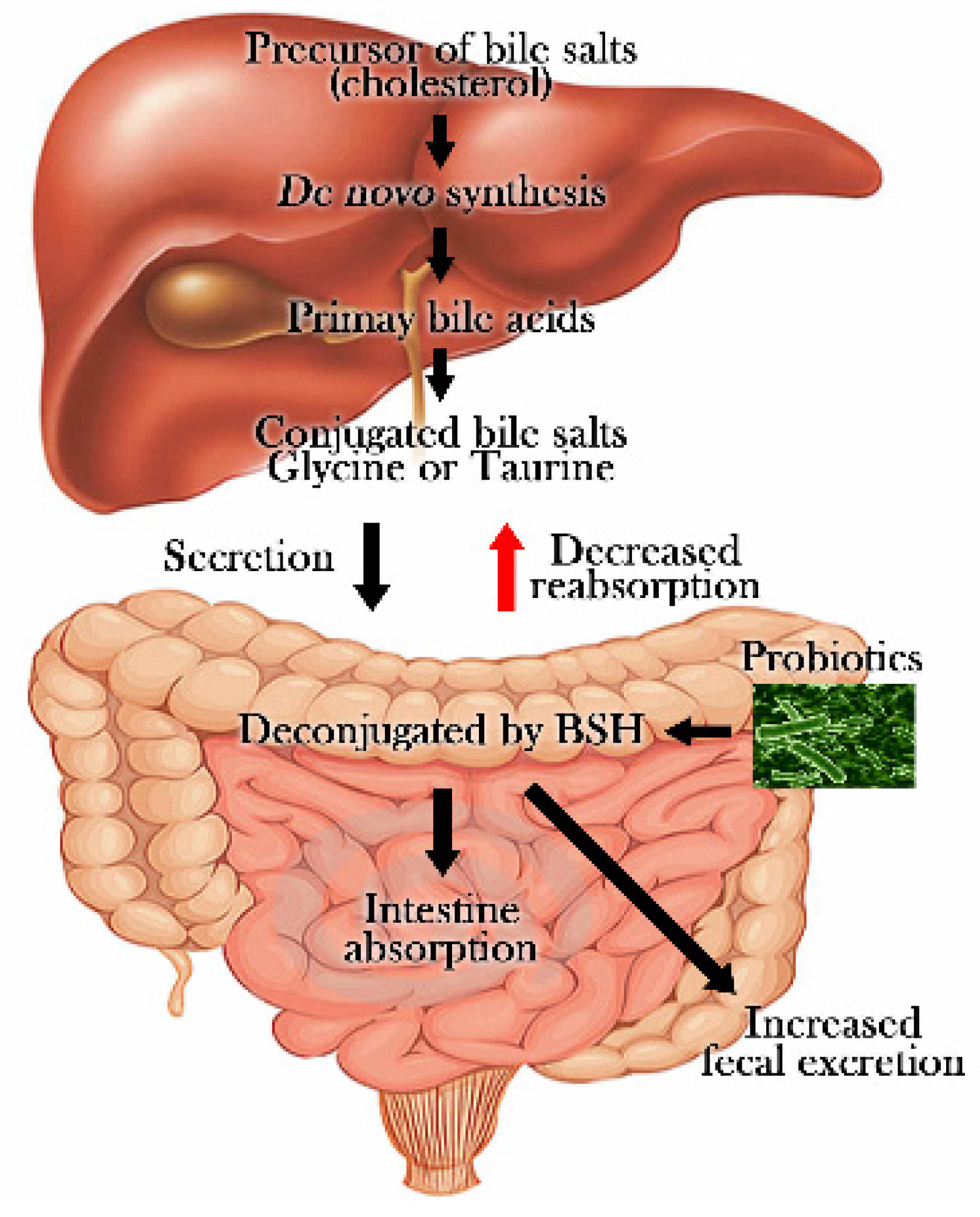
  123. Begley, M.; Hill, C.; Gahan, C.G. Bile salt hydrolase activity in probiotics. Appl. Environ. Microbiol. 2006, 72, 1729–1738. [Google Scholar] [CrossRef] [PubMed]
  124. Williams, C.M. Effects of inulin on lipid parameters in humans. J. Nutr. 1999, 129, 1471S–1473S. [Google Scholar] [PubMed]
  125. Tan, K.P.; Yang, M.; Ito, S. Activation of nuclear factor (erythroid-2 like) factor 2 by toxic bile acids provokes adaptive defense responses to enhance cell survival at the emergence of oxidative stress. Mol. Pharmacol. 2007, 72, 1380–1390. [Google Scholar] [CrossRef] [PubMed]
  126. Trauner, M.; Meier, P.J.; Boyer, J.L. Molecular pathogenesis of cholestasis. N. Engl. J. Med. 1998, 339, 1217–1227. [Google Scholar] [PubMed]
  127. Beilke, L.D.; Besselsen, D.G.; Cheng, Q.; Kulkarni, S.; Slitt, A.L.; Cherrington, N.J. Minimal role of hepatic transporters in the hepatoprotection against lca-induced intrahepatic cholestasis. Toxicol. Sci. 2008, 102, 196–204. [Google Scholar] [CrossRef] [PubMed]
  128. Deichelbohrer, I.; Alonso, J.C.; Luder, G.; Trautner, T.A. Plasmid transduction by Bacillus subtilis bacteriophage SPP1: Effects of DNA homology between plasmid and bacteriophage. J. Bacteriol. 1985, 162, 1238–1243. [Google Scholar] [PubMed]
  129. Merryweather, A.; Barth, P.T.; Wilkins, B.M. Role and specificity of plasmid RP4-encoded DNA primase in bacterial conjugation. J. Bacteriol. 1986, 167, 12–17. [Google Scholar] [PubMed]
  130. Mathur, S.; Singh, R. Antibiotic resistance in food lactic acid bacteria—A review. Int. J. Food Microbiol. 2005, 105, 281–295. [Google Scholar] [CrossRef] [PubMed]
  131. Zvenigorodskii, V.I.; Pozdniakov, V.N.; Bugaichuk, IuD.; Zhdanov, V.G. Transformation of Bacillus licheniformis by plasmid DNA. Genetika 1983, 19, 1036–1038. [Google Scholar] [PubMed]
Figure 1. Molecular mechanisms of short-chain fatty acid (SCFA) receptors.
Nutrients 08 00173 g001
Figure 2. bile-salt hydrolase (BSH) effects on lowering cholesterol by probiotics.
Nutrients 08 00173 g002
Table 1. Characteristics of the included animal studies.
Intervention TypeName of Pro/Prebiotic StrainsStudy TypePro/Prebiotic Type and Dose (Per Day)Duration of InterventionOutcomesParameter without ChangeReference
ProbioticsBacillus,
Lactobacillus,
Streptococcus,
Clostridium,
Saccharomyces,
Candida
RatsRice bran (107 CFU/g) 30 g/kg4 weeksDecreased serum total cholesterol Increase Δ6-desaturase activity and serum arachidonic acid Fukushima et al., 1999 [74]
ProbioticsB. lactis Bb-12,
B. longum Bb-46
RatsBuffalo milk yoghurt and soy-yoghurt4 weeksDecreased total cholesterol and LDL-C
Increasedfecal excretions of bile acids
Abd El-Gawad et al., 2005 [75]
ProbioticsL. plantarum PH04MiceHuman isolate (107 CFU/day)14 daysDecreased total cholesterol and TG
Increased fecal lactic acid bacteria
Nguyen et al., 2007 [76]
ProbioticsL. acidophilus,
L. casei,
L. lactis biovar diacetylactis
RatsDahi 15% (150g/kg)8 weeksDecreased glucose intolerance, hyperglycemia, hyperinsulinemia, dyslipidemia and oxidative stressHDL-CYadav et al., 2007 [70]
ProbioticsL. acidophilus NCDC14,
L. casei NCDC19
RatsDahi (73 × 108 CFU/g)28 daysInhibition of insulin depletion, lipid peroxidation and nitrite formation Yadav et al., 2008 [69]
ProbioticsB. animalis lactis 420Mice(109 CFU/day)6 weeks Decreased glucose intolerance, tissue inflammation, insulin resistance and secondarily glycaemia Amar et al., 2011 [48]
PrebioticsInulinRats5%4 weeksDecrease LDL-C, total cholesterol, Liver lipid and TG concentrations
Increased HDL-C, and faecal excretions of bile acids
Kim et al., 1998 [77]
Abbreviations: Bifidobacterium (B), lactobacillus (L), streptococcus (S), colony forming units (CFU), tab (tablet), low-density lipoprotein cholesterol (LDL-C), high-density lipoprotein (HDL-C), triglycerides (TG).
Table 2. Characteristics of the included human studies.
Intervention TypeName of Pro/Prebiotic StrainsStudy TypePro/Prebiotic Type and Dose (Per Day)Duration of InterventionOutcomesParameter without ChangeReference
ProbioticsL. acidophilus L1,HumanFermented milk 200 mL/day4 weeksDecreased total cholesterol Anderson et al., 1999 [78]
ProbioticsB. longum BL1Human/RatsFermented milk 100 mL/3 ×day4 weeksDecreased total cholesterol, LDL-C and TGHDL-CXiao et al., 2003 [79]
ProbioticsL. acidophilus LA-1HumanFreeze-dried Two tablet/day (3 × 10³ CFU/tab)6 weeks Total cholesterol, HDL-C, LDL-C, TGLewis et al., 2005 [73]
ProbioticsL. fermentumHumanFreeze-dried Two tablet/2 × day (2 × 10⁹ CFU/tab)10 weeks Total cholesterol, HDL-C, LDL-C, TG liver enzymesSimons et al., 2006 [80]
ProbioticsL. casei subsp. casei.HumanYogurt 100 g/day and 200 g/day6 weeksDecreased total cholesterol and LDL-C
Increased HDL-C
Fabian et al., 2006 [81]
ProbioticsL. rhamnosus LC705,
Propionibacterium freudenreichiissp shermaniistrain JS
HumanTwo tablet/day (2 × 10¹⁰ CFU/tab)4 weeks Total cholesterol, HDL-C, LDL-C, TGHatakka et al., 2008 [82]
ProbioticsL. acidophilus La5,
B. lactis Bb12
HumanYogurt 300 g/day (2 × 10⁶ CFU/g)6 weeksDecreased total cholesterol and LDL-CHDL-C, TGEjtahed et al., 2011 [22]
ProbioticsL. acidophilus La5,
B. lactis Bb12
HumanYogurt containing 300 g/day (2 × 10⁶ CFU/g) 6 weeksDecreased fasting blood glucose levels and HbA1c, Increased erythrocyte superoxide dismutase, glutathione peroxidase activities and total antioxidantstatusInsulin concentrationEjtahed et al., 2012 [68]
ProbioticsL. acidophilus,
L. casei,
L. rhamnosus,
L. bulgaricus,
B. breve,
B. longum,
S. thermophiles
HumanFreeze-dried One tablet/day (14 × 109 CFU/tab)8 weeksDecreased serum hs-CRP
Increased plasma total GSH Prevention of a rise in fasting plasma glucose
Asemi et al., 2013 [67]
Probiotics
Prebiotics
L. casei,
L. acidophilus,
L. rhamnosus,
L. bulgaricus,
B. breve,
B. longum,
S. thermophiles,
Fructooligosaccharid-e
HumanOne tablet/day 500 mg/tab8 weeksPositive effects on systolic blood pressureTotal cholesterol, LDL-C, HDL-C TG, TG/LDL and LDL/HDL ratiosMahboobi et al., 2014 [71]
PrebioticsInulinHumanRice-based ready-to-eat cereal (18%)4 weeksDecreased total cholesterol and TG
Increased breath H2 excretion and fecal lactic acid
Brighenti et al., 1995 [83]
PrebioticsInulinHumanOne pint of vanilla ice cream (20 g/pint)3 weeksDecreased total cholesterol and TG Causey et al., 2004 [84]
Abbreviations: Bifidobacterium (B), lactobacillus (L), streptococcus (S), colony forming units (CFU), tab (tablet), low-density lipoprotein cholesterol (LDL-C), high-density lipoprotein (HDL-C), triglycerides (TG).

