Botulinum Toxin Injections and Electrical Stimulation for Spastic Paresis Improve Active Hand Function Following Stroke

 , ,
, ,
Abstract
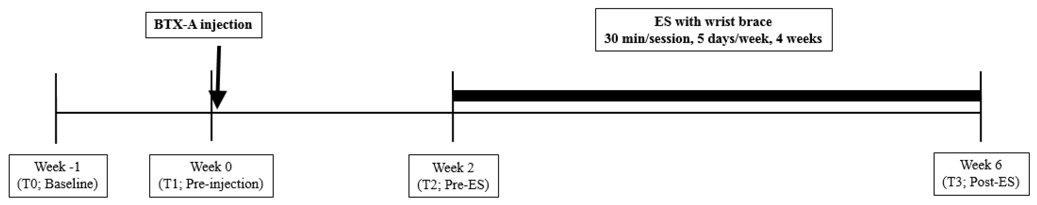
:1. Introduction
2. Results
2.1. Primary Outcomes
2.2. Secondary Outcomes
3. Discussion
4. Conclusions
5. Materials and Methods
5.1. Inclusion and Exclusion Criteria
5.2. Intervention
5.2.1. BTX-A Administration
5.2.2. ES with a Wrist Brace
5.3. Outcome Measures
5.3.1. Primary Outcomes
5.3.2. Secondary Outcomes
5.4. Statistical Analysis
Author Contributions
Funding
Conflicts of Interest
References
- Sommerfeld, D.K.; Eek, E.U.-B.; Svensson, A.-K.; Holmqvist, L.W.; von Arbin, M.H. Spasticity after stroke: Its occurrence and association with motor impairments and activity limitations. Stroke 2004, 35, 134–139. [Google Scholar] [CrossRef] [PubMed]
- Gracies, J.M. Pathophysiology of spastic paresis. I: Paresis and soft tissue changes. Muscle Nerve 2005, 31, 535–551. [Google Scholar] [CrossRef] [PubMed]
- Gracies, J.M. Pathophysiology of spastic paresis. II: Emergence of muscle overactivity. Muscle Nerve 2005, 31, 552–571. [Google Scholar] [CrossRef] [PubMed]
- Foley, N.; Pereira, S.; Salter, K.; Fernandez, M.M.; Speechley, M.; Sequeira, K.; Miller, T.; Teasell, R. Treatment with botulinum toxin improves upper-extremity function post stroke: A systematic review and meta-analysis. Arch. Phys. Med. Rehabil. 2013, 94, 977–989. [Google Scholar] [CrossRef] [PubMed]
- Esquenazi, A.; Albanese, A.; Chancellor, M.B.; Elovic, E.; Segal, K.R.; Simpson, D.M.; Smith, C.P.; Ward, A.B. Evidence-based review and assessment of botulinum neurotoxin for the treatment of adult spasticity in the upper motor neuron syndrome. Toxicon 2013, 67, 115–128. [Google Scholar] [CrossRef] [PubMed]
- Wissel, J.; Ward, A.B.; Erztgaard, P.; Bensmail, D.; Hecht, M.J.; Lejeune, T.M.; Schnider, P. European consensus table on the use of botulinum toxin type A in adult spasticity. J. Rehabil. Med. 2009, 41, 13–25. [Google Scholar] [CrossRef] [PubMed] [Green Version]
- Simpson, D.; Gracies, J.; Graham, H.; Miyasaki, J.; Naumann, M.; Russman, B.; Simpson, L.; So, Y. Assessment: Botulinum neurotoxin for the treatment of spasticity (an evidence-based review) Report of the Therapeutics and Technology Assessment Subcommittee of the American Academy of Neurology. Neurology 2008, 70, 1691–1698. [Google Scholar] [CrossRef] [PubMed]
- Demetrios, M.; Khan, F.; Turner-Stokes, L.; Brand, C.; Mc-Sweeney, S. Multidisciplinary rehabilitation following botulinum toxin and other focal intramuscular treatment for post-stroke spasticity. Cochrane Database Syst. Rev. 2012, 6, 12. [Google Scholar] [CrossRef]
- Simpson, D.M.; Hallett, M.; Ashman, E.J.; Comella, C.L.; Green, M.W.; Gronseth, G.S.; Armstrong, M.J.; Gloss, D.; Potrebic, S.; Jankovic, J. Practice guideline update summary: Botulinum neurotoxin for the treatment of blepharospasm, cervical dystonia, adult spasticity, and headache Report of the Guideline Development Subcommittee of the American Academy of Neurology. Neurology 2016, 86, 1818–1826. [Google Scholar] [CrossRef] [PubMed]
- Pirazzini, M.; Rossetto, O.; Eleopra, R.; Montecucco, C. Botulinum neurotoxins: Biology, pharmacology, and toxicology. Pharmacol. Rev. 2017, 69, 200–235. [Google Scholar] [CrossRef] [PubMed]
- Pirazzini, M. Novel Botulinum Neurotoxins: Exploring Underneath the Iceberg Tip. Toxins 2018, 10, 190. [Google Scholar] [Green Version]
- Hallett, M.; Albanese, A.; Dressler, D.; Segal, K.R.; Simpson, D.M.; Truong, D.; Jankovic, J. Evidence-based review and assessment of botulinum neurotoxin for the treatment of movement disorders. Toxicon 2013, 67, 94–114. [Google Scholar] [CrossRef] [PubMed]
- Naumann, M.; Dressler, D.; Hallett, M.; Jankovic, J.; Schiavo, G.; Segal, K.R.; Truong, D. Evidence-based review and assessment of botulinum neurotoxin for the treatment of secretory disorders. Toxicon 2013, 67, 141–152. [Google Scholar] [CrossRef] [PubMed]
- Shaw, L.; Rodgers, H.; Price, C.; van Wijck, F.; Shackley, P.; Steen, N.; Barnes, M.; Ford, G.; Graham, L. BoTULS: A multicentre randomised controlled trial to evaluate the clinical effectiveness and cost-effectiveness of treating upper limb spasticity due to stroke with botulinum toxin type A. Health Technol. Assess. 2010, 14, 1–113. [Google Scholar] [CrossRef] [PubMed]
- Gracies, J.M.; Brashear, A.; Jech, R.; McAllister, P.; Banach, M.; Valkovic, P.; Walker, H.; Marciniak, C.; Deltombe, T.; Skoromets, A.; et al. Safety and efficacy of abobotulinumtoxinA for hemiparesis in adults with upper limb spasticity after stroke or traumatic brain injury: A double-blind randomised controlled trial. Lancet Neurol. 2015, 14, 992–1001. [Google Scholar] [CrossRef]
- Dong, Y.; Wu, T.; Hu, X.; Wang, T. Efficacy and safety of Botulinum Toxin type A for upper limb spasticity after stroke or traumatic brain injury: A systematic review with meta-analysis and trial sequential analysis. Eur. J. Phys. Rehabil. Med. 2016, 53. [Google Scholar] [CrossRef]
- Rosales, R.L.; Efendy, F.; Teleg, E.S.; Santos, M.M.D.; Rosales, M.C.; Ostrea, M.; Tanglao, M.J.; Ng, A.R. Botulinum toxin as early intervention for spasticity after stroke or non-progressive brain lesion: A meta-analysis. J. Neurol. Sci. 2016, 371, 6–14. [Google Scholar] [CrossRef] [PubMed]
- Mills, P.B.; Finlayson, H.; Sudol, M.; O’Connor, R. Systematic review of adjunct therapies to improve outcomes following botulinum toxin injection for treatment of limb spasticity. Clin. Rehabil. 2016, 30, 537–548. [Google Scholar] [CrossRef] [PubMed]
- Sun, S.-F.; Hsu, C.-W.; Sun, H.-P.; Hwang, C.-W.; Yang, C.-L.; Wang, J.-L. Combined botulinum toxin type A with modified constraint-induced movement therapy for chronic stroke patients with upper extremity spasticity: A randomized controlled study. Neurorehabil. Neural Repair 2010, 24, 34–41. [Google Scholar] [CrossRef] [PubMed]
- Meythaler, J.M.; Vogtle, L.; Brunner, R.C. A preliminary assessment of the benefits of the addition of botulinum toxin a to a conventional therapy program on the function of people with longstanding stroke. Arch. Phys. Med. Rehabil. 2009, 90, 1453–1461. [Google Scholar] [CrossRef] [PubMed]
- Broeks, J.G.; Lankhorst, G.J.; Rumping, K.; Prevo, A.J. The long-term outcome of arm function after stroke: Results of a follow-up study. Disabil. Rehabil. 1999, 21, 357–364. [Google Scholar] [CrossRef] [PubMed]
- Mayer, N.H.; Esquenazi, A.; Childers, M.K. Common patterns of clinical motor dysfunction. Muscle Nerve 1997, 20, 21–35. [Google Scholar] [CrossRef]
- Picelli, A.; Lobba, D.; Midiri, A.; Prandi, P.; Melotti, C.; Baldessarelli, S.; Smania, N. Botulinum toxin injection into the forearm muscles for wrist and fingers spastic overactivity in adults with chronic stroke: A randomized controlled trial comparing three injection techniques. Clin. Rehabil. 2014, 28, 232–242. [Google Scholar] [CrossRef] [PubMed]
- Brashear, A.; Gordon, M.F.; Elovic, E.; Kassicieh, V.D.; Marciniak, C.; Do, M.; Lee, C.-H.; Jenkins, S.; Turkel, C. Intramuscular injection of botulinum toxin for the treatment of wrist and finger spasticity after a stroke. N. Engl. J. Med. 2002, 347, 395–400. [Google Scholar] [CrossRef] [PubMed]
- Weber, D.J.; Skidmore, E.R.; Niyonkuru, C.; Chang, C.-L.; Huber, L.M.; Munin, M.C. Cyclic functional electrical stimulation does not enhance gains in hand grasp function when used as an adjunct to onabotulinumtoxinA and task practice therapy: A single-blind, randomized controlled pilot study. Arch. Phys. Med. Rehabil. 2010, 91, 679–686. [Google Scholar] [CrossRef] [PubMed]
- Gracies, J.M.; Bayle, N.; Goldberg, S.; Simpson, D.M. Botulinum toxin type B in the spastic arm: A randomized, double-blind, placebo-controlled, preliminary study. Arch. Phys. Med. Rehabil. 2014, 95, 1303–1311. [Google Scholar] [CrossRef] [PubMed]
- Billian, C.; Gorman, P.H. Upper extremity applications of functional neuromuscular stimulation. Assist. Technol. 1992, 4, 31–39. [Google Scholar] [CrossRef] [PubMed]
- Chae, J.; Bethoux, F.; Bohinc, T.; Dobos, L.; Davis, T.; Friedl, A. Neuromuscular stimulation for upper extremity motor and functional recovery in acute hemiplegia. Stroke 1998, 29, 975–979. [Google Scholar] [CrossRef] [PubMed]
- Simpson, D.; Alexander, D.; O’brien, C.; Tagliati, M.; Aswad, A.; Leon, J.; Gibson, J.; Mordaunt, J.; Monaghan, E. Botulinum toxin type A in the treatment of upper extremity spasticity A randomized, double-blind, placebo-controlled trial. Neurology 1996, 46, 1306. [Google Scholar] [CrossRef] [PubMed]
- Smith, S.; Ellis, E.; White, S.; Moore, A. A double-blind placebo-controlled study of botulinum toxin in upper limb spasticity after stroke or head injury. Clin. Rehabil. 2000, 14, 5–13. [Google Scholar] [CrossRef] [PubMed]
- Sheean, G.L. Botulinum treatment of spasticity: Why is it so difficult to show a functional benefit? Curr. Opin. Neurol. 2001, 14, 771–776. [Google Scholar] [CrossRef] [PubMed]
- Kamper, D.; Rymer, W. Impairment of voluntary control of finger motion following stroke: Role of inappropriate muscle coactivation. Muscle Nerve 2001, 24, 673–681. [Google Scholar] [CrossRef] [PubMed]
- Li, S.; Francisco, G.E. New insights into the pathophysiology of post-stroke spasticity. Front. Hum. Neurosci. 2015, 9, 192. [Google Scholar] [CrossRef] [PubMed]
- Picelli, A.; Baricich, A.; Cisari, C.; Paolucci, S.; Smania, N.; Sandrini, G. The Italian real-life post-stroke spasticity survey: Unmet needs in the management of spasticity with botulinum toxin type A. Funct. Neurol. 2017, 32, 89–96. [Google Scholar] [CrossRef] [PubMed]
- Simpson, D.M.; Patel, A.T.; Alfaro, A.; Ayyoub, Z.; Charles, D.; Dashtipour, K.; Esquenazi, A.; Graham, G.D.; McGuire, J.R.; Odderson, I. OnabotulinumtoxinA Injection for Poststroke Upper-Limb Spasticity: Guidance for Early Injectors from a Delphi Panel Process. PM R 2017, 9, 136–148. [Google Scholar] [CrossRef] [PubMed]
- Filippi, G.M.; Errico, P.; Santarelli, R.; Bagolini, B.; Manni, E. Botulinum A toxin effects on rat jaw muscle spindles. Acta Oto-Laryngol. 1993, 113, 400–404. [Google Scholar] [CrossRef]
- Manni, E.; Bagolini, B.; Pettorossi, V.E.; Errico, P. Effect of botulinum toxin on extraocular muscle proprioception. Doc. Ophthalmol. 1989, 72, 189–198. [Google Scholar] [CrossRef] [PubMed]
- Wöber, C.; Schnider, P.; Steinhoff, N.; Trattnig, S.; Zebenholzer, K.; Auff, E. Posturographic findings in patients with idiopathic cervical dystonia before and after local injections with botulinum toxin. Eur. Neurol. 1999, 41, 194–200. [Google Scholar] [CrossRef] [PubMed]
- Kaňovský, P.; Rosales, R.L. Debunking the pathophysiological puzzle of dystonia–with special reference to botulinum toxin therapy. Parkinsonism Relat. Disord. 2011, 17, S11–S14. [Google Scholar] [CrossRef] [PubMed]
- Veverka, T.; Hluštík, P.; Hok, P.; Otruba, P.; Zapletalová, J.; Tüdös, Z.; Krobot, A.; Kaňovský, P. Sensorimotor modulation by botulinum toxin A in post-stroke arm spasticity: Passive hand movement. J. Neurol. Sci. 2016, 362, 14–20. [Google Scholar] [CrossRef] [PubMed]
- Dresel, C.; Bayer, F.; Castrop, F.; Rimpau, C.; Zimmer, C.; Haslinger, B. Botulinum toxin modulates basal ganglia but not deficient somatosensory activation in orofacial dystonia. Mov. Disord. 2011, 26, 1496–1502. [Google Scholar] [CrossRef] [PubMed]
- Zakin, E.; Simpson, D. Evidence on botulinum toxin in selected disorders. Toxicon 2018, 147, 134–140. [Google Scholar] [CrossRef] [PubMed]
- McCrory, P.; Turner-Stokes, L.; Baguley, I.J.; De Graaff, S.; Katrak, P.; Sandanam, J.; Davies, L.; Munns, M.; Hughes, A. Botulinum toxin A for treatment of upper limb spasticity following stroke: A multi-centre randomized placebo-controlled study of the effects on quality of life and other person-centred outcomes. J. Rehabil. Med. 2009, 41, 536–544. [Google Scholar] [CrossRef] [PubMed]
- Fheodoroff, K.; Ashford, S.; Jacinto, J.; Maisonobe, P.; Balcaitiene, J.; Turner-Stokes, L. Factors influencing goal attainment in patients with post-stroke upper limb spasticity following treatment with botulinum toxin A in real-life clinical practice: Sub-analyses from the Upper Limb International Spasticity (ULIS)-II Study. Toxins 2015, 7, 1192–1205. [Google Scholar] [CrossRef] [PubMed]
- Shaw, L.C.; Price, C.I.; van Wijck, F.M.; Shackley, P.; Steen, N.; Barnes, M.P.; Ford, G.A.; Graham, L.A.; Rodgers, H. Botulinum Toxin for the Upper Limb after Stroke (BoTULS) Trial: Effect on impairment, activity limitation, and pain. Stroke 2011, 42, 1371–1379. [Google Scholar] [CrossRef] [PubMed]
- Emerson, E.T.; Krizek, T.J.; Greenwald, D.P. Anatomy, physiology, and functional restoration of the thumb. Ann. Plast. Surg. 1996, 36, 180–191. [Google Scholar] [CrossRef] [PubMed]
- Asutay, F.; Atalay, Y.; Asutay, H.; Acar, A.H. The Evaluation of the Clinical Effects of Botulinum Toxin on Nocturnal Bruxism. Pain Res. Manag. 2017, 2017, 5. [Google Scholar] [CrossRef] [PubMed]
- Desrosiers, J.; Bravo, G.; Hébert, R.; Dutil, É.; Mercier, L. Validation of the Box and Block Test as a measure of dexterity of elderly people: Reliability, validity, and norms studies. Arch. Phys. Med. Rehabil. 1994, 75, 751–755. [Google Scholar] [PubMed]
- Van der Lee, J.H.; Beckerman, H.; Lankhorst, G.J.; Bouter, L.M. The responsiveness of the Action Research Arm test and the Fugl-Meyer Assessment scale in chronic stroke patients. J. Rebab. Med. 2001, 33, 110–113. [Google Scholar]
- Rodriquez, A.A.; McGinn, M.; Chappell, R. Botulinum toxin injection of spastic finger flexors in hemiplegic patients. Am. J. Phys. Med. Rehabil. 2000, 79, 44–47. [Google Scholar] [CrossRef] [PubMed]
- Ring, H.; Rosenthal, N. Controlled study of neuroprosthetic functional electrical stimulation in sub-acute post-stroke rehabilitation. J. Rehabil. Med. 2005, 37, 32–36. [Google Scholar] [CrossRef] [PubMed]
- Bohannon, R.W.; Smith, M.B. Interrater reliability of a modified Ashworth scale of muscle spasticity. Phys. Ther. 1987, 67, 206–207. [Google Scholar] [CrossRef] [PubMed]
- Gummesson, C.; Ward, M.M.; Atroshi, I. The shortened disabilities of the arm, shoulder and hand questionnaire (Quick DASH): Validity and reliability based on responses within the full-length DASH. BMC Musculoskelet. Disord. 2006, 7, 44. [Google Scholar] [CrossRef] [PubMed]


| Outcome | p† | ||||||
|---|---|---|---|---|---|---|---|
| T1 | T2 | T3 | p * | T1–T2 | T2–T3 | T1–T3 | |
| BB test | 3.07 ± 3.85 | 3.60 ± 4.91 | 4.67 ± 5.25 | 0.039 | 0.473 | 0.120 | 0.028 |
| ARAT-total | 11.33 ± 8.03 | 11.27 ± 7.71 | 12.73 ± 7.67 | 0.043 | 1.000 | 0.036 | 0.044 |
| ARAT-grasp | 2.87 ± 3.82 | 3.00 ± 3.98 | 3.27 ± 3.75 | 0.276 | 0.854 | 0.334 | 0.276 |
| ARAT-grip | 2.13 ± 2.07 | 1.67 ± 1.92 | 2.53 ± 1.92 | 0.120 | 0.141 | 0.059 | 0.257 |
| ARAT-gross movement | 5.67 ± 2.16 | 5.93 ± 2.15 | 6.27 ± 2.37 | 0.013 | 0.046 | 0.096 | 0.021 |
| ARAT-pinch | 0.67 ± 1.23 | 0.67 ± 0.98 | 0.67 ± 0.98 | 1.000 | 1.000 | 1.000 | 1.000 |
| Outcome | p† | ||||||
|---|---|---|---|---|---|---|---|
| T1 | T2 | T3 | p * | T1–T2 | T2–T3 | T1–T3 | |
| Active-FE | 1.73 ± 0.88 | 2.00 ± 0.85 | 2.20 ± 0.94 | 0.060 | 0.102 | 0.180 | 0.053 |
| Distance-FP (cm) | 2.58 ± 3.12 | 3.80 ± 3.02 | 3.67 ± 2.58 | 0.212 | 0.023 | 0.655 | 0.027 |
| Repeat-FE | 2.07 ± 1.58 | 2.27 ± 1.16 | 3.13 ± 1.77 | 0.007 | 0.558 | 0.017 | 0.008 |
| Thumb opposition | 0.07 ± 0.26 | 0.13 ± 0.35 | 0.33 ± 0.62 | 0.223 | 0.317 | 0.180 | 0.102 |
| MAS-WF | 2.13 ± 0.35 | 1.80 ± 0.41 | 1.73 ± 0.46 | 0.015 | 0.025 | 0.317 | 0.014 |
| MAS-WE | 0.20 ± 0.41 | 0.13 ± 0.35 | 0.07 ± 0.26 | 0.223 | 0.317 | 0.317 | 0.157 |
| MAS-FF | 2.33 ± 0.49 | 1.67 ± 0.49 | 1.60 ± 0.51 | <0.001 | 0.002 | 0.317 | 0.001 |
| MAS-FE | 0.07 ± 0.26 | 0.00 ± 0.00 | 0.00 ± 0.00 | 0.368 | 0.317 | 1.000 | 0.317 |
| MRC-WF | 2.40 ± 0.91 | 2.40 ± 0.91 | 2.53 ± 0.83 | 0.135 | 1.000 | 0.157 | 0.157 |
| MRC-WE | 2.27 ± 0.88 | 2.40 ± 0.74 | 2.53 ± 0.83 | 0.050 | 0.157 | 0.157 | 0.046 |
| MRC-FF | 2.60 ± 0.51 | 2.60 ± 0.51 | 2.67 ± 0.49 | 0.717 | 1.000 | 0.317 | 0.564 |
| MRC-FE | 1.47 ± 0.64 | 1.60 ± 0.63 | 1.67 ± 0.62 | 0.097 | 0.157 | 0.317 | 0.083 |
| AROM-WF (°) | 46.67 ± 26.37 | 46.67 ± 26.37 | 49.33 ± 23.44 | 0.050 | 1.000 | 0.102 | 0.102 |
| AROM-WE (°) | 32.67 ± 26.04 | 33.67 ± 25.53 | 38.00 ± 24.55 | 0.011 | 0.396 | 0.024 | 0.023 |
| AROM-RD (°) | 4.67 ± 7.42 | 5.33 ± 6.40 | 6.67 ± 7.24 | 0.174 | 0.564 | 0.157 | 0.083 |
| AROM-UD (°) | 4.67 ± 6.40 | 5.33 ± 6.40 | 5.33 ± 6.40 | 0.368 | 0.317 | 1.000 | 0.317 |
| Grip strength (kg) | 0.27 ± 1.03 | 0.13 ± 0.52 | 0.33 ± 1.05 | 0.223 | 0.317 | 0.180 | 0.317 |
| QDASH | 56.88 ± 17.22 | 54.65 ± 14.88 | 53.87 ± 16.54 | 0.162 | 0.220 | 0.529 | 0.058 |
| Patient | Stroke Type | Lesion Side | Time from Stroke (Months) | Dominant Hand | Injection Site (Dosage in Units) |
|---|---|---|---|---|---|
| 1 | Ischemic | Rt. | 11.5 | Rt. | FCU (25), FDP 2/3/4/5 (10/20/20/10), FDS 2/3/4/5 (20/30/25/15), FPL (25) |
| 2 | Ischemic | Rt. | 10.9 | Rt. | FCU (25), FDP 2/3/4 (15/20/15), FDS 2/3/4/5 (25/25/30/15), FPL (30) |
| 3 | Hemorrhagic | Rt. | 13.2 | Rt. | FCU (40), FDP 2/3/4 (10/15/15), FDS 2/3/4/5 (25/30/35/20), FPB (10), FPL (30), PT (50) |
| 4 | Hemorrhagic | Rt. | 17.4 | Rt. | AP (10), BB (40), brachialis (50), FDP 2/3/4/5 (10/15/15/5), FDS 2/3/4/5 (25/35/40/15), FPL (40) |
| 5 | Ischemic | Rt. | 18.9 | Rt. | AP (10), brachialis (55), FDP 2/3/4 (10/10/10), FDS 2/3/4/5 (30/40/35/20), FPL (40), PT (40) |
| 6 | Ischemic | Rt. | 10.2 | Rt. | FCU (30), FDP 2/3/4/5 (10/20/10/10), FDS 2/3/4/5 (25/25/20/15), FPB (5), FPL (25), PT (20) |
| 7 | Hemorrhagic | Lt. | 12.6 | Rt. | Brachialis (60), FCU (40), FDP 2/3/4 (10/10/15), FDS 2/3/4/5 (25/35/30/15), FPL (20), PT (40) |
| 8 | Ischemic | Lt. | 13 | Rt. | Brachialis (60), deltoid (20), FDP 2/3/4/5 (15/15/10/10), FDS 2/3/4/5 (30/40/30/10), FPB (10), FPL (30), PT (20) |
| 9 | Hemorrhagic | Rt. | 10.3 | Lt. | Brachialis (60), deltoid (20), FDP 2/3/4/5 (15/15/10/10), FDS 2/3/4/5 (30/40/30/10), FPB (10), FPL (30), PT (20) |
| 10 | Hemorrhagic | Rt. | 20.3 | Rt. | AP (10), FCU (30), FDS 2/3/4 (10/15/10), FPB (10), FPL (20), lumbricals (10) |
| 11 | Ischemic | Rt. | 8.6 | Rt. | AP (10), FCU (30), FDP 2/3/4/5 (5/10/10/5), FDS 2/3/4/5 (20/30/20/10), FPL (20) |
| 12 | Ischemic | Rt. | 7.2 | Rt. | AP (10), BB (40), FDP 2/3/5 (10/10/5), FDS 2/3/4/5 (30/35/20/10), FPL (30) |
| 13 | Hemorrhagic | Rt. | 9.8 | Rt. | AP (15), FDP 2/3/4 (5/10/5), FDS 2/3/4/5 (10/20/15/5), FPL (15) |
| 14 | Hemorrhagic | Rt. | 12.8 | Rt. | FDP 2/3/4/5 (30/30/35/25), FDS 2/3/4 (10/15/15), FPL (30), opponens (5) |
| 15 | Hemorrhagic | Rt. | 14.9 | Rt. | Brachialis (50), brachioradialis (20), FCU (30), FDP 2/3/4 (15/15/15), FDS 2/3/4/5 (35/40/30/20), FPL (30) |
© 2018 by the authors. Licensee MDPI, Basel, Switzerland. This article is an open access article distributed under the terms and conditions of the Creative Commons Attribution (CC BY) license (http://creativecommons.org/licenses/by/4.0/).
Share and Cite
Lee, J.-M.; Gracies, J.-M.; Park, S.-B.; Lee, K.H.; Lee, J.Y.; Shin, J.-H. Botulinum Toxin Injections and Electrical Stimulation for Spastic Paresis Improve Active Hand Function Following Stroke. Toxins 2018, 10, 426. https://doi.org/10.3390/toxins10110426
Lee J-M, Gracies J-M, Park S-B, Lee KH, Lee JY, Shin J-H. Botulinum Toxin Injections and Electrical Stimulation for Spastic Paresis Improve Active Hand Function Following Stroke. Toxins. 2018; 10(11):426. https://doi.org/10.3390/toxins10110426
Chicago/Turabian StyleLee, Jong-Min, Jean-Michel Gracies, Si-Bog Park, Kyu Hoon Lee, Ji Yeong Lee, and Joon-Ho Shin. 2018. "Botulinum Toxin Injections and Electrical Stimulation for Spastic Paresis Improve Active Hand Function Following Stroke" Toxins 10, no. 11: 426. https://doi.org/10.3390/toxins10110426