Article

First Genome-Based Characterisation and Staphylococcal Enterotoxin Production Ability of Methicillin-Susceptible and Methicillin-Resistant Staphylococcus aureus Strains Isolated from Ready-to-Eat Foods in Algiers (Algeria)

1
National Research Council of Italy, Institute of Sciences of Food Production (CNR-ISPA), 70126 Bari, Italy
2
Department of Microbiology and Biotechnology, Max Rubner-Institut, Hermann-Weigmann-Straße 1, 24103 Kiel, Germany
3
Research and Development, Silantes GmbH, Gollierstrasse 70C, 80339 Munich, Germany
4
Laboratory of Hygiene and Animal Pathology, Institute of Veterinary Sciences, University of Tiaret, Tiaret 14000, Algeria
5
Department of Food Hygiene and Consumer Health Protection, Wrocław University of Environmental and Life Sciences, 50-375 Wrocław, Poland
*
Author to whom correspondence should be addressed.
These authors contributed equally to this work and thus should be designated both as first authors.
Toxins 2022, 14(11), 731; https://doi.org/10.3390/toxins14110731
Submission received: 2 September 2022 / Revised: 18 October 2022 / Accepted: 19 October 2022 / Published: 25 October 2022
(This article belongs to the Collection Staphylococcus aureus Toxins)

Abstract

:
Staphylococcus aureus is a pathogenic microorganism of humans and animals, able to cause foodborne intoxication due to the production of staphylococcal enterotoxins (SEs) and to resist antibiotic treatment as in the case of methicillin-resistant S. aureus (MRSA). In this study, we performed a genomic characterisation of 12 genetically diverse S. aureus strains isolated from ready-to-eat foods in Algiers (Algeria). Moreover, their ability to produce some classical and new staphylococcal enterotoxins (SEs) was investigated. The 12 S. aureus strains resulted to belong to nine known sequence types (STs) and to the novel ST7199 and ST7200. Furthermore, S. aureus SA46 was assigned to the European clone MRSA-ST80-SCCmec-IV. The 12 strains showed a wide endowment of se and sel (staphylococcal enterotoxin-like toxin) genes (sea, seb, sed, seg, seh, sei, selj, sek, sem, sen, seo, seq, ser, selu2, selw, selx, sey, sel30; ψent1-ψent2), including variants and pseudogenes, and harboured the enterotoxin gene cluster (egc) types 1 and 5. Additionally, they produced various amounts of SEA (64.54–345.02 ng/mL), SEB (2871.28–14739.17 ng/mL), SED (322.70–398.94 ng/mL), SEH (not detectable–239.48 ng/mL), and SER (36,720.10–63,176.06 ng/mL) depending on their genotypes. The genetic determinants related to their phenotypic resistance to β-lactams (blaZ, mecA), ofloxacin (gyrA-S84L), erythromycin (ermB), lincomycin (lmrS), kanamycin (aph(3′)-III, ant(6)-I), and tetracyclin (tet(L), tet(38)) were also detected. A plethora of virulence-related genes, including major virulence genes such as the tst gene, determinant for the toxic shock syndrome toxin-1, and the lukF-PV and lukS-PV genes, encoding the panton-valentine leukocidin (PVL), were present in the S. aureus strains, highlighting their pathogenic potential. Furthermore, a phylogenomic reconstruction including worldwide foodborne S. aureus showed a clear clustering based on ST and geographical origin rather than the source of isolation.
Key Contribution: This study reported for the first time a genome-based characterisation and SEs production of foodborne S. aureus in Algeria, and helped to enlarge the knowledge on the enterotoxigenicity, overall virulence, antimicrobial resistance, as well as molecular-based epidemiology of this microorganism in the African continent.

1. Introduction

Despite the increased awareness of foodborne diseases and huge progress in food safety diagnostics, enterotoxigenic Staphylococcus (S.) aureus continues to cause staphylococcal food-poisoning (SFP) cases worldwide. Following the discovery of the classical enterotoxins (SEA-SEE), 33 staphylococcal enterotoxin (se) and staphylococcal enterotoxin-like toxin (sel) genes are known to date [1], highlighting the great enterotoxigenic potential of S. aureus. Moreover, this pathogenic bacterium may show resistance to several antibiotics [2] that may affect the antibiotic treatment when S. aureus infections occur. In particular, community-, livestock-, and healthcare-associated methicillin-resistant S. aureus (MRSA), even enterotoxigenic strains, are emerging in food products, animals used for foods, animal husbandry, and food production or processing workers [3].
Recently, we investigated the prevalence, enterotoxigenic potential, and antimicrobial resistance of 48 methicillin-susceptible S. aureus (MSSA) and MRSA isolated from 207 ready-to-eat foods randomly sampled from hotels, restaurants, fast foods, and pizzerias in Algiers, the capital of Algeria, during 2018 and 2019 [4]. The resulting S. aureus prevalence was 23.2% (48/207) [4]. For strain typing, intergenic spacer region (ISR) typing showed a higher discriminatory power than the staphylococcal enterotoxin gene (SEg) typing. Indeed, the same SEg type was found in S. aureus belonging to different ISR types. In particular, the 48 S. aureus strains were grouped in seven SEg types, while 12 ISR types were detected. Moreover, 2 out of the 48 S. aureus isolates, belonging to the same ISR type, were found to harbour the mecA gene that encodes for the penicillin-binding protein 2a (PBP2a), a transpeptidase protein with low affinity for most β-lactam antibiotics, including methicillin as well as oxacillin and cefoxitin [4,5]. Twelve strains, each belonging to one of the twelve detected ISR types, were subjected to antibiotic susceptibility testing. Some strains were resistant towards antimicrobials including benzylpenicillin, ofloxacin, erythromycin, lincomycin, tetracycline, kanamycin, oxacillin, and cefoxitin; the mecA-positive strain, being resistant to oxacillin and cefoxitin, was confirmed as MRSA, and two other tested strains showed multidrug resistance [4]. However, the performed molecular detection of se and sel genes did not permit to assess the presence of enterotoxin gene variants or pseudogenes, which may affect the enterotoxigenic activity and the virulence of the S. aureus strains, nor to distinguish among the ψent1/ψent2 pseudogenes and the selu2 gene in the enterotoxin gene cluster (egc) found in the S. aureus strains [4]. This drawback did not allow determining the egc type they harboured. This cluster plays a crucial role in the enterotoxigenicity of S. aureus since phylogenetic analyses indicated that potentially all the se and sel genes may derive from the egc [6], and, to date, at least eight egc types have been described in S. aureus [1,7]. Moreover, the presence of genetic determinants of antibiotic resistance, other than mecA, was not investigated in the 12 analysed strains [4]. Although some efforts are being made to characterise foodborne S. aureus in Africa, including the Algerian Country [8,9,10,11], specific data on S. aureus from ready-to-eat foods are scarce. Additionally, data on S. aureus molecular epidemiology are fairly increasing in the African continent, but studies are mainly performed on humans and animals rather than food isolates and MRSA rather than MSSA [12,13], underlying the need of collecting data on foodborne African isolates, including the Algerian Country. For all these reasons, the 12 Algerian MSSA and MRSA strains, representative of the different ISR types, were subjected to whole-genome sequencing. Herein, we reported a comprehensive genomic characterisation of these 12 genetically diverse S. aureus, exploring their genomic relatedness, investigating the genetic features in relation to their virulence and antibiotic resistance, and performing a phylogenomic analysis including worldwide foodborne S. aureus isolates. We also assessed the SEs production and detected all the se and sel genes known to date in order to improve the characterisation of foodborne S. aureus in the African continent.

2. Results

2.1. Genomic Statistics

The genomic features of S. aureus strains are reported in Supplementary Table S1. Genomes resulted in a median length of 2.7 Mbp and an average N50 value of 270 kbp. The average mol% GC content was 32.59%, with a median number of coding genes of 2606 and a coding density of 83.95%. The quality of the assemblies was excellent for all strains, with the exception of SA02, which resulted in only 95.5% completeness.

2.2. Multilocus Sequence and spa Typing

The sequence types (STs) of the 12 sequenced S. aureus strains are shown in Table 1, whereas the multilocus sequence type (MLST) allelic profiles are detailed in Supplementary Table S2. All strains belong to different STs, with the exception of strains SA02 and SA51, which both belong to ST5. Two novel STs have been deposited in PubMLST as ST7199 and ST7200: ST7199 differs from the known ST15 only for the novel aroE-1025 allele, whereas ST7200 differs from the known ST72 for the novel yqiL-954 allele.
Additionally, spa types of the 12 sequenced S. aureus strains are reported in Supplementary Table S3.

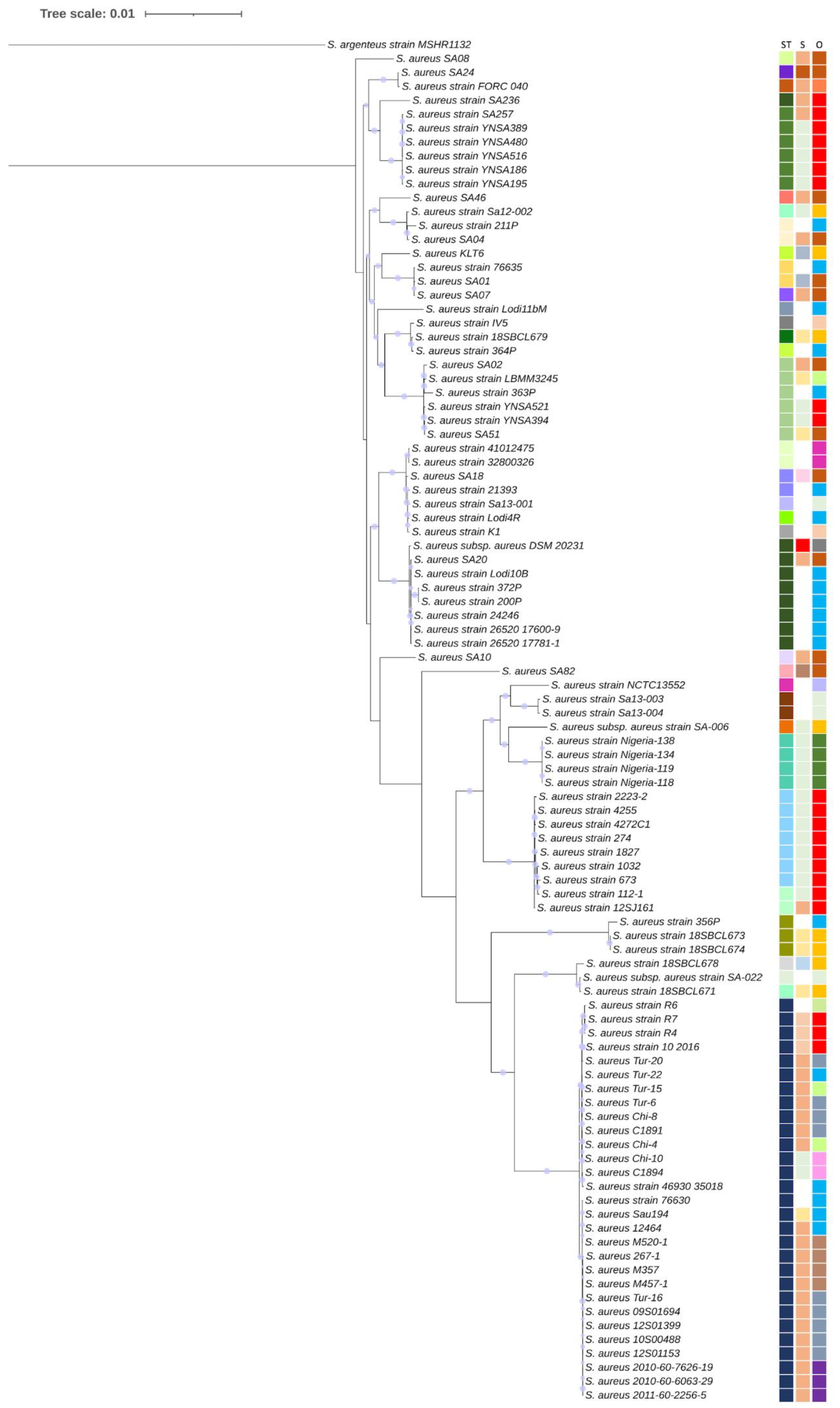
2.3. Phylogenetic Analysis

A RAxML phylogenetic tree is reported in Figure 1. It also shows the ST, source of isolation (S), and geographical origin (O) of the analysed strains.
Globally, S. aureus strains are grouped in the tree mainly depending on the ST and geographical origin rather than the source of isolation, although a certain association appears for meat, milk, and dairy products. The grouping is clear for S. aureus isolates from China (red squares in the third column origin (O)), Nigeria (dark green squares in the third column O), and Italy (light blue in the third column O).
S. aureus strains sequenced in this study clustered on different branches of the tree. Those belonging to the same ST, namely SA02 and SA51, and those belonging to STs that differ by only a single allele, namely SA01 and SA07, closely grouped in the tree, whereas SA08, SA10, and SA82, belonging to STs that differ in numerous alleles, were individually located in the tree. The genetic divergence of SA82 from the other sequenced strains was also confirmed by ANI analysis, which reported average values of 98.3% between SA82 and the other sequenced strains, which, among them, share >99% of ANI (Supplementary Table S5).

2.4. Pan-Genome Analysis

The pan-genome of all the S. aureus isolates analysed in this study, which includes orthologues and unique genes, comprised a set of 7537 genes. The core genome (shared by >99% of S. aureus isolates), on the other hand, consisted of 1596 genes. The accessory genome (total core: genes in >2 isolates but not in all) consisted of 4059 genes, whereas the unique genome was composed of 1882 genes. The soft core, shell, and cloud genomes contained 261, 1323, and 4357 genes, respectively (Supplementary Table S6). The output of the Roary analysis is shown in Supplementary Figure S1, which confirms the clustering obtained by the phylogenetic analysis.

2.5. Enterotoxin Genes Presence and Localisation

The se and sel genes harboured by the 12 sequenced S. aureus strains, including variants and pseudogenes, are shown in Table 1. The nucleotide and amino acid sequences of the detected se and sel genes and predicted proteins, as well as the percentage of identity with previously reported reference sequences, are shown in Supplementary Table S7 [1,6,7,14,15,17,18,19,20,21,22,23,24,25,26,27,28,29,30,31].
selw is located on the chromosome. In SA02, SA51, SA18, and SA24, the selw gene was predicted as a pseudogene due to a frameshift mutation (SA02 and SA51) or an internal stop codon (SA18 and SA24). It should be pointed out that for SA82, SA08, and SA10 selw, the NCBI Prokaryotic Genome Annotation Pipeline (PGAP) annotated the predicted protein as se type 26 [1]. In this study, however, we retained the selw nomenclature as proposed since its discovery by Okumura et al. [32].
selx is also located on the chromosome in all the analysed strains.
In SA02, the enterotoxin gene cluster (egc) is located in a S. aureus pathogenicity island (SAPI) of νSaβ type I (SaPim3/n3), with seg, sen, ψent1-ψent2, sei, sem, and seo (J3P54_11775-11800) (egc1 [6]) (Supplementary Figure S2). This pathogenicity island harbours genes for the bi-component leukocidin LukED, serine protease genes splABCDEF with splA being a pseudogene, genes for the restriction endonucleases M and S, and several transposase coding genes, some of which predicted as pseudogenes.
SA08 harbours the egc5 (seg, sen, selu2, sei, sem, seo: KQP42_01815-01790) [7] in a νSaβ of type IV, with lukED, serine protease genes, and a lantibiotic encoding gene cluster (KQP42_01860-01895) [33] (Supplementary Figure S2). SA24 harbours the egc5 (seg, sen, selu2, sei, sem, seo: J3135_04230-04205) located in a SAPI of νSaβ type I. We hypothesised a νSaβ of type III in SA82, but the contig is interrupted downstream the egc5 (seg, sen, selu2, sei, sem, seo: KQP39_04565-04540) (Supplementary Figure S2). In strain SA51, the egc1 (seg, sen, ψent1-ψent2, sei, sem, and seo: J3T04_07830-07855) is located in a νSaβ type I (SaPim3/n3) (Supplementary Figure S2). This genome also harbours sea (J3T04_10205) in a prophage region (ΦMu3A) (Figure 2A), harbouring the saK gene and sphingomyelin phosphodiesterase pseudogene as a result of a frameshift mutation. In the genome of SA51, we also identified the sel30 gene (J3T04_08740). The plasmid location of this gene [1] appears to be confirmed by the nearby presence of rep pseudogene and the high homology (>99%) retrieved between a 17 kb flanking region comprising sel30 in S. aureus NCTC6135 and SA51 genome.
The sea gene is also present in the genome of SA04 in a prophage region of φSA3 type (Figure 2A). This large region includes 40 kb upstream sea, seq, and sek (Figure 2A), close to a site-specific integrase, a pseudogene for a sphingomyelin phosphodiesterase, a MBL fold metallo-hydrolase, the bi-component leukocidin LukGH, and the chaperonin GroEL.
In SA04, the genomic locus including the seh gene (J3T03_06310) is shown in Figure 2B and it is typical of a phage of φSa3ms type. Close to the seh gene, a pseudogene for the exotoxin seo, and the cst operon, which protects S. aureus from sulfide toxicity [34], can be identified.
In the SA18 genome, the seb gene is located in a pathogenicity island with 99.4% similarity with the SaPIivm60 described by Sato’o et al. [35].
In the genome of strain SA20, the pseudogene sed (J3T02_03365) and the genes ser (J3T02_03360) and selj (J3T02_03355) are one by one located at the beginning of NODE_17, immediately followed by several rep protein genes, indicating the presence of a putative multiresistance pIB485-like plasmid [36] (Figure 3). This plasmid contains blaI, blaR1, and blaZ genes and shows a 100% nucleotide homology with the SAP048A plasmid (Accession: NC_019007.1), with an inversion due to the role of the two recombinase/invertase present in this locus (bin gene) [37,38].
In SA46, the sey gene (J3T00_13960) is located on the chromosome [7]. The genomic locus comprising seh (J3T00_044075), annotated as a pseudogene, also includes the staphyloferrin operon and three tandem-type lipoproteins.
Lastly, the sequences of allelic variants of sea [17], sed [18] and selx [7,15,16], different than those previously described, have been deposited in DDBJ/ENA/GenBank under the following accessions: seA-SA51 (ON205830.1); seD-SA20 (ON205834.1); selX-SA01 (ON205831.1); selX-SA08 (ON205832.1); selX-SA10 (ON205833.1); selX-SA46 (ON205835.1).

2.6. Antibiotic Resistance Genetic Determinants

Table 2 shows the antibiotic resistance profile, as assessed by Mekhloufi et al. [4], and the related genetic determinants identified in the S. aureus genomes analysed in the present study.
With the exception of SA02 and SA46, in all the other strains we identified blaZ, blaR1, and blaI genes, which encode for the penicillin-hydrolysing class A beta-lactamase BlaZ, the beta-lactam sensor/signal transducer BlaR1, and the penicillinase repressor BlaI, respectively. In S. aureus, the blaZ gene is carried by transposons [39], either located on a large plasmid or integrated into the bacterial chromosome. In strain SA24, we did not find any rep gene, which would suggest the presence of a plasmid. In contrast, such a rep gene was found in all the other blaZ-carrying strains. Moreover, the blaZ genomic context in SA24 does not appear to be similar to the other strains; indeed, it harbours few transposases, genes coding for YolD-like family proteins, integrases and recombinases, and the operon pmtABCD, coding for the transporter responsible for the export of phenol-soluble modulins (PSMs), which modulate both biofilm formation and virulence in S. aureus [40]. In strain SA20, besides the presence of beta-lactam resistance genes, this region also harbours the pseudogene sed, as well as selj and ser genes, as described above (Figure 3).
In the S. aureus strain SA02 (resistant to ofloxacin, erythromycin, lincomycin, and tetracycline), we found an S84L substitution as a result of a mutation in the gyrA gene, which was reported to confer resistance to quinolones and fluoroquinolones [41]. The SA02 genome also harbours the ermB gene encoding a 23S rRNA (adenine(2058)-N(6))-dimethyltransferase associated with erythromycin resistance, a gene coding for the multidrug efflux MFS transporter LmrS involved in lincomycin resistance, as well as tet(L) and tet(38) genes, which are related to tetracycline resistance. tet(L) and tet(38) were closely located on a putative plasmid.
In the S. aureus strain SA18, we identified the aph(3′)-IIIa and ant(6)-I genes, which encode the aminoglycoside O-phosphotransferase APH(3′)-IIIa and the aminoglycoside nucleotidyltransferase ANT(6)-Ia, respectively, involved in the aminoglycoside resistance. These genes are located near blaZ, blaR1, and blaI on a mobile element. Furthermore, the genome of the strain SA18 also harbours the tet(L) and tet(38) genes involved in tetracycline resistance.
In addition, in the genome of the S. aureus strain SA46, which is resistant to benzylpenicillin, oxacillin, cefoxitin, and kanamycin, we identified the aph(3′)-IIIa and ant(6)-I genes. The blaZ gene was not present, whereas the genome harbours the mecA gene coding for the low-affinity penicillin binding protein (PBP) 2a responsible for the β-lactam resistance [42].
The strain SA51 was susceptible to the benzylpenicillin, as well as to the other tested antimicrobials (oxacillin, cefoxitin, gentamicin, kanamycin, tobramycin, ofloxacin, erythromycin, lincomycin, clindamycin, pristinamycin, linezolid, teicoplanin, vancomycin, tetracycline, fosfomycin, nitrofurantoin, fusidic acid, rifampicin, and co-trimoxazole [4]), even though the blaZ gene was shown to be present and intact in the genome of this strain.

2.7. Staphylococcal Chromosomal Cassette mec

In the genome of SA46, we identified a locus that has a homology of 88.74% with the staphylococcal chromosomal cassette mec (SCCmec) subtype-IVc(2B) and is identical to the clinical isolate GR2 (CP010402.1). The sequencing assembly has provided the dislocation of the SCCmec elements on two different contigs: mecA (J3T00_03665, NODE_6:211567..213573), mecR1 (J3T00_03670, NODE_6:213673..214659), IS1272 (J3T00_03680*, predicted as incomplete, NODE_6:214648..216490), ccrA2 (J3T00_04455, NODE_7:179619..180968), ccrB2 (NODE_7:180969..182618), and subtype-IVc(2B) (J3T00_04435, NODE_7:174448..175602). Although the prediction of SCCmecFinder classifies the SA46 mec complex as class B, the PAG pipeline predicted mecR1 as intact, with a sequence 100% identical to WP_001549960.1, whereas IS1272 was predicted as incomplete in the middle of the contig.
In SA04, genes with 92.2% of homology with ccrB1 (NODE_2:290655..292279) and 94.37% with ccrA1 (NODE_2:292301..293650) were predicted, but no whole SCCmec cassette was identified. In SA20, genes with 98.7%, 94.0%, and 99.8% of homology, respectively, were predicted with ccrA2 (NODE_20:3675..5024), ccrB2 (NODE_20:5046..6653), and subtype-IVa(2B): (NODE_10:93114..94604); moreover, in this case, no complete SCCmec cassette can be detected.

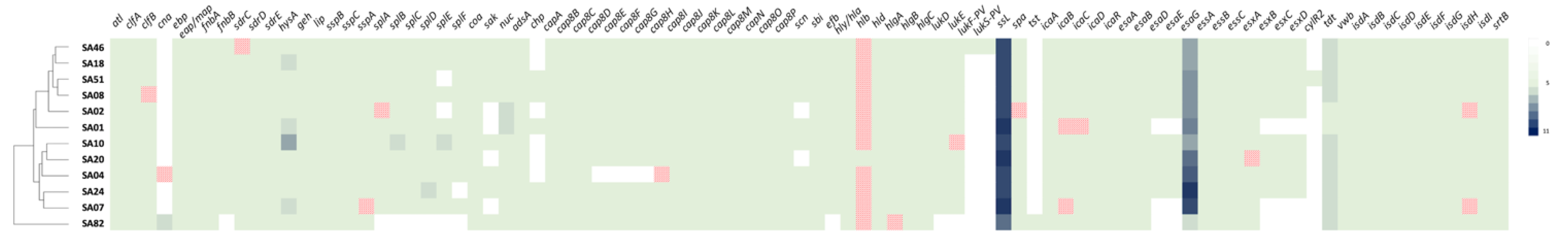
2.8. Virulence Determinants

Comparative analysis of virulence determinants is described by the heatmap shown in Figure 4, whereas the list of genetic determinants is reported in Supplementary Table S8. All strains harbour a repertoire of genes coding for (a) proteins involved in the adherence, such as the autolysin gene, clumping factors clfA and clfB (this latter is a pseudogene in SA08), and elastin binding protein gene; (b) exoenzymes such as the hyaluronate lyase gene hysA, serine proteases, lipases, and the coagulase gene vWbp (von Willebrand factor-binding protein); (c) proteins involved in the immune modulation and in the heme uptake system; and (d) several exotoxins (Figure 4; Supplementary Table S8).
The staphylococcal superantigen-like (ssl) genes are arranged as tandem repeats in the genomic island α (ssl1–11) and in the immune evasion cluster 2 (IEC2, ssl12–14) on a vSaγ island on the bacterial chromosome [44]. This last island also comprises the hyl gene, which codes for the α-haemolysin, one fibrin-binding protein, and the scn gene coding for the staphylococcal complement inhibitor [45]. Only in S. aureus SA02 the contig is interrupted downstream hyl.
S. aureus SA82 is the only strain that harbours the tst gene coding for the toxic shock syndrome toxin (TSST-1), whereas the lukF-PV and lukS-PV genes coding for the Panton–Valentine leukocidin (PVL) are present on a bacteriophage φSa2 only in the genome of S. aureus SA46.
In the heatmap built on the presence or absence of genes or pseudogenes coding for virulence determinants, the strains are clustered into two groups, with SA82 in an individual clade, as already shown by the phylogenetic analysis (Figure 1). The SA82 genome, although harbouring the tst gene, lacked genes coding for (a) four out of five secreted virulence factors (EsxB, EsxC, EscD, and EsaD); (b) the soluble cytosolic protein EsaE of the type VII secretion system; and (c) the bi-component staphylococcal leukotoxins family LukDE [46]. It also missed (d) the efb gene coding for a fibrinogen binding protein; (e) the fnbB gene, encoding a fibronectin binding protein; and (f) all the genes coding for serine proteases. The hlgA gene, coding for the gamma haemolysin, is predicted to be a pseudogene. In contrast to all the other strains, the SA82 genome harbours two cna genes, encoding the collagen binding protein, which mediates bacterial adherence to collagen substrates and collagenous tissues, and is strongly associated with the pathogenesis of osteomyelitis and septic arthritis [47].
The hlb gene is interrupted due to a prophage integration in all strains with the exception of SA20.

2.9. Growth of S. aureus Strains in BHI + YE Broth and Production of Staphylococcal Enterotoxins

The concentrations of S. aureus strains SA04, SA18, SA20, SA46, and SA51 (harbouring sea, seb, sed, seh, and ser genes) at 0, 24, and 48 h of incubation in brain heart infusion broth supplemented with yeast extract (BHI + YE) are shown in Table 3. The pH values of the corresponding broth cultures are also reported (Table 3). At 0 h (immediately after the inoculum) the initial counts of all strains were similar (ranging between 6.38 ± 0.28 and 6.91 ± 0.29 Log cfu/mL). Major changes were observed after 24 h of incubation, when all bacterial numbers increased by more than 2 Log cfu/mL (ranging between 8.97 ± 0.09 and 9.34 ± 0.23 Log cfu/mL). Concentrations after 48 h of incubation were found at similar levels to those found after 24 h of incubation (ranging between 9.08 ± 0.13 and 9.44 ± 0.28).
With the exception of SA46, for which the SEH production was 1.76 ± 2.26 ng/mL at 24 h and not detectable at 48 h, all the other strains produced higher amounts of SEA, SEB, SED, SEH and SER at both 24 h and 48 h of incubation in the BHI + YE broth, as shown in Table 4.

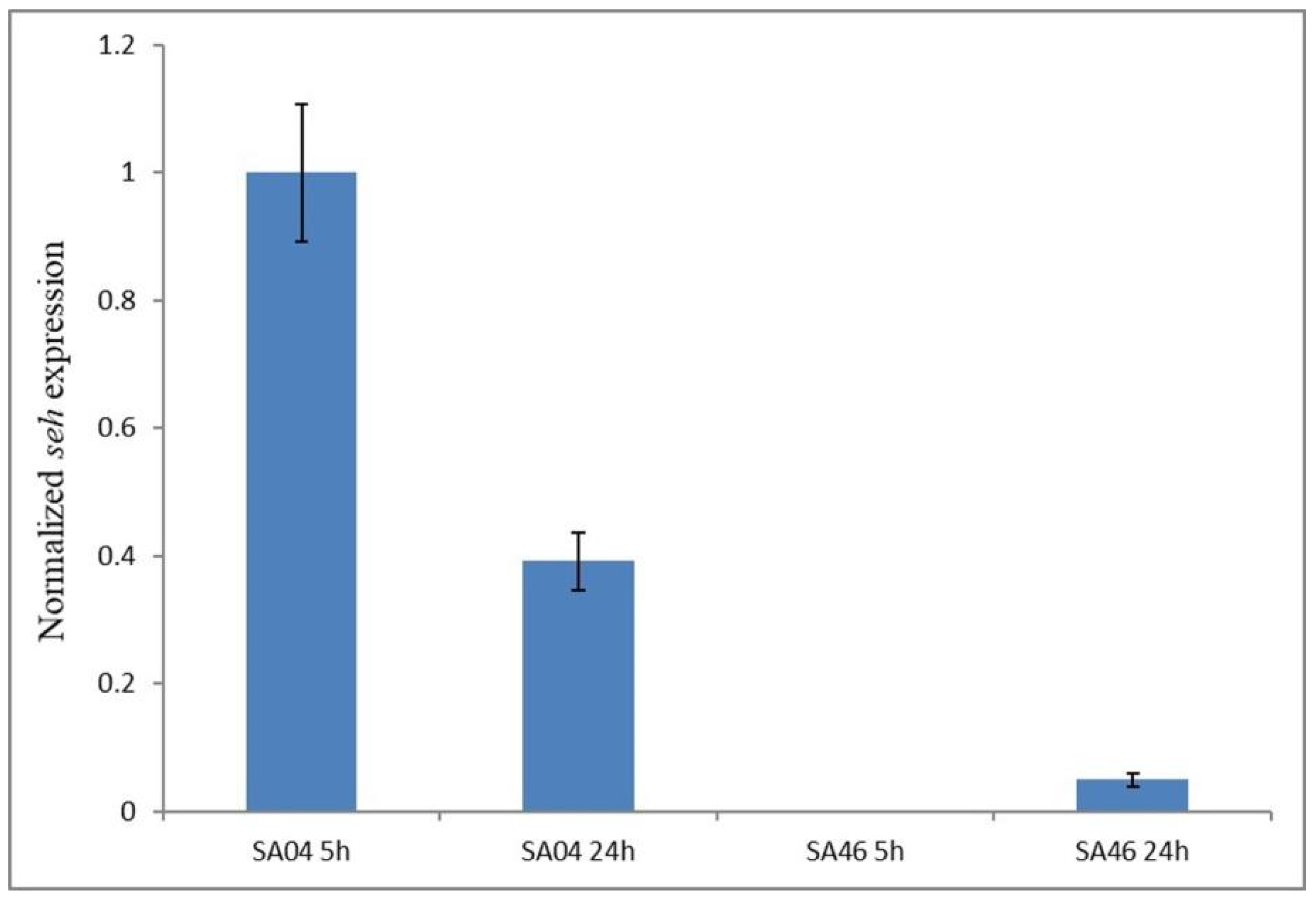
2.10. Expression of the seh Gene

The relative expression of the seh gene was evaluated in SA46 as well as in the other seh-positive strain SA04, at 5 and 24 h of incubation in the BHI + YE broth. As shown in Figure 5, in SA46 no transcription of the seh gene was observed at 5 h of incubation, whereas a very low transcription level compared to SA04 (0.13 fold) was detected at 24 h of incubation. In SA04 seh expression decreased at 24 h compared to that at 5 h (0.39-fold).

3. Discussion

In this study, we performed a comprehensive genomic analysis of 12 S. aureus strains isolated from 207 Algerian ready-to-eat foods to explore their genetic relatedness, gain further data on S. aureus molecular epidemiology, and investigate their virulence and antibiotic resistance genetic determinants that may help to better understand the features of foodborne S. aureus in Algeria as well as in the African continent.
Two novel alleles, aroE-1025 and yqiL-954, and two novel STs, 7199 and 7200, were identified and deposited in the PubMLST public database, thus contributing as new suitable resources for monitoring the global and local epidemiology of S. aureus. Among the other nine detected STs, six have been previously reported in Algeria, i.e., ST1-, ST8-, ST15-, ST22-, and ST97-MSSA clones as well as the ST80-MRSA clone [48,49], whereas ST5-, ST25-, and ST101-MSSA clones have been detected in other African countries [12,50,51]. In our study, the detection of a strain that is a member of the ST80-MRSA-SCCmec-IV lineage, also referred to as the European clone [52], is of particular interest since it has been described as having an alarming dissemination in Algeria [49]. It is worth mentioning that the clones herein found in Algerian ready-to-eat foods have been also found in humans (ST1-, ST5-, ST22-, ST25-, ST97-, and ST101-MSSA and ST80-MRSA-SCCmec-IV [12,49,50,51]) and animals (ST1-, ST8-, ST15-, ST22-, and ST97-MSSA and ST80-MRSA-SCCmec-IV [48,49]). This confirms the complexity of S. aureus epidemiology and highlights the fact that ready-to-eat foods, besides posing a risk to consumers for the onset of SFP, may also serve as means for S. aureus dissemination via food ingestion or handling, as pointed out also by other authors [3,53,54].
Considering the role of S. aureus in foodborne diseases due to the production of SEs [3], understanding the enterotoxigenic capabilities of circulating strains is a pivotal issue. Following the discovery of the classical SEs (SEA-SEE) [55,56], the number of the newly discovered SEs and SEls is constantly increasing. In particular, a total of 33 se and sel genes (sea-see; seg-selu; selu2; selv-selz; sel27-sel33) are known to date, including the last five sels recently reported (sel29 pseudogene; sel30-sel33) [1], thus indicating the wide enterotoxigenic potential of S. aureus. To the best of our knowledge, this is the first study searching for all the se and sel genes known to date and reporting the sel30 gene in Algerian S. aureus isolates from food after its recent discovery [1]. The gene, detected in SA51, shares 100% homology with the sequence reported by Dicks et al. [1]. Recently, some authors made efforts to gain data on the enterotoxigenic potential of foodborne S. aureus in Algeria, searching for some of the known se and sel genes (sea-see; seg-sek; selp and ser) [8,10,11], as well as the presence of the enterotoxin gene cluster (egc) [8]. These authors found the sea, seb, sec, see, seg, seh, sei, selj, sek, selp, ser, and the egc, although, in the latter case, no information on the actual egc gene content and the egc-type was provided [8,10,11]. We should remark that for the assessment of S. aureus enterotoxigenicity, not only the presence of se and sel genes but also the evaluation of genetic variants and the presence of pseudogenes, as herein performed, should be considered, since they may directly affect the enterotoxin production and virulence of the hosting S. aureus strains.
According to the results of our genome analysis, selw and selx genes were identified in all the sequenced strains, both located in the chromosome, in line with other studies [15,32]. selw was predicted as a pseudogene in 4 of 12 S. aureus strains, confirming that this gene, likely due to its chromosomal localisation, has a high rate of pseudogenisation [1]. Otherwise, no pseudogenes for selx were herein detected, corroborating previous reports in which an overall low prevalence of selx pseudogenes was observed, probably linked only to specific S. aureus lineages [1,16].
Most of the SFP diseases are associated with the classical SEs (SEA-SEE) [57]; furthermore, SEH has been found as a causative agent of SFP outbreaks [25,58]. Moreover, other newly discovered SEs such as SER and those belonging to the enterotoxin gene cluster (egc) are being reported with increasing evidence to have a possible role in foodborne intoxications [59,60,61].
The sea gene belonging to allele class 1 (sea1) [17] was identified in SA04, whereas SA51 harbours the sea2, a single-nucleotide sequence variant of the sea2 reported by Borst and Betley [17], that leads to no amino acid change in the predicted protein sequence (Table 1; Supplementary Table S7). SA04 produced higher amounts of SEA (156.04 ± 36.58–345.02 ± 62.67 ng/mL) compared with those produced by SA51 (64.54 ± 9.29–74.91 ± 7.36 ng/mL), being 2.4- and 4.6-folds higher at 24 h and 48 h, respectively. SEA, responsible for the majority of the SFP cases (about 80%) [57], is reported to cause human intoxication symptoms at a low dose of about 20–200 ng [62,63], and it is frequently detected in small amounts (ranging from 0.015 ng to >6 ng per gram or millilitre of food) in foods involved in SFP outbreaks [62,63,64,65]. These findings highlight the potential pathogenicity that our SEA-producing S. aureus strains may have in the contest of SFP. As regard the difference in SEA production observed in SA04 and SA51, it has been associated, by Borst and Betley [17], to the region upstream the translational start sites of sea. This region in SA04 is identical to that of the high-SEA-producing S. aureus FRI100 (harbouring sea allele class 1), whereas that in SA51 is identical to that of the low-SEA-producing S. aureus FRI281A (harbouring sea allele class 2) (Supplementary Table S7). However, the dissimilarities in the SEA amounts registered for SA04 and SA51 were not so pronounced as for S. aureus FRI100 and S. aureus FRI281A, since also other loci may affect the phage-mediated mechanism responsible for SEA expression [17].
sebv1 was identified in S. aureus SA18. This strain was able to produce SEB (2871.28 ± 811.09–14739.17 ± 5077.70 ng/mL) corroborating the findings of Johler et al. [18] who reported that all the described seb variants (sebv1-v4) are expressed, leading to the SEB production. In the literature, SEB-related SFPs are poorly documented; nevertheless, in a described outbreak involving boiled eggs, a high SEB concentration, greater than 1000 ng/g [66] and thus similar to that produced in the BHI + YE broth by our S. aureus strain, was reported. Unlike sea, differences in the seb upstream region were reported as not directly correlated with the SEB production level, which, on the other hand, may be linked to the type of the seb-harbouring SAPIs. Interestingly our seb-positive strain harbours a SAPI that has 99.4% similarity with SaPIivm60, which has been associated with strong SEB production by Sato’o et al. [35].
In SA20, sed was predicted as a pseudogene, which differs for a nucleotide substitution (leading to 1 amino-acid substitution) from the truncated sed described by Johler et al. [18] (Table 1; Supplementary Table S7). SA20 was able to produce SED (322.70 ± 41.17–398.94 ± 64.79 ng/mL), predicted as shorter than the intact protein (179 compared with the 258 amino acids of the intact SED) [18]. We should highlight that the prediction of the toxic properties based on the changes in the length and/or amino acid sequence of the SEs may be difficult, considering that the mechanism of action of SEs is not fully elucidated [18]. However, it should be highlighted that the truncated SED may have an impaired toxic function, resulting in a reduced virulence of the producing S. aureus strain. Low (0.052 ng/g [67]) to high (>200 ng/g [63]) concentrations of SED have been detected in foods involved in SFP outbreaks, although, to the best of our knowledge, no estimation of the oral dose capable of causing human symptoms has been reported. Some S. aureus strains harbouring the truncated sed gene were found either to not produce the relevant protein at detectable levels or even to not transcribe this pseudogene [18,68]. Nevertheless, Johler et al. [18] reported that certain S. aureus harbouring the truncated sed were able to produce the SED protein, being in line with our finding, although they found that the amounts were far lower than those produced by strains harbouring the intact sed gene.
selj and ser genes were also present in SA20 and located along with sed on the same putative pIB485-like plasmid as previously described [36]. Although also ser pseudogenes have been recently reported [7,69], SA20 harbours an intact ser gene and was able to produce SER (36,720.10 ± 6272.32–63,176.06 ± 14,422.93 ng/mL) in amounts more than 100-folds higher than SED. Interestingly, S. aureus strains harbouring the plasmid-related sed, selj, and ser genes have been isolated from seven SFP outbreaks in Japan [60], and their ability to produce SER in BHI + YE (523–2925 ng/mL) has been demonstrated, suggesting that SER may have played a possible role in the onset of the described SFPs [60].
The seh gene was detected as intact or as a pseudogene in two of our sequenced strains. SA04, harbouring the intact seh, was able to produce the enterotoxin SEH (221.76 ± 38.15–239.48 ± 84.83 ng/mL) (along with SEA, as discussed above), and, notably, the nucleotide sequence was identical to that of the seh gene found in S. aureus strains that caused a SEH-mediated SFP outbreak ([25]; Supplementary Table S7), indicating, therefore, the potential virulence of this strain. In particular, in the reported outbreak, a concentration of SEH equal to 55.5 ng/g was estimated in the consumed mashed potato made with raw milk, and the tested S. aureus strains, isolated from these leftovers and the farm bulk raw milk, were able to produce SEH in concentrations similar (96–108 ng/mL) to that produced by our SA04 strain, when grown in a microbial broth [25]. The seh pseudogene was instead detected in the SA46 strain belonging to the European MRSA-ST80-SCCmec-IV clone, corroborating the study of Dicks et al. [1] who recently reported this pseudogene as strongly associated with the MRSA-ST80-SCCmec-IV clone. SA46 also harboured the sey gene, whose sequence was previously detected in a ST80 strain [24]. SA46 produced SEH at almost undetectable levels. To investigate if the difference in the SEH amounts produced by SA04 and SA46 may depend on differences in seh expression, we assessed it taking into account that, in experimental conditions similar to those herein applied, the seh gene expression occurs during the exponential phase of growth [70]. The expression analysis detected no transcripts at 5 h for SA46, whereas at 24 h, their levels were much lower than those of the intact seh gene, harboured by SA04, confirming that, in this case, the failure of the SEH production in SA46 depends on transcriptional regulation. This is interesting since certain se pseudogenes may be transcribed at levels similar to the intact se genes, as found by Lis et al. [68], suggesting that the protein synthesis failure may occur later, at the translational level.
Although the involvement of MRSA in SFP has been reported [71] based on these findings and considering the relative little assortment of se and sel genes (seh pseudogene, sey, selx, and selw), it seems that our MRSA strain (SA46) may play a minor role in the onset of SFP compared with the other enterotoxin producing MSSA strains herein detected. A previous study found one S. aureus strain belonging to the European clone carrying the egc-related genes seg, sei, sem, sen, seo, and selu [72], whereas seb and sek were harboured by atypical MRSA-ST80-SCCmec-IV pvl-negative isolates [73]. Therefore, our findings may help to enrich the knowledge regarding the endowment of se and sel genes within the European clone in order to improve the understanding of the enterotoxigenic potential of this lineage.
The egc was found in 5 out of the 12 sequenced S. aureus strains, corroborating previous investigations that reported that egc-related genes are among the most prevalent genes in foodborne S. aureus isolates [74]. Beyond its correlation to SFP, the egc presence seems to be relevant also in other clinical conditions, such as chronic infections in cystic fibrosis patients [75]. This cluster may harbour several se and sel genes (i.e., seo, sem, sei, selu, sen, seg, selu2, selv, and sel33) and pseudogenes (ψent1-ψent2) in different combinations, and, to date, at least eight egc types have been described in S. aureus [1,7]. Two of our strains harbour the egc1 (seg, sei, sem, sen, seo, and ψent1-ψent2) and three strains harbour the egc5 (seg, sei, sem, sen, seo, and selu2). Both these types have been detected in foodborne S. aureus isolates [7], but there is still scarce knowledge on their actual prevalence mostly due to the fact that egc5 was only recently described [7].
The emergence of antimicrobial resistance in S. aureus is a clinical and public health challenge due to the reduction of the antimicrobial assortment that is effective when S. aureus infections occur. In addition to MRSA, whose presence has been globally reported in various ecological niches including African food, animals, and humans [13,76], also multidrug-resistant MSSA, typically showing resistance to three or more antimicrobials of different classes, as herein reported for SA02 and SA18, are being detected in various sources comprising food products (especially animal-derived unprocessed products) in Africa [23,77,78,79] and worldwide [54,80,81,82]. Genome analysis allowed us to detect the antimicrobial resistance genes related to the phenotypic resistance profile shown by our sequenced S. aureus strains. In particular, the blaZ gene and regulatory system composed of blaR1 and blaI that we found in 10 out of the 12 sequenced strains represent the genetic mechanism conferring resistance to benzylpenicillin and other penicillinase-labile penicillins. We should emphasise that in S. aureus SA51 we found the bla locus (blaZ, blaR1, and blaI), although it was reported as benzylpenicillin-susceptible. Since 2012, CLSI acknowledged that penicillin-susceptible results should be further investigated before being confirmed, performing additional phenotypic tests to detect β-lactamase production (i.e., nitrocefin-based tests and/or penicillin zone edge) [83]. However, these phenotypic methods were described as less sensitive compared with the molecular detection of the blaZ gene, and, therefore, different authors considered the latter the gold standard method for β-lactamase detection in S. aureus [84,85].
blaZ is also frequently found in MRSA [86,87], including the ST80-MRSA-SCCmec-IV lineage as previously reported for most of the analysed isolates by Monecke et al. [86]. Our ST80-MRSA-SCCmec-IV strain, SA46, although harbouring the mecA gene conferring resistance to β-lactam antimicrobials [87] such as benzylpenicillin, oxacillin, and cefoxitin, does not harbour the blaZ gene, and, in Africa (in Cape Verde), a similar feature was recently reported for other MRSA isolates belonging to the ST5-SCCmec-VI lineage [88]. Although the bla system is involved in the regulation of the mecA expression [89], it has been elucidated that the loss of the bla system may allow the constitutive expression of the mecA gene, conferring β-lactam resistance in SCCmec-IV harbouring MRSA [89].
It has also been reported that 91.6% of ST80-MRSA isolates are resistant to kanamycin, and the kanamycin-resistance gene aph(3′)-III has been detected in the ST80-MRSA clone [90], which is consistent with our finding. Beyond aph(3′)-IIIa, also the other detected genes conferring resistance to aminoglycosides (ant(6)-I), lincosamides (lmrS), tetracyclines (tet(L), tet(38)), macrolides (ermB), quinolones, and fluroquinolones (gyrA-S84L) have been previously reported in S. aureus isolates [91,92,93,94]. Nevertheless, a paucity of studies detected a range of antimicrobial resistance determinants in foodborne S. aureus in Africa, and, in particular, only few of the aforementioned genes were previously reported in Tunisia (aph(3′)-IIIa, tet(L) [95]), Kenya (aph(3′)-IIIa [96]), and South Africa (aph (3′)-1-IIIa, gyrA-S84L [77,97]). Therefore, the present study helps to further expand the knowledge on the genetic determinants harboured by antimicrobial-resistant S. aureus isolated from food in the African continent.
The results of the genomic analysis highlighted a plethora of different virulence determinants in all the sequenced strains, involved in adhesion, colonisation, spreading, and immune modulation. Adherence is the first process involved in the pathogenicity of S. aureus. It is determined by the expression of several genes such as that coding for the autolysin, which mediates adherence to immobilised fibrinogen and fibronectin [98], the clumping factors clfA and clfB that bind to different sites in fibrinogen, and fibronectin binding proteins A and B, which facilitate the attachment of S. aureus to host cells [99].These genes are present in all the sequenced strains. Exoenzymes such as the hyaluronate lyase coded by hysA are required for the degradation of hyaluronic acid, which contributes to the local dissolution of the extracellular matrix, whereas vWbp enables the bacteria to disseminate and resist to opsonophagocytic clearance by host immune cells [100]. Among the exotoxins, haemolysin genes were retrieved in all the strains. The hlb gene, which codes for a sphingomyelinase that cleaves sphingomyelin into phosphocholine and ceramide [101], was a pseudogene in all strains, with the exception of SA20. The interruption of this gene is caused by the integration of a prophage of Sa3int type. Loss and reintegration of this phage are correlated with the transmission of S. aureus from humans to livestock and vice versa [102]. The restoration of the hlb gene, which occurred in SA20, might be relevant for important specific infections [103,104].
Panton-Valentine leukocidin (PVL) is a major virulence factor of S. aureus, although it presents in less than 5% of S. aureus strains. It is a two-component toxin that induces pore formation in the leukocyte cell membrane complement receptors [105]. The results of our genomic analysis detected pvl genes only in S. aureus SA46, harbouring SCCmec of type IV. PVL has been proposed as a marker of CA-MRSA [106], although PVL-negative CA-MRSA clones have been reported [52].
According to the international working group on the Classification of Staphylococcal Cassette Chromosome [107], the genomic sequence of SA46 meets the criteria for defining an SCCmec: (i) carriage of mecA in a mec gene complex; (ii) presence of ccr gene(s) (ccrAB and/or ccrC) in the ccr gene complex; (iii) integration at a specific site in the staphylococcal chromosome, designated as the integration site sequence (ISS) for SCC, which serves as a target for ccr-mediated recombination; and (iv) presence of flanking direct repeat sequences containing the ISS. SCCmec of SA46 was predicted as SCCmec subtype-IVc(2B), although the B class of the mec complex is characterised by the presence of mecA and a truncated mecR1 resulting from the insertion of IS1272 upstream mecA [108]. On the contrary, the annotation of this genome returned an intact mecR1 and an incomplete IS1272, in the middle of a contig. Indeed, the whole cassette was dislocated in two separate nodes. This failure in reconstructing the entire SCCmec as the result of short-read sequencing technology is due to the multiple insertion sequences, which cause difficulties in the assembly.
To date, only two studies performed a genomic analysis of Algerian S. aureus strains: one by Mairi et al. [109], who reported the first occurrence of the “Maltese clone” (CC5-MRSA-IV-SCCfus) in Bat Guano, and a second by Aouati et al. [110], who uncovered the emergence of MRSA SCCmec-III Mercury in clinical isolates in Eastern Algeria. Other sequence read archives (SRAs) obtained by sequencing S. aureus isolated from Algerian dairy products were deposited in NCBI (Accession ERS4338951-ERS4338960).
This is the first genomic study performed on Algerian foodborne S. aureus strains and, to the best of our knowledge, the first comparative and phylogenetic reconstruction of foodborne S. aureus of worldwide origin. According to our data, there is a clear phylogenetic association of S. aureus, based on the ST or the geographical origin, rather than the source of isolation. This reflects the dynamics and evolution of the S. aureus population worldwide. The arising grouping based on the food source might be an effect of the sample collection rather than a clustering based on the use of a specific ecological niche.
The Algerian isolates are dispersed in the phylogenetic tree with the exception of SA51 and SA02, which both belong to the ST5, and SA01 and SA07, which differ for the aroE allele in the MLST. From an evolutionary point of view, SA08 is the less recent strain, whereas SA82, which is individually located in all the cluster analyses we have performed, is the most recent. However, such phylogenetic reconstructions are affected by the fact that most strains, although sharing the same geographical origin, have been isolated from different food products, some generically indicated as food, and no information is available on the manufacturing process nor on the origin of raw material used, thus hindering an exhaustive reconstruction of phylogenetic and epidemiological maps.
This underlines the urgent need for genomic studies on foodborne S. aureus in Algeria to have more detailed insights regarding the structure of the S. aureus population as well as the harboured genetic determinants of virulence and antibiotic resistance, in order to be able to estimate the public health burden related to the spread of this pathogen in food.

4. Conclusions

A comprehensive genomic characterisation and the production of some classical and newly described staphylococcal enterotoxins of 12 Algerian S. aureus strains, previously isolated from 207 ready-to-eat foods, were herein performed. Two novel STs were identified, and a strain belonging to the alarming MRSA-ST80-SCCmec-IV European clone was detected. An up-to-date assessment of se and sel genes in our foodborne S. aureus strains was carried out, and we found that MSSA strains produced higher amounts of enterotoxins than the MRSA-ST80-SCCmec-IV strain herein sequenced. The detection of antimicrobial resistance genetic determinants in the 12 S. aureus strains supported the results of the antibiotic susceptibility testing previously performed by Mekhoulfi et al. [4]. Furthermore, a plethora of virulence genes were found in the 12 S. aureus genomes, although SA82 lacked several determinants involved in adhesion and colonisation, confirming its outgrouping position in the taxonomic clustering.
The results of the genomic analysis, together with the production of enterotoxins investigated in this study, confirm the risk associated with the spread of this pathogen in food, which is worsened by the observation that some of these S. aureus strains are multidrug resistant, and by the detection of the tst gene, determinant for the toxic shock syndrome toxin-1, in S. aureus SA82, and the PVL-encoding genes in the methicillin-resistant SA46 strain.

5. Materials and Methods

5.1. S. aureus Strains Used in the Study

The 12 S. aureus strains sequenced in this study are included in Supplementary Table S4. These strains were cultured on Baird–Parker egg yolk (BPEY, Oxoid, France), and their purity was checked by streaking on the same agar medium. The pure cultures were stored as stock cultures at −80° C in brain heart infusion broth (BHI; Conda. Pronadisa, Spain) supplemented with 0.6% yeast extract (Biolife Italiana, Milan, Italy) and 20% glycerol. For DNA extraction, the working culture was prepared as described by Fusco et al. [111].

5.2. Whole-Genome Sequencing and Analysis

Two millilitres of fresh working cultures of each of the S. aureus strains were used for the DNA extraction following the protocol described by Chieffi et al. [7]. The integrity, purity, and quantity of DNA were assessed by agarose gel electrophoresis, a Nanodrop photometer (Peqlab-VWR International Srl, Darmstadt, Germany), and a Qubit 3.0 fluorimeter (Thermo Fisher Scientific, Waltham, MA, USA). DNA was subjected to whole-genome shotgun sequencing by using an Illumina mate pair library prep kit (Illumina, San Diego, CA, USA), according to the manufacturer’s instructions, and then sequenced on an Illumina MiSeq platform with the 2 × 250 mate pair procedure. Reads were then trimmed by NxTrim (V2) [112], and de novo assembly was performed using SPAdes version 3.10.1 (©St. Petersburg State University, St. Petersburg, Russia [113]) with the following parameters: Kmer 21, 33, 55, 77 and “careful”.
The overall contiguity of the assembly and genome statistics were determined with MIGA [114]. The completeness of the de novo assemblies was measured by the presence of 106 single-copy genes that are observed across almost all prokaryotic genomes by using MiGA [114]. Contamination was calculated based on the copy number of essential genes (in %) present in the genome. Quality scores were then calculated as completeness percentage minus five times contamination percentage.
The whole-genome shotgun projects have been deposited at DDBJ/ENA/GenBank under the accessions reported in Supplementary Table S4. The versions described in this paper are JAHLTR010000000 for S. aureus SA01, JAFNJM010000000 for S. aureus SA02, JAFNJN010000000 for S. aureus SA04, JAHLTS010000000 for S. aureus SA07, JAHLTT010000000 for S. aureus SA08, JAHLTU010000000 for S. aureus SA10, JAFNJO010000000 for S. aureus SA18, JAFNJP010000000 S. aureus SA20, JAFRED010000000 for S. aureus SA24, JAFNJQ010000000 for S. aureus SA46, JAFNJR010000000 for S. aureus SA51, and JAHLTV010000000 for S. aureus SA82.

5.3. Bioinformatic Methods

5.3.1. Gene Prediction, ANI, and Phylogenomic Analysis

Genes were predicted and annotated using the PROKKA pipeline implemented in the Galaxy platform (Galaxy Tool Version 1.14.5 [115]) and by NCBI (National Center for Biotechnology Information) Prokaryotic Genome Annotation Pipeline (PGAP [116]). Protein ID used in the manuscript indicated those obtained by PGAP.
All the protein sequences used in this study were retrieved from GenBank (NCBI). The homology-based relationship of S. aureus genes and predicted proteins towards reference sequences was determined by the BLASTN and BLASTP algorithms on the NCBI site [117]. Gene models were manually determined, and clustering and orientation were subsequently deduced for the closely linked genes.
Strains used to perform pan-genome and phylogenetic analyses are listed in Supplementary Table S4. Genomic sequences were downloaded from the NCBI and submitted to the PROKKA pipeline. The obtained .gff3 files were used as input for Roary (Galaxy Version 3.13.0 + galaxy1), the pangenome pipeline [118], to generate a core gene alignment with 95% as the minimum percentage identity for blastp analysis and 99% as the total percentage of the isolates in which the gene needs to be present for it to be considered a core gene. The pan-genome was represented as the core genome (shared by >99% of strains), accessory genome (genes present in >2 strains but not in all), and unique genome (genes unique to individual strains). The total pan-genome was also shown as core (99% ≤ strains ≤ 100%), soft core (95% ≤ strains < 99%), shell (15% ≤ strains  < 95%), and cloud (0% ≤ strains  < 15%). Visualisation of the output was achieved by using Phandango (version 1.3.0 [119]).
The genetic divergence among S. aureus species was calculated using the ANI calculator [120,121], which estimates the average nucleotide identity (ANI) using both best hits (one-way ANI) and reciprocal best hits (two-way ANI) between genomic datasets.
Genome-based phylogeny using 1000 single copy genes was reconstructed employing the Phylogenetic Tree Building Service implemented in the BV-BRC platform [122], with the maximum likelihood method RAxML (version 8.2.11) and progressive refinement [123]. S. argenteus MSHR1132 was used as an outgroup, and visualisation of the phylogenetic trees was performed by using iTOL (version 6.3.2 [124]).

5.3.2. MLST and spa Typing

Multilocus sequence typing (MLST) was performed by MLST 2.0 (software version: 2.0.4 (8 May 2019); database version: 2.0.0 (13 September 2021)) of the Center for Genomic Epidemiology [125,126]. spa typing was performed by spaTyper (Software version 1.0 Database version: 22 August 2022) of the Center for Genomic Epidemiology [127,128].

5.3.3. Antibiotic Resistance and Virulence Determinants Analyses

Antibiotic resistance determinants were computationally predicted within the BV-BRC platform [129] by a k-mer-based detection method and BLAST analysis, and then manually curated. Virulence determinants were screened by using the comparative pathogenomics-based VF analysis pipeline VFanalyzer [130] implemented in the virulence factor database (VFDB) [131], and then manually curated. A heatmap was manually constructed and visualised by using the heatmapper web server [43,132] with average linkage as the clustering method and Euclidean distance measurement method.
Enterotoxin genes were determined by homology search using previously reported reference sequences ([1]; Supplementary Table S7) using an E-value cut-off of 0.001, a minimum percentage of alignment identity of 80%, and a relative coverage threshold of >80% [41,133].
Prediction of SCCmec elements in sequenced S. aureus isolates was performed by using the SCCmecFinder v1.2 [134,135] with default parameters, then manually curated.

5.4. Cultivation of S. aureus for Staphylococcal Enterotoxin Production Assessment

S. aureus strains SA04, SA18, SA20, SA46, and SA51, harbouring the classical and some newly described se genes (namely sea, seb, sed, seh, and ser), were chosen to assess the ability to produce SEs. Bacterial cultures were prepared as described by Schubert et al. [136]. Briefly, 100 mL of brain heart infusion supplemented with 1% yeast extract (BHI + YE, Biocorp, Warsaw, Poland) were inoculated with pre-culture to reach an optical density of 0.02 at 600 nm (OD600). Prior to inoculation, the pre-cultures were washed twice with phosphate-buffered saline (PBS) to remove residual BHI broth and enterotoxins (repeated centrifugation at 12,000× g for 5 min and resuspension in PBS). Cultures were incubated at 37 °C with constant agitation at 230 rpm. The bacterial cell concentration of the broth cultures at 0, 24, and 48 h of incubation was determined by plating serial tenfold dilutions, prepared in quarter-strength Ringer’s solution (Oxoid, Basingstoke, UK), onto BHI agar. The pH of the broth cultures was measured using a FE20-FiveEasy™ pH-meter (Mettler-Toledo, Greifensee, Switzerland).

5.5. Sandwich ELISA for SEA, SEB, SED, SEH, and SER Detection

Recombinant staphylococcal enterotoxins (rSED, rSEH, and rSER) were obtained as previously described by Schubert et al. [136,137]. Rabbit polyclonal anti-A, anti-B, anti-SED, and anti-SER antibodies were purchased from OriGene Technologies GmbH (Herford, Germany), whereas rabbit polyclonal anti-H and anti-H(HRP) antibodies were purchased from Abcam (Cambridge, UK). Samples for staphylococcal enterotoxins (SEs) detection were collected after 24 and 48 h of growth and stored at −20 °C until analysed. Supernatants were pre-incubated with a 20% normal rabbit serum to bind protein A and then diluted in PBS containing 0.1% Tween-20 (Merck, Darmstadt, Germany). The enzyme linked immunosorbent assay (ELISA) was also performed as described by Schubert et al. [136,137]. The concentration of the SEs in samples was measured with SEA, SEB (Merck KGaA, Darmstadt, Germany), rSED, rSEH, and rSER as standards, using a 4-parameter logistic curve fit. The ELISA assays for each SE were run in two biological repeats with two replicates. Data analysis was carried out using GraphPad Prism software 8.0.1 (GraphPad Software Inc., La Jolla, CA, USA).

5.6. RNA Extraction and Reverse Transcription–Quantitative PCR to Assess seh Gene Expression

Expression of the seh gene was assessed in the seh-positive S. aureus SA04 and SA46 strains. Samples for RNA isolation were collected from the BHI+YE broth after 5 and 24 h of growth. RNA extraction, purification, cDNA synthesis, and reverse transcription–quantitative polymerase chain reaction (RT-qPCR) were performed as described by Schubert et al. [136]. The rpoB housekeeping gene was used for normalisation [138]. Transcript levels of seh relative to rpoB were calculated according to Pfaffl [139]. Each RT-qPCR assay consisted of two biological repeats with three replicates. Data analysis was carried out using Bio-Rad CFX Manager software.

Supplementary Materials

The following supporting information can be downloaded at: https://www.mdpi.com/article/10.3390/toxins14110731/s1, Supplementary Figure S1. Pan-genome analysis of S. aureus strains. Visualisation of pan-genome analysis of S. aureus strains was performed by using Phandango (version 1.3.0). Supplementary Figure S2. Enterotoxin gene cluster (egc) localisation in S. aureus strains. Protein IDs are indicated in the figure. Red: enterotoxins; yellow: serine proteases; brown: leukocidins; grey: hypothetical proteins; light blue: restriction endonucleases; pink: transposases; turquoise: lantibiotic cluster; capB: calcium binding protein; pepA1: glutamyl aminopeptidase; * indicates a pseudogene. Supplementary Table S1. Genomic features of S. aureus strains sequenced in this study. Supplementary Table S2. MLST allelic profile of sequenced S. aureus strains. Supplementary Table S3. spa typing of S. aureus strains. Supplementary Table S4. List of S. aureus strains used in this study. Supplementary Table S5. ANI values of S. aureus strains. Supplementary Table S6. Roary statistics. Supplementary Table S7. se and sel genes and predicted protein sequences in S. aureus strains. Supplementary Table S8. Virulence determinants in S. aureus strains.

Author Contributions

Conceptualization, V.F. and F.F.; formal analysis, F.F., D.C., J.S. and V.F.; investigation, F.F., D.C., G.-S.C., J.S. and O.A.M.; supervision, V.F., J.B. and C.M.A.P.F.; visualization, F.F., D.C. and J.S.; writing—original draft preparation, F.F., D.C. and V.F.; writing—review and editing, F.F., D.C., V.F., C.M.A.P.F., G.-S.C., J.B., J.S. and O.A.M. All authors have read and agreed to the published version of the manuscript.

Funding

This research received no external funding.

Institutional Review Board Statement

Not applicable.

Informed Consent Statement

Not applicable.

Data Availability Statement

Data is contained within the article or Supplementary Materials.

Conflicts of Interest

The authors declare no conflict of interest.

References

  1. Dicks, J.; Turnbull, J.D.; Russell, J.; Parkhill, J.; Alexander, S. Genome sequencing of a historic Staphylococcus aureus collection reveals new enterotoxin genes and sheds light on the evolution and genomic organization of this key virulence gene family. J. Bacteriol. 2021, 203, e00587-20. [Google Scholar] [CrossRef] [PubMed]
  2. Foster, T.J. Antibiotic resistance in Staphylococcus aureus. Current status and future prospects. FEMS Microbiol. Rev. 2017, 41, 430–449. [Google Scholar] [CrossRef] [PubMed]
  3. Fusco, V.; Chieffi, D.; Fanelli, F.; Logrieco, A.F.; Cho, G.-S.; Kabisch, J.; Böhnlein, C.; Franz, C.M.A.P. Microbial quality and safety of milk and milk products in the 21st century. Compr. Rev. Food Sci. Food Saf. 2020, 19, 2013–2049. [Google Scholar] [CrossRef] [PubMed]
  4. Mekhloufi, O.A.; Chieffi, D.; Hammoudi, A.; Bensefia, S.A.; Fanelli, F.; Fusco, V. Prevalence, enterotoxigenic potential and antimicrobial resistance of Staphylococcus aureus and Methicillin-Resistant Staphylococcus aureus (MRSA) isolated from Algerian ready to eat foods. Toxins 2021, 13, 835. [Google Scholar] [CrossRef]
  5. Fishovitz, J.; Hermoso, J.A.; Chang, M.; Mobashery, S. Penicillin-binding protein 2a of methicillin-resistant Staphylococcus aureus. IUBMB Life 2014, 66, 572–577. [Google Scholar] [CrossRef] [Green Version]
  6. Jarraud, S.; Peyrat, M.A.; Lim, A.; Tristan, A.; Bes, M.; Mougel, C.; Etienne, J.; Vandenesch, F.; Bonneville, M.; Lina, G. egc, a highly prevalent operon of enterotoxin gene, forms a putative nursery of superantigens in Staphylococcus aureus. J. Immunol. 2001, 166, 669–677. [Google Scholar] [CrossRef] [PubMed] [Green Version]
  7. Chieffi, D.; Fanelli, F.; Cho, G.-S.; Schubert, J.; Blaiotta, G.; Franz, C.M.A.P.; Bania, J.; Fusco, V. Novel insights into the enterotoxigenic potential and genomic background of Staphylococcus aureus isolated from raw milk. Food Microbiol. 2020, 90, 103482. [Google Scholar] [CrossRef]
  8. Achek, R.; El-Adawy, H.; Hotzel, H.; Hendam, A.; Tomaso, H.; Ehricht, R.; Neubauer, H.; Nabi, I.; Hamdi, T.M.; Monecke, S. Molecular Characterization of Staphylococcus aureus Isolated from Human and Food Samples in Northern Algeria. Pathogens 2021, 10, 1276. [Google Scholar] [CrossRef]
  9. Chaalal, W.; Chaalal, N.; Bourafa, N.; Kihal, M.; Diene, S.M.; Rolain, J.-M. Characterization of Staphylococcus aureus Isolated from Food Products in Western Algeria. Foodborne Pathog. Dis. 2018, 15, 353–360. [Google Scholar] [CrossRef]
  10. Titouche, Y.; Hakem, A.; Houali, K.; Meheut, T.; Vingadassalon, N.; Ruiz-Ripa, L.; Salmi, D.; Chergui, A.; Chenouf, N.; Hennekinne, J.A.; et al. Emergence of methicillin-resistant Staphylococcus aureus (MRSA) ST8 in raw milk and traditional dairy products in the Tizi Ouzou area of Algeria. J. Dairy Sci. 2019, 102, 6876–6884. [Google Scholar] [CrossRef]
  11. Titouche, Y.; Houali, K.; Ruiz-Ripa, L.; Vingadassalon, N.; Nia, Y.; Fatihi, A.; Cauquil, A.; Bouchez, P.; Bouhier, L.; Torres, C.; et al. Enterotoxin genes and antimicrobial resistance in Staphylococcus aureus isolated from food products in Algeria. J. Appl. Microbiol. 2020, 129, 1043–1052. [Google Scholar] [CrossRef] [PubMed]
  12. Breurec, S.; Fall, C.; Pouillot, R.; Boisier, P.; Brisse, S.; Diene-Sarr, F.; Djibo, S.; Etienne, J.; Fonkoua, M.C.; Perrier-Gros-Claude, J.D.; et al. Epidemiology of methicillin-susceptible Staphylococcus aureus lineages in five major African towns: High prevalence of Panton-Valentine leukocidin genes. Clin. Microbiol. Infect. 2011, 17, 633–639. [Google Scholar] [CrossRef] [PubMed]
  13. Lozano, C.; Gharsa, H.; Ben Slama, K.; Zarazaga, M.; Torres, C. Staphylococcus aureus in animals and food: Methicillin resistance, prevalence and population structure. A review in the African continent. Microorganisms 2016, 4, 12. [Google Scholar] [CrossRef] [PubMed] [Green Version]
  14. Aung, M.; San, T.; Aye, M.; Mya, S.; Maw, W.; Zan, K.; Htut, W.; Kawaguchiya, M.; Urushibara, N.; Kobayashi, N. Prevalence and Genetic Characteristics of Staphylococcus aureus and Staphylococcus argenteus Isolates Harboring Panton-Valentine Leukocidin, Enterotoxins, and TSST-1 Genes from Food Handlers in Myanmar. Toxins 2017, 9, 241. [Google Scholar] [CrossRef] [Green Version]
  15. Wilson, G.J.; Seo, K.S.; Cartwright, R.A.; Connelley, T.; Chuang-Smith, O.N.; Merriman, J.A.; Guinane, C.M.; Park, J.Y.; Bohach, G.A.; Schlievert, P.M.; et al. A novel core genome-encoded superantigen contributes to lethality of community-associated MRSA necrotizing pneumonia. PLoS Pathog. 2011, 7, e1002271. [Google Scholar] [CrossRef] [Green Version]
  16. Roetzer, A.; Haller, G.; Beyerly, J.; Geier, C.B.; Wolf, H.M.; Gruener, C.S.; Model, N.; Eibl, M.M. Genotypic and phenotypic analysis of clinical isolates of Staphylococcus aureus revealed production patterns and hemolytic potentials unlinked to gene profiles and source. BMC Microbiol. 2016, 16, 13. [Google Scholar] [CrossRef] [Green Version]
  17. Borst, D.W.; Betley, M.J. Phage-associated differences in staphylococcal enterotoxin A gene (sea) expression correlate with sea allele class. Infect. Immun. 1994, 62, 113–118. [Google Scholar] [CrossRef] [Green Version]
  18. Johler, S.; Sihto, H.-M.; Macori, G.; Stephan, R. Sequence variability in staphylococcal enterotoxin genes seb, sec, and sed. Toxins 2016, 8, 169. [Google Scholar] [CrossRef] [Green Version]
  19. Baba, T.; Takeuchi, F.; Kuroda, M.; Yuzawa, H.; Aoki, K.; Oguchi, A.; Nagai, Y.; Iwama, N.; Asano, K.; Naimi, T.; et al. Genome and virulence determinants of high virulence community-acquired MRSA. Lancet 2002, 359, 1819–1827. [Google Scholar] [CrossRef]
  20. Betley, M.J.; Mekalanos, J.J. Nucleotide sequence of the type A staphylococcal enterotoxin gene. J. Bacteriol. 1988, 170, 34–41. [Google Scholar] [CrossRef]
  21. Chua, K.; Seemann, T.; Harrison, P.F.; Davies, J.K.; Coutts, S.J.; Chen, H.; Haring, V.; Moore, R.; Howden, B.P.; Stinear, T.P. Complete Genome Sequence of Staphylococcus aureus Strain JKD6159, a Unique Australian Clone of ST93-IV Community Methicillin-Resistant Staphylococcus aureus. J. Bacteriol. 2010, 192, 5556–5557. [Google Scholar] [CrossRef] [PubMed] [Green Version]
  22. Collery, M.M.; Smyth, C.J. Rapid differentiation of Staphylococcus aureus isolates harbouring egc loci with pseudogenes psient1 and psient2 and the selu or seluv gene using PCR-RFLP. J. Med. Microbiol. 2007, 56, 208–216. [Google Scholar] [CrossRef] [PubMed] [Green Version]
  23. Egyir, B.; Hadjirin, N.F.; Gupta, S.; Owusu, F.; Agbodzi, B.; Adogla-Bessa, T.; Addo, K.K.; Stegger, M.; Larsen, A.R.; Holmes, M.A. Whole-genome sequence profiling of antibiotic-resistant Staphylococcus aureus isolates from livestock and farm attendants in Ghana. J. Glob. Antimicrob. Resist. 2020, 22, 527–532. [Google Scholar] [CrossRef]
  24. Gawlik, D.; Ruppelt-Lorz, A.; Muller, E.; Reißig, A.; Hotzel, H.; Braun, S.D.; Söderquist, B.; Ziegler-Cordts, A.; Stein, C.; Pletz, M.W.; et al. Molecular investigations on a chimeric strain of Staphylococcus aureus sequence type 80. PLoS ONE 2020, 15, e0232071. [Google Scholar] [CrossRef] [PubMed]
  25. Jørgensen, H.J.; Mathisen, T.; Lovseth, A.; Omoe, K.; Qvale, K.S.; Loncarevic, S. An outbreak of staphylococcal food poisoning caused by enterotoxin H in mashed potato made with raw milk. FEMS Microbiol. Lett. 2005, 252, 267–272. [Google Scholar] [CrossRef] [PubMed] [Green Version]
  26. Langley, R.J.; Ting, Y.T.; Clow, F.; Young, P.G.; Radcliff, F.J.; Choi, J.M.; Sequeira, R.P.; Holtfreter, S.; Baker, H.; Fraser, J.D. Staphylococcal enterotoxin-like X (SElX) is a unique superantigen with functional features of two major families of staphylococcal virulence factors. PLoS Pathog. 2017, 13, e1006549. [Google Scholar] [CrossRef] [PubMed] [Green Version]
  27. Sabirova, J.S.; Xavier, B.B.; Hernalsteens, J.P.; De Greve, H.; Ieven, M.; Goossens, H.; Malhotra-Kumar, S. Complete Genome Sequences of Two Prolific Biofilm-Forming Staphylococcus aureus Isolates Belonging to USA300 and EMRSA-15 Clonal Lineages. Genome Announc. 2014, 2, e00610-14. [Google Scholar] [CrossRef] [Green Version]
  28. Thomas, D.Y.; Jarraud, S.; Lemercier, B.; Cozon, G.; Echasserieau, K.; Etienne, J.; Gougeon, M.L.; Lina, G.; Vandenesch, F. Staphylococcal enterotoxin-like toxins U2 and V, two new staphylococcal superantigens arising from recombination within the enterotoxin gene cluster. Infect. Immun. 2006, 74, 4724–4734. [Google Scholar] [CrossRef] [Green Version]
  29. Tuffs, S.W.; James, D.B.A.; Bestebroer, J.; Richards, A.C.; Goncheva, M.I.; O’Shea, M.; Wee, B.A.; Seo, K.S.; Schlievert, P.M.; Lengeling, A.; et al. The Staphylococcus aureus superantigen SElX is a bifunctional toxin that inhibits neutrophil function. PLoS Pathog. 2017, 13, e1006461. [Google Scholar] [CrossRef] [Green Version]
  30. Utter, B.; Deutsch, D.R.; Schuch, R.; Winer, B.Y.; Verratti, K.; Bishop-Lilly, K.; Sozhamannan, S.; Fischetti, V.A. Beyond the Chromosome: The Prevalence of Unique Extra-Chromosomal Bacteriophages with Integrated Virulence Genes in Pathogenic Staphylococcus aureus. PLoS ONE. 2014, 9, E100502. [Google Scholar] [CrossRef]
  31. Wan, T.W.; Liu, Y.J.; Wang, Y.T.; Lin, Y.T.; Hsu, J.C.; Tsai, J.C.; Chiu, H.C.; Hsueh, P.R.; Hung, W.C.; Teng, L.J. Potentially conjugative plasmids harboring Tn6636, a multidrug-resistant and composite mobile element, in Staphylococcus aureus. J. Microbiol. Immunol. Infect. 2022, 55, 225–233. [Google Scholar] [CrossRef] [PubMed]
  32. Okumura, K.; Shimomura, Y.; Murayama, S.Y.; Yagi, J.; Ubukata, K.; Kirikae, T.; Miyoshi-Akiyama, T. Evolutionary paths of streptococcal and staphylococcal superantigens. BMC Genom. 2012, 13, 404. [Google Scholar] [CrossRef] [PubMed] [Green Version]
  33. Kläui, A.J.; Boss, R.; Graber, H.U. Characterization and comparative analysis of the Staphylococcus aureus genomic island vSaβ: An in silico Approach. J. Bacteriol. 2019, 201, e00777-18. [Google Scholar] [CrossRef] [PubMed] [Green Version]
  34. Higgins, K.A.; Peng, H.; Luebke, J.L.; Chang, F.M.; Giedroc, D.P. Conformational analysis and chemical reactivity of the multidomain sulfurtransferase, Staphylococcus aureus CstA. Biochemistry 2015, 54, 2385–2398. [Google Scholar] [CrossRef] [PubMed]
  35. Sato’o, Y.; Omoe, K.; Ono, H.K.; Nakane, A.; Hu, D.L. A novel comprehensive analysis method for Staphylococcus aureus pathogenicity islands. Microbiol. Immunol. 2013, 57, 91–99. [Google Scholar] [CrossRef]
  36. Shearer, J.E.; Wireman, J.; Hostetler, J.; Forberger, H.; Borman, J.; Gill, J.; Sanchez, S.; Mankin, A.; Lamarre, J.; Lindsay, J.A.; et al. Major families of multiresistant plasmids from geographically and epidemiologically diverse staphylococci. G3 Genes Genomes Genet. 2011, 1, 581–591. [Google Scholar] [CrossRef] [Green Version]
  37. Rowland, S.J.; Dyke, K.G. Characterization of the staphylococcal beta-lactamase transposon Tn552. EMBO J. 1989, 18, 2761–2773. [Google Scholar] [CrossRef]
  38. Rowland, S.J.; Dyke, K.G. Tn552, a novel transposable element from Staphylococcus aureus. Mol Microbiol. 1990, 4, 961–975. [Google Scholar] [CrossRef]
  39. Jensen, S.O.; Lyon, B.R. Genetics of antimicrobial resistance in Staphylococcus aureus. Future Microbiol. 2009, 4, 565–582. [Google Scholar] [CrossRef]
  40. Peschel, A.; Otto, M. Phenol-soluble modulins and staphylococcal infection. Nat. Rev. Microbiol. 2013, 11, 667–673. [Google Scholar] [CrossRef]
  41. Gordon, N.C.; Price, J.R.; Cole, K.; Everitt, R.; Morgan, M.; Finney, J.; Kearns, A.M.; Pichon, B.; Young, B.; Wilson, D.J.; et al. Prediction of Staphylococcus aureus antimicrobial resistance by whole-genome sequencing. J. Clin. Microbiol. 2014, 52, 1182–1191. [Google Scholar] [CrossRef] [PubMed] [Green Version]
  42. Dumitrescu, O.; Dauwalder, O.; Boisset, S.; Reverdy, M.É.; Tristan, A.; Vandenesch, F. Staphylococcus aureus resistance to antibiotics: Key points in 2010. Med. Sci. 2010, 26, 943–949. [Google Scholar] [CrossRef]
  43. Heatmapper. Available online: http://www.heatmapper.ca (accessed on 31 March 2022).
  44. McCarthy, A.J.; Lindsay, J.A. Staphylococcus aureus innate immune evasion is lineage-specific: A bioinfomatics study. Infect. Genet. Evol. 2013, 19, 7–14. [Google Scholar] [CrossRef] [PubMed]
  45. Aswani, V.; Najar, F.; Pantrangi, M.; Mau, B.; Schwan, W.R.; Shukla, S.K. Virulence factor landscape of a Staphylococcus aureus sequence type 45 strain, MCRF184. BMC Genom. 2019, 20, 123. [Google Scholar] [CrossRef] [PubMed]
  46. Gravet, A.; Colin, D.A.; Keller, D.; Girardot, R.; Monteil, H.; Prévost, G. Characterization of a novel structural member, LukE-LukD, of the bi-component staphylococcal leucotoxins family. FEBS Lett. 1998, 436, 202–208. [Google Scholar] [CrossRef] [Green Version]
  47. Rhem, M.N.; Lech, E.M.; Patti, J.M.; McDevitt, D.; Höök, M.; Jones, D.B.; Wilhelmus, K.R. The collagen-binding adhesin is a virulence factor in Staphylococcus aureus keratitis. Infect. Immun. 2000, 68, 3776–3779. [Google Scholar] [CrossRef] [Green Version]
  48. Agabou, A.; Ouchenane, Z.; Ngba Essebe, C.; Khemissi, S.; Chehboub, M.T.E.; Chehboub, I.B.; Sotto, A.; Dunyach-Remy, C.; Lavigne, J.P. Emergence of nasal carriage of ST80 and ST152 PVL+ Staphylococcus aureus isolates from livestock in Algeria. Toxins 2017, 9, 303. [Google Scholar] [CrossRef] [PubMed] [Green Version]
  49. Mairi, A.; Touati, A.; Pantel, A.; Zenati, K.; Martinez, A.Y.; Dunyach-Remy, C.; Sotto, A.; Lavigne, J.P. Distribution of toxinogenic Methicillin-Resistant and Methicillin-Susceptible Staphylococcus aureus from different ecological niches in Algeria. Toxins 2019, 11, 500. [Google Scholar] [CrossRef] [Green Version]
  50. Ghebremedhin, B.; Olugbosi, M.O.; Raji, A.M.; Layer, F.; Bakare, R.A.; König, B.; König, W. Emergence of a community-associated methicillin-resistant Staphylococcus aureus strain with a unique resistance profile in Southwest Nigeria. J. Clin. Microbiol. 2009, 47, 2975–2980. [Google Scholar] [CrossRef] [Green Version]
  51. Schaumburg, F.; Ngoa, U.A.; Kösters, K.; Köck, R.; Adegnika, A.A.; Kremsner, P.G.; Lell, B.; Peters, G.; Mellmann, A.; Becker, K. Virulence factors and genotypes of Staphylococcus aureus from infection and carriage in Gabon. Clin. Microbiol. Infect. 2011, 17, 1507–1513. [Google Scholar] [CrossRef]
  52. Lakhundi, S.; Zhang, K. Methicillin-Resistant Staphylococcus aureus: Molecular Characterization, Evolution, and Epidemiology. Clin. Microbiol. Rev. 2018, 31, e00020-18. [Google Scholar] [CrossRef] [PubMed] [Green Version]
  53. Thapaliya, D.; Forshey, B.M.; Kadariya, J.; Quick, M.K.; Farina, S.; O’ Brien, A.; Nair, R.; Nworie, A.; Hanson, B.; Kates, A.; et al. Prevalence and molecular characterization of Staphylococcus aureus in commercially available meat over a one-year period in Iowa, USA. Food Microbiol. 2017, 65, 122–129. [Google Scholar] [CrossRef] [PubMed] [Green Version]
  54. Mama, O.M.; Morales, L.; Ruiz-Ripa, L.; Zarazaga, M.; Torres, C. High prevalence of multidrug resistant S. aureus-CC398 and frequent detection of enterotoxin genes among non-CC398 S. aureus from pig-derived food in Spain. Int. J. Food Microbiol. 2020, 320, 108510. [Google Scholar] [CrossRef] [PubMed]
  55. Casman, E.P.; Bennett, R.W.; Dorsey, A.E.; Issa, J.A. Identification of a fourth staphylococcal enterotoxin, enterotoxin D. J. Bacteriol. 1967, 94, 1875–1882. [Google Scholar] [CrossRef] [Green Version]
  56. Bergdoll, M.S.; Borja, C.R.; Robbins, R.N.; Weiss, K.F. Identification of enterotoxin E. Infect Immun. 1971, 4, 593–595. [Google Scholar] [CrossRef] [Green Version]
  57. Benkerroum, N. Staphylococcal enterotoxins and enterotoxin-like toxins with special reference to dairy products: An overview. Crit. Rev. Food Sci. Nutr. 2018, 58, 1943–1970. [Google Scholar] [CrossRef]
  58. Ikeda, T.; Tamate, N.; Yamaguchi, K.; Makino, S. Mass outbreak of food poisoning disease caused by small amounts of staphylococcal enterotoxins A and H. Appl. Environ. Microbiol. 2005, 71, 2793–2795. [Google Scholar] [CrossRef] [Green Version]
  59. Johler, S.; Giannini, P.; Jermini, M.; Hummerjohann, J.; Baumgartner, A.; Stephan, R. Further evidence for staphylococcal food poisoning outbreaks caused by egc-encoded enterotoxins. Toxins 2015, 7, 997–1004. [Google Scholar] [CrossRef]
  60. Suzuki, Y.; Kobayashi, M.; Matsushita, S.; Uehara, S.; Kato, R.; Sato’o, Y.; Ono, H.K.; Sadamasu, K.; Kai, A.; Kamata, Y. Detection of the staphylococcal enterotoxin D-like gene from staphylococcal food poisoning isolates over the last two decades in Tokyo. J. Vet. Med. Sci. 2015, 77, 905–911. [Google Scholar] [CrossRef] [Green Version]
  61. Umeda, K.; Nakamura, H.; Yamamoto, K.; Nishina, N.; Yasufuku, K.; Hirai, Y.; Hirayama, T.; Goto, K.; Hase, A.; Ogasawara, J. Molecular and epidemiological characterization of staphylococcal foodborne outbreak of Staphylococcus aureus harboring seg, sei, sem, sen, seo, and selu genes without production of classical enterotoxins. Int. J. Food Microbiol. 2017, 256, 30–35. [Google Scholar] [CrossRef]
  62. Denayer, S.; Delbrassinne, L.; Nia, Y.; Botteldoorn, N. Food-Borne Outbreak investigation and molecular typing: High diversity of Staphylococcus aureus strains and importance of toxin detection. Toxins 2017, 9, 407. [Google Scholar] [CrossRef] [PubMed] [Green Version]
  63. Johler, S.; Weder, D.; Bridy, C.; Huguenin, M.-C.; Robert, L.; Hummerjohann, J.; Stephan, R. Outbreak of staphylococcal food poisoning among children and staff at a Swiss boarding school due to soft cheese made from raw milk. J. Dairy Sci. 2015, 98, 2944–2948. [Google Scholar] [CrossRef] [PubMed] [Green Version]
  64. Asao, T.; Kumeda, Y.; Kawai, T.; Shibata, T.; Oda, H.; Haruki, K.; Nakazawa, H.; Kozaki, S. An extensive outbreak of staphylococcal food poisoning due to low-fat milk in Japan: Estimation of enterotoxin A in the incriminated milk and powdered skim milk. Epidemiol. Infect. 2003, 130, 33–40. [Google Scholar] [CrossRef] [PubMed]
  65. Evenson, M.L.; Ward Hinds, M.; Bernstein, R.S.; Bergdoll, M.S. Estimation of human dose of staphylococcal enterotoxin A from a large outbreak of staphylococcal food poisoning involving chocolate milk. Int. J. Food Microbiol. 1988, 7, 311–316. [Google Scholar] [CrossRef]
  66. Harbrecht, D.F.; Bergdoll, M. Staphylococcal enterotoxin B production in hard-boiled eggs. J. Food Sci. 1980, 45, 307–309. [Google Scholar] [CrossRef]
  67. Guidi, F.; Duranti, A.; Gallina, S.; Nia, Y.; Petruzzelli, A.; Romano, A.; Travaglini, V.; Olivastri, A.; Calvaresi, V.; Decastelli, L.; et al. Characterization of A Staphylococcal Food Poisoning Outbreak in A Workplace Canteen during the Post-Earthquake Reconstruction of Central Italy. Toxins 2018, 10, 523. [Google Scholar] [CrossRef] [Green Version]
  68. Lis, E.; Podkowik, M.; Schubert, J.; Bystroń, J.; Stefaniak, T.; Bania, J. Production of Staphylococcal Enterotoxin R by Staphylococcus aureus Strains. Foodborne Pathog. Dis. 2012, 9, 762–766. [Google Scholar] [CrossRef]
  69. Hait, J.M.; Bennett, R.W.; Monday, S.R. Staphylococcal enterotoxin type r pseudogene presence in Staphylococcus aureus reference and outbreak strains. J. AOAC Int. 2018, 101, 216–220. [Google Scholar] [CrossRef]
  70. Lis, E.; Podkowik, M.; Bystroń, J.; Stefaniak, T.; Bania, J. Temporal expression of staphylococcal enterotoxin H in comparison with accessory gene regulator–dependent and –independent enterotoxins. J. Food Prot. 2012, 75, 238–244. [Google Scholar] [CrossRef]
  71. Jones, T.F.; Kellum, M.E.; Porter, S.S.; Bell, M.; Schaffner, W. An outbreak of community-acquired foodborne illness caused by methicillin-resistant Staphylococcus aureus. Emerg. Infect. Dis. 2002, 8, 82–84. [Google Scholar] [CrossRef]
  72. Peeters, L.E.J.; Argudín, M.A.; Azadikhah, S.; Butaye, P. Antimicrobial resistance and population structure of Staphylococcus aureus recovered from pigs farms. Vet. Microbiol. 2015, 180, 151–156. [Google Scholar] [CrossRef] [PubMed]
  73. Aung, K.T.; Hsu, L.Y.; Koh, T.H.; Hapuarachchi, H.C.; Chau, M.L.; Gutiérrez, R.A.; Ng, L.C. Prevalence of methicillin-resistant Staphylococcus aureus (MRSA) in retail food in Singapore. Antimicrob. Resist. Infect. Control. 2017, 6, 94. [Google Scholar] [CrossRef] [PubMed] [Green Version]
  74. Zhang, C.; Shen, Y.; Dong, M. Distribution, polymorphism and temporal expression of egc in Staphylococcus aureus isolates from various foods in China. Food Control 2013, 29, 279–285. [Google Scholar] [CrossRef]
  75. Fischer, A.J.; Kilgore, S.H.; Singh, S.B.; Allen, P.D.; Hansen, A.R.; Limoli, D.H.; Schlievert, P.M. High prevalence of Staphylococcus aureus enterotoxin gene cluster superantigens in cystic fibrosis clinical isolates. Genes 2019, 10, 1036. [Google Scholar] [CrossRef] [Green Version]
  76. Grema, H.A.; Geidam, Y.A.; Gadzama, G.B.; Ameh, J.A.; Suleiman, A. Methicillin resistant Staphylococcus aureus (MRSA): A review. Adv. Anim. Vet. Sci. 2015, 3, 79–98. [Google Scholar] [CrossRef] [Green Version]
  77. Pekana, A.; Green, E. Antimicrobial resistance profiles of Staphylococcus aureus isolated from meat carcasses and bovine milk in abattoirs and dairy farms of the Eastern Cape, South Africa. Int. J. Environ. Res. Public Health 2018, 15, 2223. [Google Scholar] [CrossRef] [Green Version]
  78. Okorie-Kanu, O.J.; Anyanwu, M.U.; Ezenduka, E.V.; Mgbeahuruike, A.C.; Thapaliya, D.; Gerbig, G.; Ugwuijem, E.E.; Okorie-Kanu, C.O.; Agbowo, P.; Olorunleke, S.; et al. Molecular epidemiology, genetic diversity and antimicrobial resistance of Staphylococcus aureus isolated from chicken and pig carcasses, and carcass handlers. PLoS ONE 2020, 15, e0232913. [Google Scholar] [CrossRef]
  79. Sadat, A.; Shata, R.R.; Farag, A.M.M.; Ramadan, H.; Alkhedaide, A.; Soliman, M.M.; Elbadawy, M.; Abugomaa, A.; Awad, A. Prevalence and characterization of PVL-positive Staphylococcus aureus isolated from raw cow’s milk. Toxins 2022, 14, 97. [Google Scholar] [CrossRef]
  80. Chao, G.; Zhang, X.; Zhang, X.; Huang, Y.; Xu, L.; Zhou, L.; Yang, W.; Jiang, Y.; Xue, F.; Wu, Y. Phenotypic and genotypic characterization of methicillin-resistant Staphylococcus aureus (MRSA) and methicillin-susceptible Staphylococcus aureus (MSSA) from different sources in China. Foodborne Pathog. Dis. 2013, 10, 214–221. [Google Scholar] [CrossRef]
  81. Ge, B.; Mukherjee, S.; Hsu, C.H.; Davis, J.A.; Tran, T.T.T.; Yang, Q.; Abbott, J.W.; Ayers, S.L.; Young, S.R.; Crarey, E.T.; et al. MRSA and multidrug-resistant Staphylococcus aureus in U.S. retail meats, 2010–2011. Food Microbiol. 2017, 62, 289–297. [Google Scholar] [CrossRef]
  82. Drougka, E.; Foka, A.; Giormezis, N.; Sergelidis, D.; Militsopoulou, M.; Jelastopulu, E.; Komodromos, D.; Sarrou, S.; Anastassiou, E.D.; Petinaki, E.; et al. Multidrug-resistant enterotoxigenic Staphylococcus aureus lineages isolated from animals, their carcasses, the personnel, and the environment of an abattoir in Greece. J. Food Process. Preserv. 2019, 43, e13961. [Google Scholar] [CrossRef]
  83. Clinical and Laboratory Standards Institute. Performance Standards for Antimicrobial Susceptibility Testing; 22nd Informational Supplement, CLSI document M100-S22; CLSI: Wayne, PA, USA, 2012. [Google Scholar]
  84. Pereira, L.A.; Harnett, G.B.; Hodge, M.M.; Cattell, J.A.; Speers, D.J. Real-time PCR assay for detection of blaZ genes in Staphylococcus aureus clinical isolates. J. Clin. Microbiol. 2014, 52, 1259–1261. [Google Scholar] [CrossRef] [PubMed] [Green Version]
  85. Richter, S.S.; Doern, G.V.; Heilmann, K.P.; Miner, S.; Tendolkar, S.; Riahi, F.; Diekema, D.J. Detection and prevalence of penicillin-susceptible Staphylococcus aureus in the United States in 2013. J. Clin. Microbiol. 2016, 54, 812–814. [Google Scholar] [CrossRef] [PubMed] [Green Version]
  86. Monecke, S.; Berger-Bächi, B.; Coombs, G.; Holmes, A.; Kay, I.; Kearns, A.; Linde, H.-J.; O’Brien, F.; Slickers, P.; Ehricht, R. Comparative genomics and DNA array-based genotyping of pandemic Staphylococcus aureus strains encoding Panton-Valentine leukocidin. Clin. Microbiol. Infect. 2007, 13, 236–249. [Google Scholar] [CrossRef] [Green Version]
  87. Arêde, P.; Ministro, J.; Oliveira, D.C. Redefining the role of the β-lactamase locus in methicillin-resistant Staphylococcus aureus: β-lactamase regulators disrupt the MecI-mediated strong repression on mecA and optimize the phenotypic expression of resistance in strains with constitutive mecA expression. Antimicrob. Agents Chemother. 2013, 57, 3037–3045. [Google Scholar] [CrossRef] [PubMed] [Green Version]
  88. Wysocka, M.; Monteiro, T.; de Pina, C.; Gonçalves, D.; de Pina, S.; Ludgero-Correia, A.; Moreno, J.; Zamudio, R.; Almebairik, N.; Gray, L.J.; et al. Whole-genome analysis uncovers loss of blaZ associated with carriage isolates belonging to methicillin-resistant Staphylococcus aureus (MRSA) clone ST5-VI in Cape Verde. J. Glob. Antimicrob. Resist. 2021, 26, 77–83. [Google Scholar] [CrossRef]
  89. Sabat, A.J.; Pournaras, S.; Akkerboom, V.; Tsakris, A.; Grundmann, H.; Friedrich, A.W. Whole-genome analysis of an oxacillin-susceptible CC80 mecA-positive Staphylococcus aureus clinical isolate: Insights into the mechanisms of cryptic methicillin resistance. J. Antimicrob. Chemother. 2015, 70, 2956–2964. [Google Scholar] [CrossRef] [PubMed] [Green Version]
  90. Mairi, A.; Touati, A.; Lavigne, J.P. Methicillin-resistant Staphylococcus aureus ST80 clone: A systematic review. Toxins 2020, 12, 119. [Google Scholar] [CrossRef] [Green Version]
  91. Li, G.; Wu, C.; Wang, X.; Meng, J. Prevalence and characterization of methicillin susceptible Staphylococcus aureus ST398 isolates from retail foods. Int. J. Food Microbiol. 2015, 196, 94–97. [Google Scholar] [CrossRef]
  92. Naorem, R.S.; Blom, J.; Fekete, C. Genome-wide comparison of four MRSA clinical isolates from Germany and Hungary. PeerJ 2021, 9, e10185. [Google Scholar] [CrossRef]
  93. Bruce, S.A.; Smith, J.T.; Mydosh, J.L.; Ball, J.; Needle, D.B.; Gibson, R.; Andam, C.P. Shared antibiotic resistance and virulence genes in Staphylococcus aureus from diverse animal hosts. Sci. Rep. 2022, 12, 4413. [Google Scholar] [CrossRef] [PubMed]
  94. Muneeb, K.H.; Sudha, S.; Sivaraman, G.K.; Ojha, R.; Mendem, S.K.; Murugesan, D.; Raisen, C.L.; Shome, B.; Holmes, M. Whole-genome sequence analysis of Staphylococcus aureus from retail fish acknowledged the incidence of highly virulent ST672-MRSA-IVa/t1309, an emerging Indian clone, in Assam, India. Environ. Microbiol. Rep. 2022, 14, 412–421. [Google Scholar] [CrossRef] [PubMed]
  95. Chairat, S.; Gharsa, H.; Lozano, C.; Gómez-Sanz, E.; Gómez, P.; Zarazaga, M.; Boudabous, A.; Torres, C.; Ben Slama, K. Characterization of Staphylococcus aureus from raw meat samples in Tunisia: Detection of clonal lineage ST398 from the African continent. Foodborne Pathog. Dis. 2015, 12, 686–692. [Google Scholar] [CrossRef] [PubMed]
  96. Omwenga, I.; Aboge, G.O.; Mitema, E.S.; Obiero, G.; Ngaywa, C.; Ngwili, N.; Wamwere, G.; Wainaina, M.; Bett, B. Antimicrobial usage and detection of multidrug-resistant Staphylococcus aureus, including methicillin-resistant strains in raw milk of livestock from Northern Kenya. Microb. Drug Resist. 2021, 27, 843–854. [Google Scholar] [CrossRef] [PubMed]
  97. Amoako, D.G.; Somboro, A.M.; Abia, A.L.K.; Allam, M.; Ismail, A.; Bester, L.; Essack, S.Y. Genomic analysis of methicillin-resistant Staphylococcus aureus isolated from poultry and occupational farm workers in Umgungundlovu District, South Africa. Sci. Total Environ. 2019, 670, 704–716. [Google Scholar] [CrossRef] [PubMed]
  98. Heilmann, C.; Hartleib, J.; Hussain, M.S.; Peters, G. The multifunctional Staphylococcus aureus autolysin aaa mediates adherence to immobilized fibrinogen and fibronectin. Infect. Immun. 2005, 73, 4793–4802. [Google Scholar] [CrossRef] [Green Version]
  99. Menzies, B.E. The role of fibronectin binding proteins in the pathogenesis of Staphylococcus aureus infections. Curr. Opin. Infect. Dis. 2003, 16, 225–229. [Google Scholar] [CrossRef]
  100. Bjerketorp, J.; Jacobsson, K.; Frykberg, L. The von Willebrand factor-binding protein (vWbp) of Staphylococcus aureus is a coagulase. FEMS Microbiol Lett. 2004, 234, 309–314. [Google Scholar] [CrossRef]
  101. Dinges, M.M.; Orwin, P.M.; Schlievert, P.M. Exotoxins of Staphylococcus aureus. Clin. Microbiol. Rev. 2000, 13, 16–34. [Google Scholar] [CrossRef]
  102. Rohmer, C.; Wolz, C. The role of hlb-converting bacteriophages in Staphylococcus aureus host adaption. Microb. Physiol. 2021, 31, 109–122. [Google Scholar] [CrossRef]
  103. Hayashida, A.; Bartlett, A.H.; Foster, T.J.; Park, P.W. Staphylococcus aureus beta-toxin induces lung injury through syndecan-1. Am. J. Pathol. 2009, 174, 509–518. [Google Scholar] [CrossRef] [PubMed] [Green Version]
  104. Katayama, Y.; Baba, T.; Sekine, M.; Fukuda, M.; Hiramatsu, K. Beta-hemolysin promotes skin colonization by Staphylococcus aureus. J. Bacteriol. 2013, 195, 1194–1203. [Google Scholar] [CrossRef] [PubMed]
  105. Rasigade, J.P.; Laurent, F.; Lina, G.; Meugnier, H.; Bes, M.; Vandenesch, F.; Etienne, J.; Tristan, A. Global distribution and evolution of panton-valentine leukocidin-positive methicillin-susceptible Staphylococcus aureus, 1981–2007. J. Infect. Dis. 2010, 201, 1589–1597. [Google Scholar] [CrossRef] [PubMed] [Green Version]
  106. Valle, D.L., Jr.; Paclibare, P.A.; Cabrera, E.C.; Rivera, W.L. Molecular and phenotypic characterization of methicillin-resistant Staphylococcus aureus isolates from a tertiary hospital in the Philippines. Trop. Med. Health 2016, 44, 3. [Google Scholar] [CrossRef] [PubMed] [Green Version]
  107. International Working Group on the Classification of Staphylococcal Cassette Chromosome Elements (IWG-SCC). Classification of staphylococcal cassette chromosome mec (SCCmec): Guidelines for reporting novel SCCmec elements. Antimicrob. Agents Chemother. 2009, 53, 4961–4967. [Google Scholar] [CrossRef] [Green Version]
  108. Uehara, Y. Current status of Staphylococcal Cassette Chromosome mec (SCCmec). Antibiotics 2022, 11, 86. [Google Scholar] [CrossRef]
  109. Mairi, A.; Touati, A.; Pantel, A.; Yahiaoui Martinez, A.; Ahmim, M.; Sotto, A.; Dunyach-Remy, C.; Lavigne, J.P. First Report of CC5-MRSA-IV-SCCfus “Maltese Clone” in Bat Guano. Microorganisms 2021, 9, 2264. [Google Scholar] [CrossRef]
  110. Aouati, H.; Hadjadj, L.; Aouati, F.; Agabou, A.; Ben Khedher, M.; Bousseboua, H.; Bentchouala, C.; Rolain, J.M.; Diene, S.M. Emergence of methicillin-resistant Staphylococcus aureus ST239/241 SCCmec-III mercury in Eastern Algeria. Pathogens 2021, 10, 1503. [Google Scholar] [CrossRef]
  111. Fusco, V.; Quero, G.M.; Morea, M.; Blaiotta, G.; Visconti, A. Rapid and reliable identification of Staphylococcus aureus harbouring the enterotoxin gene cluster (egc) and quantitative detection in raw milk by real time PCR. Int. J. Food Microbiol. 2011, 144, 528–537. [Google Scholar] [CrossRef]
  112. Bolger, A.M.; Lohse, M.; Usadel, B. Trimmomatic: A flexible trimmer for Illumina sequence data. Bioinformatics 2014, 30, 2114–2120. [Google Scholar] [CrossRef]
  113. Bankevich, A.; Nurk, S.; Antipov, D.; Gurevich, A.A.; Dvorkin, M.; Kulikov, A.S.; Lesin, V.M.; Nikolenko, S.I.; Pham, S.; Prjibelski, A.D.; et al. SPAdes: A new genome assembly algorithm and its applications to single-cell sequencing. J. Comput. Biol. 2021, 19, 455–477. [Google Scholar] [CrossRef] [Green Version]
  114. Rodriguez-R, L.M.; Gunturu, S.; Harvey, W.T.; Rosselló-Mora, R.; Tiedje, J.M.; Cole, J.R.; Konstantinidis, K.T. The Microbial Genomes Atlas (MiGA) webserver: Taxonomic and gene diversity analysis of Archaea and Bacteria at the whole genome level. Nucleic Acids Res. 2018, 46, W282–W288. [Google Scholar] [CrossRef] [PubMed]
  115. Seemann, T. Prokka: Rapid prokaryotic genome annotation. Bioinformatics 2014, 30, 2068–2069. [Google Scholar] [CrossRef] [PubMed] [Green Version]
  116. Tatusova, T.; DiCuccio, M.; Badretdin, A.; Chetvernin, V.; Nawrocki, E.P.; Zaslavsky, L.; Lomsadze, A.; Pruitt, K.D.; Borodovsky, M.; Ostell, J. NCBI prokaryotic genome annotation pipeline. Nucleic Acids Res. 2016, 44, 6614–6624. [Google Scholar] [CrossRef] [PubMed]
  117. BLAST. Available online: http://blast.ncbi.nlm.nih.gov/Blast.cgi (accessed on 15 April 2022).
  118. Page, A.J.; Cummins, C.A.; Hunt, M.; Wong, V.K.; Reuter, S.; Holden, M.T.; Fookes, M.; Falush, D.; Keane, J.A.; Parkhill, J. Roary: Rapid large-scale prokaryote pan genome analysis. Bioinformatics 2015, 31, 3691–3693. [Google Scholar] [CrossRef] [PubMed] [Green Version]
  119. Hadfield, J.; Croucher, N.J.; Goater, R.J.; Abudahab, K.; Aanensen, D.M.; Harris, S.R. Phandango: An interactive viewer for bacterial population genomics. Bioinformatics 2018, 34, 292–293. [Google Scholar] [CrossRef] [Green Version]
  120. Goris, J.; Konstantinidis, K.T.; Klappenbach, J.A.; Coenye, T.; Vandamme, P.; Tiedje, J.M. DNA-DNA hybridization values and their relationship to whole-genome sequence similarities. Int. J. Syst. Evol. Microbiol. 2007, 57, 81–91. [Google Scholar] [CrossRef] [Green Version]
  121. Rodriguez-R, L.M.; Konstantinidis, K.T. The enveomics collection: A toolbox for specialized analyses of microbial genomes and metagenomes. Peer J. 2016, 4, e1900v1. [Google Scholar] [CrossRef]
  122. BV-BRC: Bacterial And Viral Bioinformatics Resource Center. Available online: https://www.bv-brc.org/ (accessed on 10 May 2022).
  123. Stamatakis, A. RAxML version 8: A tool for phylogenetic analysis and post-analysis of large phylogenies. Bioinformatics 2014, 30, 1312–1313. [Google Scholar] [CrossRef] [Green Version]
  124. Letunic, I.; Bork, P. Interactive Tree of Life (iTOL) v4: Recent updates and new developments. Nucleic Acids Res. 2019, 47, W256–W259. [Google Scholar] [CrossRef]
  125. MLST. Available online: https://cge.cbs.dtu.dk/services/MLST (accessed on 8 January 2022).
  126. Larsen, M.V.; Cosentino, S.; Rasmussen, S.; Friis, C.; Hasman, H.; Marvig, R.L.; Jelsbak, L.; Sicheritz-Pontén, T.; Ussery, D.W.; Aarestrup, F.M.; et al. Multilocus sequence typing of total genome sequenced bacteria. Clin. Micobiol. 2012, 50, 1355–1361. [Google Scholar] [CrossRef] [PubMed] [Green Version]
  127. spaTyper. Available online: https://cge.cbs.dtu.dk/services/spaTyper (accessed on 8 January 2022).
  128. Bartels, M.D.; Petersen, A.; Worning, P.; Nielsen, J.B.; Larner-Svensson, H.; Johansen, H.K.; Andersen, L.P.; Jarløv, J.O.; Boye, K.; Larsen, A.R.; et al. Comparing whole-genome sequencing with Sanger sequencing for spa typing of methicillin-resistant Staphylococcus aureus. J. Clin. Microbiol. 2014, 52, 4305–4308. [Google Scholar] [CrossRef] [PubMed] [Green Version]
  129. Davis, J.J.; Boisvert, S.; Brettin, T.; Kenyon, R.W.; Mao, C.; Olson, R.; Overbeek, R.; Santerre, J.; Shukla, M.; Wattam, A.R.; et al. Antimicrobial Resistance Prediction in PATRIC and RAST. Sci Rep. 2016, 6, 27930. [Google Scholar] [CrossRef]
  130. Liu, B.; Zheng, D.; Jin, Q.; Chen, L.; Yang, J. VFDB 2019: A comparative pathogenomic platform with an interactive web interface. Nucleic Acids Res. 2019, 47, 687–692. [Google Scholar] [CrossRef] [PubMed]
  131. VFDB. Available online: http://www.mgc.ac.cn/VFs/ (accessed on 8 February 2022).
  132. Babicki, S.; Arndt, D.; Marcu, A.; Liang, Y.; Grant, J.R.; Maciejewski, A.; Wishart, D.S. Heatmapper: Web-enabled heat mapping for all. Nucleic Acids Res. 2016, 44, W147–W153. [Google Scholar] [CrossRef] [PubMed]
  133. Merda, D.; Felten, A.; Vingadassalon, N.; Denayer, S.; Titouche, Y.; Decastelli, L.; Hickey, B.; Kourtis, C.; Daskalov, H.; Mistou, M.Y.; et al. NAuRA: Genomic Tool to Identify Staphylococcal Enterotoxins in Staphylococcus aureus Strains Responsible for FoodBorne Outbreaks. Front. Microbiol. 2020, 11, 1483. [Google Scholar] [CrossRef] [PubMed]
  134. SCCmecFinder. Available online: https://cge.cbs.dtu.dk/services/SCCmecFinder-1.2 (accessed on 8 January 2022).
  135. Kaya, H.; Hasman, H.; Larsen, J.; Stegger, M.; Johannesen, T.B.; Allesøe, R.L.; Lemvigh, C.K.; Aarestrup, F.M.; Lund, O.; Larsen, A.R. SCCmecFinder, a web-based tool for typing of Staphylococcal Cassette Chromosome mec in Staphylococcus aureus using whole-genome sequence data. mSphere 2018, 3, e00612-17. [Google Scholar] [CrossRef] [Green Version]
  136. Schubert, J.; Podkowik, M.; Bystroń, J.; Bania, J. Production of staphylococcal enterotoxins in microbial broth and milk by Staphylococcus aureus strains harboring seh gene. Int. J. Food Microbiol. 2016, 235, 36–45. [Google Scholar] [CrossRef]
  137. Schubert, J.; Podkowik, M.; Bystroń, J.; Bania, J. Production of staphylococcal enterotoxins D and R in milk and meat juice by Staphylococcus aureus strains. Foodborne Pathog. Dis. 2017, 14, 223–230. [Google Scholar] [CrossRef]
  138. Valihrach, L.; Alibayov, B.; Zdenkova, K.; Demnerova, K. Expression and production of staphylococcal enterotoxin C is substantially reduced in milk. Food Microbiol. 2014, 44, 54–59. [Google Scholar] [CrossRef]
  139. Pfaffl, M.W. A new mathematical model for relative quantification in real-time RT-PCR. Nucleic Acids Res. 2001, 29, 2002–2007. [Google Scholar] [CrossRef] [PubMed]
Figure 1. Phylogeny of S. aureus strains. Genome-based phylogenetic tree inferred by using maximum likelihood method RAxML with progressive refinement. S. argenteus MSHR1132 was used as outgroup. Support values are represented by scaled circles at each node. Tree is annotated with sequence type (ST), source of isolation (S), and geographical origin (O). See Supplementary Table S4 for detailed information.
Toxins 14 00731 g001
Figure 2. sea and seh prophagic regions in S. aureus SA04 and SA51 genomes. (A) ΦMu3A region comprising sea gene in SA51 genome and φSA3 region comprising sea, sek, and seq genes in SA04 genome. se genes are shown in red; sak: staphylokinase; scn: complement inhibitor SCIN-A; spH: sphingomyelin phosphodiesterase; int: integrase; pAI: pathogenicity island family protein; ter: terminase; lukGH: bi-component leukocidin LukGH; groEL: chaperonin GroEL; * indicates pseudogenes. (B) φSa3ms region comprising seh gene in SA04 genome. act: acetyltransferase; safE: SafE family protein; cstR: persulfide-sensing transcriptional repressor CstR; cstA: persulfide response sulfurtransferase CstA; cstB: persulfide dioxygenase-sulfurtransferase CstB; * indicates pseudogenes.
Toxins 14 00731 g002
Figure 3. pIB485-like plasmid in S. aureus SA20 genome. Enterotoxin genes ser and selj are represented in red. rep: replication protein; cadD: cadmium resistance transporter CadD; RNApol: RNA-directed DNA polymerase; rec: recombinase; IS6: IS6 family transposase; blaZ: beta-lactamase; blaR1: regulatory sensor/signal transducer BlaR1; blaI: beta-lactamase repressor BlaI; marR: MarR family transcriptional regulator; ox: oxidoreductase; * indicates pseudogenes.
Toxins 14 00731 g003
Figure 4. Virulence determinants in S. aureus genomes. Heatmap of virulence determinants in S. aureus genomes. Presence and numbers of genes are indicated by green colour intensity; absence of gene was indicated by a white square; pseudogenes are indicated as red-striped squares. Heatmap was visualised by using heatmapper web server [43] with average linkage as clustering method and Euclidean distance measurement method.
Toxins 14 00731 g004
Figure 5. seh relative gene expression. seh relative gene expression in S. aureus SA04 and SA46 at 5 h and 24 h of incubation in BHI + YE broth.
Toxins 14 00731 g005
Table 1. Main genetic features of the sequenced S. aureus strains isolated from Algerian ready-to-eat foods.
StrainMSSA or MRSAOriginSTse and sel GenesmecA
(SCCmec-Type)
tstlukF-PV and lukS-PV
SA01MSSAPotato in sauce15selwa, selxh---
SA02MSSACooked meat5egc1 (seg, sei, sem, sen, seo, ψent1-ψent2 *), selw *,b, selx1---
SA04MSSACooked meat1sea1, seh, sek, seq, selwa, selx5---
SA07MSSACooked meat7199 kselwa, selxh---
SA08MSSACooked meat25egc5 (seg, sei, sem, sen, seo, selu2), selwd, selx9 i---
SA10MSSACooked meat101selwe, selxh---
SA18MSSAPizza97sebv1, selw *,f, selxbov2---
SA20MSSACooked meat8sed *,j, selj, ser, selw c, selx2---
SA24MSSALentil soup7200 kegc5 (seg, sei, sem, sen, seo, selu2), selw *,f, selx5---
SA46MRSABraised beef80seh *, selw g, selx h, sey+(SCCmec-IV)-+
SA51MSSAFermented milk5sea2 i, egc1 (seg, sei, sem, sen, seo, ψent1-ψent2 *), selw *,b, selx1, sel30---
SA82MSSASautéed beef with potato22egc5 (seg, sei, sem, sen, seo, selu2), selwd, selx6-+-
* pseudogene; + present; - absent; a selw belonging to group 3 [14]; b Nonfunctional selw belonging to group 4 [14]; c selw belonging to group 2 [14]; d 98% similarity to selw genes belonging to group 1 [14]; e 98% similarity to selw gene belonging to group 2 [14]; f 98% similarity to nonfunctional selw gene belonging to group 4 [14]; g 99% similar to selw gene belonging to group 6 [14]; h Different allelic variant than those previously described for selx [7,15,16]; i 1 nucleotide substitution compared with the described allelic variant (not leading to amino acid sequence variation in the predicted protein) [15,17]; j Different allelic variant than the truncated sedv3 described by Johler et al. [18]; k Novel ST.
Table 2. Antimicrobial resistance profiles and related genetic determinants of S. aureus strains isolated from Algerian ready-to-eat foods.
StrainResistance Profile aGenetic Determinants
SA01PblaZ
SA02OF, ERY, L, TEgyrA-S84L, ermB, lmrS, tet(L), tet(38)
SA04PblaZ
SA07PblaZ
SA08PblaZ
SA10PblaZ
SA18P, KAN, TEblaZ, aph(3′)-IIIa, ant(6)-I, tet(L), tet(38)
SA20PblaZ
SA24PblaZ
SA46P, OXA, FOX, KANmecA, aph(3′)-IIIa, ant(6)-I
SA51- bblaZ
SA82PblaZ
P: Benzylpenicillin; OF: ofloxacin; ERY: erythromycin; L: lincomycin; KAN: kanamycin; TE: tetracycline; OXA: oxacillin; FOX: cefoxitin; a As reported by Mekhloufi et al. [4]; b Sensitive to all tested antibiotics (benzylpenicillin, oxacillin, cefoxitin, gentamicin, kanamycin, tobramycin, ofloxacin, erythromycin, lincomycin, clindamycin, pristinamycin, linezolid, teicoplanin, vancomycin, tetracycline, fosfomycin, nitrofurantoin, fusidic acid, rifampicin, and co-trimoxazole) [4].
Table 3. Growth and pH values of S. aureus cultures in BHI + YE broth.
Strain0 h24 h48 h
Log cfu/mLpHLog cfu/mLpHLog cfu/mLpH
SA046.77 ± 0.587.619.22 ± 0.036.869.22 ± 0.127.53
SA186.91 ± 0.077.619.34 ± 0.236.979.44 ± 0.287.57
SA206.91 ± 0.297.618.97 ± 0.096.719.08 ± 0.137.29
SA466.39 ± 0.267.619.29 ± 0.087.059.26 ± 0.087.61
SA516.38 ± 0.287.619.15 ± 0.116.819.16 ± 0.077.23
Table 4. Production of SEA, SEB, SED, SEH, and SER (ng/mL) by S. aureus strains in BHI + YE broth.
StrainSEASEBSEDSEHSER
24 h48 h24 h48 h24 h48 h24 h48 h24 h48 h
SA04156.04 ± 36.58345.02 ± 62.67----221.76 ± 38.15239.48 ± 84.83--
SA18--2871.28 ± 811.0914,739.17 ± 5077.70------
SA20----322.70 ± 41.17398.94 ± 64.79--36,720.10 ± 6272.3263,176.06 ± 14,422.93
SA46------1.76 ± 2.26N.D.--
SA5164.54 ± 9.2974.91 ± 7.36--------
N.D.: not detectable.
Publisher’s Note: MDPI stays neutral with regard to jurisdictional claims in published maps and institutional affiliations.

