Article

Proteomics of Vespa velutina nigrithorax Venom Sac Queens and Workers: A Quantitative SWATH-MS Analysis

by
Manuela Alonso-Sampedro
1,2,†,
Xesús Feás
1,3,4,†,
Susana Belén Bravo
1,5,
María Pilar Chantada-Vázquez
1,5 and
Carmen Vidal
1,2,6,7,*
1
Fundación Instituto de Investigación Sanitaria de Santiago de Compostela (FIDIS), Hospital Clínico, 15706 Santiago de Compostela, Spain
2
Research Methods Group (RESMET), Health Research Institute of Santiago de Compostela (IDIS), Network for Research on Chronicity, Primary Care, and Health Promotion (RICAPPS-ISCIII/RD21/0016/0022), University Hospital of Santiago de Compostela, 15706 Santiago de Compostela, Spain
3
Universitat Carlemany, Av. Verge de Canòlich, 47 AD600 Sant Julià de Lòria, Andorra
4
Academy of Veterinary Sciences of Galicia, 15707 Santiago de Compostela, Spain
5
Proteomic Unit, Health Research Institute of Santiago de Compostela (IDIS), University Hospital of Santiago de Compostela, 15706 Santiago de Compostela, Spain
6
Allergy Department, University Hospital of Santiago de Compostela, 15706 Santiago de Compostela, Spain
7
Department of Psychiatry, Radiology, Public Health, Nursing and Medicine, Faculty of Medicine, University of Santiago de Compostela (USC), 15782 Santiago de Compostela, Spain
*
Author to whom correspondence should be addressed.
These authors contributed equally to this work.
Toxins 2023, 15(4), 266; https://doi.org/10.3390/toxins15040266
Submission received: 10 March 2023 / Revised: 20 March 2023 / Accepted: 29 March 2023 / Published: 3 April 2023
(This article belongs to the Special Issue Advances in Venom Immunology and Allergy)

Abstract

:
Health risks caused by stings from Vespa velutina nigrithorax (VV), also known as the yellow-legged Asian hornet, have become a public concern, but little is known about its venom composition. This study presents the proteome profile of the VV’s venom sac (VS) based on Sequential Window Acquisition of all Theoretical Mass Spectra (SWATH-MS). The study also performed proteomic quantitative analysis and examined the biological pathways and molecular functions of the proteins in the VS of VV gynes (i.e., future queens [SQ]) and workers [SW]. The total protein content per VS was significantly higher in the SW than in the SQ (274 ± 54 µg/sac vs. 175 ± 22 µg/sac; p = 0.02). We quantified a total of 228 proteins in the VS, belonging to 7 different classes: Insecta (n = 191); Amphibia and Reptilia (n = 20); Bacilli, γ-Proteobacteria and Pisoniviricetes (n = 12); and Arachnida (n = 5). Among the 228 identified proteins, 66 showed significant differential expression between SQ and SW. The potential allergens hyaluronidase A, venom antigen 5 and phospholipase A1 were significantly downregulated in the SQ venom.
Key Contribution: This study is the first to address the overall proteomic profile of Vespa velutina castes: workers and gynes, i.e., future queens.

1. Introduction

Stinging (aculeate) wasps account for approximately 33,000 species across 22 families [1]. Although universally despised, there is evidence that wasps provide similar regulatory, provisioning, supporting and cultural ecosystem services as other insects such as bees [1]. Approximately 1000 species of aculeate wasps are social, belonging to the vespid subfamilies: Polistinae, Stenogastrinae and Vespinae.
Vespa Linnaeus, 1758 (Hymenoptera: Vespidae: Vespinae) is one of four genera in the subfamily Vespinae and contains 22 species of hornets [2]. Hornets prey on a wide variety of arthropods and insects, and several hornet species are prolific honeybee hunters. In turn, hornets are preyed on by a variety of natural enemies and have evolved some defense mechanisms, including a coordinated stinging response, potent venom and aggressiveness [3].
Vespa sp. venom contains a large and broad array of compounds. The types and concentrations of proteins in hornet venom could differ from one species to another. Hornet venom mainly contains three groups of components: (1) low-molecular-mass peptides (mastoparans, chemotactic peptides and kinins), (2) high-molecular-mass proteins (hyaluronidases, phospholipases, antigen 5, serine proteases and dipeptidyl peptidase IV) and (3) other minor components (acetylcholine, histamine, serotonin, adrenaline, norepinephrine, dopamine and alarm pheromones) [4,5]. In recent years, wasp venom has been the subject of intensive studies on its biological and pharmacological (e.g., antimicrobial, anticoagulant, genotoxic and anti-inflammatory) properties [6]. Some wasp venom compounds have been reported as having beneficial effects in preventing illnesses such as rhinitis, rheumatoid arthritis, ischemia stroke, Parkinson’s disease, Alzheimer’s disease and epilepsy (reviewed in [7]).
In the last two decades, non-native hornet species such as Vespa bicolor, Vespa orientalis, Vespa tropica, Vespa mandarinia and Vespa velutina (VV), have been sighted outside of their natural range [8,9,10,11,12,13,14,15,16,17,18,19]. Regarding VV, its invasion has become a public health concern in several countries including Spain [8,9,10], France [11,12,13] Portugal [14,15], Italy [16], China [17] and South Korea [18,19] because of allergic and toxic reactions caused by its stings. Recently, the entomological and allergological characteristics of VV have been extensively reviewed by Vidal [20]. Due to its ecology, abundance and wider distribution [21,22], VV represents a major human and animal health risk compared with other native species of Hymenoptera [23,24,25].
Despite the relative lack of research on VV venom toxins, several protein components [26,27,28,29] and other volatile compounds [30,31,32] have been isolated from the venom and characterized. From an allergological point of view, Vesp v 5 and glycosylated Vesp v 1 are relevant allergens in VV anaphylaxis [8,33,34,35]. Preliminary clinical and immunological results in VV allergy had also shown a pattern of sensitization [34], consistent measurement of sIgE and the basophil activation test [35], and the efficacy of Vespula venom immunotherapy for treating patients with VV allergy [36]. However, the choice of immunotherapy could be complicated by double vespid sensitization [37].
Recent reviews have shown that hymenopteran venom is a rich cocktail of peptic and proteins with qualitative and quantitative intraspecies and interspecies variation [38,39]. The VV transcriptome in the venom gland has been analyzed and includes 293 putative toxin-encoding sequences, with the two largest families being the hemostasis-impairing toxins and the neurotoxins [40,41]. However, transcriptomics might not fully reflect the final amount of protein compounds in the venom sac. In addition, slight differences have been observed between winter and summer VV venoms using Liquid Chromatography–Mass Spectrometry (LC–MS) combined with multivariate analysis [42].
The aim of this study was to identify proteins in the venom sacs of VV. To the best of our knowledge, this is the first study to use sequential window acquisition of all theoretical mass spectra (SWATH-MS) as a protein quantification tool for the VV venom sac. SWATH-MS data acquisition combines dependent and independent data approaches to simultaneously identify and quantify proteins. We describe the distribution for the various identified protein classes (n = 228), as well as their variability in the VV venom sac castes: VV gynes (i.e., future queens [SQ]) and workers [SW]).

2. Results

2.1. Morphology of the Venom Sac of Vespa velutina Queens and Workers

An adult population of 163 VV (13% males, 63% workers and 24% gynes) was found. As can be seen in Figure 1A, both the morphology and the size of the SW and SQ differ with the SW being longer and more transparent than the SQ, which are more rounded and opaque. These differences result in significantly larger total protein content obtained per venom sac (Figure 1B) in SW than in SQ (274 ± 54 μg/sac vs. 175 ± 22 μg/sac; p = 0.02).

2.2. SDS-PAGE Analysis of the Venom Sac Potential Allergenic Proteins of Vespa velutina Queens and Workers

The electrophoretic separation of all proteins from the SQ and SW is shown in Figure 1C. The protein-banding pattern of the SQ and SW differs strongly. Clear and distinct bands corresponding to the three main allergens of VV (hyaluronidase, two isoforms of phospholipase A1 and venom antigen 5) can be identified in the SW (SW 1–4), being less prominent in the venom samples of SQ (SQ 1–4).

2.3. Proteomic Quantitative Analysis, Biological Pathways and Molecular Functions of Dysregulated Proteins in the Venom Sac of Vespa velutina Queens and Workers

To further study the differences in the electrophoretic protein profiles, the collected pooled venom sac was analyzed by LC-MS/MS mass spectrometry, and a quantitative analysis was performed to identify the proteins with differential expression in the venom sac between queens and workers. A total of 228 proteins were quantified in the SQ and SW using the SWATH-MS quantification method. The quantified proteins (Table 1) belong to 7 different classes and 23 different species comprising: (i) mites (n = 1), (ii) viruses (n = 1), (iii) snakes (n = 1), (iv) frogs (n = 2), (v) bacteria (n = 8) and (vi) insects (n = 10). The individual values of SWATH-normalized areas per protein and sample and the fold change (FC) and p-values of the t-test are available in Supplementary File S1.
Among the 228 identified proteins, 66 showed significant differential expression between SQ and SW samples (FC ≥ 1.5 and p < 0.05). Among them, 6 were downregulated and 60 were upregulated in the SQ. These findings support the differences in the electrophoretic profiles of the SQ and SW shown in Figure 1C. A volcano plot (Figure 2) was employed to represent the global quantification of the venom sac proteins in the queens and workers and indicate the dysregulated proteins between the groups.
Among the 60 proteins upregulated in the SQ, the 4 most overexpressed are LIM domain-binding protein (FC = 61.9; p = 0.004), 4.5 LIM domains protein 2 (FC = 28.6; p = 0.009), phosphoenolpyruvate carboxykinase (FC = 16.5; p = 0.002) and transgelin (FC = 16.5; p = 0.000).
Regarding the six downregulated proteins in the SQ, we observed decreased expression of beta-galactosidase (FC = 83.3; p = 0.008), venom antigen 5 (FC = 19.6; p = 0.004), zinc finger protein-like 1 homolog (FC = 3.6; p = 0.043), alpha-galactosidase (FC = 3.4; p = 0.020), alpha-glucosidase (FC = 2.8; p = 0.026), hyaluronidase A (FC = 2.3; p = 0.032) and phospholipaseA1 whose expression was 113-fold lower in the SQ than in the SW (p = 0.08).
Figure 3 shows the most common gene ontology (GO) terms of the differentially expressed proteins in VV SQ and SW, allowing for a quick comparison of protein functions at the molecular level (Figure 3B) and an assessment of the biological processes (Figure 3A) in which they are involved. The results revealed the involvement of the SW overexpressed proteins in the regulation of carbohydrate and lipid metabolic process, defense response and intracellular cholesterol transport, as well as the participation of the SQ overexpressed proteins in the glycolytic process, phosphorylation, gluconeogenesis, proteolysis, endocytosis and cell differentiation. A detailed list with GO terms is available in Supplementary File S2.

2.4. Proteomic Quantitative Analysis from Potential Vespa velutina Allergens

Five of the seven known potential allergens from VV annotated in the toxin database ToxProt (available online: http://www.uniprot.org (accessed on 10 February 2023)) were identified. As shown in Figure 4, hyaluronidase A, venom antigen 5 and phospholipase A1 were significantly downregulated in the SQ (Mann–Whitney test, p < 0.05). There were no changes in the expression of hyaluronidase B (FC = 1.12; p = 0.570) and phospholipase A1 verotoxin-2b (FC = 2.51; p = 0.270).

2.5. Proteomic Quantitative Analysis from Dysregulated Proteins of the Class Insecta

The quantified proteins from the class Insecta belong to 10 different species (Table 1), seven of them to the Hymenoptera/Apidae lineage. A total of 47, 49 and 82 proteins from Dufourea novaeangliae, Apis cerana cerana and Frieseomelitta varia, respectively, were quantified in the SQ and SW (Table 2). Overall, 42%, 26% and 29% of proteins from D. novaeangliae, A. cerana and F. varia, respectively, showed differential expressions between the two groups. For D. novaeangliae (n = 20); these proteins were upregulated in the SQ. For A. cerana (n = 13), while 11 proteins were also upregulated in the SQ, two proteins were downregulated. Similarly for F. varia (n = 24), 22 proteins were upregulated and 2 were downregulated in the SQ.
Among the identified proteins from other species such as Apis mellifera ligustica, Apis mellifera, Bombus festivus, Xylocopa caerulea, Megachile rotundata and Bombus humilis (Figure 5), the proteins phosphoenolpyruvate carboxykinase (FC = 16.56; p = 0.002) and 14-3-3 zeta (FC 2.91; p = 0.028) were significantly upregulated in the SQ (Mann–Whitney test, p < 0.05). Arginine kinase showed upregulation in the SQ (FC 1.84; p = 0.057) without reaching statistical significance.

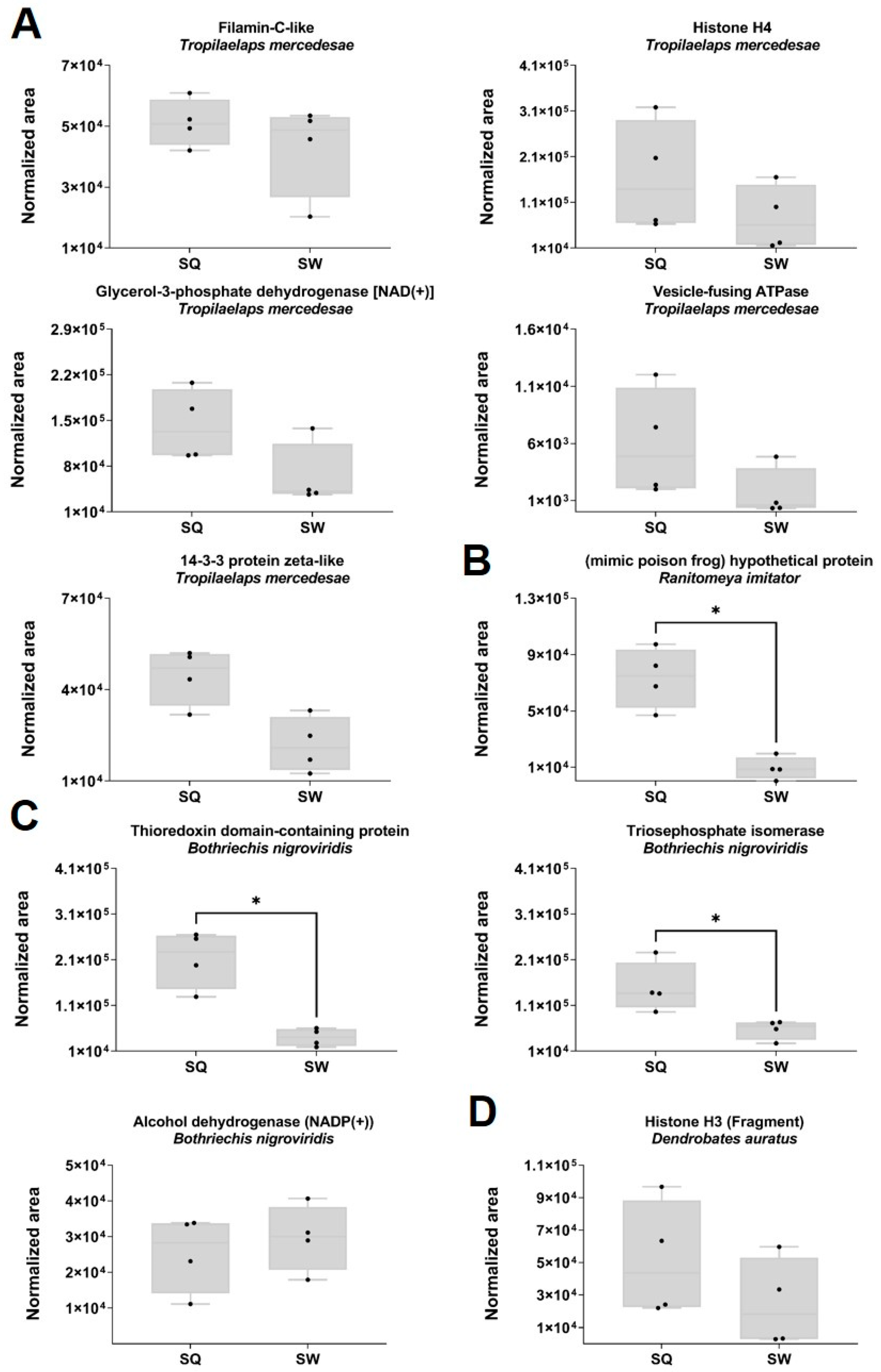
2.6. Proteomic Quantitative Analysis from Dysregulated Proteins of the Classes Amphibia, Arachnida and Reptilia

A total of 17 proteins from the class Amphibia in the SQ and SW were quantified, of which 16 belonged to the mimic poison frog Ranitomeya imitator and one to the poison dart frog Dendrobates auratus (Figure 6D). The hypothetical mimic poison frog protein (Figure 6B) is upregulated (FC 8.01; p = 0.001) in the SQ. None of the other 15 proteins from R. imitator showed statistically significant differences in expression (Mann–Whitney test, p < 0.05). As shown in Figure 6A, five proteins from the mite Tropilaelaps mercedesae (class Arachnida) and three proteins from the viper Bothriechis nigroviridis (class Reptilia) Figure 6C were quantified in the SQ and SW. For T. mercedesae, none of the proteins showed statistically significant differences in expression (Mann–Whitney test, p < 0.05). Thioredoxin domain-containing protein (FC = 5.3; p = 0.002) and triosephosphate isomerase (FC 2.6; p = 0.02), both from the viper Bothriechis nigroviridis, were significantly upregulated in the VV queens (Mann–Whitney test, p < 0.05).

2.7. Proteomic Quantitative Analysis of Dysregulated Proteins from the Classes Bacilla, γ-Proteobacteria and Pisoniviricetes

No differences were observed in the protein expression levels between the SQ and SW from the classes Bacilla (D and E), γ-Proteobacteria (A–C and F) and Pisoniviricetes (G) (Figure 7).

3. Discussion

A total of 228 proteins were quantified in the SQ and SW, belonging to seven different classes: Insecta (n = 191); Amphibia and Reptilia (n = 20); Bacilli, γ-Proteobacteria and Pisoniviricetes (n = 12); and Arachnida (n = 5). The VV is a venomous insect that can inject its biological toxins and proteins into another creature by stinging or spraying, resulting in injury [11,43].
Proteins from γ-proteobacteria, Klebsiella spp., Pantoea agglomerans, Candidatus Schmidhempelia bombi str. Bimp, Gilliamella apis and Pseudomonas spp., Bacilli (Paenibacillus larvae subsp. larvae, Lactobacillus spp., Lactobacillus bombicola) and Pisoniviricetes (deformed wing virus, DWV) were identified in the VV venom sac of both VV queens and workers. The gut bacterial compositions of the genus Vespa from various species and regions have been described in a very similar manner at the phylum and class level [44]. Pantoea agglomerans [45], Gilliamella apis [45,46] and Lactobacillus spp. [44,45], which are the main species in the gut of honeybees, are also present in VV. Klebsiella spp. [44,46] and Pseudomonas spp. [47] have also previously been reported in VV specimens. Despite the generally held view that venom is sterile, microorganisms can viably colonize venoms of vertebrates and invertebrates [48]. The venom sac is connected to the tip of the sting via a persistently open duct exposed to the environment, being comparable to clinical catheterization assemblies: a transcutaneous needle (the sting) that rests on an unsterile surface and is linked to an ongoing duct that leads to a vessel (venom sac) that contains liquid [48]. The analyzed samples could contain not only proteins from the venom itself but also from proteins released from the sac during the procedure. The microorganisms delivered through the sting differ between honeybees, wasps and hornets, with a potentially greater microbial risk for wasp stings in humans [49]. There is an emerging field for integrating microbiology as part of venomics (i.e., venom–microbiomics) for exploring venom as a microenvironment [50]. However, more studies are needed on the origin of these microorganisms, some of which are typically present in the hornet gut flora and which might be present in the extracts analyzed due to cross-contamination during the process of obtaining the sacs.
Regarding the proteins of Paenibacillus larvae subsp. larvae and genome polyprotein from DWV (Iflaviridae), it should be stated that P. larvae is the etiological agent of American foulbrood, the most dangerous brood disease for honeybees. The species P. larvae comprises four different genotypes named ERIC I to ERIC IV. The UniProt V9W8E0 quantified in the present study corresponds to ERIC II genotype, the most important genotype with the highest virulence and one that is frequently isolated from American foulbrood-diseased honeybee colonies [51,52]. To the best of our knowledge, this is the first study to report the presence of P. larvae in VV sacs. Vespa orientalis has already been shown as a potential reservoir of P. larvae [53]. DWV is now the most prevalent pathogen for bee species across the world, in particular Apis mellifera. DWV has also been found in 65 arthropod species from eight insect orders and three Arachnida orders [54]. The first report of DWV in invasive VV populations from the Iberian Peninsula since its description was in France (2008 and 2014) [55,56] and in Italy (2017) [57].
Proteins were quantified by SWATH-MS from (i) Ranitomeya imitator (n = 16), the mimic poison frog, which is naturally distributed in the north-central region of eastern Peru; (ii) Dendrobates auratus (n = 1), the green and black poison dart frog; and (iii) Bothriechis nigroviridis (n = 3), the black-speckled palm pit viper, native to central America. R. imitator mimics not one but three other Ranitomeya species of highly toxic poison frogs (R. fantastica, R. variabilis and R. ventrimaculata) [58]. Whether these proteins represent a potential risk for humans after VV stings deserves further analysis.
A total of 191 proteins belonging to class Insecta from a total of ten species were quantified by SWATH-MS in the SQ and SW (Table 1). Previous studies suggest that the entire extant Hymenoptera lineage might be descended from a “common venomous ancestor” [59].
Finally, from an allergological point of view, there were several reasons for performing the present study. First of all, allergists face patients with anaphylaxis after being stung by VV which was an unknown specimen involved in such allergic reactions until 2015 [8]. Extensive studies were designed and performed from that moment onwards to find out which allergens were responsible for these reactions and phospholipase A1 (the so-called Vesp v 1 allergen) and antigen 5 (Vesp v 5 allergen) were identified [8,33,34,35,36]. The fact that in venoms from both SQ and SW, Vesp v 1 and Vesp v 5 were isolated was in accordance with our previous studies in which specific IgE against both allergens was detected [8,33,34,35,36]. The larger amount of these allergens in SW could be of importance since the probability of being stung by a worker is much higher than that of being stung by a queen. Such differences could represent an evolutionary composition of the venom in workers to better protect the colony. Even though there are no reports on the allergenicity of hyaluronidase A or B in VV, they would seem to behave as allergens since hyaluronidases from other Hymenoptera have proven their capacity to induce specific IgE in patients [33,34,37]. Unexpectedly, while hyaluronidase A was found in a larger amount in SW, no differences were found between hyaluronidase B in SW and SQ. The meaning of this finding is uncertain.
Another relevant issue to consider in the field of allergy is the convenience of performing sting challenge tests to follow up the efficacy of allergen immunotherapy (reviewed in [60]). Since its first application, it has been used to monitor the time course of protection in patients treated with venom immunotherapy. It implies the use of live insects to cause a real sting in an allergic patient to check the reaction provoked by the venom. Due to ethical issues and since the publication of the transcriptomic of VV venom, identifying several toxins [40,41], a serious concern about inflicting severe reactions during the procedure arose and that was one of the main reasons why we decided to analyze the protein content of VV venom sacs. Thus, previous transcriptomic results suggested a high number of putative toxins in sacs venom of VV. However, transcriptomic analysis involves the study of all the messenger RNA (mRNA) molecules present in a living organism but it does not imply the actual presence of the proteins themselves. It is true that we are 100% sure that contamination with some proteins from the sac itself exists but the methods used in the present study were similar to those employed in the reports of the transcriptomic analysis [40,41]. The fact that not a great amount of toxins has been detected in our venom samples could be of importance to deciding the convenience of the sting challenge test with VV.

4. Conclusions

This is the first characterization and quantification of the proteome profile of the venom sacs of Vespa velutina nigrithorax. Differences in the electrophoretic profiles of the venom of SQ and SW, both qualitative (presence/absence) and quantitative (intensity of specific bands), have been demonstrated which could have medical relevance. Among the identified proteins (n = 228), 29% showed significant differential expression between SQ and SW. The allergen proteins hyaluronidase A, venom allergen 5 and phospholipase A1 were significantly downregulated in the SQ, which should be considered when characterizing the living organism’s response to a VV sting. Relevant differences were found with respect to the putative toxins suggested by the previous transcriptomic analysis, implying a safer scenario when dealing with allergic patients.

5. Materials and Methods

5.1. Source of Insects

A nest of the yellow-legged Asian hornet, VV was obtained on November 2021 in Galicia (NW Spain). Insects’ castes were identified using their external morphological characteristics [61,62]. A detailed description of the sampling area, nest architecture and identification of VV caste individuals are found in Supplementary File S3.

5.2. Venom Collection

After sex and female castes differentiation, the venom sac was extracted from frozen insects by pulling the stinger apparatus from the hornet abdomen by forceps. The venom sac was dissected from the stinger. The venom of 3 sacs (worker or queen) was pooled (4 pooles were obtained) and homogenized in PBS (phosphate-buffered saline). Residual tissues were removed by centrifugation at 3000 rpm × 3 min. The supernatant was moved to a new tube and stored at −20 °C. The venom proteins were quantified by the Bradford method (Bio-Rad, Hercules, CA, USA). The venom proteins were analyzed by 12% SDS-PAGE (sodium dodecyl sulfate polyacrylamide gel electrophoresis) under reducing conditions and Sypro Ruby-stained (Bio-Rad, Hercules, CA, USA). The protein bands were visualized using the ChemiDoc MP imaging system (Bio-Rad, Hercules, CA, USA).

5.3. Sample Preparation for Mass Spectrometric Analysis

In relation to tryptic digestion for mass spectrometry, 24 μg protein was concentrated in an SDS-PAGE single band [63,64] and submitted to manual digestion as described elsewhere [65]. Finally, the peptides were dissolved in 0.1% formic acid for further analysis.

5.4. Sequential Window Acquisition of All Theoretical Mass Spectra (SWATH-MS) Quantification

For quantitative proteomic analysis, a hybrid quadrupole-TOF mass spectrometer 6600+ (SCIEX, Framingham, MA, USA) coupled to a micro-liquid chromatography (LC) system Ekspert nLC425 (Eksigen, Dublin, CA, USA) was used. Data were collected using aProteinPilot v.5.0.1, PeakView v.2.2, MarkerView and SWATH Acquisition MicroApp v.2.0 software package (SCIEX, Framingham, MA, USA). A customizer database including Vespa + Vespa velutina + Apis mellifera + venom + toxins Uniprot databases (Available online: https://www.uniprot.org/ (accessed on 2 February 2023)) was employed. The obtained peptide mixtures from sample pools were chromatographed for a total time of 40 min operating with data-dependent acquisition in positive ion mode to build the MS/MS spectral libraries, as previously described [66,67,68,69]. The false discovery rate was set to 1% with a confidence score above 99% [66]. This spectral library was used to create the spectral window acquisition used in the SWATH-MS method. Then, 4 μL from each sample were individually analysed, since SWATH-MS technology does not need sample labeling. The SWATH-MS method is based on repeating a cycle that consists of the acquisition of 100-flight mass spectrometry (TOF MS/M) windows. The quantitative analyses of SWATH-MS are supported by extracted ion chromatograms at both the MS1 and MS2 levels. Proteins with more than 10 peptides and seven transitions were selected for quantification. Any shared or modified peptides were excluded. These advantages of SWATH-MS result from retrospectively targeting fragmentation maps to monitor peptides of interest, as well as extendable spectra and virtual libraries. In combination, results identified by SWATH-MS have greater reproducibility, consistency, and sensitivity. These properties contribute to a superior ability to carry out proteomic quantification in a single profiling experiment, especially with higher sensitivity for identifying low-abundance proteins. A Student’s t-test analysis was performed for comparison among the samples based on the averaged area sums of all the transitions derived for each protein. The t-test was used to indicate how well each variable distinguishes the two groups (SQ and SW), reported as a p value. In the set of differentially expressed proteins (p < 0.05), a 1.5-fold increase or 0.66-fold decrease was selected as the cut-off point.

5.5. Protein Functional Analysis

The differentially regulated proteins were subjected to functional analysis for biological information related to molecular functions, biological processes, cellular components and protein families. We searched them against the protein databases UniProtKB and toxin database ToxProt (Available online: http://www.uniprot.org (accessed on 10 February 2023)).

5.6. Statistical Analysis

Graphic expressions of the comparisons between SWATH normalized area of expressed proteins in the sac venom of queens and workers were made using box plots with median, whiskers min to max, and black dots for the individual values per sample. A Volcano plot was generated by plotting the log2 fold change (FC) for the identified proteins against their corresponding adjusted log10 p value. FC indicates up- or downregulated proteins if FC > 1.5. A p < 0.05 value was considered statistically significant in all tests. Graphics were performed using the GraphPad Prism software (GraphPad Software, San Diego, CA, USA).

Supplementary Materials

The following supporting information can be downloaded at: https://www.mdpi.com/article/10.3390/toxins15040266/s1, Supplementary File S1: Excel file with all quantified proteins with the individual values of SWATH normalized areas per protein and sample, and the FC and p value of the t-test. Supplementary File S2: Excel file download from UniProtKB with the GO terms for the biological process, the molecular function, protein family as well as the cellular component of the 228 quantified proteins in the venom sacs from VV. Supplementary File S3: Detailed description of the: (1) sampling area; (2) nest collection; (3) nest architecture and colony composition; and (4) identification of Vespa velutina nigrithorax caste individuals.

Author Contributions

Conceptualization, C.V. and X.F.; methodology, M.A.-S.; software, S.B.B.; sample preparation, M.P.C.-V.; formal analysis, M.A.-S.; investigation, X.F.; visualization, M.A.-S.; resources, C.V.; data curation, M.A.-S., S.B.B. and X.F.; writing—original draft preparation, X.F., M.A.-S. and C.V.; writing—review and editing, X.F. and C.V.; supervision, C.V.; project administration, M.A.-S.; funding acquisition, C.V.; M.A.-S. and X.F. contributed equally to this work. All authors have read and agreed to the published version of the manuscript.

Funding

This research was supported by grants from the Carlos III Institute of Health (Instituto de Salud Carlos III-ISCIII/PI19/01023/ (Co-funded by the European Union), and the Network for Research on Chronicity, Primary Care, and Health Promotion (Instituto de Salud Carlos III-ISCIII/RD21/0016/0022/ (Co-funded by the European Union). M.A.-S. was supported by the Galician Innovation Agency-Competitive Benchmark Groups (GAIN-GRC/IN607A/2021/02/Xunta de Galicia). X.F. was supported by Spanish Network for Addictive Disorders (Instituto de Salud Carlos III-ISCIII/RD16/0017/0018/ (Co-funded by the European Union).

Institutional Review Board Statement

The study protocol was approved by the “Comité Ético de Investigación Clínica (CEIC) de Galicia” (protocol code 2018/622 on 19 December 2018).

Informed Consent Statement

Not applicable.

Data Availability Statement

The mass spectrometry proteomics data have been deposited to the ProteomeXchange Consortium via the PRIDE partner repository with the dataset identifier PXD040764.

Acknowledgments

Very specially thanks to Pierre Anquet for the Vespa velutina photo provided for the graphical abstract. X.F. would like to acknowledge Dani Slizt the high effort for obtaining the Vespa velutina nest. Thanks also to Carmen Cabadas Amoedo for providing the necessary facilities for the field work in Fornelos de Montes, Galicia. X.F. appreciates the support of the Universitat Carlemany; Universitat Carlemany is part of Planeta Formación y Universidades.

Conflicts of Interest

The authors declare no conflict of interest. The funders had no role in the design of the study; in the collection, analyses or interpretation of data; in the writing of the manuscript; or in the decision to publish the results.

References

  1. Brock, R.E.; Cini, A.; Sumner, S. Ecosystem services provided by aculeate wasps. Biol. Rev. Camb. Philos. Soc. 2021, 96, 1645–1675. [Google Scholar] [CrossRef] [PubMed]
  2. Perrard, A.; Pickett, K.; Villemant, C.; Kojima, J.; Carpenter, J. Phylogeny of hornets: A total evidence approach (Hymenoptera, Vespidae, Vespinae, Vespa). J. Hymenopt. Res. 2013, 2, 1–15. [Google Scholar] [CrossRef]
  3. Detoni, M.; Féas, X.; Jeanne, R.L.; Loope, K.J.; O’Donnell, S.; Santoro, D.; Sumner, S.; Jandt, J.M. Evolutionary and ecological pressures shaping social wasps collective defenses. Ann. Entomol. Soc. Am. 2020, 113, 407–424. [Google Scholar] [CrossRef]
  4. Herrera, C.; Leza, M.; Martínez-López, E. Diversity of compounds in Vespa spp. venom and the epidemiology of its sting: A global appraisal. Arch. Toxicol. 2020, 94, 3609–3627. [Google Scholar] [CrossRef] [PubMed]
  5. Dongol, T.; Dhananjaya, B.L.; Shrestha, R.K.; Aryal, G. Pharmacological and immunological properties of wasp venom. In Pharmacology and Therapeutics; Joghi, S., Gowder, T., Eds.; Intech: Rijeka, Croatia, 2014; pp. 47–81. [Google Scholar] [CrossRef] [Green Version]
  6. Abd El-Wahed, A.; Yosri, N.; Sakr, H.H.; Du, M.; Algethami, A.F.M.; Zhao, C.; Abdelazeem, A.H.; Tahir, H.E.; Masry, S.H.D.; Abdel-Daim, M.M.; et al. Wasp Venom Biochemical Components and Their Potential in Biological Applications and Nanotechnological Interventions. Toxins 2021, 13, 206. [Google Scholar] [CrossRef]
  7. Silva, J.; Monge-Fuentes, V.; Gomes, F.; Lopes, K.; dos Anjos, L.; Campos, G.; Arenas, C.; Biolchi, A.; Gonçalves, J.; Galante, P.; et al. Pharmacological Alternatives for the Treatment of Neurodegenerative Disorders: Wasp and Bee Venoms and Their Components as New Neuroactive Tools. Toxins 2015, 7, 3179–3209. [Google Scholar] [CrossRef] [PubMed] [Green Version]
  8. Chugo, S.; Lizaso, M.T.; Alvarez, M.J.; Arroabaren, E.; Lizarza, S.; Tabar, A.I. Vespa velutina nigritorax: A New Causative Agent in Anaphylaxis. J. Investig. Allergol. Clin. Immunol. 2015, 25, 231–232. [Google Scholar] [PubMed]
  9. Vega, J.M.; Ortiz-Sánchez, F.J.; Martínez-Arcediano, A.; Castro, L.; Alfaya, T.; Carballada, F.; Cruz, S.; Marqués, L.; Vega, A.; Ruiz-León, B. Social wasps in Spain: The who and where. Allergol. Immunopathol. 2022, 50, 58–64. [Google Scholar] [CrossRef] [PubMed]
  10. Carriazo, S.; Ortiz, A. Wasp stings and plasma exchange. Clin. Kidney J. 2022, 15, 1455–1458. [Google Scholar] [CrossRef]
  11. Laborde-Castérot, H.; Darrouzet, E.; Le Roux, G.; Labadie, M.; Delcourt, N.; de Haro, L.; Vodovar, D.; Langrand, J.; French Poison Control Centers Research Group. Ocular Lesions Other Than Stings Following Yellow-Legged Hornet (Vespa velutina nigrithorax) Projections, as Reported to French Poison Control Centers. JAMA Ophthalmol. 2021, 139, 105–108. [Google Scholar] [CrossRef]
  12. de Haro, L.; Labadie, M.; Chanseau, P.; Cabot, C.; Blanc-Brisset, I.; Penouil, F.; National Coordination Committee for Toxicovigilance. Medical consequences of the Asian black hornet (Vespa velutina) invasion in Southwestern France. Toxicon 2010, 55, 650–652. [Google Scholar] [CrossRef] [PubMed]
  13. Van der Brempt, X. Les anaphylaxies aux piqûres de frelons asiatiques (Vespa velutina): Cas déclarés au Réseau d’Allergo-Vigilance® (RAV) de 2011 à 2021 et conduite à tenir. Rev. Fr. Allergol. 2022, 62, 166–170. [Google Scholar] [CrossRef]
  14. Miranda, J.; Mesquita, A.M.; Plácido, J.; Coimbra, A. Vespa velutina nigrithorax venom allergy: Real-life clinical practice. Ann. Allergy Asthma Immunol. 2022, 129, 524–525. [Google Scholar] [CrossRef] [PubMed]
  15. Esteves Caldeira, L.; Silva, M.I.T.; Pedro, E.; Cosme, J. Hypersensitivity to Vespa velutina nigrithorax: An emerging problem in Portugal? Eur. Ann. Allergy Clin. Immunol. 2023. [Google Scholar] [CrossRef]
  16. Grossi, V.; Severino, M.; Massolo, A.; Infantino, M.; Laureti, F.; Macchia, D.; Meucci, E.; Francescato, E.; Pantera, B.; Ebbli, A.; et al. Vespa velutina nigrithorax venom allergy: Inhibition studies approach for the choice of specific immunotherapy. Eur. Ann. Allergy Clin. Immunol. 2022. [Google Scholar] [CrossRef]
  17. Xie, C.; Xu, S.; Ding, F.; Xie, M.; Lv, J.; Yao, J.; Pan, D.; Sun, Q.; Liu, C.; Chen, T.; et al. Clinical features of severe wasp sting patients with dominantly toxic reaction: Analysis of 1091 cases. PLoS ONE 2013, 8, e83164. [Google Scholar] [CrossRef] [Green Version]
  18. Choi, M.B.; Hong, E.J.; Kwon, O. Defensive behavior of the invasive alien hornet, Vespa velutina, against color, hair and auditory stimuli of potential aggressors. PeerJ 2021, 9, e11249. [Google Scholar] [CrossRef]
  19. Choi, M.B.; Kim, T.G.; Kwon, O. Recent Trends in Wasp Nest Removal and Hymenoptera Stings in South Korea. J. Med. Entomol. 2019, 56, 254–260. [Google Scholar] [CrossRef]
  20. Vidal, C. The Asian wasp Vespa velutina nigrithorax: Entomological and allergological characteristics. Clin. Exp. Allergy 2022, 52, 489–498. [Google Scholar] [CrossRef]
  21. Feás Sánchez, X.; Charles, R.J. Notes on the nest architecture and colony composition in winter of the yellow-legged Asian Hornet, Vespa velutina Lepeletier 1836 (Hym.: Vespidae), in its introduced habitat in Galicia (NW Spain). Insects 2019, 10, 237. [Google Scholar] [CrossRef] [Green Version]
  22. Feás, X.; Vázquez-Tato, M.P.; Seijas, J.A.; Pratima GNikalje, A.; Fraga-López, F. Extraction and Physicochemical Characterization of Chitin Derived from the Asian Hornet, Vespa velutina Lepeletier 1836 (Hym.: Vespidae). Molecules 2020, 25, 384. [Google Scholar] [CrossRef] [PubMed] [Green Version]
  23. Feás, X.; Vidal, C.; Remesar, S. What We Know about Sting-Related Deaths? Human Fatalities Caused by Hornet, Wasp and Bee Stings in Europe (1994–2016). Biology 2022, 11, 282. [Google Scholar] [CrossRef] [PubMed]
  24. Feás, X.; Vidal, C.; Vázquez-Tato, M.P.; Seijas, J.A. Asian Hornet, Vespa velutina Lepeletier 1836 (Hym.: Vespidae), Venom Obtention Based on an Electric Stimulation Protocol. Molecules 2021, 27, 138. [Google Scholar] [CrossRef]
  25. Feás, X. Human Fatalities Caused by Hornet, Wasp and Bee Stings in Spain: Epidemiology at State and Sub-State Level from 1999 to 2018. Biology 2021, 10, 73. [Google Scholar] [CrossRef] [PubMed]
  26. Yun, H.S.; Oh, J.; Lim, J.S.; Kim, H.J.; Kim, J.S. Anti-Inflammatory Effect of Wasp Venom in BV-2 Microglial Cells in Comparison with Bee Venom. Insects 2021, 12, 297. [Google Scholar] [CrossRef]
  27. Meng, Y.C.; Mo, X.G.; He, T.T.; Wen, X.X.; Nieh, J.C.; Yang, X.W.; Tan, K. New bioactive peptides from the venom gland of a social hornet Vespa velutina. Toxicon 2021, 199, 94–100. [Google Scholar] [CrossRef]
  28. Chae, J.H.; Oh, J.; Lim, J.S.; Jeong, Y.A.; Yun, H.S.; Jang, C.H.; Kim, H.J.; Kim, J.S. Wasp Venom Ameliorates Scopolamine-Induced Learning and Memory Impairment in Mice. Toxins 2022, 14, 256. [Google Scholar] [CrossRef]
  29. Le, T.N.; Da Silva, D.; Colas, C.; Darrouzet, E.; Baril, P.; Leseurre, L.; Maunit, B. Asian hornet Vespa velutina nigrithorax venom: Evaluation and identification of the bioactive compound responsible for human keratinocyte protection against oxidative stress. Toxicon 2020, 176, 1–9. [Google Scholar] [CrossRef]
  30. Cappa, F.; Cini, A.; Pepiciello, I.; Petrocelli, I.; Inghilesi, A.F.; Anfora, G.; Dani, F.R.; Bortolotti, L.; Wen, P.; Cervo, R. Female volatiles as sex attractants in the invasive population of Vespa velutina nigrithorax. J. Insect. Physiol. 2019, 119, 103952. [Google Scholar] [CrossRef]
  31. Cheng, Y.N.; Wen, P.; Dong, S.H.; Tan, K.; Nieh, J.C. Poison and alarm: The Asian hornet. J. Exp. Biol. 2017, 220 Pt 4, 645–651. [Google Scholar] [CrossRef] [Green Version]
  32. Thiéry, D.; Bonnard, O.; Riquier, L.; De Revel, G.; Monceau, K. An alarm pheromone in the venom gland of Vespa velutina: Evidence revisited from the European invasive population. Entomol. Gen. 2018, 38, 145–156. [Google Scholar] [CrossRef]
  33. Vidal, C.; Armisén, M.; Monsalve, R.; Gómez-Rial, J.; González-Fernández, T.; Carballada, F.; Lombardero, M.; González-Quintela, A. Vesp v 5 and glycosylated Vesp v 1 are relevant allergens in Vespa velutina nigrithorax anaphylaxis. Clin. Exp. Allergy 2020, 50, 1424–1427. [Google Scholar] [CrossRef] [PubMed]
  34. Vidal, C.; Armisén, M.; Monsalve, R.; González-Vidal, T.; Lojo, S.; López-Freire, S.; Méndez, P.; Rodríguez, V.; Romero, L.; Galán, A.; et al. Anaphylaxis to Vespa velutina nigrithorax: Pattern of Sensitization for an Emerging Problem in Western Countries. J. Investig. Allergol. Clin. Immunol. 2021, 31, 228–235. [Google Scholar] [CrossRef] [PubMed] [Green Version]
  35. Rodríguez-Vázquez, V.; Gómez-Rial, J.; Monsalve, R.I.; Vidal, C. Consistency of Determination of sIgE and the Basophil Activation Test in Vespa velutina nigrithorax Allergy. J. Investig. Allergol. Clin. Immunol. 2022, 32, 143–145. [Google Scholar] [CrossRef] [PubMed]
  36. Rodríguez-Vázquez, V.; Armisén, M.; Gómez-Rial, J.; Lamas-Vázquez, B.; Vidal, C. Immunotherapy with Vespula venom for Vespa velutina nigrithorax anaphylaxis: Preliminary clinical and immunological results. Clin. Exp. Allergy 2022, 52, 345–347. [Google Scholar] [CrossRef]
  37. Ruiz-Leon, B.; Serrano, P.; Vidal, C.; Moreno-Aguilar, C. Management of Double Sensitization to Vespids in Europe. Toxins 2022, 14, 126. [Google Scholar] [CrossRef] [PubMed]
  38. Guido-Patiño, J.C.; Plisson, F. Profiling hymenopteran venom toxins: Protein families, structural landscape, biological activities, and pharmacological benefits. Toxicon X 2022, 14, 100119. [Google Scholar] [CrossRef]
  39. Dos Santos-Pinto, J.R.A.; Perez-Riverol, A.; Lasa, A.M.; Palma, M.S. Diversity of peptidic and proteinaceous toxins from social Hymenoptera venoms. Toxicon 2018, 148, 172–196. [Google Scholar] [CrossRef] [Green Version]
  40. Liu, Z.; Chen, S.; Zhou, Y.; Xie, C.; Zhu, B.; Zhu, H.; Liu, S.; Wang, W.; Chen, H.; Ji, Y. Deciphering the venomic transcriptome of killer-wasp Vespa velutina. Sci. Rep. 2015, 5, 9454. [Google Scholar] [CrossRef]
  41. Tan, J.; Wang, W.; Wu, F.; Li, Y.; Fan, Q. Transcriptome profiling of venom gland from wasp species: De novo assembly, functional annotation, and discovery of molecular markers. BMC Genom. 2020, 21, 427. [Google Scholar] [CrossRef]
  42. Le, T.N.; da Silva, D.; Colas, C.; Darrouzet, E.; Baril, P.; Leseurre, L.; Maunit, B. Development of an LC-MS multivariate nontargeted methodology for differential analysis of the peptide profile of Asian hornet venom (Vespa velutina nigrithorax): Application to the investigation of the impact of collection period variation. Anal. Bioanal. Chem. 2020, 412, 1419–1430. [Google Scholar] [CrossRef]
  43. Nelsen, D.R.; Nisani, Z.; Cooper, A.M.; Fox, G.A.; Gren, E.C.; Corbit, A.G.; Hayes, W.K. Poisons, toxungens, and venoms: Redefining and classifying toxic biological secretions and the organisms that employ them. Biol. Rev. Camb. Philos. Soc. 2014, 89, 450–465. [Google Scholar] [CrossRef]
  44. Zhang, L.; Liu, F.; Wang, X.L.; Wang, P.H.; Ma, S.L.; Yang, Y.; Ye, W.-G.; Diao, K.-Y.; Dai, P.L. Midgut Bacterial Communities of Vespa velutina Lepeletier (Hymenoptera: Vespidae). Front. Ecol. Evol. 2022, 10, 934054. [Google Scholar] [CrossRef]
  45. Suenami, S.; Konishi, N.M.; Miyazaki, R. Community analysis of gut microbiota in hornets, the largest eusocial wasps, Vespa mandarinia and V. simillima. Sci. Rep. 2019, 9, 9830. [Google Scholar] [CrossRef] [PubMed] [Green Version]
  46. Cini, A.; Meriggi, N.; Bacci, G.; Cappa, F.; Vitali, F.; Cavalieri, D.; Cervo, R. Gut microbial composition in different castes and developmental stages of the invasive hornet Vespa velutina nigrithorax. Sci. Total Environ. 2020, 745, 140873. [Google Scholar] [CrossRef] [PubMed]
  47. Kim, E.; Seo, J.; Yang, S.H.; Kim, I.-S.; Koo, Y. Intestine bacterial microbiota of Asian hornet (Vespa velutina nigrithorax) and honey bee. Korean J. Environ. Agric. 2018, 37, 135–140. [Google Scholar] [CrossRef] [Green Version]
  48. Esmaeilishirazifard, E.; Usher, L.; Trim, C.; Denise, H.; Sangal, V.; Tyson, G.H.; Barlow, A.; Redway, K.F.; Taylor, J.D.; Kremyda-Vlachou, M.; et al. Bacterial Adaptation to Venom in Snakes and Arachnida. Microbiol. Spectr. 2022, 10, e0240821. [Google Scholar] [CrossRef]
  49. Gkitsaki, I.; Papachristoforou, A.; Michailidou, S.; Karamvalis, N.; Iliadis, I.; Graikini, D.; Sakarikou, C.; Tsoukis, E.; Argyriou, A.; Giaouris, E. The transmittable through stinging microbiota differs between honeybees and wasps: A potentially greater microbial risk of the wasp sting for humans. Int. Microbiol. 2023. [Google Scholar] [CrossRef]
  50. Ul-Hasan, S.; Rodríguez-Román, E.; Reitzel, A.M.; Adams, R.M.M.; Herzig, V.; Nobile, C.J.; Saviola, A.J.; Trim, S.A.; Stiers, E.E.; Moschos, S.A.; et al. The emerging field of venom-microbiomics for exploring venom as a microenvironment, and the corresponding Initiative for Venom Associated Microbes and Parasites (iVAMP). Toxicon X 2019, 4, 100016. [Google Scholar] [CrossRef]
  51. Djukic, M.; Brzuszkiewicz, E.; Fünfhaus, A.; Voss, J.; Gollnow, K.; Poppinga, L.; Liesegang, H.; Garcia-Gonzalez, E.; Genersch, E.; Daniel, R. How to kill the honey bee larva: Genomic potential and virulence mechanisms of Paenibacillus larvae. PLoS ONE 2014, 9, e90914. [Google Scholar] [CrossRef]
  52. Beims, H.; Bunk, B.; Erler, S.; Mohr, K.I.; Spröer, C.; Pradella, S.; Günther, G.; Rohde, M.; von der Ohe, W.V.; Steinert, M. Discovery of Paenibacillus larvae ERIC V: Phenotypic and genomic comparison to genotypes ERIC I-IV reveal different inventories of virulence factors which correlate with epidemiological prevalences of American Foulbrood. Int. J. Med. Mictrobiol. 2020, 310, 151394. [Google Scholar] [CrossRef]
  53. Nowar, E.E. Oriental Hornet (Vespa orientalis) as AFB Disease Vector to Honeybee (Apis mellifera L.). Colonies. Middle East J. Appl. Sci. 2021, 06, 934–940. [Google Scholar]
  54. Martin, S.J.; Brettell, L.E. Deformed Wing Virus in Honeybees and Other Insects. Annu. Rev. Virol. 2019, 6, 49–69. [Google Scholar] [CrossRef] [PubMed] [Green Version]
  55. Chauzat, M.-P.; Schurr, F.; Faucon, J.-P.; Ribière-Chabert, M.; Blanchard, P.; Drajnudel, P. First detections of honey bee pathogens in nest of the Asian hornet (Vespa velutina) collected in France. CIHEAM Watch. Lett. 2015, 33, 1–5. [Google Scholar]
  56. Dalmon, A.; Gayral, P.; Decante, D.; Klopp, C.; Bigot, D.; Thomasson, M.; Herniou, E.A.; Alaux, C.; Le Conte, Y. Viruses in the Invasive Hornet. Viruses 2019, 11, 1041. [Google Scholar] [CrossRef] [Green Version]
  57. Mazzei, M.; Forzan, M.; Cilia, G.; Sagona, S.; Bortolotti, L.; Felicioli, A. First detection of replicative deformed wing virus (DWV) in Vespa velutina nigrithorax. Bull. Insectol. 2018, 71, 211–216. [Google Scholar]
  58. Brown, J.L.; Twomey, E.; Amézquita, A.; Souza, M.B.; Caldwell, J.P.; Lötters, S.; May, R.V.; Melo-Sampaio, P.R.; Mejia-Vargas, D.; Pérez-Peña, P.; et al. A taxonomic revision of the Neotropical poison frog genus Ranitomeya (Amphibia: Dendrobatidae). Zootaxa 2011, 3083, 1–120. [Google Scholar] [CrossRef] [Green Version]
  59. Koludarov, I.; Velasque, M.; Timm, T.; Greve, C.; Ben Hamadou, A.; Kumar Gupta, D.; Lochnit, G.; el Heinzinger, M.; Vilcinskas, A.; Gloag, R.; et al. A common venomous ancestor? Prevalent bee venom genes evolved before the aculeate stinger while few major toxins are bee-specific. bioRxiv 2022. [Google Scholar] [CrossRef]
  60. Ruiz-León, B.; Martínez San Ireneo, M.; de la Roca, F.; Arenas, L.; Alfaya Arias, T.; Cordobés, C.; Marqués, L.; Vega, A.; Moreno-Aguilar, C. The Lights and the Shadows of Controlled Sting Challenge with Hymenoptera. J. Investig. Allergol. Clin. Immunol. 2022, 32, 357–366. [Google Scholar] [CrossRef]
  61. Feás, X. The Yellow-Legged Asian Hornet (Vespa velutina): Reproductive Caste—Drone. Available online: https://youtu.be/JgMyW1v5H1Y (accessed on 20 March 2023).
  62. Feás, X. The Yellow-Legged Asian Hornet (Vespa velutina) collecting water. Available online: https://youtu.be/d9lJFNXtES0 (accessed on 20 March 2023).
  63. Bonzon-Kulichenko, E.; Pérez-Hernández, D.; Núñez, E.; Martínez-Acedo, P.; Navarro, P.; Trevisan-Herraz, M.; Ramos Mdel, C.; Sierra, S.; Martínez-Martínez, S.; Ruiz-Meana, M.; et al. A robust method for quantitative high-throughput analysis of proteomes by 18O labeling. Mol. Cell. Proteom. 2011, 10, M110.003335. [Google Scholar] [CrossRef] [Green Version]
  64. Perez-Hernandez, D.; Gutiérrez-Vázquez, C.; Jorge, I.; López-Martín, S.; Ursa, A.; Sánchez-Madrid, F.; Vázquez, J.; Yáñez-Mó, M. The intracellular interactome of tetraspanin-enriched microdomains reveals their function as sorting machineries toward exosomes. J. Biol. Chem. 2013, 288, 11649–11661. [Google Scholar] [CrossRef] [PubMed] [Green Version]
  65. Shevchenko, A.; Wilm, M.; Vorm, O.; Jensen, O.N.; Podtelejnikov, A.V.; Neubauer, G.; Shevchenko, A.; Mortensen, P.; Mann, M. A strategy for identifying gel-separated proteins in sequence databases by MS alone. Biochem. Soc. Trans. 1996, 24, 893–896. [Google Scholar] [CrossRef] [Green Version]
  66. Peñas-Martínez, J.; Barrachina, M.N.; Cuenca-Zamora, E.J.; Luengo-Gil, G.; Bravo, S.B.; Caparrós-Pérez, E.; Teruel-Montoya, R.; Eliseo-Blanco, J.; Vicente, V.; García, Á.; et al. Qualitative and quantitative comparison of plasma exosomes from neonates and adults. Int. J. Mol. Sci. 2021, 22, 1926. [Google Scholar] [CrossRef]
  67. Camino, T.; Lago-Baameiro, N.; Sueiro, A.; Bravo, S.B.; Couto, I.; Santos, F.F.; Teruel-Montoya, R.; Eliseo-Blanco, J.; Vicente, V.; García, Á.; et al. Brown Adipose Tissue Sheds Extracellular Vesicles That Carry Potential Biomarkers of Metabolic and Thermogenesis Activity Which Are Affected by High Fat Diet Intervention. Int. J. Mol. Sci. 2022, 23, 10826. [Google Scholar] [CrossRef] [PubMed]
  68. Chantada-Vázquez, M.D.P.; Conde-Amboage, M.; Graña-López, L.; Vázquez-Estévez, S.; Bravo, S.B.; Núñez, C. Circulating Proteins Associated with Response and Resistance to Neoadjuvant Chemotherapy in HER2-Positive Breast Cancer. Cancers 2022, 14, 1087. [Google Scholar] [CrossRef]
  69. Shilov, I.V.; Seymour, S.L.; Patel, A.A.; Loboda, A.; Tang, W.H.; Keating, S.P.; Hunter, C.L.; Nuwaysir, L.M.; Schaeffer, D.A. The Paragon Algorithm, a Next Generation Search Engine That Uses Sequence Temperature Values and Feature Probabilities to Identify Peptides from Tandem Mass Spectra. Mol. Cell. Proteom. 2007, 6, 1638–1655. [Google Scholar] [CrossRef] [PubMed] [Green Version]
Figure 1. (A) Images of the VV venom sac of SQ and SW (scale in mm); (B) Box plot with median, whiskers min. to max., and black dots for the individual values per venom sac (* p = 0.02); (C) Electrophoretic separation of total proteins from SQ and SW. Arrows indicate the bands corresponding to hyaluronidase, phospholipase A1 and venom antigen 5, according to their molecular mass and later confirmed by SWATH-MS. kDa, kilodalton; ST, precision plus protein standard. Bands 1–4 correspond to pooled samples of SQ and SW, respectively. Each pool contains 3 sacs so a total of 12 SQ and 12 SW were analyzed. Photo Author: Manuela Alonso-Sampedro.
Toxins 15 00266 g001
Figure 2. Volcano plot of the venom sac quantitative proteomics data of the VV queens and workers. Volcano plot shows the significantly differentially abundant proteins in the venom sacs by quantitative proteomics analysis. Proteins are ranked in a volcano plot according to their statistical p-value (y-axis) as—log10 and their relative abundance ratio (log2 FC) between SQ and SW (x-axis). Off-centered spots are those that vary the most between the two groups. The cut-offs for significant changes are FC ≥ 1.5 and p < 0.05 (t-test). Green spots show the upregulated proteins in SQ, red spots show the downregulated proteins in SQ, and blue spots show the non-dysregulated proteins between the two groups. Vesp v 5, venom antigen 5; Vesp v 1, phospholipase A1; Vesp v 2A, hyaluronidase A; Vesp v 2B, hyaluronidase B; VT 2b, phospholipase A1 verotoxin-2b; SQ, VV queen venom sac; SW, VV worker venom sac. Sample size: SQ = 4 pooled (3 sacs each); SW = 4 pooled (3 sacs each).
Toxins 15 00266 g002
Figure 3. (A) Biological processes and (B) molecular functions mainly related to the differentially expressed proteins in the venom sac of VV queens (SQ) and workers (SW). The histograms represent the main categories for each gene ontology (GO) term in which differentially expressed proteins were involved (p < 0.05). The y-axis shows the number of individual proteins in each GO term, and the x-axis shows the GO term.
Toxins 15 00266 g003
Figure 4. Representation of the SWATH-MS normalized area in potential VV queen and worker allergens. The normalized area was obtained from the SWATH-MS method for each individual sample. Box plot with median, whiskers min to max, and black dots for the individual values per sample. Statistical differences by Mann–Whitney test, * p < 0.05. SQ, VV queen venom sac; SW, VV worker venom sac. Sample size: SQ = 4 pooled (3 sacs each); SW = 4 pooled (3 sacs each).
Toxins 15 00266 g004
Figure 5. Representation of SWATH-MS normalized area in the SQ and SW quantified proteins from (A) Apis mellifera ligustica, (B) Apis mellifera, (C) Bombus festivus, (D) Xylocopa caerulea, (E) Megachile rotundata and (F) Bombus humilis. The normalized area was obtained with the SWATH-MS method for each individual sample. Box plot with median, whiskers min. to max., black dots for the individual values per sample. Statistical differences by Mann–Whitney test, * p < 0.05. SQ, VV SW VV SW. Sample size: SQ = 4 pooled (3 sacs each); SW = 4 pooled (3 sacs each).
Toxins 15 00266 g005
Figure 6. Representation of the SWATH-MS normalized area in the VV SQ and SW quantified proteins from (A) Tropilaelaps mercedesae; (B) Ranitomeya imitator; (C) Bothriechis nigroviridis; and (D) Dendrobates auratus. The normalized area was obtained from the SWATH-MS method for each individual sample. Box plot with median, whiskers min. to max., and black dots for the individual values per sample. Statistical differences by Mann–Whitney test, * p < 0.05. Sample size: SQ = 4 pooled (3 sacs each); SW = 4 pooled (3 sacs each).
Toxins 15 00266 g006
Figure 7. Representation of the SWATH-MS normalized area in Vespa velutina (VV) (Hym.: Vespidae) SQ andSW quantified proteins from (A) Enterobacter agglomerans, (B) Pseudomonas sp. RIT-PI-a, (C) Klebsiella spp., (D) Lactobacillus spp., (E) Paenibacillus larvae subsp. Larvae, (D) Gilliamella apis, (F) Candidatus Schmidhempelia bombi str. Bimp and (G) Deformed wing virus. The normalized area was obtained from the SWATH-MS method for each individual sample. Box plot with median, whiskers min to max, and black dots for the individual values per sample. Sample size: SQ = 4 pooled (3 sacs each); SW = 4 pooled (3 sacs each).
Toxins 15 00266 g007
Table 1. Classification of the 228 proteins (pp) found in the SQ and the SW by SWATH-MS, according to the species and group to which they belong.
ClassOrder/FamilyScientific Name/Common NameppGroup
AmphibiaAnura/DendrobatidaeRanitomeya imitator (Schulte, 1986)/Mimic poison frog.169NEOB
Dendrobates auratus (Girard, 1955)/Green and black poison dart frog1DENAT
ArachnidaMesostigmata/LaelapidaeTropilaelaps mercedesae (Delfinado & Baker, 1961)/Tropilaelaps mite59ACAR
BacilliBacillales/PaenibacillaceaePaenibacillus larvae subsp. larvae (White 1906)/American Foulbrood19BACL
Lactobacillales/LactobacillaceaeLactobacillus sp. 19LACO
Lactobacillus bombicola (Praet et al., 2015)19LACO
γ-proteobacteriaEnterobacterales/EnterobacteriaceaeKlebsiella sp. 39ENTR
Enterobacterales/ErwiniaceaePantoea agglomerans (Ewing & Fife, 1972) Gavini et al., 1989 2ENTAG
Orbales/OrbaceaeCandidatus Schmidhempelia bombi str. Bimp19GAMM
Gilliamella apis (Ludvigsen et al., 2018)19GAMM
Pseudomonadales/PseudomonadaceaPseudomonas sp. 19PSED
InsectaHymenoptera/ApidaeApis cerana cerana (Fabricius, 1793)/Asian honeybee49APICC
Apis mellifera ligustica (Spinola, 1806)/Italian honeybee2APILI
Apis mellifera (Linnaeus, 1758)/European honeybee2APIME
Bombus festivus (Smith, 1861)/Bumblebee of Sichuan19HYME
Bombus humilis (Illiger, 1806)/Brown-banded carder bumblebee1BOMHU
Frieseomelitta varia (Lepeletier, 1836)/Yellow marmalade bee829HYME
Xylocopa caerulea (Fabricius, 1804)/Blue carpenter bee19HYME
Hymenoptera/ HalictidaeDufourea novaeangliae (Robertson, 1897)/Sweat bee47DUFNO
Hymenoptera/ MegachilidaeMegachile rotundata (Fabricius, 1787)/Alfalfa leafcutting bee1MEGRT
Hymenoptera/VespidaeVespa velutina (Lepeletier, 1836)/Yellow-legged Asian hornet5VESVE
PisoniviricetesPicornavirales/IflaviridaeDeformed wing virus19VIRU
ReptiliaSquamata/ViperidaeBothriechis nigroviridis (Peters, 1859)/Black-speckled palm pit viper3BOTNI
Table 2. Proteins from Dufourea novaeangliae, Apis cerana cerana and Frieseomelitta varia with differential expression between VV SQ and SW by SWATH-MS.
SpeciesUniprot CodeProteinp Value
(t Test)
FC
(SQ/SW)
Dufourea novaeangliaeA0A154P4T7Four and a half LIM domains protein 20.01028.572
A0A154NYM3Transgelin 0.00116.538
A0A154PS72Profilin 0.0119.233
A0A154NWX1Laminin subunit β-1 0.0206.452
A0A154PD98Nidogen-1 0.0024.470
A0A154PLJ1Heat shock 70 kDa protein cognate 5 0.0074.375
A0A154PJJ4Annexin0.0023.627
A0A154PP88α-1,4 glucan phosphorylase 0.0083.360
A0A154PQH0Glycogenin-1 0.0042.693
A0A154P296Acetyl-CoA hydrolase0.0272.644
A0A154P796Spectrin α chain 0.0062.612
A0A154PAT6Malate dehydrogenase 0.0442.552
A0A154PNQ0Sortilin-related receptor0.0172.535
A0A154P1L6ATP synthase subunit β0.0192.389
A0A154P3S2Muscle M-line assembly protein unc-89 0.0032.335
A0A154PJP5Titin 0.0192.170
A0A154PQ65Multifunctional fusion protein 0.0412.158
A0A154P3Q8Elongation factor 2 0.0282.003
A0A154P289Vacuolar proton pump subunit B 0.0161.994
A0A154PPS02-phospho-D-glycerate hydro-lyase 0.0311.930
Apis cerana ceranaA0A2A3EBK6LIM domain-binding protein 0.00461.974
A0A2A3EA22Four and a half LIM domains protein 0.0087.737
A0A2A3EL86Elongation factor 1-γ 0.0295.917
A0A2A3EC59Calpain-A 0.0003.745
A0A2A3E2W8Pyruvate dehydrogenase E1 component subunit α 0.0193.234
A0A2A3EGD2Arginine kinase 0.0222.724
A0A2A3EMK6α -actinin, sarcomeric 0.0012.719
A0A2A3EKS3Fructose-bisphosphate aldolase 0.0492.583
A0A2A3E5U7Talin-2 0.0202.285
A0A2A3EPX9Glutamate dehydrogenase (NAD(p)(+)) 0.0262.042
A0A2A3E464ATP synthase subunit α0.0252.028
A0A2A3EAL0Zn finger protein-like 1 homolog 0.0440.281
A0A2A3EJN3Acid β-galactosidase 0.0080.012
Frieseomelitta variaA0A833VQ2840S ribosomal protein S40.0106.475
A0A833VR32EF-1-γ-C-terminal domain-containing protein0.0226.293
A0A833S6V3Neurochondrin homolog0.0015.899
A0A833VVD0Isocitrate dehydrogenase [NAD] subunit, mitochondrial0.0085.509
A0A833R5C8Lipocln_cytosolic_FA-bd_dom domain-containing protein0.0014.562
A0A833VMV6Eukaryotic translation initiation factor 5A (eIF-5A)0.0174.163
A0A833RMB6Muscle LIM protein Mlp84B0.0084.161
A0A833RMX9Filamin-A0.0114.147
A0A833W6L4Small nuclear ribonucleoprotein-associated protein0.0414.017
A0A5P1MU32Glyceraldehyde-3-phosphate dehydrogenase0.0004.000
A0A833WD27Pyruvate dehydrogenase E1 component subunit β0.0493.428
A0A833S5I5Phosphoglycerate kinase0.0013.175
A0A833R5H5DJ-1_PfpI domain-containing protein0.0083.170
A0A833RCZ1Thioredoxin domain-containing protein0.0093.081
A0A833S1K4Malic enzyme0.0212.587
A0A833VV22WD_REPEATS_REGION domain-containing protein0.0152.522
A0A833W6G6Pyruvate kinase0.0022.501
A0A833S8P7ADF-H domain-containing protein0.0092.389
A0A833RUN1Heat shock 70 kDa protein cognate 40.0132.205
A0A833W2V9Fructose-bisphosphate aldolase0.0212.173
A0A833WDK7Calreticulin0.0242.003
A0A833WFT3Thioredoxin domain-containing protein0.0281.975
A0A833RKT2α-glucosidase0.0270.358
A0A833W1X0α-galactosidase 0.0210.298
FC (SQ/SW), fold change of queen/worker; FC > 1 overexpressed protein in SQ; FC < 1 downregulated protein in SQ; p value < 0.05: statistically significant.
Disclaimer/Publisher’s Note: The statements, opinions and data contained in all publications are solely those of the individual author(s) and contributor(s) and not of MDPI and/or the editor(s). MDPI and/or the editor(s) disclaim responsibility for any injury to people or property resulting from any ideas, methods, instructions or products referred to in the content.

