A Streamlined Method to Obtain Biologically Active TcdA and TcdB Toxins from Clostridioides difficile

 , and
, and 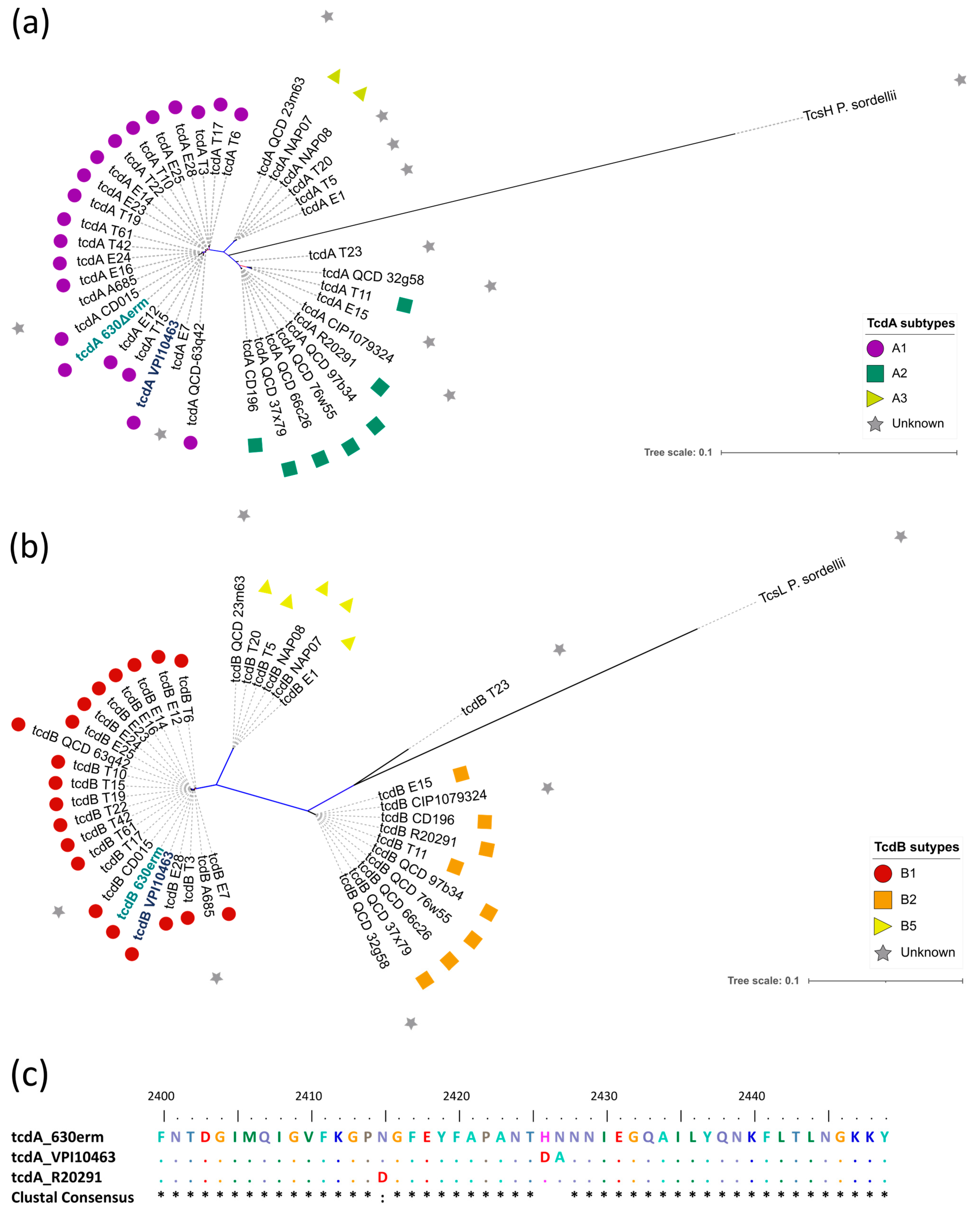
Abstract
:1. Introduction
2. Results
2.1. Selection of a C. difficile Strain
2.2. Expression and Purification of Recombinant Toxins
2.3. Recombinant Purified Toxins Possess Similar Biological Activity Compared to Native Toxins
2.4. Recombinant Toxins Can Be Used as Substitutes for Native Toxins in Clinical CDI Investigational Studies
3. Discussion and Conclusions
4. Materials and Methods
4.1. Bacterial Strains, Plasmids, and Growth Conditions
4.2. Constructs and Cloning in the C. difficile 630∆erm Strain
4.3. Expression and Purification of Recombinant Toxins
4.3.1. From AD1 and AD2 Strains
4.3.2. From AD3 and AD4 Strains
4.4. Cytotoxicity Assay
4.5. Disruption of the Actin Cytoskeleton by Purified Recombinant Toxins
4.6. Comparison of Native or Recombinant Toxins for the Quantification of Serum Antibodies by Quantitative ELISA
4.7. Neutralization Antibody Assay
4.8. Phylogenetic Analysis
4.9. Statistical Analysis and Software
Supplementary Materials
Author Contributions
Funding
Institutional Review Board Statement
Informed Consent Statement
Data Availability Statement
Acknowledgments
Conflicts of Interest
References
- Lawson, P.A.; Citron, D.M.; Tyrrell, K.L.; Finegold, S.M. Reclassification of Clostridium difficile as Clostridioides difficile (Hall and O’Toole 1935) Prévot 1938. Anaerobe 2016, 40, 95–99. [Google Scholar] [CrossRef] [PubMed]
- Hall, I.C. Intestinal flora in new-born infants: With a description of a new pathogenic anaerobe Bacillus difficilis. Am. J. Dis. Child. 1935, 49, 390. [Google Scholar] [CrossRef]
- Bartlett, J.G. Clostridium difficile: History of its role as an enteric pathogen and the current state of knowledge about the organism. Clin. Infect. Dis. 1994, 18 (Suppl. 4), S265–S272. [Google Scholar] [CrossRef] [PubMed]
- Garey, K.W.; Sethi, S.; Yadav, Y.; DuPont, H.L. Meta-analysis to assess risk factors for recurrent Clostridium difficile infection. J. Hosp. Infect. 2008, 70, 298–304. [Google Scholar] [CrossRef] [PubMed]
- Johnson, S. Recurrent Clostridium difficile infection: A review of risk factors, treatments, and outcomes. J. Infect. 2009, 58, 403–410. [Google Scholar] [CrossRef] [PubMed]
- Balsells, E.; Shi, T.; Leese, C.; Lyell, I.; Burrows, J.; Wiuff, C.; Campbell, H.; Kyaw, M.H.; Nair, H. Global burden of Clostridium difficile infections: A systematic review and meta-analysis. J. Glob. Health 2019, 9, 010407. [Google Scholar] [CrossRef] [PubMed]
- Jones, A.M.; Kuijper, E.J.; Wilcox, M.H. Clostridium difficile: A European perspective. J. Infect. 2013, 66, 115–128. [Google Scholar] [CrossRef]
- McGlone, S.M.; Bailey, R.R.; Zimmer, S.M.; Popovich, M.J.; Tian, Y.; Ufberg, P.; Muder, R.R.; Lee, B.Y. The economic burden of Clostridium difficile. Clin. Microbiol. Infect. 2012, 18, 282–289. [Google Scholar] [CrossRef]
- Brown, K.A.; Khanafer, N.; Daneman, N.; Fisman, D.N. Meta-analysis of antibiotics and the risk of community-associated Clostridium difficile infection. Antimicrob. Agents Chemother. 2013, 57, 2326–2332. [Google Scholar] [CrossRef]
- Savidge, T.C.; Pan, W.; Newman, P.; O’Brien, M.; Anton, P.M.; Pothoulakis, C. Clostridium difficile toxin B is an inflammatory enterotoxin in human intestine. Gastroenterology 2003, 125, 413–420. [Google Scholar] [CrossRef]
- Braun, V.; Hundsberger, T.; Leukel, P.; Sauerborn, M.; von Eichel-Streiber, C. Definition of the single integration site of the pathogenicity locus in Clostridium difficile. Gene 1996, 181, 29–38. [Google Scholar] [CrossRef]
- Dupuy, B.; Sonenshein, A.L. Regulated transcription of Clostridium difficile toxin genes. Mol. Microbiol. 1998, 27, 107–120. [Google Scholar] [CrossRef]
- Orrell, K.E.; Melnyk, R.A. Large Clostridial Toxins: Mechanisms and Roles in Disease. Microbiol. Mol. Biol. Rev. 2021, 85, e0006421. [Google Scholar] [CrossRef] [PubMed]
- Carroll, K.C.; Bartlett, J.G. Biology of Clostridium difficile: Implications for epidemiology and diagnosis. Annu. Rev. Microbiol. 2011, 65, 501–521. [Google Scholar] [CrossRef]
- Aktories, K.; Schwan, C.; Jank, T. Clostridium difficile Toxin Biology. Annu. Rev. Microbiol. 2017, 71, 281–307. [Google Scholar] [CrossRef] [PubMed]
- Chandrasekaran, R.; Lacy, D.B. The role of toxins in Clostridium difficile infection. FEMS Microbiol. Rev. 2017, 41, 723–750. [Google Scholar] [CrossRef]
- Mansfield, M.J.; Tremblay, B.J.-M.; Zeng, J.; Wei, X.; Hodgins, H.; Worley, J.; Bry, L.; Dong, M.; Doxey, A.C. Phylogenomics of 8,839 Clostridioides difficile genomes reveals recombination-driven evolution and diversification of toxin A and B. PLoS Pathog. 2020, 16, e1009181. [Google Scholar] [CrossRef] [PubMed]
- Luo, J.; Yang, Q.; Zhang, X.; Zhang, Y.; Wan, L.; Zhan, X.; Zhou, Y.; He, L.; Li, D.; Jin, D.; et al. TFPI is a colonic crypt receptor for TcdB from hypervirulent clade 2 C. difficile. Cell 2022, 185, 980–994.e15. [Google Scholar] [CrossRef]
- Tian, S.; Xiong, X.; Zeng, J.; Wang, S.; Tremblay, B.J.-M.; Chen, P.; Chen, B.; Liu, M.; Chen, P.; Sheng, K.; et al. Identification of TFPI as a receptor reveals recombination-driven receptor switching in Clostridioides difficile toxin B variants. Nat. Commun. 2022, 13, 6786. [Google Scholar] [CrossRef]
- Sullivan, N.M.; Pellett, S.; Wilkins, T.D. Purification and characterization of toxins A and B of Clostridium difficile. Infect. Immun. 1982, 35, 1032–1040. [Google Scholar] [CrossRef]
- Popoff, M.R.; Rubin, E.J.; Gill, D.M.; Boquet, P. Actin-specific ADP-ribosyltransferase produced by a Clostridium difficile strain. Infect. Immun. 1988, 56, 2299–2306. [Google Scholar] [CrossRef]
- Just, I.; Selzer, J.; Hofmann, F.; Aktories, K. Clostridium difficile Toxin B as a Probe for Rho GTPases. In Bacterial Toxins; Aktories, K., Ed.; Wiley: Hoboken, NJ, USA, 1997; pp. 159–168. [Google Scholar] [CrossRef]
- Johnson, S.; Gerding, D.N.; Janoff, E.N. Systemic and Mucosal Antibody Responses to Toxin A in Patients Infected with Clostridium difficile. J. Infect. Dis. 1992, 166, 1287–1294. [Google Scholar] [CrossRef]
- van Opstal, E.; Kolling, G.L.; Moore, J.H., II; Coquery, C.M.; Wade, N.S.; Loo, W.M.; Bolick, D.T.; Shin, J.H.; Erickson, L.D.; Warren, C.A. Vancomycin Treatment Alters Humoral Immunity and Intestinal Microbiota in an Aged Mouse Model of Clostridium difficile Infection. J. Infect. Dis. 2016, 214, 130–139. [Google Scholar] [CrossRef] [PubMed]
- Wei, Y.; Yang, F.; Wu, Q.; Gao, J.; Liu, W.; Liu, C.; Guo, X.; Suwal, S.; Kou, Y.; Zhang, B.; et al. Protective Effects of Bifidobacterial Strains Against Toxigenic Clostridium difficile. Front. Microbiol. 2018, 9, 888. [Google Scholar] [CrossRef]
- Torres, J.; Camorlinga-Ponce, M.; Muñoz, O. Sensitivity in culture of epithelial cells from rhesus monkey kidney and human colon carcinoma to toxins A and B from Clostridium difficile. Toxicon 1992, 30, 419–426. [Google Scholar] [CrossRef]
- Johnson, S.; Sypura, W.D.; Gerding, D.N.; Ewing, S.L.; Janoff, E.N. Selective neutralization of a bacterial enterotoxin by serum immunoglobulin A in response to mucosal disease. Infect. Immun. 1995, 63, 3166–3173. [Google Scholar] [CrossRef] [PubMed]
- Zhang, B.-Z.; Cai, J.; Yu, B.; Hua, Y.; Lau, C.C.; Kao, R.Y.-T.T.; Sze, K.-H.; Yuen, K.-Y.; Huang, J.-D. A DNA vaccine targeting TcdA and TcdB induces protective immunity against Clostridium difficile. BMC Infect. Dis. 2016, 16, 596. [Google Scholar] [CrossRef] [PubMed]
- Ballard, J.; Bryant, A.; Stevens, D.; Tweten, R.K. Purification and characterization of the lethal toxin (alpha-toxin) of Clostridium septicum. Infect. Immun. 1992, 60, 784–790. [Google Scholar] [CrossRef]
- Martinez, R.D.; Wilkins, T.D. Purification and characterization of Clostridium sordellii hemorrhagic toxin and cross-reactivity with Clostridium difficile toxin A (enterotoxin). Infect. Immun. 1988, 56, 1215–1221. [Google Scholar] [CrossRef]
- Meador, J.; Tweten, R.K. Purification and characterization of toxin B from Clostridium difficile. Infect. Immun. 1988, 56, 1708–1714. [Google Scholar] [CrossRef]
- Popoff, M.R. Purification and characterization of Clostridium sordellii lethal toxin and cross-reactivity with Clostridium difficile cytotoxin. Infect. Immun. 1987, 55, 35–43. [Google Scholar] [CrossRef]
- Phelps, C.J.; Lyerly, D.L.; Johnson, J.L.; Wilkins, T.D. Construction and expression of the complete Clostridium difficile toxin A gene in Escherichia coli. Infect. Immun. 1991, 59, 150–153. [Google Scholar] [CrossRef]
- Letourneur, O.; Ottone, S.; Delauzun, V.; Bastide, M.-C.; Foussadier, A. Molecular cloning, overexpression in Escherichia coli, and purification of 6x his-tagged C-terminal domain of Clostridium difficile toxins A and B. Protein Expr. Purif. 2003, 31, 276–285. [Google Scholar] [CrossRef]
- Burger, S.; Tatge, H.; Hofmann, F.; Genth, H.; Just, I.; Gerhard, R. Expression of recombinant Clostridium difficile toxin A using the Bacillus megaterium system. Biochem. Biophys. Res. Commun. 2003, 307, 584–588. [Google Scholar] [CrossRef] [PubMed]
- Yang, G.; Zhou, B.; Wang, J.; He, X.; Sun, X.; Nie, W.; Tzipori, S.; Feng, H. Expression of recombinant Clostridium difficile toxin A and B in Bacillus megaterium. BMC Microbiol. 2008, 8, 192. [Google Scholar] [CrossRef] [PubMed]
- DiBenedetto, N.; Oberkampf, M.; Cersosimo, L.; Yeliseyev, V.; Bry, L.; Peltier, J.; Dupuy, B. The TcdE holin drives toxin secretion and virulence in Clostridioides difficile. bioRxiv 2023. [Google Scholar] [CrossRef]
- Hemmi, H.; Takeuchi, O.; Kawai, T.; Kaisho, T.; Sato, S.; Sanjo, H.; Matsumoto, M.; Hoshino, K.; Wagner, H.; Takeda, K.; et al. A Toll-like receptor recognizes bacterial DNA. Nature 2000, 408, 740–745. [Google Scholar] [CrossRef]
- Jose, S.; Madan, R. Neutrophil-mediated inflammation in the pathogenesis of Clostridium difficile infections. Anaerobe 2016, 41, 85–90. [Google Scholar] [CrossRef] [PubMed]
- Nusrat, A.; von Eichel-Streiber, C.; Turner, J.R.; Verkade, P.; Madara, J.L.; Parkos, C.A. Clostridium difficile toxins disrupt epithelial barrier function by altering membrane microdomain localization of tight junction proteins. Infect. Immun. 2001, 69, 1329–1336. [Google Scholar] [CrossRef]
- Paparella, A.S.; Aboulache, B.L.; Harijan, R.K.; Potts, K.S.; Tyler, P.C.; Schramm, V.L. Inhibition of Clostridium difficile TcdA and TcdB toxins with transition state analogues. Nat. Commun. 2021, 12, 6285. [Google Scholar] [CrossRef]
- Maniatis, T.; Fritsch, E.F.; Sambrook, J. Molecular Cloning: A Laboratory Manual; Cold Spring Harbor; Cold Spring Harbor Laboratory Press: New York, NY, USA, 1982. [Google Scholar]
- Trieu-Cuot, P.; Carlier, C.; Poyart-Salmeron, C.; Courvalin, P. A pair of mobilizable shuttle vectors conferring resistance to spectinomycin for molecular cloning in Escherichia coli and in gram-positive bacteria. Nucleic Acids Res. 1990, 18, 4296. [Google Scholar] [CrossRef] [PubMed]
- Peltier, J.; Hamiot, A.; Garneau, J.R.; Boudry, P.; Maikova, A.; Hajnsdorf, E.; Fortier, L.-C.; Dupuy, B.; Soutourina, O. Type I toxin-antitoxin systems contribute to the maintenance of mobile genetic elements in Clostridioides difficile. Commun. Biol. 2020, 3, 718. [Google Scholar] [CrossRef] [PubMed]
- Engler, C.; Kandzia, R.; Marillonnet, S. A one pot, one step, precision cloning method with high throughput capability. PLoS ONE 2008, 3, e3647. [Google Scholar] [CrossRef]
- Fagan, R.P.; Fairweather, N.F. Clostridium difficile Has Two Parallel and Essential Sec Secretion Systems. J. Biol. Chem. 2011, 286, 27483–27493. [Google Scholar] [CrossRef]
- Malet-Villemagne, J.; Yucheng, L.; Evanno, L.; Denis-Quanquin, S.; Hugonnet, J.-E.; Arthur, M.; Janoir, C.; Candela, T. Polysaccharide II Surface Anchoring, the Achilles’ Heel of Clostridioides difficile. Microbiol. Spectr. 2023, 11, e04227-22. [Google Scholar] [CrossRef] [PubMed]
- Cartman, S.T.; Kelly, M.L.; Heeg, D.; Heap, J.T.; Minton, N.P. Precise manipulation of the Clostridium difficile chromosome reveals a lack of association between the tcdC genotype and toxin production. Appl. Environ. Microbiol. 2012, 78, 4683–4690. [Google Scholar] [CrossRef] [PubMed]
- Schindelin, J.; Arganda-Carreras, I.; Frise, E.; Kaynig, V.; Longair, M.; Pietzsch, T.; Preibisch, S.; Rueden, C.; Saalfeld, S.; Schmid, B.; et al. Fiji: An open-source platform for biological-image analysis. Nat. Methods 2012, 9, 676–682. [Google Scholar] [CrossRef] [PubMed]
- Péchiné, S.; Gleizes, A.; Janoir, C.; Gorges-Kergot, R.; Barc, M.-C.; Delmée, M.; Collignon, A. Immunological properties of surface proteins of Clostridium difficile. J. Med. Microbiol. 2005, 54, 193–196. [Google Scholar] [CrossRef]
- Launay, O.; Sadorge, C.; Jolly, N.; Poirier, B.; Béchet, S.; Van Der Vliet, D.; Seffer, V.; Fenner, N.; Dowling, K.; Giemza, R.; et al. Safety and immunogenicity of SC599, an oral live attenuated Shigella dysenteriae type-1 vaccine in healthy volunteers: Results of a Phase 2, randomized, double-blind placebo-controlled trial. Vaccine 2009, 27, 1184–1191. [Google Scholar] [CrossRef]
- Société Française de Microbiologie. Référentiel en Microbiologie Médicale, 7th ed.; Société Française de Microbiologie: Paris, France, 2022; ISBN 978-2-87805-041-7. [Google Scholar]
- Moura, I.; Monot, M.; Tani, C.; Spigaglia, P.; Barbanti, F.; Norais, N.; Dupuy, B.; Bouza, E.; Mastrantonio, P. Multidisciplinary Analysis of a Nontoxigenic Clostridium difficile Strain with Stable Resistance to Metronidazole. Antimicrob. Agents Chemother. 2014, 58, 4957–4960. [Google Scholar] [CrossRef]
- Madeira, F.; Pearce, M.; Tivey, A.R.N.; Basutkar, P.; Lee, J.; Edbali, O.; Madhusoodanan, N.; Kolesnikov, A.; Lopez, R. Search and sequence analysis tools services from EMBL-EBI in 2022. Nucleic Acids Res. 2022, 50, W276–W279. [Google Scholar] [CrossRef] [PubMed]
- Price, M.N.; Dehal, P.S.; Arkin, A.P. FastTree 2—Approximately Maximum-Likelihood Trees for Large Alignments. PLoS ONE 2010, 5, e9490. [Google Scholar] [CrossRef] [PubMed]
- Letunic, I.; Bork, P. Interactive Tree of Life (iTOL) v5: An online tool for phylogenetic tree display and annotation. Nucleic Acids Res. 2021, 49, W293–W296. [Google Scholar] [CrossRef] [PubMed]
- Hall, T. BioEdit: A user-friendly biological sequence alignment editor and analysis program for Windows 95/98/NT. Nucleic Acids Symp. Ser. 1999, 41, 95–98. [Google Scholar]
- Mutterer, J.; Zinck, E. Quick-and-clean article figures with FigureJ. J. Microsc. 2013, 252, 89–91. [Google Scholar] [CrossRef]
- Pantaléon, V.; Soavelomandroso, A.P.; Bouttier, S.; Briandet, R.; Roxas, B.; Chu, M.; Collignon, A.; Janoir, C.; Vedantam, G.; Candela, T. The Clostridium difficile Protease Cwp84 Modulates both Biofilm Formation and Cell-Surface Properties. PLoS ONE 2015, 10, e0124971. [Google Scholar] [CrossRef]


Disclaimer/Publisher’s Note: The statements, opinions and data contained in all publications are solely those of the individual author(s) and contributor(s) and not of MDPI and/or the editor(s). MDPI and/or the editor(s) disclaim responsibility for any injury to people or property resulting from any ideas, methods, instructions or products referred to in the content. |
© 2024 by the authors. Licensee MDPI, Basel, Switzerland. This article is an open access article distributed under the terms and conditions of the Creative Commons Attribution (CC BY) license (https://creativecommons.org/licenses/by/4.0/).
Share and Cite
Sapa, D.; Brosse, A.; Coullon, H.; Péan de Ponfilly, G.; Candela, T.; Le Monnier, A. A Streamlined Method to Obtain Biologically Active TcdA and TcdB Toxins from Clostridioides difficile. Toxins 2024, 16, 38. https://doi.org/10.3390/toxins16010038
Sapa D, Brosse A, Coullon H, Péan de Ponfilly G, Candela T, Le Monnier A. A Streamlined Method to Obtain Biologically Active TcdA and TcdB Toxins from Clostridioides difficile. Toxins. 2024; 16(1):38. https://doi.org/10.3390/toxins16010038
Chicago/Turabian StyleSapa, Diane, Anaïs Brosse, Héloïse Coullon, Gauthier Péan de Ponfilly, Thomas Candela, and Alban Le Monnier. 2024. "A Streamlined Method to Obtain Biologically Active TcdA and TcdB Toxins from Clostridioides difficile" Toxins 16, no. 1: 38. https://doi.org/10.3390/toxins16010038