Article

Sequence Variability in Staphylococcal Enterotoxin Genes seb, sec, and sed

1
Institute for Food Safety and Hygiene, Vetsuisse Faculty University of Zurich, Winterthurerstrasse 272, 8057 Zurich, Switzerland
2
National Reference Laboratory for Coagulase-Positive Staphylococci including Staphylococcus aureus, Istituto Zooprofilattico Sperimentale del Piemonte, Liguria e Valle d’Aosta, Via Bologna 148, 10154 Torino, Italy
*
Author to whom correspondence should be addressed.
Toxins 2016, 8(6), 169; https://doi.org/10.3390/toxins8060169
Submission received: 3 May 2016 / Revised: 26 May 2016 / Accepted: 27 May 2016 / Published: 1 June 2016
(This article belongs to the Collection Staphylococcus aureus Toxins)

Abstract

:
Ingestion of staphylococcal enterotoxins preformed by Staphylococcus aureus in food leads to staphylococcal food poisoning, the most prevalent foodborne intoxication worldwide. There are five major staphylococcal enterotoxins: SEA, SEB, SEC, SED, and SEE. While variants of these toxins have been described and were linked to specific hosts or levels or enterotoxin production, data on sequence variation is still limited. In this study, we aim to extend the knowledge on promoter and gene variants of the major enterotoxins SEB, SEC, and SED. To this end, we determined seb, sec, and sed promoter and gene sequences of a well-characterized set of enterotoxigenic Staphylococcus aureus strains originating from foodborne outbreaks, human infections, human nasal colonization, rabbits, and cattle. New nucleotide sequence variants were detected for all three enterotoxins and a novel amino acid sequence variant of SED was detected in a strain associated with human nasal colonization. While the seb promoter and gene sequences exhibited a high degree of variability, the sec and sed promoter and gene were more conserved. Interestingly, a truncated variant of sed was detected in all tested sed harboring rabbit strains. The generated data represents a further step towards improved understanding of strain-specific differences in enterotoxin expression and host-specific variation in enterotoxin sequences.

Graphical Abstract

1. Introduction

Staphylococcal food poisoning (SFP) is the most prevalent foodborne intoxication worldwide. The Centers for Disease Control estimate that 240,000 cases per year occur in the US alone, leading to 1000 hospitalizations and six deaths [1]. Upon ingestion, staphylococcal enterotoxins (SEs) secreted by Staphylococcus (S.) aureus during growth in the food matrix elicit symptoms of acute gastroenteritis such as violent vomiting and diarrhea [2]. S. aureus strains can produce one or several of the five major SEs (SEA, SEB, SEC, SED, SEE).
Pronounced strain-specific variation of SE mRNA and protein levels has been reported, in particular under conditions of environmental stress encountered in the food matrix [3,4,5,6]. Expression of the phage-encoded SEA was shown to be linked to the life cycle of the phage [7,8] and to nucleotide sequence variation in the sea gene and upstream promoter region [9].
There is some data on the variation of the enterotoxin gene and promoter sequences of SEB, SEC, and SED. Previous studies characterizing the seb and sed promoters have shown that the region from −98 to −59 is required for the expression and regulation of seb [10] and that the region from −34 to +18 is required for sed promoter function [11].
The seb gene resides in one of seven different S. aureus pathogenicity islands (SaPIs) [12,13,14]. Strains harboring different SaPIs carrying seb were reported to vary in SEB levels produced [13]. To date, five different allelic variants of SEB have been described that vary in biological activity [15].
The sec gene can also be located in different SaPIs, including SaPIn1, SaPIm1, SaPImw2, and SaPIbov1. Four variants of SEC (SEC1-4) associated with human S. aureus strains have been described, as well as the host-specific variants SEC-bovine and SEC-ovine [16,17,18,19].
The sed gene and reporter sequences seem to be highly conserved and are located on a pIB485-related 27.6 kb plasmid [20]. However, strains harboring a single base deletion in various locations in the sed sequence have been reported in S. aureus isolates obtained from human hosts [21,22,23,24].
The aim of this study was to analyze promoter and gene sequences of seb, sec, and sed from S. aureus strains originating from different sources. Data on the variability of enterotoxin nucleotide sequences in strains from different hosts can represent an important further step in understanding strain-specific variation in SE expression, and in monitoring the evolution of S. aureus pathogenicity and host adaptation.

2. Results

2.1. Seb Promoter and Gene Sequences

The seb promoter and gene sequences of 12 strains were determined and alignments of all sequences are provided as a supplementary file (Figure S1). Five variants of the seb promoter (sebp v1–v5) were detected that differed at several nucleotide positions. While −35 (TGAATA) and −10 (TATATT) seb promoter elements were identical in all tested strains, sequence variation was detected in the region essential for seb expression that is located between 59 and 93 nucleotides upstream of the transcription start site. The seb promoter variants sebp v1, v2, and v5 exhibited nucleotides GT (positions −47, −46), AT (positions −23, −22), and A (position −18), while sebp v3 and v4 exhibited nucleotides AA (position −47, −46), GA (positions −23, −22), and G (position −18). Promoter variant sebp v4 and v5 did not correspond to any known seb promoter sequences in GenBank.
The seb gene ORF exhibited a length of 801 bp in all 12 strains. Nucleotide sequence variation was found at numerous positions (9, 19, 26, 44, 52, 62, 84, 87, 121, 162, 165, 351, 393, 405, 456, 484, 513, 522, 543, 621, 656, 738, 745), leading to the identification of four different variants (seb v1–v4). Two strains (RKI4 and SAI33) harbored the novel variant v3. The different seb nucleotide sequences resulted in three different amino acid variants (266 amino acid precursor), which were identical to known amino acid variants of reference strains (COL, IVM10, No. 10). An alignment of the respective amino acid sequences and reference sequences is provided in Figure 1.
Screening of strains representing the different seb variants for production of SEB using SET-RPLA (Oxoid, Pratteln, Switzerland) showed that all variants are expressed.

2.2. Sec Promoter and Gene Sequences

The sec promoter and gene sequences were determined in 10 strains and alignments of all sequences are provided as a supplementary file (Figure S2). The −35 (TTGAA) and −10 (TATATTT) sec promoter elements were identical in all tested strains.
The sec ORF exhibited a length of 801 bp in all strains. Isolates obtained from nasal colonization and foodborne outbreaks harbored a sec variant (v1) identical to the previously described SEC-2 subtype. All bovine strains exhibited sec v2 identical with the SEC-bovine subtype. For SAI3, a human infection isolate, a sec variant (sec v3) identical to subtype SEC-1 was found. For SAI48, a strain also linked to an infection in a human patient, a novel nucleotide sequence similar to SEC-2, with the exception of a point mutation at position 87 (T -> C), was identified. The four nucleotide sequence variants resulted in three different predicted variants of the 266-amino-acid precursor. Nucleotide sequence variants v1 and the novel variant v4 both resulted in the amino acid variant secaa v1 (SEC-2), while v2 resulted in secaa v2 (SEC-bovine), and v3 resulted in secaa v3 (SEC-1), respectively. An alignment of the respective amino acid sequences and reference sequences is provided as Figure 2.
Screening the strains exhibiting sec for SEC production using SET-RPLA led to detection of SEC in all strains, showing that all sec variants are expressed.

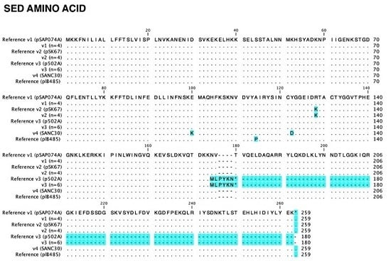
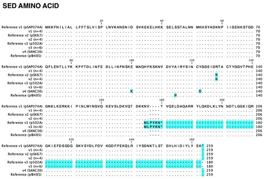
2.3. Sed Promoter and Gene Sequences

The sequences of the sed promoter and gene were determined in 12 strains and alignments of all sequences are provided as a supplementary file (Figure S3). The −35 (ATGAAA) and −10 (TATAA) promoter elements were identical in all tested strains.
The sed sequences were also highly conserved. However, point mutations were observed in strain SANC30 (position 198 G -> A, position 364 T -> G) and in strains BW10, RKI1, RKI2, and SAR35 (position 383 G -> A). In total, four different amino acid variants were detected, none of which was 100% identical to the common sed plB485 reference sequence (Genbank accession number M28521.1). SANC30 exhibited a novel amino acid sequence (variant 4) with two amino acid changes (position 100 E -> K, position 121 Y -> D) that did not correspond to any sequence in the GenBank database. An alignment of the respective amino acid sequences and reference sequences is provided in Figure 3.
All three tested rabbit isolates harbored the same sed nucleotide sequence variant (v3), which exhibits a deletion in sed at nucleotide position 521, resulting in a premature stop codon at amino acid position 180. To confirm the possibility of a host-specific variant, an additional three sed+ rabbit strains were sequenced, which harbored the same truncated variant.
Screening of strains representing the different sed variants for production of SED by SET-RPLA showed that not only the complete sed variants but also the truncated sed v3 were expressed. However, for the strains harboring the truncated sed variant, SED levels were far lower, with only the first dilution in the dilution series yielding a weakly positive test result. Four rabbit strains harboring the truncated sed v3 variant were screened. While SED was detected in three of these strains (SAK8, SAK11, SAK13), one strain (SAK64) did not yield a positive result for SED production. While all sed+ rabbit strains represent the same clonal complex (CC5), SAK64 was the only rabbit strain of spa type t160.

3. Discussion

In this study, several new variants of seb, sec, and sed enterotoxin genes and promoters were detected. Sequencing of seb, sec, and sed promoter regions revealed that promoter sequences were highly conserved in sec and sed. In contrast, several variable positions were observed in the seb promoter region, including the region required for seb transcription and expression. This is consistent with findings by Sato’o et al. [13], reporting high variability in seb upstream sequences from different strains. In the same study, several novel SaPIs carrying seb were identified and linked to differences in SEB production levels. However, the differences in seb promoter regions did not correlate with SEB production in a statistically significant manner [13].
Comparative analysis of the nucleotide sequences of the seb, sec, and sed genes showed that sed sequences were more conserved than seb and sec sequences. The length of the seb and sec coding sequences determined in this study was consistent with previous reports [18,25,26].
Most of the residues that are conserved throughout all SEs are either centrally located or can be found at the C-terminal end [27]. This is also consistent with the findings in this study for the amino acid prediction of seb, and sec variants, indicating that amino acid exchanges were more likely to occur at the N-terminus. However, for SED, the highest degree of amino acid variability detected in this study was centrally located.
Predicting altered functionality based on the detected amino acid exchanges is challenging, as emetic activity is still poorly understood. While lack of the disulfide loop was suggested to result in no or lower emetic activity, it has been shown that the disulfide bond is not a prerequisite for emetic activity [28]. Concerning superantigenic activity, ovine and bovine SEC variants were reported to be strongly altered in function due to only three amino acid changes resulting in host-dependent superantigenicity [27]. With regard to antigenicity, it was shown that SE variants differing in several residues, such as the SEC variants identified in FRI909 and FRI913 (9 differing residues), can still be antigenetically indistinguishable [27].
For SEC, four variants (SEC1-4) associated with human S. aureus isolates have been reported, as well as the host-specific variants SEC-bovine and SEC-ovine [16,17,18,19]. In contrast, subtypes of seb and the different SaPIs associated with these subtypes have only recently gained attention [13,15]. Kohler et al. demonstrated the existence of multiple SEB variants that differed in their ability to activate subsets of T cells and in their effects on the proliferation of peripheral blood mononuclear cells and rabbit splenocytes [15].
In this study, a novel seb nucleotide sequence variant (seb v3) was identified in two of the tested strains (RKI4 and SAI33). However, the seb v3 nucleotide sequence variant results in a known SEB amino acid sequence identical to reference strain No. 10. For sec, one novel sec nucleotide sequence (sec v4) was detected in one strain (SAI48). Amino acid sequence prediction showed that sec v4 results in an amino acid sequence identical to the one of sec v1, which is also known as SEC-2 (reference strain 79_S10). For sed, one novel nucleotide sequence (sed v4) was determined in a strain associated with human nasal colonization (SANC30). The novel sed v4 nucleotide sequence variant results in a novel amino acid sequence variant of SED that was not previously described elsewhere (sedaa v4).
In this study, a variant of sed was identified which was present in all tested rabbit isolates (n = 6). This variant sed v3 exhibited a deletion that resulted in a premature stop codon and a truncated sed amino acid precursor. In foodborne outbreak isolates, deletions at nucleotide positions 150 [21] and 514 [22] resulting in a premature stop codon have been reported. A deletion in sed identical to the one seen in the rabbit isolates in this study (nucleotide position 521) has been reported in S. aureus isolates originating from humans and from food [23,24]. While Lis et al. confirmed transcription of sed by qPCR, they could not detect SED protein by ELISA or Western blotting [24]. In contrast, in this study, three of four rabbit strains tested with truncated sed variants yielded a weak, but positive result for SED in the SET-RPLA assay. The deletion in sed may impair the functionality of the protein and recognition by various detection methods.

4. Conclusions

The sequence data generated in this study extends the current knowledge on sequence variation in enterotoxin genes of S. aureus strains isolated from various sources. Several novel variants of enterotoxin promoter and gene nucleotide sequences were described, and a novel amino acid sequence variant of SED was identified in a strain obtained from a nasal carrier. In addition, the results presented in this study confirm previous reports of host-specific enterotoxin variants such as SEC-bovine. Interestingly, all sed+ rabbit strains tested in this study harbored a sed variant that exhibited a deletion in sed leading to a premature stop codon.
The data generated represents a further step towards improved understanding of strain-specific differences in enterotoxin expression and host-specific variation in enterotoxin sequences.

5. Materials and Methods

5.1. Bacterial Strains

The S. aureus isolates used in this study originated from SFP outbreaks, asymptomatic nasal colonization or cases of infections in humans, as well as rabbit carcasses and bovine mastitis milk. Isolates were selected from a large collection of well-characterized S. aureus strains, for which DNA microarray enterotoxin hybridization patterns, spa types, and clonal complexes had been previously determined and published [14,28,29,30,31]. Detailed information on all S. aureus strains used in this study is provided in Table 1.

5.2. DNA Extraction and PCR Amplification

Frozen stock cultures (−80 °C) of S. aureus strains were resuscitated by plating on 5% sheep blood agar and incubation at 37 °C over night. Bacterial DNA was extracted using the DNeasy Blood and Tissue Kit (Qiagen, Hilden, Germany) following the manufacturer′s instructions.
PCR was performed using the Phusion High-Fidelity System (Thermo Scientific, Reinach, Switzerland) using a total reaction volume of 50 μL. All primers and primer-pair specific annealing temperatures are provided as supplemental material (Table S1). For each reaction, 5 μL buffer, 2 μL DMSO, 1 μL dNTP mix, 2 μL of each primer (c = 10 μM), 0.5 μL Phusion High-Fidelity DNA polymerase, 36.5 μL Aq.B., and 1 μL DNA template were used. PCR cycling conditions included: 5 min hot start at 95 °C, followed by 30 amplification cycles (denaturation at 95 °C for 30 s, annealing at the primer-specific annealing temperature for 30 s, elongation at 72 °C for 75 s), a final elongation step at 72 °C for 10 min, and a cooling step. Target-specific amplification was confirmed by electrophoresis using a 1% agarose gel.

5.3. PCR Purification and Sequencing

PCR amplicons were purified using the MinElute PCR Purification Kit (Qiagen, Hilden, Germany) and sequencing was outsourced (Microsynth, Balgach, Switzerland). The acquired sequences were analyzed using CLC Main Workbench software (Version 6.9, CLC Bio/Qiagen, Aarhus, Denmark, 2012) and were compared to reference nucleotide sequences imported from GenBank (NCBI). Novel variants of enterotoxin promoter or gene sequences were subsequently submitted to GenBank.

5.4. Toxin Detection by SET-RPLA

Expression of different seb, sec, and sed variants was assessed in selected strains using the SET-RPLA kit (Oxoid). Enterotoxins were detected using bacterial culture filtrates (0.2 μm filter, Whatman, Sigma-Aldrich, Buchs, Switzerland) from stationary phase cultures of each strain in Luria Bertrani (LB, Becton Dickinson, Allschwil, Switzerland) broth (37 °C, 225 rpm shaking, 20 h of incubation) in accordance with the manufacturer’s instructions. Culture filtrates were diluted in a five-fold dilution series for semi-quantitative detection of SEB, SEC, and SED. For seb, strains KLT6, SANC49, SANC14, SAI45, RKI4, and SAI40 were tested for SEB expression. All sec strains were assayed for SEC expression. For sed, strains BW10, RKI1, RKI2, KLT8, SAK8, SAK11, SAK13, and SAK64 were tested for SED expression.

5.5. Amino Acid Identity

A pairwise amino acid identity comparison between all SEB, SEC, and SED enterotoxin variants is provided in Table 2.

5.6. Accession Numbers

All variants of promoter and gene sequences were submitted to GenBank and can be accessed using accession numbers KX168612–KX168635. Promoter sequence variants are available for seb variants (sebp v1 = KX168623, sebp v2 = KX168624, sebp v3 = KX168625, sebp v4 = KX168626, sebp v5 = KX168627), sec variants (secp v1 = KX168633, secp v2 = KX168634, secp v3 = KX168635), and sed variants (sedp v1 = KX168616, sedp v2 = KX168617, sedp v3 = KX168618). Sequence variants are available for the seb gene (seb v1 = KX168628, seb v2 = KX168629, seb v3 = KX168630, seb v4 = KX168631, seb v5 = KX168632), the sec gene (sec v1 = KX168612, sec v2 = KX168613, sec v3 = KX168614, sec v4 = KX168615), and the sed gene (sed v1 = KX168619, sed v2 = KX168620, sed v3 = KX168621, sed v4 = KX168622).

Supplementary Materials

The following are available online at www.mdpi.com/2072-6651/8/6/169/s1, Figure S1: Sequence alignments for seb. Figure S2: Sequence alignments for sec. Figure S3: Sequence alignments for sed. Table S1: Primers used in this study.

Acknowledgments

We thank Mirjam Moser for laboratory work. This study was supported by the Swiss National Research Program 69 (40690_145211/1) and the Forschungskredit of the University of Zurich (grant no. FK-13-059 and FK-15-055).

Author Contributions

S.J. and R.S. conceived and designed the study. H.-M.S., G.M. and S.J. performed the experiments and analyzed the data. S.J. and H.-M.S. wrote the paper.

Conflicts of Interest

The authors declare no conflict of interest.

References

  1. Scallan, E.; Hoekstra, R.M.; Angulo, F.J.; Tauxe, R.V.; Widdowson, M.-A.; Roy, S.L.; Jones, J.L.; Griffin, P.M. Foodborne illness acquired in the United States—Major pathogens. Emerg. Infect. Dis. 2011, 17, 7–15. [Google Scholar] [CrossRef] [PubMed]
  2. Tranter, H.S. Foodborne staphylococcal illness. Lancet 1990, 336, 1044–1046. [Google Scholar] [CrossRef]
  3. Sihto, H.M.; Tasara, T.; Stephan, R.; Johler, S. Growth behavior and temporal enterotoxin D expression of Staphylococcus aureus strains under glucose and lactic acid stress. Food Control 2016, 62, 69–73. [Google Scholar] [CrossRef]
  4. Sihto, H.-M.; Budi Susilo, Y.; Tasara, T.; Rådström, P.; Stephan, R.; Schelin, J.; Johler, S. Effect of sodium nitrite and regulatory mutations Δagr, ΔsarA, and ΔsigB on the mRNA and protein levels of staphylococcal enterotoxin D. Food Control 2016, 65, 37–45. [Google Scholar] [CrossRef]
  5. Sihto, H.-M.; Tasara, T.; Stephan, R.; Johler, S. Temporal expression of the staphylococcal enterotoxin D gene under NaCl stress conditions encountered during food production and preservation. FEMS Microbiol. Lett. submitted for publication. 2015. [Google Scholar] [CrossRef] [PubMed]
  6. Wallin-Carlquist, N.; Cao, R.; Márta, D.; da Silva, A.S.; Schelin, J.; Rådström, P. Acetic acid increases the phage-encoded enterotoxin A expression in Staphylococcus aureus. BMC Microbiol. 2010, 10. [Google Scholar] [CrossRef] [PubMed]
  7. Cao, R.; Zeaki, N.; Wallin-Carlquist, N.; Skandamis, P.N.; Schelin, J.; Rådström, P. Elevated enterotoxin A expression and formation in Staphylococcus aureus and its association with prophage Induction. Appl. Environ. Microbiol. 2012, 78, 4942–4948. [Google Scholar] [CrossRef] [PubMed]
  8. Sumby, P.; Waldor, M.K. Transcription of the toxin genes present within the staphylococcal phage φSa3ms is intimately linked with the phage’s life cycle. J. Bacteriol. 2003, 185, 6841–6851. [Google Scholar] [CrossRef] [PubMed]
  9. Borst, D.W.; Betley, M.J. Phage-associated differences in staphylococcal enterotoxin A gene (sea) expression correlate with sea allele class. Infect. Immun. 1994, 62, 113–118. [Google Scholar] [PubMed]
  10. Mahmood, R.; Khan, S.A. Role of upstream sequences in the expression of the staphylococcal enterotoxin B gene. J. Biol. Chem. 1990, 265, 4652–4656. [Google Scholar] [PubMed]
  11. Zhang, S.; Stewart, G.C. Characterization of the promoter elements for the staphylococcal enterotoxin D gene. J. Bacteriol. 2000, 182, 2321–2325. [Google Scholar] [CrossRef] [PubMed]
  12. Shafer, W.M.; Iandolo, J.J. Chromosomal locus for staphylococcal enterotoxin B. Infect. Immun. 1978, 20, 273–278. [Google Scholar] [PubMed]
  13. Sato’o, Y.; Omoe, K.; Ono, H.K.; Nakane, A.; Hu, D.-L. A novel comprehensive analysis method for Staphylococcus aureus pathogenicity islands. Microbiol. Immunol. 2013, 57, 91–99. [Google Scholar] [CrossRef] [PubMed]
  14. Stevens, M.J.A.; Stephan, R.; Johler, S. Complete and assembled genome sequence of Staphylococcus aureus RKI4, a food-poisoning strain exhibiting a novel S. aureus pathogenicity island carrying seb. Genome Announc. 2015, 3. [Google Scholar] [CrossRef] [PubMed]
  15. Kohler, P.L.; Greenwood, S.D.; Nookala, S.; Kotb, M.; Kranz, D.M.; Schlievert, P.M. Staphylococcus aureus isolates encode variant staphylococcal enterotoxin B proteins that are diverse in superantigenicity and lethality. PLoS ONE 2012, 7, e41157. [Google Scholar] [CrossRef] [PubMed]
  16. Couch, J.L.; Betley, M.J. Nucleotide sequence of the type C3 staphylococcal enterotoxin gene suggests that intergenic recombination causes antigenic variation. J. Bacteriol. 1989, 171, 4507–4510. [Google Scholar] [PubMed]
  17. Marr, J.C.; Lyon, J.D.; Roberson, J.R.; Lupher, M.; Davis, W.C.; Bohach, G.A. Characterization of novel type C staphylococcal enterotoxins: biological and evolutionary implications. Infect. Immun. 1993, 61, 4254–4262. [Google Scholar] [PubMed]
  18. Bohach, G.A.; Schlievert, P.M. Nucleotide sequence of the staphylococcal enterotoxin C1 gene and relatedness to other pyrogenic toxins. Mol. Gen. Genet. 1987, 209, 15–20. [Google Scholar] [CrossRef] [PubMed]
  19. Fitzgerald, J.R.; Monday, S.R.; Foster, T.J.; Bohach, G.A.; Hartigan, P.J.; Meaney, W.J.; Smyth, C.J. Characterization of a putative pathogenicity island from bovine Staphylococcus aureus encoding multiple superantigens. J. Bacteriol. 2001, 183, 63–70. [Google Scholar] [CrossRef] [PubMed]
  20. Bayles, K.W.; Iandolo, J.J. Genetic and molecular analyses of the gene encoding staphylococcal enterotoxin D. J. Bacteriol. 1989, 171, 4799–4806. [Google Scholar] [PubMed]
  21. Kauffman, N.M.; Roberts, R.F. Staphylococcal enterotoxin D production by Staphylococcus aureus FRI 100. J. Food Prot. 2006, 69, 1448–1451. [Google Scholar] [PubMed]
  22. Suzuki, Y.; Kobayashi, M.; Matsushita, S.; Uehara, S.; Kato, R.; Sato’o, Y.; Ono, H.K.; Sadamasu, K.; Kai, A.; Kamata, Y. Detection of the staphylococcal enterotoxin D-like gene from staphylococcal food poisoning isolates over the last two decades in Tokyo. J. Vet. Med. Sci. 2015, 77, 905–911. [Google Scholar] [CrossRef] [PubMed]
  23. Parker, D.; Narechania, A.; Sebra, R.; Deikus, G.; Larussa, S.; Ryan, C.; Smith, H.; Prince, A.; Mathema, B.; Ratner, A.J.; et al. Genome sequence of bacterial interference strain Staphylococcus aureus 502A. Genome Announc. 2014, 2, 5–6. [Google Scholar] [CrossRef] [PubMed]
  24. Lis, E.; Podkowik, M.; Schubert, J.; Bystroń, J.; Stefaniak, T.; Bania, J. Production of staphylococcal enterotoxin R by Staphylococcus aureus strains. Foodborne Pathog. Dis. 2012, 9, 762–766. [Google Scholar] [CrossRef] [PubMed]
  25. Jones, C.L.; Khan, S.A. Nucleotide sequence of the enterotoxin B gene from Staphylococcus aureus. J. Bacteriol. 1986, 166, 29–33. [Google Scholar] [PubMed]
  26. Johns, M.B., Jr.; Khan, S.A. Staphylococcal enterotoxin B gene is associated with a discrete genetic element. J. Bacteriol. 1988, 170, 4033–4039. [Google Scholar] [PubMed]
  27. Dinges, M.M.; Orwin, P.M.; Schlievert, P.M. Exotoxins of Staphylococcus aureus. Clin. Microbiol. Rev. 2000, 13, 16–34. [Google Scholar] [CrossRef] [PubMed]
  28. Hovde, C.; Marr, C.; Hoffmann, M.; Hackett, S.; Chi, Y.; Crum, K.; Stevens, D.; Stauffacher, C.; Bohach, G. Investigation of the role of the disulphide bond in the activity and structure of staphylococcal enterotoxin C1. Mol. Microbiol. 1994, 15, 897–909. [Google Scholar] [CrossRef]
  29. Tobes, R.; Manrique, M.; Brozynska, M.; Stephan, R.; Pareja, E.; Johler, S. Noncontiguous finished genome sequence of Staphylococcus aureus KLT6, a staphylococcal enterotoxin B-positive strain involved in a food poisoning outbreak in Switzerland. Genome Announc. 2013, 1. [Google Scholar] [CrossRef] [PubMed]
  30. Wattinger, L.; Stephan, R.; Layer, F.; Johler, S. Comparison of Staphylococcus aureus isolates associated with food intoxication with isolates from human nasal carriers and human infections. Eur. J. Clin. Microbiol. Infect. Dis. 2012, 31, 455–464. [Google Scholar] [CrossRef] [PubMed]
  31. Merz, A.; Stephan, R.; Johler, S. Genotyping and DNA microarray based characterization of Staphylococcus aureus isolates from rabbit carcasses. Meat Sci. 2016, 112, 86–89. [Google Scholar] [CrossRef] [PubMed]
  32. Johler, S.; Layer, F.; Stephan, R. Comparison of virulence and antibiotic resistance genes of food poisoning outbreak isolates of Staphylococcus aureus with isolates obtained from bovine mastitis milk and pig carcasses. J. Food Prot. 2011, 74, 1852–1859. [Google Scholar] [CrossRef] [PubMed]
Figure 1. Amino acid variants of SEB. Amino acid exchanges compared to the most common amino acid detected are highlighted in blue (n = number of strains representing each variant).
Toxins 08 00169 g001
Figure 2. Amino acid variants of SEC. Amino acid exchanges compared to the most common amino acid detected are highlighted in blue (n = number of strains representing each variant).
Toxins 08 00169 g002
Figure 3. Amino acid variants of SED. Amino acid exchanges compared to the most common amino acid detected are highlighted in blue (n = number of strains representing each variant).
Toxins 08 00169 g003
Table 1. Detailed overview of sequence variants of enterotoxin promoters and genes of all S. aureus strains used in this study. In addition, information on other major enterotoxin genes harbored by each strain, the source of the strain, and its assignment to a spa type and clonal complex are provided.
GeneStrain IDIdentical Reference 1Promoter Variant 2 (Reference)Gene Variant 2 (Reference)Amino Acid Variant (Reference)SourceClonal Complex/spa Type Reference
sebKLT6COLsebp v1 seb v1 sebaa v1 SFP 3CC12/t160[29]
SANC31COLsebp v1 seb v1 sebaa v1 Human nasal colonizationCC59/t216[30]
SANC49COLsebp v1 seb v1 sebaa v1 Human nasal colonizationCC59/t216[30]
SAK9COLsebp v1 seb v1 sebaa v1 Rabbit CC5/t8456[31]
SAK18COLsebp v1 seb v1 sebaa v1 Rabbit CC5/t8456[31]
SAI10COLsebp v1 seb v1 sebaa v1 Human infectionCC59/t216[30]
SAI50COLsebp v1 seb v1 sebaa v1 Human infectionCC59/t015[30]
SANC14IVM10sebp v2 seb v2 sebaa v2 Human nasal colonizationCC45/t630[30]
RKI4novel 4sebp v3 (No. 10)seb v3 (novel) 4sebaa v3 (No. 10)SFPCC9/t733 [14]
SAI40novel 4sebp v4 (novel) 4seb v3 (No. 10)sebaa v3 (No. 10) Human infectionCC15/t084[30]
SAI33novel 4sebp v4 (novel) 4seb v3 (novel) 4sebaa v3 (No. 10)Human infectionCC20/t164[30]
SAI45novel 4sebp v5 (novel) 4seb v4 (IVM10)sebaa v2 (IVM10)Human infectionCC121/t272[30]
secBW1079_S10secp v1sec v1 = SEC-2secaa v1 = SEC-2SFPCC45/t383Medical Department of the German Federal Armed Forces, Germany
LRA179_S10secp v1sec v1 = SEC-2secaa v1 = SEC-2SFPCC73/t015Bavarian State Office of Health and Food Safety, Germany
SANC2379_S10secp v1sec v1 = SEC-2secaa v1 = SEC-2Human nasal colonizationCC8/t8016[30]
SANC4879_S10secp v1sec v1 = SEC-2secaa v1 = SEC-2Human nasal colonizationCC45/t015[30]
NB679_S10secp v1sec v1 = SEC-2secaa v1 = SEC-2SFPCC45/t6969Bavarian State Office of Health and Food Safety, Germany
SAR1RF122secp v2sec v2 = SEC-bovine secaa v2 = SEC-bovineBovine mastitis milk CC151/t529[32]
SAR38RF122secp v2sec v2 = SEC-bovine secaa v2 = SEC-bovineBovine mastitis milkCC151/t529[32]
SAR50RF122secp v2sec v2 = SEC-bovine secaa v2 = SEC-bovineBovine mastitis milkCC151/t529[32]
SAI3novel 4secp v3 (H-EMRSA-15)sec v3 = SEC-1 (B1085)secaa v3 = SEC-1 (B1085)Human infectionCC8/t148[30]
SAI48novel 4secp v1 (79_S10)sec v4 (novel) 4secaa v1 = SEC-2 (79_S10)Human infectionCC5/t002[30]
sedKLT8pSAP074Asedp v1sed v1 sedaa v1SFPCC5/t8017Cantonal Laboratory Thurgau, Switzerland
SAI8pSAP074Asedp v1sed v1 sedaa v1Human infectionCC5/t954[30]
SAI41pSAP074Asedp v1sed v1 sedaa v1Human infectionCC5/t8017[30]
SAI48novel 4sedp v3 (novel) 4sed v1 (pSAP074A)sedaa v1 (pSAP074A)Human infectionCC5/t002[30]
BW10pSK67sedp v2sed v2 sedaa v2SFPCC45/t383Medical Department of the German Federal Armed Forces, Germany
RKI1pSK67sedp v2sed v2 sedaa v2SFPCC8/t648Robert Koch Institute, Germany
RKI2pSK67sedp v2sed v2 sedaa v2SFPCC8/t008Robert Koch Institute, Germany
SAR35novel 4sedp v1 (pSAP074A)sed v2 (pSK67) sedaa v2 (pSK67)Bovine mastitis milkCC8/t2953[32]
SAK8novel 4sedp v1 (pSAP074A)sed v3 (p502A)sedaa v3 (p502A)RabbitCC5/t179[31]
SAK9novel 4ND 5sed v3 (p502A)sedaa v3 (p502A)Rabbit CC5/t8456[31]
SAK11novel 4sedp v1 (pSAP074A)sed v3 (p502A)sedaa v3 (p502A)RabbitCC5/t179[31]
SAK13novel 4sedp v1 (pSAP074A)sed v3 (p502A)sedaa v3 (p502A)RabbitCC5/t179[31]
SAK18novel 4ND 5sed v3 (p502A)sedaa v3 (p502A)Rabbit CC5/t8456[31]
SAK64novel 4ND 5sed v3 (p502A)sedaa v3 (p502A)RabbitCC5/t160[31]
SANC30novel 4sedp v1 (pSAP074A)sed v4 (novel)4sedaa v4 (novel) 4Human nasal colonizationCC5/t002[30]
1 Reference sequences were obtained from GenBank. Accession numbers: CP000046.1 (strain COL), AB716349.1 (strain IVM10), AB716351.1 (strain No. 10), CP010952.1 (strain 93b_S9), CP010944.1 (strain 79_S10), AJ938182.1 (strain RF122), KF386012.1 (strain B1085), CP007659.1 (strain H-EMRSA-15), GQ900426.1, (plasmid pSAP074A), CP007455.1 (plasmid p502A), GQ900447.1 (plasmid pSK67); 2 Nucleotide sequence; 3 SFP = Staphylococcal Food Poisoning; 4 Novel variant with no identical reference sequence available in GenBank; 5 Not determined.
Table 2. Overview of pairwise amino acid identity of the different SEB, SEC, and SED variants. The novel variant 4 of SED (sedaa v4) was detected in a strain isolated from a nasal carrier and has not been previously described.
VariantSEB v1SEB v2SEB v3SEC-1SEC-2SEC-3SEC-4SEC-bovineSEC-ovineSED v1SED v2SED v3SED v4
SEB v1100999768676967696736352536
SEB v2-1009867666766686736362536
SEB v3--10067666766686736352435
SEC-1---100979497999832322232
SEC-2----1009699979633322232
SEC-3-----10096949333332333
SEC-4------100979633332233
SEC-bovine-------1009932322232
SEC-ovine--------10032322132
SED v1---------100996699
SED v2----------1006699
SED v3-----------10065
SED v4------------100

