Development of Bioelectronic Devices Using Bionanohybrid Materials for Biocomputation System

1
Department of Chemical & Biomolecular Engineering, Sogang University, 35 Baekbeom-Ro, Mapo-Gu, Seoul 04107, Korea
2
Department of Chemical Engineering, Kwangwoon University, Wolgye-dong, Nowon-gu, Seoul 01899, Korea
*
Author to whom correspondence should be addressed.
Micromachines 2019, 10(5), 347; https://doi.org/10.3390/mi10050347
Submission received: 17 April 2019
/
Revised: 16 May 2019
/
Accepted: 22 May 2019
/
Published: 27 May 2019
(This article belongs to the Special Issue Emerging Memory and Computing Devices in the Era of Intelligent Machines)
Abstract
:Bioelectronic devices have been researched widely because of their potential applications, such as information storage devices, biosensors, diagnosis systems, organism-mimicking processing system cell chips, and neural-mimicking systems. Introducing biomolecules including proteins, DNA, and RNA on silicon-based substrates has shown the powerful potential for granting various functional properties to chips, including specific functional electronic properties. Until now, to extend and improve their properties and performance, organic and inorganic materials such as graphene and gold nanoparticles have been combined with biomolecules. In particular, bionanohybrid materials that are composed of biomolecules and other materials have been researched because they can perform core roles of information storage and signal processing in bioelectronic devices using the unique properties derived from biomolecules. This review discusses bioelectronic devices related to computation systems such as biomemory, biologic gates, and bioprocessors based on bionanohybrid materials with a selective overview of recent research. This review contains a new direction for the development of bioelectronic devices to develop biocomputation systems using biomolecules in the future.
1. Introduction
Bioelectronics is defined as the combined field of biology and electronics that has recently been greatly developed to overcome the current limitation of silicon-based electronics and biology-based engineering [1]. By introducing biomolecules on the silicon-substrate, electrical functions have been demonstrated on the chip using the unique properties of biomolecules, such as specific target molecule detection and optoelectrical properties, that can be applied in bioelectronic devices such as biosensors, biophotodiodes, and biotransistors [2,3,4,5]. Various biomolecules including metalloprotein possess a metal ion at their core, and functional DNA with specific chemical group modifications such as amine and carboxyl groups have advantages for applications to develop bioelectronic devices because of their unique properties such as redox properties that are derived from the metal ion in the protein and the specific binding properties of DNA with its complementary DNA at the nanometer scale [6,7]. By fusing biomolecules with organic materials, electronic functions have been widely studied to develop bioelectronic devices with enhanced performance such as more sensitive target detection and increased signal [8,9,10,11]. Until now, many functional bioelectronic devices including protein-based bioelectronic chips that use the electron transfer mechanism of proteins and biophotodiode devices that use the photoelectric effect of rhodopsin have been reported [12,13,14]. However, current bioelectronic devices have certain critical limitations for practical application because the use of biomolecules inevitably accompanies limitations such as the low electrical/electrochemical signal-to-noise ratio derived from biomolecules, instability in harsh conditions, and narrow functionalization [15,16]. To overcome the limitations of biomolecules, innovative methods have been developed introducing nanoparticles to enhance the signal induced from biomolecules, combine biomolecules with carbon-based materials such as carbon nanotubes (CNT) or graphene for electrochemical signal increment and long-term stability using the biocompatibility of carbon-based materials, and the use of nanoscale-patterned chips as a platform for the extension of the functionality of bioelectronic devices such as by demonstrating nanoscale electronic functions and immobilizing different biomolecules independently at the nanometer scale to use these biomolecules simultaneously [17,18,19,20,21,22].
Recently, bionanohybrid materials composed of biomolecules and other nanomaterials have been developed widely for applications in bioelectronic devices. Bionanohybrid materials have received much attention for their wide application in developing delicate bioelectronic devices that accompany enhanced electronic functions or highly sensitive target detection for biosensors. As mentioned above, biomolecules have unique properties at the nanometer scale and nanomaterials such as nanoparticles, CNT, and biocompatible polymers that improve the properties of biomolecules can be hybridized precisely at the nanometer scale while retaining the properties of biomolecules and nanomaterials [23,24,25].
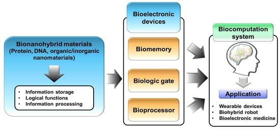
Among the various bioelectronic devices, certain bioelectronic devices that are capable of performing information storage or signal processing similar to memory or logic gates in conventional electronic devices have shown a new perspective and direction for the development of biocomputation systems [26,27]. Biomemory devices based on metalloprotein or redox-controllable linker have been reported [28,29,30] that can demonstrate the memory function using biomolecules through controlling two apparently distinguished biomolecular states reversibly. In addition, using the above-mentioned bionanohybrid materials as the core component, bioprocessor devices have been reported that can process the input signal to process the out signal using bionanohybrid materials as the processing platform [31]. In addition, to develop sophisticated and improved functional bioelectronic devices, various advanced materials have been studied and introduced to fabricate modern devices such as new functionalized structural graphene and two-dimensional materials [32,33,34]. Through these efforts, various bionanohybrid materials that are capable of performing information storage, logical functions, and information processing have been developed for the development of bioelectronic devices including biomemory, biologic gates, and bioprocessors. Such bioelectronic devices can be used as core components to develop a biocomputation system that is capable of performing computation similar to conventional computers that are common in our surroundings as depicted in Figure 1.
In this review, bioelectronic devices based on bionanohybrid materials that are capable of performing information storage and signal processing for computation systems are discussed with a selective overview of recent research. Although there are many extensive reviews of bioelectronic devices, this review discusses in detail recent reports of various specific types of bioelectronic device for biocomputation systems. This review will suggest a new inspirable direction and aspects of bioelectronic devices to develop a biocomputation system [35,36].
2. Biomemory
Information storage is an important function for the operation of electronic devices. Until now, various information storage function devices have been developed in conventional silicon-based electronic devices through controlling two apparently distinguished states such as “1” and “0” states for the demonstration of conventional memory functions. From the bioelectronics perspective, some specific biomolecules have properties of existing in two distinguished states by external stimulation such as metal ion states that control metalloprotein, which can be utilized to develop biomolecular memory devices [37,38]. In addition, the hybridization of more than two types of biomolecule and bionanohybrid materials has been proposed to demonstrate multiple states control and increase the electrochemical signal derived from biomolecules for biomemory. In this chapter, we provide research related to biomemory devices including protein-based biomemory and resistive switching memory devices.
2.1. Multilevel Biomemory Devices
Metalloproteins have metal ions in their body that can be utilized for electrochemical investigation [39,40]. For example, the metal ion of a metalloprotein can be used to affect the redox reactions of specific materials, which can be measured using electrochemical techniques for developing biosensors [18,41]. In addition, this can be applied to develop biotransistors using redox properties [42]. This metal ion can exist in two different states like the Cu+ and Cu2+ states of azurin, a metalloprotein that possesses copper ion, which shows the potential of metalloprotein-based biomemory devices [28] by controlling metal ions with distinguished states. Various research groups have developed metalloprotein-based biomemory devices [29,37,38]. Among them, our group developed various biomemory devices using metalloproteins such as azurin and cytochrome c, which have never been reported before. Beyond just controlling the ion states of one type of metalloprotein for biomemory, we suggested multilevel biomemory devices using two kinds of metalloproteins to achieve the incremental memory density [43]. By controlling isoelectric points of metalloproteins via pH control, we immobilized two different metalloproteins, recombinant azurin modified with cysteine group and cytochrome c, directly on to the gold substrate by self-assembly through the electrostatic bond without any chemical linkers for the control of multiple redox states [44,45]. This simple immobilization process could reduce the immobilizing time of biomolecules, and remove the introduction of the other chemical materials for immobilization. Figure 2A shows the schematic image and demonstration of multilevel biomemory using the direct immobilization of two kinds of metalloproteins. We confirmed multilevel memory device fabrication by surface plasmon resonance (SPR) and scanning tunneling microscopy (STM) to verify the metalloprotein double layer formation through morphological changes. Then, an electrochemical investigation was performed using cyclic voltammetry (CV) and chronoamperometry (CA). By introducing two different metalloproteins, this device showed the multiple redox states that could be derived from copper ions of azurin and iron ions of cytochrome c as shown in Figure 2B. This showed oxidation potential peaks at 0.294 V and 0.184 V that were derived from cytochrome c and azurin, respectively, and the reduction of potential peaks at 0.131 V and 0.062 V from cytochrome c and azurin, respectively. These potential values for each metalloprotein were used as input potentials to control the metal ion states of the two metalloproteins. Then, we estimated the memory performance for this device using the obtained redox potential peak values of two metalloproteins for the “writing step” and “erasing step” and obtained the open circuit potential (OCP) values of metalloproteins for the “reading step” for multilevel biomemory demonstration. As shown in Figure 2C, this device showed apparently distinguished states when applying a potential to the device following expected schematic images with two different forms of “writing step”, “reading step”, and “erasing step”. From these results, we successfully developed new-concept multilevel biomemory devices using two different metalloproteins for multiple information storage biodevices.
2.2. Electrochemical Signal-Enhanced Biomemory Device
As mentioned in the introduction, bioelectronic devices have certain limitations like the low electrical or electrochemical signal induced from biomolecules and low stability in harsh conditions [15,16]. To overcome these problems, various researchers have proposed the introduction of functional biocompatible nanomaterials for improved signal and stability [18,19,20]. Through these suggestions, biosensors and biofuel cells have been developed with advanced performance. In the case of biomemory devices, the extremely low electrochemical signal from biomolecules should be solved for application in practical applications. To achieve this, introducing metal nanoparticles can be a solution for signal enhancement. Gold nanoparticles (GNP) have been reported as an enhancer for the electron transfer reaction with metalloprotein [46]. Using the reported results, our group proposed a biomemory device using metalloprotein (azurin, Azu) and GNP to increase the electrochemical signal derived from metalloprotein (Figure 3A) [17]. To develop this electrochemical signal enhanced biomemory device, various GNP of nanometer size of diameter in the range 5–60 nm was introduced to find the optimized size for the GNP diameter. Based on the electrochemical signal increasing the redox potential peak values from CV results (Figure 3B), we found the optimized GNP size (5 nm) that showed smaller redox potential peak values compared to the results using the 60 nm GNP. However, in the case of the 60 nm GNP, the enhanced signal was not derived from Azu–GNP but directly induced from the immobilized GNP to the gold substrate without Azu. Therefore, the 5 nm GNP was chosen as the optimized size for biomemory fabrication. In addition, we assumed that the proposed increment of the electron transfer mechanism followed the equation below:
In Equation (1), k1 and k−1 are the electron transfer rate constants between the GNP and Azu and k2 and k−2 are the electron transfer rate constants between Azu and the gold substrate. By introducing the GNP, the electrochemical signal from Azu could be enhanced through the better electric coupling between azurin and GNP and between Azu and the gold substrate. Furthermore, the better coupling between Azu and GNP compared to that between Azu and the gold substrate induced a remarkably enhanced signal. After verifying the signal enhancement, the biomemory function of the proposed device was estimated. As shown in Figure 3C,D, the biomemory device composed of Azu and GNP (Azu–GNP) showed enhanced memory function compared to biomemory prepared with only Azu. The stored charge amounts were calculated by the following equation,
The current value (i) and time value (dt) were obtained by CA technique. To acquire the CA results, the redox potential peak values of Azu–GNP obtained by CV analysis were applied. From the calculation of the area underneath the CA graphs, the stored charge amounts of biomemory composed of Azu–GNP was about 4.503 µC, approximately four times higher than that of biomemory prepared with only Azu (about 1.1413 µC). This difference originated from the electric coupling between Azu and the GNP. Through this research, electrochemical signal-enhanced biomemory was developed for the first time, and this approach may demonstrate the possibility of developing accurate nanoscale biomemory devices that can overcome the problems associated with low electrochemical signals.
2.3. Resistive Biomemory Device
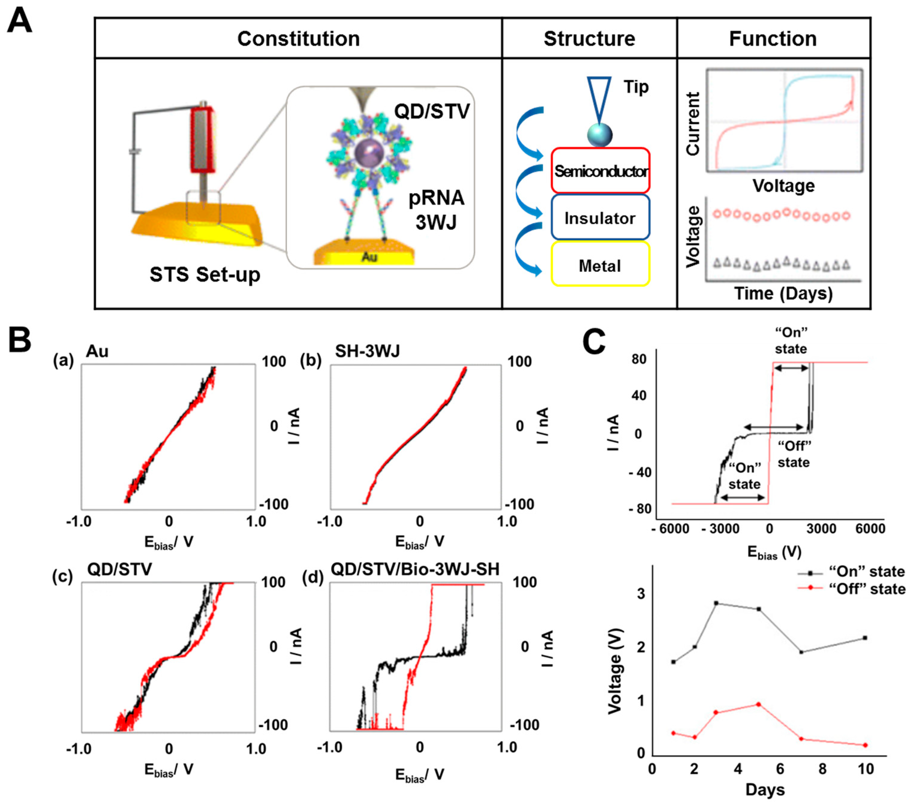
In conventional silicon-based electronic fields, huge attention has been paid to the development of resistive memory devices for resistive switching function demonstration. Resistive memory devices have been researched widely for commercialization due to their advantages such as fast processing and response and low energy requirement. In the case of the existence of metal–insulator–metal layers or semiconductor–insulator–metal layers on the substrate, there are specific unique hysteresis properties at some voltage range with two apparently different resistance values (extremely high resistance value and extremely low resistance value) following theories such as ohmic conduction, thermionic emission, Schottky emission, or tunneling current [47]. Various research groups have developed organic material-based resistive memory devices [48,49]. Biomolecules are suitable for demonstrating resistive switching functionality at the nanometer scale because they possess unique properties at such scale. Guo’s group developed a resistive biomemory device using the RNA structure and quantum dot (QD) by collaboration with our group [50]. In previous research, they developed a packaging RNA (pRNA) three-way junction structure (pRNA-3WJ) that showed thermodynamically stability [51]. This pRNA-3WJ could overcome the critical limitations of conventional RNA such as extremely low stability even at room temperature. In resistive biomemory research, they introduced the developed pRNA-3WJ as a stable insulator to demonstrate the resistive switching function. Figure 4A shows the schematic images and resistive function in this device. Using the biological binding properties between streptavidin and biotin, they developed a nanoscale bionanohybrid material composed of pRNA-3WJ and QD. The conjugation of pRNA-3WJ and QD for bionanohybrid materials was verified by electrophoresis through the existence of the upper located band due to the increased total weight and size by QD introduction compared to the band in only pRNA-3WJ without QD. After immobilizing this bionanohybrid material on the gold substrate, pRNA-3WJ performed a role as an insulating layer and QD as the semiconducting layer on the conducting gold layer. Using a scanning tunneling spectroscopy (STS) technique, they estimated the resistive switching function of this device at the nanometer scale using the platinum tip as the probe located on this bionanohybrid material. Figure 4B displays the I–V curve of a bionanohybrid material on a gold substrate. Compared to the gold substrate alone, only pRNA-3WJ, and only QD on a gold substrate, a bionanohybrid material composed of pRNA-3WJ and QD on a gold substrate showed apparently distinguished resistance values with extremely high and low resistance at the voltage range of +3 to −3 V. This bistable behavior could be defined as “On state” and “Off state” for resistive memory applications.
Our group also developed a resistive biomemory device based on two-dimensional material. A bionanohybrid material composed of molybdenum disulfide nanoparticles (MoS2) and a DNA layer on a gold substrate was developed to demonstrate resistive switching functionality at the nanometer scale [52]. To develop this resistive biomemory at the nanometer scale, we immobilized DNA and synthesized MoS2 sequentially on a complementary DNA modified gold substrate. Then, a semiconductor (MoS2)–insulator (DNA)–metal layer (gold substrate) was formed that could demonstrate resistive switching functionality through specific unique hysteresis properties at a certain voltage range with two apparently different resistance values. MoS2 is a metal dichalcogenide material that has been widely used to develop bioelectronic devices because of its unique properties including biocompatibility, excellent semiconductivity, and its optical properties [53,54]. To demonstrate resistive switching functionality at the nanometer scale, MoS2 nanoparticles with surface modification (carboxyl group) was synthesized for the first time to conjugate efficiently with amine-tagged DNA via EDC/NHS bonding. The synthesis of surface-modified MoS2 nanoparticles and the fabrication of bionanohybrid materials were verified by transmission electron microscopy (TEM) for MoS2 synthesized nanoparticles, energy-dispersive X-ray spectroscopy (EDS) for elemental analysis, electrophoresis for the conjugation of MoS2 and DNA, and STM techniques to immobilize this bionanohybrid material on the gold substrate. Using STS analysis, the proposed resistive biomemory device based on MoS2 and DNA showed the resistive switching function with bistable states at a wide voltage range (4 to −4 V) and long-term stability as shown in Figure 4C. Figure 4C shows that the resistance value dramatically decreased when the voltage reached 2.4 V; on the other hand, the resistance value abruptly increased when the voltage reached 0.01 V. In addition, by introducing DNA as the insulating layer, which is more stable than RNA, it showed resistive switching function for about 10 days. As with these studies, bionanohybrid material-based resistive biomemory devices have been researched to demonstrate resistive switching functionality at the nanometer scale, which suggests a future direction for the development of the next generation of memory devices using biomolecules.
3. Biologic Gate
Among the various components of computing systems, logic gates are a core component in the computing process. These electrical circuits implement Boolean functions that can perform logical operations by converting more than two inputs to one binary output. Until now, many logic gates have been developed including AND (the gate that performing the logical conjugation), OR (the gate that performing the logical disjunction), NOT (the gate that performing the logical negation), XOR (the gate that giving the true outputted signal when the number of true inputs is odd), and NAND (the gate that giving the false outputted signal only when all inputs are true) gates [55,56]. In bioelectronics, some biomolecules can interact with specific chemical materials or biomolecules. For example, myoglobin can react with hydrogen peroxide [18] and glucose oxidase can react with glucose [57]. These properties can be utilized to demonstrate a logic gate using biomolecular interactions by controlling the input materials. In addition, conformational changes of biomolecules can be utilized to develop logic gates such as the conformational change of G-quadruplex DNA (G-rich DNA) as a bending shape and straightening shape that is dependent on the pH value [58]. Furthermore, these logic functions based on biomolecules can provide opportunities to mimic the analog human decision-making process [59] through controlling the combination of biomolecules and organic and inorganic materials. In this chapter, we will introduce research into biologic gates using biomolecules such as proteins and DNA and bioelectronic devices that mimic the analog human decision-making process.
3.1. DNA-Based Biologic Gate
DNA is the smallest level at which the composition of living organisms is developed. There have been reports related to DNA research such as DNA sequencing, immunoassay, and DNA structure formation for wider applications [60,61,62]. From the bioelectronics perspective, the unique properties of DNA have received attention for their granting of functionality to bioelectronic devices. DNA can specifically bind with complementary DNA and the structure of DNA can be controlled by external responses [58]. Until now, various DNA-based logic gates have been reported based on colorimetric or fluorescence investigations. However, electrochemical techniques are better suited to bioelectronic device fabrication due to their fast response, minimal required reagents, and simplified outputs compared to colorimetric-based bioelectronic devices. In addition, a report found that mismatched sequences of double strand DNA such as cytosine–cytosine (C–C) and thymine–thymine (T–T) mismatched pairs could possess metal ions in such locations [63]. From this perspective, Qiu’s group developed a biologic gate using this unique property of DNA [64]. Figure 5A showed the schematic process and results of AND logic gates based on DNA mismatching. Silver ions (Ag+) and mercury ions (Hg2+) could enter the mismatched C–C and T–T pairs, respectively. Using these properties, they designed T- and C-rich DNA sequences with ferrocenecarboxylic acid (Fc) as the redox generator. In this device, metal ions were used as input molecules and the electrochemical signal from Fc was the output signal from the logic gate. By controlling the DNA sequences, they developed AND, NAND, and NOR logic gates through controlling the output signal using the unique electrochemical signals derived from the inserted Ag+ and Hg2+ ions located inside the mismatched pairs in the DNA. From the results, in the case of the insertion of only both Ag+ and Hg2+ ions, the electrochemical signal was detected by the differential pulse voltammetry (DPV), which was defined as “1” due to the co-existence of Ag+ and Hg2+ ions as shown in Figure 5A. Furthermore, this logic gate based on DNA mismatching can be operated reversibly compared to DNA cleavage-based logic gates. This result showed the possibility of applying bionanohybrid materials based on specifically designed DNA sequences and metal ions for both bioelectronic devices and for the development of a multiplexed biosensing platform.
3.2. Protein/DNA-Based Biologic Gate
Their distinct properties mean that proteins are widely utilized in bioelectronics. As mentioned in the introduction, proteins have certain unique advantages such as distinctive redox properties that can target specific molecules and reactions. Through these properties, protein-based biologic gates, particularly enzyme-based biologic gates, have been broadly developed [66,67]. Aida’s group developed a biologic gate by protein folding [68] and Schöning’s group proposed a biologic gate using a membrane composed of multiple enzymes [69]. Recently, Katz’s group developed reversible biologic gates based on both enzymes and DNA [65]. They developed and combined an enzyme-based biologic gate with a reversible DNA-based biologic gate through a biomolecular electrode to create complex reversible logical computing systems. This proposed system was composed of an enzyme-based Fredkin gate that was capable of converting three input signals to three output signals and a DNA-based Feynman gate that was capable of converting two input signals to two output signals [70,71]. To demonstrate this complex biologic gate, they introduced the optical, electrochemical, and fluorescent measurement techniques. Figure 5B shows a schematic diagram of this complex biologic system that is composed of a protein-based biologic gate and a DNA-based biologic gate as demonstrated by the enzyme reaction and connected DNA reaction. Glucose (Glc), lactic acid (Lac), and β-nicotinamide adenine dinucleotide hydrate (NAD+) were used as three input signals for the enzyme-based Fredkin biologic gate and glucose dehydrogenase (GDH), lactate dehydrogenase (LDH), glucose oxidase (GOx), and horseradish peroxidase (HRP) were utilized for enzyme-based biologic operation. After reacting in the first enzyme-based biologic gate, the generated signal that produced NADH through enzymatic reactions was measured by an optical technique and transferred to the electrochemical system for electrochemical enzymatic reaction. Then, in the final stage at the connected DNA-based biologic gate, the transferred signal was converted to fluorescent final outputs. Pyrroloquinoline quinone (PQQ)-modified electrode and iron ion (Fe3+) crosslinked alginate-modified electrode with entrapped DNA were used for the electrochemical system and final DNA-based biologic system. By the electrochemical reaction, the Fe3+ ion of the crosslinked alginate-modified electrode with entrapped DNA was oxidized to Fe2+ and the entrapped DNA was released from the alginate-modified electrode to a DNA-based biologic gate for the final fluorescence output signal. Although many components and complex biological reactions were utilized for this biologic gate, they developed a complex biologic system that was composed of two different kinds of biomolecule-based biologic gates that were more complex than the reported biologic gate to accurately mimic a conventional silicon-based electronic logic system.
3.3. Analog Decision Mimicking Bioelectronic Device
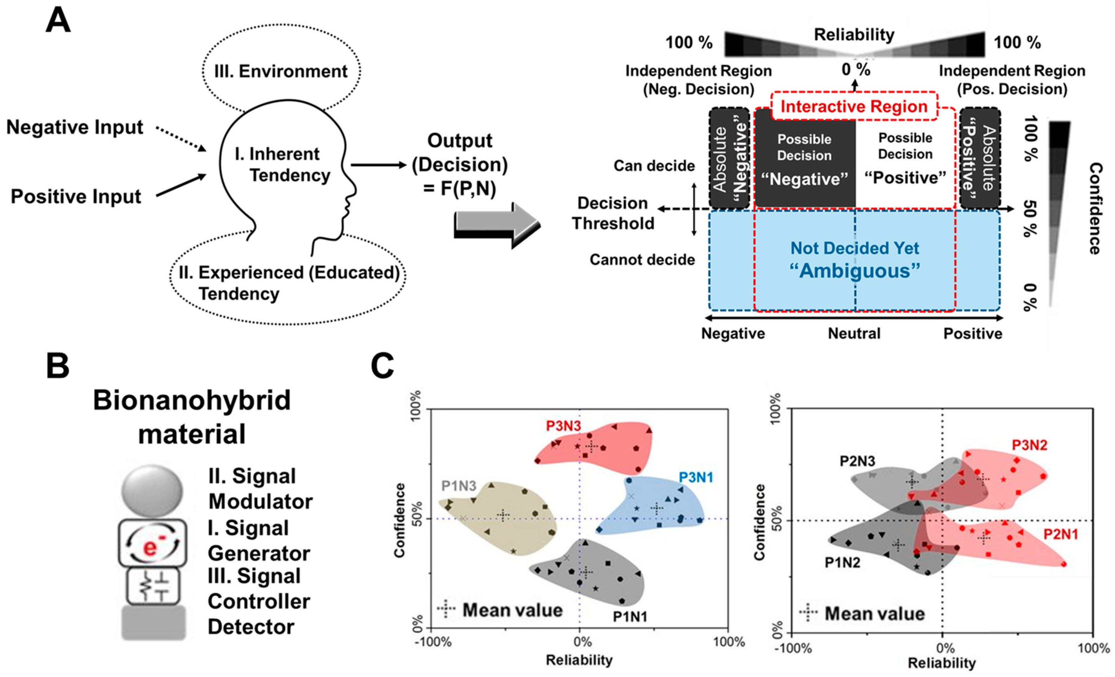
In conventional silicon-based electronics, only digitalized processing, logic, and arithmetic operations have been developed and utilized in all devices [72,73]. These operations have certain advantages for the development of electronic devices in which the binary coded digital signals “1” and “0” can be distinguished, defined, and operated easily by converting input signals into an integrated single output signal. However, digital signal-based conventional electronic devices have limitations for the demonstration of human logic systems or other analog decision-making processes because these systems are not decided or operated by one simple and single digital input and output, but are instead affected by a myriad of complex factors such as personality, experience, and intelligence. The critical difference between conventional electronic devices and real human logic systems can hinder the development of biocomputation systems. Therefore, in bioelectronic fields, there have been studies to develop bioelectronic devices that are capable of mimicking analog decisions or analog logic systems [74] by considering various factors for analog calculation. Liu’s group developed four analog computing systems and extended the range of computing to real numbers based on DNA by connecting DNA-based biologic gates using unique properties of DNA such as DNA strand displacement. This result showed the potential of DNA-based real number calculation such as a calculator and by extension a combination of various DNA-based biologic gates; this could demonstrate more complex number calculation. In addition, bioelectronic noses and tongues based on biomolecular receptors have been researched recently to mimic the processes of real living organisms [75,76]. To demonstrate the analog decision-making process on a bioelectronic chip, our group developed an electrochemical bioelectronic device based on a bionanohybrid material composed of metalloprotein and organic/inorganic nanomaterials or metal ions [59]. Figure 6A shows a conceptual image of their research for mimicking analog decision-making through the analogously processed output signals by inputting two different external factors (negative input and positive input) via electrochemical investigation. We defined specific regions of the acquired signal as the degree of confidence and reliability of a human following defined threshold values. Myoglobin (Mb) that is a metalloprotein used as signal generator and defined as an inherent human tendency, organic chemical linkers that are used as signal controllers and defined as experience-induced human tendencies, and inorganic materials that are used for signal modulation and defined as environment-dependent signal modulators were combined to demonstrate analog decision-making by signal control and modulation (Figure 6B). As shown in Figure 6C, the plotted results of analog decision-making based on the analysis of electrochemical signals by defined external factors showed the decision variation of 12 people based on defined threshold values. This research shows the conceptual potential for the development of analog-based bioelectronic devices which has never been reported to apply for biocomputation systems. Of course, many subjective definitions exist that can demonstrate the analog decision-making process on a bioelectronic chip. This research shows one potential development route for a human mimicking analog computation system.
4. Bioprocessor
Until now, numerous molecular electronic devices have been developed to miniaturize electronic devices at the molecular scale for overcoming the physical or technological limitations of conventional silicon-based electronic devices, such as difficulty to achieve compact integration at nanometer or molecular scale [77]. Biomolecules have unique properties even at the nanometer scale that are suitable to complement the molecular electronic devices with delicate functional processing properties. Accordingly, some researchers developed bioprocessors that could control the biological output signals, such as the expressed gene level, through the biological reaction process by specific inputted biomolecules [78,79]. To mimic the conventional silicon-based electronic processors, especially, the functional bionanohybrid material composed of biomolecules and various nanomaterials can used for processing the input signal converted to the processed output signal such as electrochemical signal. In addition, specifically designed microchips can be a powerful tool to control the biologically processed output signals through the control of biological reactions. In this chapter, we will provide the recently developed bioprocessor devices that could mimic the processing in electronic devices.
4.1. DNA-Based Bioprocessor
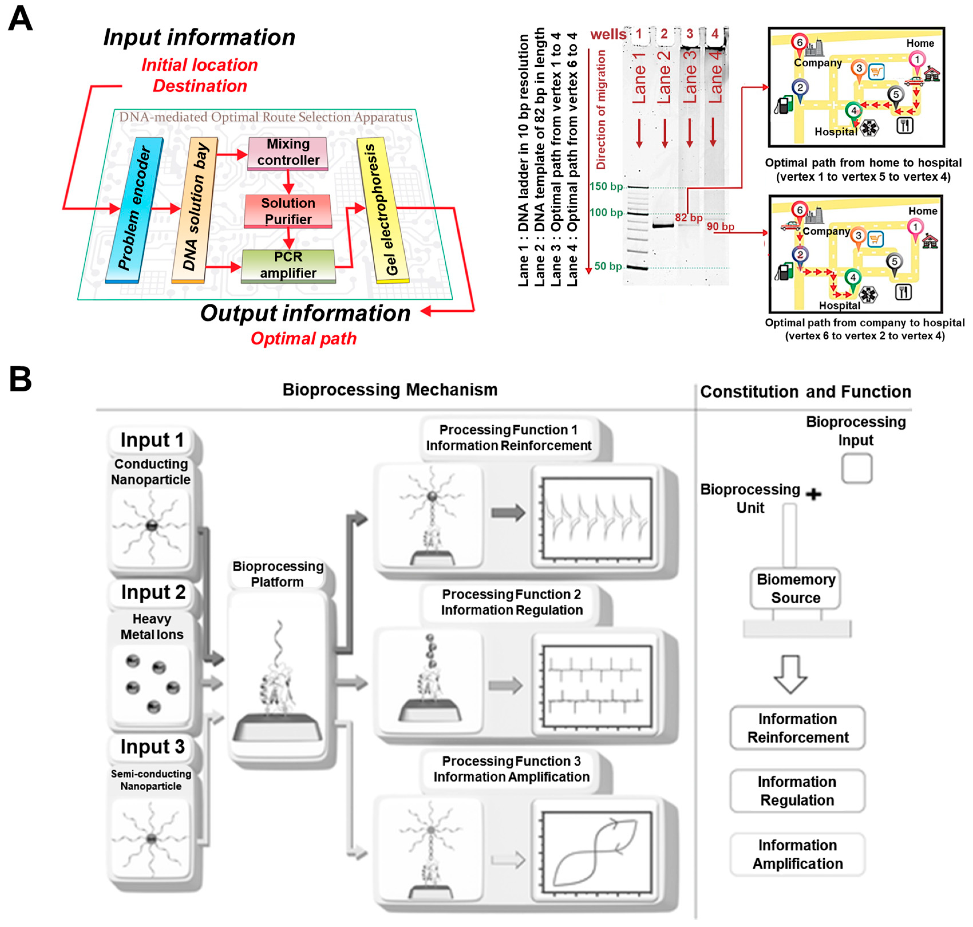
As mentioned in the above chapters, DNA is a suitable biomaterial for bioelectronic applications. Especially, the massive parallelism of DNA hybridization exhibits tremendous potential, which can be utilized to develop feasible electronic devices capable of performing processing or computing operation to fulfill the demands of monolithic parallel computing system with specific computational algorithm [80]. From this point of view, DNA-based bioprocessors or applications have been reported [81]. Lee’s group proposed a novel programmable DNA-mediated processor to solve the optimal route planning problems [82]. To achieve this functional DNA-mediated bioprocessor, they fabricated the programmable optimal route planning apparatus comprising six stages, as shown in Figure 7A. Also, the routes shown in Figure 7A were defined following the specific DNA sequences (20mer single strand DNA) to find the optimal route based on DNA processing through the conventional PCR reaction using the inputted DNA sequences, which determine distance between specific locations as shown in the map of the right side of Figure 7A. To operate this PCR system, they defined the first stage as problem encoder for conversion of vertices and weighted edges of the designed route to DNA sequences, and all distances between each of the six locations were defined as specific DNA sequences (20mer single strand DNA) to apply for the program encoder. They defined the second stage as DNA solution bay for converted DNA preparation, the third as mixing controller for mixing and ligase of appropriate DNA sequences to make the template of DNA duplexes that represents the possible routes, the fourth as solution purifier for isolation of optimal DNA template from impurities such as the incompletely hybridized oligonucleotides or enzymes, and the fifth as PCR amplifier for amplification of optimal DNA template which is the optimal route, final as gel electrophoresis to acquire the final electrophoresis data for optimal DNA template as the find of the defined optimal route. Using these stages for optimal-route finding, they performed the DNA reactions at these mentioned stages by binding and amplification of the combined six defined DNA sequences for six locations. They obtained the results of electrophoresis to find the optimal route from home or company to the hospital as shown in Figure 7A using subjectively defined factors. Although there existed too many subjective definitions for operation, these results showed the possibility of DNA-based bioprocessing for solving the practical problems; this could be demonstrated with much fewer components and materials compared to the conventional silicon-based electronic devices. Until now, DNA-based bioprocessors remain at the early stage. However, due to the huge researches for DNA-based bioprocessors, the more sophisticate and functional processable bioprocessors will be developed.
4.2. Bioprocessor Based on Bionanohybrid Material
Advancing from only DNA-based bioprocessors, bionanohybrid material based on DNA can be used as the platform to develop the bioprocessor with more intuitive bioprocessing operation, without too much subjective definition seen in DNA-based bioprocessors, such as definition of the specific DNA sequences as the specific distance between home or company to hospital for solving the optimal-route finding. Our group developed the bioprocessing device based on bionanohybrid materials composed of protein, DNA, and inorganic nanomaterials to demonstrate the various bioprocessing functions using electrochemical/electrical investigation [31]. To develop this bioprocessor, the recombinant protein (azurin, Azu) and single-strand DNA were conjugated through the organic linker as the electrochemical signal generating bioprocessing unit (Azu/DNA hybrid) by the redox properties derived from recombinant azurin. Then, the complementary DNA (cDNA) and gold nanoparticle (GNP) hybrid (cDNA/GNP), heavy metal ions, and cDNA and quantum dot (QD) hybrid (cDNA/QD) were introduced to the bioprocessing unit as the input materials for electrochemical signal reinforcement, regulation, and amplification. Figure 7B showed the schematic image of this bioprocessor, which processed the three different outputs by introduced each input material. In the case of cDNA/GNP introduction, the electrochemical signal from bioprocessing unit was reinforced by the existence of conducting GNP. Moreover, in the case of introduction of heavy metal ions, the electrochemical signal was regulated by existed heavy metal ions such as Cu, Zn, Ni, Co, Fe, and Mn through the movement of redox peak values compared with the peak values of only Azu/DNA hybrid without metal ions. In addition, in the case of introduction of the cDNA/QD as the semiconducting nanoparticle to the bioprocessing unit, the processed electrical signal showed the electrical bistable properties as the resistive memory function by STS investigation compared to the result of only the bioprocessing unit without cDNA/QD. This developed bioprocessor device can process three different functions in the single bionanohybrid material using electrochemical and electrical signals intuitively compared to the bioprocessors demonstrated based on subjective definitions. It showed the possibility of the development of the biocomputation system in a single-biomolecular hybrid at nanometer scale.
5. Future Perspective
Since the 1960s, silicon-based electronic devices have been developed widely to demonstrate more complex functions with faster and more efficient processing on nanoscale-size chips. However, until now, the demonstration of a computation system on the single-molecular level has been impossible in the electronics field. To develop the single-molecular computation system, bioelectronic devices present new possibilities in the development of single biomolecular computation systems based on bionanohybrid materials. Bionanohybrid materials composed of biomolecules such as protein or DNA, and organic/inorganic nanomaterials can perform sophisticated functions at the single-biomolecular level to apply for bioelectronic devices. In this review, authors discussed the various research areas related to the bioelectronic devices including biomemory, biologic gates, and bioprocessors, which are the core components of the computation system. First, we discussed biomemory device based on the metalloprotein heterolayer, metalloprotein-nanoparticle hybrids, and nucleic acids-semiconducting nanoparticle hybrids. To achieve the memory function, developed bionanohybrid materials should demonstrate the two distinctive bistable states, which can be defined as ‘1’ and ‘0’ states for memory. As shown in results, those bionanohybrid materials showed apparently distinguished bistable states by electrochemical or electrical investigation. Next, we examined the studies about biologic gates based on the DNA–metal ion hybrids, protein–DNA connected reaction, and protein–organic/inorganic nanomaterial hybrids. Using these bionanohybrid material, various logic functions including the AND, NAND, Fredkin, or Feynman logic gates were demonstrated. Furthermore, the human analog decision-mimicking device was developed. In addition, we discussed about bioprocessors capable of processing of the inputted signals to the output signals such as finding of optimal routes and processing of different electrochemical signals through the DNA reactions and metalloprotein, DNA and inorganic nanomaterial hybrids. In addition to the results discussed in this review, many research groups have studied to develop the delicate functional bionanohybrid materials to apply for biomemory, biologic gates, and bioprocessors. The bioelectronic devices comprised with bionanohybrid materials would be a milestone for biomolecular-computation systems in the near future. Moreover, this will provide a useful way of bioelectronic devices to apply in development of wearable devices [83,84], biohybrid robots [85,86,87], and bioelectronic medicine [88,89].
Author Contributions
The manuscript was written through contributions of all authors. J.Y. reviewed and wrote the biologic gates and bioprocessors. T.L. reviewed and wrote the biomemory. J.-W.C. directed entire manuscript and contributed to this work as the corresponding author. All authors read and approved the submitted manuscript.
Funding
This research was supported by the Leading Foreign Research Institute Recruitment Program, through the National Research Foundation of Korea (NRF), funded by the Ministry of Science, ICT and Future Planning (MSIP) (2013K1A4A3055268), and the Basic Science Research Program through the National Research Foundation of Korea (NRF) funded by the Ministry of Education(2016R1A6A1A03012845).
Conflicts of Interest
The authors declare no conflict of interest.
References
- Willner, I.; Willner, B. Biomaterials integrated with electronic elements: En route to bioelectronics. Trends Biotechnol. 2001, 19, 222–230. [Google Scholar] [CrossRef]
- Goode, J.A.; Rushworth, J.V.H.; Millner, P.A. Biosensor Regeneration: A Review of Common Techniques and Outcomes. Langmuir 2015, 31, 6267–6276. [Google Scholar] [CrossRef]
- Lee, T.; Lee, Y.; Park, S.Y.; Hong, K.; Kim, Y.; Park, C.; Chung, Y.-H.; Lee, M.-H.; Min, J. Fabrication of Electrochemical Biosensor Composed of Multi-functional DNA Structure/Au Nanospike on Micro-gap/PCB System for Detecting Troponin I in Human Serum. Colloids Surf. B 2019, 175, 343–350. [Google Scholar] [CrossRef] [PubMed]
- Lee, T.; Park, S.Y.; Jang, H.; Kim, G.H.; Lee, Y.; Park, C.; Mohammadniaei, M.; Lee, M.-H.; Min, J. Fabrication of Electrochemical Biosensor consisted of Multi-functional DNA Structure/porous Au Nanoparticle for Avian Influenza Virus (H5N1) in Human Serum. Mater. Sci. Eng. C 2019, 99, 511–519. [Google Scholar] [CrossRef] [PubMed]
- Brown, M.A.; Barker, L.; Semprini, L.; Minot, E.D. Graphene Biotransistor Interfaced with a Nitrifying Biofilm. Environ. Sci. Technol. Lett. 2015, 2, 118–122. [Google Scholar] [CrossRef]
- Gorton, L.; Lindgren, A.; Larsson, T.; Munteanu, F.D.; Ruzgas, T.; Gazaryan, I. Direct electron transfer between heme-containing enzymes and electrodes as basis for third generation biosensors. Anal. Chim. Acta 1999, 400, 91–108. [Google Scholar] [CrossRef]
- Akkilic, N.; Kamran, M.; Stan, R.; Sanghamitra, N.J.M. Voltage-controlled fluorescence switching of a single redox protein. Biosens. Bioelectron. 2015, 67, 747–751. [Google Scholar] [CrossRef]
- Macchia, E.; Alberga, D.; Manoli, K.; Mangiatordi, G.F.; Magliulo, M.; Palazzo, G.; Giordano, F.; Lattanzi, G.; Torsi, L. Organic bioelectronics probing conformational changes in surface confined proteins. Sci. Rep. 2016, 6, 28085. [Google Scholar] [CrossRef] [Green Version]
- Berggren, M.; Richter-Dahlfors, A. Organic Bioelectronics. Adv. Mater. 2007, 19, 3201–3213. [Google Scholar] [CrossRef]
- Rivnay, J.; Owens, R.M.; Malliaras, G.G. The Rise of Organic Bioelectronics. Chem. Mater. 2014, 26, 679–685. [Google Scholar] [CrossRef]
- Zhang, A.; Lieber, C.M. Nano-Bioelectronics. Chem. Rev. 2016, 116, 215–257. [Google Scholar] [CrossRef]
- Bostick, C.D.; Mukhopadhyay, S.; Pecht, I. Protein bioelectronics: A review of what we do and do not know. Rep. Prog. Phys. 2018, 81, 026601. [Google Scholar] [CrossRef]
- Kim, M.J.; Yang, M.-S.; Kwon, H.T.; Song, J.M. Low noise bipolar photodiode array protein chip based on on-chip bioassay for the detection of E. coli O157:H7. Biomed. Microdevices 2007, 9, 565–572. [Google Scholar] [CrossRef]
- Vistas, C.R.; Soares, S.S.; Rodrigues, R.M.M.; Chu, V.; Conde, J.P.; Ferreira, G.N.M. An amorphous silicon photodiode microfluidic chip to detect nanomolar quantities of HIV-1 virion infectivity factor. Analyst 2014, 139, 3709–3713. [Google Scholar] [CrossRef]
- Ingebrandt, S. Bioelectronics: Sensing beyond the limit. Nat. Nanotechnol. 2015, 10, 734–735. [Google Scholar] [CrossRef]
- Thudi, L.; Jasti, L.S.; Swarnalatha, Y.; Fadnavis, N.W.; Mulani, K.; Deokar, S.; Ponrathnam, S. Adsorption induced enzyme denaturation: The role of protein surface in adsorption induced protein denaturation on allyl glycidyl ether (AGE)-ethylene glycol dimethacrylate (EGDM) copolymers. Colloids Surf. B 2012, 90, 184–190. [Google Scholar] [CrossRef]
- Lee, T.; Yoo, S.-Y.; Chung, Y.-H.; Min, J.; Choi, J.-W. Signal Enhancement of Electrochemical Biomemory Device Composed of Recombinant Azurin/Gold Nanoparticle. Electroanalysis 2011, 23, 2023–2029. [Google Scholar] [CrossRef]
- Yoon, J.; Lee, T.; Bapurao, G.B.; Jo, J.; Oh, B.-K.; Choi, J.-W. Electrochemical H2O2 biosensor composed of myoglobin on MoS2 nanoparticle-graphene oxide hybrid structure. Biosens. Bioelectron. 2017, 93, 14–20. [Google Scholar] [CrossRef]
- Zebda, A.; Gondran, C.; Goff, A.L.; Holzinger, M.; Cinquin, P.; Cosnier, S. Mediatorless high-power glucose biofuel cells based on compressed carbon nanotube-enzyme electrodes. Nat. Commun. 2011, 2, 370. [Google Scholar] [CrossRef] [Green Version]
- Zuo, X.; He, S.; Li, D.; Peng, C.; Huang, Q.; Song, S.; Fan, C. Graphene Oxide-Facilitated Electron Transfer of Metalloproteins at Electrode Surfaces. Langmuir 2010, 26, 1936–1939. [Google Scholar] [CrossRef]
- Kwak, S.K.; Lee, G.S.; Ahn, D.J.; Choi, J.-W. Pattern formation of cytochrome c by microcontact printing and dip-pen nanolithography. Mater. Sci. Eng. C 2004, 24, 151–155. [Google Scholar] [CrossRef]
- Wu, X.; Xiao, T.; Luo, Z.; He, R.; Cao, Y.; Guo, Z.; Zhang, W.; Chen, Y. A micro-/nano-chip and quantum dots-based 3D cytosensor for quantitative analysis of circulating tumor cells. J. Nanobiotechnol. 2018, 16, 65. [Google Scholar] [CrossRef]
- Mahyad, B.; Janfaza, S.; Hosseini, E.S. Bio-nano hybrid materials based on bacteriorhodopsin: Potential applications and future strategies. Adv. Colloid Interface Sci. 2015, 225, 194–202. [Google Scholar] [CrossRef]
- Liu, Y.; Turner, A.P.F.; Zhao, M.; Mak, W.C. Processable enzyme-hybrid conductive polymer composites for electrochemical biosensing. Biosens. Bioelectron. 2018, 100, 374–381. [Google Scholar] [CrossRef]
- Eguílaz, M.; Villalonga, R.; Rivas, G. Electrochemical biointerfaces based on carbon nanotubes-mesoporous silica hybrid material: Bioelectrocatalysis of hemoglobin and biosensing applications. Biosens. Bioelectron. 2018, 111, 144–151. [Google Scholar] [CrossRef]
- Lv, Z.; Wang, Y.; Chen, Z.; Sun, L.; Wang, J.; Chen, M.; Xu, Z.; Liao, Q.; Zhou, L.; Chen, X.; et al. Phototunable Biomemory Based on Light-Mediated Charge Trap. Adv. Sci. 2018, 5, 1800714. [Google Scholar] [CrossRef] [Green Version]
- Kramer, B.P.; Fischer, C.; Fussenegger, M. BioLogic gates enable logical transcription control in mammalian cells. Biotechnol. Bioeng. 2004, 87, 478–484. [Google Scholar] [CrossRef] [Green Version]
- Choi, J.-W.; Oh, B.-K.; Kim, Y.J. Protein-based biomemory device consisting of the cysteine-modified azurin. Appl. Phys. Lett. 2007, 91, 263902. [Google Scholar] [CrossRef]
- Güzel, R.; Ersöz, A.; Dolak, I.; Say, R. Multistate proteinous biomemory device based on redox controllable hapten cross-linker. Mater. Sci. Eng. C 2017, 79, 336–342. [Google Scholar] [CrossRef]
- Min, J.; Kim, S.-U.; Kim, Y.J.; Yea, C.-H.; Choi, J.-W. Fabrication of Recombinant Azurin Self-assembled Layer for the Application of Bioelectronic Device. J. Nanosci. Nanotechnol. 2008, 8, 4982–4987. [Google Scholar] [CrossRef]
- Lee, T.; Yagati, A.K.; Min, J.; Choi, J.-W. Bioprocessing Device Composed of Protein/DNA/Inorganic Material Hybrid. Adv. Funct. Mater. 2014, 24, 1781–1789. [Google Scholar] [CrossRef]
- Kang, P.; Wang, M.C.; Nam, S.W. Bioelectronics with two-dimensional materials. Microelectron. Eng. 2016, 161, 18–35. [Google Scholar] [CrossRef] [Green Version]
- Zhang, T.; Liu, J.; Wang, C.; Leng, X.; Xiao, Y.; Fu, L. Synthesis of graphene and related two-dimensional materials for bioelectronics devices. Biosens. Bioelectron. 2017, 89, 28–42. [Google Scholar] [CrossRef]
- Choi, C.; Lee, Y.; Cho, K.W.; Koo, J.H.; Kim, D.-H. Wearable and Implantable Soft Bioelectronics Using Two-Dimensional Materials. Acc. Chem. Res. 2019, 52, 73–81. [Google Scholar] [CrossRef]
- Dunn, K.E.; Trefzer, M.A.; Johnson, S.; Tyrrell, A.M. Towards a Bioelectronic Computer: A Theoretical Study of a Multi-Layer Biomolecular Computing System That Can Process Electronic Inputs. Int. J. Mol. Sci. 2018, 19, 2620. [Google Scholar] [CrossRef]
- Katz, E. Biocomputing- tools, aims, perspectives. Curr. Opin. Biotechnol. 2015, 34, 202–208. [Google Scholar] [CrossRef]
- Choi, J.-W.; Kim, J.S.; Kim, S.-U.; Min, J. Charge Storage in Redox-active Azurin Monolayer on 11-MUA Modified Gold Surface. Biochip J. 2009, 3, 157–163. [Google Scholar]
- Güzel, R.; Ersöz, A.; Ziyadanoğulları, R.; Say, R. Nano-hemoglobin film based sextet state biomemory device by cross-linked photosensitive hapten monomer. Talanta 2018, 176, 85–91. [Google Scholar] [CrossRef]
- Hwang, H.J.; Carey, J.R.; Brower, E.T.; Gengenbach, A.J.; Abramite, J.A.; Lu, Y. Blue Ferrocenium Azurin: An Organometalloprotein with Tunable Redox Properties. J. Am. Chem. Soc. 2005, 127, 15356–15357. [Google Scholar] [CrossRef]
- Liu, J.; Chakraborty, S.; Hosseinzadeh, P.; Yu, Y.; Tian, S.; Petrik, I.; Bhagi, A.; Lu, Y. Metalloproteins Containing Cytochrome, Iron–Sulfur, or Copper Redox Centers. Chem. Rev. 2014, 114, 4366–4469. [Google Scholar] [CrossRef]
- Suárez, G.; Santschi, C.; Martin, O.J.F.; Slaveykova, V.I. Biosensor based on chemically-designed anchorable cytochrome c for the detection of H2O2 released by aquaticcells. Biosens. Bioelectron. 2013, 42, 385–390. [Google Scholar] [CrossRef]
- Artés, J.M.; Díez-Pérez, I.; Gorostiza, P. Transistor-like Behavior of Single Metalloprotein Junctions. Nano Lett. 2012, 12, 2679–2684. [Google Scholar] [CrossRef]
- Lee, T.; Kim, S.-U.; Min, J.; Choi, J.-W. Multilevel Biomemory Device Consisting of Recombinant Azurin/Cytochrome c. Adv. Mater. 2010, 22, 510–514. [Google Scholar] [CrossRef]
- Balabushevich, N.G.; Sholina, E.A.; Mikhalchik, E.V.; Filatova, L.Y.; Vikulina, A.S.; Volodkin, D. Self-Assembled Mucin-Containing Microcarriers via Hard Templating on CaCO3 Crystals. Micromachines 2018, 9, 307. [Google Scholar] [CrossRef]
- Norris, K.; Mishukova, O.I.; Zykwinska, A.; Colliec-Jouault, S.; Sinquin, C.; Koptioug, A.; Cuenot, S.; Kerns, J.G.; Surmeneva, M.A.; Surmenev, R.A.; Douglas, T.E.L. Marine Polysaccharide-Collagen Coatings on Ti6Al4V Alloy Formed by Self-Assembly. Micromachines 2019, 10, 68. [Google Scholar] [CrossRef]
- Jensen, P.S.; Chi, Q.; Zhang, J.; Ulstrup, J. Long-Range Interfacial Electrochemical Electron Transfer of Pseudomonas aeruginosa Azurin−Gold Nanoparticle Hybrid Systems. J. Phys. Chem. C 2009, 113, 13993–14000. [Google Scholar] [CrossRef]
- Cho, B.; Song, S.; Ji, Y.; Kim, T.-W.; Lee, T. Organic Resistive Memory Devices: Performance Enhancement, Integration, and Advanced Architectures. Adv. Funct. Mater. 2011, 21, 2806–2829. [Google Scholar] [CrossRef]
- Li, L.; Li, G. High-Performance Resistance-Switchable Multilayers of Graphene Oxide Blended with 1,3,4-Oxadiazole Acceptor Nanocomposite. Micromachines 2019, 10, 140. [Google Scholar] [CrossRef]
- Li, L. Tunable Memristic Characteristics Based on Graphene Oxide Charge-Trap Memory. Micromachines 2019, 10, 151. [Google Scholar] [CrossRef]
- Lee, T.; Yagati, A.K.; Pi, F.; Sharma, A.; Choi, J.-W.; Guo, P. Construction of RNA-Quantum Dot Chimera for Nanoscale Resistive Biomemory Application. ACS Nano 2015, 9, 6675–6682. [Google Scholar] [CrossRef]
- Shu, D.; Shu, Y.; Haque, F.; Abdelmawla, S.; Guo, P. Thermodynamically Stable RNA three-way junctions as platform for constructing multi-functional nanoparticles for delivery of therapeutics. Nat. Nanotechnol. 2011, 6, 658–667. [Google Scholar] [CrossRef]
- Yoon, J.; Mohammadniaei, M.; Choi, H.K.; Shin, M.; Bharate, B.G.; Lee, T.; Choi, J.-W. Resistive switching biodevice composed of MoS2-DNA heterolayer on the gold electrode. Appl. Surf. Sci. 2019, 478, 134–141. [Google Scholar] [CrossRef]
- Lembke, D.; Bertolazzi, S.; Kis, A. Single-Layer MoS2 Electronics. Acc. Chem. Res. 2015, 48, 100–110. [Google Scholar] [CrossRef]
- Barua, S.; Dutta, H.S.; Gogoi, S.; Devi, R.; Khan, R. Nanostructured MoS2-Based Advanced Biosensors: A Review. ACS Appl. Nano Mater. 2018, 1, 2–25. [Google Scholar] [CrossRef]
- Andersson, M.; Sinks, L.E.; Hayes, R.T.; Zhao, Y.; Wasielewski, M.R. Bio-Inspired Optically Controlled Ultrafast Molecular AND Gate. Angew. Chem. Int. Ed. 2003, 42, 3139–3143. [Google Scholar] [CrossRef]
- Sivan, S.; Tuchman, S.; Lotan, N. A biochemical logic gate using an enzyme and its inhibitor. Part II: The logic gate. BioSystems 2003, 70, 21–33. [Google Scholar] [CrossRef]
- Dung, N.Q.; Patil, D.; Duong, T.-T.; Jung, H.; Kim, D.; Yoon, S.-G. An amperometric glucose biosensor based on a GOx-entrapped TiO2–SWCNT composite. Sens. Actuator B-Chem. 2012, 166–167, 103–109. [Google Scholar] [CrossRef]
- Chen, Q.; Yoo, S.-Y.; Chung, Y.-H.; Lee, J.-Y.; Min, J.; Choi, J.-W. Control of electrochemical signals from quantum dots conjugated to organic materials by using DNA structure in an analog logic gate. Bioelectrochemistry 2016, 111, 1–6. [Google Scholar] [CrossRef]
- Chung, Y.-H.; Lee, T.; Yoo, S.-Y.; Min, J.; Choi, J.-W. Electrochemical Bioelectronic Device Consisting of Metalloprotein for Analog Decision Making. Sci. Rep. 2015, 5, 14501. [Google Scholar] [CrossRef] [Green Version]
- Gurunathan, S.; Klinman, D.M.; Seder, R.A. DNA Vaccines: Immunology, Application, and Optimization. Annu. Rev. Immunol. 2000, 18, 927–974. [Google Scholar] [CrossRef] [Green Version]
- França, L.T.; Carrilho, E.; Kist, T.B. A review of DNA sequencing techniques. Q. Rev. Biophys. 2002, 35, 169–200. [Google Scholar] [CrossRef]
- Pei, H.; Lu, N.; Wen, Y.; Song, S.; Liu, Y.; Yan, H.; Fan, C. A DNA Nanostructure-based Biomolecular Probe Carrier Platform for Electrochemical Biosensing. Adv. Mater. 2010, 22, 4754–4758. [Google Scholar] [CrossRef] [Green Version]
- Ono, A.; Cao, S.; Togashi, H.; Tashiro, M.; Fujimoto, T.; Machinami, T.; Oda, S.; Miyake, Y.; Okamotoa, I.; Tanaka, Y. Specific interactions between silver(I) ions and cytosine–cytosine pairs in DNA duplexes. Chem. Commun. 2008, 39, 4825–4827. [Google Scholar] [CrossRef]
- Zhang, Y.M.; Zhang, L.; Liang, R.P.; Qiu, J.D. DNA Electronic: Logic Gates Based on Metal-Ion-Dependent Induction of Oligonucleotide Structural Motifs. Chem. Eur. J. 2013, 19, 6961–6965. [Google Scholar] [CrossRef]
- Guz, N.; Fedotova, T.A.; Fratto, B.E.; Schlesinger, O.; Alfonta, L.; Kolpashchikov, D.M.; Katz, E. Bioelectronic Interface Connecting Reversible Logic Gates Based on Enzyme and DNA Reactions. ChemPhysChem 2016, 17, 2247–2255. [Google Scholar] [CrossRef]
- Katz, E.; Minko, S. Enzyme-based logic systems interfaced with signal-responsive materials and electrodes. Chem. Commun. 2015, 51, 3493–3500. [Google Scholar] [CrossRef]
- Katz, E.; Poghossian, A.; Schöning, M.J. Enzyme-based logic gates and circuits-analytical applications and interfacing with electronics. Anal. Bioanal. Chem. 2017, 409, 81–94. [Google Scholar] [CrossRef]
- Muramatsu, S.; Kinbara, K.; Taguchi, H.; Ishii, N.; Aida, T. Semibiological Molecular Machine with an Implemented “AND” Logic Gate for Regulation of Protein Folding. J. Am. Chem. Soc. 2006, 128, 3764–3769. [Google Scholar] [CrossRef]
- Poghossian, A.; Katz, E.; Schöning, M.J. Enzyme logic AND-Reset and OR-Reset gates based on a field-effect electronic transducer modified with multi-enzyme membrane. Chem. Commun. 2015, 51, 6564–6567. [Google Scholar] [CrossRef]
- Fredkin, E.; Toffoli, T. Conservative Logic. In Collision-Based Computing; Adamatzky, A., Ed.; Springer: London, UK, 2002; pp. 47–81. [Google Scholar]
- O’Brien, J.L.; Pryde, G.J.; White, A.G.; Ralph, T.C.; Branning, D. Demonstration of an all-optical quantum controlled-NOT gate. Nature 2003, 426, 264–267. [Google Scholar] [CrossRef] [Green Version]
- Sun, D.-M.; Timmermans, M.Y.; Kaskela, A.; Nasibulin, A.G.; Kishimoto, S.; Mizutani, T.; Kauppinen, E.I.; Ohno, Y. Mouldable all-carbon integrated circuits. Nat. Commun. 2013, 4, 2302. [Google Scholar] [CrossRef] [PubMed] [Green Version]
- Kelley, T.W.; Baude, P.F.; Gerlach, C.; Ender, D.E.; Muyres, D.; Haase, M.A.; Vogel, D.E.; Theiss, S.D. Recent Progress in Organic Electronics: Materials, Devices, and Processes. Chem. Mater. 2004, 16, 4413–4422. [Google Scholar] [CrossRef]
- Zou, C.; Wei, C.; Zhang, Q.; Liu, C.; Zhou, C.; Liu, Y. Four-Analog Computation Based on DNA Strand Displacement. ACS Omega 2017, 2, 4143–4160. [Google Scholar] [CrossRef]
- Son, M.; Park, T.H. The bioelectronic nose and tongue using olfactory and taste receptors: Analytical tools for food quality and safety assessment. Biotechnol. Adv. 2018, 36, 371–379. [Google Scholar] [CrossRef]
- Lim, J.-H.; Park, J.; Ahn, J.H.; Jin, H.J.; Hong, S.; Park, T.H. A peptide receptor-based bioelectronic nose for the real-time determination of seafood quality. Biosens. Bioelectron. 2013, 39, 244–249. [Google Scholar] [CrossRef] [PubMed]
- Szaciłowski, K. Digital Information Processing in Molecular Systems. Chem. Rev. 2008, 108, 3481–3548. [Google Scholar] [CrossRef] [PubMed]
- Toriello, N.M.; Douglas, E.S.; Thaitrong, N.; Hsiao, S.C.; Francis, M.B.; Bertozzi, C.R.; Mathies, R.A. Integrated microfluidic bioprocessor for single-cell gene expression analysis. Proc. Natl. Acad. Sci. USA 2008, 105, 20173–20178. [Google Scholar] [CrossRef] [Green Version]
- Evans, A.C.; Thadani, N.N.; Suh, J. Biocomputing nanoplatforms as therapeutics and diagnostics. J. Controll. Release 2016, 240, 387–393. [Google Scholar] [CrossRef] [Green Version]
- Yurke, B.; Turberfield, A.J.; Mills, A.P., Jr.; Simmel, F.C.; Neumann, J.L. A DNA-fuelled molecular machine made of DNA. Nature 2000, 406, 605–608. [Google Scholar] [CrossRef]
- MacConnell, A.B.; Price, A.K.; Paegel, B.M. An Integrated Microfluidic Processor for DNA-Encoded Combinatorial Library Functional Screening. ACS Comb. Sci. 2017, 19, 181–192. [Google Scholar] [CrossRef]
- Shu, J.-J.; Wang, Q.-W.; Yong, K.-Y.; Shao, F.; Lee, K.J. Programmable DNA-Mediated Multitasking Processor. J. Phys. Chem. B 2015, 119, 5639–5644. [Google Scholar] [CrossRef] [Green Version]
- Kim, J.; Jeerapan, I.; Sempionatto, J.R.; Barfidokht, A.; Mishra, R.K.; Campbell, A.S.; Hubble, L.J.; Wang, J. Wearable Bioelectronics: Enzyme-Based Body-Worn Electronic Devices. Acc. Chem. Res. 2018, 51, 2820–2828. [Google Scholar] [CrossRef]
- Lopes, P.A.; Paisana, H.; De Almeida, A.T.; Majidi, C.; Tavakoli, M. Hydroprinted Electronics: Ultrathin Stretchable Ag-In-Ga E-Skin for Bioelectronics and Human-Machine Interaction. ACS Appl. Mater. Interfaces 2018, 10, 38760–38768. [Google Scholar] [CrossRef]
- Nawroth, J.C.; Lee, H.; Feinberg, A.W.; Ripplinger, C.M.; McCain, M.L.; Grosberg, A.; Dabiri, J.O.; Parker, K.K. A tissue-engineered jellyfish with biomimetic propulsion. Nat. Biotechnol. 2012, 30, 792–797. [Google Scholar] [CrossRef]
- Williams, B.W.; Anand, S.V.; Rajagopalan, J.; Saif, M.; Taher, A. A self-propelled biohybrid swimmer at low Reynolds number. Nat. Commun. 2014, 5, 3081. [Google Scholar] [CrossRef] [Green Version]
- Shin, S.R.; Zihlmann, C.; Akbari, M.; Assawes, P.; Cheung, L.; Zhang, K.; Manoharan, V.; Zhang, Y.S.; Yüksekkaya, M.; Wan, K.-T.; Nikkhah, M.; Dokmeci, M.R.; Tang, X.S.; Khademhosseini, A. Reduced Graphene Oxide-GelMA Hybrid Hydrogels as Scaffolds for Cardiac Tissue Engineering. Small 2016, 12, 27. [Google Scholar] [CrossRef]
- Olofsson, P.S.; Tracey, K.J. Bioelectronic medicine: Technology targeting molecular mechanisms for therapy. J. Intern. Med. 2017, 282, 3–4. [Google Scholar] [CrossRef]
- Löffler, S.; Melican, K.; Nilsson, K.P.R.; Richter-Dahlfors, A. Organic bioelectronics in medicine. J. Intern. Med. 2017, 282, 24–36. [Google Scholar] [CrossRef] [Green Version]
Figure 1.
Bioelectronic devices based on bionanohybrid materials to develop biomemory, biologic gates, and bioprocessors for biocomputation systems.
Figure 1.
Bioelectronic devices based on bionanohybrid materials to develop biomemory, biologic gates, and bioprocessors for biocomputation systems.
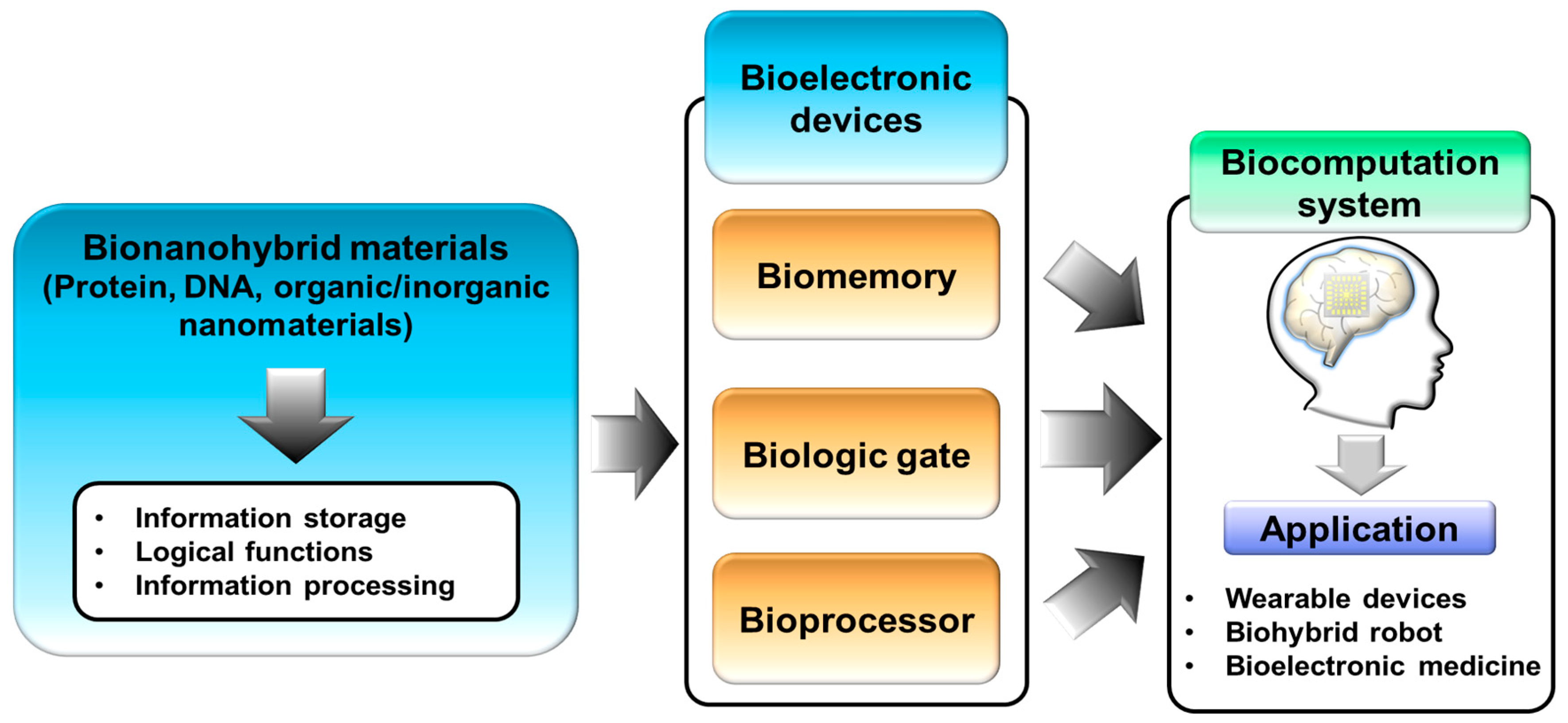
Figure 2.
Multilevel biomemory device. (A) Schematic image demonstrating a multilevel biomemory device using metal ions states to control two different kinds of metalloprotein. (B) Cyclic voltammogram of a multilevel biomemory device composed of recombinant azurin and cytochrome c that shows two apparently distinguished reduction potential peaks and two oxidation potential peaks. (C) Memory performance of a multilevel biomemory device including writing, reading, and erasing steps by applying the potential values of reduction and oxidation potential peak values and the OCP values of metalloproteins. (Reproduced with permission from [43], published by John Wiley and Sons, 2010).

Figure 3.
Electrochemical signal enhanced biomemory device. (A) Schematic image of the biomemory device composed of Azu and gold nanoparticles (GNP). (B) Redox potential peak values for optimizing the GNP diameter. (C) Cyclic voltammogram of Azu–GNP and Azu. (D) memory performance of Azu–GNP and Azu. (Reproduced with permission from [17], published by John Wiley and Sons, 2011).

Figure 4.
Resistive biomemory device. (A) Schematic image of a resistive biomemory device composed of pRNA-3WJ and quantum dot (QD) on a gold substrate, (B) I–V curves of bare Au, pRNA-3WJ, QD and pRNA-3WJ, and QD. (C) Resistive switching function and stability test for a resistive biomemory device composed of MoS2 and DNA on a gold substrate with apparently distinguished resistance states and long-term stability for 10 days. (Reproduced with permission from [50], published by the American Chemical Society, 2015, and reproduced with permission from [52], published by Elsevier, 2019).

Figure 5.
Biologic gates. (A) Schematic image of a DNA-based biologic gate based on metal ions inserted inside mismatched DNA pairs and differential pulse voltammetry (DPV) results of this device by controlling output signals through Ag+ and Hg2+ ions inserted inside mismatched DNA pairs. (B) Schematic image of a protein/DNA-based biologic gate through the signal transduction of a protein-based biologic gate to a DNA-based biologic gate for the final outputted fluorescence signal (Reproduced with permission from [64], published by John Wiley and Sons, 2013 and reproduced with permission from [65], published by John Wiley and Sons, 2016).
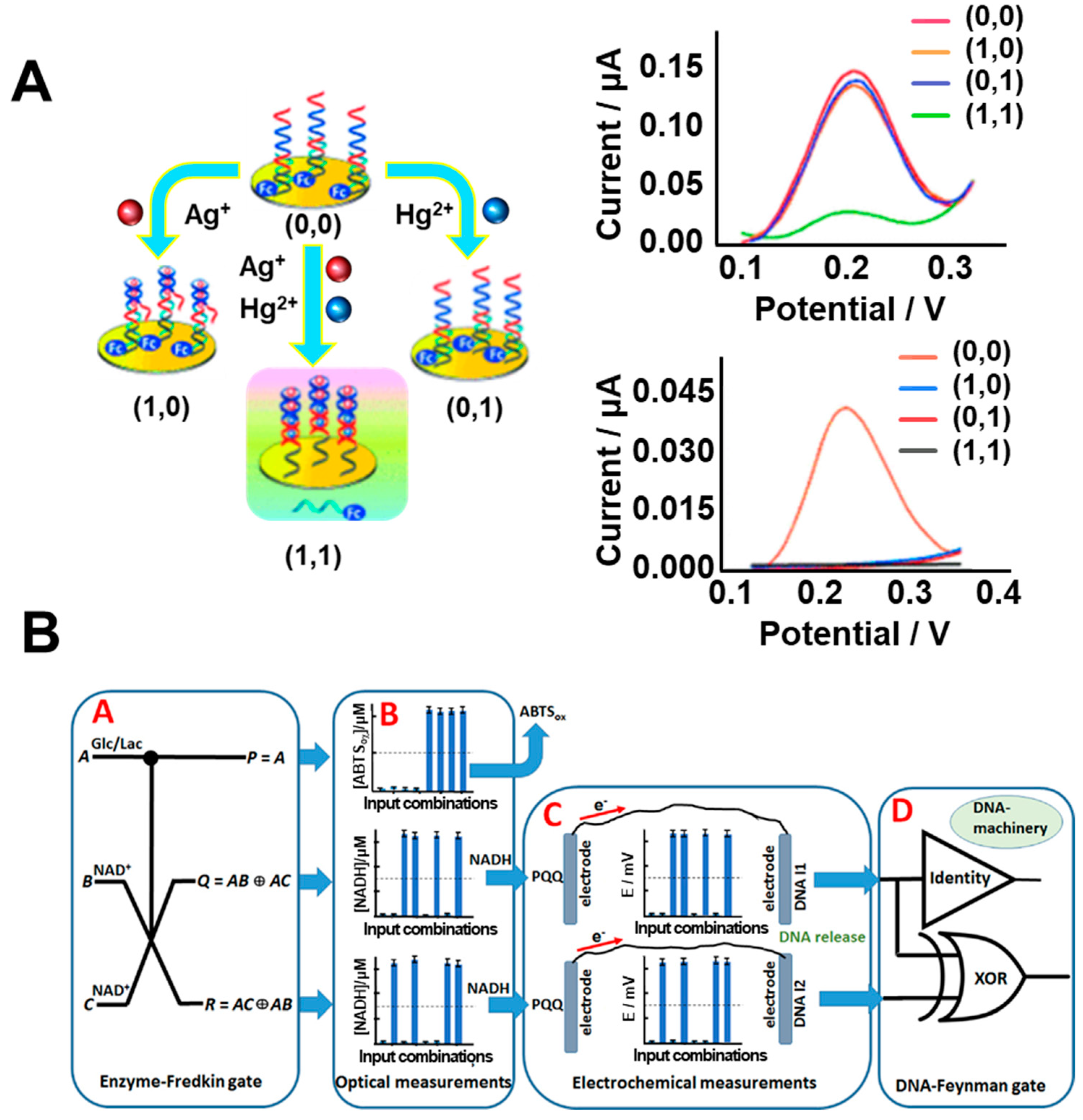
Figure 6.
Analog decision mimicking bioelectronic device. (A) Schematic image and theory of this bioelectronic device through the analogously processed output signals by two different external factors inputted (negative input and positive input) by electrochemical investigation. (B) Bionanohybrid material used for this device composed of metalloprotein used as signal generator, organic chemical linkers as signal controller, and inorganic materials used for signal modulation. (C) The plotted results of analog decision-making based on the analysis of electrochemical signal by defined external factors showed the decision variation of 12 persons based on the defined threshold values. (Reproduced with permission from [59], the figures follow the terms of use under a Creative Commons Attribution 4.0 International License.).

Figure 7.
Bioprocessors. (A) Schematic image and electrophoresis results of DNA-based bioprocessor composed of six stages, including the first stage as problem encoder, the second stage as DNA solution bay for converted DNA preparation, the third as mixing controller for mixing and ligase of appropriate DNA sequences to make the template of DNA duplexes, the fourth as solution purifier for isolation of optimal DNA template from impurities such as the incompletely hybridized oligonucleotides or enzymes, the fifth as PCR amplifier for amplification of optimal DNA template which is the optimal route, and the sixth as gel electrophoresis to acquire the final electrophoresis data for solving optimal-route-planning problems, (B) Schematic image of bioprocessor based on bionanohybrid materials to demonstrate the specific processing functions including the electrochemical signal reinforcement, regulation, and amplification. (Reproduced with permission from [82], published by American Chemical Society, 2015, and reproduced with permission from [31], published by John Wiley and Sons, 2013).
© 2019 by the authors. Licensee MDPI, Basel, Switzerland. This article is an open access article distributed under the terms and conditions of the Creative Commons Attribution (CC BY) license (http://creativecommons.org/licenses/by/4.0/).
Share and Cite
MDPI and ACS Style
Yoon, J.; Lee, T.; Choi, J.-W. Development of Bioelectronic Devices Using Bionanohybrid Materials for Biocomputation System. Micromachines 2019, 10, 347. https://doi.org/10.3390/mi10050347
AMA Style
Yoon J, Lee T, Choi J-W. Development of Bioelectronic Devices Using Bionanohybrid Materials for Biocomputation System. Micromachines. 2019; 10(5):347. https://doi.org/10.3390/mi10050347
Chicago/Turabian StyleYoon, Jinho, Taek Lee, and Jeong-Woo Choi. 2019. "Development of Bioelectronic Devices Using Bionanohybrid Materials for Biocomputation System" Micromachines 10, no. 5: 347. https://doi.org/10.3390/mi10050347
Note that from the first issue of 2016, this journal uses article numbers instead of page numbers. See further details here.