Next Issue
Volume 10, September
Previous Issue
Volume 10, July


Cancers, Volume 10, Issue 8 (August 2018) – 36 articles

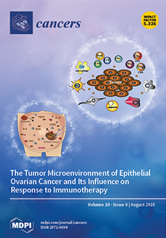
Cover Story (view full-size image): The tumor microenvironment (TME) of epithelial ovarian cancer is a complex network of cancer cells surrounded by immune cells, such as T (CD4+, CD8+) and B lymphocytes (B cells); regulatory lymphocytes (CD4+ Treg, CD8+ Treg and Breg cells); natural killer cells (NKs); pro-inflammatory type 1 macrophages (M1) and tumor associated macrophages (TAMs); dendritic cells (DCs); myeloid-derived suppressor cells (MDSCs); and other components including cancer associated fibroblasts (CAFs) and the adipocytes in the omentum. Primary tumors, metastasis, and ascites are enriched with different components, making each TME a unique case. In this issue, we describe the composition of the TME, how it can influence antitumoral responses, and current EOC models. View this paper
  • Issues are regarded as officially published after their release is announced to the table of contents alert mailing list.
  • You may sign up for e-mail alerts to receive table of contents of newly released issues.
  • PDF is the official format for papers published in both, html and pdf forms. To view the papers in pdf format, click on the "PDF Full-text" link, and use the free Adobe Reader to open them.
Order results
Result details
Section
Select all
Export citation of selected articles as:
15 pages, 2051 KiB  
Article
Sperm Protein 17 Expression by Murine Epithelial Ovarian Cancer Cells and Its Impact on Tumor Progression
by Qian Gao, Sue D. Xiang, Kirsty Wilson, Mutsa Madondo, Andrew N. Stephens and Magdalena Plebanski
Cancers 2018, 10(8), 276; https://doi.org/10.3390/cancers10080276 - 20 Aug 2018
Cited by 11 | Viewed by 4445
Abstract
The cancer testis antigen sperm protein 17 (Sp17) is a promising antigenic target in epithelial ovarian cancer (EOC) vaccine development. However, its role in ovarian cancer is unclear. We isolated and expanded Sp17+ and Sp17 clones from the murine EOC cell [...] Read more.
The cancer testis antigen sperm protein 17 (Sp17) is a promising antigenic target in epithelial ovarian cancer (EOC) vaccine development. However, its role in ovarian cancer is unclear. We isolated and expanded Sp17+ and Sp17 clones from the murine EOC cell line ID8, and compared their in-vitro cell growth characteristics and in-vivo tumorigenicity. We also examined the potential co-expression of molecules that may influence cancer cell survival and interaction with immune cells. These include stimulatory and immunosuppressive molecules, such as major histocompatibility class I molecules (MHC I), MHC II, cytotoxic T lymphocyte associated antigen-4 (CTLA-4), CD73, CD39, tumor necrosis factor receptor II (TNFRII), signal transducer and activator of transcription 3 (STAT3) and programmed death-ligand 1 (PD-L1). Whilst the presence of Sp17 was not correlated with the ID8 cell proliferation/growth capacity in vitro, it was critical to enable progressive tumor formation in vivo. Flow cytometry revealed that Sp17+ ID8 cells displayed higher expression of both STAT3 and PD-L1, whilst MHC II expression was lower. Moreover, Sp17high (PD-L1+MHCII) cell populations showed significantly enhanced resistance to Paclitaxel-induced cell death in vitro compared to Sp17low (PD-L1MHCII+) cells, which was associated in turn with increased STAT3 expression. Together, the data support Sp17 as a factor associated with in-vivo tumor progression and chemo-resistance, validating it as a suitable target for vaccine development. Full article
(This article belongs to the Special Issue Cancer Vaccines: Research and Applications)
Show Figures

Figure 1

13 pages, 1432 KiB  
Article
The Impact of p53 Dysfunction in ATR Inhibitor Cytotoxicity and Chemo- and Radiosensitisation
by Fiona K. Middleton, John R. Pollard and Nicola J. Curtin
Cancers 2018, 10(8), 275; https://doi.org/10.3390/cancers10080275 - 20 Aug 2018
Cited by 28 | Viewed by 5049
Abstract
Ataxia telangiectasia mutated and Rad3 related kinase (ATR) signals replication stress and DNA damage to S and G2 arrest and promotes DNA repair. Mutations in p53, critical for G1 checkpoint control, are common in cancer and predicted to confer vulnerability to ATR inhibitors. [...] Read more.
Ataxia telangiectasia mutated and Rad3 related kinase (ATR) signals replication stress and DNA damage to S and G2 arrest and promotes DNA repair. Mutations in p53, critical for G1 checkpoint control, are common in cancer and predicted to confer vulnerability to ATR inhibitors. Reported data on the impact of p53 status are variable possibly because of the use of unmatched cells and surrogate endpoints of survival. The cytotoxicity of VE-821 alone and its ability to potentiate radiation and gemcitabine cytotoxicity was determined in isogenic and unmatched p53 wild-type (wt) and null/mutant cells, as well as immortalised nonmalignant MCF10 (immortalised non-neoplastic) cells, by colony-forming assay. The effect on cell cycle checkpoints was determined by flow cytometry. The isogenic p53 defective cells were not more sensitive to VE-821 alone. Defective p53 consistently conferred greater chemo- and radiosensitisation, particularly at high dose levels in isogenic cells but not unmatched cells. VE-821 did not sensitise MCF10 cells. We conclude that p53 status is just one factor contributing to chemo- and radiosensitisation by ATR inhibition, the lack of chemo- or radiosensitisation in the noncancerous cells suggests an element of tumour-specificity that warrants further investigation. The greater sensitisation at high-dose irradiation suggests that ATR inhibitors may be most effective with hypofractionated radiotherapy. Full article
(This article belongs to the Special Issue New Developments in Radiotherapy)
Show Figures

Figure 1

21 pages, 3134 KiB  
Article
Genetics and Expression Profile of the Tubulin Gene Superfamily in Breast Cancer Subtypes and Its Relation to Taxane Resistance
by Babak Nami and Zhixiang Wang
Cancers 2018, 10(8), 274; https://doi.org/10.3390/cancers10080274 - 18 Aug 2018
Cited by 75 | Viewed by 7695
Abstract
Taxanes are a class of chemotherapeutic agents that inhibit cell division by disrupting the mitotic spindle through the stabilization of microtubules. Most breast cancer (BC) tumors show resistance against taxanes partially due to alterations in tubulin genes. In this project we investigated tubulin [...] Read more.
Taxanes are a class of chemotherapeutic agents that inhibit cell division by disrupting the mitotic spindle through the stabilization of microtubules. Most breast cancer (BC) tumors show resistance against taxanes partially due to alterations in tubulin genes. In this project we investigated tubulin isoforms in BC to explore any correlation between tubulin alterations and taxane resistance. Genetic alteration and expression profiling of 28 tubulin isoforms in 6714 BC tumor samples from 4205 BC cases were analyzed. Protein-protein, drug-protein and alterations neighbor genes in tubulin pathways were examined in the tumor samples. To study correlation between promoter activity and expression of the tubulin isoforms in BC, we analyzed the ChIP-seq enrichment of active promoter histone mark H3K4me3 and mRNA expression profile of MCF-7, ZR-75-30, SKBR-3 and MDA-MB-231 cell lines. Potential correlation between tubulin alterations and taxane resistance, were investigated by studying the expression profile of taxane-sensitive and resistant BC tumors also the MDA-MB-231 cells acquired resistance to paclitaxel. All genomic data were obtained from public databases. Results showed that TUBD1 and TUBB3 were the most frequently amplified and deleted tubulin genes in the BC tumors respectively. The interaction analysis showed physical interactions of α-, β- and γ-tubulin isoforms with each other. The most of FDA-approved tubulin inhibitor drugs including taxanes target only β-tubulins. The analysis also revealed sex tubulin-interacting neighbor proteins including ENCCT3, NEK2, PFDN2, PTP4A3, SDCCAG8 and TBCE which were altered in at least 20% of the tumors. Three of them are tubulin-specific chaperons responsible for tubulin protein folding. Expression of tubulin genes in BC cell lines were correlated with H3K4me3 enrichment on their promoter chromatin. Analyzing expression profile of BC tumors and tumor-adjacent normal breast tissues showed upregulation of TUBA1A, TUBA1C, TUBB and TUBB3 and downregulation of TUBB2A, TUBB2B, TUBB6, TUBB7P pseudogene, and TUBGCP2 in the tumor tissues compared to the normal breast tissues. Analyzing taxane-sensitive versus taxane-resistant tumors revealed that expression of TUBB3 and TUBB6 was significantly downregulated in the taxane-resistant tumors. Our results suggest that downregulation of tumor βIII- and βV-tubulins is correlated with taxane resistance in BC. Based on our results, we conclude that aberrant protein folding of tubulins due to mutation and/or dysfunction of tubulin-specific chaperons may be potential mechanisms of taxane resistance. Thus, we propose studying the molecular pathology of tubulin mutations and folding in BC and their impacts on taxane resistance. Full article
(This article belongs to the Special Issue Drug Resistance in Cancers)
Show Figures

Figure 1

18 pages, 1760 KiB  
Article
Functional Linkage of RKIP to the Epithelial to Mesenchymal Transition and Autophagy during the Development of Prostate Cancer
by Mahmoud Ahmed, Trang Huyen Lai, Sahib Zada, Jin Seok Hwang, Trang Minh Pham, Miyong Yun and Deok Ryong Kim
Cancers 2018, 10(8), 273; https://doi.org/10.3390/cancers10080273 - 16 Aug 2018
Cited by 29 | Viewed by 4592
Abstract
Raf kinase inhibitor protein (RKIP) plays a critical role in many signaling pathways as a multi-functional adapter protein. In particular, the loss of RKIP’s function in certain types of cancer cells results in epithelial to mesenchymal transition (EMT) and the promotion of cancer [...] Read more.
Raf kinase inhibitor protein (RKIP) plays a critical role in many signaling pathways as a multi-functional adapter protein. In particular, the loss of RKIP’s function in certain types of cancer cells results in epithelial to mesenchymal transition (EMT) and the promotion of cancer metastasis. In addition, RKIP inhibits autophagy by modulating LC3-lipidation and mTORC1. How the RKIP-dependent inhibition of autophagy is linked to EMT and cancer progression is still under investigation. In this study, we investigated the ways by which RKIP interacts with key gene products in EMT and autophagy during the progression of prostate cancer. We first identified the gene products of interest using the corresponding gene ontology terms. The weighted-gene co-expression network analysis (WGCNA) was applied on a gene expression dataset from three groups of prostate tissues; benign prostate hyperplasia, primary and metastatic cancer. We found two modules of highly co-expressed genes, which were preserved in other independent datasets of prostate cancer tissues. RKIP showed potentially novel interactions with one EMT and seven autophagy gene products (TGFBR1; PIK3C3, PIK3CB, TBC1D25, TBC1D5, TOLLIP, WDR45 and WIPI1). In addition, we identified several upstream transcription modulators that could regulate the expression of these gene products. Finally, we verified some RKIP novel interactions by co-localization using the confocal microscopy analysis in a prostate cancer cell line. To summarize, RKIP interacts with EMT and autophagy as part of the same functional unit in developing prostate cancer. Full article
(This article belongs to the Special Issue Targeting Raf Kinase Inhibitor Protein (RKIP) in Cancer)
Show Figures

Figure 1

14 pages, 2577 KiB  
Article
miR-1246 Targets CCNG2 to Enhance Cancer Stemness and Chemoresistance in Oral Carcinomas
by Shih-Shen Lin, Chih-Yu Peng, Yi-Wen Liao, Ming-Yung Chou, Pei-Ling Hsieh and Cheng-Chia Yu
Cancers 2018, 10(8), 272; https://doi.org/10.3390/cancers10080272 - 16 Aug 2018
Cited by 54 | Viewed by 4848
Abstract
MiRNAs have been recognized as crucial components in carcinogenesis, but whether miR-1246 affects the cancer stemness and drug resistance in oral squamous cell carcinoma (OSCC) has not been fully understood and its downstream targets still need to be unraveled. In the present work, [...] Read more.
MiRNAs have been recognized as crucial components in carcinogenesis, but whether miR-1246 affects the cancer stemness and drug resistance in oral squamous cell carcinoma (OSCC) has not been fully understood and its downstream targets still need to be unraveled. In the present work, we employed miRNAs RT-PCR analysis to evaluate the expression of miR-1246 in tumor tissues and oral cancer stem cells (OCSC). Stemness phenotypes, including self-renewal, migration, invasion, colony formation capacities, and in vivo oncogenicity of oral cancer cells following transfected with miR-1246 inhibitors or mimics were examined. Our results suggested that the expression level of miR-1246 was significantly upregulated in the tumor tissues and OCSC. Kaplan-Meier survival analysis of OSCC patients with high levels of miR-1246 had the worst survival rate compared to their low-expression counterparts. Inhibition of miR-1246 in OCSC significantly reduced the stemness hallmarks, while overexpression of miR-1246 enhanced these characteristics. Moreover, we showed that downregulation of miR-1246 decreased chemoresistance. In addition, we verified that miR-1246-inhibited CCNG2 contributed to the cancer stemness of OSCC. These results demonstrated the significance of miR-1246 in the regulation of OSCC stemness. Targeting miR-1246-CCNG2 axis may be beneficial to suppress cancer relapse and metastasis in OSCC patients. Full article
(This article belongs to the Special Issue Cancer Chemoresistance)
Show Figures

Figure 1

11 pages, 257 KiB  
Review
The Role of Direct Oral Anticoagulants in Treatment of Cancer-Associated Thrombosis
by Hanny Al-Samkari and Jean M. Connors
Cancers 2018, 10(8), 271; https://doi.org/10.3390/cancers10080271 - 15 Aug 2018
Cited by 32 | Viewed by 5794
Abstract
Venous thromboembolism (VTE) complicates the clinical course of approximately 5–10% of all cancer patients. Anticoagulation of the cancer patient often presents unique challenges as these patients have both a higher risk of recurrent VTE and a higher risk of bleeding than patients without [...] Read more.
Venous thromboembolism (VTE) complicates the clinical course of approximately 5–10% of all cancer patients. Anticoagulation of the cancer patient often presents unique challenges as these patients have both a higher risk of recurrent VTE and a higher risk of bleeding than patients without cancer. Although low molecular weight heparins (LMWH) are the standard of care for the management of cancer-associated VTE, their use requires once or twice daily subcutaneous injections, which can be a significant burden for many cancer patients who often require a long duration of anticoagulation. The direct oral anticoagulants (DOACs) are attractive options for patients with malignancy. DOACs offer immediate onset of action and short half-lives, properties similar to LMWH, but the oral route of administration is a significant advantage. Given the higher risks of recurrent VTE and bleeding, there has been concern about the efficacy and safety of DOACs in this patient population. Data are now emerging for the use of DOACs in the cancer patient population from dedicated clinical trials. While recently published data suggest that DOACs hold promise for the treatment of cancer associated VTE, additional studies are needed to establish DOACs as the standard-of-care treatment. Many such studies are currently underway. The available data for the use of DOACs in the treatment of cancer-associated VTE will be reviewed, focusing on efficacy, safety, and other considerations relevant to the cancer patient. Full article
(This article belongs to the Special Issue The Role of Thrombosis and Haemostasis in Cancer)
17 pages, 3225 KiB  
Article
Assessment of the Mutational Status of NSCLC Using Hypermetabolic Circulating Tumor Cells
by Matteo Turetta, Michela Bulfoni, Giulia Brisotto, Gianpiero Fasola, Andrea Zanello, Eva Biscontin, Laura Mariuzzi, Agostino Steffan, Carla Di Loreto, Daniela Cesselli and Fabio Del Ben
Cancers 2018, 10(8), 270; https://doi.org/10.3390/cancers10080270 - 14 Aug 2018
Cited by 13 | Viewed by 4097
Abstract
Molecular characterization is currently a key step in NSCLC therapy selection. Circulating tumor cells (CTC) are excellent candidates for downstream analysis, but technology is still lagging behind. In this work, we show that the mutational status of NSCLC can be assessed on hypermetabolic [...] Read more.
Molecular characterization is currently a key step in NSCLC therapy selection. Circulating tumor cells (CTC) are excellent candidates for downstream analysis, but technology is still lagging behind. In this work, we show that the mutational status of NSCLC can be assessed on hypermetabolic CTC, detected by their increased glucose uptake. We validated the method in 30 Stage IV NSCLC patients: peripheral blood samples were incubated with a fluorescent glucose analog (2-NBDG) and analyzed by flow cytometry. Cells with the highest glucose uptake were sorted out. EGFR and KRAS mutations were detected by ddPCR. In sorted cells, mutated DNA was found in 85% of patients, finding an exact match with primary tumor in 70% of cases. Interestingly, in two patients multiple KRAS mutations were detected. Two patients displayed different mutations with respect to the primary tumor, and in two out of the four patients with a wild type primary tumor, new mutations were highlighted: EGFR p.746_750del and KRAS p.G12V. Hypermetabolic CTC can be enriched without the need of dedicated equipment and their mutational status can successfully be assessed by ddPCR. Finally, the finding of new mutations supports the possibility of probing tumor heterogeneity. Full article
(This article belongs to the Special Issue Circulating Tumor Cells (CTCs))
Show Figures

Graphical abstract

20 pages, 5582 KiB  
Article
4-Acetyl-Antroquinonol B Suppresses SOD2-Enhanced Cancer Stem Cell-Like Phenotypes and Chemoresistance of Colorectal Cancer Cells by Inducing hsa-miR-324 re-Expression
by Oluwaseun Adebayo Bamodu, Ching-Kuo Yang, Wei-Hong Cheng, David T.W. Tzeng, Kuang-Tai Kuo, Chun-Chih Huang, Li Deng, Michael Hsiao, Wei-Hwa Lee and Chi-Tai Yeh
Cancers 2018, 10(8), 269; https://doi.org/10.3390/cancers10080269 - 10 Aug 2018
Cited by 30 | Viewed by 4858
Abstract
Background: Colorectal cancer (CRC) remains a leading cause of cancer-related morbidity and mortality in both sexes globally. This is not unconnected with the heterogeneity and plasticity of CRC stem cells (CRC-SCs) which stealthily exploit the niche-related and (epi)genetic factors to facilitate metastasis, chemoresistance, [...] Read more.
Background: Colorectal cancer (CRC) remains a leading cause of cancer-related morbidity and mortality in both sexes globally. This is not unconnected with the heterogeneity and plasticity of CRC stem cells (CRC-SCs) which stealthily exploit the niche-related and (epi)genetic factors to facilitate metastasis, chemoresistance, tumor recurrence, and disease progression. Despite the accumulating evidence of the role of dysregulated microRNAs in malignancies, the therapeutic efficacy of pharmacological-targeting of CRC-SC-associated microRNAs is relatively under-explored. Experimental approach: In this present study, we employed relatively new bioinformatics approaches, analyses of microarray data, Western blot, real-time polymerase chain reaction (RT-PCR), and functional assays to show that hsa-miR-324-5p expression is significantly suppressed in CRC cells, and inversely correlates with the aberrant expression of SOD2. Results: This converse hsa-miR-324-5p/SOD2 relationship is associated with enhanced oncogenicity, which is effectively inhibited by 4-acetylantroquinonol B (4-AAQB), as evidenced by inhibited cell viability and proliferation, as well as attenuated migration, invasion, and clonogenicity in 4-AAQB-treated DLD1 and HCT116 cells. Interestingly, 4-AAQB did not affect the viability and proliferation of normal colon cells. We also showed that 4-AAQB-induced re-expression of hsa-miR-324-5p, akin to short-interfering RNA, reduced SOD2 expression, correlates with the concurrent down-regulation of SOD2, N-cadherin, vimentin, c-Myc, and BcL-xL2, with concomitant up-regulation of E-cadherin and BAX2 proteins. Enhanced expression of hsa-miR-324-5p in the CRC cells suppressed their tumorigenicity in vitro and in vivo. Additionally, 4-AAQB synergistically potentiates the FOLFOX (folinate (leucovorin), fluorouracil (5FU), and oxaliplatin) anticancer effect by eliciting the re-expression of SOD2-suppressed hsa-miR-324, and inhibiting SOD2-mediated tumorigenicity. Conclusion: Our findings highlight the pre-clinical anti-CSC efficacy of 4-AAQB, with or without FOLFOX in CRC, and suggest a potential novel therapeutic strategy for CRC patients. Full article
Show Figures

Figure 1

25 pages, 1924 KiB  
Review
The Methylation Status of the Epigenome: Its Emerging Role in the Regulation of Tumor Angiogenesis and Tumor Growth, and Potential for Drug Targeting
by Luciano Pirola, Oskar Ciesielski and Aneta Balcerczyk
Cancers 2018, 10(8), 268; https://doi.org/10.3390/cancers10080268 - 10 Aug 2018
Cited by 32 | Viewed by 6948
Abstract
Approximately 50 years ago, Judah Folkman raised the concept of inhibiting tumor angiogenesis for treating solid tumors. The development of anti-angiogenic drugs would decrease or even arrest tumor growth by restricting the delivery of oxygen and nutrient supplies, while at the same time [...] Read more.
Approximately 50 years ago, Judah Folkman raised the concept of inhibiting tumor angiogenesis for treating solid tumors. The development of anti-angiogenic drugs would decrease or even arrest tumor growth by restricting the delivery of oxygen and nutrient supplies, while at the same time display minimal toxic side effects to healthy tissues. Bevacizumab (Avastin)—a humanized monoclonal anti VEGF-A antibody—is now used as anti-angiogenic drug in several forms of cancers, yet with variable results. Recent years brought significant progresses in our understanding of the role of chromatin remodeling and epigenetic mechanisms in the regulation of angiogenesis and tumorigenesis. Many inhibitors of DNA methylation as well as of histone methylation, have been successfully tested in preclinical studies and some are currently undergoing evaluation in phase I, II or III clinical trials, either as cytostatic molecules—reducing the proliferation of cancerous cells—or as tumor angiogenesis inhibitors. In this review, we will focus on the methylation status of the vascular epigenome, based on the genomic DNA methylation patterns with DNA methylation being mainly transcriptionally repressive, and lysine/arginine histone post-translational modifications which either promote or repress the chromatin transcriptional state. Finally, we discuss the potential use of “epidrugs” in efficient control of tumor growth and tumor angiogenesis. Full article
(This article belongs to the Special Issue Tumor Angiogenesis: An Update)
Show Figures

Figure 1

13 pages, 263 KiB  
Review
The Current Role of Salvage Surgery in Recurrent Head and Neck Squamous Cell Carcinoma
by Marc Hamoir, Sandra Schmitz, Carlos Suarez, Primoz Strojan, Kate A Hutcheson, Juan P Rodrigo, William M Mendenhall, Ricard Simo, Nabil F Saba, Anil K D‘Cruz, Missak Haigentz, Jr., Carol R Bradford, Eric M Genden, Alessandra Rinaldo and Alfio Ferlito
Cancers 2018, 10(8), 267; https://doi.org/10.3390/cancers10080267 - 10 Aug 2018
Cited by 78 | Viewed by 5437
Abstract
Chemoradiotherapy has emerged as a gold standard in advanced squamous cell carcinoma of the head and neck (SCCHN). Because 50% of advanced stage patients relapse after nonsurgical primary treatment, the role of salvage surgery (SS) is critical because surgery is generally regarded as [...] Read more.
Chemoradiotherapy has emerged as a gold standard in advanced squamous cell carcinoma of the head and neck (SCCHN). Because 50% of advanced stage patients relapse after nonsurgical primary treatment, the role of salvage surgery (SS) is critical because surgery is generally regarded as the best treatment option in patients with recurrent resectable SCCHN. Surgeons are increasingly confronted with considering operation among patients with significant effects of failed non-surgical primary treatment. Wide local excision to achieve clear margins must be balanced with the morbidity of the procedure, the functional consequences of organ mutilation, and the likelihood of success. Accurate selection of patients suitable for surgery is a major issue. It is essential to establish objective criteria based on functional and oncologic outcomes to select the best candidates for SS. The authors propose first to understand preoperative prognostic factors influencing survival. Predictive modeling based on preoperative information is now available to better select patients having a good chance to be successfully treated with surgery. Patients with a high comorbidity index, advanced oropharyngeal or hypopharyngeal primary tumors, and both local and regional recurrence have a very limited likelihood of success with salvage surgery and should be strongly considered for other treatments. Following SS, identifying patients with postoperative prognostic factors predicting high risk of recurrence is essential because those patients could benefit of adjuvant treatment or be included in clinical trials. Finally, defining HPV tumor status is needed in future studies including recurrent oropharyngeal SCC patients. Full article
22 pages, 2399 KiB  
Review
Targeting the Microenvironment in High Grade Serous Ovarian Cancer
by Nkechiyere G. Nwani, Livia E. Sima, Wilberto Nieves-Neira and Daniela Matei
Cancers 2018, 10(8), 266; https://doi.org/10.3390/cancers10080266 - 10 Aug 2018
Cited by 31 | Viewed by 5412
Abstract
Cancer–stroma interactions play a key role in cancer progression and response to standard chemotherapy. Here, we provide a summary of the mechanisms by which the major cellular components of the ovarian cancer (OC) tumor microenvironment (TME) including cancer-associated fibroblasts (CAFs), myeloid, immune, endothelial, [...] Read more.
Cancer–stroma interactions play a key role in cancer progression and response to standard chemotherapy. Here, we provide a summary of the mechanisms by which the major cellular components of the ovarian cancer (OC) tumor microenvironment (TME) including cancer-associated fibroblasts (CAFs), myeloid, immune, endothelial, and mesothelial cells potentiate cancer progression. High-grade serous ovarian cancer (HGSOC) is characterized by a pro-inflammatory and angiogenic signature. This profile is correlated with clinical outcomes and can be a target for therapy. Accumulation of malignant ascites in the peritoneal cavity allows for secreted factors to fuel paracrine and autocrine circuits that augment cancer cell proliferation and invasiveness. Adhesion of cancer cells to the mesothelial matrix promotes peritoneal tumor dissemination and represents another attractive target to prevent metastasis. The immunosuppressed tumor milieu of HGSOC is permissive for tumor growth and can be modulated therapeutically. Results of emerging preclinical and clinical trials testing TME-modulating therapeutics for the treatment of OC are highlighted. Full article
(This article belongs to the Special Issue The Tumor Microenvironment of High Grade Serous Ovarian Cancer)
Show Figures

Figure 1

10 pages, 4783 KiB  
Review
Organotypic 3D Models of the Ovarian Cancer Tumor Microenvironment
by Karen M. Watters, Preety Bajwa and Hilary A. Kenny
Cancers 2018, 10(8), 265; https://doi.org/10.3390/cancers10080265 - 09 Aug 2018
Cited by 37 | Viewed by 6834
Abstract
Ovarian cancer progression involves multifaceted and variable tumor microenvironments (TMEs), from the in situ carcinoma in the fallopian tube or ovary to dissemination into the peritoneal cavity as single cells or spheroids and attachment to the mesothelial-lined surfaces of the omentum, bowel, and [...] Read more.
Ovarian cancer progression involves multifaceted and variable tumor microenvironments (TMEs), from the in situ carcinoma in the fallopian tube or ovary to dissemination into the peritoneal cavity as single cells or spheroids and attachment to the mesothelial-lined surfaces of the omentum, bowel, and abdominal wall. The TME comprises the tumor vasculature and lymphatics (including endothelial cells and pericytes), in addition to mesothelial cells, fibroblasts, immune cells, adipocytes and extracellular matrix (ECM) proteins. When generating 3D models of the ovarian cancer TME, researchers must incorporate the most relevant stromal components depending on the TME in question (e.g., early or late disease). Such complexity cannot be captured by monolayer 2D culture systems. Moreover, immortalized stromal cell lines, such as mesothelial or fibroblast cell lines, do not always behave the same as primary cells whose response in functional assays may vary from donor to donor; 3D models with primary stromal cells may have more physiological relevance than those using stromal cell lines. In the current review, we discuss the latest developments in organotypic 3D models of the ovarian cancer early metastatic microenvironment. Organotypic culture models comprise two or more interacting cell types from a particular tissue. We focus on organotypic 3D models that include at least one type of primary stromal cell type in an ECM background, such as collagen or fibronectin, plus ovarian cancer cells. We provide an overview of the two most comprehensive current models—a 3D model of the omental mesothelium and a microfluidic model. We describe the cellular and non-cellular components of the models, the incorporation of mechanical forces, and how the models have been adapted and utilized in functional assays. Finally, we review a number of 3D models that do not incorporate primary stromal cells and summarize how integration of current models may be the next essential step in tackling the complexity of the different ovarian cancer TMEs. Full article
(This article belongs to the Special Issue The Tumor Microenvironment of High Grade Serous Ovarian Cancer)
Show Figures

Graphical abstract

20 pages, 1694 KiB  
Review
The SUMO System and TGFβ Signaling Interplay in Regulation of Epithelial-Mesenchymal Transition: Implications for Cancer Progression
by Ayan Chanda, Anusi Sarkar and Shirin Bonni
Cancers 2018, 10(8), 264; https://doi.org/10.3390/cancers10080264 - 08 Aug 2018
Cited by 22 | Viewed by 4813
Abstract
Protein post-translational modification by the small ubiquitin-like modifier (SUMO), or SUMOylation, can regulate the stability, subcellular localization or interactome of a protein substrate with key consequences for cellular processes including the Epithelial-Mesenchymal Transition (EMT). The secreted protein Transforming Growth Factor beta (TGFβ) is [...] Read more.
Protein post-translational modification by the small ubiquitin-like modifier (SUMO), or SUMOylation, can regulate the stability, subcellular localization or interactome of a protein substrate with key consequences for cellular processes including the Epithelial-Mesenchymal Transition (EMT). The secreted protein Transforming Growth Factor beta (TGFβ) is a potent inducer of EMT in development and homeostasis. Importantly, the ability of TGFβ to induce EMT has been implicated in promoting cancer invasion and metastasis, resistance to chemo/radio therapy, and maintenance of cancer stem cells. Interestingly, TGFβ-induced EMT and the SUMO system intersect with important implications for cancer formation and progression, and novel therapeutics identification. Full article
(This article belongs to the Special Issue TGF-Beta Signaling in Cancer)
Show Figures

Figure 1

14 pages, 3349 KiB  
Article
Real-World Implications of Nonbiological Factors with Staging, Prognosis and Clinical Management in Colon Cancer
by Qi Liu, Dakui Luo, Sanjun Cai, Qingguo Li and Xinxiang Li
Cancers 2018, 10(8), 263; https://doi.org/10.3390/cancers10080263 - 08 Aug 2018
Cited by 14 | Viewed by 3894
Abstract
Background: The present study analyzed the nonbiological factors (NBFs) together with the American Joint Committee on Cancer (AJCC) Tumor-Node-Metastasis (TNM) staging system to generate a refined, risk-adapted stage for the clinical treatment of colon cancer. Methods: Eligible patients (N = 28,818) with [...] Read more.
Background: The present study analyzed the nonbiological factors (NBFs) together with the American Joint Committee on Cancer (AJCC) Tumor-Node-Metastasis (TNM) staging system to generate a refined, risk-adapted stage for the clinical treatment of colon cancer. Methods: Eligible patients (N = 28,818) with colon cancer between 1 January 2010 and 31 December 2014, were identified from the Surveillance, Epidemiology, and End Results (SEER) database. Kaplan-Meier curves and Cox proportional hazards regression, analyzed the probabilities of cancer-specific survival (CSS) in patients with colon cancer, with different NBF-TNM stages. Results: Insurance status, marital status, and median household income were significant prognostic NBFs in the current study (p < 0.05). The concordance index of NBF-TNM stage was 0.857 (95% confidence interval (CI) = 0.8472–0.8668). Multivariate Cox analyses, indicated that NBF1-stage was independently associated with a 50.4% increased risk of cancer-specific mortality in colon cancer (p < 0.001), which increased to 77.1% in non-metastatic colon cancer. NBF0-stage improved in CSS as compared to the NBF1-stage in the respective stages (p < 0.05). Conclusions: The new proposed NBF-stage was an independent prognostic factor in colon cancer. Effect of NBFs on the survival of colon cancer necessitates further clinical attention. Moreover, the incorporation of NBF-stage into the AJCC TNM staging system is essential for prognostic prediction, and clinical guidance of adjuvant chemotherapy in stage II and III colon cancer. Full article
Show Figures

Figure 1

11 pages, 376 KiB  
Review
UnPAXing the Divergent Roles of PAX2 and PAX8 in High-Grade Serous Ovarian Cancer
by Laura R. Hardy, Amrita Salvi and Joanna E. Burdette
Cancers 2018, 10(8), 262; https://doi.org/10.3390/cancers10080262 - 08 Aug 2018
Cited by 23 | Viewed by 4959
Abstract
High-grade serous ovarian cancer is a deadly disease that can originate from the fallopian tube or the ovarian surface epithelium. The PAX (paired box) genes PAX2 and PAX8 are lineage-specific transcription factors required during development of the fallopian tube but not in the [...] Read more.
High-grade serous ovarian cancer is a deadly disease that can originate from the fallopian tube or the ovarian surface epithelium. The PAX (paired box) genes PAX2 and PAX8 are lineage-specific transcription factors required during development of the fallopian tube but not in the development of the ovary. PAX2 expression is lost early in serous cancer progression, while PAX8 is expressed ubiquitously. These proteins are implicated in migration, invasion, proliferation, cell survival, stem cell maintenance, and tumor growth. Hence, targeting PAX2 and PAX8 represents a promising drug strategy that could inhibit these pro-tumorigenic effects. In this review, we examine the implications of PAX2 and PAX8 expression in the cell of origin of serous cancer and their potential efficacy as drug targets by summarizing their role in the molecular pathogenesis of ovarian cancer. Full article
(This article belongs to the Special Issue The Tumor Microenvironment of High Grade Serous Ovarian Cancer)
Show Figures

Figure 1

29 pages, 872 KiB  
Review
The Endometriotic Tumor Microenvironment in Ovarian Cancer
by Jillian R. Hufgard Wendel, Xiyin Wang and Shannon M. Hawkins
Cancers 2018, 10(8), 261; https://doi.org/10.3390/cancers10080261 - 07 Aug 2018
Cited by 35 | Viewed by 7445
Abstract
Women with endometriosis are at increased risk of developing ovarian cancer, specifically ovarian endometrioid, low-grade serous, and clear-cell adenocarcinoma. An important clinical caveat to the association of endometriosis with ovarian cancer is the improved prognosis for women with endometriosis at time of ovarian [...] Read more.
Women with endometriosis are at increased risk of developing ovarian cancer, specifically ovarian endometrioid, low-grade serous, and clear-cell adenocarcinoma. An important clinical caveat to the association of endometriosis with ovarian cancer is the improved prognosis for women with endometriosis at time of ovarian cancer staging. Whether endometriosis-associated ovarian cancers develop from the molecular transformation of endometriosis or develop because of the endometriotic tumor microenvironment remain unknown. Additionally, how the presence of endometriosis improves prognosis is also undefined, but likely relies on the endometriotic microenvironment. The unique tumor microenvironment of endometriosis is composed of epithelial, stromal, and immune cells, which adapt to survive in hypoxic conditions with high levels of iron, estrogen, and inflammatory cytokines and chemokines. Understanding the unique molecular features of the endometriotic tumor microenvironment may lead to impactful precision therapies and/or modalities for prevention. A challenge to this important study is the rarity of well-characterized clinical samples and the limited model systems. In this review, we will describe the unique molecular features of endometriosis-associated ovarian cancers, the endometriotic tumor microenvironment, and available model systems for endometriosis-associated ovarian cancers. Continued research on these unique ovarian cancers may lead to improved prevention and treatment options. Full article
(This article belongs to the Special Issue The Tumor Microenvironment of High Grade Serous Ovarian Cancer)
Show Figures

Figure 1

15 pages, 2175 KiB  
Article
LY2157299 Monohydrate, a TGF-βR1 Inhibitor, Suppresses Tumor Growth and Ascites Development in Ovarian Cancer
by Qing Zhang, Xiaonan Hou, Bradley J. Evans, Jamison L. VanBlaricom, Saravut J. Weroha and William A. Cliby
Cancers 2018, 10(8), 260; https://doi.org/10.3390/cancers10080260 - 07 Aug 2018
Cited by 43 | Viewed by 5705
Abstract
Transforming growth factor beta (TGF-β) signaling has pleiotropic functions regulating cancer initiation, development, and metastasis, and also plays important roles in the interaction between stromal and cancer cells, making the pathway a potential therapeutic target. LY2157299 monohydrate (LY), an inhibitor of TGF-β receptor [...] Read more.
Transforming growth factor beta (TGF-β) signaling has pleiotropic functions regulating cancer initiation, development, and metastasis, and also plays important roles in the interaction between stromal and cancer cells, making the pathway a potential therapeutic target. LY2157299 monohydrate (LY), an inhibitor of TGF-β receptor I (TGFBRI), was examined for its ability to inhibit ovarian cancer (OC) growth both in high-grade serous ovarian cancer (HGSOC) cell lines and xenograft models. Immunohistochemistry, qRT-PCR, and Western blot were performed to study the effect of LY treatment on expression of cancer- and fibroblast-derived genes. Results showed that exposure to TGF-β1 induced phosphorylation of SMAD2 and SMAD3 in all tested OC cell lines, but this induction was suppressed by pretreatment with LY. LY alone inhibited the proliferation, migration, and invasion of HGSOC cells in vitro. TGF-β1-induced fibroblast activation was blocked by LY. LY also delayed tumor growth and suppressed ascites formation in vivo. In addition, independent of tumor inhibition, LY reduces ascites formation in vivo. Using OVCAR8 xenograft specimens we confirmed the inhibitory effect of LY on TGF-β signaling and tumor stromal expression of collagen type XI chain 1 (COL11A1) and versican (VCAN). These observations suggest a role for anti-TGF-β signaling-directed therapy in ovarian cancer. Full article
Show Figures

Figure 1

12 pages, 701 KiB  
Article
Radiotherapy for T1N0M0 Esophageal Cancer: Analyses of the Predictive Factors and the Role of Endoscopic Submucosal Dissection in the Local Control
by Gen Suzuki, Hideya Yamazaki, Norihiro Aibe, Koji Masui, Daisuke Shimizu, Takuya Kimoto, Takeshi Nishimura, Akihiro Nakashima, Tadashi Takenaka, Osamu Dohi, Takeshi Ishikawa and Kei Yamada
Cancers 2018, 10(8), 259; https://doi.org/10.3390/cancers10080259 - 03 Aug 2018
Cited by 10 | Viewed by 3640
Abstract
Several therapeutic options are available for clinical T1N0M0 thoracic esophageal squamous cell carcinoma (stage I ESCC); however, the studies on the treatment results are limited. This study assessed the outcomes of stage I ESCC treated with radiotherapy (RT), determined predictive factors, and evaluated [...] Read more.
Several therapeutic options are available for clinical T1N0M0 thoracic esophageal squamous cell carcinoma (stage I ESCC); however, the studies on the treatment results are limited. This study assessed the outcomes of stage I ESCC treated with radiotherapy (RT), determined predictive factors, and evaluated the benefits of endoscopic submucosal dissection (ESD) combined with RT. We retrospectively analyzed the data of 50 patients (41 men, 9 women; median age, 66 years) with stage I ESCC treated with RT. The median total irradiation dose was 50 Gy. Elective nodal irradiation (ENI) was performed in 17 patients and ESD in 29 patients (ESD group). Forty-six patients concurrently received chemotherapy with RT. The median tumor length of ESD and non-ESD groups was 2.3 and 5 cm, respectively. The median follow-up was 33 months. The 3-year overall survival, disease-free survival (DFS), and local control (LC) rates were 77.3%, 61.1%, and 88.1%, respectively. Grade 3 adverse events occurred in 14 patients. T stage and tumor length were significant prognostic factors for 3-year DFS and 3-year LC, respectively. ESD appeared to be an important prognostic factor for LC. ENI and total irradiation dose above 50.4 Gy were not predictive factors. Our findings might help in treatment decisions for stage I ESCC. Full article
Show Figures

Figure 1

22 pages, 735 KiB  
Review
Circular RNAs: Characteristics, Function and Clinical Significance in Hepatocellular Carcinoma
by Man Wang, Fei Yu and Peifeng Li
Cancers 2018, 10(8), 258; https://doi.org/10.3390/cancers10080258 - 02 Aug 2018
Cited by 101 | Viewed by 6142
Abstract
Hepatocellular carcinoma (HCC) is one of the leading causes of cancer-related deaths worldwide. HCC patients are commonly diagnosed at an advanced stage, for which highly effective therapies are limited. Moreover, the five-year survival rate of HCC patients remains poor due to high frequency [...] Read more.
Hepatocellular carcinoma (HCC) is one of the leading causes of cancer-related deaths worldwide. HCC patients are commonly diagnosed at an advanced stage, for which highly effective therapies are limited. Moreover, the five-year survival rate of HCC patients remains poor due to high frequency of tumor metastasis and recurrence. These challenges give rise to the emergent need to discover promising biomarkers for HCC diagnosis and identify novel targets for HCC therapy. Circular RNAs (circRNAs), a class of long-overlook non-coding RNA, have been revealed as multi-functional RNAs in recent years. Growing evidence indicates that circRNA expression alterations have a broad impact in biological characteristics of HCC. Most of these circRNAs regulate HCC progression by acting as miRNA sponges, suggesting that circRNAs may function as promising diagnostic biomarkers and ideal therapeutic targets for HCC. In this review, we summarize the current progress in studying the functional role of circRNAs in HCC pathogenesis and present their potential values as diagnostic biomarkers and therapeutic targets. In-depth investigations on the function and mechanism of circRNAs in HCC will enrich our knowledge of HCC pathogenesis and contribute to the development of effective diagnostic biomarkers and therapeutic targets for HCC. Full article
(This article belongs to the Special Issue Cancer Biomarkers)
Show Figures

Graphical abstract

11 pages, 2222 KiB  
Article
Stereotactic Body Radiation Therapy for Patients with Pulmonary Interstitial Change: High Incidence of Fatal Radiation Pneumonitis in a Retrospective Multi-Institutional Study
by Hiroshi Onishi, Hideomi Yamashita, Yoshiyuki Shioyama, Yasuo Matsumoto, Kenji Takayama, Yukinori Matsuo, Akifumi Miyakawa, Haruo Matsushita, Masahiko Aoki, Keiji Nihei, Tomoki Kimura, Hiromichi Ishiyama, Naoya Murakami, Kensei Nakata, Atsuya Takeda, Takashi Uno, Takuma Nomiya, Tuyoshi Takanaka, Yuji Seo, Takafumi Komiyama, Kan Marino, Shinichi Aoki, Ryo Saito, Masayuki Araya, Yoshiyasu Maehata, Licht Tominaga and Kengo Kuriyamaadd Show full author list remove Hide full author list
Cancers 2018, 10(8), 257; https://doi.org/10.3390/cancers10080257 - 02 Aug 2018
Cited by 29 | Viewed by 3668
Abstract
Pretreatment pulmonary interstitial change (PIC) has been indicated as a risk factor of severe radiation pneumonitis (RP) following stereotactic body radiation therapy (SBRT) for early-stage lung cancer, but details of its true effect remain unclear. This study aims to evaluate treatment outcomes of [...] Read more.
Pretreatment pulmonary interstitial change (PIC) has been indicated as a risk factor of severe radiation pneumonitis (RP) following stereotactic body radiation therapy (SBRT) for early-stage lung cancer, but details of its true effect remain unclear. This study aims to evaluate treatment outcomes of SBRT for stage I non-small cell lung cancer in patients with PIC. A total of 242 patients are included in this study (88% male). The median age is 77 years (range, 55–92 years). A total dose of 40–70 Gy is administered in 4 to 10 fractions during a 4-to-25 day period. One, two, and three-year overall survival (OS) rates are 82.1%, 57.1%, and 42.6%, respectively. Fatal RP is identified in 6.9% of all patients. The percent vital capacity <70%, mean percentage normal lung volume receiving more than 20 Gy (>10%), performance status of 2–4, presence of squamous cell carcinoma, clinical T2 stage, regular use of steroid before SBRT, and percentage predicting forced expiratory volume in one second (<70%) are associated with worse prognoses for OS. Our results indicate that fatal RP frequently occurs after SBRT for stage I lung cancer in patients with PIC. Full article
(This article belongs to the Special Issue Recent Advances in Non-Small Cell Lung Cancer)
Show Figures

Figure 1

14 pages, 1128 KiB  
Review
Role of Pseudogenes in Tumorigenesis
by Xinling Hu, Liu Yang and Yin-Yuan Mo
Cancers 2018, 10(8), 256; https://doi.org/10.3390/cancers10080256 - 01 Aug 2018
Cited by 70 | Viewed by 6948
Abstract
Functional genomics has provided evidence that the human genome transcribes a large number of non-coding genes in addition to protein-coding genes, including microRNAs and long non-coding RNAs (lncRNAs). Among the group of lncRNAs are pseudogenes that have not been paid attention in the [...] Read more.
Functional genomics has provided evidence that the human genome transcribes a large number of non-coding genes in addition to protein-coding genes, including microRNAs and long non-coding RNAs (lncRNAs). Among the group of lncRNAs are pseudogenes that have not been paid attention in the past, compared to other members of lncRNAs. However, increasing evidence points the important role of pseudogenes in diverse cellular functions, and dysregulation of pseudogenes are often associated with various human diseases including cancer. Like other types of lncRNAs, pseudogenes can also function as master regulators for gene expression and thus, they can play a critical role in various aspects of tumorigenesis. In this review we discuss the latest developments in pseudogene research, focusing on how pseudogenes impact tumorigenesis through different gene regulation mechanisms. Given the high sequence homology with the corresponding parent genes, we also discuss challenges for pseudogene research. Full article
Show Figures

Figure 1

11 pages, 1617 KiB  
Commentary
Viability Assessment Following Anticancer Treatment Requires Single-Cell Visualization
by Razmik Mirzayans, Bonnie Andrais and David Murray
Cancers 2018, 10(8), 255; https://doi.org/10.3390/cancers10080255 - 01 Aug 2018
Cited by 19 | Viewed by 3756
Abstract
A subset of cells within solid tumors become highly enlarged and enter a state of dormancy (sustained proliferation arrest) in response to anticancer treatment. Although dormant cancer cells might be scored as “dead” in conventional preclinical assays, they remain viable, secrete growth-promoting factors, [...] Read more.
A subset of cells within solid tumors become highly enlarged and enter a state of dormancy (sustained proliferation arrest) in response to anticancer treatment. Although dormant cancer cells might be scored as “dead” in conventional preclinical assays, they remain viable, secrete growth-promoting factors, and can give rise to progeny with stem cell-like properties. Furthermore, cancer cells exhibiting features of apoptosis (e.g., caspase-3 activation) following genotoxic stress can undergo a reversal process called anastasis and survive. Consistent with these observations, single-cell analysis of adherent cultures (solid tumor-derived cell lines with differing p53 status) has demonstrated that virtually all cells—irrespective of their size and morphology—that remain adherent to the culture dish for a long time (weeks) after treatment with anticancer agents exhibit the ability to metabolize 3-(4,5-dimethylthiazol-2-yl)-2,5-diphenyl- tetrazolium bromide (MTT). The purpose of this commentary is to briefly review these findings and discuss the significance of single-cell (versus population averaged) observation methods for assessment of cancer cell viability and metabolic activity. Full article
Show Figures

Figure 1

17 pages, 3045 KiB  
Article
TGF-βRII Knock-down in Pancreatic Cancer Cells Promotes Tumor Growth and Gemcitabine Resistance. Importance of STAT3 Phosphorylation on S727
by Vincent Drubay, Nicolas Skrypek, Lucie Cordiez, Romain Vasseur, Céline Schulz, Nihad Boukrout, Belinda Duchêne, Lucie Coppin, Isabelle Van Seuningen and Nicolas Jonckheere
Cancers 2018, 10(8), 254; https://doi.org/10.3390/cancers10080254 - 31 Jul 2018
Cited by 16 | Viewed by 4912
Abstract
Pancreatic adenocarcinoma (PDAC) is one of the most deadly cancers in the Western world because of a lack of early diagnostic markers and efficient therapeutics. At the time of diagnosis, more than 80% of patients have metastasis or locally advanced cancer and are [...] Read more.
Pancreatic adenocarcinoma (PDAC) is one of the most deadly cancers in the Western world because of a lack of early diagnostic markers and efficient therapeutics. At the time of diagnosis, more than 80% of patients have metastasis or locally advanced cancer and are therefore not eligible for surgical resection. Pancreatic cancer cells also harbour a high resistance to chemotherapeutic drugs such as gemcitabine that is one of the main palliative treatments for PDAC. Proteins involved in TGF-β signaling pathway (SMAD4 or TGF-βRII) are frequently mutated in PDAC (50–80%). TGF-β signalling pathway plays antagonistic roles during carcinogenesis by initially inhibiting epithelial growth and later promoting the progression of advanced tumors and thus emerged as both tumor suppressor and oncogenic pathways. In order to decipher the role of TGF-β in pancreatic carcinogenesis and chemoresistance, we generated CAPAN-1 and CAPAN-2 cell lines knocked down for TGF-βRII (first actor of TGF-β signaling). The impact on biological properties of these TGF-βRII-KD cells was studied both in vitro and in vivo. We show that TGF-βRII silencing alters tumor growth and migration as well as resistance to gemcitabine. TGF-βRII silencing also leads to S727 STAT3 and S63 c-Jun phosphorylation, decrease of MRP3 and increase of MRP4 ABC transporter expression and induction of a partial EMT phenotype. These markers associated with TGF-β signaling pathways may thus appear as potent therapeutic tools to better treat/manage pancreatic cancer. Full article
(This article belongs to the Special Issue Cancer Chemoresistance)
Show Figures

Graphical abstract

22 pages, 906 KiB  
Review
Antithrombotic Agents and Cancer
by Annalisa Bruno, Melania Dovizio, Stefania Tacconelli, Annalisa Contursi, Patrizia Ballerini and Paola Patrignani
Cancers 2018, 10(8), 253; https://doi.org/10.3390/cancers10080253 - 31 Jul 2018
Cited by 29 | Viewed by 4943
Abstract
Platelet activation is the first response to tissue damage and, if unrestrained, may promote chronic inflammation-related cancer, mainly through the release of soluble factors and vesicles that are rich in genetic materials and proteins. Platelets also sustain cancer cell invasion and metastasis formation [...] Read more.
Platelet activation is the first response to tissue damage and, if unrestrained, may promote chronic inflammation-related cancer, mainly through the release of soluble factors and vesicles that are rich in genetic materials and proteins. Platelets also sustain cancer cell invasion and metastasis formation by fostering the development of the epithelial-mesenchymal transition phenotype, cancer cell survival in the bloodstream and arrest/extravasation at the endothelium. Furthermore, platelets contribute to tumor escape from immune elimination. These findings provide the rationale for the use of antithrombotic agents in the prevention of cancer development and the reduction of metastatic spread and mortality. Among them, low-dose aspirin has been extensively evaluated in both preclinical and clinical studies. The lines of evidence have been considered appropriate to recommend the use of low-dose aspirin for primary prevention of cardiovascular disease and colorectal cancer by the USA. Preventive Services Task Force. However, two questions are still open: (i) the efficacy of aspirin as an anticancer agent shared by other antiplatelet agents, such as clopidogrel; (ii) the beneficial effect of aspirin improved at higher doses or by the co-administration of clopidogrel. This review discusses the latest updates regarding the mechanisms by which platelets promote cancer and the efficacy of antiplatelet agents. Full article
(This article belongs to the Special Issue The Role of Thrombosis and Haemostasis in Cancer)
Show Figures

Figure 1

15 pages, 1962 KiB  
Article
CSF1R-Expressing Tumor-Associated Macrophages, Smoking and Survival in Lung Adenocarcinoma: Analyses Using Quantitative Phosphor-Integrated Dot Staining
by Kentaro Inamura, Yasuyuki Shigematsu, Hironori Ninomiya, Yasuhiro Nakashima, Maki Kobayashi, Haruyuki Saito, Katsuhiro Takahashi, Etsuko Futaya, Sakae Okumura, Yuichi Ishikawa and Hiroaki Kanda
Cancers 2018, 10(8), 252; https://doi.org/10.3390/cancers10080252 - 31 Jul 2018
Cited by 17 | Viewed by 4201
Abstract
CSF1R-expressing tumor-associated macrophages (TAMs) induce a tumor-promoting microenvironment by regulating immunity. Evidence demonstrates that the expression and single nucleotide polymorphisms of CSF1R relate with survival and risk of lung cancer in never smokers. However, no previous studies have examined the association of CSF1R [...] Read more.
CSF1R-expressing tumor-associated macrophages (TAMs) induce a tumor-promoting microenvironment by regulating immunity. Evidence demonstrates that the expression and single nucleotide polymorphisms of CSF1R relate with survival and risk of lung cancer in never smokers. However, no previous studies have examined the association of CSF1R expression in TAMs with mortality or whether the prognostic association differs according to smoking status in lung adenocarcinoma. Quantitative phosphor-integrated dot staining was used to precisely assess CSF1R expression in TAMs. Using 195 consecutive cases of lung adenocarcinoma, we examined the association of CSF1R expression with mortality and whether the prognostic association differs according to smoking status. We observed high expression levels of CSF1R in TAMs in 65 of 195 (33%) cases of lung adenocarcinoma. High expression levels of CSF1R were associated with high lung cancer-specific mortality (log-rank p = 0.037; hazard ratio (HR) = 1.61, 95% confidence interval (CI) = 1.02−2.52, p = 0.043). This prognostic association differed according to smoking status (p for interaction = 0.049, between never-smoking and ever-smoking patients). The association between high expression levels of CSF1R and lung cancer-specific mortality was stronger in never-smoking patients (log-rank p = 0.0027; HR = 2.90, 95% CI = 1.41−6.11, p = 0.0041) than in ever-smoking patients (log-rank p = 0.73; HR = 1.11, 95% CI = 0.59−2.00, p = 0.73). The findings suggest that CSF1R-expressing TAMs may exert stronger tumor-promoting immunity in never-smoking patients with lung adenocarcinoma and serve as a therapeutic target in precision immunotherapies. Full article
Show Figures

Figure 1

30 pages, 1378 KiB  
Review
The Role of Inflammation and Inflammatory Mediators in the Development, Progression, Metastasis, and Chemoresistance of Epithelial Ovarian Cancer
by Sudha S. Savant, Shruthi Sriramkumar and Heather M. O’Hagan
Cancers 2018, 10(8), 251; https://doi.org/10.3390/cancers10080251 - 30 Jul 2018
Cited by 106 | Viewed by 10140
Abstract
Inflammation plays a role in the initiation and development of many types of cancers, including epithelial ovarian cancer (EOC) and high grade serous ovarian cancer (HGSC), a type of EOC. There are connections between EOC and both peritoneal and ovulation-induced inflammation. Additionally, EOCs [...] Read more.
Inflammation plays a role in the initiation and development of many types of cancers, including epithelial ovarian cancer (EOC) and high grade serous ovarian cancer (HGSC), a type of EOC. There are connections between EOC and both peritoneal and ovulation-induced inflammation. Additionally, EOCs have an inflammatory component that contributes to their progression. At sites of inflammation, epithelial cells are exposed to increased levels of inflammatory mediators such as reactive oxygen species, cytokines, prostaglandins, and growth factors that contribute to increased cell division, and genetic and epigenetic changes. These exposure-induced changes promote excessive cell proliferation, increased survival, malignant transformation, and cancer development. Furthermore, the pro-inflammatory tumor microenvironment environment (TME) contributes to EOC metastasis and chemoresistance. In this review we will discuss the roles inflammation and inflammatory mediators play in the development, progression, metastasis, and chemoresistance of EOC. Full article
(This article belongs to the Special Issue The Tumor Microenvironment of High Grade Serous Ovarian Cancer)
Show Figures

Figure 1

15 pages, 1611 KiB  
Review
The Tip of an Iceberg: Replication-Associated Functions of the Tumor Suppressor p53
by Vanesa Gottifredi and Lisa Wiesmüller
Cancers 2018, 10(8), 250; https://doi.org/10.3390/cancers10080250 - 28 Jul 2018
Cited by 15 | Viewed by 5812
Abstract
The tumor suppressor p53 is a transcriptional factor broadly mutated in cancer. Most inactivating and gain of function mutations disrupt the sequence-specific DNA binding domain, which activates target genes. This is perhaps the main reason why most research has focused on the relevance [...] Read more.
The tumor suppressor p53 is a transcriptional factor broadly mutated in cancer. Most inactivating and gain of function mutations disrupt the sequence-specific DNA binding domain, which activates target genes. This is perhaps the main reason why most research has focused on the relevance of such transcriptional activity for the prevention or elimination of cancer cells. Notwithstanding, transcriptional regulation may not be the only mechanism underlying its role in tumor suppression and therapeutic responses. In the past, a direct role of p53 in DNA repair transactions that include the regulation of homologous recombination has been suggested. More recently, the localization of p53 at replication forks has been demonstrated and the effect of p53 on nascent DNA elongation has been explored. While some data sets indicate that the regulation of ongoing replication forks by p53 may be mediated by p53 targets such as MDM2 (murine double minute 2) and polymerase (POL) eta other evidences demonstrate that p53 is capable of controlling DNA replication by directly interacting with the replisome and altering its composition. In addition to discussing such findings, this review will also analyze the impact that p53-mediated control of ongoing DNA replication has on treatment responses and tumor suppressor abilities of this important anti-oncogene. Full article
(This article belongs to the Special Issue p53 Signaling in Cancers)
Show Figures

Graphical abstract

12 pages, 1945 KiB  
Article
Multimodal Radiomic Features for the Predicting Gleason Score of Prostate Cancer
by Ahmad Chaddad, Michael J Kucharczyk and Tamim Niazi
Cancers 2018, 10(8), 249; https://doi.org/10.3390/cancers10080249 - 28 Jul 2018
Cited by 89 | Viewed by 7331
Abstract
Background: Novel radiomic features are enabling the extraction of biological data from routine sequences of MRI images. This study’s purpose was to establish a new model, based on the joint intensity matrix (JIM), to predict the Gleason score (GS) of prostate cancer (PCa) [...] Read more.
Background: Novel radiomic features are enabling the extraction of biological data from routine sequences of MRI images. This study’s purpose was to establish a new model, based on the joint intensity matrix (JIM), to predict the Gleason score (GS) of prostate cancer (PCa) patients. Methods: A retrospective dataset comprised of the diagnostic imaging data of 99 PCa patients was used, extracted from The Cancer Imaging Archive’s (TCIA) T2-Weighted (T2-WI) and apparent diffusion coefficient (ADC) images. Radiomic features derived from JIM and the grey level co-occurrence matrix (GLCM) were extracted from the reported tumor locations. The Kruskal-Wallis test and Spearman’s rank correlation identified features related to the GS. The Random Forest classifier model was implemented to identify the best performing signature of JIM and GLCM radiomic features to predict for GS. Results: Five JIM-derived features: contrast, homogeneity, difference variance, dissimilarity, and inverse difference were independent predictors of GS (p < 0.05). Combined JIM and GLCM analysis provided the best performing area-under-the-curve, with values of 78.40% for GS ≤ 6, 82.35% for GS = 3 + 4, and 64.76% for GS ≥ 4 + 3. Conclusion: This retrospective study produced a novel predictive model for GS by the incorporation of JIM data from standard diagnostic MRI images. Full article
(This article belongs to the Special Issue Cancer Biomarkers)
Show Figures

Figure 1

81 pages, 2255 KiB  
Review
Lung Cancers: Molecular Characterization, Clonal Heterogeneity and Evolution, and Cancer Stem Cells
by Ugo Testa, Germana Castelli and Elvira Pelosi
Cancers 2018, 10(8), 248; https://doi.org/10.3390/cancers10080248 - 27 Jul 2018
Cited by 248 | Viewed by 14570
Abstract
Lung cancer causes the largest number of cancer-related deaths in the world. Most (85%) of lung cancers are classified as non-small-cell lung cancer (NSCLC) and small-cell lung cancer (15%) (SCLC). The 5-year survival rate for NSCLC patients remains very low (about 16% at [...] Read more.
Lung cancer causes the largest number of cancer-related deaths in the world. Most (85%) of lung cancers are classified as non-small-cell lung cancer (NSCLC) and small-cell lung cancer (15%) (SCLC). The 5-year survival rate for NSCLC patients remains very low (about 16% at 5 years). The two predominant NSCLC histological phenotypes are adenocarcinoma (ADC) and squamous cell carcinoma (LSQCC). ADCs display several recurrent genetic alterations, including: KRAS, BRAF and EGFR mutations; recurrent mutations and amplifications of several oncogenes, including ERBB2, MET, FGFR1 and FGFR2; fusion oncogenes involving ALK, ROS1, Neuregulin1 (NRG1) and RET. In LSQCC recurrent mutations of TP53, FGFR1, FGFR2, FGFR3, DDR2 and genes of the PI3K pathway have been detected, quantitative gene abnormalities of PTEN and CDKN2A. Developments in the characterization of lung cancer molecular abnormalities provided a strong rationale for new therapeutic options and for understanding the mechanisms of drug resistance. However, the complexity of lung cancer genomes is particularly high, as shown by deep-sequencing studies supporting the heterogeneity of lung tumors at cellular level, with sub-clones exhibiting different combinations of mutations. Molecular studies performed on lung tumors during treatment have shown the phenomenon of clonal evolution, thus supporting the occurrence of a temporal tumor heterogeneity. Full article
Show Figures

Figure 1

23 pages, 1054 KiB  
Review
The Dynamic Roles of TGF-β Signalling in EBV-Associated Cancers
by Sharmila Velapasamy, Christopher W. Dawson, Lawrence S. Young, Ian C. Paterson and Lee Fah Yap
Cancers 2018, 10(8), 247; https://doi.org/10.3390/cancers10080247 - 27 Jul 2018
Cited by 22 | Viewed by 11347
Abstract
The transforming growth factor-β (TGF-β) signalling pathway plays a critical role in carcinogenesis. It has a biphasic action by initially suppressing tumorigenesis but promoting tumour progression in the later stages of disease. Consequently, the functional outcome of TGF-β signalling is strongly context-dependent and [...] Read more.
The transforming growth factor-β (TGF-β) signalling pathway plays a critical role in carcinogenesis. It has a biphasic action by initially suppressing tumorigenesis but promoting tumour progression in the later stages of disease. Consequently, the functional outcome of TGF-β signalling is strongly context-dependent and is influenced by various factors including cell, tissue and cancer type. Disruption of this pathway can be caused by various means, including genetic and environmental factors. A number of human viruses have been shown to modulate TGF-β signalling during tumorigenesis. In this review, we describe how this pathway is perturbed in Epstein-Barr virus (EBV)-associated cancers and how EBV interferes with TGF-β signal transduction. The role of TGF-β in regulating the EBV life cycle in tumour cells is also discussed. Full article
(This article belongs to the Special Issue Epstein–Barr Virus Associated Cancers)
Show Figures

Figure 1

Previous Issue
Back to TopTop