Next Issue
Volume 10, October
Previous Issue
Volume 10, August


Cancers, Volume 10, Issue 9 (September 2018) – 65 articles

Cover Story (view full-size image): 
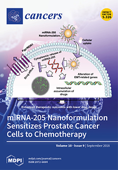
MicroRNA-based therapeutics have gained prominence as a cancer therapy due to their multiple targeting abilities. However, their rapid degradation in serum and low cellular internalization makes them ineffective for treatment. This study reports an efficient nanoplatform that delivers microRNA (we chose miR-205) to prostate cancer cells. Our nanoformulation (MPEI-PEG) efficiently delivered miRNA-205 and suppressed cell growth and the metastasis of cancer cells. Further, it selectively inhibited P-gp/MDR1 and thus prevented the drug efflux causing the intracellular drug accumulation of chemotherapy drugs. Overall, this data is evidence of the chemo-sensitizing ability of miR-205 nanoformulation for chemotherapy. View this paper
  • Issues are regarded as officially published after their release is announced to the table of contents alert mailing list.
  • You may sign up for e-mail alerts to receive table of contents of newly released issues.
  • PDF is the official format for papers published in both, html and pdf forms. To view the papers in pdf format, click on the "PDF Full-text" link, and use the free Adobe Reader to open them.
Order results
Result details
Section
Select all
Export citation of selected articles as:
18 pages, 5269 KiB  
Article
Deficiency in STAT1 Signaling Predisposes Gut Inflammation and Prompts Colorectal Cancer Development
by Sonia Leon-Cabrera, Armando Vázquez-Sandoval, Emmanuel Molina-Guzman, Yael Delgado-Ramirez, Norma L. Delgado-Buenrostro, Blanca E. Callejas, Yolanda I. Chirino, Carlos Pérez-Plasencia, Miriam Rodríguez-Sosa, Jonadab E. Olguín, Citlaltepetl Salinas, Abhay R. Satoskar and Luis I. Terrazas
Cancers 2018, 10(9), 341; https://doi.org/10.3390/cancers10090341 - 19 Sep 2018
Cited by 20 | Viewed by 4547
Abstract
Signal transducer and activator of transcription 1 (STAT1) is part of the Janus kinase (JAK/STAT) signaling pathway that controls critical events in intestinal immune function related to innate and adaptive immunity. Recent studies have implicated STAT1 in tumor–stroma interactions, and its expression and [...] Read more.
Signal transducer and activator of transcription 1 (STAT1) is part of the Janus kinase (JAK/STAT) signaling pathway that controls critical events in intestinal immune function related to innate and adaptive immunity. Recent studies have implicated STAT1 in tumor–stroma interactions, and its expression and activity are perturbed during colon cancer. However, the role of STAT1 during the initiation of inflammation-associated cancer is not clearly understood. To determine the role of STAT1 in colitis-associated colorectal cancer (CAC), we analyzed the tumor development and kinetics of cell recruitment in wild-type WT or STAT1−/− mice treated with azoxymethane (AOM) and dextran sodium sulfate (DSS). Following CAC induction, STAT1−/− mice displayed an accelerated appearance of inflammation and tumor formation, and increased damage and scores on the disease activity index (DAI) as early as 20 days after AOM-DSS exposure compared to their WT counterparts. STAT1−/− mice showed elevated colonic epithelial cell proliferation in early stages of injury-induced tumor formation and decreased apoptosis in advanced tumors with over-expression of the anti-apoptotic protein Bcl2 at the colon. STAT1−/− mice showed increased accumulation of Ly6G+Ly6CCD11b+ cells in the spleen at 20 days of CAC development with concomitant increases in the production of IL-17A, IL-17F, and IL-22 cytokines compared to WT mice. Our findings suggest that STAT1 plays a role as a tumor suppressor molecule in inflammation-associated carcinogenesis, particularly during the very early stages of CAC initiation, modulating immune responses as well as controlling mechanisms such as apoptosis and cell proliferation. Full article
Show Figures

Figure 1

12 pages, 838 KiB  
Review
Autologous Transplantation for Older Adults with AML
by Beatrice U. Mueller, Katja Seipel, Ulrike Bacher and Thomas Pabst
Cancers 2018, 10(9), 340; https://doi.org/10.3390/cancers10090340 - 19 Sep 2018
Cited by 18 | Viewed by 4095
Abstract
While the majority of patients with acute myeloid leukemia (AML) are above the age of 65 years at diagnosis, the outcome of older AML patients remains disappointing. Even if standard intensive chemotherapy induces morphologic complete remission (CR1), relapses in older AML patients are [...] Read more.
While the majority of patients with acute myeloid leukemia (AML) are above the age of 65 years at diagnosis, the outcome of older AML patients remains disappointing. Even if standard intensive chemotherapy induces morphologic complete remission (CR1), relapses in older AML patients are common leading to poor long-term survival outcomes. Since autologous hematopoietic stem cell transplantation (HCT) offers distinct anti-leukemic effectiveness while avoiding graft-versus-host disease associated with allogeneic transplantation, it represents an option for consolidation treatment in selected older AML patients. However, prospective studies in older AML patients assessing the benefit of autologous HCT compared to chemotherapy consolidation or allogeneic transplantation are lacking. Consequently, clinicians face the dilemma that there is considerable ambiguity on the most appropriate consolidation treatment for older AML patients in CR1. This review highlights the possible role of autologous HCT for consolidation in older AML patients reaching CR1 after induction treatment. Full article
(This article belongs to the Special Issue Treatment of Older Adults with Acute Myeloid Leukemia)
Show Figures

Figure 1

17 pages, 268 KiB  
Review
Current Immunotherapeutic Approaches in T Cell Non-Hodgkin Lymphomas
by Teresa Poggio, Justus Duyster and Anna L. Illert
Cancers 2018, 10(9), 339; https://doi.org/10.3390/cancers10090339 - 18 Sep 2018
Cited by 14 | Viewed by 4206
Abstract
T cell non-Hodgkin lymphoma (T-NHL) is a rare and heterogeneous group of neoplasms of the lymphoid system. With the exception of a few relatively indolent entities, T-NHL is typically aggressive, treatment resistant, and associated with poor prognosis. Relatively few options with proven clinical [...] Read more.
T cell non-Hodgkin lymphoma (T-NHL) is a rare and heterogeneous group of neoplasms of the lymphoid system. With the exception of a few relatively indolent entities, T-NHL is typically aggressive, treatment resistant, and associated with poor prognosis. Relatively few options with proven clinical benefit are available for patients with relapsed or refractory disease. Immunotherapy has emerged as a promising treatment for the management of patients with hematological malignancies. The identification of tumor antigens has provided a large number of potential targets. Therefore, several monoclonal antibodies (alemtuzumab, SGN-30, brentuximab vedotin, and mogamulizumab), directed against tumor antigens, have been investigated in different subtypes of T-NHL. In addition to targeting antigens involved in cancer cell physiology, antibodies can stimulate immune effector functions or counteract immunosuppressive mechanisms. Chimeric antigen receptor (CAR)-T cells directed against CD30 and immune checkpoint inhibitors are currently being investigated in clinical trials. In this review, we summarize the currently available clinical evidence for immunotherapy in T-NHL, focusing on the results of clinical trials using first generation monoclonal antibodies, new immunotherapeutic agents, immune checkpoint inhibitors, and CAR-T cell therapies. Full article
13 pages, 1753 KiB  
Article
Phase I Study of Carbon Ion Radiotherapy and Image-Guided Brachytherapy for Locally Advanced Cervical Cancer
by Tatsuya Ohno, Shin-ei Noda, Kazutoshi Murata, Yuya Yoshimoto, Noriyuki Okonogi, Ken Ando, Tomoaki Tamaki, Shingo Kato, Takashi Hirakawa, Tatsuya Kanuma, Takashi Minegishi, Takashi Nakano and On behalf of the Working Group for Gynecologic Cancer
Cancers 2018, 10(9), 338; https://doi.org/10.3390/cancers10090338 - 18 Sep 2018
Cited by 17 | Viewed by 4641
Abstract
A phase I study was performed to determine the recommended dose of carbon ion radiotherapy and 3D image-guided brachytherapy for histologically confirmed stage II (≥4 cm), III, or IVA cervical cancer. Dose-limiting toxicities (treatment-related toxicities occurring within three months from the start of [...] Read more.
A phase I study was performed to determine the recommended dose of carbon ion radiotherapy and 3D image-guided brachytherapy for histologically confirmed stage II (≥4 cm), III, or IVA cervical cancer. Dose-limiting toxicities (treatment-related toxicities occurring within three months from the start of carbon ion radiotherapy) included Grade 3 non-hematological toxicity, Grade 4 hematological toxicity, or interruption of treatment for more than two weeks due to treatment-related toxicities. Carbon ion radiotherapy consisted of whole-pelvic irradiation with 36.0 Gy (relative biological effectiveness) in 12 fractions and local boost with 19.2 Gy in four fractions for the primary site, and for positive lymph nodes. Three sessions of three-dimensional (3D) image-guided brachytherapy were administered after completion of carbon ion radiotherapy. Weekly cisplatin at a dose of 40 mg/m2 was given concurrently. At a dose level of one, a total rectosigmoid D2cc dose between 67.2 Gy and 71.3 Gy at a biological equivalent dose of 2 Gy per fraction from carbon ion radiotherapy and 3D image-guided brachytherapy was prescribed. Six patients were enrolled into this dose level. No patients developed the pre-defined dose-limiting toxicities. For late toxicities, however, one patient developed Grade 3 rectal hemorrhage requiring transfusion at 10 months after treatment. The median survival time was 50.0 months for the five surviving patients. No further dose escalation was performed, and we determined the dose of level one as the recommended rectosigmoid dose. Although our results are preliminary, the study regimen encourages further investigation (registration: UMIN000013340). Full article
(This article belongs to the Special Issue New Developments in Radiotherapy)
Show Figures

Figure 1

15 pages, 1078 KiB  
Review
Oxidative Phosphorylation: A Target for Novel Therapeutic Strategies Against Ovarian Cancer
by Amruta P. Nayak, Arvinder Kapur, Lisa Barroilhet and Manish S. Patankar
Cancers 2018, 10(9), 337; https://doi.org/10.3390/cancers10090337 - 18 Sep 2018
Cited by 82 | Viewed by 7639
Abstract
Aerobic glycolysis is an important metabolic adaptation of cancer cells. There is growing evidence that oxidative phosphorylation is also an active metabolic pathway in many tumors, including in high grade serous ovarian cancer. Metastasized ovarian tumors use fatty acids for their energy needs. [...] Read more.
Aerobic glycolysis is an important metabolic adaptation of cancer cells. There is growing evidence that oxidative phosphorylation is also an active metabolic pathway in many tumors, including in high grade serous ovarian cancer. Metastasized ovarian tumors use fatty acids for their energy needs. There is also evidence of ovarian cancer stem cells privileging oxidative phosphorylation (OXPHOS) for their metabolic needs. Metformin and thiazolidinediones such as rosiglitazone restrict tumor growth by inhibiting specific steps in the mitochondrial electron transport chain. These observations suggest that strategies to interfere with oxidative phosphorylation should be considered for the treatment of ovarian tumors. Here, we review the literature that supports this hypothesis and describe potential agents and critical control points in the oxidative phosphorylation pathway that can be targeted using small molecule agents. In this review, we also discuss potential barriers that can reduce the efficacy of the inhibitors of oxidative phosphorylation. Full article
(This article belongs to the Special Issue The Tumor Microenvironment of High Grade Serous Ovarian Cancer)
Show Figures

Figure 1

13 pages, 437 KiB  
Review
Revisiting Neutrophil Gelatinase-Associated Lipocalin (NGAL) in Cancer: Saint or Sinner?
by Brigitte Bauvois and Santos A. Susin
Cancers 2018, 10(9), 336; https://doi.org/10.3390/cancers10090336 - 18 Sep 2018
Cited by 41 | Viewed by 5336
Abstract
Human neutrophil gelatinase-associated lipocalin (NGAL) is a glycoprotein present in a wide variety of tissues and cell types. NGAL exists as a 25 kDa monomer, a 46 kDa homodimer (the most abundant form in healthy subjects) and a 130 kDa disulfide-linked heterodimer bound [...] Read more.
Human neutrophil gelatinase-associated lipocalin (NGAL) is a glycoprotein present in a wide variety of tissues and cell types. NGAL exists as a 25 kDa monomer, a 46 kDa homodimer (the most abundant form in healthy subjects) and a 130 kDa disulfide-linked heterodimer bound to latent matrix metalloproteinase-9. Dysregulated expression of NGAL in human malignancies suggests its value as a clinical marker. A growing body of evidence is highlighting NGAL’s paradoxical (i.e., both beneficial and detrimental) effects on cellular processes associated with tumor development (proliferation, survival, migration, invasion, and multidrug resistance). At least two distinct cell surface receptors are identified for NGAL. This review (i) summarizes our current knowledge of NGAL’s expression profiles in solid tumors and leukemias, and (ii) critically evaluates the beneficial and detrimental activities of NGAL having been documented in a diverse range of cancer-derived cell lines. A better understanding of the causal relationships between NGAL dysregulation and tumor development will require a fine analysis of the molecular aspects and biological role(s) of NGAL both in primary tumors and at different stages of disease. Having an accurate picture of NGAL’s contribution to tumor progression is a prerequisite for attempting to modulate this protein as a putative therapeutic target. Full article
(This article belongs to the Special Issue Cancer Biomarkers)
Show Figures

Graphical abstract

17 pages, 6891 KiB  
Article
Combination of EGFR Inhibitor Lapatinib and MET Inhibitor Foretinib Inhibits Migration of Triple Negative Breast Cancer Cell Lines
by Aleksandra Simiczyjew, Ewelina Dratkiewicz, Marleen Van Troys, Christophe Ampe, Ilona Styczeń and Dorota Nowak
Cancers 2018, 10(9), 335; https://doi.org/10.3390/cancers10090335 - 17 Sep 2018
Cited by 25 | Viewed by 5965
Abstract
Triple-negative breast cancer (TNBC) is the most challenging subtype to treat due to the lack of estrogen receptor, progesterone receptor, and HER2 expression, which excludes the usage of directed targeted therapy against them. Promising therapeutic targets are the hepatocyte growth factor receptor (MET) [...] Read more.
Triple-negative breast cancer (TNBC) is the most challenging subtype to treat due to the lack of estrogen receptor, progesterone receptor, and HER2 expression, which excludes the usage of directed targeted therapy against them. Promising therapeutic targets are the hepatocyte growth factor receptor (MET) and epidermal growth factor receptor (EGFR), which expression is frequently elevated in TNBC. Inhibitors of these receptors used as monotherapy are often ineffective. Due to that, we studied the efficacy of combined therapy targeting MET and EGFR simultaneously. Two TNBC cell lines were treated with lapatinib (a dual EGFR and HER2 inhibitor), foretinib (a MET inhibitor), or a combination of the two. After the inhibitors treatment, we verified the cell viability (XTT assay), distribution of the cell cycle phases, the activation of signaling pathways (Western blotting), distribution of invadopodia, fluorescent gelatin digestion (immunofluorescence), and the invasion capacity of cells. A combination of foretinib and lapatinib effectively reduced the viability of examined cells, led to G2/M arrest and reduction of pAKT. There was also a decreasein number of invadopodia formed by cells, their ability to digest gelatin and reduction of cells migration/invasion capacity. Therapy targeting of both EGFR and MET receptors was much more effective against tested cells than monotherapy. We selected a combination of drugs that could be successfully used against this breast cancer subtype. Full article
(This article belongs to the Special Issue Tyrosine Kinase Signaling Pathways in Cancer)
Show Figures

Figure 1

17 pages, 2762 KiB  
Article
Factors Secreted by Cancer-Associated Fibroblasts that Sustain Cancer Stem Properties in Head and Neck Squamous Carcinoma Cells as Potential Therapeutic Targets
by Saúl Álvarez-Teijeiro, Cristina García-Inclán, M. Ángeles Villaronga, Pedro Casado, Francisco Hermida-Prado, Rocío Granda-Díaz, Juan P. Rodrigo, Fernando Calvo, Nagore Del-Río-Ibisate, Alberto Gandarillas, Francisco Morís, Mario Hermsen, Pedro Cutillas and Juana M. García-Pedrero
Cancers 2018, 10(9), 334; https://doi.org/10.3390/cancers10090334 - 17 Sep 2018
Cited by 40 | Viewed by 4717
Abstract
This study investigates for the first time the crosstalk between stromal fibroblasts and cancer stem cell (CSC) biology in head and neck squamous cell carcinomas (HNSCC), with the ultimate goal of identifying effective therapeutic targets. The effects of conditioned media from cancer-associated fibroblasts [...] Read more.
This study investigates for the first time the crosstalk between stromal fibroblasts and cancer stem cell (CSC) biology in head and neck squamous cell carcinomas (HNSCC), with the ultimate goal of identifying effective therapeutic targets. The effects of conditioned media from cancer-associated fibroblasts (CAFs) and normal fibroblasts (NFs) on the CSC phenotype were assessed by combining functional and expression analyses in HNSCC-derived cell lines. Further characterization of CAFs and NFs secretomes by mass spectrometry was followed by pharmacologic target inhibition. We demonstrate that factors secreted by CAFs but not NFs, in the absence of serum/supplements, robustly increased anchorage-independent growth, tumorsphere formation, and CSC-marker expression. Modulators of epidermal growth factor receptor (EGFR), insulin-like growth factor receptor (IGFR), and platelet-derived growth factor receptor (PDGFR) activity were identified as paracrine cytokines/factors differentially secreted between CAFs and NFs, in a mass spectrometry analysis. Furthermore, pharmacologic inhibition of EGFR, IGFR, and PDGFR significantly reduced CAF-induced tumorsphere formation and anchorage-independent growth suggesting a role of these receptor tyrosine kinases in sustaining the CSC phenotype. These findings provide novel insights into tumor stroma–CSC communication, and potential therapeutic targets to effectively block the CAF-enhanced CSC niche signaling circuit. Full article
(This article belongs to the Special Issue Targeting Head and Neck Cancer)
Show Figures

Figure 1

12 pages, 1035 KiB  
Article
Proton Beam Therapy in Combination with Intra-Arterial Infusion Chemotherapy for T4 Squamous Cell Carcinoma of the Maxillary Gingiva
by Hiromasa Endo, Kanako Takayama, Kenji Mitsudo, Tatsuya Nakamura, Ichiro Seto, Hisashi Yamaguchi, Takashi Ono, Motohisa Suzuki, Yusuke Azami, Hitoshi Wada, Masao Murakami and Iwai Tohnai
Cancers 2018, 10(9), 333; https://doi.org/10.3390/cancers10090333 - 15 Sep 2018
Cited by 4 | Viewed by 3473
Abstract
This study aimed to evaluate the therapeutic effect and toxicity of proton beam therapy in combination with intra-arterial infusion chemotherapy in patients with squamous cell carcinoma of the maxillary gingiva. Between December 2010 and March 2016, 30 patients with T4 squamous cell carcinoma [...] Read more.
This study aimed to evaluate the therapeutic effect and toxicity of proton beam therapy in combination with intra-arterial infusion chemotherapy in patients with squamous cell carcinoma of the maxillary gingiva. Between December 2010 and March 2016, 30 patients with T4 squamous cell carcinoma of the maxillary gingiva were treated with radiotherapy and retrograde intra-arterial infusion chemotherapy using cisplatin (20–40 mg/m2, 4–6 times). Radiotherapy was basically administered using boost proton beam therapy for primary tumor and neck lymph node tumors, following 36–40 Gy photon radiation therapy delivered to the prophylactic area, to a total dose of 70.4–74.8 Gy. The median follow-up was 33 months. The 3-year local control and overall survival rates were 69% and 59%, respectively. Major grade 3 or higher acute toxicities included mucositis, neutropenia, and dermatitis in 12 (40%), 5 (17%), and 3 (10%) patients, respectively. No grade 3 or higher late toxicities were observed. These results suggested that proton beam therapy in combination with intra-arterial infusion chemotherapy was not inferior to other treatment protocols and should be considered as a safe and effective option in patients with T4 squamous cell carcinoma of the maxillary gingiva. Full article
(This article belongs to the Special Issue Proton and Carbon Ion Therapy)
Show Figures

Figure 1

15 pages, 1324 KiB  
Article
Clonal Heterogeneity Reflected by PI3K-AKT-mTOR Signaling in Human Acute Myeloid Leukemia Cells and Its Association with Adverse Prognosis
by Ina Nepstad, Kimberley Joanne Hatfield, Tor Henrik Anderson Tvedt, Håkon Reikvam and Øystein Bruserud
Cancers 2018, 10(9), 332; https://doi.org/10.3390/cancers10090332 - 14 Sep 2018
Cited by 24 | Viewed by 3919
Abstract
Clonal heterogeneity detected by karyotyping is a biomarker associated with adverse prognosis in acute myeloid leukemia (AML). Constitutive activation of the phosphatidylinositol-3-kinase-Akt-mechanistic target of rapamycin (PI3K-Akt-mTOR) pathway is present in AML cells, and this pathway integrates signaling from several upstream receptors/mediators. We suggest [...] Read more.
Clonal heterogeneity detected by karyotyping is a biomarker associated with adverse prognosis in acute myeloid leukemia (AML). Constitutive activation of the phosphatidylinositol-3-kinase-Akt-mechanistic target of rapamycin (PI3K-Akt-mTOR) pathway is present in AML cells, and this pathway integrates signaling from several upstream receptors/mediators. We suggest that this pathway reflects biologically important clonal heterogeneity. We investigated constitutive PI3K-Akt-mTOR pathway activation in primary human AML cells derived from 114 patients, together with 18 pathway mediators. The cohort included patients with normal karyotype or single karyotype abnormalities and with an expected heterogeneity of molecular genetic abnormalities. Clonal heterogeneity reflected as pathway mediator heterogeneity was detected for 49 patients. Global gene expression profiles of AML cell populations with and without clonal heterogeneity differed with regard to expression of ectopic olfactory receptors (a subset of G-protein coupled receptors) and proteins involved in G-protein coupled receptor signaling. Finally, the presence of clonal heterogeneity was associated with adverse prognosis for patients receiving intensive antileukemic treatment. The clonal heterogeneity as reflected in the activation status of selected mediators in the PI3K-Akt-mTOR pathway was associated with a different gene expression profile and had an independent prognostic impact. Biological heterogeneity reflected in the intracellular signaling status should be further investigated as a prognostic biomarker in human AML. Full article
(This article belongs to the Special Issue Cancer Biomarkers)
Show Figures

Figure 1

18 pages, 4315 KiB  
Article
TRPC6 Channels Are Required for Proliferation, Migration and Invasion of Breast Cancer Cell Lines by Modulation of Orai1 and Orai3 Surface Exposure
by Isaac Jardin, Raquel Diez-Bello, Jose J. Lopez, Pedro C. Redondo, Ginés M. Salido, Tarik Smani and Juan A. Rosado
Cancers 2018, 10(9), 331; https://doi.org/10.3390/cancers10090331 - 14 Sep 2018
Cited by 66 | Viewed by 5248
Abstract
Transient receptor potential channels convey signaling information from a number of stimuli to a wide variety of cellular functions, mainly by inducing changes in cytosolic Ca2+ concentration. Different members of the TRPC, TRPM and TRPV subfamilies have been reported to play a [...] Read more.
Transient receptor potential channels convey signaling information from a number of stimuli to a wide variety of cellular functions, mainly by inducing changes in cytosolic Ca2+ concentration. Different members of the TRPC, TRPM and TRPV subfamilies have been reported to play a role in tumorigenesis. Here we show that the estrogen receptor positive and triple negative breast cancer cell lines, MCF7 and MDA-MB-231, respectively, exhibit enhanced expression of the TRPC6 channel as compared to the non-tumoral MCF10A cell line. In vitro TRPC6 knockdown using shRNA impaired MCF7 and MDA-MB-231 cell proliferation, migration and invasion detected by BrdU incorporation, wound healing and Boyden chamber assays, respectively. Using RNAi-mediated TRPC6 silencing as well as overexpression of the pore-dead dominant-negative TRPC6 mutant we have found that TRPC6 plays a relevant role in the activation of store-operated Ca2+ entry in the breast cancer cell lines but not in non-tumoral breast cells. Finally, we have found that TRPC6 interacts with Orai1 and Orai3 in MCF7 and MDA-MB-231 cells and is required for the translocation of Orai1 and Orai3 to the plasma membrane in MDA-MB-231 and MCF7 cells, respectively, upon Ca2+ store depletion. These findings introduce a novel mechanism for the modulation of Ca2+ influx and the development of different cancer hallmarks in breast cancer cells. Full article
(This article belongs to the Special Issue Ion Channels in Cancer)
Show Figures

Graphical abstract

19 pages, 7298 KiB  
Article
Proliferative and Invasive Colorectal Tumors in Pet Dogs Provide Unique Insights into Human Colorectal Cancer
by Jin Wang, Tianfang Wang, Yanfang Sun, Yuan Feng, William C. Kisseberth, Carolyn J. Henry, Irene Mok, Susan E. Lana, Kevin Dobbin, Nicole Northrup, Elizabeth W. Howerth and Shaying Zhao
Cancers 2018, 10(9), 330; https://doi.org/10.3390/cancers10090330 - 14 Sep 2018
Cited by 17 | Viewed by 5531
Abstract
Spontaneous tumors in pet dogs represent a valuable but undercharacterized cancer model. To better use this resource, we performed an initial global comparison between proliferative and invasive colorectal tumors from 20 canine cases, and evaluated their molecular homology to human colorectal cancer (CRC). [...] Read more.
Spontaneous tumors in pet dogs represent a valuable but undercharacterized cancer model. To better use this resource, we performed an initial global comparison between proliferative and invasive colorectal tumors from 20 canine cases, and evaluated their molecular homology to human colorectal cancer (CRC). First, proliferative canine tumors harbor overactivated WNT/β-catenin pathways and recurrent CTNNB1 (β-catenin) mutations S45F/P, D32Y and G34E. Invasive canine tumors harbor prominent fibroblast proliferation and overactivated stroma. Both groups have recurrent TP53 mutations. We observed three invasion patterns in canine tumors: collective, crypt-like and epithelial–mesenchymal transition (EMT). We detected enriched Helicobacter bilis and Alistipes finegoldii in proliferative and crypt-like tumors, but depleted mucosa-microbes in the EMT tumor. Second, guided by our canine findings, we classified 79% of 478 human colon cancers from The Cancer Genome Atlas into four subtypes: primarily proliferative, or with collective, crypt-like or EMT invasion features. Their molecular characteristics match those of canine tumors. We showed that consensus molecular subtype 4 (mesenchymal) of human CRC should be further divided into EMT and crypt-like subtypes, which differ in TGF-β activation and mucosa-microbe content. Our canine tumors share the same pathogenic pathway as human CRCs. Dog-human integration identifies three CRC invasion patterns and improves CRC subtyping. Full article
Show Figures

Figure 1

19 pages, 5029 KiB  
Review
Repurposing of Drugs Targeting YAP-TEAD Functions
by Gian Marco Elisi, Matteo Santucci, Domenico D’Arca, Angela Lauriola, Gaetano Marverti, Lorena Losi, Laura Scalvini, Maria Laura Bolognesi, Marco Mor and Maria Paola Costi
Cancers 2018, 10(9), 329; https://doi.org/10.3390/cancers10090329 - 14 Sep 2018
Cited by 32 | Viewed by 8402
Abstract
Drug repurposing is a fast and consolidated approach for the research of new active compounds bypassing the long streamline of the drug discovery process. Several drugs in clinical practice have been reported for modulating the major Hippo pathway’s terminal effectors, namely YAP (Yes1-associated [...] Read more.
Drug repurposing is a fast and consolidated approach for the research of new active compounds bypassing the long streamline of the drug discovery process. Several drugs in clinical practice have been reported for modulating the major Hippo pathway’s terminal effectors, namely YAP (Yes1-associated protein), TAZ (transcriptional co-activator with PDZ-binding motif) and TEAD (transcriptional enhanced associate domains), which are directly involved in the regulation of cell growth and tissue homeostasis. Since this pathway is known to have many cross-talking phenomena with cell signaling pathways, many efforts have been made to understand its importance in oncology. Moreover, this could be relevant to obtain new molecular tools and potential therapeutic assets. In this review, we discuss the main mechanisms of action of the best-known compounds, clinically approved or investigational drugs, able to cross-talk and modulate the Hippo pathway, as an attractive strategy for the discovery of new potential lead compounds. Full article
Show Figures

Graphical abstract

20 pages, 3506 KiB  
Article
Clinical Impact of Epithelial-to-Mesenchymal Transition Regulating MicroRNAs in Pancreatic Ductal Adenocarcinoma
by Sameer Abdallah Dhayat, Max Michael Traeger, Jan Rehkaemper, Anda Jana Stroese, Konrad Steinestel, Eva Wardelmann, Iyad Kabar and Norbert Senninger
Cancers 2018, 10(9), 328; https://doi.org/10.3390/cancers10090328 - 13 Sep 2018
Cited by 18 | Viewed by 3797
Abstract
Pancreatic ductal adenocarcinoma (PDAC) is one of the most aggressive carcinoma entities worldwide with early and rapid dissemination. Recently, we discussed the role of microRNAs as epigenetic regulators of Epithelial-to-Mesenchymal Transition (EMT) in PDAC. In this study, we investigated their value as diagnostic [...] Read more.
Pancreatic ductal adenocarcinoma (PDAC) is one of the most aggressive carcinoma entities worldwide with early and rapid dissemination. Recently, we discussed the role of microRNAs as epigenetic regulators of Epithelial-to-Mesenchymal Transition (EMT) in PDAC. In this study, we investigated their value as diagnostic and prognostic markers in tissue and blood samples of 185 patients including PDAC, non-malignant pancreatic disorders, and age-matched healthy controls. Expression of the microRNA-200-family (microRNAs -141, -200a, -200b, -200c, -429) and microRNA-148a was significantly downregulated in tissue of PDAC Union internationale contre le cancer (UICC) Stage II. Correspondingly, stromal PDAC tissue showed strong expression of Fibronectin, Vimentin, and ZEB-1 (Zinc finger E-box-binding homeobox) versus low expression of E-cadherin. Transient transfection of microRNA-200b and microRNA-200c mimics resulted in the downregulation of their key target ZEB-1. Inversely, blood serum analyses of patients with PDAC UICC Stages II, III, and IV showed a significant over-expression of microRNA-200-family members, microRNA-148a, microRNA-10b, and microRNA-34a. Correspondingly, Enzyme-linked Immunosorbent Assay (ELISA) analyses revealed a significant over-expression of soluble E-cadherin in serum samples of PDAC patients versus healthy controls. The best diagnostic accuracy to distinguish between PDAC and non-PDAC in this patient collective could be achieved in tissue by microRNA-148a with an area under the receiver-operating-characteristic (ROC) curve (AUC) of 0.885 and in blood serum by a panel of microRNA-141, -200b, -200c, and CA.19-9 with an AUC of 0.890. Both diagnostic tools outreach the diagnostic performance of the currently most common diagnostic biomarker CA.19-9 (AUC of 0.834). Kaplan Meier survival analysis of this patient collective revealed an improved overall survival in PDAC patients with high expression of tissue-related microRNA-34a, -141, -200b, -200c, and -429. In conclusion, EMT-regulating microRNAs have great potential as liquid and solid biopsy markers in PDAC patients. Their prognostic and therapeutic benefits remain important tasks for future studies. Full article
(This article belongs to the Special Issue MicroRNA-Associated Cancer Metastasis)
Show Figures

Figure 1

17 pages, 1359 KiB  
Review
The Role of Signal Transducer and Activator of Transcription 3 (STAT3) and Its Targeted Inhibition in Hematological Malignancies
by Loukik Arora, Alan Prem Kumar, Frank Arfuso, Wee Joo Chng and Gautam Sethi
Cancers 2018, 10(9), 327; https://doi.org/10.3390/cancers10090327 - 13 Sep 2018
Cited by 100 | Viewed by 6526
Abstract
Signal transducer and activator of transcription 3 (STAT3), a member of the STAT protein family, can be phosphorylated by receptor-associated Janus kinases (JAKs) in response to stimulation by cytokines and growth factors. It forms homo- or heterodimers that can translocate to the cell [...] Read more.
Signal transducer and activator of transcription 3 (STAT3), a member of the STAT protein family, can be phosphorylated by receptor-associated Janus kinases (JAKs) in response to stimulation by cytokines and growth factors. It forms homo- or heterodimers that can translocate to the cell nucleus where they act as transcription activators. Constitutive activation of STAT3 has been found to be associated with initiation and progression of various cancers. It can exert proliferative as well as anti-apoptotic effects. This review focuses on the role of STAT3 in pathogenesis i.e., proliferation, differentiation, migration, and apoptosis of hematological malignancies viz. leukemia, lymphoma and myeloma, and briefly highlights the potential therapeutic approaches developed against STAT3 activation pathway. Full article
Show Figures

Figure 1

14 pages, 1648 KiB  
Article
Chromogenic Multiplex Immunohistochemistry Reveals Modulation of the Immune Microenvironment Associated with Survival in Elderly Patients with Lung Adenocarcinoma
by Marius Ilié, Mélanie Beaulande, Saima Ben Hadj, Emmanuel Chamorey, Renaud Schiappa, Elodie Long-Mira, Sandra Lassalle, Catherine Butori, Charlotte Cohen, Sylvie Leroy, Olivier Guérin, Jérôme Mouroux, Charles-Hugo Marquette, Jean-François Pomerol, Gilles Erb, Véronique Hofman and Paul Hofman
Cancers 2018, 10(9), 326; https://doi.org/10.3390/cancers10090326 - 13 Sep 2018
Cited by 23 | Viewed by 5571
Abstract
With underrepresentation of elderly patients with lung adenocarcinoma (LADC) in anti-PD-1/PD-L1 clinical trials, better understanding of the interplay of PD-L1 and tumor-associated immune cells (TAICs) could assist clinicians in stratifying these patients for immunotherapy. One hundred and one patients with LADCs, stratified by [...] Read more.
With underrepresentation of elderly patients with lung adenocarcinoma (LADC) in anti-PD-1/PD-L1 clinical trials, better understanding of the interplay of PD-L1 and tumor-associated immune cells (TAICs) could assist clinicians in stratifying these patients for immunotherapy. One hundred and one patients with LADCs, stratified by age, were included for analysis of PD-L1 expression and density of TAICs expressing CD4, CD8, and CD33, by using multiplex chromogenic immunohistochemistry (IHC) assays and automated digital quantification. The CD4+/CD8+ ratio was significantly higher in elderly patients. In patients <75 years, the density of CD4+, CD8+, and PD-L1 in TAICs showed a positive significant correlation with PD-L1 expression in tumor cells (TCs), while a lower correlation was observed in the elderly population. In the latter, a high CD4+/CD8+ ratio, and combined PD-L1 expression ≥1% TCs with a low CD8+ density, low CD33+ density, and a high CD4+ density correlated to worse overall survival. We identified differences according to age in the CD4+/CD8+ ratio and in correlation between PD-L1 expression and the density of TAICs in LADC patients. Distinct groups of tumor microenvironments had an impact on the OS of elderly patients with LADC. Full article
Show Figures

Figure 1

15 pages, 2186 KiB  
Article
G9a Correlates with VLA-4 Integrin and Influences the Migration of Childhood Acute Lymphoblastic Leukemia Cells
by Elena Madrazo, David Ruano, Lorea Abad, Estefanía Alonso-Gómez, Carmen Sánchez-Valdepeñas, África González-Murillo, Manuel Ramírez and Javier Redondo-Muñoz
Cancers 2018, 10(9), 325; https://doi.org/10.3390/cancers10090325 - 12 Sep 2018
Cited by 11 | Viewed by 3784
Abstract
Acute lymphoblastic leukemia (ALL) is the most common pediatric cancer. As ALL progresses, leukemic cells cross the endothelial barrier and infiltrate other tissues. Epigenetic enzymes represent novel therapeutic targets in hematological malignancies, and might contribute to cells’ capacity to migrate across physical barriers. [...] Read more.
Acute lymphoblastic leukemia (ALL) is the most common pediatric cancer. As ALL progresses, leukemic cells cross the endothelial barrier and infiltrate other tissues. Epigenetic enzymes represent novel therapeutic targets in hematological malignancies, and might contribute to cells’ capacity to migrate across physical barriers. Although many molecules drive this process, the role of the nucleus and its components remain unclear. We report here, for the first time, that the expression of G9a (a histone methyltransferase related with gene silencing) correlates with the expression of the integrin subunit α4 in children with ALL. We have demonstrated that G9a depletion or its inhibition with BIX01294 abrogated the ability of ALL cells to migrate through an endothelial monolayer. Moreover, G9a-depleted and BIX01294-treated cells presented bigger nuclei and more adherent phenotype than control cells on endothelial monolayers. Blocking G9a did not affect the cell cytoskeleton or integrin expression of ALL cell lines, and only its depletion reduced slightly F-actin polymerization. Similarly to the transendothelial migration, G9a inhibition impaired the cell migration induced by the integrin VLA-4 (α4β1) of primary cells and ALL cell lines through narrow spaces in vitro. Our results suggest a cellular connection between G9a and VLA-4, which underlies novel functions of G9a during ALL cell migration. Full article
(This article belongs to the Special Issue Epigenetic Influence on Cancer Metastasis and/or Treatment Resistance)
Show Figures

Figure 1

5 pages, 548 KiB  
Perspective
No Increase in Colon Cancer Risk Following Induction with Neu5Gc-Bearing Rabbit Anti-T Cell IgG (ATG) in Recipients of Kidney Transplants
by Jean-Paul Soulillou, Caner Süsal, Bernd Döhler and Gerhard Opelz
Cancers 2018, 10(9), 324; https://doi.org/10.3390/cancers10090324 - 12 Sep 2018
Cited by 10 | Viewed by 4742
Abstract
Because of a mutation of the gene allowing the synthesis of the Neu5Gc form of neuraminidic acid, humans lack the Neu5Gc present in other mammals and develop anti-Neu5Gc. However, humans can absorb dietary Neu5Gc and normal colon epithelium displays minute amounts of Neu5Gc. [...] Read more.
Because of a mutation of the gene allowing the synthesis of the Neu5Gc form of neuraminidic acid, humans lack the Neu5Gc present in other mammals and develop anti-Neu5Gc. However, humans can absorb dietary Neu5Gc and normal colon epithelium displays minute amounts of Neu5Gc. The potential “physiological” formation of in situ immune complexes has been proposed as a risk factor for colon cancer and as the link between red meat-rich diet and colon carcinoma. In this article, we took advantage of evidence that polyclonal rabbit IgG (ATG) elicits an immune response against Neu5Gc and we consulted a large data base of allograft recipients treated or not with animal-derived IgG to discuss this hypothesis. Based on data from 173,960 and 38,505 patients without and with ATG induction, respectively, we found no evidence that exposure to higher levels of anti-Neu5Gc is associated with a higher incidence of colon carcinoma. Full article
Show Figures

Figure 1

16 pages, 3225 KiB  
Article
CAR-T Cells Based on Novel BCMA Monoclonal Antibody Block Multiple Myeloma Cell Growth
by Robert Berahovich, Hua Zhou, Shirley Xu, Yuehua Wei, Jasper Guan, Jian Guan, Hizkia Harto, Shuxiang Fu, Kaihuai Yang, Shuying Zhu, Le Li, Lijun Wu and Vita Golubovskaya
Cancers 2018, 10(9), 323; https://doi.org/10.3390/cancers10090323 - 11 Sep 2018
Cited by 24 | Viewed by 8821
Abstract
The cell-surface protein B cell maturation antigen (BCMA, CD269) has emerged as a promising target for CAR-T cell therapy for multiple myeloma. In order to create a novel BCMA CAR, we generated a new BCMA monoclonal antibody, clone 4C8A. This antibody exhibited strong [...] Read more.
The cell-surface protein B cell maturation antigen (BCMA, CD269) has emerged as a promising target for CAR-T cell therapy for multiple myeloma. In order to create a novel BCMA CAR, we generated a new BCMA monoclonal antibody, clone 4C8A. This antibody exhibited strong and selective binding to human BCMA. BCMA CAR-T cells containing the 4C8A scFv were readily detected with recombinant BCMA protein by flow cytometry. The cells were cytolytic for RPMI8226, H929, and MM1S multiple myeloma cells and secreted high levels of IFN-γ in vitro. BCMA-dependent cytotoxicity and IFN-γ secretion were also observed in response to CHO (Chinese Hamster Ovary)-BCMA cells but not to parental CHO cells. In a mouse subcutaneous tumor model, BCMA CAR-T cells significantly blocked RPMI8226 tumor formation. When BCMA CAR-T cells were given to mice with established RPMI8226 tumors, the tumors experienced significant shrinkage due to CAR-T cell activity and tumor cell apoptosis. The same effect was observed with 3 humanized BCMA-CAR-T cells in vivo. These data indicate that novel CAR-T cells utilizing the BCMA 4C8A scFv are effective against multiple myeloma and warrant future clinical development. Full article
(This article belongs to the Special Issue CAR-T Cell Therapy-Novel Approaches and Challenges)
Show Figures

Figure 1

12 pages, 1260 KiB  
Article
High-Dose-Rate Brachytherapy Monotherapy versus Image-Guided Intensity-Modulated Radiotherapy with Helical Tomotherapy for Patients with Localized Prostate Cancer
by Hideya Yamazaki, Koji Masui, Gen Suzuki, Satoaki Nakamura, Daisuke Shimizu, Tatsuyuki Nishikawa, Haruumi Okabe, Ken Yoshida, Tadayuki Kotsuma, Eiichi Tanaka, Keisuke Otani, Yasuo Yoshioka and Kazuhiko Ogawa
Cancers 2018, 10(9), 322; https://doi.org/10.3390/cancers10090322 - 10 Sep 2018
Cited by 6 | Viewed by 3423
Abstract
The aim of this paper is to compare outcomes between high-dose-rate interstitial brachytherapy (HDR-BT) monotherapy and image-guided intensity-modulated radiotherapy (IG-IMRT) for localized prostate cancer. We examined 353 HDR-BT and 270 IG-IMRT patients. To reduce background selection bias, we used the method of inverse [...] Read more.
The aim of this paper is to compare outcomes between high-dose-rate interstitial brachytherapy (HDR-BT) monotherapy and image-guided intensity-modulated radiotherapy (IG-IMRT) for localized prostate cancer. We examined 353 HDR-BT and 270 IG-IMRT patients. To reduce background selection bias, we used the method of inverse probability treatment weighting (IPTW) with propensity scores. The actuarial five-year biochemical failure-free survival rates were 92.9% and 96.7% (p = 0.1847; p = 0.077 in IPTW) for HDR-BT and IG-IMRT, respectively; they were 100% and 95.8% (p = 0.286) for the low-risk group, 95.6% and 92% (p = 0.42) for the intermediate-risk group, 90.4% and 84.9% (p = 0.1059; p = 0.04 in IPTW) for the high-risk group, and 87.1% and 89.2% (p = 0.3816) for the very-high-risk group. In the assessment of accumulated incidences of grade ≥ 2 toxicity (Common Terminology Criteria for Adverse Events version 4.0) at five years, HDR-BT monotherapy showed higher genitourinary toxicity (11.9%) than IG-IMRT (3.3%) (p < 0.0001). The gastrointestinal toxicity was equivalent for HDR-BT (2.3%) and IG-IMRT (5.5%) (p = 0.063). No Grade 4 or 5 toxicity was detected in either modality. HDR-BT showed higher genitourinary toxicity than IG-IMRT. HDR-BT and IG-IMRT showed equivalent outcomes in low-, intermediate-, and very-high-risk groups. For high-risk patients, HDR-BT showed potential to improve prostate-specific antigen (PSA) control rate compared to IG-IMRT. Full article
(This article belongs to the Special Issue New Developments in Radiotherapy)
Show Figures

Figure 1

18 pages, 997 KiB  
Article
miR-9-5p in Nephrectomy Specimens is a Potential Predictor of Primary Resistance to First-Line Treatment with Tyrosine Kinase Inhibitors in Patients with Metastatic Renal Cell Carcinoma
by Bernhard Ralla, Jonas Busch, Anne Flörcken, Jörg Westermann, Zhongwei Zhao, Ergin Kilic, Sabine Weickmann, Monika Jung, Annika Fendler and Klaus Jung
Cancers 2018, 10(9), 321; https://doi.org/10.3390/cancers10090321 - 10 Sep 2018
Cited by 16 | Viewed by 3718
Abstract
Approximately 20–30% of patients with metastatic renal cell carcinoma (mRCC) in first-line treatment with tyrosine kinase inhibitors (TKIs) do not respond due to primary resistance to this drug. At present, suitable robust biomarkers for prediction of a response are not available. Therefore, the [...] Read more.
Approximately 20–30% of patients with metastatic renal cell carcinoma (mRCC) in first-line treatment with tyrosine kinase inhibitors (TKIs) do not respond due to primary resistance to this drug. At present, suitable robust biomarkers for prediction of a response are not available. Therefore, the aim of this study was to evaluate a panel of microRNAs (miRNAs) in nephrectomy specimens for use as predictive biomarkers for TKI resistance. Archived formalin-fixed, paraffin embedded nephrectomy samples from 60 mRCC patients treated with first-line TKIs (sunitinib, n = 51; pazopanib, n = 6; sorafenib, n = 3) were categorized into responders and non-responders. Using the standard Response Evaluation Criteria in Solid Tumors, patients with progressive disease within 3 months after the start of treatment with TKI were considered as non-responders and those patients with stable disease and complete or partial response under the TKI treatment for at least 6 months as responders. Based on a miRNA microarray expression profile in the two stratified groups of patients, seven differentially expressed miRNAs were validated using droplet digital reverse-transcription quantitative real-time polymerase chain reaction (RT-qPCR) assays in the two groups. Receiver operating characteristic curve analysis and binary logistic regression of response prediction were performed. MiR-9-5p and miR-489-3p were able to discriminate between the two groups. MiR-9-5p, as the most significant miRNA, improved the correct prediction of primary resistance against TKIs in comparison to that of conventional clinicopathological variables. The results of the decision curve analyses, Kaplan-Meier analyses and Cox regression analyses confirmed the potential of miR-9-5p in the prediction of response to TKIs and the prediction of progression-free survival after the initiation of TKI treatment. Full article
(This article belongs to the Special Issue Cancer Biomarkers)
Show Figures

Figure 1

17 pages, 3483 KiB  
Article
Melatonin Sensitizes Hepatocellular Carcinoma Cells to Chemotherapy Through Long Non-Coding RNA RAD51-AS1-Mediated Suppression of DNA Repair
by Chin-Chuan Chen, Chi-Yuan Chen, Shu-Huei Wang, Chau-Ting Yeh, Shih-Chi Su, Shir-Hwa Ueng, Wen-Yu Chuang, Chuen Hsueh and Tong-Hong Wang
Cancers 2018, 10(9), 320; https://doi.org/10.3390/cancers10090320 - 10 Sep 2018
Cited by 39 | Viewed by 5593
Abstract
DNA repair systems are abnormally active in most hepatocellular carcinoma (HCC) cells due to accumulated mutations, resulting in elevated DNA repair capacity and resistance to chemotherapy and radiotherapy. Thus, targeting DNA repair mechanisms is a common treatment approach in HCC to sensitize cancer [...] Read more.
DNA repair systems are abnormally active in most hepatocellular carcinoma (HCC) cells due to accumulated mutations, resulting in elevated DNA repair capacity and resistance to chemotherapy and radiotherapy. Thus, targeting DNA repair mechanisms is a common treatment approach in HCC to sensitize cancer cells to DNA damage. In this study, we examined the anti-HCC effects of melatonin and elucidated the regulatory mechanisms. The results of functional assays showed that in addition to inhibiting the proliferation, migration, and invasion abilities of HCC cells, melatonin suppressed their DNA repair capacity, thereby promoting the cytotoxicity of chemotherapy and radiotherapy. Whole-transcriptome and gain- and loss-of-function analyses revealed that melatonin induces expression of the long noncoding RNA RAD51-AS1, which binds to RAD51 mRNA to inhibit its translation, effectively decreasing the DNA repair capacity of HCC cells and increasing their sensitivity to chemotherapy and radiotherapy. Animal models further demonstrated that a combination of melatonin and the chemotherapeutic agent etoposide (VP16) can significantly enhance tumor growth inhibition compared with monotherapy. Our results show that melatonin is a potential adjuvant treatment for chemotherapy and radiotherapy in HCC. Full article
Show Figures

Graphical abstract

15 pages, 2466 KiB  
Article
Impact of Tumor Regression Grade as a Major Prognostic Factor in Locally Advanced Rectal Cancer after Neoadjuvant Chemoradiotherapy: A Proposal for a Modified Staging System
by Changhoon Song, Joo-Hyun Chung, Sung-Bum Kang, Duck-Woo Kim, Heung-Kwon Oh, Hye Seung Lee, Jin Won Kim, Keun-Wook Lee, Jee Hyun Kim and Jae-Sung Kim
Cancers 2018, 10(9), 319; https://doi.org/10.3390/cancers10090319 - 07 Sep 2018
Cited by 45 | Viewed by 4277
Abstract
There is ongoing debate regarding the significance of complete or near-complete response after neoadjuvant chemoradiotherapy (CRT) for rectal cancer. This study assessed the prognostic value of the Dworak tumor regression grade (TRG) following neoadjuvant CRT and surgery primarily in patients with pathological stage [...] Read more.
There is ongoing debate regarding the significance of complete or near-complete response after neoadjuvant chemoradiotherapy (CRT) for rectal cancer. This study assessed the prognostic value of the Dworak tumor regression grade (TRG) following neoadjuvant CRT and surgery primarily in patients with pathological stage (ypStage) II and III rectal cancer. The records of 331 patients who underwent neoadjuvant CRT followed by total mesorectal excision between 2004 and 2015 were retrospectively reviewed. Patients were categorized as having a good response (GR, TRG 3/4, n = 122) or a poor response (PR, TRG 1/2, n = 209). At a median follow-up of 65 months, five-year disease-free survival (DFS) was higher in the GR group than in the PR group (91.3% vs. 66.6%, p < 0.001). Patients with a GR and ypStage II disease had a five-year DFS that was indistinguishable from that of patients with ypStage 0–I disease (92.3% vs. 90.7%, p = 0.885). Likewise, patients with a GR and ypStage III disease had a five-year DFS similar to those with ypStage II disease (76.0% vs. 75.9%, p = 0.789). A new modified staging system that incorporates grouped TRG (GR vs. PR) was developed. The prognostic performance of this modified stage and the ypStage was compared with the Harrell C statistic. C statistic of the modified stage was higher than that of the ypStage (0.784 vs. 0.757, p = 0.012). The results remained robust after multivariate Cox regression analyses. In conclusion, a GR to neoadjuvant CRT is an independent predictor of good DFS and overall survival and further stratifies patients so as to estimate the risk of recurrence and survival among patients with ypStage II and III rectal cancer. Full article
Show Figures

Figure 1

10 pages, 1685 KiB  
Article
Nutrition Impact Symptoms Are Prognostic of Quality of Life and Mortality After Surgery for Oesophageal Cancer
by Poorna Anandavadivelan, Lena Martin, Therese Djärv, Asif Johar and Pernilla Lagergren
Cancers 2018, 10(9), 318; https://doi.org/10.3390/cancers10090318 - 07 Sep 2018
Cited by 16 | Viewed by 4417
Abstract
We aimed to clarify the influence of nutritional problems after surgery for oesophageal cancer on functional health related quality of life (HRQOL) and survival. A prospective nationwide cohort of oesophageal cancer patients operated 2001–2005 in Sweden with 6 months postoperative follow up was [...] Read more.
We aimed to clarify the influence of nutritional problems after surgery for oesophageal cancer on functional health related quality of life (HRQOL) and survival. A prospective nationwide cohort of oesophageal cancer patients operated 2001–2005 in Sweden with 6 months postoperative follow up was used. Nutritional problems were categorized as low/moderate/severe/very severe based on weight loss and nutrition impact symptoms. An ANCOVA model calculated mean score differences (MD) with 95% confidence intervals (CI) of global quality of life (QOL), social and physical function scores, stratified by preoperative body mass index (BMI) <25 and ≥25. A Cox proportional hazards model produced hazard ratios (HR) with 95% CI for overall 5-year survival. Of 358 patients, 196 (55%) had preoperative BMI ≥25. Very severe and severe nutritional problems were associated with worse HRQOL in both BMI groups. E.g. MD’s for global QOL among ‘very severe’ group was −29 (95% CI −39–−19) and −20 (95% CI −29–−11) for <25 and ≥25 BMI, respectively, compared to the ‘low’ group. Overall 5-year survival among ‘very severe’ and BMI ≥ 25 was worse; HR 4.6 (95% CI 1.4–15.6). Intense nutritional problems negatively impact postoperative HRQOL and combined with preoperative BMI ≥ 25 are associated with poorer 5-year overall survival representing a group needing greater clinical attention. Full article
Show Figures

Figure 1

19 pages, 5652 KiB  
Article
Transcriptome Signatures of Canine Mammary Gland Tumors and Its Comparison to Human Breast Cancers
by Kang-Hoon Lee, Hyoung-Min Park, Keun-Hong Son, Tae-Jin Shin and Je-Yoel Cho
Cancers 2018, 10(9), 317; https://doi.org/10.3390/cancers10090317 - 07 Sep 2018
Cited by 21 | Viewed by 4987
Abstract
Breast cancer (BC)/mammary gland carcinoma (MGC) is the most frequently diagnosed and leading cause of cancer-related mortality in both women and canines. To better understand both canine MGC and human BC-specific genes, we sequenced RNAs obtained from eight pairs of carcinomas and adjacent [...] Read more.
Breast cancer (BC)/mammary gland carcinoma (MGC) is the most frequently diagnosed and leading cause of cancer-related mortality in both women and canines. To better understand both canine MGC and human BC-specific genes, we sequenced RNAs obtained from eight pairs of carcinomas and adjacent normal tissues in dogs. By comprehensive transcriptome analysis, 351 differentially expressed genes (DEGs) were identified in overall canine MGCs. Based on the DEGs, comparative analysis revealed correlation existing among the three histological subtypes of canine MGC (ductal, simple, and complex) and four molecular subtypes of human BC (HER2+, ER+, ER&HER2+, and TNBC). Eight DEGs shared by all three subtypes of canine MGCs had been previously reported as cancer-associated genes in human studies. Gene ontology and pathway analyses using the identified DEGs revealed that the biological processes of cell proliferation, adhesion, and inflammatory responses are enriched in up-regulated MGC DEGs. In contrast, fatty acid homeostasis and transcription regulation involved in cell fate commitment were down-regulated in MGC DEGs. Moreover, correlations are demonstrated between upstream promoter transcripts and DEGs. Canine MGC- and subtype-enriched gene expression allows us to better understand both human BC and canine MGC, yielding new insight into the development of biomarkers and targets for both diseases. Full article
Show Figures

Figure 1

20 pages, 687 KiB  
Review
The Extracellular Matrix and Pancreatic Cancer: A Complex Relationship
by Maximilian Weniger, Kim C. Honselmann and Andrew S. Liss
Cancers 2018, 10(9), 316; https://doi.org/10.3390/cancers10090316 - 06 Sep 2018
Cited by 173 | Viewed by 11503
Abstract
Pancreatic ductal adenocarcinoma (PDAC) has an extraordinarily dense fibrotic stroma that impedes tumor perfusion and delivery of anticancer drugs. Since the extracellular matrix (ECM) comprises the bulk of the stroma, it is primarily responsible for the increased interstitial tissue pressure and stiff mechanical [...] Read more.
Pancreatic ductal adenocarcinoma (PDAC) has an extraordinarily dense fibrotic stroma that impedes tumor perfusion and delivery of anticancer drugs. Since the extracellular matrix (ECM) comprises the bulk of the stroma, it is primarily responsible for the increased interstitial tissue pressure and stiff mechanical properties of the stroma. Besides its mechanical influence, the ECM provides important biochemical and physical cues that promote survival, proliferation, and metastasis. By serving as a nutritional source, the ECM also enables PDAC cells to survive under the nutrient-poor conditions. While therapeutic strategies using stroma-depleting drugs have yielded disappointing results, an increasing body of research indicates the ECM may offer a variety of potential therapeutic targets. As preclinical studies of ECM-targeted drugs have shown promising effects, a number of clinical trials are currently investigating agents with the potential to advance the future treatment of PDAC. Thus, the present review seeks to give an overview of the complex relationship between the ECM and PDAC. Full article
(This article belongs to the Special Issue Latest Development in Pancreatic Cancer)
Show Figures

Figure 1

18 pages, 5956 KiB  
Article
Could miRNA Signatures be Useful for Predicting Uterine Sarcoma and Carcinosarcoma Prognosis and Treatment?
by Laura Gonzalez dos Anjos, Bruna Cristine De Almeida, Thais Gomes de Almeida, André Mourão Lavorato Rocha, Giovana De Nardo Maffazioli, Fernando Augusto Soares, Isabela Werneck da Cunha, Edmund Chada Baracat and Katia Candido Carvalho
Cancers 2018, 10(9), 315; https://doi.org/10.3390/cancers10090315 - 06 Sep 2018
Cited by 24 | Viewed by 4181
Abstract
Changes in microRNA (miRNA) expression may lead to cancer development and/or contribute to its progression; however, their role in uterine sarcomas is poorly understood. Uterine sarcomas (US) belong to a rare class of heterogeneous tumors, representing about 1% of all gynecologic neoplasms. This [...] Read more.
Changes in microRNA (miRNA) expression may lead to cancer development and/or contribute to its progression; however, their role in uterine sarcomas is poorly understood. Uterine sarcomas (US) belong to a rare class of heterogeneous tumors, representing about 1% of all gynecologic neoplasms. This study aimed to assess the expression profile of 84 cancer-related miRNAs and to evaluate their correlation with clinical pathological features. Eighty-two formalin-fixed paraffin-embedded (FFPE) samples were selected. In leiomyosarcoma (LMS), there was an association of lower cancer-specific survival (CSS) with the downregulation of miR-125a-5p and miR-10a-5p, and the upregulation of miR-196a-5p and miR-34c-5p. In carcinosarcoma (CS), lower CSS was associated with the upregulation of miR-184, and the downregulation of let-7b-5p and miR-124. In endometrial stromal sarcomas (ESS), the upregulation of miR-373-3p, miR-372-3p, and let-7b-5p, and the down-expression of let-7f-5p, miR-23-3p, and let-7b-5p were associated with lower CSS. Only miR-138-5p upregulation was associated with higher survival rates. miR-335-5p, miR-301a-3p, and miR-210-3p were more highly expressed in patients with tumor metastasis and relapse. miR-138-5p, miR-146b-5p, and miR-218-5p expression were associated with higher disease-free survival (DFS) in treated patients. These miRNAs represent potential prediction markers for prognosis and treatment response in these tumors. Full article
Show Figures

Graphical abstract

15 pages, 1790 KiB  
Article
PTPRT and PTPRD Deleterious Mutations and Deletion Predict Bevacizumab Resistance in Metastatic Colorectal Cancer Patients
by Hung-Chih Hsu, Nina Lapke, Shu-Jen Chen, Yen-Jung Lu, Ren-Shiang Jhou, Chien-Yuh Yeh, Wen-Sy Tsai, Hsin-Yuan Hung, Jason Chia-Hsun Hsieh, Tsai-Sheng Yang, Tan Kien Thiam and Jeng-Fu You
Cancers 2018, 10(9), 314; https://doi.org/10.3390/cancers10090314 - 06 Sep 2018
Cited by 30 | Viewed by 5974
Abstract
Background: Bevacizumab-based regimens are used as standard treatments for colorectal cancer. Unfortunately, there are no established predictive markers for bevacizumab response. Methods: Tumor samples from 36 metastatic colorectal cancer patients treated with bevacizumab plus chemotherapy were analyzed by next-generation sequencing of all coding [...] Read more.
Background: Bevacizumab-based regimens are used as standard treatments for colorectal cancer. Unfortunately, there are no established predictive markers for bevacizumab response. Methods: Tumor samples from 36 metastatic colorectal cancer patients treated with bevacizumab plus chemotherapy were analyzed by next-generation sequencing of all coding exons of more than 400 genes. Single gene and signaling pathway analyses were performed to correlate genomic data with response. Results: Among the genes most frequently mutated in our cohort, only mutations in PTPRT, a phosphatase involved in JAK/STAT signaling, were associated with response status, with deleterious mutations being enriched in non-responders. Pathway analysis revealed that deleterious mutations in genes of the JAK/STAT pathway, namely in PTPRT and the related gene PTPRD, correlated with resistance. Mutations in RTK/PI3K/RAS, Wnt and TGFβ pathways did not associate with response. Lack of response was observed in all patients with deleterious mutations or copy number loss of PTPRT/PTPRD (n = 10), compared to only 30.8% (n = 8) of patients without such alterations (relative risk, 3.25; 95% CI, 1.83–5.79, p = 0.0003). Similarly, PTPRT/PTPRD deleterious alterations were associated with shorter progression-free survival, an association that was retained in multivariate analysis (HR, 3.33; 95% CI, 1.47–7.54; p = 0.0038). Conclusion: Deleterious alterations in PTPRT/PTPRD are potential biomarkers for bevacizumab resistance. Full article
(This article belongs to the Special Issue Drug Resistance in Cancers)
Show Figures

Figure 1

14 pages, 2561 KiB  
Article
Non-Invasive Assessment of Lactate Production and Compartmentalization in Renal Cell Carcinomas Using Hyperpolarized 13C Pyruvate MRI
by Renuka Sriram, Jeremy Gordon, Celine Baligand, Fayyaz Ahamed, Justin Delos Santos, Hecong Qin, Robert A. Bok, Daniel B. Vigneron, John Kurhanewicz, Peder E. Z. Larson and Zhen J. Wang
Cancers 2018, 10(9), 313; https://doi.org/10.3390/cancers10090313 - 05 Sep 2018
Cited by 21 | Viewed by 3842
Abstract
Optimal treatment selection for localized renal tumors is challenging due to their variable biological behavior and limited ability to pre-operatively assess their aggressiveness. We investigated hyperpolarized (HP) 13C pyruvate MRI to noninvasively assess tumor lactate production and compartmentalization, which are strongly associated [...] Read more.
Optimal treatment selection for localized renal tumors is challenging due to their variable biological behavior and limited ability to pre-operatively assess their aggressiveness. We investigated hyperpolarized (HP) 13C pyruvate MRI to noninvasively assess tumor lactate production and compartmentalization, which are strongly associated with renal tumor aggressiveness. Orthotopic tumors were created in mice using human renal cell carcinoma (RCC) lines (A498, 786-O, UOK262) with varying expression of lactate dehydrogenase A (LDHA) which catalyzes the pyruvate-to-lactate conversion, and varying expression of monocarboxylate transporter 4 (MCT4) which mediates lactate export out of the cells. Dynamic HP 13C pyruvate MRI showed that the A498 tumors had significantly higher 13C pyruvate-to-lactate conversion than the UOK262 and 786-O tumors, corresponding to higher A498 tumor LDHA expression. Additionally, diffusion-weighted HP 13C pyruvate MRI showed that the A498 tumors had significantly higher 13C lactate apparent diffusion coefficients compared to 786-O tumors, with corresponding higher MCT4 expression, which likely reflects more rapid lactate export in the A498 tumors. Our data demonstrate the feasibility of HP 13C pyruvate MRI to inform on tumor lactate production and compartmentalization, and provide the scientific premise for future clinical investigation into the utility of this technique to noninvasively interrogate renal tumor aggressiveness and to guide treatment selection. Full article
Show Figures

Figure 1

7 pages, 1288 KiB  
Brief Report
Serial MRI Imaging Reveals Minimal Impact of Ketogenic Diet on Established Liver Tumor Growth
by Frances L. Byrne, Stefan R. Hargett, Sujoy Lahiri, R. Jack Roy, Stuart S. Berr, Stephen H. Caldwell and Kyle L. Hoehn
Cancers 2018, 10(9), 312; https://doi.org/10.3390/cancers10090312 - 05 Sep 2018
Cited by 14 | Viewed by 7318
Abstract
Rodent models of liver tumorigenesis have reproducibly shown that dietary sugar intake is a powerful driver of liver tumor initiation and growth. In contrast, dietary sugar restriction with ketogenic diets or calorie restriction generally prevents liver tumor formation. Ketogenic diet is viewed positively [...] Read more.
Rodent models of liver tumorigenesis have reproducibly shown that dietary sugar intake is a powerful driver of liver tumor initiation and growth. In contrast, dietary sugar restriction with ketogenic diets or calorie restriction generally prevents liver tumor formation. Ketogenic diet is viewed positively as a therapeutic adjuvant; however, most ketogenic diet studies described to date have been performed in prevention mode rather than treatment mode. Therefore, it remains unclear whether a ketogenic diet can be administered in late stages of disease to stall or reverse liver tumor growth. To model the clinically relevant treatment mode, we administered a ketogenic diet to mice after liver tumor initiation and monitored tumor growth by magnetic resonance imaging (MRI). Male C57BL/6 mice were injected with diethylnitrosamine (DEN) at 2 weeks of age and fed a chow diet until 39 weeks of age, when they underwent MRI imaging to detect liver tumors. Mice were then randomised into two groups and fed either a chow diet or switched to a ketogenic diet from 40–48 weeks of age. Serial MRIs were performed at 44 and 48 weeks of age. All mice had tumors at study completion and there were no differences in total tumor burden between diet groups. Although a ketogenic diet has marked protective effects against DEN-induced liver tumourigenesis in this mouse model, these data demonstrate that ketogenic diet cannot stop the progression of established liver tumors. Full article
Show Figures

Figure 1

Previous Issue
Next Issue
Back to TopTop