
Promotion Effect of Palladium on BiVO4 Sensing Material for Epinephrine Detection
by
 , and
, and
Hsiang-Ning Luk
1 ,
,
Tsong-Yung Chou
2,
Bai-Hao Huang
3,
Yu-Syuan Lin
3,
Hui Li
4 and
Ren-Jang Wu
3,* 1
Department of Anesthesia, Hualien Tzu-Chi Hospital, Hualien 97002, Taiwan
2
Department of Laboratory Medicine and Biotechnology, Graduate Institute of Medical Biotechnology, Tzu Chi University, 701, Zhongyang Road Section 3, Hualien 97004, Taiwan
3
Department of Applied Chemistry, Providence University, Shalu, Taichung 43301, Taiwan
4
Center for Advanced Thin Films and Devices, School of Materials and Energy, Southwest University, Chongqing 400715, China
*
Author to whom correspondence should be addressed.
Catalysts 2021, 11(9), 1083; https://doi.org/10.3390/catal11091083
Submission received: 20 August 2021
/
Revised: 4 September 2021
/
Accepted: 6 September 2021
/
Published: 8 September 2021
(This article belongs to the Special Issue Nanobiocatalysis and Its Potential Applications)
Abstract
:In this study, the Pd/BiVO4 composite was prepared by hydrothermal method as an electrochemical sensing material for epinephrine. X-ray diffraction, scanning electron microscopy, and a transmission electron microscope were used to characterize the samples. In the electrochemical detection system, cyclic voltammetry and differential pulse voltammetry were applied to measure the concentration of the epinephrine solution (0.9–27.5 µM) with the Pd/BiVO4-coated glassy carbon electrode. As a result, the oxidation peak current of Pd/BiVO4/GCE demonstrated good linearity with the epinephrine concentration. The detection limit of the epinephrine concentration by cyclic voltammetry and differential pulse voltammetry were 0.262 µM and 0.154 µM, respectively. Additionally, the proposed sensing material exhibited good reproducibility, stability, and selectivity. A plausible sensing mechanism was proposed.
1. Introduction
Epinephrine (EP) is the neurotransmitter material essential for human body functions. The chemical structure of EP (adrenaline) is illustrated in Figure 1a. Generally, EP is one of the most important neurotransmitters, which plays a vital role in the transmission of nerve impulses. Numerous diseases are related to abnormal EP concentrations, such as Parkinson’s disease, Alzheimer’s disease, and stress and thyroid hormone diseases. Moreover, EP is used as an emergency medicine to treat seizures with cardio tissues. Thus, the accurate quantitative determination of EP is necessary for diagnoses of related diseases and the preparation of intravenous solution for clinical practice [1,2]. There are various ways to determine the EP concentrations, such as high-performance liquid chromatography-mass spectrometer (HPLC-MS) [3], fluorescence spectrometry [4], capillary electrophoresis [5], and electrochemical detection [6,7]. Among them, the electrochemical system exhibits advantages of low cost, easy fabrication, rapid detection, and easy operation.
The electrochemical sensors have good properties due to their high surface area and good electrical conductivity [6,7,8,9]. Especially in the past decade, there have been many nanomaterials applied in electrochemical measurement for EP detection, such as CuFe2O4, Au, graphene, polyaniline, Au-Pd decorated reduced graphene oxide, and so on [8,9,10,11,12]. Table 1 summarizes the sensing properties of some reported electrochemical sensors [8,9,10,11]. A nanostructured Au electrode was applied for sensing a wide range of epinephrine concentrations by linear sweep voltammetry (LSV) and differential pulse voltammetry (DPV) methods [8]. The anodic aluminum oxide structure depositing with a highly ordered Au film was served as a sensitive electrode for a simple and fast electrochemical determination of epinephrine. The LSV and DPV have shown good linearity with the epinephrine concentration range of 60–600 μM, and 10–150 μM, respectively. The tetrahexahedral Au-Pd core–shell on the reduced graphene oxide was prepared for epinephrine detection [9]. The cyclic voltammetry (CV) and DPV methods were applied, and consequently presented a lower limit of detection to 0.0012 μM, and wide linear detection ranging from 0.001 μM to 1000 μM. A modified electrode based on graphene quantum was prepared for application to the electrochemical detection of epinephrine; the detection concentration range was measured from 0.36 to 380 μM [10]. The gold nanoparticle-polyaniline nanocomposite was prepared onto the surface of the glassy carbon electrode (GCE) to fabricate a voltammetric sensor for epinephrine detection [11]. The detection concentration range was measured from 0.4 to 10 μM, and the detection limit was obtained as 0.08 μM.
ABO4-type metal oxide exhibits efficient electrocatalytic behavior toward EP. Therefore, ABO4 has great research and development potential as an electrochemical sensor with good sensitivity [13]. Recently, BiVO4 has been applied as a supercapacitor material in energy storage systems, photoanode material in water pollution monitoring, and photocatalyst in wastewater treatment owing to its narrow bandgap (2.4 eV), non-toxicity, good chemical stability, and outstanding photocatalytic response with low production cost [14,15]. Moreover, it can be easily doped with other material to improve the electrocatalytic properties [16]. BiVO4 with modification shows a good future in electrochemical applications. However, the application of BiVO4 for electrochemical detection of EP has been barely reported.
Therefore, the object of this work is to develop a simple prepared BiVO4-based electrochemical sensor for EP detection. The electrochemical detection system is displayed in Figure 1b. The fabricated Pd doped BiVO4 was prepared onto a carbon paste electrode as the working electrode. The material characterization and the electrochemical behavior of the Pd/BiVO4 composite were systematically studied. Moreover, the possible EP detection mechanism was discussed. The proposed Pd/BiVO4 is a promising sensing material for EP detection with a wide range and low detection limit as presented in Table 1.
2. Results and Discussion
2.1. Characterization of the Sensing Materials
Figure 2 presents the XRD patterns of (a) BiVO4, (b) 0.5% Pd/BiVO4, (c) 1% Pd/BiVO4, and (d) 2% Pd/BiVO4, respectively. The main diffraction peaks at 2θ of 18.7°, 28.6°, and 30.5° are assigned to the (110), (121) and (040) crystal planes of monoclinic scheelite BiVO4, respectively [15]. All the diffraction peaks correspond to the monoclinic scheelite BiVO4 phase (JCPDS No. 14-0133) regardless of the Pd loading. Neither the peak attributed to the Pd species nor the significant peak shift were observed in Figure 2, indicating no effect of the presence of Pd on the crystalline lattice of the BiVO4 due to the low amount of the loaded Pd.
Figure 3 shows SEM images of (a) BiVO4, (b) 1% Pd/BiVO4 with magnification ×5000, and (c) of 1% Pd/BiVO4 with magnification ×20,000. The SEM images present well crystallized samples possessing a uniform morphology of rod and sphere-like structures with the agglomeration of particles. The grain size was around 100 nm. After Pd doping, there appears to be spots attached to the surface of the BiVO4 plates in Figure 3b. Furthermore, this phenomenon is clearly observed in Figure 3c with larger magnification (×20,000), revealing the formation of Pd nanoparticles. This result indicates the successful loading of Pd nanoparticles to afford Pd/BiVO4 with relatively high dispersion by the low-cost and simple hydrothermal method.
TEM images of 1% Pd/BiPO4 are displayed in Figure 4a,b, and the Pd nanoparticles loaded on the surface of BiVO4 are observed. It seems that BiVO4 has palladium not only on the surface but also in the inner structure. Figure 4a shows a nearly uniform distribution of black spots. In the TEM image with a larger magnification (Figure 4b), the crystalline lattice space obtained from the line width was 0.29 nm, which was identified as the (040) crystal plane of BiVO4. In addition, a 0.23 nm interfringe distance was observed, which was close to the lattice spacing of the (111) crystal plane of Pd. This result indicates the high dispersion of the Pd nanoparticles which could favor the electron collections. The elemental composition of the synthesized 1% Pd/BiVO4 was elucidated using energy-dispersive X-ray spectroscopy analysis, and the result is shown in Figure 4c. Pd, Bi, V, and O elements were detected from the proposed Pd/BiVO4 composite. This EDS result reveals that the obtained Pd/BiVO4 does not contain other impurities. Combined with the aforementioned TEM and XRD results, the successful fabrication of high-purity Pd/BiVO4 composite material can be confirmed.
2.2. Electrochemical Sensor Behavior
A cyclic voltammetry (CV) quantitative test was conducted using phosphate buffered saline solution (PBS) as the solvent, and a bare glassy carbon electrode, BiVO4 and 1% Pd/BiVO4 coated glassy carbon electrode as the work electrode, respectively. The potential range was set from −0.8 V to 0.8 V with a scan rate of 1.0 mV·s−1. The test results are displayed in Figure 5A; 1% Pd/BiVO4 exhibited better electrochemical performance with a significantly higher redox peak current than those of bare GCE and BiVO4. The electrochemical activity of different modified electrodes with various Pd loading amounts were examined by CV in 27.5 μM epinephrine (Figure 5B). The obtained 1% Pd/BiVO4 processes the highest redox peak current value as Ipa = 6.0 µA and Ipc = −12.0 µA, which means a better electrochemical performance for EP detection than 0.5% Pd and 2% Pd on BiVO4. Appropriate amounts of addition Pd can promote the sensing signals; in this study, Pd existed not only on top of the structure but also sometimes in the inner structure. The overloading palladium of 2% might block the surface-active sites of BiVO4. It should be noted that the potential of 0.40 V, where the oxidation peak current appeared, could be identified as the potential of oxidation action of epinephrine to the quinone [17].
The effect of the variation of epinephrine concentrations on the sensing performance of 1% Pd/BiVO4 was studied. PBS with epinephrine concentrations of 0.9, 1.7, 3.4, 6.9, 13.8 and 27.5 μM were applied in the electrochemical system, respectively, and the peak current value gradually increased with the increase in the epinephrine concentrations (Figure 5C). The plot of the oxidative peak current (@ 0.40 V) against EP concentration takes two linear relationships as shown in Figure 5D.In each instance, the resultant linear regression equation has a favorable linearity of 0.9998. The limit of detection (LOD) was calculated by the detection limit formula, and the LOD value was determined as 0.262 μM.
Differential pulse volts (DPV) testing was also employed to estimate the electrochemical sensing property of 1% Pd/BiVO4 for EP detection in the same EP concentration range. The potential ranges from −0.2 V to 0.9 V, with a scanning rate of 1.0 mVs−1 and a pulse height of 50 mV. As a result, the oxidative peak current value gradually increases with the continuous increase in the EP concentration, and the C9H13NO5 oxidation peak appears at 0.35 V (Figure 6A). The oxidative peak current shows two good linear relationships to the EP concentration from 0.9 to 3.4 μM and 6.9 to 27.5 μM accordingly with the linear regression equations of Ipa (µA) = 0.2632 C (µM) + 0.597 (R2 = 0.9998), Ipa (µA) = 0.1153 C (µM) + 1.1007 (R2 = 1.00), respectively (Figure 6B). DPV presents a better linearity than the CV method and the detection limit is calculated as 0.154 μM.
The repeatability of the 1% Pd/BiVO4/GCE was investigated by continuously measuring DPV responses with the same electrode in the presence of 0.9 µM EP for 15 days in Figure 7. The obtained Ipa values were maintained around 1.6 μA for about half a month, suggesting that the proposed electrodes have an excellent repeatability. A long-time stability of 1% Pd/BiVO4 in the detection of EP has been proven in Figure 7.
The interference test of 1% Pd/BiVO4 electrode was performed, and the results are shown in Figure 8a,b. The influence of biological and pharmaceutical interferences on the epinephrine CV oxidation signal was studied in the presence of 3.4 µM ascorbic acid, L-cysteine, and dopamine at 0.42, 0.38, and 0.22 V, respectively. Figure 8 reveals that the CV currents to 3.4 µM of ascorbic acid, L-cysteine, dopamine and epinephrine were obtained as 0.09, 0.14, 0.13 and 1.25 µA, respectively. The current signal of epinephrine was almost ten times greater than the interferences. These results confirm that the 1% Pd/BiVO4 electrode has good selectivity for the determination of epinephrine.
2.3. Sensing Mechanism
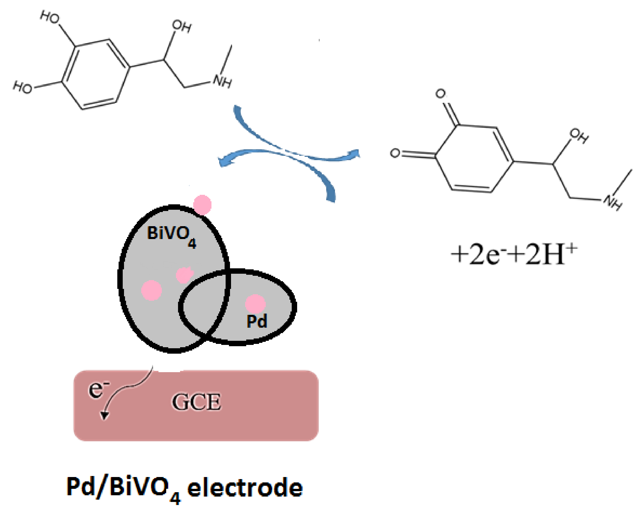
The synthesized 1%Pd/BiVO4 was used as sensing material for epinephrine detection in electrochemical system. The electrocatalytic redox mechanism of EP at 1% Pd/BiVO4/GCE is presented in Figure 9. The sensing materials catalyzed the oxidation of EP to form epinephrine quinone, as illustrated in Equation (1) [18,19,20].
C9H13NO3 => C9H11NO3 + 2 H+ + 2e−
The improvement of the electrochemical sensing performance might be attributed to the highly dispersed Pd nanoparticles on the surface of BiVO4 efficiently collecting the EP oxidation-induced electrons during the electrocatalytic reaction to transfer to BiVO4 and then the surface of GCE.
In the meantime, the presence of an appropriate amount of Pd (1%) nanoparticles can accelerate the electrocatalysis-induced electron–hole pair separation and superoxide radical formation [18]. Appropriate amounts of additional Pd can promote the sensing signals; the overloading palladium of 2% might block the surface-active sites of BiVO4. Therefore, the 1% Pd/BiVO4 composite exhibits a quicker electrochemical detection of epinephrine.
3. Experiment
3.1. Materials
Bismuth(III) nitrate pentahydrate (Bi(NO3)3·5H2O, 98%), palladium (II) nitrate (Pd(NO3)2, 99.9%), ammonium metavanadate (NH4VO3, >99%), and ethylene dinitrilotetraacetic acid (EDTA, 99%) were purchased from Sigma-Aldrich. Nitric acid (HNO3, 78–80%), sodium hydroxide (NaOH, 99.9%), and methanol (CH3OH, 99.9%) were procured from J.T. Baker (Phillipsburg, NJ, USA). Epinephrine (98%, Aladdin Industrial Corporation (Shanghai, China)) and lidocaine were offered by Hualien Tzu-Chi Hospital. The above materials were used without further purification. Distilled and deionized (DI) water was used in the experiments prepared by a Milli-Q water purification system (Millipore).
3.2. Preparation of Sensing Materials
The amount of 4.37 g Bi(NO3)3·5H2O, 2.63 g EDTA and 1 ml HNO3 were dissolved in 50 mL DI water with magnetic stirring at 90 °C for 30 min to form solution A, and 1.05 g NH4VO3 was dissolved in 50 mL DI water under continuous stirring for 30 min at 60 °C to form solution B. Solutions A and B were mixed together with vigorous stirring at 50 °C for 1 h. Following this, the mixed solution’s pH value was adjusted to 7.0 by adding 1 M NH4OH solution. After that, the solution was poured into a Teflon-lined stainless-steel autoclave and then transferred into an oven then heated at 180 °C for 6 h. The obtained precipitate was filtered and washed with DI water and ethanol several times, and then dried at 100 °C for 12 h and calcined at 450 °C for 4 h to obtain pure BiVO4 powder. The amount of 0.5 g BiVO4 and appropriate amounts of Pd(NO3)2 were added into 100 mL DI water and stirred in an ice bath for 2 h; subsequently, 0.1 M NaBH4 was added whilst stirring, then the mixture was stirred in an ice bath for 4 h. Finally, the precipitate was washed with ethanol, DI water, and then dried at 80 °C in an oven, until the dark green Pd/BiVO4 powder was obtained.
3.3. Preparation of Pd-BiVO4/GCE
The glassy carbon electrode (GCE, 3 mm diameter.) was first polished carefully with 0.01 mm α-Al2O3 powder and then a fine emery paper. Al2O3 and other residues were then removed by rinsing the surface with distilled water and ethanol, and then the electrode was sonic cleaned in distilled water and ethanol for 5 min, respectively. An appropriate amount of Pd-BiVO4 with 5% chitosan and 5 mL acetone were mixed ultrasonically for more than 30 min. Then, around 50–75 μL of this slurry was dropped onto the freshly polished GCE via a syringe. After being dried in an oven at 80 °C, making the acetone evaporate completely, the prepared electrode (Pd-BiVO4/GCE) was used as the working electrode.
3.4. XRD, SEM and TEM/EDX
The crystal structure of the Pd/BiVO4 composite material was characterized through X-ray diffractometer (Shimadzu XRD-6000, Cu X-ray tube (Cu Kα 1 = 1.54060 Å)). The characterization of the particle planes were conducted through XRD at a voltage of 40.0 keV, with the 2θ scanning range from 10 to 80°, at a scanning rate of 4 °/min. The surface microstructure of the samples was studied by a field emission scanning electron microscope (FE-SEM) (JEOL JSM-7000F). The morphology of the sensing materials was investigated through a high-resolution transmission electron microscope (HR-TEM) (JEM-2010). The elements of the samples were analyzed by an energy dispersive spectrometer (EDS) (England EXFORD Inca X-Stream).
3.5. Electrochemical Detection System
The electrochemical detection system is shown in Figure 1b. The potentiostat/galvanostat (PG-stat) (ZIVE SP1 compact type Electrochemical Workstation) was applied for the electrochemical measurements by cyclic voltammetry (CV) and differential pulse voltammetry (DPV) analysis. In this electrochemical system, three electrodes were used; BiVO4- and Pd/BiVO4-coated glassy carbon electrode (GCE) as the working electrode, a saturated Ag/AgCl electrode as the reference electrode, and platinum wire as the counter electrode.
4. Conclusions
In this research, the Pd/BiVO4 composite was fabricated for electrochemical sensing of epinephrine and was characterized using XRD, SEM, TEM and EDS. High-purity BiVO4 doping with Pd nanoparticles was obtained. The 1% Pd/BiVO4-modified GCE electrode presented favorable electrocatalytic behavior using both CV and DPV methods with a good redox sensing signal. The concentration of epinephrine was determined with a linear range of 0.9 µM to 27.5 µM; high correlation coefficient (R2 = 0.9998 for CV, 1.00 for DPV) and low limit of detection (0.262 µM for CV, 0.154 µM for DPV) were obtained. The proposed modified electrode also demonstrates excellent reproducibility, stability, and selectivity. Combing the simple fabrication process and low cost, 1% Pd/BiVO4 has great potential as a sensing element in practical applications of epinephrine detection.
Author Contributions
Conceptualization, H.-N.L. and R.-J.W.; methodology, R.-J.W.; software, Y.-S.L.; validation, B.-H.H.; formal analysis, B.-H.H.; investigation, B.-H.H.; resources, B.-H.H.; data curation, B.-H.H.; writing—original draft preparation, R.-J.W.; writing—review and editing, H.L. and R.-J.W.; supervision, H.-N.L. and T.-Y.C.; project administration, R.-J.W.; funding acquisition, H.-N.L. All authors have read and agreed to the published version of the manuscript.
Funding
This research was funded by Ministry of Science and Technology (Grant No.: MOST 110-2113-M-126-001), Taiwan.
Acknowledgments
The authors would like to thank the Ministry of Science and Technology (Grant No.: MOST 110-2113-M-126-001), Taiwan, R.O.C., for the financial support that it has provided for this study.
Conflicts of Interest
The authors declare no conflict of interest.
References
- Emran, M.Y.; Khalifa, H.; Gomaa, H.; Shenashen, M.; Akhtar, N.; Mekawy, M.; Faheem, A.; El-Safty, S.A. Hierarchical C-N doped NiO with dual-head echinop flowers for ultrasensitive monitoring of epinephrine in human blood serum. Microchim. Acta 2017, 184, 4553–4562. [Google Scholar] [CrossRef]
- Goldberg, E.; Grau, J.B.; Fortier, J.; Salvati, E.; Levy, R.J.; Ferrari, G. Serotonin and catecholamines in the development and progression of heart valve diseases. Cardiovasc. Res. 2017, 113, 849–857. [Google Scholar] [CrossRef] [PubMed] [Green Version]
- Zhang, G.; Zhang, Y.; Ji, C.; McDonald, T.; Walton, J.; Groeber, E.A.; Steenwyk, R.C.; Lin, Z. Ultra sensitive measurement of endogenous epinephrine and norepinephrine in human plasma by semi-automated SPE-LC–MS/MS. J. Chromatogr. B 2012, 895–896, 186–190. [Google Scholar] [CrossRef] [PubMed]
- Davletbaeva, P.; Falkova, M.; Safonova, E.; Moskvin, L.; Bulatov, A. Flow method based on cloud point extraction for fluorometric determination of epinephrine in human urine. Anal. Chim. Acta 2016, 911, 69–74. [Google Scholar] [CrossRef] [PubMed]
- Li, T.; Wang, Z.; Xie, H.; Fu, Z. Highly sensitive trivalent copper chelate-luminol chemiluminescence system for capillary electrophoresis detection of epinephrine in the urine of smoker. J. Chromatogr. B 2012, 911, 1–5. [Google Scholar] [CrossRef] [PubMed]
- Wierzbicka, E.; Sulka, G.D. Nanoporous spongelike Au–Ag films for electrochemical epinephrine sensing. J. Electroanal. Chem. 2016, 762, 43–50. [Google Scholar] [CrossRef]
- Wierzbicka, E.; Szultka-Młyńska, M.; Buszewski, B.; Sulka, G.D. Epinephrine sensing at nanostructured Au electrode and determination its oxidative metabolism. Sens. Actuators B Chem. 2016, 237, 206–215. [Google Scholar] [CrossRef]
- Wierzbicka, E.; Sulka, G.D. Fabrication of highly ordered nanoporous thin Au films and their application for electrochemical determination of epinephrine. Sens. Actuators B Chem. 2016, 222, 270–279. [Google Scholar] [CrossRef]
- Dong, W.; Ren, Y.; Bai, Z.; Jiao, J.; Chen, Y.; Han, B.; Chen, Q. Synthesis of tetrahexahedral Au-Pd core–shell nanocrystals and reduction of graphene oxide for the electrochemical detection of epinephrine. J. Colloid Interface Sci. 2018, 512, 812–818. [Google Scholar] [CrossRef] [PubMed]
- Tashkhourian, J.; Nami-Ana, S.F.; Shamsipur, M. Designing a modified electrode based on graphene quantum dot-chitosan application to electrochemical detection of epinephrine. J. Mol. Liq. 2018, 266, 548–556. [Google Scholar] [CrossRef]
- Zou, L.; Li, Y.; Cao, S.; Ye, B. Gold nanoparticles/polyaniline Langmuir–Blodgett Film modified glassy carbon electrode as voltammetric sensor for detection of epinephrine and uric acid. Talanta 2013, 117, 333–337. [Google Scholar] [CrossRef] [PubMed]
- Bavandpour, R.; Karimi-Maleh, H.; Asif, M.; Gupta, V.K.; Atar, N.; Abbasghorbani, M. Liquid phase determination of adrenaline uses a voltammetric sensor employing CuFe2O4 nanoparticles and room temperature ionic liquids. J. Mol. Liq. 2016, 213, 369–373. [Google Scholar] [CrossRef]
- Zhang, J.; Geng, S.; Li, R.; Zhang, X.; Zhou, Y.; Yu, T.; Wang, Y.; Song, S.; Shao, Z. Novel monoclinic ABO4 oxide with single-crystal structure as next generation electrocatalyst for oxygen evolution reaction. Chem. Eng. J. 2021, 420, 130492. [Google Scholar] [CrossRef]
- Srinivasan, R.; Elaiyappillai, E.; Anandaraj, S.; Duvaragan, B.K.; Johnson, P.M. Study on the electrochemical behavior of BiVO4/PANI composite as a high performance supercapacitor material with excellent cyclic stability. J. Electroanal. Chem. 2020, 861, 113972. [Google Scholar] [CrossRef]
- Kangkun, N.; Ponchio, C. Photoelectrodeposition of BiVO4 layer on FTO/WO3 photoanodes for highly efficient photoelectrocatalytic chemical oxygen demand sensor applications. Appl. Surf. Sci. 2020, 526, 146686. [Google Scholar] [CrossRef]
- Chen, L.; Miao, L.; Chen, Y.; Gao, Y.; Di, J. An enzyme-free photoelectrochemical glucose sensor based on coupling BiVO4 with gold nanoparticles. Mater. Sci. Semicond. Process. 2021, 125, 105632. [Google Scholar] [CrossRef]
- Wang, L.; Bian, Z. Photocatalytic degradation of paracetamol on Pd-BiVO4 under visible light irradiation. Chemosphere 2020, 239, 124815. [Google Scholar] [CrossRef] [PubMed]
- Shaikshavali, P.; Reddy, T.M.; Gopal, T.V.; Venkataprasad, G.; Kotakadi, V.S.; Palakollu, V.; Karpoormath, R. A simple sonochemical assisted synthesis of nanocomposite (ZnO/MWCNTs) for electrochemical sensing of Epinephrine in human serum and pharmaceutical formulation. Colloids Surfaces A Physicochem. Eng. Asp. 2020, 584, 124038. [Google Scholar] [CrossRef]
- Luk, H.-N.; Dai, T.-H.; Wu, R.-J.; Chavali, M. Sensing properties of Pt@SnO2 core-shell nanocomposite detecting Epinephrine. J. Chin. Chem. Soc. 2020, 67, 1431–1436. [Google Scholar] [CrossRef]
- Pradhan, S.; Banerjee, M.B.; Biswas, S.; Hamizi, N.A.; Das, D.K.; Bhar, R.; Bandyopadhyay, R.; Pramanik, P. An Efficient Simultaneous Electrochemical Detection of Nanomolar Epinephrine and Uric Acid using Low Temperature Synthesized Nano-sized Copper Telluride. Electroanalysis 2021, 33, 383–392. [Google Scholar] [CrossRef]
Figure 1.
(a) Chemical structure of epinephrine, and (b) Electrochemical detection system for epinephrine.
Figure 1.
(a) Chemical structure of epinephrine, and (b) Electrochemical detection system for epinephrine.

Figure 2.
XRD patterns of obtained samples.
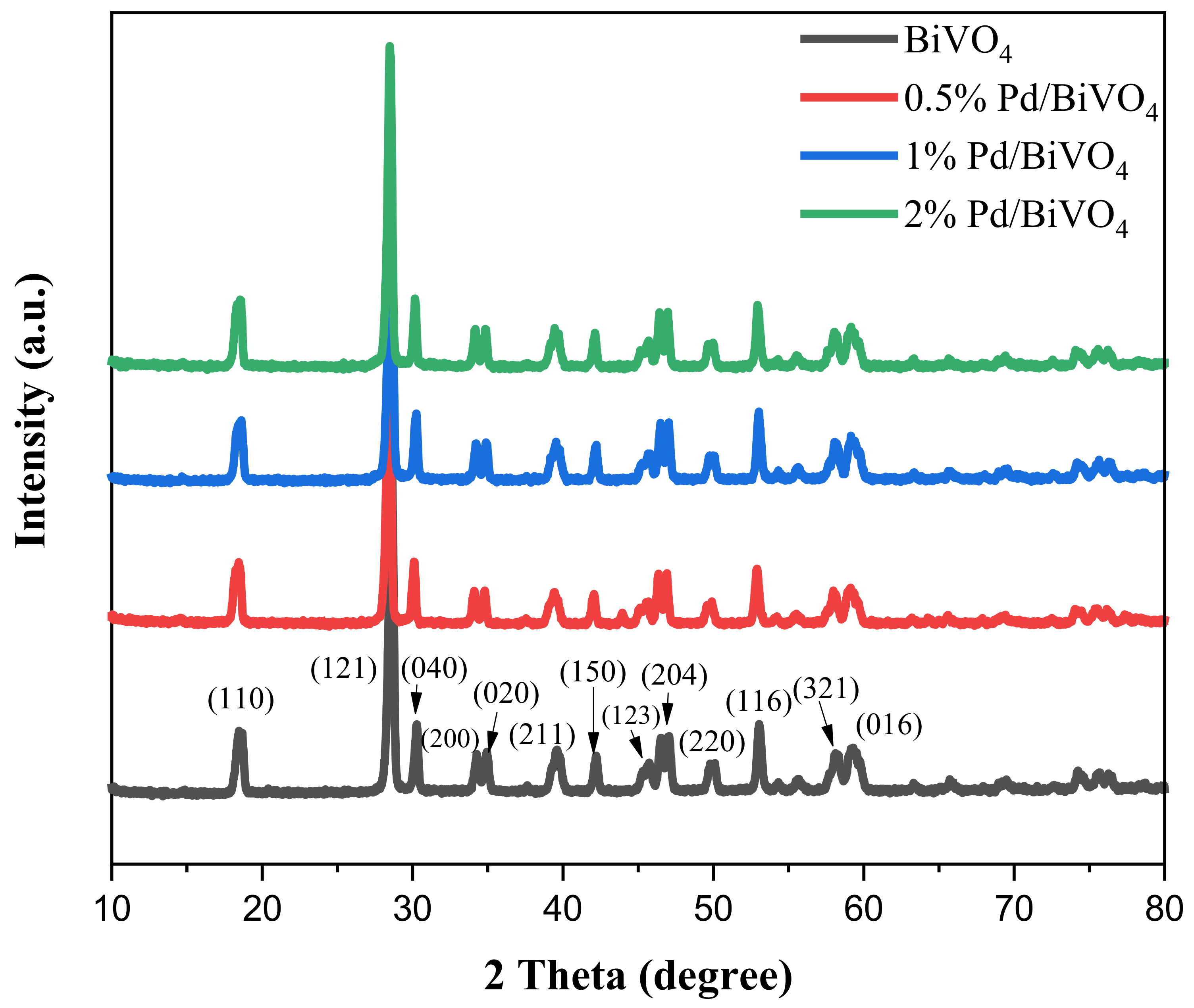
Figure 3.
SEM images of (a) BiVO4, (b) 1% Pd/BiVO4 (×5000), and (c) 1% Pd/BiVO4 (×20,000).
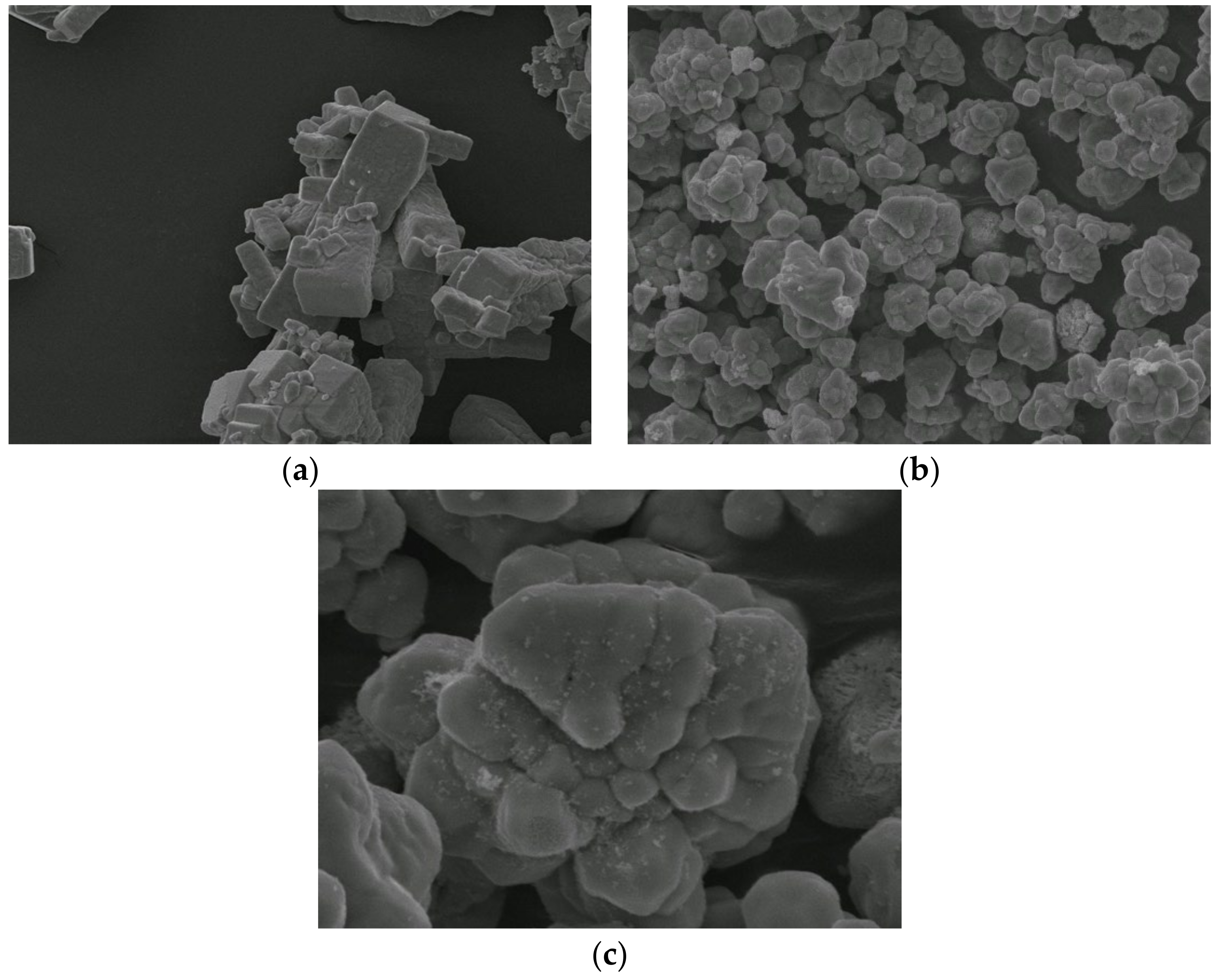
Figure 4.
(a) TEM images of 1%Pd/BiVO4; (b) TEM images of 1%Pd/BiVO4 with high magnification, and (c) EDS spectrum of 1%Pd/BiVO4.
Figure 4.
(a) TEM images of 1%Pd/BiVO4; (b) TEM images of 1%Pd/BiVO4 with high magnification, and (c) EDS spectrum of 1%Pd/BiVO4.
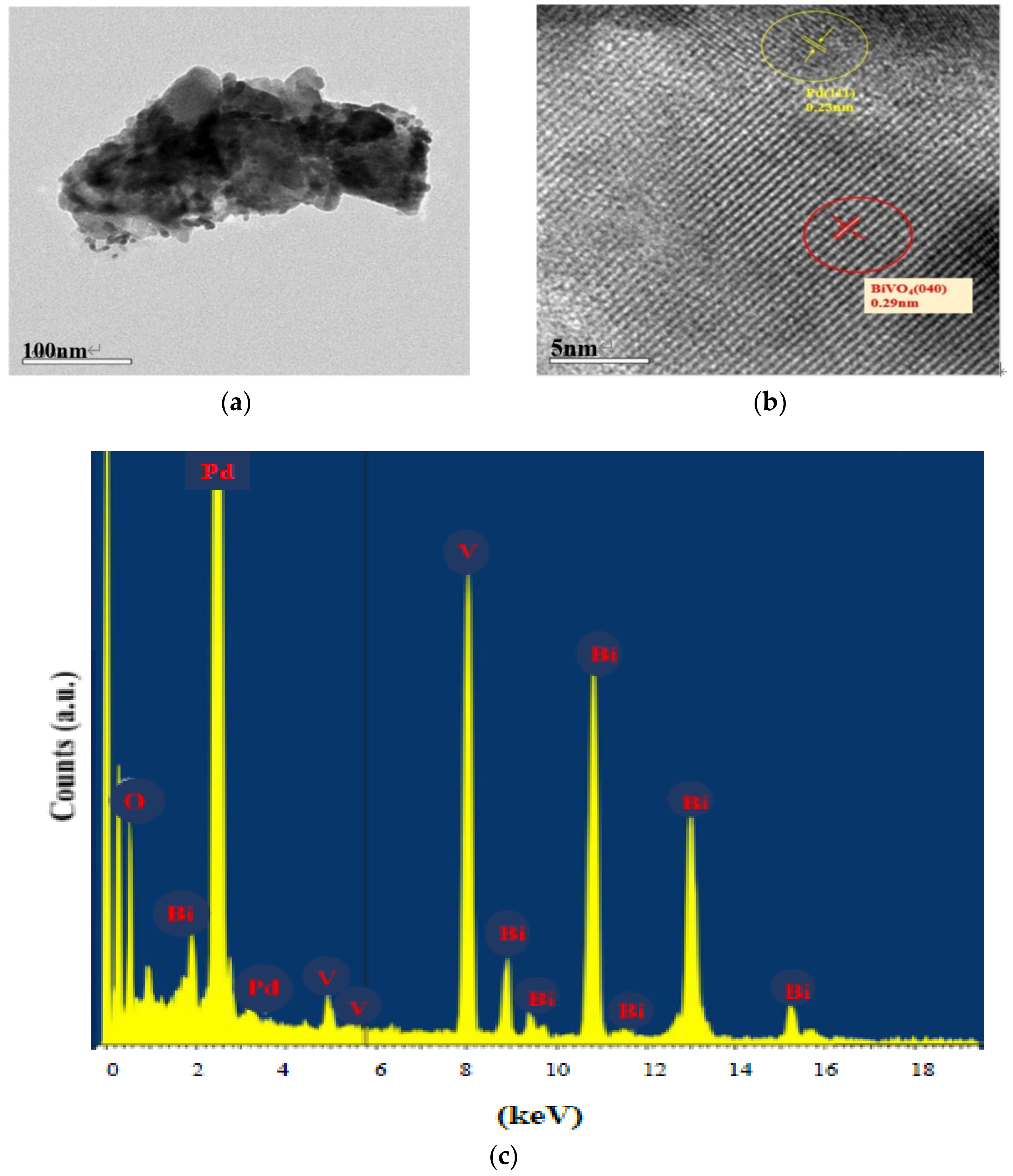
Figure 5.
(A) CV response of (a) bare GCE (b) BiPO4/GCE (c) 1%Pd/BiVO4/GCE at scan rate of 1.0 mVs−1; (B) CV response of 27.5 µM epinephrine at Pd/BiVO4/GCE with various Pd amounts; (C) CV response at 1%Pd/BiVO4/GCE with various epinephrine concentrations (a) 0.9 µM (b) 1.7 µM (c) 3.4 µM (d) 6.9 µM (e) 13.8 µM (f) 27.5 µM; (D) The plot of Ipa against various epinephrine concentrations at 1%Pd/BiVO4/GCE.
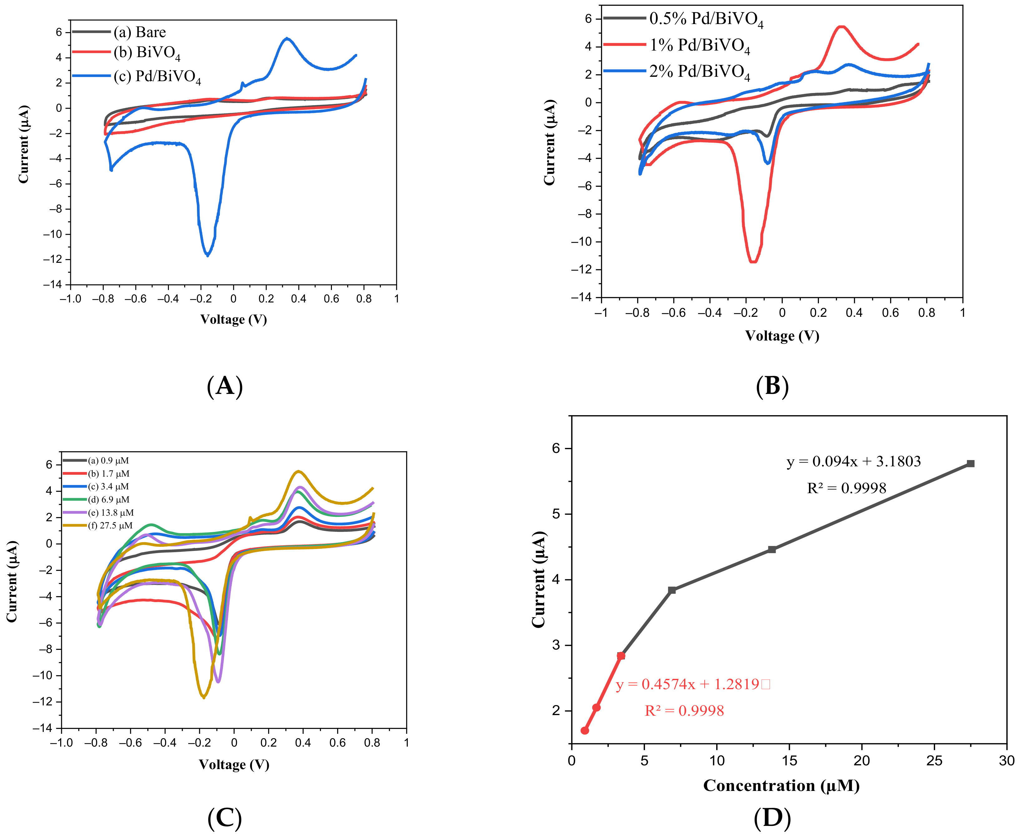
Figure 6.
(A) DPV I-V curve of 1%Pd/BiVO4/GCE with various concentrations of epinephrine (a) 0.9 µM (b) 1.7 µM (c) 3.4 µM (d) 6.9 µM (e) 13.8 µM (f) 27.5 µM; (B) The plot of DPV current against various epinephrine concentrations at 1%Pd/BiVO4/GCE.
Figure 6.
(A) DPV I-V curve of 1%Pd/BiVO4/GCE with various concentrations of epinephrine (a) 0.9 µM (b) 1.7 µM (c) 3.4 µM (d) 6.9 µM (e) 13.8 µM (f) 27.5 µM; (B) The plot of DPV current against various epinephrine concentrations at 1%Pd/BiVO4/GCE.
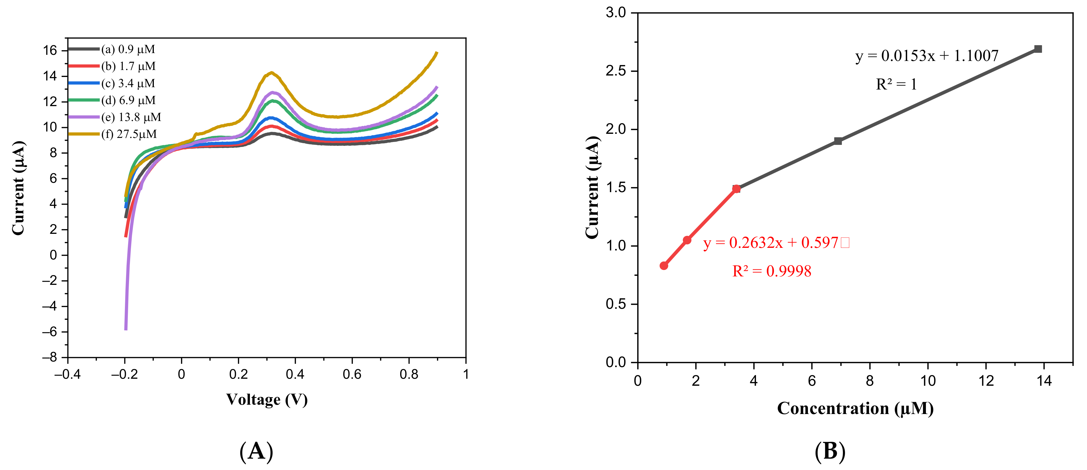
Figure 7.
The stability test of the 1% Pd/BiVO4/GCE on 3.4 µM EP.
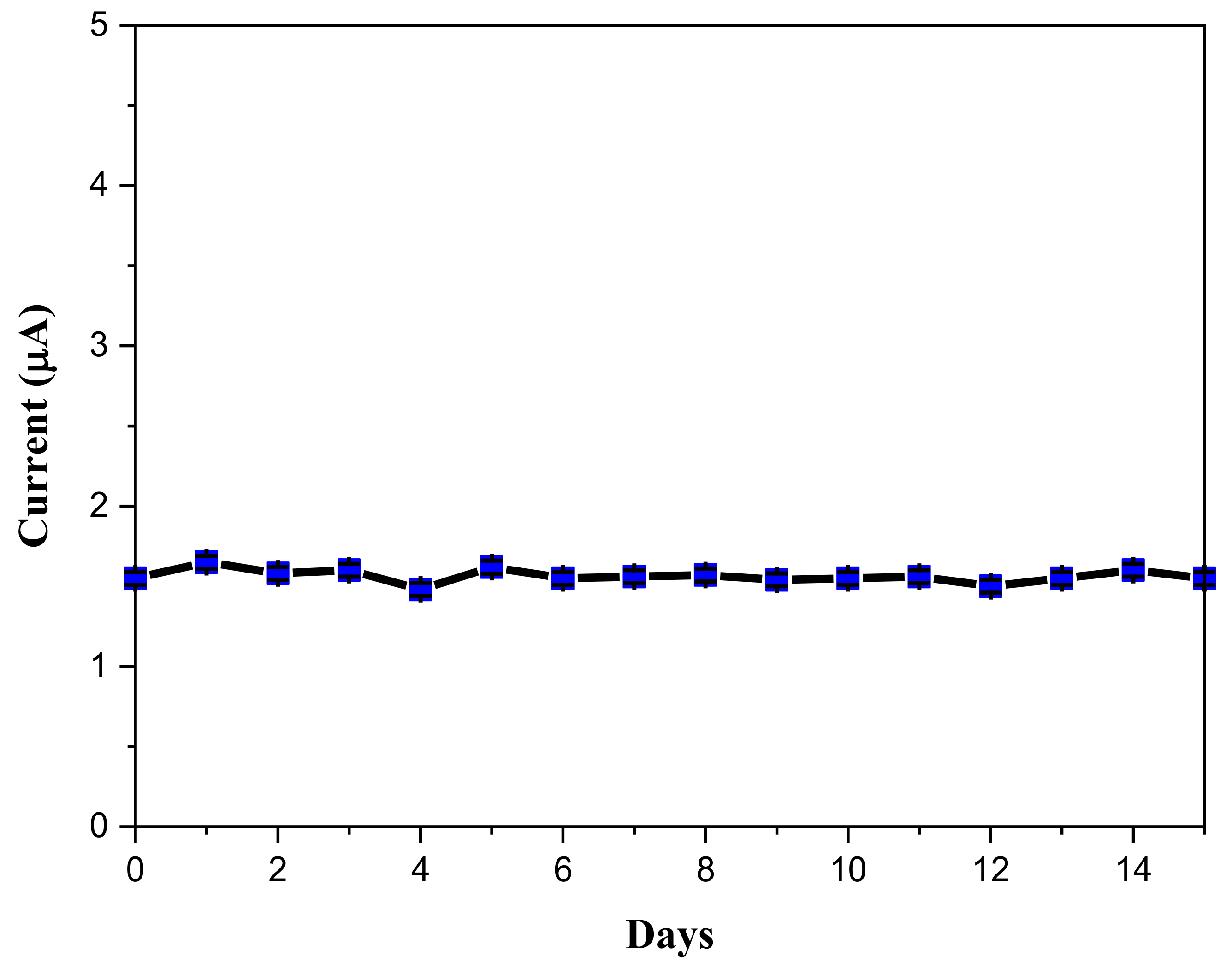
Figure 8.
(a) and (b) Interference test of CV current to 3.4 µM ascorbic acid (0.42 V), L-cysteine (0.38 V) and dopamine (0.22 V) comparing to epinephrine (0.40 V).
Figure 8.
(a) and (b) Interference test of CV current to 3.4 µM ascorbic acid (0.42 V), L-cysteine (0.38 V) and dopamine (0.22 V) comparing to epinephrine (0.40 V).
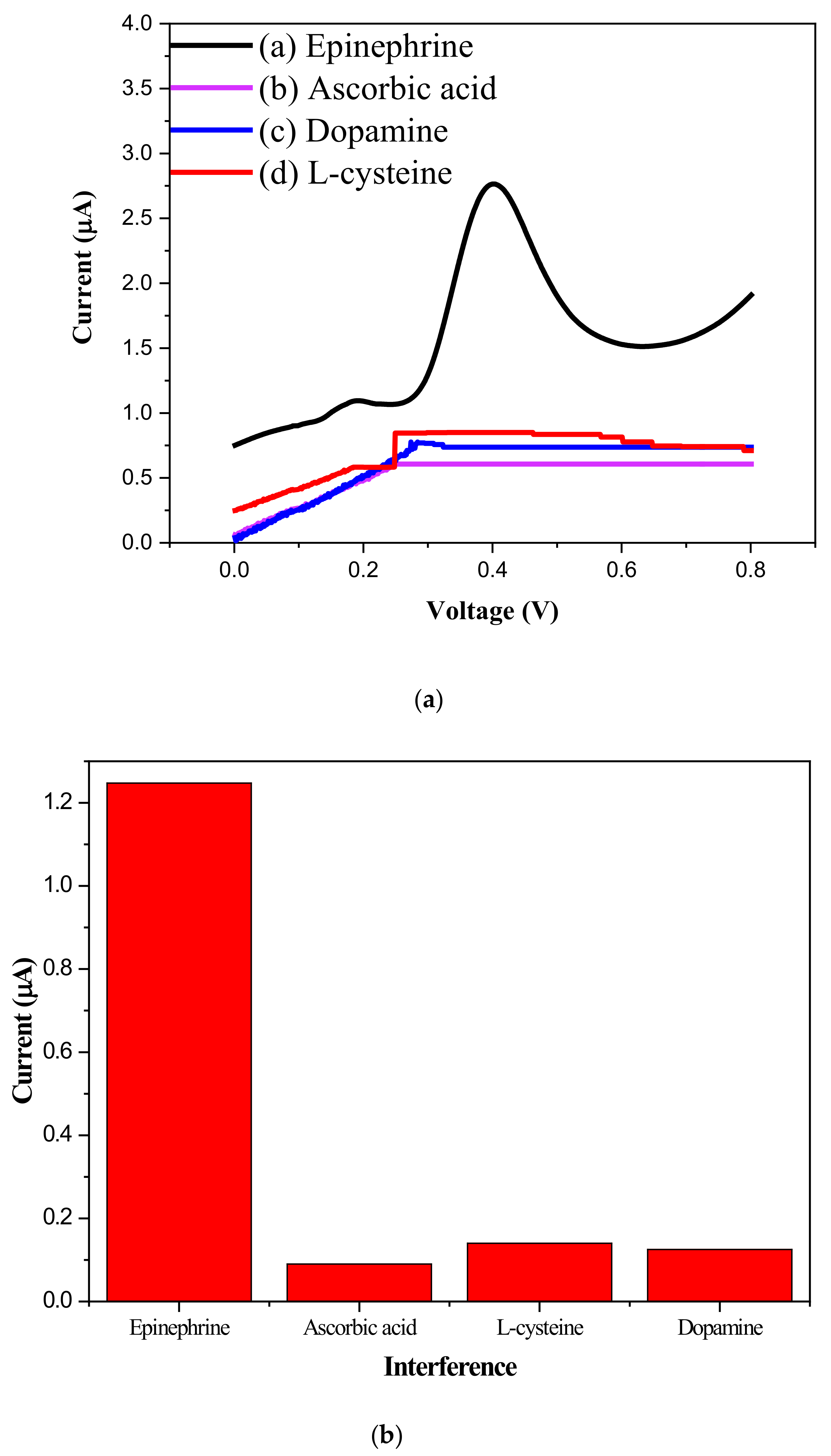
Figure 9.
Electrochemical oxidation mechanism of epinephrine on Pd/BiVO4/GCE modified electrode.

Table 1.
Comparison with epinephrine sensing properties on various sensing materials.
| Author/Year/Reference | Sensing Material | Range of Detection (µM) | Detection Limit (µM) |
|---|---|---|---|
| E. Wierzbicka /2016/[8] | Nano Au/high ordered anodic Al2O3 | 10–150 | 3 |
| W. Dong/2018/[9] | Au-Pd@reduced graphene oxide | 0.001–1000 | 0.0012 |
| J. Tashkhourian /2018/[10] | Graphene | 0.36–380 | 0.16 |
| L. Zou/2018/[11] | Au-polyaniline | 0.4–10 | 0.08 |
| This work | 1% Pd/BiVO4 | 0.9–27.5 | CV: 0.262 (R2 = 0.9998) DPV: 0.154 (R2 = 0.9998) |
Publisher’s Note: MDPI stays neutral with regard to jurisdictional claims in published maps and institutional affiliations. |
© 2021 by the authors. Licensee MDPI, Basel, Switzerland. This article is an open access article distributed under the terms and conditions of the Creative Commons Attribution (CC BY) license (https://creativecommons.org/licenses/by/4.0/).
Share and Cite
MDPI and ACS Style
Luk, H.-N.; Chou, T.-Y.; Huang, B.-H.; Lin, Y.-S.; Li, H.; Wu, R.-J. Promotion Effect of Palladium on BiVO4 Sensing Material for Epinephrine Detection. Catalysts 2021, 11, 1083. https://doi.org/10.3390/catal11091083
AMA Style
Luk H-N, Chou T-Y, Huang B-H, Lin Y-S, Li H, Wu R-J. Promotion Effect of Palladium on BiVO4 Sensing Material for Epinephrine Detection. Catalysts. 2021; 11(9):1083. https://doi.org/10.3390/catal11091083
Chicago/Turabian StyleLuk, Hsiang-Ning, Tsong-Yung Chou, Bai-Hao Huang, Yu-Syuan Lin, Hui Li, and Ren-Jang Wu. 2021. "Promotion Effect of Palladium on BiVO4 Sensing Material for Epinephrine Detection" Catalysts 11, no. 9: 1083. https://doi.org/10.3390/catal11091083
Note that from the first issue of 2016, this journal uses article numbers instead of page numbers. See further details here.