Article

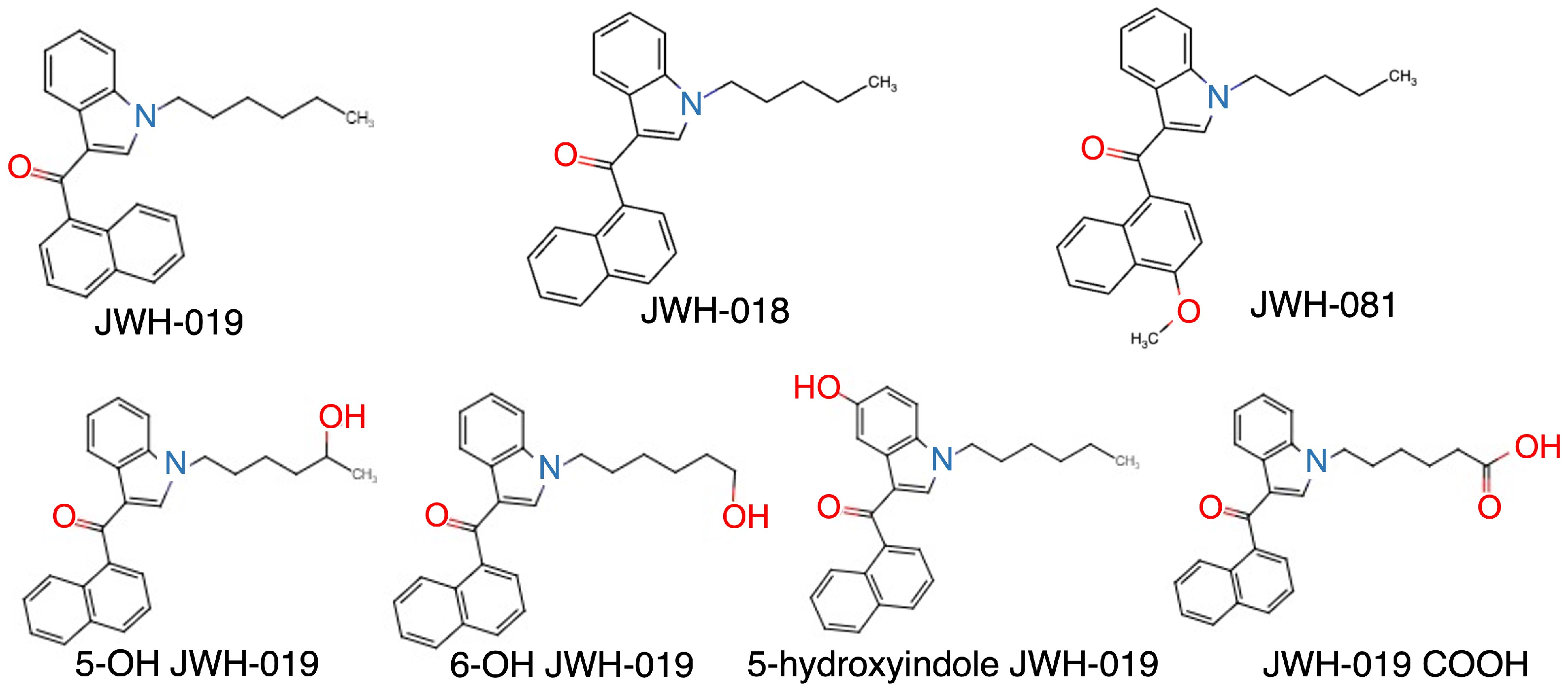
Identification of Cytochrome P450 Enzymes Responsible for Oxidative Metabolism of Synthetic Cannabinoid (1-Hexyl-1H-Indol-3-yl)-1-naphthalenyl-methanone (JWH-019)

1
Department of Pharmaceutical Sciences, College of Pharmacy, University of Arkansas for Medical Sciences, Little Rock, AR 72205, USA
2
Department of Pharmacology & Toxicology, College of Medicine, University of Arkansas for Medical Sciences, Little Rock, AR 72205, USA
3
Department of Pharmacy Practice, College of Pharmacy, University of Arkansas for Medical Sciences, Little Rock, AR 72205, USA
4
Department of Pharmaceutical Sciences, College of Pharmacy, Northeast Ohio Medical University, Rootstown, OH 44272, USA
*
Author to whom correspondence should be addressed.
Catalysts 2023, 13(6), 1008; https://doi.org/10.3390/catal13061008
Submission received: 14 April 2023 / Revised: 8 June 2023 / Accepted: 13 June 2023 / Published: 15 June 2023
(This article belongs to the Special Issue Enzymes in Biomedical, Cosmetic and Food Application)

Abstract

:
(1-Hexyl-1H-indol-3-yl)-1-naphthalenyl-methanone (JWH-019) is one of the second-generation synthetic cannabinoids which as a group have been associated with severe adverse reactions in humans. Although metabolic activation can be involved in the mechanism of action, the metabolic pathway of JWH-019 has not been fully investigated. In the present study, we aimed to identify the enzymes involved in the metabolism of JWH-019. JWH-019 was incubated with human liver microsomes (HLMs) and recombinant cytochrome P450s (P450s or CYPs). An animal study was also conducted to determine the contribution of the metabolic reaction to the onset of action. Using an ultra-performance liquid chromatography system connected to a single-quadrupole mass detector, we identified 6-OH JWH-019 as the main oxidative metabolite in HLMs supplemented with NADPH. JWH-019 was extensively metabolized to 6-OH JWH-019 in HLMs with the KM and Vmax values of 31.5 µM and 432.0 pmol/min/mg. The relative activity factor method estimated that CYP1A2 is the primary contributor to the metabolic reaction in the human liver. The animal study revealed that JWH-019 had a slower onset of action compared to natural and other synthetic cannabinoids. CYP1A2 mediates the metabolic activation of JWH-019, contributing to the slower onset of its pharmacological action.

1. Introduction

Marijuana, Cannabis sativa, is one of the most pervasive drugs in the world due to its popularity in both medicinal and recreational use, with a significant increase in consumption being reported in the United States during the 21st century [1]. Synthetic cannabinoids (SCBs) are a large and chemically diverse group of substances. SCBs were initially created as investigatory cannabinoid receptor agonists, which activate central cannabinoid (CB1) and peripheral cannabinoid (CB2) receptors, the same receptors on which the naturally occurring cannabinoids found in marijuana exert their effects [2]. While researchers study the therapeutic potential of SCBs, the use of SCBs has deviated from its original intent, leading to abuse by the general public due to their initial uncontrolled legal status, low cost, ease of availability, difficulty of detection, and pronounced psychoactive effects [3,4,5]. SCBs are known as being natural and safe to consumers; however, they are neither natural nor safe, causing serious adverse reactions, including seizures, nephrotoxicity, and, in some cases, death [6,7,8]. With growing awareness and popularity, the number of SC users reporting adverse events dramatically increased over the last decade [9,10]. Case reports and user experiences indicate there is extreme variability in the pattern, duration, and severity of individual clinical courses following SC use. The continued limited understanding of the basic pharmacokinetic and dynamic specifics of individual SC compounds significantly complicates the clinical management of patients seeking medical attention. Care for these patients is largely symptomatic and supportive as, to this point, no specific antidote or therapy is available that targets SCBs in intoxicated users.
The substance (1-Hexyl-1H-indol-3-yl)-1-naphthalenyl-methanone (JWH-019) is a member of the second generation of SCBs with the molecular formula C25H25NO and a molecular mass of 355.5 [11]. It is one of the cannabinoid receptor agonists from the napthoylindole family that were synthesized during the 1990s and has seen widespread human recreational use in the U.S. since 2010 [12]. This synthetic compound is more potent and addictive than the natural cannabinoids and is associated with concerns of causing severe complications for human health [13]. While significant limitations in clinical testing have hampered clear delineation and complete characterization of toxicity among SCBs, the medical literature does contain several reports of analytically confirmed involvement of JWH-019 in patients experiencing adverse medical consequence. In 2013, there were 29 subjects reporting to the ER with restlessness, hallucination, vertigo, and anxiousness after ingesting SCBs including JWH-019 [14]. In a 2014 report, a twenty-year-old male is described as having driving skill impairment, vestibular disorder, enlarged pupils, and blunt mood during a traffic stop. In this case a toxicology blood sample was taken after 80 min and the result was positive for four SCs including JWH-019 (1.7 ng/mL) [15]. In 2015, a fifty-year-old man is described as losing consciousness while working and died after being transferred to the hospital. In his initial toxicology findings there was no detection of common psychoactive substances or drug of abuse but, three years later, using the same sample on gas chromatography–mass spectrometry urinalyses the reanalysis found the presence of JWH-019 (278 ng/mL) [16]. Additionally, calls to the United States Poison Control Centers involving exposure to SCBs increased from 14 in 2009 to 6968 by year end of 2011. As the significant harms from SCBs became apparent, an initial attempt in the United States to curb the open availability associated with the sale of many SCBs in the early portion of the last decade was the passage of the Synthetic Drug Abuse Prevention Act in June of 2012. This legislation categorized several chemical structure classes of cannabinoids as schedule I substances. Fifteen SCBs were named in the Act and JWH-019 was included among them [17].
Xenobiotics are transformed in the body by phase I and II drug-metabolizing enzymes (DMEs), such as cytochrome P450s (CYPs) and UDP-glucuronosyltransferases (UGTs), respectively [18,19]. CYPs and UGTs are the most important enzymes that catalyze oxidation and glucuronidation of xenobiotics; therefore, it was hypothesized that CYPs and UGTs are the main enzymes contributing to the metabolism of SCBs. They metabolize lipophilic xenobiotics by introducing a hydrophilic functional group [18,19]. The metabolites formed by DMEs are more readily excreted from the body due to increased water solubility. These detoxification enzymes are abundantly expressed in the liver and extrahepatic tissues, playing an important role in systemic and local metabolism of xenobiotics [19,20,21]. Oxidized and glucuronidated SCB metabolites have been identified in the blood and urine of SCB users [22,23], suggesting an involvement of CYPs and UGTs in the clearance of SCBs.
The JWH-019 homolog JWH-018 was identified as one of the earliest compounds detected early in the development of increased SCB recreational use [24]. As such, there has been significantly more investigation into this compound and there are many studies on its metabolic process and enzymes involved in its metabolism. JWH-018 shares a similar structure to JWH-019, with the only difference being the length of the alkyl chain. Currently, the process of metabolism of JWH-019 remains unclear. However, based on the similarity in the structure [25], it was hypothesized that JWH-019 would follow the same metabolic pathway as JWH-018 without supporting evidence. Based on the hypothesis, JWH-019 could undergo oxidation to form hydroxyindole (5-hydroxyindole JWH-019), N-hexanoic acid (JWH-019 COOH) and two monohydroxylated at the N-alkyl chain (5-OH JWH-019 and 6-OH JWH-019) metabolites (Figure 1). According to a recent study that analyzed urine samples from drug users that were positive for JWH-019, three out of four metabolites, 5-OH JWH-019, 6-OH JWH-019, and JWH-019 COOH, have been confirmed [26]. However, it is still unclear which is the primary metabolite or which drug metabolizing enzyme is responsible for the oxidation reaction.
The aim of this study is to identify the main metabolite of JWH-019 and the P450 isoforms that are responsible for the metabolite formation in vitro. We also conducted an animal study to determine the contribution of the metabolic reaction to the onset of action.

2. Results

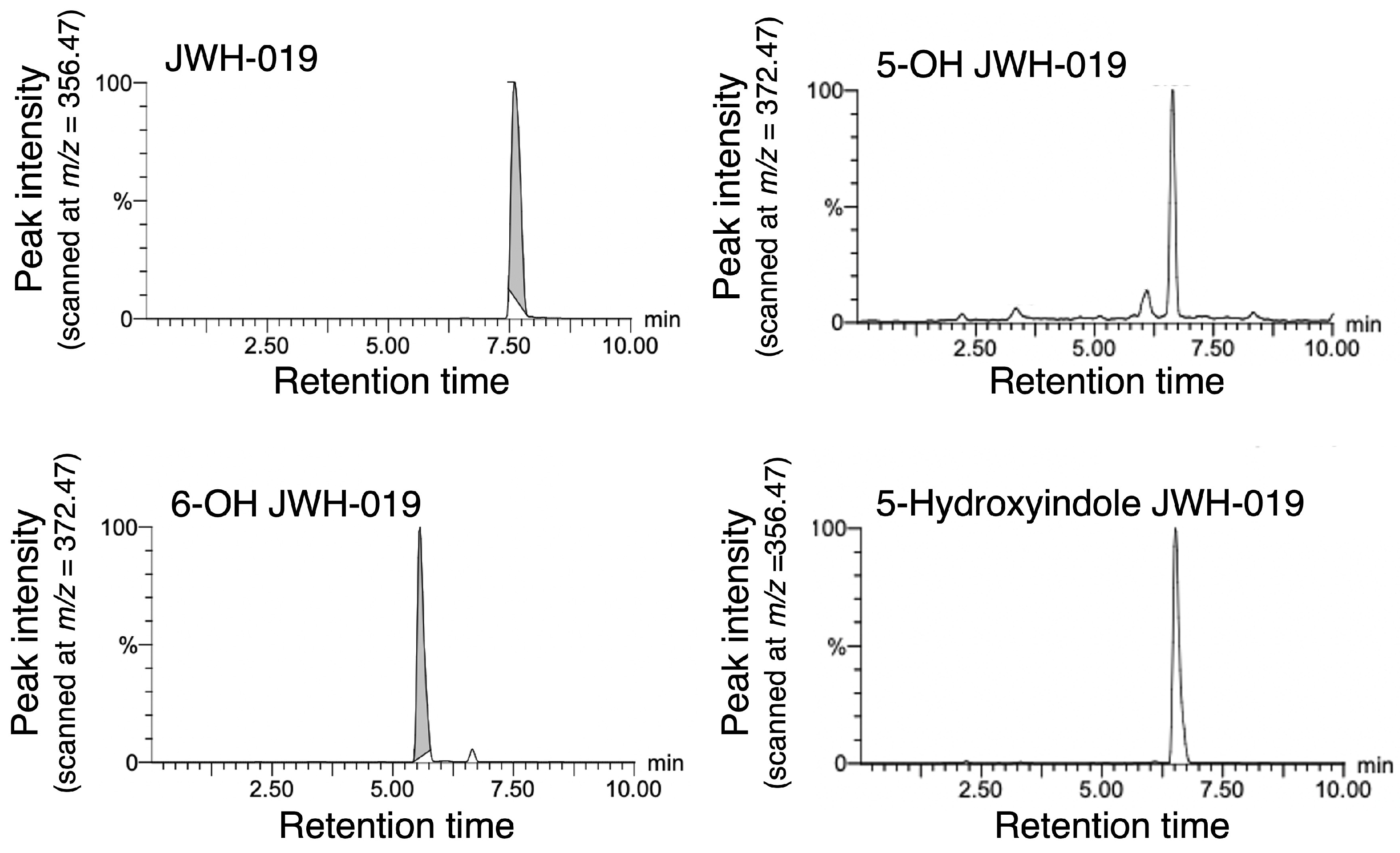
2.1. Development of a UPLC Method for JWH-019 and Its Metabolites

Currently, four oxidative metabolites of JWH-019 have been suggested, which are 5-OH JWH-019, 6-OH JWH-019, JWH-019 COOH, and 5-hydroxyindole JWH-019. Out of the four postulated metabolites, three, 5-OH JWH-019, 6-OH JWH-019, and JWH-019 COOH, have been previously detected in human urine samples [26]. However, the study was not quantitative. Furthermore, the enzymes that mediate the metabolic reactions of JWH-019 have not been determined to date. The liver is the most important tissue for the metabolism of xenobiotics. Among a number of hepatic detoxification enzymes, CYP is the most abundant and functional enzyme responsible for oxidative metabolism. Therefore, the first aim of this investigation was to identify metabolites of JWH-019 produced in HLMs where CYPs are located.
As a preliminary study, we tested the detection of JWH-019 and its metabolites with positive and negative modes of ionization using incubation mixtures. While the negative mode did not produce any significant peak of any compounds, the positive mode detected JWH-019 and metabolites. The data indicated that the highest sensitivity would be obtained at m/z = 356.47 (potentially JWH-019), 386.45 (potentially JWH-019 COOH), and 372.47 (potentially 5-hydroxyindole JWH-019, 5-OH JWH-019, and 6-OH JWH-019). Using the chemical standard, it was confirmed that the peak of m/z = 356.47 was JWH-019 with the retention time of 7.6 min. Similarly, we confirmed that a peak at the retention time of 5.5 min was 6-OH JWH-019. The retention times of hydroxyindole JWH-019 and 5-OH JWH-019 were 6.5 min and 6.6 min, respectively (Figure 2). Although we believe that JWH-019 COOH will be detected at m/z = 386.45, we were not able to determine the retention time of JWH-019 COOH due to the unavailability of the standard molecule on the market.
To investigate which form of metabolites can be produced by CYPs, JWH-019 (400 µM) was incubated with HLMs for 15 min in the presence and absence of the NADPH regenerating system. A significant peak was observed at 7.6 min regardless of the NADPH regenerating system when the incubated mixture was analyzed at m/z = 356.47, indicating that the substrate was not depleted at all with the 15 min incubation with HLMs (Figure 3A,B). A unique peak was observed at 5.5 min only in a sample that was incubated with NADPH when it was analyzed at m/z = 372.47 (Figure 3C,D), indicating that CYPs are responsible for producing 6-OH JWH-019. The peak size was incubation time dependent, supporting the involvement of enzyme reaction in the production of 6-OH JWH-019 in HLMs. In the same detection setting (m/z = 372.47), another peak was detected at 6.6 min in both incubated mixtures. The peak size, which was much smaller than that of 6-OH JWH-019, was not incubation time dependent, suggesting that a small amount of 5-OH JWH-019 was non-enzymatically produced in HLM regardless of the NADPH regenerating system. There was no peak at 6.5 min when incubation mixtures were analyzed at m/z = 372.47. In addition, there was no significant peak when samples were analyzed at m/z = 386.45 (Figure 3E,F). These data indicate that hydroxyindole JWH-019 or JWH-019 COOH was not produced, or the produced amount was below the limits of detection, in HLMs even with the presence of NADPH.

2.2. Kinetic Analysis of 6-OH JWH-019 Formation in HLMs

After the initial screening, it was determined that HLMs produced 6-OH JWH-019 as a main metabolite of JWH-019 when supplemented with NADPH. To further investigate the enzyme–substrate binding affinity and reaction rate in HLMs, we conducted the enzyme assay at a wide range of substrate concentrations (1–400 µM). The metabolic rate-substrate concentration plots showed a Michaelis–Menten curve (Figure 4). By fitting the data into the Michaelis–Menten equation, the KM and Vmax values were estimated to be 31.5 ± 3.3 µM and 432 ± 40 pmol/min/mg protein.

2.3. Identification of JWH-019-Oxidating Enzymes

CYPs are super-family enzymes. CYP1A2, -2A6, -2B6, -2C8, -2C9, -2C19, -2D6, -2E1, -2J2, -3A4, and -3A5 have been reported as main CYP isoforms expressed and functional in the human liver [27,28]. To identify the CYP isoforms responsible for the formation of 6-OH JWH-019 in HLMs, we used recombinant CYPs and assessed their respective abilities to metabolize JWH-019 using a substrate concentration of 400 μM. Of all the recombinant proteins that were screened, CYP2J2 showed the greatest activity for the formation of 6-OH JWH-019 at a reaction rate of 8.0 ± 0.3 nmol/min/nmol P450 (Table 1). CYP1A2, -2C19, -2D6, and -3A5 showed moderate activities with their reaction rates of 2.4 ± 0.1, 4.2 ± 0.04, 1.6 ± 0.1, and 2.0 ± 0.1 nmol/min/nmol P450, respectively. While CYP2B6, -2C8, -2C9, and -3A4 showed slight activities of the 6-OH JWN-019 formation, CYP2A6 or CYP2E1 barely showed the activity.
Each CYP isoform is differently expressed in the human liver. While CYP3A4 has been known as the isoform that is dominantly expressed in the liver, other isoforms such as CYP2J2 are hardly expressed in hepatic tissue [27,28]. To quantitatively determine the contribution of each CYP isoform to the formation of 6-OH JWH-019 in HLMs, we employed a relative activity factor (RAF) method. The contribution rate (%) was estimated by considering the enzyme activity of the recombinant enzyme and the relative expression level of each CYP isoform in the human liver. Even though CYP2J2 showed the highest activity in the earlier investigation, it was estimated to be the fifth contributor to the overall metabolic reaction in the liver (Table 1). The enzyme that contributes to the metabolism the most was estimated to be CYP1A2 with the contribution rate of 33.6%. CYP2C19, -2C9, and -3A4 were the second, third, and fourth contributors to the formation of 6-OH JWH-019, respectively, with their contribution rates ranging from 10.9 to 15.8%. While CYP2B6, -2C8, -2D6, and -3A5 made a slight contribution (1.1–6.1%), CYP2A6 and CYP2E1 made almost no contribution due to their lowest enzyme activity.

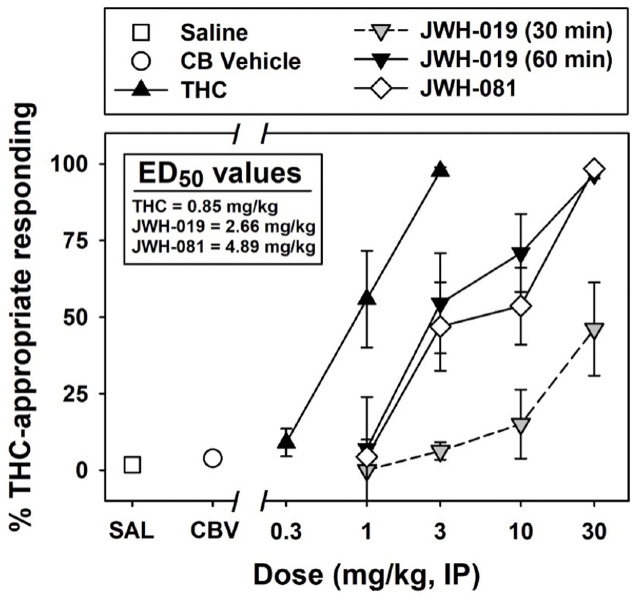
2.4. Pharmacological Activity of JWH-019

Metabolic reaction usually results in reduction of the pharmacological activity of the substances. However, in rare cases, metabolic activation occurs where metabolites show stronger pharmacological activity, which is evidenced by an enhanced pharmacological activity with increased pretreatment time.
Sprague Dawley rats were administered THC (0.3–3.0 mg/kg, I.P., N = 8), JWH-019 (1.0–30 mg/kg, I.P., N = 8), or JWH-081 (1.0–30 mg/kg, I.P., N = 8). All injections were made 30 min prior to sessions. THC showed the strongest activity with its ED50 values of 0.85 mg/kg (Figure 5). JWH-081 fully substituted for the discriminative stimulus effects produced by 3 mg/kg of THC (ED50 = 4.89 mg/kg). In contrast, JWH-019 only partially substituted for THC when tested 30 min after injection (Emax = 46% at 3 mg/kg). We also tested the pharmacological activity of JWH-019 with a pretreatment time of 60 min. This time, interestingly, JWH-019 fully substituted for the discriminative stimulus effects produced by 3 mg/kg of THC (Emax = 97% and ED50 = 2.7 mg/kg). Prolonged pretreatment time enhanced the pharmacological activity of JWH-019, indicating that an active metabolite was produced in the JWH-019-treated rats.

3. Materials and Methods

3.1. Materials

Cannabinoids used in the study, JWH-019, JWH-081, and Δ9-THC, were obtained from the NIDA Drug Supply Program (Research Triangle Institute, Durham, NC, USA). Oxidative metabolites of JWH-019 were obtained from Cayman Chemical (Ann Arbor, MI). Pooled human liver microsomes (HLMs), recombinant P450s, and NADPH Regenerating System Solutions A and B were purchased from Corning (Corning, NY, USA). Recombinant P450 systems used in the study included the expression of cytochrome b5. Water, methanol, and acetic acid at LC/MS grade were purchased from Thermo Fisher Scientific (Hampton, NH, USA). All other chemicals and reagents used for this study were of at least reagent grade and were purchased from Sigma-Aldrich (St. Louis, MO, USA) or Thermo Fisher Scientific, unless specified otherwise.

3.2. Separation and Detection of JWH-019 and Its Metabolites

The parent compound and its oxidized metabolites were separated and identified by the ACQUITY UPLC System with a QDa single-quadrupole mass detector (Waters, Milford, MA, USA). The mobile phases were 0.1% acetic acid (A) and 100% methanol (B), and the flow rate was 0.5 mL/min with an elution gradient of 100% A (0–0.1 min) and a linear gradient from 100% A to 10% A–90% B (0.1–8.5 min). An ACQUITY UPLC BEH C18 column (130 Å, 1.7 µm, 2.1 mm × 50 mm) was re-equilibrated at initial conditions for 1.5 min between runs. The elution was monitored using the MassLynx software (Waters). Injected standards and samples (10 μL) were analyzed with the QDa detector at a positive mode. The optimized parameters of the QDa interface were: source temperature, 120 °C; vaporizer temperature, 600 °C; drying gas (nitrogen) temperature, 600 °C; cone voltage, 15 V; capillary voltage, 800 mV; Gain 1. JWH-019 and its metabolites 5-hydroxyindole JWH-019, JWH-019 COOH, 5-OH JWH-019, and 6-OH JWH-019 were monitored at their m/z = 356.47 (JWH-019), 386.45 (JWH-019 COOH), and 372.47 (5-hydroxyindole JWH-019, 5-OH JWH-019, and 6-OH JWH-019). The retention times were 7.6 min for JWH-019, 5.5 min for 6-OH JWH-019, 6.5 min for hydroxyindole JWH-019, and 6.6 min for 5-OH JWH-019, respectively.

3.3. Metabolism of JWH-019 in HLMs and Recombinant P450s

The metabolism of JWH-019 was examined by analyzing the activity of HLMs and recombinant human P450 enzymes (CYP1A2, -2A6, -2B6, -2C8, -2C9, -2C19, -2D6, -2E1, -2J2, -3A4, and -3A5). The substrate (final concentration 1–400 µM) was added to each tube (50 µL) along with protein, water, and buffer (final concentration 0.1 M KPO4, pH 7.4); the reactions were started with the addition of an NADPH-regenerating system (1 mM NADP+, 3 mM glucose 6-phosphate, 3 mM MgCl2; 1 U/mL glucose 6-phosphate dehydrogenase) to ensure the saturation of NADPH, thus enabling cytochrome P450-mediated reactions. As described previously in Jones et al. (2020) [29], we used up to 50 µg of enzyme sources for each incubation tube. We confirmed that we observed the enzyme activity in a protein amount-dependent manner. Controls omitting the substrate, protein, and NADPH were included with each assay. Reactions were incubated at 37 °C for 0–60 min and were terminated by the addition of three-time volume of methanol. Protein and other particulates were precipitated by centrifugation at 12,000× g for 5 min, and the supernatant was subsequently analyzed by the UPLC-QDa system as described above. All reactions were performed in triplicate.

3.4. Kinetic Assay and Data Analysis

Incubation conditions were optimized for time and protein concentration, which is typically 15–30 min of incubation time and 1.0 mg/mL of protein concentration. All reactions were performed within the linear range of metabolite formation. Other than substrate concentrations and incubation times, the reaction mixture composition and analytical methods were identical to those described for the above screening assays. Incubations were carried out with HLMs in the presence of various concentrations of the substrate (1–400 µM) for 15 min at 37 °C.
Kinetic parameters were estimated from fitted curves using a program (http://www.ic50.tk, accessed on 1 July 2021) designed for non-linear regression analysis. The Michaelis–Menten equation,
V = Vmax · [S]/(KM + [S]),
was used to calculate the KM and Vmax values, where V is the velocity of the reaction, S is the substrate concentration, KM is the Michaelis–Menten constant, and Vmax is the maximum velocity. Kinetic constants are reported as the mean ± SD of triplicate experiments. Quantification of 6-OH JWH-019 was performed by comparing the peak height to that of the authentic standard.

3.5. Assessment of the Pharmacological Activity

All procedures were identical, with some modifications, to those in previous methods [30,31]. All procedures were carried out in accordance with the Guide for Care and Use of Laboratory Animals as adopted and promulgated by the National Institutes of Health (NIH). The experimental protocol was approved by the Institutional Animal Care and Use Committee (IACUC) at the University of Arkansas for Medical Sciences. The detailed method for the animal study can be found as a supplemental material.
The percentage of drug-appropriate responding was calculated by dividing the total number of responses on the drug-appropriate lever by the total number of responses. Rate of responding was calculated by dividing the total number of responses by the session time (excluding the post-response TO periods) and expressed as percentage of saline (control) rate of responding. These data were shown as mean values (±S.E.M.) for groups of subjects at each drug dose. If subjects were tested repeatedly under a single condition, the data were pooled for an individual subject and then averaged into a group mean.
Full substitution was operationally defined as: (1) 85% or more of the group responses on the drug-appropriate lever and (2) the group mean was significantly different from that of saline. For all analyses, p < 0.05 was considered statistically significant. The statistical significance was assessed by appropriate one-way repeated-measures analyses of variance (ANOVA). A post hoc Bonferroni t test was used for all pairwise comparisons. Dose–effect curves (DECs) for percent drug-appropriate responding were analyzed using standard linear regression techniques, from which ED50 values (50% effective dose for the substitution) with 95% confidence limit (95% Cl) values [32] will be calculated. Only points on the linear part of the ascending portions of the DECs were used. For test substances that did not fully substitute for the training drug, standard ANOVA was conducted to determine whether drug substitution differed from saline controls. The maximal substitution (Emax) was also compared.

4. Discussion

While a few SCB based drug products (dronabinol and nabilone) have obtained U.S. Food and Drug Administration approval for human medicinal use, the number of unapproved SCBs used recreationally has rapidly expanded in popularity, due to their low cost, ease of availability, and difficulty of detection [4,5,33]. Importantly, a significant portion of patients admitted to emergency rooms after use of SCBs are adolescents [34]. However, currently there are limited data to explain the high risk of SCB toxicity in adolescents. Moreover, there is no emergency therapy that can be used to target SCBs in intoxicated users. In order to develop therapeutic treatments for detoxification of SCBs, it is essential to determine whether the metabolic process results in detoxification of SCBs or production of their active metabolite., Additionally, the understanding of SCBs’ toxicokinetic properties is unknown due to the uncontrolled and rapid emergence of SCBs, These knowledge deficits make investigations into their metabolic profiles vital for developing improved risk assessment and prognosis of clinical course following recreational use misadventure.
A previous study extensively investigated the metabolic pathway of JWH-018, finding that at least 13 metabolites were formed in NADPH-supplemented HLMs [35]. One of the major metabolic reactions of JWH-018 was mono-hydroxylation at the naphthalene ring system, the indole moiety, or the alkyl side chain. Carboxylation of the alkyl chain produced carboxylated JWH-018 in HLMs. Meanwhile, in a case of JWH-019, the chemical structure of which is almost identical to that of JWH-018 except for the length of the alkyl chain, 6-OH JWH-019 was identified as the main oxidative metabolite in HLMs. This observation might appear inconsistent with the earlier investigation that detected 5-OH JWH-019 and JWH-019 COOH in urine samples from drug users that were positive for JWH-019 [26]. One explanation for this is a potential involvement of cytosolic enzymes or extrahepatic tissues in the production of 5-OH JWH-019 and JWH-019 COOH. In fact, DMEs including CYPs and carboxylases are widely expressed and functional in various extrahepatic tissues [36,37].
One of the most significant findings in our study was the identification of CYP1A2 as the major contributor to the mono-hydroxylation of JWH-019. The expression of CYP1A2 is highly regulated by an aryl hydrocarbon receptor (AhR), which is a ligand-activated transcription factor. Environmental contaminants such as dioxin, polycyclic aromatic hydrocarbons, and a component of ambient air pollution and cigarette smoke activate the function of AhR [38], causing induction of the CYP1A2 expression. Moreover, CYP1A2 is a polymorphic enzyme. While many of the gene polymorphisms on human CYP1A2 result in reducing its enzyme activity, some genetic mutation can induce the activity [39]. Although increased metabolism might seem helpful, that is not the case if the metabolic reaction produces an active metabolite. As evidenced by our investigation, 6-OH JWH-019 seems to be an active metabolite (Figure 5). Therefore, genetic mutation and/or exposure to AhR-activating environmental contaminants might be an important contributing factor associated with development of JWH-019 toxic reactions in some individuals.
Another interesting observation in our study was the great ability of CYP2J2 to form 6-OH JWH-019 (Figure 4). Due to the lowest expression in the human liver, its contribution to the overall metabolism of JWH-019 in the body was lower (Table 1). However, CYP2J2 is primarily expressed in the cardiovascular system, especially cardiomyocytes and endothelial cells [40,41]. Due to its abundance in the heart, along with the ability to form the active metabolite, CYP2J2 might be involved in an increased susceptibility to cardiac toxicity of JWH-019.
In conclusion, we investigated the CYP-mediated oxidation metabolism of JWH-019 in NADPH-supplemented HLMs. We identified 6-OH JWH-019 as the main oxidative metabolite in contrast to the case of JWH-018, which is metabolized to at least 13 oxidative metabolites in HLMs. The RAF method concluded that CYP1A2 was the highest contributor to the metabolism of JWH-019 in the liver. Prolonged metabolic process resulted in increased pharmacological activity of JWH-019 in our animal study, suggesting that JWH-019 is metabolized to an active metabolite, which is most likely 6-OH JWH-019. Smoking and genetic polymorphisms in the CYP1A2 gene might increase the risk of JWH-019-inducing toxic reactions in individuals.

Author Contributions

Conceptualization, W.E.F. and R.F.; methodology, N.T., W.E.F. and R.F.; validation, N.T., W.E.F. and R.F.; formal analysis, N.T.; investigation, N.T., W.E.F. and R.F.; resources, W.E.F.; data curation, N.T.; writing—original draft preparation, N.T., W.E.F. and R.F.; writing—review and editing, N.T., W.E.F., K.R.M., X.W. and R.F.; visualization, N.T.; supervision, W.E.F. and R.F.; project administration, N.T., W.E.F. and R.F.; funding acquisition, W.E.F. and R.F. All authors have read and agreed to the published version of the manuscript.

Funding

This work was supported in part by the National Institutes of Health National Institute on Drug Abuse [Grants DA039143_W.E.F.]. The study was also supported by the University of Arkansas for Medical Sciences (UAMS) COBRE program, with a grant from the National Institute of General Medical Sciences (NIGMS) [2P20GM109005_R.F.].

Data Availability Statement

All data supporting the reported results can be found in the published article.

Acknowledgments

We thank (UAMS) Analytical and Bioanalytical Core (CORE B) for supporting quantification of small molecules.

Conflicts of Interest

The authors declare that the research was conducted in the absence of any commercial or financial relationships that could be construed as a potential conflict of interest.

References

  1. Hasin, D.S.; Saha, T.D.; Kerridge, B.T.; Goldstein, R.B.; Chou, S.P.; Zhang, H.; Jung, J.; Pickering, R.P.; Ruan, W.J.; Smith, S.M.; et al. Prevalence of marijuana use disorders in the United States between 2001–2002 and 2012–2013. JAMA Psychiatry 2015, 72, 1235–1242. [Google Scholar] [CrossRef]
  2. Howlett, A.C.; Abood, M.E. CB1 and CB2 Receptor Pharmacology. Adv. Pharmacol. 2017, 80, 169–206. [Google Scholar]
  3. Schneir, A.B.; Cullen, J.; Ly, B.T. “Spice” girls: Synthetic cannabinoid intoxication. J. Emerg. Med. 2011, 40, 296–299. [Google Scholar] [CrossRef]
  4. Palamar, J.J.; Acosta, P. Synthetic cannabinoid use in a nationally representative sample of US high school seniors. Drug. Alcohol. Depend. 2015, 149, 194–202. [Google Scholar] [CrossRef] [PubMed] [Green Version]
  5. Winstock, A.R.; Barratt, M.J. Synthetic cannabis: A comparison of patterns of use and effect profile with natural cannabis in a large global sample. Drug. Alcohol. Depend. 2013, 131, 106–111. [Google Scholar] [CrossRef]
  6. Lapoint, J.; James, L.P.; Moran, C.L.; Nelson, L.S.; Hoffman, R.S.; Moran, J.H. Severe toxicity following synthetic cannabinoid ingestion. Clin. Toxicol. 2011, 49, 760–764. [Google Scholar] [CrossRef]
  7. Law, R.; Schier, J.; Martin, C.; Chang, A.; Wolkin, A. Increase in reported adverse health effects related to synthetic cannabinoid use—United States, January–May 2015. MMWR Morb. Mortal. Wkly. Rep. 2015, 64, 618. [Google Scholar]
  8. Kasper, A.M.; Ridpath, A.D.; Gerona, R.R.; Cox, R.; Galli, R.; Kyle, P.B.; Parker, C.; Arnold, J.K.; Chatham-Stephens, K.; Morrison, M.A.; et al. Severe illness associated with reported use of synthetic cannabinoids: A public health investigation (Mississippi, 2015). Clin. Toxicol. 2019, 57, 10–18. [Google Scholar] [CrossRef]
  9. American Association of Poison Control Centers. Synthetic Cannabinoid Data. 23 September 2015. Available online: http://www.aapcc.org/ (accessed on 1 March 2022).
  10. Connors, J.M. Hemorrhagic highs from synthetic cannabinoids–A new epidemic. N. Engl. J. Med. 2018, 379, 1275–1277. [Google Scholar] [CrossRef] [PubMed]
  11. Shanks, K.G.; Dahn, T.; Behonick, G.; Terrell, A. Analysis of First and Second Generation Legal Highs for Synthetic Cannabinoids and Synthetic Stimulants by Ultra-Performance Liquid Chromatography and Time of Flight Mass Spectrometry. J. Anal. Toxicol. 2012, 36, 360–371. [Google Scholar] [CrossRef] [PubMed] [Green Version]
  12. Logan, B.K.; Reinhold, L.E.; Xu, A.; Diamond, F.X. Identification of synthetic cannabinoids in herbal incense blends in the United States. J. Forensic Sci. 2012, 57, 1168–1180. [Google Scholar] [CrossRef]
  13. Müller, H.H.; Kornhuber, J.; Sperling, W. The behavioral profile of spice and synthetic cannabinoids in humans. Brain Res. Bull. 2016, 126, 3–7. [Google Scholar] [CrossRef]
  14. Hermanns-Clausen, M.; Kneisel, S.; Szabo, B.; Auwärter, V. Acute toxicity due to the confirmed consumption of synthetic cannabinoids: Clinical and laboratory findings. Addiction 2013, 108, 534–544. [Google Scholar] [CrossRef]
  15. Musshoff, F.; Madea, B.; Kernbach-Wighton, G.; Bicker, W.; Kneisel, S.; Hutter, M.; Auwärter, V. Driving under the influence of synthetic cannabinoids (“Spice”): A case series. Int. J. Legal Med. 2014, 128, 59–64. [Google Scholar] [CrossRef] [PubMed]
  16. Anzillotti, L.; Marezza, F.; Calò, L.; Banchini, A.; Cecchi, R. A case report positive for synthetic cannabinoids: Are cardiovascular effects related to their protracted use? Leg. Med. 2019, 41, 101637. [Google Scholar] [CrossRef] [PubMed]
  17. Brents, L.K.; Prather, P.L. The K2/Spice phenomenon: Emergence, identification, legislation and metabolic characterization of synthetic cannabinoids in herbal incense products. Drug. Metab. Rev. 2014, 46, 72–85. [Google Scholar] [CrossRef] [Green Version]
  18. Rendic, S. Summary of information on human CYP enzymes: Human P450 metabolism data. Drug. Metab. Rev. 2002, 34, 83–448. [Google Scholar] [CrossRef]
  19. Fisher, M.B.; Paine, M.F.; Strelevitz, T.J.; Wrighton, S.A. The role of hepatic and extrahepatic UDP-glucuronosyltransferases in human drug metabolism. Drug. Metab. Rev. 2001, 33, 273–297. [Google Scholar] [CrossRef]
  20. Hirth, J.; Watkins, P.B.; Strawderman, M.; Schott, A.; Bruno, R.; Baker, L.H. The effect of an individual’s cytochrome CYP3A4 activity on docetaxel clearance. Clin. Cancer Res. 2000, 6, 1255–1258. [Google Scholar] [PubMed]
  21. Ding, X.; Kaminsky, L.S. Human extrahepatic cytochromes P450: Function in xenobiotic metabolism and tissue-selective chemical toxicity in the respiratory and gastrointestinal tracts. Annu. Rev. Pharmacol. Toxicol. 2003, 43, 149–173. [Google Scholar] [CrossRef]
  22. Vikingsson, S.; Josefsson, M.; Gréen, H. Identification of AKB-48 and 5F-AKB-48 Metabolites in Authentic Human Urine Samples Using Human Liver Microsomes and Time of Flight Mass Spectrometry. J. Anal. Toxicol. 2015, 39, 426–435. [Google Scholar] [CrossRef] [PubMed]
  23. Diao, X.; Scheidweiler, K.B.; Wohlfarth, A.; Pang, S.; Kronstrand, R.; Huestis, M.A. In Vitro and In Vivo Human Metabolism of Synthetic Cannabinoids FDU-PB-22 and FUB-PB-22. AAPS J. 2016, 18, 455–464. [Google Scholar] [CrossRef] [Green Version]
  24. Patton, A.L.; Seely, K.A.; Yarbrough, A.L.; Fantegrossi, W.; James, L.P.; McCain, K.R.; Fujiwara, R.; Prather, P.L.; Moran, J.H.; Radominska-Pandya, A. Altered metabolism of synthetic cannabinoid JWH-018 by human cytochrome P450 2C9 and variants. Biochem. Biophys. Res. Commun. 2018, 498, 597–602. [Google Scholar] [CrossRef]
  25. Aung, M.M.; Griffin, G.; Huffman, J.W.; Wu, M.; Keel, C.; Yang, B.; Showalter, V.M.; Abood, M.E.; Martin, B.R. Influence of the N-1 alkyl chain length of cannabimimetic indoles upon CB(1) and CB(2) receptor binding. Drug. Alcohol. Depend. 2000, 60, 133–140. [Google Scholar] [CrossRef] [PubMed]
  26. Hutter, M.; Broecker, S.; Kneisel, S.; Franz, F.; Brandt, S.D.; Auwärter, V. Metabolism of synthetic cannabinoid receptor agonists encountered in clinical casework: Major in vivo phase I metabolites of JWH-007, JWH-019, JWH-203, JWH-307, UR-144, XLR-11, AM-2201, MAM-2201 and AM-694 in human urine using LC-MS/MS. Curr. Pharm. Biotechnol. 2018, 19, 144–162. [Google Scholar] [CrossRef] [PubMed]
  27. Jonsson-Schmunk, K.; Schafer, S.C.; Croyle, M.A. Impact of nanomedicine on hepatic cytochrome P450 3A4 activity: Things to consider during pre-clinical and clinical studies. J. Pharm. Investig. 2018, 48, 113–134. [Google Scholar] [CrossRef]
  28. Wu, Z.; Lee, D.; Joo, J.; Shin, J.H.; Kang, W.; Oh, S.; Lee, D.Y.; Lee, S.J.; Yea, S.S.; Lee, H.S.; et al. CYP2J2 and CYP2C19 are the major enzymes responsible for metabolism of albendazole and fenbendazole in human liver microsomes and recombinant P450 assay systems. Antimicrob. Agents Chemother. 2013, 57, 5448–5456. [Google Scholar] [CrossRef] [Green Version]
  29. Jones, S.; Yarbrough, A.L.; Fantegrossi, W.E.; Prather, P.L.; Bush, J.M.; Radominska-Pandya, A.; Fujiwara, R. Identifying cytochrome P450s involved in oxidative metabolism of synthetic cannabinoid N-(adamantan-1-yl)-1-(5-fluoropentyl)-1H-indole-3-carboxamide (STS-135). Pharmacol. Res. Perspect. 2020, 8, e00561. [Google Scholar] [CrossRef] [Green Version]
  30. Gannon, B.M.; Williamson, A.; Suzuki, M.; Rice, K.C.; Fantegrossi, W.E. Stereoselective Effects of Abused “Bath Salt” Constituent 3,4-Methylenedioxypyrovalerone in Mice: Drug Discrimination, Locomotor Activity, and Thermoregulation. J. Pharmacol. Exp. Ther. 2016, 356, 615–623. [Google Scholar] [CrossRef] [Green Version]
  31. Hiranita, T.; Soto, P.L.; Tanda, G.; Katz, J.L. Lack of cocaine-like discriminative-stimulus effects of σ-receptor agonists in rats. Behav. Pharmacol. 2011, 22, 525–530. [Google Scholar] [CrossRef]
  32. Snedecor, G.W.; Cochran, W.G. Statistical Methods, 6th ed.; Iowa State University Press: Ames, IA, USA, 1967. [Google Scholar]
  33. Keyes, K.M.; Rutherford, C.; Hamilton, A.; Palamar, J.J. Age, period, and cohort effects in synthetic cannabinoid use among US adolescents, 2011–2015. Drug. Alcohol. Depend. 2016, 166, 159–167. [Google Scholar] [CrossRef] [PubMed] [Green Version]
  34. Bush, D.M.; Woodwell, D.A. Update: Drug-related emergency department visits involving synthetic cannabinoids. In The CBHSQ Report; Substance Abuse and Mental Health Services Administration (US): Rockville, MD, USA, 2014. [Google Scholar]
  35. Wintermeyer, A.; Moller, I.; Thevis, M.; Jubner, M.; Beike, J.; Rothschild, M.A.; Bender, K. In vitro phase I metabolism of the synthetic cannabimimetic JWH-018. Anal. Bioanal. Chem. 2010, 398, 2141–2153. [Google Scholar] [CrossRef] [PubMed]
  36. Zhang, Q.Y.; Dunbar, D.; Ostrowska, A.; Zeisloft, S.; Yang, J.; Kaminsky, L.S. Characterization of human small intestinal cytochromes P-450. Drug Metab. Dispos. 1999, 27, 804–809. [Google Scholar] [PubMed]
  37. Di, L. The Impact of Carboxylesterases in Drug Metabolism and Pharmacokinetics. Curr. Drug. Metab. 2019, 20, 91–102. [Google Scholar] [CrossRef]
  38. Rogers, S.; de Souza, A.R.; Zago, M.; Iu, M.; Guerrina, N.; Gomez, A.; Matthews, J.; Baglole, C.J. Aryl hydrocarbon receptor (AhR)-dependent regulation of pulmonary miRNA by chronic cigarette smoke exposure. Sci. Rep. 2017, 7, 40539. [Google Scholar] [CrossRef] [PubMed] [Green Version]
  39. Laika, B.; Leucht, S.; Heres, S.; Schneider, H.; Steimer, W. Pharmacogenetics and olanzapine treatment: CYP1A2*1F and serotonergic polymorphisms influence therapeutic outcome. Pharmacogenomics J. 2010, 10, 20–29. [Google Scholar] [CrossRef] [PubMed] [Green Version]
  40. Delozier, T.C.; Kissling, G.E.; Coulter, S.J.; Dai, D.; Foley, J.F.; Bradbury, J.A.; Murphy, E.; Steenbergen, C.; Zeldin, D.C.; Goldstein, J.A. Detection of human CYP2C8, CYP2C9, and CYP2J2 in cardiovascular tissues. Drug. Metab. Dispos. 2007, 35, 682–688. [Google Scholar] [CrossRef] [Green Version]
  41. Evangelista, E.A.; Kaspera, R.; Mokadam, N.A.; Jones, J.P., 3rd; Totah, R.A. Activity, inhibition, and induction of cytochrome P450 2J2 in adult human primary cardiomyocytes. Drug. Metab. Dispos. 2013, 41, 2087–2094. [Google Scholar] [CrossRef] [Green Version]
Figure 1. Structures of JWH-019 and its potential metabolites. Chemical structures of SCB JWH-019, -018, and -081 (top) and predicted metabolites of JWH-019 (bottom), 5-OH JWH-019, 6-OH JWH-019, 5-hydroxyindole JWH-019, and JWH-019 COOH, are shown.
Catalysts 13 01008 g001
Figure 2. Representative chromatogram of JWH-019 and its main metabolites. Standard solution of JWH-019 and its metabolites was injected to the UPLC-MS system. JWH-019 was detected at m/z = 356.47. 5-OH JWH-019, 6-OH JWH-019, and 5-hydroxyindole JWH-019 were detected at m/z = 372.47. The retention times were 7.6 min for JWH-019, 5.5 min for 6-OH JWH-019, 6.5 min for hydroxyindole JWH-019, and 6.6 min for 5-OH JWH-019, respectively.
Catalysts 13 01008 g002
Figure 3. Incubation of JWH-019 in HLMs with and without NADPH. After the incubation of JWH-019 with (A) and without (B) NADPH, the sample was separated and monitored at m/z = 356.47 to detect the presence of JWH-019. The incubated sample was also monitored at m/z = 372.47 and 386.45 to detect the presence of JWH-019 mono-hydroxy and carboxylated metabolites. A unique peak (6-OH JWH-019) was detected when the parent compound was incubated with NADPH (C) but not when incubated without NADPH (D). JWH-019 COOH was not detected when JWH-019 was incubated with (E) or without (F) NADPH.
Catalysts 13 01008 g003
Figure 4. Steady-state kinetic analysis in HLMs. Kinetic constant of 6-OH JWH-019 formation in HLMs was estimated. The kinetic profile followed classical Michaelis–Menten kinetics with a Vmax value of 432 ± 40 pmol/min/mg and a KM value of 31.5 ± 3.3 µM.
Catalysts 13 01008 g004
Figure 5. Dose–effect curves for substitution of JWH-019 (1.0–30 mg/kg, I.P., N = 8), THC (0.3–3.0 mg/kg, I.P., N = 8), JWH-081 (1.0–30.0 mg/kg, I.P., N = 8), and cannabinoid vehicle (0.9% saline containing 5% ethanol and 5% emulphor, I.P., N = 8) for THC (3 mg/kg, I.P.). All injections were made 30 min prior to sessions, except for JWH-019, which was also tested following 60 min pretreatment (black inverted triangles). JWH-019 dose dependently produced THC-like discriminative-stimulus effects as reflected in dose-dependent increases in % drug responding when administered 30 or 60 min before sessions, but was more potent and effective when injected 60 min before testing. Vertical lines represent S.E.M. unless the S.E.M. is smaller than the size of the symbol.
Catalysts 13 01008 g005
Table 1. Estimated contribution of CYPs to the 6-OH JWH-019 formation in HLMs.
Activity (pmol/min/nmol P450)Abundance (%)Contribution (%)
CYP1A22359 ± 6812.133.0 ± 0.9
CYP2A625 ± 78.30.25 ± 0.07
CYP2B6395 ± 482.51.14 ± 0.14
CYP2C81238 ± 175.57.86 ± 0.11
CYP2C9559 ± 1716.810.8 ± 0.3
CYP2C194195 ± 433.215.5 ± 0.2
CYP2D61637 ± 651.83.40 ± 0.14
CYP2E12.8 ± 0.614.10.05 ± 0.01
CYP2J28000 ± 3341.1610.7 ± 0.4
CYP3A4366 ± 830.512.9 ± 0.3
CYP3A52014 ± 1041.94.41 ± 0.23
Disclaimer/Publisher’s Note: The statements, opinions and data contained in all publications are solely those of the individual author(s) and contributor(s) and not of MDPI and/or the editor(s). MDPI and/or the editor(s) disclaim responsibility for any injury to people or property resulting from any ideas, methods, instructions or products referred to in the content.

