
Role of Nanocellulose in Light Harvesting and Artificial Photosynthesis


1
Department of Circular Economy and Renewable Materials, SIRRIS, Gaston Geenslaan 8, 3001 Leuven, Belgium
2
Department of Paper Technology, Indian Institute of Technology Roorkee, Roorkee 247667, India
3
Department of Applied Physics and Photonics, Vrije Universiteit Brussel, Pleinlaan 2, 1050 Brussels, Belgium
*
Author to whom correspondence should be addressed.
Catalysts 2023, 13(6), 986; https://doi.org/10.3390/catal13060986
Submission received: 26 April 2023
/
Revised: 23 May 2023
/
Accepted: 30 May 2023
/
Published: 8 June 2023
(This article belongs to the Special Issue Towards Artificial Photosynthesis: Sustainable Hydrogen Utilization for Photocatalytic Reduction of CO2 to High-Value Renewable Fuels)
Abstract
:Artificial photosynthesis has rapidly developed as an actual field of research, mimicking natural photosynthesis processes in plants or bacteria to produce energy or high-value chemicals. The nanocelluloses are a family of biorenewable materials that can be engineered into nanostructures with favorable properties to serve as a host matrix for encapsulation of photoreactive moieties or cells. In this review, the production of different nanocellulose structures such as films, hydrogels, membranes, and foams together with their specific properties to function as photosynthetic devices are described. In particular, the nanocellulose’s water affinity, high surface area and porosity, mechanical stability in aqueous environment, and barrier properties can be tuned by appropriate processing. From a more fundamental viewpoint, the optical properties (transparency and haze) and interaction of light with nanofibrous structures can be further optimized to enhance light harvesting, e.g., by functionalization or appropriate surface texturing. After reviewing the basic principles of natural photosynthesis and photon interactions, it is described how they can be transferred into nanocellulose structures serving as a platform for immobilization of photoreactive moieties. Using photoreactive centers, the isolated reactive protein complexes can be applied in artificial bio-hybrid nanocellulose systems through self-assembly, or metal nanoparticles, metal-organic frameworks, and quantum dots can be integrated in nanocellulose composites. Alternatively, the immobilization of algae or cyanobacteria in nanopaper coatings or a porous nanocellulose matrix allows to design photosynthetic cell factories and advanced artificial leaves. The remaining challenges in upscaling and improving photosynthesis efficiency are finally addressed in order to establish a breakthrough in utilization of nanocellulose for artificial photosynthesis.
1. Introduction
In a societal transformation to develop sustainable pathways for production of energy and chemicals, artificial photosynthesis has become a rapidly expanding research area that aims at biomimicking the naturally occurring photosynthesis taking place in green plants, bacteria or algae. During photosynthesis, water molecules and carbon dioxide captured from the atmosphere are converted by living organisms into various products under the absorption of solar light, providing chemical and biological energy from the conversion [1]. On one hand, chemical energy produced through water splitting into oxygen and hydrogen offers an alternative to fossil-based energy from petroleum, oil or gas [2]. In parallel with the daily increasing energy demand, our dependence on fossil fuels needs to be phased out to reduce greenhouse gas releases in the atmosphere as the absorption rate of carbon dioxide by plants is limited [3]. Moreover, energy sources are becoming scarce at the current consumption rate as it takes several centuries to convert the stored energy into fuels. On the other hand, the carbohydrate monomers, building blocks, or high-value chemicals (e.g., cellulose, fructose, starch, ethylene) can be synthesized through the assimilation of carbon dioxide and water as renewable feedstock. By these processes, carbon becomes fixed and photosynthetically active components function as carbon sinks [4]. The urgent need to mimic the natural photosynthetic system to produce materials and fuels with low carbon footprint for industrial use can thus be resolved by artificial photosynthesis [5].
In artificial photosynthesis, a favorable environment must be created for the completion of all the stages of the photosynthetic process, where the initial requirement is a light-harvesting unit to absorb the light with excellent capacity and efficiency, utilizing scaffolds with photoreactive moieties [6]. Light harvesting in nature is a highly efficient process regulated by the hierarchical assembly of enzymatic subunits [7]: theoretical quantum efficiencies for water splitting in biological systems are almost 95 to 98% [8]. However, the efficiency for conversion of light into carbon-based products is only around 0.5 to 2% [9], or potential efficiency for hydrogen production by green algae is around 10 to 12% [10] due to interference with intrinsic cell metabolisms. At present, a 5% minimum efficiency is required for commercialization, while it is in reality often in the range of 1 to 2% for photosynthetic systems [11], with exceptions in lab-scale pilots rising up to a highest possible efficiency of 22% [12]. The efficiency of artificial systems such as photobioreactors should be further optimized by maximizing light utilization in suspension cultures, improving light harvesting by adapting environmental factors that affect photosynthetic organisms, or by reducing energy spills for cell cultivation, harvesting and reactor maintenance. The artificial photocatalytic scaffolds are currently made from transition metals [13], or metal oxide alloys [14], but they are non-biodegradable, toxic, expensive, difficult to produce, and exhibit problems with holding water and harvesting enzymes properly. Biomolecular scaffolds including organic dyes, metal chelates, fluorescent proteins, dye-doped and noble metal nanoparticles, or photoactive polymers have been developed; however, there are challenges in including precise dye location and performance [15]. The further optimization of artificial photosynthesis relies on the selection of appropriate matrixes to incorporate photoreactive moieties or cells. Furthermore, advances in nanotechnology and nanomaterial research have helped the development of more efficient artificial photosynthetic systems [16]. It is an urgent quest to create biohybrid and biomimetic systems with more efficient photosynthesis methods compared to existing natural and artificial devices [17].
Cellulosic biomass is widely available as a sustainable feedstock obtained through biosynthesis from lignocellulosic plants, bacteria or algae [18]. Lignocellulosic materials consist of cellulose and hemicellulose, with lignin acting as a binder that provides resistance to decay and water impermeability and supports the integrity and strength of plants through bonding with hemicelluloses as an interface compatibilizer. Cellulose is the major component in the secondary cell wall of green plants, characterized as a fiber structure with low density, biodegradability and high specific strength and stiffness. Due to a hierarchical structuring of micro- and nanofibrils in crystalline and amorphous cellulose domains and intramolecular hydrogen bonds with adjacent glucose molecules in the same or adjacent polymer chains, nanocellulose (NC) can be produced by isolating the individual fibrils through a combination of mechanical, chemical and biological routes [19], resulting in shapes of cellulose nanocrystals (CNC) [20], or cellulose nanofibrils (CNF) [21]. The nanosized cellulose fibrils can also directly be produced by bacteria or artificial spinning, resulting in bacterial nanocellulose (BNC) [22], or cellulose nano yarns (CNY) [23], respectively. Given the large number of reviews available focusing on NC sources [24], production, properties, and applications [25,26,27], the fabrication of NC materials is not further covered in this review. Depending upon the origin, isolation, processing conditions, and pre- or post-treatments, the NC’s intrinsic properties can be tailored. Related application areas for NC are continuously under development, e.g., in energy capture and storage devices [28], solar cells [29], or photocatalytic conversion of pollutants [30].
This review focusses on the unique structures obtained by processing NC into adequate morphologies for artificial photosynthesis, such as films, hydrogels, aerogels, foams and membranes with controlled porosity, whose fabrication involves: (i) dispersion of NC into a suitable solvent; (ii) casting the solution into molds or glass plate based on the required structure; and (iii) solvent removal through heat, vacuum or other chemicals and/or structuring with physical or chemical templates to create porous structures. As a host matrix, the assemblies of CNF and/or CNC in water form scaffolds for the incorporation of photosynthetic cells. The arrangements of CNC and/or CNF in two- and three-dimensional structures can be particularly steered depending on the dimensions and size distribution, resulting in functionalities that cover the plant cell wall. Moreover, tailoring those NC properties important to photosynthesis is addressed with special attention to optical characteristics and light interactions in nanostructured materials. The assembly of NC structures for artificial photosynthesis provides a promising platform either through the incorporation of extracted photoreactive moieties or entire cells. A state-of-the-art overview of recent advances in the field is given; however, future challenges and outlook for improvement in photosynthetic efficiency should be considered in order to achieve the final breakthroughs.
2. Processing of Nanocellulose Structures for Artificial Photosynthesis
2.1. Nanocellulose Films
The NC can be converted into thin films that are lightweight, durable and flexible in order to serve as artificial leaves for photosynthesis. The films have higher mechanical stability, with higher density, lower water-holding capacity and low porosity compared to other structures such as hydrogels, foams and membranes [31]. Several methods used to fabricate isometric films of NC are schematically represented in Figure 1.
Vacuum filtration is a traditional method comparable to papermaking, where the NC suspension is poured onto a filter membrane followed by evacuation for solvent removal without clogging the filter [32]. The film is allowed to settle and rinsed to remove any impurities, followed by drying and peeling off from the filter paper (Figure 1a). During solution casting [33], the NC solution is cast onto a glass plate, followed by drying in an oven, vacuum oven, or air to remove solvent, in addition to crosslinking the fibers (Figure 1b). By controlling the quantity of the solution used, the thickness of the film can be varied, whereas adding plasticizers such as glycerol and sorbitol in the NC suspension increases the flexibility of films.
Figure 1.
Production of NC films by: (a) vacuum filtration, (b) solvent casting, (c) electrostatic spinning [34], and (d) solution coagulation [35].

Electrostatic spinning is a more sophisticated technique in which the NC solution is extruded as droplets and stretched by static charge, followed by spinning with a spinneret and precipitated into continuous filaments to form a NC film (Figure 1c). In electrostatic spinning, solvent separation happens rapidly, resulting in a better distribution of cellulose fibers within the film without any irregularities compared to previously mentioned methods, thus achieving control over the directional properties of the film [34]. Alternatively, the solution coagulation resembles solution casting, but film solidification happens in a liquid bath. The NC solution is cast onto a glass plate, followed by placing the glass plate in a solvent bath that does not dissolve NC but absorbs all the solvent, coagulating it onto the glass plate. The coagulant is usually obtained as a hydrogel of NC that is further dried under controlled conditions to form a regenerated NC film (Figure 1d) [35]: depending on coagulation conditions and selected solvent, cellulose crystallinity and mechanical performance of the films can be adopted. More recently, spray-coating applications resulted in the formation of smoother and more homogeneous NC films [36].
2.2. Nanocellulose Hydrogels
The hydrogels are 3D crosslinked hydrophilic networks formed by gelation of NC with at least 10 wt.% residual water within the structure, in addition to expanding and retaining the structure in water without dissolving. The NC hydrogels possess a high aspect ratio and optimum surface area, with good mechanical properties, biocompatibility, hydrophilicity, and water retention [37], thus making them as suitable media to create photosynthetic cell factories (PCF). However, the hydrogels have inferior surface area, porosity, and light-penetration properties compared to foams and membranes as a better alternative [38]. The NC hydrogels are prepared by a selection of methods summarized in Figure 2.
Free-radical polymerization uses free radicals to form crosslinked NC polymers by linking hydrogen bonds of NC with monomers or crosslinkers, and includes: (i) initiation or generation of free radicals by application of heat or light energy; (ii) propagation of the chain by linking the cellulosic monomer units; and (iii) termination of the chain to end the growth of the chain, where these chains combine to form NC hydrogels by crosslinking (Figure 2a). Free radicals from potassium sulfonate (KPS) can propagate the polymerization reaction in a NC suspension, but removing KPS after forming the hydrogel is mandatory. Alternatively, gamma or UV radiation also produces pure and initiator-free hydrogels [39], where the radiation-initiated free radicals, such as N,N-dimethyl acrylamide or methacrylates [40], generate free radicals. The introduction of hydroxyl groups terminates the chains of NC, which combine into the NC hydrogels (Figure 2b). The polymerization results in a high degree of crosslinking of NC, achieving hydrogels with high mechanical strength and stability. Hydrogels with uniform crosslinking density and a lower degree of crosslinking can be produced through covalent binding of the NC fibers with crosslinking agents (Figure 2c).
Directional freeze-casting is a method in which a CNC suspension, together with eventual reinforcing agents such as, e.g., xylan, is frozen under a unidirectional temperature gradient to evacuate the solvent (Figure 2d). This results in a well-controlled microporous hydrogel structure with crosslinking bonds providing higher stability and structural integrity [41]. The freeze-thawing is a two-stage process in which the density of the CNC suspension increases on freezing due to ionic bonds, H-bonds, and hydrophobic interactions between the individual crystals [42], followed by a thawing cycle at room temperature to retain the hydrogel shape through formation of stable hydrogen bonds between the fibers (Figure 2e). Polyvinyl alcohol (PVA) is frequently added to improve the mechanical strength by forming more hydrogen bonds, while chitosan enhances the stability of hydrogels by forming covalent bonds [43].
The NC hydrogels may serve as a matrix to embed algae [44], as it enables the proper supply of light, nutrients, water, and CO2 to the encapsulated living matter, resulting in direct photocatalysts. Algae interact with NC through ionic electrostatic interactions; however, both algae and NC are anionic, and NC surface modification with Ca2+ and K2+ or coating with chitosan is required to introduce positive charges (Figure 2f). Furthermore, a patterned arrangement of NC fibers with embedded algae can be controlled by varying the pH, temperature, crosslinking agent and ionic strength of the solution. The reinforced composite hydrogels can also be achieved by freeze-thawing in the presence of PVA [45], or by dual crosslinking [44,46]. In conclusion, the CNF hydrogels possess a better inherited network geometry compared to CNCs; hence they do not require additional surface modification to harvest algae and are a better host through physical entrapment.
Figure 2.
Formation of NC hydrogels by: (a) free radical polymerization [42] (reproduced with permission from WILEY-VCH Verlag GmbH & Co. KGaA ©2016), (b) polymerization irradiation [39], (c) crosslinking [46] (reproduced with permission from WILEY-VCH Verlag GmbH & Co. KGaA ©2015), (d) directional freeze casting [41] , (e) freeze-thawing, (f) ionic electrostatic interactions between algae and NC [44].

2.3. Nanocellulose Foams and Membranes
Foams are 3D sponge-like porous structures of NC, while membranes are 2D porous films of NC with interconnected fibers [47]. Both foams and membranes have high porosity, low density, and large surface area, and hence are used for fabricating photosynthetic cell factories with good water-retention capacity. Films are typically thin, flat, and continuous solid structures while membranes are comparatively thick and possess a porous structure with interconnected cellulose nanofibrils or sheets of NC. Moreover, films only exhibit mechanical properties such as strength and flexibility which make the films useful for coatings and artificial leaves, while membranes with a porous structure assist in filtration and separation applications. Membranes possess a higher surface area to volume ratio compared to films, allowing for capture of small particles while large molecules may permeate [48]. With good surface adsorption properties, high porosity, and surface area, NC foams and membranes can replace photosynthetic scaffolds made of metals, ceramics and polymers [49]. Some techniques for the processing of NC foams and membranes are summarized in Figure 3.
Templated self-assemblies are used to make foams using physical templates, resulting in foams with controlled pores with the required position, size, shape, and direction. The NC suspension is formed by casting onto a mold containing templates, settling the solution and removing the templates from the molds to produce porous foams [54]. In contrast, emulsion templating utilizes the amphiphilic property of NC to create porous gels and foams. The NC is suspended in a mixture of water, mineral oil and surfactants, followed by vigorous stirring to form a water–oil emulsion. As NC is both hydrophilic and hydrophobic, it is deposited at the interface of stable water–oil emulsions preventing the mixture and polymerizing at the interface (Figure 3a). The oil is evaporated by heat, forming a porous gel of NC, followed by solvent removal to produce foams [55]. In addition, emulsion templating with NC suspension as an aqueous phase and a monomer with organic solvent as an organic phase produces membranes (Figure 3b). With changes in monomer unit or concentration, required properties of the membrane can be achieved. In addition, the membrane properties depend on the type of catalyst used, temperature, pH and time. Pickering emulsions are a variant of emulsion templating, which uses mechanical agitation force to break and disperse the oil droplets, resulting in the foaming of NC. After removal of the oil by centrifugation, the NC foam is preserved while the oil phase is recovered [50].
Foaming or supercritical drying is applied to produce foams in which NC is suspended in supercritical CO2 or N2 along with solvent, followed by pressurizing the solution to evaporate the solvent creating bubbles inside the solution. The bubbles formed inside the suspension of NC multiply in number and grow until they merge with each other, resulting in a NC solution completely occupied by bubbles or cells. These bubbles are responsible for the porous structure of the NC foams and membranes (Figure 3c). The control of porosity and surface area can be achieved easily by using supercritical drying [56]. The freeze-drying produces both NC foams and membranes, where the NC suspension is frozen in liquid nitrogen, followed by solvent sublimation under vacuum to make dry foams and membranes with the desired structure, porosity and surface area. If the suspension is cast onto a glass plate followed by freeze-drying, it results in membranes (Figure 3d). The fabrication of NC films with proper control in compaction of the layers of NC fibers results in NC membranes with the desired porosity, surface area and thickness. Hence, vacuum filtration and electrospinning can also be employed to produce membranes. A variant of solution coagulation for making films is modified as phase inversion, where a NC-casted glass plate is immersed in a solvent bath followed by heating to evaporate the solvent in solution, leaving thick NC gel on the surface of the glass plate. This gel is then dried at a controlled temperature and humidity to obtain the membrane (Figure 3e). Varying the boiling points of solvents varies the pore size, and altering the substrate regulates the surface morphology of the membrane [51].
A method for processing of high-performance NC membranes with a more open porous structure was recently presented [53], where a hybrid NC foam was first created by freeze-drying of the NC suspension in presence of lignosulfonate and polycations, followed by hot-pressing into a membrane structure (Figure 3f): as a result, the membrane showed high mechanical stability through crosslinking and low resistance for water flow. Through this method, a 3D foam can be converted into a 2D NC membrane with specific functionalities and higher mechanical strength, porosity, robustness and high flexibility, in contrast with traditional processing of NC membranes through solvent evaporation that results in more rigid and dense structures.
3. Nanocellulose Properties for Artificial Photosynthesis
The main expected properties of NC films, hydrogels, foam or membranes serving as a suitable host matrix for artificial photosynthesis include high surface area, high porosity, good mechanical properties and barrier properties, in addition to better-controlled water interactions and diffusion for influx and outflux of water and gases. A summary of governing properties is illustrated in Figure 4, where undesired properties are marked in boxes with a dotted line while methods to develop desired properties are mentioned in bullet points.
The excellent affinity to water in liquid and vapor state provides NC with hydrophilic and hygroscopic characteristics [57]. The hygroscopy, owing to the presence of a multitude of hydroxyl groups and large surface area of the fibrils, forms ideal conditions to serve under an aqueous environment through local binding of water molecules. Pore size, pore volume, large surface area and surface modifications determine the water-holding capacity through binding of water molecules inside a NC structure [58]. The water transport properties driven by diffusion, permeability, and capillary forces can adequately be controlled depending on the design and processing of NC structures, enabling the diffusion of reactants, nutrients and other products [59]. With an increase in the water content, photosynthetic activity increases, whereas too high a water content might dilute the concentration of reactants and reduce productivity. However, excess water removal from NC structures leads to irreversible drying without possibilities for rewetting and the unavailability of water molecules for photosynthesis. Therefore, proper control over the water-holding ability of NC is required to prevent the degradation of enzymes, thereby protecting their stability and functionality. If required, NC can be hydrophobized to limit water-holding capacity through selective inclusion of lignin, silylation, grafting, and solvent exchange in ionic liquids [60]. In contrast, the hydrophilicity and water-holding ability of NC as a photosynthetic medium can be enhanced by modifying fibrils with oligosaccharides, gelatin, chitosan, and montmorillonite, as similarly required for biomedical applications, wound dressings, or water-treatment membranes [61].
The high surface area and porosity of NC scaffolds provides better photosynthesis efficiency, as it enhances the possibility for absorption of photons by encapsulated chromophores and regulates diffusion processes of nutrients and metabolic products [62], which may stimulate cell growth and proliferation and/or cause damage. With an increase in porosity, the surface area, surface adhesion, and cell interactions increase, thereby encouraging the diffusion of larger molecules during photosynthesis. Low porosity results in the entrapment of large molecules secreted by cyanobacterial and algal cells, restricting the diffusion of molecules into the NC structure during photosynthesis [63]. The accumulation of produced oxygen during photosynthesis may cause damage through oxidation of the photosynthetic moieties present in the hydrogel. Enzymes embedded in NC consume water and carbon dioxide during photosynthesis, emitting oxygen and carbohydrates. If the movement of water/CO2 within the NC is slow, the enzymes have less water/CO2 to react. In contrast, if water movement within the NC is high, the water dilutes the reactants, affecting photosynthesis efficiency [64]. Besides this, entrapped oxygen can affect light absorption and other processes within the NC. Furthermore, water and gases act as heat-transferring devices to maintain the optimal temperature of the NC structure, where a temperature rise can affect photosynthesis [65]. Hence the proper control of water and gas diffusion inside the structure of NC is achieved by modifying surface properties with different chemical groups for controlling the surface area, alignment of fibers, and porosity. Depending on the selected sources and production methods of NC, aspect ratios are different, and NC derived from tunicates or rice straw by sonication and homogenization possess higher surface area compared to NC from wood origin [66]. The control in porosity of NC structures is achieved by freeze-casting of anisotropic hydrogels, freeze-drying by replacing water with 2-propanol in aerogels, vacuum filtration, and electrospinning of membranes and films [67].
The robustness and mechanical properties such as high tensile strength and retained stability in water define the integrity of NC scaffolds to integrate photocatalysts. The NC films should possess good flexibility that enable bending and folding when used as films, coatings, or artificial leaves for photosynthesis [68]. Therefore, the mechanical properties of the NC network can be tuned by introducing single crosslinking (e.g., ionic crosslinking with Ca2+ or Mg2+) [69], dual cross-linking (e.g., PVA/borax combined crosslinker) [70], or dynamic self-healing properties (e.g., hydrogen bonding in presence of tannic acid and reversible formation of catechol–metal coordination complexes) [71]. The chemical approaches and kinetics for crosslinking of TEMPO-CNF networks have been well described [72], along with the principles for controlling both physical and chemical gelation mechanisms [73]. In parallel, the crosslinking affects swelling, adsorbing properties and biodegradability of NC [74], and has to be balanced in parallel with available studies on effects of cell interactions [75].
Finally, NC should provide a good barrier for microbial and chemical contamination as it can easily affect the productivity and deteriorate photosynthetic enzymes or catalyst. By modifying pore size and increasing crosslinking of NC fibers, it can act as a first physical barrier to external sediments, chemical contaminants and pollutants. In particular, the high oxygen barrier properties of NC are well studied [76], while antimicrobial properties of NC can be improved by organic surface treatments or deposition of colloidal titania [77]. The low oxygen transmission of NC transparent films is sensitive to humidity, but can be improved by controlling the pressing conditions and increasing their density [78]. The recent advances in antimicrobial protection of NC have mainly focused on modification with naturally occurring biocides [79].
4. Nanocellulose for Light Harvesting and Light Interactions
4.1. Efficiency of Light Interactions in Nanofiber Structures
Scattering of light on particles can be categorized as either Rayleigh scattering (if the size of the particles is only a fraction of the wavelength of the incident light) or Mie scattering (if the particle size is of the order of the wavelength). Macrofibers therefore exhibit Mie scattering. In contrast to Rayleigh scattering, which is omnidirectional and highly dependent on the wavelength, Mie scattering is roughly independent of wavelength and it is larger in the forward direction than in the reverse direction. Indeed, the greater the particle size, the more light is scattered in the forward direction.
Since the diameter of the nanofibers is equivalent to the visible light wavelength, Mie scattering will occur as a beam of visible light impinges on a nanofiber material. A generic model of such light scattering is illustrated in Figure 5, and is described below.
At the interface of the material, part of the incident light I0 will be reflected, and another part of the light will enter into the nanofiber material, where it gradually loses intensity by absorption, and the remaining light will exit the material at the other interface and will be transmitted. After impinging on the first interface, the incident light can be divided into three parts, i.e., backscattered reflected light (RS), forward scattered transmitted light (TS), and absorbed light. The reflected light RS is in turn comprised of parallel reflected light (RP) and diffuse reflected light (RD), and, similarly, transmitted light (TS) is composed of parallel transmitted light (TP, having the same direction as the incident light) and the diffuse transmitted light (TD). The relationship between the scattered light components is shown in the following Formulas (1)–(3):
where I0 represents the intensity of incident light; represents the (total) reflectance; represents the absorption; represents (total) transmittance; and are parallel reflectance and diffuse reflectance, respectively; and and are parallel transmittance and diffuse transmittance, respectively. The amount of absorption naturally depends on the material of the nanofibers. The amount of scattering is directly proportional to the surface roughness of the material interface (so-called surface scattering) and to the thickness of the material (so-called bulk or volume scattering).
Another essential quantity is the refractive index of the nanofiber material, as it determines how much reflection will occur on the interface through the simplified Fresnel reflectance equation according to Formula (4):
where R is the reflection coefficient, n1 is the refractive index of the surrounding medium (air), and n2 is the refractive index of the nanofiber material.
4.2. Optical Properties in Nanofiber Structures: Transparency, Translucence, Haze
The quality of NC films used in applications involving optical interactions with light are attributed to high optical transmittance and haze, which are favorable to control photon interactions and absorption. Transparency is the physical property of allowing light to pass through a uniform material without appreciable scattering of light, whereas translucence also refers to the light passing through a material, but where the material is made up of several components with different indices of refraction (i.e., where a significant amount of scattering occurs). The light transmission in the photosynthetically active region (PAR) permits growth of photosynthetic microorganisms and generally refers to the wavelength ranges of 390 nm to 750 nm, or preferably 400 nm to 700 nm, in which photosynthetic pigments such as chlorophyll absorb. In general, a material can be considered to be sufficiently transparent if light absorbance in the PAR is below 0.6: e.g., the nanocellulose films have an absorbance of light at a wavelength within the PAR of not more than 0.4, or preferably not more than 0.3. Haze, on the other hand, refers to the amount of light that is subject to wide-angle scattering (i.e., deviating by an angle of more than 2.5° from the incident light beam direction).
The reduction in efficiency for in- or outcoupling of light in optical devices might be attributed to the mismatch in refractive index between the substrate and air. The NC can be produced in the form of films or nanopapers with favorable optical properties, that can be tuned depending on the selected nanocellulose fibers and processing conditions [80]. The improvements in optical properties of NC films may result in control of the interaction with light and better light-harvesting efficiency. Indeed, the nanopapers with 1D nanostructures, porosity, and non-uniformity result in light-scattering effects, but the size of fibers and interfibrillar pores mainly results in a forward scattering with elimination of back-scattering [81]. For transparent nanopapers, it was demonstrated that the transmission haze can be controlled depending on the dimensions of the nanofibers and ratio of cellulose nanofibers to wood pulp fibers [82]. The optical properties of nanopapers with a unique combination of high transparency (up to 90%) and ultrahigh haze (60%) to increase the light-scattering interactions are important to deliver strong interactions and high absorption of light in active materials [83], thus offering superior properties in collecting ambient light with great efficiency.
In general, the haze for nanocellulose CNF films or nanopapers with functionalized cellulose nanofibers can be tuned for use in optical applications such as optical sensors, light diffusers, solar cells, organic light-emitting diodes, or photosynthetic devices. The haze is determined by the diffuse scattering of the transmitted light and can be increased by either (i) inhomogeneous internal scattering, or (ii) surface scattering induced by the surface roughness. For hazy materials, the length of the light path of incident light is favorably increased and the likeliness for interactions between photons and the active material is enhanced through maximizing the light-scattering effects. Therefore, a high haze of cellulose nanopapers is beneficial for coupling of the light independently of its incidence angle. Indeed, nanocellulose paper does not exhibit strong angle-dependent reflection and transmission, which is drastically different from regular transparent substrates [81]. Depending on the nanocellulose fiber diameter, the nanocellulose films present high haze due to the light-scattering of the nanofibers [84]. Due to the high density of the CNF films or nanopapers, the strong light-scattering is mostly controlled by the surface scattering, as a result of the surface roughness and porosity. The optical haze is therefore primarily determined by the nanofiber sizes and adjustable through production. The highly transparent cellulose nanopapers with low haze were primarily fabricated by mixing optimized ratios of short-fiber CNC and long-fiber CNF [85], thus being able to tailor total transmittance, direct transmittance and diffuse transmittance: by increasing the CNC content, the optical haze of the hybrid nanopapers could be decreased and its transparency could be increased. The visual aspect and transmission haze of transparent papers consisting of wood pulp fibers with different concentrations of CNF is illustrated in Figure 6 [82], where the angular distribution of scattered light was measured: for the transparent papers consisting of 100% and 80% CNF, the angular distribution of the transmitted light mainly concentrates in the incident direction. As the CNF content decreases, the peak of the scattering angle distribution curve reduces dramatically, which indicates that the transparent paper with a lower CNF content may cause more intense light scattering which uniformly distributes transmitted light in all directions. The lowest scattering was observed for nanopapers with 100% CNF.
The gradual increase in concentrations of CNF (e.g., 0.24 to 1.8%) in the dispersion monotonically increases in the haze of transparent nanopapers during conventional oven-drying [86], which can afterwards be further adapted through controlled high-humidity drying for longer times. The controlled production of isotropic transparent nanopapers directly from anisotropic wood can be performed through controlled delignification (bleaching) of wood fibers followed by a controlled pressurization process [87], where light-reflecting and light-scattering substances are removed from the structure. As such, the isotropic transparent paper with high transmittance (about 90%) and high haze (above 80%) were obtained. Alternatively, the haze of hybrid Zn-cellulose nanopapers was artificially controlled by introducing instable wrinkling and swelling patterns in combination with an alcohol solvent [88], where the dense packing of the CNF is altered after repeated swelling cycles. The increase in optical haze for nanopapers which were fabricated through the self-assembly of lanthanide complexes and TEMPO-oxidized CNF during a blending-vacuum filtration method (Figure 7a,b) resulted in better UV light harvesting efficiency [89]: the functionalized nanopapers present excellent optical haze (100%) due to the efficient light-scattering effects caused by the formation of a layered film morphology in presence of the lanthanide complexes, which in addition functions as a light emitter to induce soft-fluorescence under UV radiation. The multiluminiscent nanopapers of TEMPO-CNF with grafted lanthanide complexes were also produced through a simple papermaking process of press-controlled extrusion, where the fluorescent properties were significantly influenced by the amount of lanthanide complexes and the solvent medium during the extrusion (Figure 7c) [90]. Alternatively, the luminescent cellulose fibers with favorable properties under UV-C illumination were fabricated by incorporating ZrO2/Y2O3 nanoparticles directly in the doping solution used for wet spinning [91].
The transparent properties of nanopapers are attributed to the diameters of the nanocellulose fibrils, which are smaller than the wavelength of visible light and reduce light-scattering as compared to regular micron-sized fibers [84]. High optical transparency of CNF films can be obtained by densely stacking the fibrils, reducing the size of cavities or voids between the fibrils, and avoiding light-scattering as compared to the micron-sized wood fibers in regular paper. The small pore dimensions between the nanocellulose fibers result in a more homogeneous refractive index and reduced back-scattering. In addition, the light transmittance for CNC films or nanopapers can be controlled by the orientation of the nanocrystals [92], while they demonstrate birefringence [93], and liquid crystalline behavior above certain concentrations present in the suspension, which can be transferred into dried films [94]. The transparent and flexible nanocellulose papers have attracted interest in industrial applications such as touch sensors, solar cells, transistors and organic light emitting diodes [95]. Depending on the drying and processing conditions of the nanocellulose films, opaque properties change into high transparency (Figure 8a). In particular, the presence of residual lignin or intentional addition of modified lignin in the transparent nanocellulose films acts as an excellent absorber in the UV-A or UV-B spectral range [96]. The fabrication of transparent nanocellulose composite films or papers recently received more attention, e.g., in combination with TiO2 nanoparticles [97], or CaCO3 pigments [98]. The high transparency of nanocellulose films can be combined with reduced brittleness and enhanced mechanical properties through tailoring of the interfacial bonding interactions in presence of a plasticizer (e.g., glycerol) and cross-linking agent (e.g., glutaraldehyde) [99]. The transparency of the nanocellulose films was mainly controlled by the degree of mechanical fibrillation and TEMPO-oxidation of the nanocellulose fibers [100]. Depending on the fiber source and resulting nanofiber dimensions, a light transmittance of 90% was obtained for a CNF film from softwood pulp, while it was around 78% for a CNF film from hardwood pulp [101]. As nanofibrillation is an energy-intensive process and the paper formation with CNF fibers is time-consuming, an alternative route for fabrication of transparent papers from microfibrillated cellulose (MFC) was developed [102], incorporating a combination of micron- and nano-sized fibers obtained by a refining process. The modification of CNF nanopapers by incorporating hydrophobic silica nanoparticles slightly reduces the light transmittance from 90% to 82% (Figure 8b) [103]. Besides the complicated and energy-consuming processes for extraction of the nanocellulose fibers, the fabrication of transparent nanocellulose papers could also be achieved by the design of a bilayer hybrid paper structure with unbeaten wood fibers and cellulose nanofibers following a regular papermaking process [104]. In more advanced processing, transparent nanocellulose films were obtained after partial dissolution of the lignin through microwave liquefaction, where a residual lignin fraction from the cellulose-rich bamboo additionally provided the UV-light absorbing ability [96]. The optical properties and transparency of CNF and CNC films can be reversibly regulated by thermochromic effects between 90% and 4% (CNF) to 17% (CNC), while the films regained transparency to up to 60% when heated above the thermochromic transition temperature (31 °C) (Figure 9) [105]. The latter thermoresponsive hybrid nanocomposite films were fabricated with a black thermochromic pigment that turned from black to transparent at the transition temperature, where optical properties such as transparency were tuned by varying pigment concentrations.
The nanocellulose films or coatings were particularly characterized by their translucent light-diffusion properties, resulting in a better coupling of the light in optoelectronic devices [106] and consequently providing higher light absorption and higher photocurrent densities (Figure 10a). In particular, a combination of high optical transmittance (around 90%) and tunable diffuse transmission (up to 78%) of nanocellulose films could be obtained in the visible and near-infrared spectrum region, where the combination of high optical transmittance and haze was adjusted through selection of a high packaging density of TEMPO-oxidized CNF fibrils and wood pulp fibers in the film (Figure 10b). The formation of films with high-packing-density modified CNF is characterized by the dense parallel stacking of fibrillar layers due to the strong interactions between the fibers and better-controlled interaction between the carboxylic groups, in contrast with the more random interactions between the unmodified hydroxylic groups. The translucent nanocellulose film consequently shows large total and diffuse light transmission owing to the high density by filling of large micropores between the fibers (e.g., avoiding reflection and backward scattering), and the controlled surface roughness (e.g., maximizing transmission and forward scattering). The haze properties are mainly controlled through the roughness of the films. As an example, the total and diffuse transmission for a 40 µm freestanding TEMPO-CNF film were measured (Figure 10c,d), where thick translucent films can be obtained with diffuse transmission (or haze) by tuning the thickness of the film. Apart from free-standing films, CNF films coated onto glass substrates improve light absorption and light coupling by the substrate, due to the increased light path length and lower reflection due to the smaller refractive index difference. Therefore, the nanocellulose films provide enhanced light absorption and in turn the resulting photocurrent density.
4.3. Optimizing Light Harvesting in Nanostructures
The light harvesting of cellulose papers can be optimized through the formation of nanocomposite papers and/or chemical modification of nanocellulose fibers. The opaque properties of regular papers can be transformed into higher transparency by reducing the interfibrous voids through infiltration with transparent material [107], controlled swelling or dissolution of the cellulose fibers followed by pressing [108], formation of cellulose composite papers [109], or nanocellulose papers [110]. However, the sensitivity of nanocellulose to humid environments needs to be considered and chemical modifications to retain its efficiency in light-harvesting devices are required, as reviewed before [111].
The conventional technologies for more efficient light trapping and controlling optical properties rely on surface texturing, with feature sizes in the order of the wavelength, that provide anti-reflection functionality or increase light scattering. For cellulose substrates or nanopapers, however, the common high-temperature processes used for such surface texturing cannot be applied as thermal degradation temperatures are at around 150 to 200 °C. Therefore, patterning technologies operating at low temperatures should be adopted for paper-like devices. One example is the creation of plasmonic back-reflector structures that are composed of arrays with ordered metal nanoparticles that enhance light scattering. Surface plasmon polariton modes are strongly confined to the interface between such a plasmonic surface and the surrounding dielectric material, which gives rise to very strong light-matter interactions [112]. The self-assembly of plasmonic nano-colloids allows for intense light scattering of the metal nanoparticles and can mainly be obtained for monodisperse physical properties [113]. The deposition of ordered arrays with monodisperse Au or Ag nanoparticles (NPs) has been demonstrated [114], but often requires a high-temperature annealing step to transform the metallic precursor into an assembled nanoparticle layer through a solid-state dewetting procedure. Alternatively, for cellulose papers, a wet coating application at low temperature (<120 °C) was developed for the precise patterning of colloidal Au nanoparticles through self-assembly into uniform long-range arrays [115]. As a result, the organized structures operate as a plasmonic light-trapping structure whose efficiency depends on the size of the nanoparticle. Colloidal lithography was used as another low-temperature process to fabricate an arranged array of TiO2 nanostructures [116], where common wet-coating techniques using doctor blade can be applied on paper-like substrates. As a result, surface patterns with feature sizes in the range of the wavelength of the light perform as a high-index anti-reflection coating that suppresses the reflection at short wavelengths and increases the optical path length of higher wavelengths through increased light scattering (Figure 11). Alternatively, the chemical surface modification of cellulose fibers by inserting plasmonic nanoparticles such as gold nanorods can be performed due to the surface reactivity of hydroxyl groups for direct immobilization without the need for additional surfactants [117]. As such, the light-harvesting properties are not hindered by the formation of an insulating intermolecular layer and hence a higher efficiency for light absorption in the visible light range was demonstrated. The opportunities in adapting cellulose-based materials for better light trapping and photonic reactivity were mainly developed in relation with their efficiency in light-harvesting for solar cells and smart sensors [118], while similar technologies for control of light interaction are used to enhance photosynthetic processes.
5. Nanocellulose in Artificial Photosynthesis
5.1. Mimicking Nature for Artificial Photosynthesis Platforms
In nature, biological photosynthesis is regulated inside the thylakoids that form separate compartments (lumen) surrounded by a protein membrane present in chloroplasts of plants, cyanobacteria and algae [119]. The resulting products of photosynthesis, i.e., NADPH and ATP proteins, are used by the photosynthetic cells to produce organic molecules [120]. The integral membrane proteins form different complexes in the thylakoid membrane (e.g., photosystems PSI and PSII) and initiate a range of catalytic light-dependent reactions for photosynthesis [121]. The photosystems comprising protein complexes are embedded photo-active pigments in the thylakoid membrane composed of a lipid bilayer [122]: depending on the composition, the PSI mainly absorbs light in the far-red region and the PSII is active in the red-light region. The PSII is principally active in splitting water into oxygen molecules and protons and consequently provides a continuous stream of electrons towards the PSI for collection and conversion into energy and hydrogen. The photosystems consist of a reactive core for photochemical conversion, which is surrounded by antenna complexes containing light harvesting complexes [123]. The latter capture the excitation energy introduced by chlorophyl molecules and transfer it towards the reaction center, where the energy will be bundled for transfer into chemical energy or molecules of higher order.
The thylakoid membranes (TM) contribute to the subsequent stages in the photosynthesis process including light harvesting, followed by electron excitation, electron conversion, electron hopping and electron transfer reactions [124]. Nature has shaped the complexity of TM to operate for optimized light harvesting and conversion of photon energy with highest possible efficiency. The conversion mechanisms for light by TM have been well understood and described by nanophotonic mechanisms [125], as their intrinsic properties allow for the absorption of light over a broad spectrum coupled with efficient water splitting and high conversion ratio of light in line with an appropriate excitation energy. As they are formed through biogenesis of macromolecular proteins in the photosystems and light-harvesting complexes, the membranes form an ordered stacked structure through self-organization into supramolecular protein arrangements [126]. The ability for repair of a distorted arrangement of the TM protein complexes has been proven through change of the solvent environment, and indicates the possibility for self-healing by controlled stacking of the membranes. The self-organization of TM was investigated by comparing their distribution in the photosystem PS II and antenna organization in isolated untreated stacked thylakoids with restacked membranes after unstacking [127]. The structural arrangements and molecular organization of photosynthetic proteins in TM that occur in living cells highly contribute to the enhanced efficiency of the photosynthesis process [128]. It was indeed observed that the photosynthetic activity of the non-ordered proteins localized outside of a living cell was highly downgraded [129], or their functionality and stability were lost in homogenized systems where they were randomly mixed and stirred [130]. Therefore, the utilization of whole cells instead of isolated PSI or PSII for photosynthesis might be more desirable because of the better definition of the routes for electron conversion within the cell. Otherwise, the transfer of the ordered TM structures towards artificial systems remains more challenging, because of (i) the complex structure that needs to mimic specific organization and interactions at molecular scale, (ii) poor stability depending on the matrix and environmental conditions, and (iii) limitations in photocurrent under synthetic conditions.
As a disadvantage of biological photosynthesis systems, however, the replication of the entire photosynthetic activities happening in living cells remains difficult under isolated conditions without the border conditions implied by the biological environment [131]. In particular, the complex architecture and large dimensions of entire cells imply difficulties organizing them into synthetic systems in a controlled way [132]. Furthermore, the continuity of photosynthesis in living cells requires the supplement of nutrients for cell growth while part of the generated energy by photoconversion is consumed for metabolism and cellular respiration processes to keep the plant cells alive [133]. Moreover, the biological and photosynthetic activity in cells vanishes over time owing to the finite lifetime of living systems. For reasons of metabolic constraints in living organisms, however, the efficiency of photosynthetic conversion in plants, bacteria, and algae remains far below the theoretical limit [134]. Thus, the incorporation of TM or isolated photosynthesis proteins as a biological material into synthetic architectures and devices requires the design of biohybrid structures that are compatible with the TM and ensure their functionality. On the other hand, the incorporation of entire cells rather than isolated photosystems is preferred because of the chloroplasts are difficult to isolate and may lose their functionality due to complementary action of different photosystems PSI and PSII in the living cell. In particular, the intrinsic hierarchical ordering of biomaterials is imposed by complex intermolecular interactions between the individual constituents and results in superior functional properties compared to individualized proteins [135].
During recent research, artificial bio-hybrid systems were adopted to combine the biotic functional materials into an abiotic environment that needs to be designed and optimized to preserve photosynthetic activity in so-called hybrid biocomposites. The design of artificial photosynthesis cells can be realized through the incorporation of isolated protein complexes [136], or may include entire cells from cyanobacteria [137]. However, the extraction of active biomolecules or living cells from their natural environment can cause denaturation, decomposition, and cell death. Moreover, the small dimensions of cells and their fragility cause problems in handling or provide low mechanical stability [138]. During transfer of the biological photosynthetic molecules into artificial structures, the intrinsic hierarchical organization of the biomolecules should also be taken into account: in particular, the arrangement of the photosynthetic pigments in so-called reaction centers (RC) [139]. Therefore, the selection of a suitable biomimetic (inert) matrix or surrounding medium for stabilization and controlled organization of TM is a prime requirement to ensure a favorable environment and create durable photosynthetic reactions, where the molecular and nanoscale interactions between the active molecules should be replicated [140]. The nanoscale conformation of photosynthetic systems has been well investigated and artificial chemical assemblies with efficient photosynthetic ability were created [141]. It has also been demonstrated that the selection of a suitable matrix for cell immobilization also prevents cell death or offers protection against toxic components, while it stimulates the creation of secondary metabolites [142]. After extraction of the various photosynthetic materials such as PSI, PSII, light-harvesting antenna complexes and entire TM from green plant chloroplasts (e.g., bean leaves, spinach, rockcress [143]), algae [144], or cyanobacteria [145], they have been incorporated into artificial photosynthetic devices such as porous electrodes [146], photovoltaic cells [147], biosensors [148], detection systems [149], photobioreactors [150], hydrogen production [151], or self-regenerating conversion equipment [152]. The biohybrids incorporating TM may include coupling with various host matrixes such as, e.g., porous silica gels [153], microcapsules [154], printable hydrogels [155], electrolytes [156], carbon nanotubes [147], or conductive polymers [157]. The suitable matrixes for immobilization should generally be mechanically and chemically stable, inert to the environment, non-toxic, porous, photo-transparent to allow penetration of sunlight, and preferably show good interaction with the cells or photosynthetic moieties due to their hydrophilic nature [158]. Therefore, the mimicking of the natural environment though the selection of a natural biopolymer host material can favorably enhance compatibility.
The organization of biomaterials or isolated protein complexes (TM) in synthetic bio-hybrid nanostructures can favorably be controlled during processing through layer-by-layer (LbL) assembly [159], that enables the creation of mechanically stable and robust structures with controlled architecture and enhanced photosynthetic efficiency through a more dense packing of the photoreactive compounds [160]: in particular, the charge distribution around TM molecules can be exploited to maximize their loading density during self-organization in combination with polycation complexes [161]. During LbL assembly, a defined number of charged layers with photosynthetic proteins are deposited in alternation with charged polyelectrolytes from appropriate polymers in order to immobilize the reactive photosynthetic moieties within a protective matrix. The latter can be selected to be either bio-based, biocompatible, or biodegradable in order to prove eventual compatibility with human cells (e.g., for applications in medicine, disposable or biodegradable electronics, bioelectronics, artificial implants), or to serve as free-standing photosynthetic devices (e.g., environmental biosensors). In some illustrative works, the thylakoids were isolated from cyanobacteria (Spirulina platensis) and deposited through LbL (layer-by-layer) assembly in combination with poly(ethyleneimine) [162]: as a result, the photoactivity of the thylakoid membrane/PEI assembly was dependent on thickness of the film and increased with the number of bilayers, and the photosynthetic electron transfer process in the thylakoid membrane/PEI single-bilayer film was demonstrated by an inhibition of the photoactivity by herbicide. In another study, the photosynthetic proteins isolated from the reaction centers of Rhodobacter sphaeroides were assembled in combination with poly(diallyldimethylammonium chloride) [163], where linear film growth was observed up to about 20 absorption cycles. During assembly, the increase in packaging density of thylakoids is an important factor in enhancing stability and functional durability of the assembled films [164], which could be achieved through better control of the molecular charge distribution of the thylakoids in combination with an insulating polymer (e.g., polyethyleneimine PEI), or conducting polymer (e.g., polyaniline PANI) [165]: as such, the times for continuous generation of photosynthetic energy were prolonged and photobleaching processes were decelerated in LbL films compared to random arrangements. After multiple deposit cycles (Figure 12), the photo-active function of the thylakoids can be maintained within the artificially assembled structures due to an excellent balance of the charged layers in the assembly. As such, cell-free and robust artificial photosynthetic devices can be fabricated in a stable abiotic environment with controllable properties.
5.2. Assembled Nanocellulose Composites in Artificial Photosynthetic Systems
The artificial photosynthesis reactions in the production of chemicals, hydrogen, or energy take place in so-called photoelectrochemical cells (PEC), where the incident light on photosensitive material introduces the chemical reactions [166]. The structural features of nanocellulose assemblies, with hydrophilic mesopores, three-dimensional porous fiber network, and consequently a high specific surface area of reactive sites for coupling of photosensitive materials, make them as an ideal host material for embedding photoreactive nanoparticle catalysts with low risk for agglomeration. The favorable dispersion and immobilization of photoreactive metal nanoparticles within a nanocellulose matrix prevents their agglomeration and hence improves the photosynthesis efficiency. The high availability of reactive hydroxyl sites within a nanocellulose matrix makes it a good medium to serve as a biotemplate. The nanocellulose composites include opportunities for the combination with metallic nanoparticle photocatalysts [167], through formation of nanocomposite structures or surface modifications. The mechanistic effects of inorganic nanoparticles as biocatalytic enablers for the photosynthesis reaction have been detailed before [168]. Alternatively, the incorporation of organic photoreactive dyes is discussed in following paragraphs.
Photosensitive metallic nanoparticles can be deposited from respective precursor solutions with in-situ reduction within a porous nanocellulose structure and deposition on the cellulose surface through covalent attachment. As an example, the photoreactive CdS nanoparticles were immobilized into a porous nanocellulose composite film through deposition from a CdCl2 solution [169], offering a new platform for photocatalytic reactions in chemical and/or energy conversion. The latter CdS/nanocellulose composites can interestingly be converted into inks for screen-printing, where nanocellulose/CdS quantum dot composites were fabricated by controlling the carboxylate content of the nanocellulose and the molar ratio of Cd2+/–COOH [170]. The combination of CdS@MoS2 hetero-structured nanocomposites promotes the oxidation–reduction reaction in the photocatalytic process [171], and can be further incorporated into a porous nanocellulose coating for enhancing the efficiency of photosynthesis reactions in degradation of organic dyes. The transport efficiency of the photogenerated electrons required during the photochemical reaction was further improved by combining the CdS/MoS2 heterojunction with a strongly electronegative montmorillonite within a nanocellulose hydrogel [172]. The latter can also be applied in combination with other photocatalyst nanoparticles, such as ZnO/CuO [173], TiO2 [174], CdS [175], or CuO/BiVO4 [176]. These nanomaterials can be incorporated into the nanocellulose hydrogels through direct coagulation casting of 1.5 wt.% TEMPO-CNF fiber suspensions, where the additives also improve the mechanical robustness through spontaneous crosslinking. The combination of ZnO nanoparticles with nanocellulose enhanced its photosensitivity due to the templating features of the nanocellulose with good binding and controlled growth of ZnO in aligned structures [177]. The favorable opto-electronic properties of ZnO are comparable to TiO2 with low band-gap energy [178], offering in addition photoreactive and catalytic properties against chemical and biological compounds [179]. The deposition of photoreactive ZnO nanoparticles on the nanocellulose surface is typically accomplished by the in situ reduction reaction during solution casting of a mono-chemical solution of zinc nitrate as a precursor and sodium hydroxide as a reducing agent [180]. The latter method is more efficient, low-cost, simpler, and has lower environmental impact in comparison with traditional routes, while being easily scalable to an industrial process. The upgrading of photoreactivity was found in nanocellulose-supported MoS2 nanostructures as compared to bare MoS2 nano-petals due to the slow recombination of electron–hole pairs [181]. Alternatively, the photoreactivity of TiO2 nanoparticles is well-documented [182], and direct deposition into nanocellulose composites can be achieved through a facile one-pot approach based on the sol-gel method with in situ reduction from a titanium(IV)-n-butoxide precursor under stirring in acetic acid: compared to hydrothermal methods and other sol–gel synthesis methods that normally require high-temperature calcination steps or harmful solvents, the novel approach is simple, environmentally friendly, low-cost, and energy-saving [183]. A mesoporous CNC network could also be functionalized with hollow TiO2 nanofibers by low-temperature atomic layer deposition [184], where the film was employed as an anode in the PEC with supply of the electrolyte through micro/nanochannels in the CNF film and contact with a light source was made (Figure 13a). Furthermore, it was interestingly shown that the thermal annealing in vacuum resulted in the removal of the cellulose templating structure while the transformation into black color improved the light absorption. A favorable control of size and morphology of synthesized TiO2 crystals within a three-dimensional CNC templating structure enhances its light-harvesting properties and photochemical activity as a result of the charge-enriched nanocellulose and modulation of the TiO2 structure [185]. The deposition of high density TiO2 nanorods with 3D nanostructure on CNF further enhanced the porosity and resulted in more efficient photosynthetic conversion in PEC: the latter can be combined in the fabrication of hierarchical meso- to nanoscale CNF structures in combination with a ZnO interlayer on the nanocellulose [186]. The enhanced light absorption of CNC with TiO2 nanorods and Au nanoparticles was demonstrated, where a light-absorption layer was formed with high sensitivity and enhanced photoreactive response under solar radiation (Figure 13b,c) [187]. An illustrative selection of photocatalytic metal nanoparticles or combinations thereof incorporated in nanocellulose composites is given in Table 1. The photosynthesis ideally occurs by capturing low energy photons from the spectrum. Some photocatalyst nanoparticles (such as CdS, CuO, …) have inherently a favorable absorption at longer wavelengths, while the others (such as TiO2, ZnO) are indeed mainly absorbing mainly in the UV region of the spectrum, while dopants can be used to shift the bandgap and thus the absorption of light into the VIS part of the spectrum.
The chemical synthesis of semiconducting colloidal quantum dots has proven to enhance the artificial photosynthesis [202]. The combination of carbon quantum dots with nanocellulose has been explored already in the literature to make chiral fluorescent materials [203], or graphite oxide quantum dots are applied in nanocellulose humidity sensors [204]. Interestingly, the carbon quantum dots could also be fabricated from cyanobacteria and combined in CNF/PVA nanocomposite films [205], resulting in high quantum yields of 5.3% and efficient emission of bright blue light under UV irradiation. The lows fluorescence quantum yield can be improved through doping with hetero-atoms, as e.g., nitrogen-carbon quantum dots have been directly deposited onto oxidized CNC for photochemical sensors [206]. The development of CNF hydrogels with carbon dots such as utilized, e.g., in the fabrication of fluorescent nanocellulosic gels with good sensitivity to heavy metals [207], can be further exploited for the fabrication of photosynthetic devices and has not been presented before. The luminescent carbon quantum dots can theoretically also be extracted from cyanobacteria by a hydrothermal method and incorporated into nanocellulose composites [205].
Metal organic frameworks (MOF) are a class of coordination polymers that have been applied as an interesting platform for the organization of light-harvesting antennae and catalytic centers for solar light conversion, owing to their structural regularity and hierarchical organization [208]. Different attributes such as light absorption, charge transport, H2O oxidation, and CO2 reduction have been described in MOFs, which has suggested the use of fully MOF-based assemblies and arrays for artificial photosynthesis [209]. Recently, the integration of MOFs into a monolayer structure of catalytic enzymes has enhanced efficiency and selectivity for photosynthetic conversions, based on the combination of active metal centers, proximal amino acids and various positive cofactors [210]. The TEMPO-CNF porous aerogels with hierarchical structure were created through encapsulation of MOF at high loadings (over 50 wt.%) and no leakage, utilizing its function in catalytic conversion [211], or adsorption [212]. The hybrid NC-MOF nanocomposites were synthesized as multifunctional materials for use in sewage treatment, gas separation, and energy storage [213], but major challenges remain to be resolved, such as stability in the water environment and better knowledge of the design, structural organization and optimized loading of MOF in the different morphologies of CNC, CNF, and BNC, amongst others.
5.3. Nanocellulose as Support for Artificial Leave Structures and Assemblies
5.3.1. Basic Artificial Leaf Structures
The biological structure of photosynthetic reaction centers in thylakoids, as described before, has been utilized for the design of more efficient photosynthetic nanopapers based on cellulose. In particular, the compartmentalisation of photoreactive substances can be realized by adopting encapsulation strategies in nanocellulose-based capsules. The encapsulation of active ingredients [214], or photosensitive dyes [215] in nanocellulose host matrixes has been performed in several fields of medicine or environmental sciences through emulsification.
Encapsulation of hydrophobic or oil-like compounds in nanocellulose can be achieved due to the stabilization properties of nanocellulose in Pickering emulsions [216], providing a method for creating optically transparent composites [217]: this technique provides potential applications in drug delivery, food, and composite materials. Furthermore, the operation of many biological systems in an aqueous environment such as are needed for photosynthetic activity reproducing the plant structure may be replicated through the use of nanocellulose as an intermediating component between the aqueous environment and photosynthetic dyes [57]. In particular, the organization of the nanocellulose at the oil/water interface enables the creation of more stable systems than surfactant-stabilized emulsions. The encapsulation of optically active molecules into a liquid core of nanocellulose capsules can be used as an efficient platform for the development of photosynthetic papers [218]: with the liquid core of the nanocellulose microcapsules consisting of hexadecane and the capsule walls consisting of an organized blend of nanocellulose fibrils and crystals, the appropriate dyes for photon energy up-conversion could be distributed in the core of the capsules. In addition, the efficiency of the photosynthetic capsules was improved by the encapsulation of multiple chromophores [219], which include energetically optimized combinations of photosensitizers and photoemittors. The selected chromophore molecules may allow for the up-conversion of low-energy photons into photons of higher energy by triplet-triplet annihilation, e.g., using Pd(II) porphyrin coordination complexes as sensitizer (i.e., it is responsible for efficient absorption of the photon energy) and dimethylbutyne as emitter (i.e., it undergoes excitation and annihilation during interaction between two emitter molecules, in which one of the exited emitters in the singlet state emits a fluorescence photon of higher energy upon decay to the ground state) [220]. The latter allow for efficient light harvesting in the entire sunlight region including the deep-red wavelengths and retain sufficient mobility when supplied in a liquid core. The encapsulation strategy for both photosensitizers and emitters in nanocellulose microcapsules is depicted in Figure 14, where the oil core/nanocellulose capsules are typically synthesized through mini-emulsion polymerization [221]. For creating the capsule walls with optimum porosity, it was critical to obtain an optimized ratio of CNC and CNF with controlled charge densities obtained after TEMPO-oxidation for introduction of carboxylic groups. In contrast with the encapsulation in traditional more glassy or rigid polymer systems, the molecular mobility of the chromophores is less hindered and the interaction efficiency for energy transfer is enhanced in soft or porous systems.
The embedding of nanocellulose liquid-core capsules as photoreactive centers within a scaffold or solid matrix needs to provide mechanical stability and protection, mimicking the embedment of photosynthetic complexes in the plant structures or entire plant cell assemblies [222]. The nanocellulose-based capsules can be typically used for the encapsulation of hydrophobic compounds [223], and serve as micro-containers with functionalities of a synthetic plant cell [224]. In plants, the cellulose fibers form the inner scaffold, providing mechanical strength due to their hierarchical structure as primary building blocks of the plant cell walls. Therefore, a protective matrix for embedding photosynthetic elements can be formed of parallel organized layers with CNF that are compatibilized at the surface to provide optimal stiffness and adhesion with the wall of the capsules. The latter nanocomposite films with NFC matrix and embedded microcapsules can be fabricated through simple solvent casting from a mixed aqueous solution of microcapsules and CNF followed by drying [225]. As another advantage, the protective oxygen-barrier properties of a nanocellulose may further enhance the efficiency of liquid-core capsules or photocatalytic cells [226], while protecting them against degradation. In particular, the presence of oxygen is disruptive for photochemical reactions due to quenching of the intermediate triplet state of photons and preventing the photon transfer reactions. The penetration of oxygen and creation of highly reactive singlet oxygen should be prevented to increase the lifetime of a photosynthetic cell. In plant leaves, the upper epidermis layer provides a protective barrier layer containing cuticles with waxy coating [227]. Therefore, the dense structure of nanocellulose is particularly known to provide additional barrier properties with low oxygen-transmission rates, as utilized in packaging systems [228]. The LbL assembly of CNF microcapsules with control of the wall morphology enables one to change the permeability [229,230]. In addition to the protective layer of the microcapsule walls, the oxygen-barrier properties of nanocellulose layers can be further exploited to build protective stacked layers on top of the embedded microcapsules [231]. In conclusion, the durability and lifetime of the composite nanopaper can thus be prolonged through optimization of the thickness of the protective NFC coating, without sacrificing for light transmission through the semi-transparent nanocomposite film.
5.3.2. Biomimetic Artificial Photosynthesis
The formation of nanocellulose composites with organic photosynthetically active compounds serves as a direct modelling of the photosynthetic system in green plants, including nature-derived photosensitizers such as chlorophyll [232], porphyrin [233], or artificial photosensitizes such as proflavine [234]. As these are relatively small molecules with low mechanical stability of their structure and difficulties in re-use due to separation problems with the photosynthesized products, immobilization in a robust matrix provides a good environment for practical use. In particular, their molecular structure is more sensitive than the previously discussed inorganic photosensitizes. The lignocellulose materials are preferably used as a supporting host matrix due to its natural origin and ideal mimicking the structure of green plants. Due to their high chemical reactivity and presence of functional groups within a lignocellulosic matrix, the entrapment of photosensitizers can be accomplished through physical entrapment [235], or chemical grafting [236].
The physical encapsulation of porphyrins including the chlorophyll pigments was accomplished within a synthetic wood-like matrix consisting of a mesoporous fiber structure that was obtained after dissolving the respective wood components (i.e., lignin, cellulose, and xylan) into an ionic liquid followed by freeze-drying (Figure 15) [237]: the presence of a remaining fraction of redox-active lignin in the nanofibrous cellulose matrix assists in the electron transfer reactions and therefore enhances the photosynthesis reaction. Similar to its natural environment, the re-assembly of light-harvesting pigments into a lignocellulosic matrix provided high reaction efficiency in the generation of redox enzymes that are active in the biochemical cycle of photochemical processes under capture of solar light. After dissolving the hydrophobic porphyrins in suitable ionic liquid [238], they were further entrapped into the wood-like gel matrix through in situ precipitation, resulting in weak interactions with the hydrophobic lignin matrix [237]. As a drawback of the synthetic wood structures, however, the complex hierarchical structure of the natural wood assembly cannot be fully mimicked. Therefore, porphyrin photosensitizer monomers were further incorporated into bio-inspired peptide nanotubes synthesized by self-assembly of diphenylalanine [239], in order to enhance the separation and electron transfer efficiency in artificial photosynthesis. The other active organic compounds such as spiroxamine also served as artificial photoreactive dyes and were chemically coupled to CNC to form photosensitive paper coatings [240]. In particular, the water-dispersibility of the hydrophobic photoreactive dyes was improved through encapsulation in a hydrophilic carrier material. The latter can also directly be dispersed in a polystyrene acrylic latex emulsion for paper coating [241]. In general, the creation of photoreactive nanocellulose can be achieved through covalent chemical coupling of artificial photoreactive monomers or molecules due to the reactivity of the hydroxyl groups on the CNC surface [242]. A huge number of photoreactive organic dyes with photo-induced activity are reported, including azo-dyes [243] and azobenzenes [244], forming nanocellulose conjugates through covalent grafting [245]. The photoreactivity and conversion efficiency of photosynthetic systems highly increased for MFC/CNF with encapsulated dyes, owing to the increased light-scattering effects introduced by the alternations in crystalline and amorphous domains of the fibers acting as light-scattering and light-reflecting domains, respectively [246].
6. Nanocellulose as Template for Photosynthetic Cell Factories
6.1. Incorporation of Photosynthetic Active Cells
The immobilization of entire biological cells in a suitable environment mimicking the biological surroundings may bring advantages over the isolation of single photosynthetic proteins [138]. Due to their hydrophilic nature and hygroscopic properties, the nanocellulose assemblies may serve as a suitable host material for creation of photosynthetic cell factories (PCF) through incorporation of algae or cyanobacteria as photosynthetic micro-organisms. At present, the PCFs are mostly created in bioreactors by mixing the living cells in a suspension culture [247], which poses several operational problems that limit the production efficiency: e.g., high energy requirement for mixing, high water use, limited light penetration, low volumetric cell densities, and interference with metabolic processes that causes the excessive growth and densification of biomass. Therefore, new technologies for immobilization of full biological cells into a biofilm or well-defined templating materials may be more beneficial for high photosynthesis productivity [248]. The immobilization of photosynthetically active matter within biologically inert matrixes has been an extensive area of research [249], mainly in relation to the transformation of solar energy into chemical energy [140], which involved the design of photosynthetic reaction centers through encapsulation of pigments, proteins and complexes.
The immobilization of cells offers better protection against severe environments and simplifies operational conditions in a photobioreactor. The controlled immobilization of phototropic micro-organisms such as microalgae and cyanobacteria has been driven by the need for process intensification, taking their cultivation beyond formation of a pure biofilm [250]. The spatial arrangement of living cells within porous matrixes such as silica provides an inert medium for photosynthetic organelles [251]. Alternatively, hydrogel encapsulation and latex-polymer immobilization provides active biocomposites or biocoatings that can be optimized with controlled presentation of active ingredients and designed for optimized mass diffusion. The various natural polymers and carbohydrate matrixes such as agar [252], alginate [253], carrageenan or chitin [254] were used for immobilization of micro-algae. Contrarily, the organic polymers such as PV (polyvinyl), or PU (polyurethane) foams cause adverse toxic reactions to cyanobacteria and death of immobilized cells [255]. At present, the nanocellulose-alginate hydrogels are generally developed for cell encapsulation [256]. The alginate hydrogels or films are most commonly used for the immobilization of algae, resulting in an increase in power output after optimization of the biophotovoltaic cells [257,258]. The constraints on photosynthetic efficiency that alginate matrixes pose, however, are due to the relatively low mechanical stability, low wet strength and porosity of the hydrogels. Therefore, the cross-linked alginate gel matrix or beads using divalent cations such as Ca2+ or Ba2+ are more durable and show less cell leakage [259]. However, the stability of the crosslinked alginate in different environments—particularly at high ionic strength—is low due the reversibility of the ionic bonding [254]. Furthermore, the precipitation of Ca2+ as CaCO3 in unfavorable under certain conditions requires for the photosynthetic production of fuels or chemicals [260].
The incorporation of photoreactive bacteria and algae into a polymer latex coating indeed revealed longer operational times for hydrogen production and higher production yields compared to the suspension cultures [261]. The nanoporous latex coatings may serve as a favorable environment for the hydrogen production by photo-autotropic bacteria under optimized conditions of temperature, humidity, and environment, allowing for good stability after drying and rehydration [262]. In particular, the controlled processing of the nanoporous latex coatings allowed for a more uniform light distribution and limited shading effects, which improved photoreactive productivity as compared to the suspension cultures in a bioreactor [263]. The transfer towards thin-layer deposition technology may improve the efficiency of PCFs. The immobilization of living cells as a thin layer onto a supporting structure or scaffold can be realized through interactions based on mechanical entrapment or physico-chemical forces [264]. The thin layers may allow for more uniform light distribution and higher cell densities, which restricts cell division and growth while increasing the photosynthetic conversion. While the immobilized algae can be used for various applications such as wastewater treatment [265], or environmental cleaning [266], however, the combination into photosynthetic cell factories has rarely been described.
6.2. Photosynthetic Cell Factories on Paper
Cellulose matrix and paper structures were primarily used as an inexpensive and accessible substrate for immobilization of photoreactive cells including cyanobacteria or micro-algae. The functionalization of filter papers by growth of aerial micro-algae (e.g., Trentepohlia aurea) was realized in batch systems, forming an algal biofilm on the paper surface after incubated dampening with medium under continuous illumination by cool-white fluorescent lamps [267]. The latter were mainly used for the removal of inorganic nitrogen compounds in purification of wastewater. The embedding of photoactive cells into hybrid polymer latex coatings can been applied as a straightforward technique [263], in parallel with coating application procedures known in paper technology. The latex wet-coalescence extrusive coating method with incorporated cyanobacteria efficiently increased the photosynthetic rate, where it was illustrated that the spatial arrangement of the cells and distribution on the paper highly affect photoreactivity [268]: most importantly, however, the illumination conditions of the coating layer and the porous substrate allowed for an optimization of the gas-phase photobioreactor design. From detailed microscopic observations, the local organization of the latex coating with embedded cells onto the different parts of the macroscopic paper surface indicates non-homogeneous exposure of the cells (Figure 16) [269]. Therefore, coating uniformity can be further improved by using more sophisticated methods for coating nonwovens, such as spin coating, roll printing, spraying, convective sedimentation assembly, and ink-jet printing. Alternatively, the partial filling of the paper pores with a photosynthetic coating creates additional active surface area, while maintaining the microporosity for adequate diffusion processes. For the different types of cells embedded into a latex coating on paper, the nanoporous microstructure, porosity, and properties were altered by the size and shape of the embedded cells [270].
The processing for biocatalytic coatings (10 to 65 µm) with immobilized micro-organisms onto cellulosic paper substrates have been further elaborated without the need for extensive drying of the latex, using a gas-phase deposition which reduces the drying stresses on the bacteria and ensures good adhesion [271]. In parallel, the photosynthetic products favorably diffused into the hydrated paper pores and could be collected in a liquid phase supporting the substrate. In general, the engineering of paper substrate as cellulose photocomposite materials can be tuned towards industrial-scale photoreactive systems by steering the interactions between colloidal sciences and cell biology, as illustrated in an approach for convective assembly [272]. Alternatively, the hollow macroscale cellulose fibers were also used as an immobilization matrix for cyanobacteria. The functionalized cellulose fibers were subsequently introduced as a support into a tubular photobioreactor [273], which was primarily used in wastewater treatment. However, similar set-ups for immobilization of photosynthetic species under optimized conditions could be further developed to operate as photosynthetic systems. The use of hydrophilic hollow cellulose fibers (rayon) for immobilization of cyanobacteria was more successful compared to cell attachment on hydrophobic polysulfone fibers [274]: the latter was proven in the hydrogen photoproduction and carbon-dioxide uptake in a continuous-flow, laboratory-scale hollow-fiber photobioreactor that operated for over one year with maximized hydrogen production. As advantages, the cellulose photobioreactors utilize a relatively cheap immobilization platform available from waste products of agricultural industry and can be used independently for both hydrogen production and CO2 capture.
6.3. Photosynthetic Cell Factories on Nanopaper
Alternatively, the route of using nanocellulose as a matrix for cultivation and confinement of living cells can be followed; however, it has not yet been extensively explored. In many review papers on immobilization strategies for micro-algae, the use of cellulose or nanocellulose as a suitable scaffold was not yet mentioned [250]. However, the application of nanocellulose as an ideal matrix for PCFs can be realized owing to its intrinsic properties discussed above, offering functionalities that promote photosynthetic efficiency, such as (i) not interfering with the photosynthetic metabolism of the cells by promoting the availability of light and nutrients, (ii) efficient diffusion of chemicals and reagents, (iii) enhancement of cellular growth and biomass aggregation. Some developed strategies for cell immobilization within different types of nanocellulose (BNC, CNF, CNC, or CNY), as found in the literature, are described below.
6.3.1. Bacterial Nanocellulose (BNC)
The transparent bacterial nanocellulose (BNC) was used as a host matrix for the culturing of photosynthetic microorganisms such as algae and cyanobacteria, according to an early patent (2010) [275]. The BNC was additionally modified through impregnation with a reinforcing material (e.g., water-soluble or polar-solvent-soluble materials, or gel-forming materials such as agar, dextran, polyacrylamide, polyvinylpyrrolidone, alginic acid salts, chitin, hyaluronic acid, curdlan, polyacrylic acid salts, pullulan, carrageenan, glucomannan, cellulose derivatives and polyethylene glycol), following the complexation reactions to enhance mechanical stability. The culturing of green or purple algae could be achieved on several forms of BNC as a gel or pellicles, by soaking it in a selected suitable minimal-salts medium [276], and seeding the photosynthetic microbe. After adding a diluted pre-culture of the photosynthetic microbe to the culture dish with BNC, growth under white fluorescent light and incubation at appropriate temperature was followed by the increase in characteristic absorbance at 680 nm corresponding to the maximum absorption intensity of the chlorophyll. In a very recent study, Chlorella sorokiniana AK-1 micro-algae were immobilized in BNC through an in situ reaction by co-culturing, where an alkaline culture medium influenced the 3-D structure of cellulose, making it porous and more suitable for microalgal attachment [277].
6.3.2. Cellulose Nanofibrils (CNF)
TEMPO-oxidized CNF could be used in different forms as a template for embedding cyanobacteria or green algae and photosynthetic production of H2, i.e., (i) as a pure hydrogel, (ii) as a stabilized hydrogel through crosslinking with Ca2+, or (iii) as a solid film after crosslinking with polyvinyl alcohol (PVA) [278]. The fabrication methods of immobilized microbial layers (often using a paper support) are illustrated in Figure 17: (i) the CNF hydrogel with entrapped cyanobacteria was constructed as a bar-coated layer onto a paper support and placed onto a sponge structure for water supply (Figure 17a), (ii) spraying with CaCl2 formed a stable polymer network with higher mechanical stability of the hydrogel (Figure 17b), and (iii) an intermediate drying step in the PVA stabilized solid film enhanced the wet strength and cell recovery happened during subsequent rewetting with an appropriate medium in combination with photo-illumination (Figure 17c). At least similar photoproduction was found as compared to a reference alginate hydrogel, but the production efficiency for green algae within the Ca2+ crosslinked hydrogel provided superior photosynthetic performance owing to better mechanical stability and favorable porosity of the matrix. Otherwise, the PVA-crosslinked matrix remained stable and embedded cyanobacteria continued efficient photosynthetic activity after several drying and rewetting cycles, owing to the high water retention within the CNF films that protected the cells during drying.
When embedded in the crosslinked CNF matrix, the embedded cells are well-protected against environmental stress owing to its porous and hygroscopic properties. Therefore, a double cross-linked CNF fiber network of Ca2+/PVA (Figure 18a) has been designed for the immobilization of cyanobacteria in mechanically more stable PCFs [279]. The combination of crosslinking through both chemical bonding (PVA) and ionic bonding (Ca2+ complexation) significantly improved wet strength and mechanical stability under severe chemical photosynthetic conditions or under adapted nutrient conditions [70], allowing for better long-term efficiency of the PCF by preventing cell leakage and matrix disintegration. The double-crosslinked network with Ca2+ shows better compatibility with a photosynthetic environment in contrast to the possibility for crosslinking with citric acid, as the latter may interfere with photosynthetic processes [280]. The high mechanical strength of double-crosslinked PVA/Ca2+ CNF networks compared to Ca2+ alginate hydrogels in wet conditions with immobilized cyanobacteria cells can be proven by large-deformation testing or rheological characterization (Figure 18b) [279]: the critical stress characterized as the starting point for weakening is higher for PVA/Ca2+ hydrogels with immobilized cells (70 MPa) compared to Ca2+ alginate hydrogels with immobilized cells (20 MPa). The presence of cyanobacteria cells causes significant weaking of the Ca2+ alginate gels as they may compete for the Ca2+ complex formation and consequently disrupt the formation of a polymer network in the alginate matrix. The better stability of the immobilized cells in the PVA/Ca2+ favorably indicates the high specificity of the hydroxyl/carbonyl bonds in the nanocellulose network, as the percolation structure of the fibrous nanocellulose network is not hindered in presence of the immobilized cells.
At present, the affinity between cyanobacteria filaments and CNF attributed to the presence of cationic charges near the carboxylic groups at the fibrillar TEMPO-CNF surface and organizations has only been achieved through mixing of the cells into a hydrogel template. However, this system is still referred to as a “passive” entrapment with random cell distribution in the CNF matrix and there is no specific guidance towards controlled deposition in gels with tailored porosity and architecture. In a further development, the use of CNF gels with controlled pore size, water content, and dewatering characteristics can be steered through dedicated surface modification and results in better light harvesting and chemical production efficiency.
6.3.3. Electrospun Cellulose Nanoyarns (CNY)
Porous fiber mats of cellulose nanoyarns (CNY) are obtained by electrospinning of cellulose acetate (CA). They were previously used for immobilization of living cells of Acinetobacter calcoaceticus, for the removal of ammonium from aquatic systems [281], or bacteria were incorporated for removal of methylene blue dye [282]. The incorporation of cells or micro-organisms within electrospun nanofibers has been demonstrated, but the conditions used during electrospinning may limit the viability/stability of the biologically encapsulated products [283]. The incorporation of photosynthetic bacteria within electrospun cellulose acetate nanofiber membranes has been realized through the weak attachment (adherence) to the surface of the fibers or encapsulation (entrapment) inside the nanofiber matrix [284] (Figure 19a). Following the adherence-incubation method, the electrospinning of a BNC solution in combination with a PVA co-binder provided a suitable matrix of composite nanocellulose fibers for the adherence of the probiotic bacteria Lactobacillus acidophilus (Figure 19b) [285], where good stability of the immobilized cells is ensured by viability testing and high immobilization versus release ratio. The efficient adherence of the photoreactive bacteria onto PVA nanofibers or binder was illustrated [286], where the prepared nanofibers were submerged within the bacterial broth for favorable attachment. For the other technique following direct electrospinning of cellulose acetate CNY, the immobilization of Bacillus paramycoides bacteria by encapsulation in electrospun cellulose acetate nanofibers resulted in nanofibrous membranes (Figure 19c), which are appropriate for the photocatalytic removal of methylene blue from wastewaters [287].
Alternatively, the electrospinning is a well-controlled process where additional properties can be provided to the electrospun cellulose fiber mat, such as electrical conductivity that improves the photosynthetic efficiency. The incorporation of thylakoid vesicles in conductive nanofibers of PEDOT/PSS (poly(3,4-ethylenedioxythiophene)/poly(styrene sulfonate) and PEO (polyethylene oxide) has been used to successfully encapsulate and stabilize thylakoid membrane vesicles isolated from spinach [288]. Due to the electron-generating photosynthetic reactions, the nanofibers with immobilized thylakoids show higher electrical conductivity compared with nanofibers without thylakoids under white-light conditions. The other methods of electrospinning of conductive nanocellulose scaffolds have already been developed as reviewed above [289], mainly including the core-shell nanofibrous membraned with incorporated carbon nanotubes [290], or polypyrrole-coated BNC scaffolds [291]. The incorporation of carbon nanomaterials in electrospun nanocellulose membranes was beneficial for photosynthetic removal of dyes from wastewaters [292]. The electrically conductive PEDOT–PSS/CNF/PEO composite fibers (i.e., poly(3,4-ethylenedioxythiophene) doped with poly(styrenesulfonate) (PEDOT–PSS) and CNF) were fabricated by electrospinning (Figure 20a), in combination with crosslinking in presence of poly(ethylene oxide) or PEO to improve the stability of the fibrous mats in water [293]: in parallel, the biocompatibility of the composite fibers enabled the immobilization of micro-algae and cyanobacteria (Figure 20b), while the resulting substrates could be applied in a photosynthetic microbial electrochemical cell. In particular, compatibility of the fibrous mats with Chlamydomonas and Synechocystis cells was demonstrated, where the maximum quantum efficiency of photosystem II (Fv/Fm) was maintained for green algae and cyanobacteria deposited on the PEDOT–PSS/CNF/PEO membranes, similar to that of the control cells (deposited on ITO glass without PEDOT–PSS/CNF/PEO) for the duration of the experiment (72 h) (Figure 20c). The quantification of photosynthetic efficiency for such systems is determined by the normalized ratio Fv/Fm, representing the ratio of variable fluorescence to maximum fluorescence: it is about 0.75 to 0.85 for most optimum conditions representing the number of reaction centers that can perform photochemistry. The morphologies of the conductive nanoyarns should be elaborated depending on the selection of suitable solvents, polymer concentrations, and electrospinning parameters. However, further exploitation of tailored photosynthetic cells with tunable nanocellulose membranes needs to be carried out, in particular because the change of electrospinning parameters allows close control of the porosity of electrospun nanocellulose membranes, as demonstrated for the construction of CNC composite membranes [294].
6.4. Photosynthetic Nanocellulose Cell Factories with Improved Light Interactions
The efficiency of photosynthetic membranes and electrodes for harvesting photonic energy may be improved by broadening the spectrum for light adsorption. In nature, green light penetrates further into the plant leaf compared to red or blue light components present in strong white light, as any additional green light absorbed by the lower chloroplasts would increase leaf photosynthesis to a greater extent than would additional red or blue light [295]. Therefore, the harvesting of light for photosynthesis is usually concentrated within the visible light wavelength region, while the infrared wavelengths of the solar irradiation spectrum remain less involved. Contrarily, the yield of artificial photosynthesis could be improved by better employment of the IR light. Enhanced light harvesting and solar energy conversion has been achieved with dual-emissive carbon dots, which show strong absorption in the UV light region and exhibit intense blue and red light in water that exactly match the absorption spectrum of chloroplasts [296]. The improvements in light capture through selection of light-harvesting pigments also contributed to a higher photosynthesis efficiency [297].
Improved light capture has also been achieved by the construction of biohybrid photosynthetic cells that contain thylakoid membranes (TM) and redox mediators (RM) in combination with an anode that is built from a composite of CNF blended with PEDOT:PSS (i.e., poly(3,4-ethylenedioxythiophene):polystyrene sulfonate) as conductive polymer [298]: as a result, the artificial irradiation of the conducting polymer in the electrode through an IR-emitting diode and local heat absorption results in a sixfold increase in the harvested bio-photovoltaic power. According to the set-up of a bio-photoelectrochemical cell (Figure 21), the diffusion of redox mediators (RM) is enhanced by heating the CNF/PEDOT:PSS electrode and charge transfer between the bio-photocatalysts and anode is consequently enhanced. As such, the designed system is compatible for incorporation with living whole-cell systems, where additional control of porosity of the electrode through control of the nanocellulose processing may further enhance its performance. The combination of artificial photosynthesis with biocatalytic redox mediators is an expanding field of research to further enhance the light interactions and photosynthesis efficiency [299].
6.5. Advanced Artificial Leaf Structures with Controlled Morphologies
Besides the incorporation of photosynthetic moieties, the advanced design of artificial leaf structures should take into account the different features of a plant leaf including the network of veins that is responsible for the supply of water and nutrients in order to fulfil the metabolic activities. It is known that the creation of nanoarray structures demonstrated improved solar energy conversion due to the nanosize dimensions and array alignment [300]. The patterning of photosynthetic devices may improve photosynthesis efficiency and capacity by mimicking the natural occurrence of patterns in natural leaves, as the cell division during growth and development of the leaf determines the pattern and volume of airspace in a leaf [301]. The fabrication of micro-photosynthetic devices with different array configurations of series or parallel alignments of living photosynthetic micro-organisms enhanced the light-harvesting efficiency and output [302].
Besides the incorporation of microalgae into a nanocellulose hydrogel, therefore, the additional patterning with formation of microfluidic channels in the hydrogel allows the integration of the living cells in combination with a system for continuous supply of nutrients [303]. The latter structures enable the growth and proliferation of the micro-organisms, resulting in a longer lifetime and more efficient metabolic activity in comparison with a reference flat hydrogel sheet that is cultivated in a bulk solution. Moreover, the photosynthetically produced species can be evacuated in a controlled way and do not accumulate within the hydrogel, which could eventually lead to damage by mechanical stress or chemical oxidation potential. Such advanced artificial leaf structures enabling multiple functionalities were recently fabricated through the incorporation of green algae (e.g., Chlorella vulgaris) into a crosslinked CNF/alginate hydrogel, where the presence of alginate improves the porosity of the hydrogel and Ca2+ crosslinking provides better mechanical stability. Therefore, the carboxymethylation process of CNF is favorable to introduce negative charges at the surface of cellulose nanofibrils, which also enable to control the crosslinking density. The patterning of a CNF hydrogel was accomplished through a 3D imprint stamp with various pattern designs and channel depths to control the diffusion rate of nutrients and gasses as metabolic products through the hydrogel. Therefore, the optimized patterns for efficient photosynthesis corresponded with the largest cell growth and proliferation, and were selected in parallel with appropriate capillary flow and molecular diffusion profiles. The resulting structures of patterned CNF/alginate hydrogels with incorporated algae are illustrated (Figure 22), providing a structure with microfluidic channels for continuous water flow under capillary action after coverage with a transparent glass cover [303]. The controlled patterning of nanocellulose hydrogels with anisotropic microstructures has frequently been applied in biomedical sciences, e.g., involving the combination of 3D printing of a nanocellulose-based polymer ink and post-treatment for controlled curing [304].
7. Challenges and Outlook
As the opportunities for nanocellulose in light harvesting and artificial photosynthesis were demonstrated in this review through the intense activity of academic research during the last decade, further breakthroughs are particularly expected in the near future if some of the main issues can be addressed in respect to both production of artificial photosynthesis devices and improving their photosynthesis efficiency. Therefore, closer interdisciplinary collaboration will be required between biologists (understanding the fundamental aspects of photosynthesis in nature), materials engineers (designing novel photosynthetic devices), and physicists (understanding photonic interactions within complex structures).
After decades of academic trials and upscaling efforts, few types and morphologies of nanocellulose have become available in recent years in either semi-industrial batches or a number of limited industrial grades. The high energy requirements in production of nanocellulose are still a cost-dominating factor that needs to be lowered in combination with development of scalable pre-treatment conditions. The latter, however, remain mainly limited to batch-scale production sites rather than continuous processing. The tuning of nanocellulose morphologies may be well controlled for traditional wood-based resources, but production from alternative biomass streams at industrial scale still needs to be established. In particular, for production of photosynthetic nanocellulose applications, industrial extraction of nanocellulose from algae is a promising route [305], which is compatible with the extraction of photosynthetic cells from algae: the latter provides a pure nanocellulose structure with low requirements for purification or post-processing. However, algae-derived nanocellulose has not yet been explored in combination with photosynthetic nanocellulose structures. In parallel, the upscaling of appropriate production technologies for photosynthetic membranes is not developed yet. In particular, the control of the membrane morphologies with definition of the most appropriate porosity and adaptable porosities for photosynthesis devices needs to be further established in order to improve the efficiency of artificial photosynthetic processes. The processing of nanocelluloses in porous membranes or foams is a major technological and economic challenge that needs to be further developed to ensure long lifetime of the membranes or foams, in combination with appropriate mechanical stability. The additional surface modification of nanocellulose membranes with photosensitive nanoparticles has been mentioned in this review, but this still requires an additional processing step that is often performed in the presence of organic chemicals/solvents and/or elaborated at laboratory scale. In future, efficiency for surface modification should be improved by developing an in-line process in the presence of renewable or recyclable solvents such as ionic liquids. The durability of the latter photosynthetic membranes in different environments (e.g., long-term water-submersed conditions) should also be further documented, as the membranes are intrinsically biodegradable and strongly hygroscopic, which may change their properties and conversion efficiencies over time due to swelling of the nanofibers or disintegration. Resolving some of the above-mentioned issue related to production would enable the faster commercialization of nanocellulose-based photosynthetic devices.
Otherwise, the continuous striving for increasing photosynthesis efficiency should be combined with possibilities for incorporation in nanocellulose-based devices. Artificial photosynthetic cycles may occur at higher intrinsic efficiencies, but they typically terminate at hydrogen, with a limited number of processes installed to complete the cycle for carbon fixation. The major challenges in improving photosynthesis require multidisciplinary efforts for the implementation of new technologies at molecular to supramolecular scale, in combination with the characterization of natural photosynthetic systems [306]. Hybrid biological–inorganic structures have therefore been created to use sunlight, air, and water as the only starting materials to accomplish carbon fixation in the form of biomass and liquid fuels [307]. Therefore, both the processes of light harvesting and downstream conversion into chemicals should be incorporated into nanocellulose-based photosynthetic cells. Although the most efficient plants do not exceed solar storage efficiencies of 1% due to energy-conversion bottlenecks in natural systems, artificial photosynthetic systems are continuously further engineered to improve conversion efficiency. The design of hybrid (organic–inorganic) nanocellulose devices allows the incorporation of photoreactive sites or biocatalysts to advance the conversion process of solar light into energy or chemicals. In particular, the combination of NC with metal-organic frameworks as quantum dots is promising, but the practical applicability of those devices should be further exploited in improving the efficiency of artificial photosynthesis. As such, novel platforms of nanocellulose for both improved light harvesting and photosynthetic conversion can be developed in future.
8. Conclusions
The favorable attributes and application of engineered nanocellulose structures for artificial photosynthesis have been described in this review. The versatility of nanocellulose processing in films, hydrogels, foams and membranes allows one to tune their mechanical, physical and chemical properties, in order to create an appropriate host matrix for artificial photosynthesis. The combination of properties such as affinity for water, high porosity and high surface area, good mechanical stability in aqueous media, flexibility, and barrier properties are intrinsic nanocellulose features that make them applicable. The foams and membranes possess higher porosity than hydrogels and films, and intense research is ongoing to further optimize the porosity in combination with appropriate surface modification. In particular, gradual increases in haze and transparency depending on the nanocellulose types increase light-scattering interactions and are important features for a better light harvesting. The conventional routes for efficient light harvesting mostly rely on surface texturing, and low-temperature processes can be adopted for nanopapers, including assembly of plasmonic nano-colloids, colloidal lithography, or templating of wet coatings. Furthermore, the enhancement of photosynthetic systems relies on the fabrication of hybrid nanocellulose composites with metal nanoparticles. Alternative concepts for nanocellulose composites using metal organic frameworks or quantum dots have proven potential to increase the photosynthetic efficiency and separate studies are available on the functionalization of nanocellulose with these substances. However, their combination in order to improve photosynthesis still has to be further developed.
The nanocellulose structures provide excellent opportunities to encapsulate either extracted photosynthetic reaction centers, pigments, chromophores, or entire cyanobacteria and algae cells in various configurations functioning as photoelectrochemical cells. As such, artificial leaf structures can be designed starting from the original biological materials present in green plant leaves, or fabrication of mesoporous fiber structures. Cell immobilization has been demonstrated on four different nanocellulose materials (BNC, CNF, CNC, or CNY), where efforts were made to improve the light interactions by enlarging the spectrum for light adsorption, by enhancing conductive properties, or by additional patterning of microfluidic channels. The main challenges include upscaling of the presented technologies and the further increase in efficiency in photosynthetic conversions. In conclusion, the NCs can be used as natural materials from plants, algae or bacteria in combination with photoreactive moieties extracted from same natural resources, which provide sustainable media for future development of artificial photosynthetic devices that can optimize the adsorption and utilization of light energy and convert this light energy into valuable chemical energy to produce renewable fuels and chemicals without using fossil or non-renewable sources.
Author Contributions
Conceptualization, P.S.; formal analysis, P.S.; writing—original draft preparation, P.S. and N.S.S.B.; writing—review and editing, P.S., V.K.R. and J.V.E.; visualization, P.S., N.S.S.B. and J.V.E.; supervision, P.S.; project administration, P.S. All authors have read and agreed to the published version of the manuscript.
Funding
This research received no external funding.
Data Availability Statement
Data sharing not applicable.
Conflicts of Interest
The authors declare no conflict of interest.
References
- Archer, M.D.; Barber, J. Photosynthesis and Photoconversion. In Molecular to Global Photosynthesis; Barber, J., Ed.; Imperial College Press: London, UK, 2004; Volume 2, pp. 1–41. [Google Scholar]
- Kumaravel, V.; Barlett, J.; Pillai, S.C. Photoelectrochemical Conversion of Carbon Dioxide (CO2) into Fuels and Value-Added Products. ACS Energy Lett. 2020, 5, 486–519. [Google Scholar] [CrossRef] [Green Version]
- Barber, J.; Tran, P.D. From Natural to Artificial Photosynthesis. J. R. Soc. Interface 2013, 10, 20120984. [Google Scholar] [CrossRef] [PubMed] [Green Version]
- Lawson, T.; Emmerson, R.; Battle, M.; Pullin, J.; Wall, S.; Hosmann, T.A. Carbon Fixation. In Photosynthesis in Action; Ruban, A., Foyer, C., Muchie, E., Eds.; Elsevier: Amsterdam, The Netherlands, 2022; pp. 31–58. [Google Scholar]
- Kornienko, N.; Zhang, J.Z.; Sakimoto, K.K.; Yang, P.; Reisner, E. Interfacing Nature’s Catalytic Machinery with Synthetic Materials for Semi-Artificial Photosynthesis. Nat. Nanotechnol. 2018, 13, 890–899. [Google Scholar] [CrossRef] [PubMed]
- Fleming, G.R.; Schlau-Cohen, G.S.; Amarnath, K.; Zaks, J. Design Principles of Photosynthetic Light-Harvesting. Faraday Discuss. 2012, 155, 27–41. [Google Scholar] [CrossRef]
- Li, G.; Fei, J.; Xu, Y.; Li, Y.; Li, J. Bioinspired Assembly of Hierarchical Light-Harvesting Architectures for Improved Photophosphorylation. Adv. Funct. Mater. 2018, 28, 1706557. [Google Scholar] [CrossRef]
- McConnell, I.; Li, G.; Brudvig, G.W. Energy Conversion in Natural and Artificial Photosynthesis. Chem. Biol. 2010, 17, 434–447. [Google Scholar] [CrossRef] [Green Version]
- Singh, M.R.; Clark, E.L.; Bell, A.T. Thermodynamic and achievable efficiencies for solar-driven electrochemical reduction of carbon dioxide to transportation fuels. Proc. Natl. Acad. Sci. USA 2015, 112, E5111–E6118. [Google Scholar] [CrossRef] [Green Version]
- Ghirardi, M.L.; Dubini, A.; Yu, J.; Maness, P.C. Photobiological hydrogen-producing systems. Chem. Soc. Rev. 2009, 38, 52–61. [Google Scholar] [CrossRef]
- Guan, X.; Chowdhury, F.A.; Wang, Y.; Pant, N.; Vanka, S.; Trudeau, M.L.; Guo, L.; Vayssieres, L.; Mi, Z. Making of an Industry-Friendly Artificial Photosynthesis Device. ACS Energy Lett. 2018, 3, 2230–2231. [Google Scholar] [CrossRef]
- Bonke, S.A.; Wiechen, M.; MacFarlane, D.R.; Spiccia, L. Renewable fuels from concentrated solar power: Towards practical artificial photosynthesis. Energy Environ. Sci. 2015, 8, 2791–2796. [Google Scholar] [CrossRef]
- Chen, S.; Qi, L.; Li, C.; Domen, K.; Zhang, F. Surface Strategies for Particulate Photocatalysts toward Artificial Photosynthesis. Joule 2018, 2, 2260–2288. [Google Scholar] [CrossRef] [Green Version]
- Ikram, M.; Qumar, U.; Ali, S.; Ul-Hamid, A. Impact of Metal Oxide Nanoparticles on Adsorptive and Photocatalytic Schemes. In Advanced Materials for Wastewater Treatment and Desalination; CRC Press: Boca Raton, FL, USA, 2022; pp. 53–77. [Google Scholar]
- Spillmann, C.M.; Medintz, I.L. Use of biomolecular scaffolds for assembling multistep light harvesting and energy transfer devices. J. Photochem. Photobiol. C Photochem. Rev. 2015, 23, 1–24. [Google Scholar] [CrossRef]
- Lewis, N.S. Developing a scalable artificial photosynthesis technology through nanomaterials by design. Nat. Nanotechnol. 2016, 11, 1010–1019. [Google Scholar] [CrossRef]
- Liu, G.; Gao, F.; Gao, V.; Xiong, Y. Bioinspiration toward efficient photosynthetic systems: From biohybrids to biomimetics. Chem. Catal. 2021, 1, 1367–1377. [Google Scholar] [CrossRef]
- Saxena, I.M.; Brown, J.R.M. Biosynthesis of Cellulose. Prog. Biotechnol. 2001, 18, 69–76. [Google Scholar]
- Barhoum, A.; Rastogi, V.K.; Mahur, B.K.; Rastogi, A.; Abdel-Haleem, F.M.; Samyn, P. Nanocelluloses as New Generation Materials: Natural Resources, Structure-Related Properties, Engineering Nanostructures, and Technical Challenges. Mater. Today Chem. 2022, 26, 101247. [Google Scholar] [CrossRef]
- Trache, D.; Hussin, M.H.; Haafiz, M.K.M.; Thakur, V.K. Recent progress in cellulose nanocrystals: Sources and production. Nanoscale 2017, 9, 1763–1786. [Google Scholar] [CrossRef] [Green Version]
- Nechyporchuk, O.; Belgacem, M.H.; Bras, J. Production of cellulose nanofibrils: A review of recent advances. Ind. Crops. Prod. 2016, 93, 2–25. [Google Scholar] [CrossRef]
- Samyn, P.; Meftahi, A.; Geravand, S.A.; Heravi, M.E.; Najarzadeh, H.; Sabery, M.S.K.; Barhoum, A. Opportunities for bacterial nanocellulose in biomedical applications: Review on biosynthesis, modification and challenges. Int. J. Biol. Macromol. 2023, 15, 123316. [Google Scholar] [CrossRef]
- Meftahi, A.; Samyn, P.; Geravand, S.A.; Khajavi, R.; Alibkshi, S.; Bechelany, M.; Barhoum, A. Nanocelluloses as skin biocompatible materials for skincare, cosmetics, and healthcare: Formulations, regulations, and emerging applications. Carbohydr. Polym. 2021, 278, 118956. [Google Scholar] [CrossRef]
- Kargarzadeh, H.; Ioelovich, M.; Ahmad, I.; Thomas, S.; Dufresne, A. Methods for Extraction of Nanocellulose from Various Sources. In Handbook of Nanocellulose and Cellulose Nanocomposites; Wiley-VCH Verlag GmbH & Co. KGaA: Weinheim, Germany, 2017; pp. 1–49. [Google Scholar]
- Thomas, P.; Duolikun, T.; Rumjit, N.P.; Moosavi, S.; Lai, C.W.; Bin Johan, M.R.; Fen, L.B. Comprehensive Review on Nanocellulose: Recent Developments, Challenges and Future Prospects. J. Mech. Behav. Biomed. Mater. 2020, 110, 103884. [Google Scholar] [CrossRef] [PubMed]
- Isogai, A. Emerging Nanocellulose Technologies: Recent Developments. Adv. Mater. 2021, 33, 2000630. [Google Scholar] [CrossRef] [PubMed]
- Moon, R.J.; Martini, A.; Nairn, J.; Simonsen, J.; Youngblood, J. Cellulose Nanomaterials Review: Structure, Properties and Nanocomposites. Chem. Soc. Rev. 2011, 40, 3941. [Google Scholar] [CrossRef] [PubMed]
- Wang, X.; Yao, C.; Wang, F.; Li, Z. Cellulose-Based Nanomaterials for Energy Applications. Small 2017, 13, 1702240. [Google Scholar] [CrossRef] [PubMed]
- Lasrado, D.; Ahankari, S.S.; Kar, K. Nanocellulose-based polymer composites for energy applications—A review. J. Appl. Polym. Sci. 2020, 137, 48959. [Google Scholar] [CrossRef]
- Varghese, R.T.; Cherian, R.M.; Antony, T.; Tharayil, A.; Das, H.; Kargarzadeh, H.; Chirayil, C.J.; Thomas, S. A Review on the Best Bioadsorbent Membrane- Nanocellulose for Effective Removal of Pollutants from Aqueous Solutions. Carbohydr. Polym. Technol. Appl. 2022, 3, 100209. [Google Scholar] [CrossRef]
- Fang, Z.; Hou, G.; Chen, C.; Hu, L. Nanocellulose-Based Films and Their Emerging Applications. Curr. Opin. Solid State Mater. Sci. 2019, 23, 100764. [Google Scholar] [CrossRef]
- Wang, D.; Yao, Q.; Liu, J.; Sun, J.; Zhu, Q.; Chen, H. Processing nanocellulose to bulk materials: A review. Cellulose 2019, 26, 7585–7617. [Google Scholar] [CrossRef]
- Canham, R. Practical Techniques for Creating Nanocellulose Film. J. Paper Conserv. 2022, 23, 142–143. [Google Scholar] [CrossRef]
- Norrrahim, M.N.F.; Kasim, N.A.M.; Knight, V.F.; Ong, K.K.; Noor, S.A.M.; Halim, N.A.; Shah, N.A.A.; Jamal, S.H.; Janudin, N.; Misenan, M.S.M.; et al. Emerging Developments Regarding Nanocellulose-Based Membrane Filtration Material against Microbes. Polymers 2021, 13, 3249. [Google Scholar] [CrossRef]
- Kawano, T.; Iikubo, S.; Andou, Y. The Relationship between Crystal Structure and Mechanical Performance for Fabrication of Regenerated Cellulose Film through Coagulation Conditions. Polymers 2021, 13, 4450. [Google Scholar] [CrossRef]
- Shanmugam, K.; Varanasi, S.; Garnier, G.; Batchelor, W. Rapid preparation of smooth nanocellulose films using spray coating. Cellulose 2017, 24, 2669–2676. [Google Scholar] [CrossRef]
- Kabir, S.M.F.; Sikdar, P.P.; Haque, B.; Bhuiyan, M.A.R.; Ali, A.; Islam, M.N. Cellulose-Based Hydrogel Materials: Chemistry, Properties and Their Prospective Applications. Prog. Biomater. 2018, 7, 153–174. [Google Scholar] [CrossRef] [Green Version]
- De France, K.J.; Hoare, T.; Cranston, E.D. Review of Hydrogels and Aerogels Containing Nanocellulose. Chem. Mater. 2017, 29, 4609–4631. [Google Scholar] [CrossRef] [Green Version]
- Ishak, W.H.W.; Jia, O.Y.; Akmad, I. pH-Responsive Gamma-Irradiated Poly (Acrylic Acid)-Cellulose-Nanocrystal-Reinforced Hydrogels. Polymers 2020, 12, 1932. [Google Scholar] [CrossRef]
- Blazic, R.; Marusic, K.; Vidovic, E. Swelling and Viscoelastic Properties of Cellulose-Based Hydrogels Prepared by Free Radical Polymerization of Dimethylaminoethyl Methacrylate in Cellulose Solution. Gels 2023, 9, 94. [Google Scholar] [CrossRef]
- Lavoine, N.; Bergström, L. Nanocellulose-Based Foams and Aerogels: Processing, Properties, and Applications. J. Mater. Chem. A Mater. 2017, 5, 16105–16117. [Google Scholar] [CrossRef] [Green Version]
- Kang, H.; Liu, R.; Huang, Y. Cellulose-Based Gels. Macromol. Chem. Phys. 2016, 217, 1322–1334. [Google Scholar] [CrossRef]
- Kim, J.; Choi, J.; Hyun, J. Free-Form Three-Dimensional Nanocellulose Structure Reinforced with Poly (Vinyl Alcohol) Using Freeze-Thaw Process. Carbohydr. Polym. 2022, 298, 120055. [Google Scholar] [CrossRef]
- Lin, J.; Jiao, G.; Kermanshahi-Pour, A. Algal Polysaccharides-Based Hydrogels: Extraction, Synthesis, Characterization, and Applications. Mar. Drugs 2022, 20, 306. [Google Scholar] [CrossRef]
- Abitbol, T.; Johnstone, T.; Quinn, T.M.; Gray, D.G. Reinforcement with cellulose nanocrystals of poly (vinyl alcohol) hydrogels prepared by cyclic freezing and thawing. Soft Matter 2011, 7, 2373–2376. [Google Scholar] [CrossRef]
- Lin, P.; Ma, S.; Wang, X.; Zhou, F. Molecularly Engineered Dual-Crosslinked Hydrogel with Ultrahigh Mechanical Strength, Toughness, and Good Self-Recovery. Adv. Mater. 2015, 27, 2054–2059. [Google Scholar] [CrossRef] [PubMed]
- Ferreira, F.V.; Otoni, C.G.; De France, K.J.; Barud, H.S.; Lona, L.M.F.; Cranston, E.D.; Rojas, O.J. Porous Nanocellulose Gels and Foams: Breakthrough Status in the Development of Scaffolds for Tissue Engineering. Mater. Today 2020, 37, 126–141. [Google Scholar] [CrossRef]
- Wang, Z.; Chen, Z.; Zheng, Z.; Liu, H.; Zhu, L.; Yang, M.; Chen, Y. Nanocellulose-Based Membranes for Highly Efficient Molecular Separation. Chem. Eng. J. 2023, 451, 138711. [Google Scholar] [CrossRef]
- Karim, Z.; Claudpierre, S.; Grahn, M.; Oksman, K.; Mathew, A.P. Nanocellulose Based Functional Membranes for Water Cleaning: Tailoring of Mechanical Properties, Porosity and Metal Ion Capture. J. Membr. Sci. 2016, 514, 418–428. [Google Scholar] [CrossRef]
- Parajuli, S.; Ureña-Benavides, E.E. Fundamental Aspects of Nanocellulose Stabilized Pickering Emulsions and Foams. Adv. Colloid Interface Sci. 2022, 299, 102530. [Google Scholar] [CrossRef]
- Mbakop, S.; Nthunya, L.N.; Onyango, M.S. Recent Advances in the Synthesis of Nanocellulose Functionalized-Hybrid Membranes and Application in Water Quality Improvement. Processes 2021, 9, 611. [Google Scholar] [CrossRef]
- Jin, F.L.; Zhao, M.; Park, M.; Park, S.-J. Recent Trends of Foaming in Polymer Processing: A Review. Polymers 2019, 11, 953. [Google Scholar] [CrossRef] [Green Version]
- Zhang, F.; Li, Y.; Cai, H.; Liu, Q.; Tong, G. Processing nanocellulose foam into high-performance membranes for harvesting energy from nature. Carbohydr. Polym. 2020, 241, 116253. [Google Scholar] [CrossRef]
- Torres-Rendon, J.G.; Femmer, T.; De Laporte, L.; Tigges, T.; Rahimi, K.; Gremse, F.; Zafarnia, S.; Lederle, W.; Ifuku, S.; Wessling, M.; et al. Bioactive Gyroid Scaffolds Formed by Sacrificial Templating of Nanocellulose and Nanochitin Hydrogels as Instructive Platforms for Biomimetic Tissue Engineering. Adv. Mater. 2015, 27, 2989–2995. [Google Scholar] [CrossRef] [Green Version]
- Salas, C.; Nypelö, T.; Rodriguez-Abreu, C.; Carrillo, C.; Rojas, O.J. Nanocellulose Properties and Applications in Colloids and Interfaces. Curr. Opin. Colloid Interface Sci. 2014, 19, 383–396. [Google Scholar] [CrossRef]
- Wei, B.; Chen, S.; Tian, Q.; Lu, J.; Xu, X. In-Situ Generation and Propagation of Well-Defined Nanocellulose Strengthened Gaseous and Supercritical Carbon Dioxide Foams in Tight Formation Fractures for Conformance Control. SPE Reserv. Eval. Eng. 2020, 23, 1219–1232. [Google Scholar] [CrossRef]
- Solhi, L.; Guccini, V.; Heise, K.; Solala, I.; Niinivaara, E.; Xu, W.; Mihhels, K.; Kröger, M.; Meng, Z.; Wohlert, J.; et al. Understanding Nanocellulose-Water Interactions: Turning a Detriment into an Asset. Chem. Rev. 2023, 123, 1925–2015. [Google Scholar] [CrossRef]
- Ul-Islam, M.; Khan, T.; Park, J.K. Water Holding and Release Properties of Bacterial Cellulose Obtained by in Situ and Ex Situ Modification. Carbohydr. Polym. 2012, 88, 596–603. [Google Scholar] [CrossRef]
- Ansaloni, L.; Salas-Gay, J.; Ligi, S.; Baschetti, M.G. Nanocellulose-based membranes for CO2 capture. J. Membr. Sci. 2017, 522, 216–225. [Google Scholar] [CrossRef]
- Sun, L.; Zhang, X.; Huayu, L.; Liu, K.; Du, H.; Kumar, A.; Sharma, G.; Chuanling, S. Recent Advances in Hydrophobic Modification of Nanocellulose. Curr. Org. Chem. 2020, 25, 417–436. [Google Scholar] [CrossRef]
- Liu, Y.; Liu, H.; Shen, Z. Nanocellulose Based Filtration Membrane in Industrial Waste Water Treatment: A Review. Materials 2021, 14, 5398. [Google Scholar] [CrossRef]
- Bukowski, B.C.; Keil, F.J.; Ravikovitch, P.I.; Sastre, G.; Snurr, R.Q.; Coppens, M.-O. Connecting Theory and Simulation with Experiment for the Study of Diffusion in Nanoporous Solids. Adsorption 2021, 27, 683–760. [Google Scholar] [CrossRef]
- Kosourov, S.; Murukesan, G.; Seibert, M.; Allahverdiyeva, Y. Evaluation of Light Energy to H2 Energy Conversion Efficiency in Thin Films of Cyanobacteria and Green Alga under Photoautotrophic Conditions. Algal Res. 2017, 28, 253–263. [Google Scholar] [CrossRef]
- Kärkäs, M.D.; Verho, O.; Johnston, E.V.; Åkermark, B. Artificial Photosynthesis: Molecular Systems for Catalytic Water Oxidation. Chem. Rev. 2014, 114, 11863–12001. [Google Scholar] [CrossRef]
- Abas, N.; Kalair, E.; Kalair, A.; Ul Hasan, Q.; Khan, N. Nature Inspired Artificial Photosynthesis Technologies for Hydrogen Production: Barriers and Challenges. Int. J. Hydrogen Energy 2020, 45, 20787–20799. [Google Scholar] [CrossRef]
- Dilamian, M.; Noroozi, B. A combined homogenization-high intensity ultrasonication process for individualizaion of cellulose micro-nano fibers from rice straw. Cellulose 2019, 26, 5831–5849. [Google Scholar] [CrossRef]
- Ashrafi, Z.; Lucia, L.; Krause, W. Bioengineering Tunable Porosity in Bacterial Nanocellulose Matrices. Soft Matter 2019, 15, 9359–9367. [Google Scholar] [CrossRef] [PubMed]
- Dias, O.A.T.; Konar, S.; Leao, A.L.; Yang, W.; Tjong, J.; Sain, M. Current state of applications of nanocellulose in flexible energy and electronic devices. Front. Chem. 2020, 8, 420. [Google Scholar] [CrossRef] [PubMed]
- Wan, C.; Zhang, L.; Yong, K.T.; Li, J.; Wu, Y. Recent progress in flexible nanocellulosic structures for wearable piezoresistive strain sensors. J. Mater. Chem. 2021, 9, 11001–11029. [Google Scholar] [CrossRef]
- Zhang, L.; Wan, C.; Sun, J.; Zhang, C.; Wei, S.; Tian, W.; Liu, X.; Cheng, W.; Li, X.; Li, X.; et al. A dual-crosslinked self-healing and antibacterial nanocellulose hydrogel for monitoring of human motions. Mater. Des. 2022, 215, 110464. [Google Scholar] [CrossRef]
- Heidarian, P.; Gharaie, S.; Yousefi, H.; Paulino, M.; Kaynak, A.; Varley, R.; Kouzani, A.Z. A 3D printable dynamic nanocellulose/nanochitin self-healing hydrogel and soft strain sensor. Carbohydr. Polym. 2022, 291, 119545. [Google Scholar] [CrossRef]
- Levanic, J.; Gericke, M.; Heinze, T.; Poljanski, I.; Oven, P. Stable nanocellulose gels prepared by crosslinking of surface charged cellulose nanofibrils with di- and triiodoalkanes. Cellulose 2020, 27, 2053–2068. [Google Scholar] [CrossRef]
- Liu, Y.; Schütz, C.; Dalazar-Alvares, G.; Bergström, L. Assembly, Gelation, and Helicoidal Consolidation of Nanocellulose Dispersions. Langmuir 2019, 35, 3600–3606. [Google Scholar] [CrossRef] [Green Version]
- Hossain, L.; Ledesma, R.M.B.; Tanner, J.; Garnier, G. Effect of crosslinking on nanocellulose superabsorbent biodegradability. Carbohydr. Polym. Technol. Appl. 2022, 3, 100199. [Google Scholar] [CrossRef]
- Curvello, R.; Garnier, G. Cationic Cross-Linked Nanocellulose-Based Matrices for the Growth and Recovery of Intestinal Organoids. Biomacromolecules 2021, 11, 701–709. [Google Scholar] [CrossRef]
- Wang, J.; Gardner, D.J.; Stark, N.M.; Bousfield, D.W.; Tajvidi, M.; Cai, Z. Moisture and Oxygen Barrier Properties of Cellulose Nanomaterial-Based Films. ACS Sustain. Chem. Eng. 2018, 6, 49–70. [Google Scholar] [CrossRef]
- Evdokimova, O.; Svensson, F.; Agafonov, A.; Håkansson, S.; Seisenbaeva, G.; Kessler, V. Hybrid Drug Delivery Patches Based on Spherical Cellulose Nanocrystals and Colloid Titania—Synthesis and Antibacterial Properties. Nanomaterials 2018, 8, 228. [Google Scholar] [CrossRef] [Green Version]
- Tayeb, A.H.; Tajvedi, M.; Bousfield, D. Enhancing the Oxygen Barrier Properties of Nanocellulose at High Humidity: Numerical and Experimental Assessment. Sustain. Chem. 2020, 1, 198–208. [Google Scholar] [CrossRef]
- Norrahim, M.N.F.; Nurazzi, N.M.; Jenol, M.A.; Farid, M.A.A.; Janudin, N.; Ujang, F.A.; Yasim-Anuar, T.A.; Najmuddin, S.U.; Ilyas, R.A. Emerging development of nanocellulose as an antimicrobial material: An overview. Mater. Adv. 2021, 2, 3538–3551. [Google Scholar] [CrossRef]
- Kim, J.; Agumba, D.O.; Kim, H.C.; Hoa, P.D. Optical Properties of Nanocellulose Film and Its Applications. In Nano-, Bio-, Info-Tech Sensors and Wearable Systems; SPIE: Bellingham, WA, USA, 2021; Volume 11590, p. 115900S. [Google Scholar] [CrossRef]
- Hu, L.; Zheng, G.; Yao, J.; Liu, N.; Weil, B.; Eskilsson, M.; Karabulut, E.; Ruan, Z.; Fan, S.; Bloking, J.T.; et al. Transparent and conductive paper from nanocellulose fibers. Energy Environ. Sci. 2013, 6, 513–518. [Google Scholar] [CrossRef]
- Fang, Z.; Zhu, H.; Bao, W.; Preston, C.; Liu, Z.; Dai, J.; Li, Y.; Hu, L. Highly transparent paper with tunable haze for green electronics. Energy Environ. Sci. 2014, 7, 3313–3319. [Google Scholar] [CrossRef]
- Fang, Z.; Zhu, H.; Yuan, Y.; Ha, D.; Zhu, S.; Preston, C.; Chen, Q.; Li, Y.; Han, X.; Lee, S.; et al. Novel nanostructured paper with ultrahigh transparency and ultrahigh haze for solar cells. Nano Lett. 2014, 14, 765–773. [Google Scholar] [CrossRef]
- Zhu, H.; Parvinian, S.; Preston, C.; Vaaland, O.; Ruan, Z.; Hu, L. Transparent Nanopaper with Tailored Optical Properties. Nanoscale 2013, 5, 3787–3792. [Google Scholar] [CrossRef] [Green Version]
- Xu, X.; Zhou, J.; Jiang, L.; Lubineau, G.; Ng, T.; Ooi, B.S.; Liao, H.Y.; Shen, C.; Chen, L.; Zhu, J.Y. Highly transparent, low-haze, hybrid cellulose nanopaper as electrodes for flexible electronics. Nanoscale 2016, 8, 12294–12306. [Google Scholar] [CrossRef]
- Isobe, N.; Kasuga, T.; Nogi, M. Clear transparent cellulose nanopaper prepared from a concentrated dispersion by high-humidity drying. RSV Adv. 2018, 8, 1833–1837. [Google Scholar] [CrossRef] [Green Version]
- Zhu, M.; Jia, C.; Wang, Y.; Fang, Z.; Dai, J.; Xu, L.; Huang, D.; Wu, J.; Li, Y.; Song, J.; et al. Isotropic Paper Directly from Anisotropic Wood: Top-Down Green Transparent Substrate Toward Biodegradable Electronics. ACS Appl. Mater. Interfaces 2018, 10, 28566–28571. [Google Scholar] [CrossRef] [PubMed]
- Zhang, Z.; Song, F.; Zhang, M.; Chang, H.; Zhang, X.; Li, X.; Zhu, X.; Lü, X.; Wang, Y.; Li, K. Cellulose nanopaper with controllable optical haze and high efficiency ultraviolet blocking for flexible optoelectronics. Cellulose 2019, 26, 2201–2208. [Google Scholar] [CrossRef]
- Zhang, S.; Liu, G.; Cheng, H.; Li, X.; Zhang, Z. Optical Haze Nanopaper Enhanced Ultraviolet Harvesting for Direct Soft-Fluorescent Emission Based on Lanthanide Complex Assembly and Oxidized Cellulose Nanofibrils. ACS Sustain. Chem. Eng. 2019, 7, 9966–9975. [Google Scholar] [CrossRef]
- Miao, M.; Zhao, J.; Feng, X.; Cao, Y.; Cao, S.; Zhao, Y.; Ge, X.; Sun, L.; Shi, L.; Fang, J. Fast fabrication of transparent and multi-luminescent TEMPO-oxidized nanofibrillated cellulose nanopaper functionalized with lanthanide complexes. J. Mater. Chem. C 2015, 3, 2511–2517. [Google Scholar] [CrossRef]
- Kulpinski, P.; Erdman, A.; Namyslak, M.; Fidelus, J.D. Cellulose fibers modified by Eu3+-doped yttria-stabilized zirconia nanoparticles. Cellulose 2012, 19, 1259–1269. [Google Scholar] [CrossRef] [Green Version]
- Tatsumi, M.; Teramoto, Y.; Nishio, Y. Different orientation patterns of cellulose nanocrystal films prepared from aqueous suspensions by shearing under evaporation. Cellulose 2015, 22, 2983–2992. [Google Scholar] [CrossRef]
- Mendoza-Galvan, A.; Tejada-Galan, T.; Dominguez-Gomez, A.B.; Mauricio-Sanchez, R.A.; Järrendahl, K.; Arwin, H. Linear Birefringent Films of Cellulose Nanocrystals Produced by Dip-Coating. Nanomaterials 2019, 9, 45. [Google Scholar] [CrossRef] [Green Version]
- Moud, A.A.; Moud, A.A. Cellulose Nanocrystals (CNC) Liquid Crystalline State in Suspension: An Overview. Appl. Biosci. 2022, 13, 244–278. [Google Scholar] [CrossRef]
- Li, S.; Lee, P.S. Development and applications of transparent conductive nanocellulose paper. Sci. Technol. Adv. Mater. 2017, 18, 620–633. [Google Scholar] [CrossRef] [Green Version]
- Shao, H.; He, L.; Xiang, L.; Tang, K.; Li, X.; Qi, J.; Xie, J. Transparent and UV-absorbing nanocellulose films prepared by directly dissolving microwave liquified bamboo in TBAA/DMSO co-solvent system. Ind. Crops Prod. 2021, 171, 113899. [Google Scholar] [CrossRef]
- Schütz, C.; Sort, J.; Bacsik, Z.; Oliynyk, V.; Pellicer, E.; Fall, A.; Wagberg, L.; Berglund, L.; Bergström, L.; Salazar-Alvarez, G. Hard and transparent films formed by nanocellulose-TiO2 nanoparticle hybrids. PLoS ONE 2012, 7, e45828. [Google Scholar] [CrossRef] [Green Version]
- Honorato, C.; Kumar, V.; Liu, J.; Koivula, H.; Xu, C.; Toivakka, M. Transparent nanocellulose-pigment composite films. J. Mater. Sci. 2015, 50, 7343–7352. [Google Scholar] [CrossRef]
- Roy, Z.; Ghosh, B.D.; Goh, K.L.; Muthoka, R.M.; Kim, J. Modulation of interfacial interactions toward strong and tough cellulose nanofiber-based transparent thin films with antifogging feature. Carbohydr. Polym. 2022, 278, 118974. [Google Scholar] [CrossRef]
- Wakabayashi, M.; Fujisawa, S.; Saito, T.; Isogai, A. Nanocellulose Film Properties Tunable by Controlling Degree of Fibrillation of TEMPO-Oxidized Cellulose. Front. Chem. 2020, 8, 37. [Google Scholar] [CrossRef]
- Fukuzumi, H.; Saito, T.; Iwata, T.; Kumamoto, Y.; Isogai, A. Transparent and high gas barrier films of cellulose nanofibers prepared by TEMPO-mediated oxidation. Biomacromolecules 2009, 10, 162–165. [Google Scholar] [CrossRef]
- Li, Z.; Liu, W.; Guan, F.; Li, G.; Song, Z.; Yu, D.; Wang, H.; Liu, H. Using cellulose fibers to fabricate transparent paper by microfibrillation. Carbohydr. Polym. 2019, 214, 26–33. [Google Scholar] [CrossRef]
- Qi, Y.; Zhang, H.; Xu, D.; He, Z.; Pan, X.; Gui, S.; Dai, X.; Fan, J.; Dong, X.; Li, Y. Screening of Nanocellulose from Different Biomass Resources and Its Integration for Hydrophobic Transparent Nanopaper. Molecules 2020, 25, 227. [Google Scholar] [CrossRef] [PubMed] [Green Version]
- Fang, Z.; Zhu, H.; Preston, C.; Han, X.; Li, Y.; Lee, S.; Chai, X.; Chen, G.; Hu, L. Highly transparent and writable wood all-cellulose hybrid nanostructured paper. J. Mater. Sci. C 2013, 1, 6191–6197. [Google Scholar] [CrossRef]
- Jaiswal, A.K.; Hokkanen, A.; Kumar, V.; Makela, T.; Harlin, A.; Orelma, H. Thermoresponsive Nanocellulose Films as an Optical Modulation Device: Proof-of-Concept. ACS Appl. Mater. Interfaces 2021, 13, 25346–25356. [Google Scholar] [CrossRef]
- Wu, W.; Tassi, N.G.; Zhu, H.; Fang, Z.; Hu, L. Nanocellulose-based translucent diffuser for optoelectronic device applications with dramatic improvement in light coupling. ACS Appl. Mater. Interfaces 2015, 7, 26860–26864. [Google Scholar] [CrossRef] [PubMed]
- Yao, Y.; Tao, J.; Zou, J.; Zhang, B.; Li, T.; Dai, J.; Zhu, M.; Wang, S.; Fu, K.K.; Henderson, D.; et al. Light management in plastic-paper hybrid substrate towards high-performance optoelectronics. Energy Environ. Sci. 2016, 9, 2278–2285. [Google Scholar] [CrossRef]
- Moriwaki, S.; Hanasaki, I. Swelling-based gelation of wet cellulose nanopaper evaluated by single particle tracking. Sci. Technol. Adv. Mater. 2023, 24, 2153622. [Google Scholar] [CrossRef] [PubMed]
- Hu, W.; Chen, G.; Liu, Y.; Liu, Y.; Li, B.; Fang, Z. Transparent and hazy all-cellulose composite films with superior mechanical properties. ACS Sustain. Chem. Eng. 2018, 6, 6974–6980. [Google Scholar] [CrossRef]
- Nogi, M.; Iwamoto, S.; Nakagaito, A.N.; Yano, H. Optically transparent nanofiber paper. Adv. Mater. 2009, 21, 1595–1598. [Google Scholar] [CrossRef]
- Operamolla, A. Recent Advances on Renewable and Biodegradable Cellulose Nanopaper Substrates for Transparent Light-Harvesting Devices: Interaction with Humid Environment. Int. J. Photoeng. 2019, 2019, 3057929. [Google Scholar] [CrossRef] [Green Version]
- Maier, S.A.; Brongersma, M.L.; Kik, P.G.; Meltzer, S.; Requicha, A.A.G.; Atwater, H.A. Plasmonics-A Route to Nanoscale Optical Devices. Adv. Mater. 2001, 13, 1501–1505. [Google Scholar] [CrossRef]
- Mendes, M.J.; Morawiec, S.; Mateus, T.; Lyubchyk, A.; Aguas, H.; Ferreira, I.; Fortunato, E.; Martin, R.; Priolo, F.; Crupi, I. Broadband light trapping in thin film solar cells with self-organized plasmonic nano-colloids. Nanotechnology 2015, 26, 135202. [Google Scholar] [CrossRef]
- Zhang, Y.; Xu, Y.; Chen, S.; Lu, H.; Chen, K.; Cao, Y.; Miroshnichenko, A.E.; Gu, M.; Li, X. Ultra-Broadband Directional Scattering by Colloidally Lithographed High-Index Mie Resonant Oligomers and Their Energy-Harvesting Application. ACS Appl. Mater. Interfaces 2018, 10, 16776–16782. [Google Scholar] [CrossRef]
- Mendes, M.J.; Morawiec, S.; Simone, F.; Priolo, F.; Crupi, I. Colloidal plasmonic back reflectors for light trapping in solar cells. Nanoscale 2014, 6, 4796–4805. [Google Scholar] [CrossRef] [Green Version]
- Sanchez-Sobrado, O.; Mendes, M.J.; Haque, S.; Mateus, T.; Araujo, A.; Aguas, H.; Fortunato, E.; Martins, R. Colloidal-lithographed TiO2 photonic nanostructures for solar cell light trapping. J. Mater. Chem. C 2017, 5, 6852–6861. [Google Scholar] [CrossRef]
- Tarnowicz-Staniak, N.; Vazquez-Diaz, S.; Pavlov, V.; Matczyszyn, K.; Grzelczak, M. Cellulose as an inert scaffold in plasmon-assisted photoregeneration of cofactor molecules. ACS Appl. Mater. Interfaces 2020, 12, 19377–19383. [Google Scholar] [CrossRef]
- Vicente, A.T.; Araujo, A.; Mendes, M.J.; Nunes, D.; Oliveira, M.J.; Sanchez-Sobrado, O.; Ferreira, M.P.; Aguas, H.; Fortunato, E.; Martins, R. Multifunctional cellulose-paper for light harvesting and smart sensing applications. J. Mater. Chem. C 2018, 6, 3143–3181. [Google Scholar] [CrossRef]
- Järvi, S.; Gollan, P.J.; Aro, E.M. Understanding the roles of the thylakoid lumen in photosynthesis regulation. Front. Plant Sci. 2013, 4, 434. [Google Scholar] [CrossRef] [PubMed] [Green Version]
- Tong, X.; Kim, E.J.; Lee, J. Sustainability of in vitro light-dependent NADPH generation by the thylakoid membrane of Synechocystis sp. PCC6803. Microbiol. Cell Fact. 2022, 21, 94. [Google Scholar] [CrossRef]
- Koochak, H.; Puthiyaveetil, S.; Mullendore, D.; Li, M.; Kirchhoff, H. The structural and functional domains of plant thylakoid membranes. Plant J. 2019, 97, 412–429. [Google Scholar] [CrossRef] [Green Version]
- Zhao, L.S.; Wang, P.; Li, K.; Zhang, Q.B.; He, F.Y.; Li, C.Y.; Su, H.N.; Chen, X.L.; Liu, L.N.; Zhang, Y.Z. Structural basis and evolution of the photosystem I-light-harvesting supercomplex of cryptophyte algae. Plant Cell 2023, koad087. [Google Scholar] [CrossRef]
- Lokstein, H.; Renger, G.; Götze, J.P. Photosynthetic Light-Harvesting (Antenna) Complexes—Structures and Functions. Molecules 2021, 26, 3378. [Google Scholar] [CrossRef]
- Stirbet, A.; Lazar, D.; Guo, Y.; Govindjee, G. Photosynthesis: Basics, history and modelling. Ann. Bot. 2020, 126, 511–537. [Google Scholar] [CrossRef] [Green Version]
- Capretti, A.; Ringsmuth, A.K.; van Velzen, J.F.; Rosnik, A.M.; Croce, R.; Gregorkiewicz, T. Nanophotonics of higher-plant photosynthetic membranes. Light Sci. Appl. 2019, 8, 5. [Google Scholar] [CrossRef] [Green Version]
- Kirchhoff, H.; Haase, W.; Haferkamp, S.; Schott, T.; Borinski, M.; Kubitscheck, U.; Rönger, M. Structural and functional self-organization of Photosystem II in grana thylakoids. Biochim. Biophys. Acta Bioenenerg. 2007, 1767, 1180–1188. [Google Scholar] [CrossRef] [PubMed]
- Dekker, J.P.; Boekema, E.J. Supramolecular organization of thylakoid membrane proteins in green plants. Biochim. Biophys. Acta Bioenerg. 2005, 1706, 12–39. [Google Scholar] [CrossRef] [PubMed] [Green Version]
- Jia, H.; Liggins, J.R.; Chow, W.S. Entropy and biological systems: Experimentally-investigated entropy-driven stacking of plant photosynthetic membranes. Sci. Rep. 2014, 4, 4142. [Google Scholar] [CrossRef] [Green Version]
- Mullineaux, C.W. Factors Controlling the Mobility of Photosynthetic Proteins. Photochem. Photobiol. 2008, 84, 1310–1316. [Google Scholar] [CrossRef]
- Zsiros, O.; Unnep, R.; Nagy, G.; Almasy, L.; Patai, R.; Szekely, N.K.; Kohlbrecher, J.; Garab, G.; Der, A.; Kovacs, L. Role of Protein-Water Interface in the Stacking Interactions of Granum Thylakoid Membranes—As Revealed by the Effects of Hofmeister Salts. Front. Plant Sci. 2020, 11, 1257. [Google Scholar] [CrossRef]
- Asher, A.; Ganesan, I.; Klasek, L.; Theg, S.M. Isolation of Physiologically Active Thylakoids and Their Use in Energy-Dependent Protein Transport Assays. J. Vis. Exp. 2018, 139, 58393. [Google Scholar]
- Nelson, N.; Ben-Shem, A. The complex architecture of oxygenic photosynthesis. Nat. Rev. Mol. Cell Biol. 2005, 5, 971–982. [Google Scholar] [CrossRef]
- Hornyak, M.; Dziurka, M.; Kula-Maximenko, M.; Pastuszak, J.; Szczerba, A.; Szklarczyk, M.; Plazek, A. Photosynthetic efficiency, growth and secondary metabolism of common buckwheat (Fagopyrum esculentum Moench) in different controlled-environment production systems. Sci. Rep. 2022, 12, 257. [Google Scholar] [CrossRef]
- Zhu, X.G.; Long, S.P.; Ort, D.R. What is the maximum efficiency with which photosynthesis can convert solar energy into biomass. Curr. Opin. Biotechnol. 2008, 19, 153–159. [Google Scholar] [CrossRef]
- Garab, G. Hierarchical organization and structural flexibility of thylakoid membranes. Biochim. Boiphys. Acta Bioenerg. 2014, 1837, 481–494. [Google Scholar] [CrossRef] [Green Version]
- Gannt, E. Pigment protein complexes and the concept of the photosynthetic unit: Chlorophyll complexes and phycobilisomes. Photosynth. Res. 1996, 48, 47–53. [Google Scholar] [CrossRef] [PubMed]
- Li, L.; Xu, Z.; Huang, X. Whole-Cell-Based Photosynthetic Biohybrid Systems for Energy and Environmental Applications. ChemPlusChem 2021, 86, 1021–1036. [Google Scholar] [CrossRef] [PubMed]
- Moreno-Garrido, I. Microalgae immobilization: Current techniques and uses. Bioresour. Technol. 2008, 99, 3949–3964. [Google Scholar] [CrossRef]
- Gisriel, C.J.; Azai, C.; Cardona, T. Recent advances in the structural diversity of reaction centers. Photosynth. Res. 2021, 149, 329–343. [Google Scholar] [CrossRef]
- Rooke, J.C.; Meunier, C.; Leonard, A.; Su, B.L. Energy from photobioreactors: Bioencapsulation of photosynthetically active molecules, organelles, and whole cells within biologically inert matrices. Pure Appl. Chem. 2008, 80, 2345–2376. [Google Scholar] [CrossRef]
- Calzaferri, G.; Huber, S.; Maas, H.; Minkoski, C. Host–Guest Antenna Materials. Angew. Chem. Int. Ed. 2003, 42, 3732–3758. [Google Scholar] [CrossRef]
- Galazzo, J.L.; Bailey, J.E. Growing Saccharomyces cerevisiae in calcium-alginate beads induces cell alterations which accelerate glucose conversion to ethanol. Biotechnol. Bioeng. 1990, 36, 417–426. [Google Scholar] [CrossRef]
- Chen, Y.E.; Yuan, S.; Schröder, W.P. Comparison of methods for extracting thylakoid membranes of Arabidopsis plants. Physiol. Plant. 2016, 156, 3–12. [Google Scholar] [CrossRef]
- Yokthongwattana, K.; Savchenko, T.; Polle, J.E.; Melis, A. Isolation and characterization of a xanthophyll-rich fraction from the thylakoid membrane of Dunaliella salina (green algae). Photochem. Photobiol. Sci. 2005, 45, 1028–1034. [Google Scholar] [CrossRef]
- Komenda, J.; Krynicka, V.; Zakar, T. Isolation of Thylakoid Membranes from the Cyanobacterium Synechocystis sp. PCC 6803 and Analysis of Their Photosynthetic Pigment-protein Complexes by Clear Native-PAGE. Bio Protoc. 2019, 5, e3126. [Google Scholar] [CrossRef]
- Ciesielski, P.N.; Scott, A.M.; Faulkner, C.J.; Berron, B.J.; Cliffel, D.E.; Jennings, G.K. Functionalized nanoporous gold leaf electrode films for the immobilization ofphotosystem I. ACS Nano 2008, 2, 2465–2472. [Google Scholar] [CrossRef]
- Calkins, J.O.; Umasankar, Y.; O’Neill, H.; Ramasamy, R.P. High photo-electrochemical activity of thylakoid-carbon nanotube composites for photosynthetic energy conversion. Energy Environ. Sci. 2013, 6, 1891–1900. [Google Scholar] [CrossRef]
- Ventrella, A.; Catucci, L.; Placido, T.; Longobardi, F.; Agostiano, A. Biomaterials based on photosynthetic membranes as potential sensors for herbicides. Biosens. Bioelectron. 2011, 26, 4747–4752. [Google Scholar] [CrossRef] [PubMed]
- Piletskaya, E.V.; Piletsky, S.A.; Sergeyeva, T.A.; El’skaya, A.V.; Sozinov, A.A.; Marty, J.L.; Rouillon, R. Thylakoid membranes-based test-system for detecting of trace quantities of the photosynthesis-inhibiting herbicides in drinking water. Anal. Chim. Acta 1999, 391, 1–7. [Google Scholar] [CrossRef]
- Lim, S.H.; La, S.W.; Hoang, T.T.H.; Le, Q.T.; Jang, S.; Choo, J.; Vasseghian, Y.; Son, S.J.; Joo, S.W. Carbon capture and biocatalytic oxygen production of photosystem II from thylakoids and microalgae on nanobiomaterials. Bioresour. Technol. 2023, 368, 128279. [Google Scholar]
- Iwuchukwu, I.J.; Vaughn, M.; Myers, N.; O’Neill, H.; Frymier, P.; Bruce, B.D. Self-organized photosynthetic nanoparticle for cell-free hydrogenproduction. Nat. Nanotechnol. 2010, 5, 73–79. [Google Scholar] [CrossRef]
- Ham, M.H.; Choi, J.H.; Boghossian, A.A.; Jeng, E.S.; Graff, R.A.; Heller, D.A.; Chang, A.C.; Mattis, A.; Bayburt, T.H.; Grinkova, Y.V.; et al. Photoelectro-chemical complexes for solar energy conversion that chemically and autonomously regenerate. Nat. Chem. 2010, 2, 929–936. [Google Scholar] [CrossRef] [PubMed]
- Meunier, C.F.; Van Cutsem, P.; Kwon, Y.U.; Su, B.L. Thylakoids entrapped within porous silica gel: Towards living matter able to convert energy. J. Mater. Chem. 2009, 19, 1535–1542. [Google Scholar] [CrossRef]
- Sun, Y.; Shi, J.; Wang, Z.; Wang, H.; Zhang, S.; Wu, Y.; Wang, H.; Li, S.; Jiang, Z. Thylakoid Membrane-Inspired Capsules with Fortified Cofactor Shuttling for Enzyme-Photocoupled Catalysis. J. Am. Chem. Soc. 2022, 144, 4168–4177. [Google Scholar] [CrossRef]
- Zhao, H.; Huang, Y.; Lv, F.; Liu, L.; Gu, Q.; Wang, S. Biomimetic 4D-printed breathing hydrogel actuators by nanothylakoid and thermoresponsive polymer networks. Adv. Healthc. Mater. 2021, 31, 2105544. [Google Scholar] [CrossRef]
- Amao, Y.; Fujimura, M.; Miyazaki, M.; Tadokoro, A.; Nakamura, M.; Shuto, N. A visible light driven electrochemical unctio cell with a function of CO2 conversion to formic acid coupled thylakoid from microalgae and biocatalyst immobilized electrodes. New J. Chem. 2018, 42, 9269–9280. [Google Scholar] [CrossRef]
- Zhou, X.; Zhou, L.; Zhang, P.; Lv, F.; Liu, L.; Qi, R.; Wang, Y.; Shen, M.Y.; Yu, H.H.; Bazan, G.; et al. Conducting polymers-thylakoid hybrid materials for water oxidation and photoelectric conversion. Adv. Electr. Mater. 2019, 5, 1800789. [Google Scholar] [CrossRef]
- Mallick, N. Biotechnological potential of immobilized algae for wastewater N, P and metal removal: A review. Biometals 2002, 15, 377–390. [Google Scholar] [CrossRef] [PubMed]
- Ariga, K.; Nakanishi, T.; Michinobu, T. Immobilization of biomaterials tonano-assembled films (self-assembled monolayers, Langmuir-Blodgett films, and layer-by-layer assemblies) and their related functions. J. Nanosci. Nanotechnol. 2006, 6, 2278–2301. [Google Scholar] [CrossRef] [PubMed]
- Hoogeveen, N.G.; Stuart, M.A.C.; Fleer, G.J.; Bohmer, M.R. Formation and stability of multilayers of polyelectrolytes. Langmuir 1996, 12, 3675–3681. [Google Scholar] [CrossRef]
- Dementiev, A.A.; Baikov, A.A.; Ptushenko, V.V.; Khomutov, G.B.; Tikhonov, A.N. Biological and polymeric self-assembled hybrid systems: Structure and properties of thylakoid/polyelectrolyte complexes. Biochim. Biophys. Acta Biomembr. 2005, 1712, 9–16. [Google Scholar] [CrossRef] [Green Version]
- Abe, K.; Ishii, A.; Hirano, M.; Rusling, J.F. Photoactivity characteristics of abiodevice using primary photosynthetic reaction centers. Electroanalysis 2005, 17, 2266–2272. [Google Scholar] [CrossRef]
- Zhao, J.Q.; Liu, B.H.; Zou, Y.L.; Xu, C.H.; Kong, J.L. Photoelectric conversion of photosynthetic reaction center in multilayered films fabricated bylayer-by-layer assembly. Electrochim. Acta 2002, 47, 2013–2017. [Google Scholar] [CrossRef]
- Krywko-Cendrowska, A.; di Leone, S.; Bina, M.; Yorulmaz-Avsar, S.; Palivan, C.G.; Meier, W. Recent Advances in Hybrid Biomimetic Polymer-Based Films: From Assembly to Applications. Polymers 2020, 12, 1003. [Google Scholar] [CrossRef]
- Shon, Y.; Kim, H.; Hwang, H.S.; Bae, E.S.; Eom, T.; Park, A.J.; Ahn, W.S.; Wie, J.J.; Shim, B.S. A nanostructured cell-free photosynthetic biocomposite via molecularly controlled layer-by-layer assembly. Sens. Acta B Chem. 2017, 244, 1–10. [Google Scholar] [CrossRef]
- Ng, A.Y.R.; Boruah, B.; Chin, K.F.; Modak, J.M.; Soo, H.S. Photoelectrochemical cells for artificial photosynthesis: Alternatives to water oxidation. ChemNanpoMat 2019, 6, 185–203. [Google Scholar]
- Chai, Y.D.; Pang, Y.L.; Lim, S.; Chong, W.C.; Lai, C.W.; Abdullah, A.Z. Recent Progress on Tailoring the Biomass-Derived Cellulose Hybrid Composite Photocatalysts. Polymers 2022, 14, 5244. [Google Scholar] [CrossRef]
- Wang, X.; Saba, T.; Yiu, H.H.; Howe, R.F.; Anderson, J.A.; Shi, J. Cofactor NAD(P)H Regeneration Inspired by Heterogeneous Pathways. Chem 2017, 2, 621–654. [Google Scholar] [CrossRef] [Green Version]
- Ke, D.; Liu, S.; Dai, K.; Zhou, J.; Zhang, L.; Peng, T. CdS/Regenerated Cellulose Nanocomposite Films for Highly Efficient Photocatalytic H2 Production under Visible Light Irradiation. J. Phys. Chem. C 2009, 113, 16021–16026. [Google Scholar] [CrossRef]
- Tang, A.; Liu, Y.; Wang, Q.; Chen, R.; Liu, W.; Fang, Z.; Wang, L. A new photoelectric ink based on nanocellulose/CdS quantum dots for screen-printing. Carbohydr. Polym. 2016, 148, 29–35. [Google Scholar] [CrossRef] [Green Version]
- Liu, X.; Li, J.; Yao, W. CdS@MoS2 Hetero-structured Nanocomposites Are Highly Effective Photo-Catalysts for Organic Dye Degradation. ACS Omega 2020, 5, 27463–27469. [Google Scholar] [CrossRef]
- Yue, Y.; Shen, S.; Cheng, W.; Han, G.; Wu, Q.; Jiang, J. Construction of mechanically robust and recyclable photocatalytic hydrogel based on nanocellulose-supported CdS/MoS2/MMT hybrid for antibiotic degradation. Coll. Surf. A 2022, 636, 128035. [Google Scholar] [CrossRef]
- Długosz, O.; Wąsowicz, N.; Szostak, K.; Banach, M. Photocatalytic properties of coating materials enriched with bentonite/ZnO/CuO nanocomposite. Mater. Chem. Phys. 2021, 260, 124150. [Google Scholar] [CrossRef]
- Wang, S.Q.; Li, J.M.; Liu, W.B. Effect of F, V and Mn co-doping on the catalytic performance of TiO2-pillared bentonite in the photocatalytic denitration. J. Fuel Chem. Technol. 2020, 48, 1131–1139. [Google Scholar] [CrossRef]
- Boukhatem, H.; Djouadi, L.; Abdelaziz, N.; Khalaf, H. Synthesis, characterization and photocatalytic activity of CdS–montmorillonite nanocomposites. Appl. Clay Sci. 2013, 72, 44–48. [Google Scholar] [CrossRef]
- Cui, J.; Wang, G.; Liu, W.; Ke, P.; Tian, Q.; Li, X.; Tian, Y. Synthesis BiVO4 modified by CuO supported onto bentonite for molecular oxygen photocatalytic oxidative desulfurization of fuel under visible light. Fuel 2021, 290, 120066. [Google Scholar] [CrossRef]
- Farooq, A.; Patoary, M.K.; Zhang, M.; Mussana, H.; Li, M.; Naeem, M.A.; Mushtaq, M.; Farooq, A.; Liu, L. Cellulose from sources to nanocellulose and an overview of synthesis and properties of nanocellulose/zinc oxide nanocomposite materials. Int. J. Biol. Macromol. 2020, 154, 1050–1073. [Google Scholar] [CrossRef]
- Reddy, K.R.; Hassan, M.; Gomes, V.G. Hybrid nanostructures based on titanium dioxide for enhanced photocatalysis. Appl. Catal. A Gen. 2015, 489, 1–16. [Google Scholar] [CrossRef]
- Sampaio, M.J.; Basca, R.R.; Benyounes, A.; Axet, R.; Serp, P.; Silva, C.G.; Simva, A.M.T.; Faria, J.L. Synergistic effect between carbon nanomaterials and ZnO for photocatalytic water decontamination. J. Catal. 2015, 331, 172–180. [Google Scholar] [CrossRef]
- Lafatshe, K.; Muiva, C.M.; Kebaabetswe, L.P. Extraction of nanocellulose and in-situ casting of ZnO/cellulose nanocomposite with enhanved photocatalytic and antibacterial activity. Carbohydr. Polym. 2017, 164, 301–308. [Google Scholar] [CrossRef] [PubMed]
- Aziz, T.; Farid, A.; Haq, F.; Kiran, M.; Ullah, A.; Zhang, K.; Li, C.; Ghazanfar, S.; Sun, H.; Ullah, R.; et al. A Review on the Modification of Cellulose and Its Applications. Polymers 2022, 14, 3206. [Google Scholar] [CrossRef]
- Schneider, J.; Matsuoka, M.; Takeuchi, M.; Zhang, J.; Horiuchi, Y.; Anpo, M.; Bahnemann, D.W. Understanding TiO2 photocatalysis: Mechanisms and materials. Chem. Rev. 2014, 114, 9919–9986. [Google Scholar] [CrossRef]
- Toro, R.G.; Adel, A.M.; de Caro, T.; Brunetti, B.; Al-Shemy, M.T.; Caschera, D. A Facile One-Pot Approach to the Fabrication of Nanocellulose–Titanium Dioxide Nanocomposites with Promising Photocatalytic and Antimicrobial Activity. Materials 2022, 15, 5789. [Google Scholar] [CrossRef]
- Li, Z.; Yao, C.; Yu, Y.; Cai, Z.; Wang, X. Highly Efficient Capillary Photoelectrochemical Water Splitting Using Cellulose Nanofiber-Templated TiO2 Photoanodes. Adv. Mater. 2014, 26, 2262–2267. [Google Scholar] [CrossRef]
- Li, Y.; Zhang, J.; Zhan, C.; Kong, F.; Li, W.; Yang, C.; Hsiao, B.S. Facile synthesis of TiO2/CNC nanocomposites for enhanced Cr(VI) photoreduction: Synergistic roles of cellulose nanocrystals. Carbohydr. Polym. 2020, 233, 115838. [Google Scholar] [CrossRef]
- Li, Z.; Yao, C.; Wang, F.; Cai, Z.; Wang, X. Cellulose nanofiber-templated three-dimension TiO2 hierarchical nanowire network for photoelectrochemical photoanode. Nanotechnology 2014, 25, 504005. [Google Scholar] [CrossRef] [Green Version]
- Nair, S.S.; Chen, J.; Slabon, A.; Mathew, A.P. Converting cellulose nanocrystals into photocatalysts by functionalization with titanium dioxide nanorods and gold nanocrystals. RSC Adv. 2020, 10, 37374–37381. [Google Scholar] [CrossRef]
- Guo, Z.; Fu, S. Preparation of gold nanoparticles on nanocellulose and their catalytic activity for reduction of NO2−. BioResources 2019, 14, 3057–3068. [Google Scholar] [CrossRef]
- Zhang, K.; Shen, M.; Liu, H.; Shang, S.; Wang, S.; Liimatainen, H. Facile synthesis of palladium and gold nanoparticles by using dialdehyde nanocellulose as template and reducing agent. Carbohydr. Polym. 2018, 186, 132–139. [Google Scholar] [CrossRef]
- Tavker, N.; Sharma, M. Enhanced photocatalytic activity of nanocellulose supported zinc oxide composite for RhB dye as well as ciprofloxacin drug under sunlight/visible light. AIP Conf. Proc. 2018, 1961, 030013. [Google Scholar]
- Zhou, Y.; Ding, E.; Li, W. Synthesis of TiO2 nanocubes induced by cellulose nanocrystal (CNC) at low temperature. Mater. Lett. 2007, 61, 5050–5052. [Google Scholar] [CrossRef]
- Carrasco, S.; Epinosa, E.; Gonzalez, Z.; Cruz-Yusta, M.; Sanchez, L.; Rodrigues, A. Simple Route to Prepare Composite Nanocellulose Aerogels: A Case of Photocatalytic De-Nox Materials Application. ACS Sustain. Chem. Eng. 2023, 11, 2354–2363. [Google Scholar] [CrossRef]
- Evdokimova, O.L.; Belousova, M.E.; Evdokimova, A.V.; Kusova, T.V.; Baranchikov, A.E.; Antonets, K.S.; Nizhnikov, A.A.; Agafonov, A.V. Fast and simple approach for production of antibacterial nanocellulose/cuprous oxide hybrid films. Cellulose 2021, 28, 2931–2945. [Google Scholar] [CrossRef]
- Zheng, A.; Sabidi, S.; Ohno, T.; Maeda, T.; Andou, Y. Cu2O/TiO2 decorated on cellulose nanofiber/reduced graphene hydrogel for enhanced photocatalytic activity and its antibacterial applications. Chemosphere 2022, 286, 131731. [Google Scholar] [CrossRef]
- Luo, Y.; Xing, L.; Hu, C.; Zhang, W.; Lin, X.; Gu, J. Facile synthesis of nanocellulose-based Cu2O/Ag heterostructure as a surface-enhanced Raman scattering substrate for trace dye detection. Int. J. Biol. Macromol. 2022, 205, 366–375. [Google Scholar] [CrossRef]
- Lu, H.; Zhang, L.; Ma, J.; Alam, N.; Zhou, X.; Ni, Y. Nano-Cellulose/MOF Derived Carbon Doped CuO/Fe3O4 Nanocomposite as High Efficient Catalyst for Organic Pollutant Remedy. Nanomaterials 2019, 9, 277. [Google Scholar] [CrossRef] [PubMed] [Green Version]
- Farahani, M.S.V.; Nikzad, M.; Ghorbani, M. Fabrication of Fe-doped ZnO/nanocellulose nanocomposite as an efficient photocatalyst for degradation of methylene blue under visible light. Cellulose 2022, 29, 7277–7299. [Google Scholar] [CrossRef]
- Li, H.; Zhang, L.; Lu, H.; Ma, J.; Zhou, X.; Wang, Z.; Yi, C. Macro-/nanoporous Al-doped ZnO/cellulose composites based on tunable cellulose fiber sizes for enhancing photocatalytic properties. Carbohydr. Polym. 2020, 250, 116873. [Google Scholar] [CrossRef] [PubMed]
- Pathania, D.; Kumari, M.; Gupta, V.K. Fabrication of ZnS–cellulose nanocomposite for drug delivery, antibacterial and photocatalytic activity. Mater. Des. 2015, 87, 1056–1064. [Google Scholar] [CrossRef]
- Thi, H.N.; Thi, N.L.; Van, T.V.; Xuam, N.P. Anchoring Ag3PO4 nanoparticles on MIL-101(Fe)@nanocellulose composite for tetracycline degradation. Viet. J. Catl. Adsorp. 2021, 10, 125–136. [Google Scholar]
- Shan, Y.; Guo, Y.; Wang, Y.; Du, X.; Yu, J.; Juo, H.; Wu, H.; Boury, B.; Xiao, H.; Huang, L.; et al. Nanocellulose-derived carbon/g-C3N4 heterojunction with a hybrid electron transfer pathway for highly photocatalytic hydrogen peroxide production. J. Colloid Interf. Sci. 2021, 599, 507–518. [Google Scholar] [CrossRef]
- Li, X.B.; Tung, C.H.; Wu, L.Z. Semiconducting quantum dots for artificial photosynthesis. Nat. Rev. Chem. 2018, 2, 160–173. [Google Scholar] [CrossRef]
- Xiong, R.; Yu, S.; Smith, M.J.; Zhou, J.; Krecker, M.; Zhang, L.; Nepal, D.; Bunning, T.J.; Tsukruk, V.V. Self-Assembly of Emissive Nanocellulose/Quantum Dot Nanostructures for Chiral Fluorescent Materials. ACS Nano 2019, 18, 9074–9081. [Google Scholar] [CrossRef]
- Huang, L.; Yang, Y.; Ti, P.; Su, G.; Yuan, Q. Graphene oxide quantum dots attached on wood-derived nanocellulose to fabricate a highly sensitive humidity sensor. Carbohydr. Polym. 2022, 288, 119312. [Google Scholar] [CrossRef]
- Xu, L.; Li, Y.; Gao, S.; Niu, Y.; Liu, H.; Mei, C.; Cai, J.; Xu, C. Preparation and Properties of Cyanobacteria-Based Carbon Quantum Dots/Polyvinyl Alcohol/Nanocellulose Composite. Polymers 2020, 12, 1143. [Google Scholar] [CrossRef]
- Afsharipour, R.; Shabani, A.M.H.; Dadfarnia, S. A selective off–on fluorescent aptasensor for alpha-fetoprotein determination based on N-carbon quantum dots and oxidized nanocellulose. J. Photochem. Photobiol. A 2022, 428, 113872. [Google Scholar] [CrossRef]
- Guo, X.; Xu, D.; Yuan, H.; Luo, Q.; Tang, S.; Liu, L.; Wu, Y. A novel fluorescent nanocellulosic hydrogel based on carbon dots for efficient adsorption and sensitive sensing in heavy metals. J. Mater. Chem. A 2019, 7, 27081–27088. [Google Scholar] [CrossRef]
- Zhang, T.; Lin, W. Metal–organic frameworks for artificial photosynthesis and photocatalysis. Chem. Soc. Rev. 2014, 43, 5982–5993. [Google Scholar] [CrossRef]
- Gibbons, B.; Cai, M.; Morrios, A.J. A Potential Roadmap to Integrated Metal Organic Framework Artificial Photosynthetic Arrays. J. Am. Chem. Soc. 2022, 144, 17723–17736. [Google Scholar] [CrossRef]
- Lan, G.; Fan, Y.; Shi, W.; You, E.; Veroneau, S.S.; Lin, W. Biomimetic active sites on monolayered metal–organic frameworks for artificial photosynthesis. Nat. Catal. 2022, 5, 1006–1018. [Google Scholar] [CrossRef]
- Seo, J.Y.; Song, Y.; Lee, J.H.; Kim, H.; Cho, S.; Baek, K.Y. Robust Nanocellulose/Metal–Organic Framework Aerogel Composites: Superior Performance for Static and Continuous Disposal of Chemical Warfare Agent Simulants. ACS Appl. Mater. Interfaces 2021, 13, 33516–33523. [Google Scholar] [CrossRef]
- Zhao, Q.; Chen, X.; Zhang, G.L.; Hao, H.; Zhu, B.W.; Hou, H.M.; Bi, J. Hierarchical Porous Nanocellulose Aerogels Loaded with Metal–Organic Framework Particles for the Adsorption Application of Heterocyclic Aromatic Amines. ACS Appl. Mater. Interfacace 2022, 14, 29131–29143. [Google Scholar] [CrossRef]
- Mai, T.; Li, D.D.; Ma, M.G. Collaboration of two-star nanomaterials: The applications of nanocellulose-based metal organic frameworks composites. Carbohydr. Polym. 2023, 302, 120359. [Google Scholar] [CrossRef]
- Zhu, H.; Zhu, E.; Xie, Y.; Liu, D.; Hu, Y.; Shi, Z.; Xiong, C.; Yang, Q. Hydrangea-like nanocellulose microspheres with high dye adsorption and drug encapsulation prepared by emulsion method. Carbohydr. Polym. 2022, 296, 119947. [Google Scholar] [CrossRef]
- Ruiz-Palmero, C.; Soriano, M.L.; Valcarcel, M. Gels based on nanocellulose with photosensitive ruthenium bipyridine moieties as sensors for silver nanoparticles in real samples. Sens. Actuators 2016, 299, 31–37. [Google Scholar] [CrossRef]
- Fujisawa, S.; Togawa, E.; Kuroda, K. Nanocellulose-stabilized Pickering emulsions and their applications. Sci. Technol. Adv. Mater. 2017, 18, 959–971. [Google Scholar] [CrossRef] [PubMed]
- Tanpichai, S.; Biswas, S.K.; Withayakran, S.; Yano, H. Optically Transparent Nanocellulose-Reinforced Composites via Pickering Emulsification. Compos. A 2020, 132, 105811. [Google Scholar] [CrossRef]
- Svagan, A.J.; Busko, D.; Avlasevich, Y.; Glasser, G.; Baluschev, S.; Landfester, K. Photon energy upconversion nanopaper: A bioinspired oxygen protection strategy. ACS Nano 2014, 8, 8198–8207. [Google Scholar] [CrossRef] [PubMed]
- Bencurova, E.; Shityakov, S.; Schaack, D.; Kaltdor, M.; Sarukhanyan, E.; Hilgarth, A.; Rath, C.; Montenegro, S.; Roth, G.; Lopes, D.; et al. Nanocellulose Composites as Smart Devices with Chassis, Light-Directed DNA Storage, Engineered Electronic Properties, and Chip Integration. Front. Bioeng. Biotechnol. 2022, 10, 869111. [Google Scholar] [CrossRef] [PubMed]
- Singh-Rachford, T.N.; Castellano, F.N. Photon upconversion based on sensitized triplet–triplet annihilation. Coord. Chem. Rev. 2010, 254, 2560–2573. [Google Scholar] [CrossRef]
- Mabrouk, A.B.; Salon, M.C.B.; Magnin, A.; Belgacem, M.N.; Boufi, S. Cellulose-based nanocomposites prepared via mini-emulsion polymerization: Understanding the chemistry of the nanocellulose/matrix interface. Colloids Surf. A 2014, 448, 1–8. [Google Scholar] [CrossRef]
- Paulraj, T.; Wennmalm, S.; Wieland, D.C.F.; Riazanova, A.V.; Dėdinaitė, A.; Pomorski, T.G.; Cárdenas, M.; Svagan, A.J. Primary cell wall inspired micro containers as a step towards a synthetic plant cell. Nat. Commun. 2020, 11, 958. [Google Scholar] [CrossRef] [Green Version]
- Casanova, F.; Pereira, C.F.; Ribeiro, A.B.; Freixa, R.; Costa, E.; Pintado, M.E.; Fernandes, J.C.; Ramos, O.L. Novel Micro-and Nanocellulose-Based Delivery Systems for Liposoluble Compounds. Nanomaterials 2021, 11, 2593. [Google Scholar] [CrossRef]
- Paulraj, T.; Wennmalm, S.; Wieland, D.C.F.; Dėdinaitė, A.; Pomorski, T.G.; Cardenas, M.; Svagan, A.J. Assembly of Primary Cell-Wall inspired Micro-containers, Plantosomes, as a step towards a Synthetic Plant-Cell. Ph.D. Thesis, KTH, Stockholm, Sweden, 2019. [Google Scholar]
- Lombardo, S.; Villares, A. Engineered Multilayer Microcapsules Based on Polysaccharides Nanomaterials. Molecules 2020, 25, 4420. [Google Scholar] [CrossRef]
- Svagan, A.J.; Koch, C.B.; Hedenqvist, M.S.; Nilsson, F.; Glasser, G.; Baluschev, S.; Andersen, M.L. Liquid-core nanocellulose-shell capsules with tunable oxygen permeability. Carbohydr. Polym. 2016, 136, 292–299. [Google Scholar] [CrossRef]
- Buschhaus, C.; Jetter, R. Composition differences between epicuticular and intracuticular wax substructures: How do plants seal their epidermal surfaces? J. Exp. Bot. 2011, 62, 841–853. [Google Scholar] [CrossRef] [Green Version]
- Nair, S.S.; Zhu, J.Y.; Deng, Y.; Ragauskas, A.J. High performance green barriers based on nanocellulose. Sustain. Chem. Proc. 2014, 2, 23. [Google Scholar] [CrossRef] [Green Version]
- Paulraj, T.; Riazanova, A.V.; Yao, K.; Andersson, R.L.; Müllertz, A.; Svagan, A.J. Bioinspired Layer-by-Layer Microcapsules Based on Cellulose Nanofibers with Switchable Permeability. Biomacromolecules 2017, 18, 1401–1410. [Google Scholar] [CrossRef]
- Paulraj, T.; Riazanova, A.V.; Svagan, A.J. Bioinspired capsules based on nanocellulose, xyloglucan and pectin—The influence of capsule wall composition on permeability properties. Acta Biomater. 2018, 69, 196–205. [Google Scholar] [CrossRef]
- Kak, A.; Parhi, A.; Rasco, B.A.; Tang, J.; Sablani, S.S. Improving the Oxygen Barrier of Microcapsules using Cellulose Nanofibers. Int. J. Food Sci. Technol. 2021, 56, 4258–4267. [Google Scholar] [CrossRef]
- Oppelt, K.T.; Wöß, E.; Stiftinger, M.; Schöfberger, W.; Buchberger, W.; Knör, G. Photocatalytic reduction of artificial and natural nucleotide co-factors with a chlorophyll-like tin-dihydroporphyrin sensitizer. Inorg. Chem. 2013, 52, 11910–11922. [Google Scholar] [CrossRef]
- Alvarado, D.R.; Argyropoulos, D.S.; Scholle, F.; Peddinti, B.S.; Ghiladi, R.A. A facile strategy for photoactive nanocellulose-based antimicrobial materials. Green Chem. 2019, 21, 3424–3435. [Google Scholar] [CrossRef]
- Nam, D.H.; Park, C.B. Visible light driven NADH regeneration sensitized by proflavine for biocatalysis. ChemBioChem 2012, 13, 1278–1282. [Google Scholar] [CrossRef]
- Spagnul, C.; Turner, L.C.; Boyle, R.W. Immobilized photosensitizers for antimicrobial applications. J. Photochem. Photobiol. B 2015, 150, 11–30. [Google Scholar] [CrossRef]
- Monteiro, C.J.; Neves, M.G.P.; Nativi, C.; Almeida, A.; Faustino, M.A. Porphyrin Photosensitizers Grafted in Cellulose Supports: A Review. Int. J. Mol. Sci. 2023, 24, 3475. [Google Scholar] [CrossRef]
- Lee, M.; Kim, J.H.; Lee, S.H.; Lee, S.H.; Park, C.B. Biomimetic artificial photosynthesis by light-harvesting synthetic wood. ChemSusChem 2011, 4, 581–586. [Google Scholar] [CrossRef] [PubMed]
- Ali, M.; Kumar, V.; Baker, S.N.; Baker, G.A.; Pandey, S. J-aggregation of ionic liquid solutions of meso-tetrakis (4-sulfonatophenyl)porphyrin. Phys. Chem. Chem. Phys. 2010, 12, 1886–1894. [Google Scholar] [CrossRef] [PubMed]
- Kim, J.H.; Lee, M.; Lee, J.S.; Park, C.B. Self-assembled light-harvesting peptide nanotubes for mimicking natural photosynthesis. Angew. Chem. Int. Ed. 2012, 51, 517–520. [Google Scholar] [CrossRef] [PubMed]
- Sun, B.; Hou, Q.; He, Z.; Liu, Z.; Ni, Y. Cellulose nanocrystals (CNC) as carriers for a spirooxazine dye and its effect on photochromic efficiency. Carbohydr. Polym. 2014, 111, 419–424. [Google Scholar] [CrossRef]
- Sun, B.; He, Z.; Hou, Q.; Liu, Z.; Cha, R.; Ni, Y. Interaction of a spirooxazine dye with latex and its photochromic efficiency on cellulosic paper. Carbohydr. Polym. 2013, 95, 598–605. [Google Scholar] [CrossRef]
- Filipponen, I.; Sadeghifar, H.; Argyrpoulos, D.S. Photoresponsive Cellulose Nanocrystals. Nanomater. Nanotechnol. 2011, 1, 34–43. [Google Scholar] [CrossRef]
- Wang, E.Z.; Wang, Y.; Xiao, D. Polymer Nanocomposites for Photocatalytic Degradation and Photoinduced Utilizations of Azo-Dye. Polymers 2021, 13, 1215. [Google Scholar] [CrossRef]
- Rashid, M.A.M.; Hayati, D.; Kwak, K.; Hong, J. Theoretical Investigation of Azobenzene-Based Photochromic Dyes for Dye-Sensitized Solar Cell. Nanomaterials 2020, 10, 914. [Google Scholar] [CrossRef]
- Chauhan, P.; Hadad, C.; Lopez, A.H.; Silvestrini, S.; La Parola, V.; Frison, E.; Maggini, M.; Prato, M.; Carofiglio, T. A nanocellulose–dye conjugate for multi-format optical pH-sensing. Chem. Commun. 2014, 50, 9493–9496. [Google Scholar] [CrossRef]
- Chiappone, A.; Bella, F.; Nair, J.R.; Meligrana, G.; Bongiovanni, R.; Gerbaldi, C. Structure-performance correlation of nanocellulose-based polymer electrolytes for efficient quasi-solid DSSCs. ChemElectroChem 2014, 1, 1350–1358. [Google Scholar] [CrossRef]
- Bapat, V.A.; Kishor, P.B.; Jalaja, N.; Jaion, S.M.; Penna, S. Plant Cell Cultures: Biofactories for the Production of Bioactive Compounds. Agronomy 2023, 13, 858. [Google Scholar] [CrossRef]
- Zhang, C.S.; Zhu, X.; Liao, Q.; Wang, Y.; Li, J.; Ding, Y.; Wang, H. Performance of a groove-type photobioreactor for hydrogen production by immobilized photosynthetic bacteria. Int. J. Hydrogen Energy 2010, 35, 5284–5292. [Google Scholar] [CrossRef]
- Meunier, C.F.; Yang, X.Y.; Rooke, J.C.; Su, B.L. Biofuel cells Based on the Immobilization of Photosynthetically Active Bioentities. ChemCatChem 2011, 3, 476–488. [Google Scholar] [CrossRef]
- Caldwell, G.S.; In-na, P.; Hart, R.; Sharp, E.; Stefanova, A.; Pickersgill, M.; Walker, M.; Unthank, M.; Perry, J.; Lee, J.G.M. Immobilising Microalgae and Cyanobacteria as Biocomposites: New Opportunities to Intensify Algae Biotechnology and Bioprocessing. Energies 2021, 14, 2566. [Google Scholar] [CrossRef]
- Meunier, C.F.; Dandoy, P.; Su, B.L. Encapsulation of cells within silica matrixes: Towards a new advance in the conception of living hybrid materials. J. Coll. Interf. Sci. 2010, 342, 211–224. [Google Scholar] [CrossRef]
- Song, W.; Rashid, N.; Choi, W.; Lee, K. Biohydrogen production by immobilized Chlorella sp. using cycles of oxygenic photosynthesis and anaerobiosis. Biores. Technol. 2011, 102, 8676–8681. [Google Scholar] [CrossRef]
- Leino, H.; Kosourov, S.N.; Saari, L.; Sivonen, K.; Tsygankov, A.A.; Aro, E.M.; Allahverdiyeva, Y. Extended H2 photoproduction by N2-fixing cyanobacteria immobilized in thin alginate films. Int. J. Hydrogen Energy 2012, 37, 151–161. [Google Scholar] [CrossRef]
- De-Bashan, L.E.; Bashan, Y. Immobilized microalgae for removing pollutants: Review of practical aspects. Biores. Technol. 2010, 101, 1611–1627. [Google Scholar] [CrossRef]
- Garbisu, C.; Gil, J.M.; Bazin, M.J.; Hall, D.O.; Serra, J.L. Removal of nitrate from water by foam-immobilized Phormidium laminosum in batch and continuous-flow bioreactors. J. Appl. Phycol. 1991, 3, 221–234. [Google Scholar] [CrossRef]
- Park, M.; Lee, D.; Hyun, J. Nanocellulose-alginate hydrogel for cell encapsulation. Carbohydr. Polym. 2015, 116, 223–228. [Google Scholar] [CrossRef]
- Ng, F.L.; Phang, S.M.; Periasamy, V.; Yunus, K.; Fisher, A.C. Enhancement of Power Output by using Alginate Immobilized Algae in Biophotovoltaic Devices. Sci. Rep. 2017, 7, 16237. [Google Scholar] [CrossRef] [PubMed] [Green Version]
- Kosourov, S.N.; Seibert, M. Hydrogen photoproduction by nutrient-deprived Chlamydomonas reinhardtii cells immobilized within thin alginate films under aerobic and anaerobic conditions. Biotechnol. Bioeng. 2009, 102, 50–58. [Google Scholar] [CrossRef]
- Touloupakis, E.; Rontogiannis, G.; Benavides, A.M.S.; Cicchi, B.; Ghanotakis, D.F.; Torzillo, G. Hydrogen production by immobilized Synechocystis sp. PCC 6803. Int. J. Hydrogen Energy 2016, 41, 15181–15186. [Google Scholar] [CrossRef]
- Vajravel, S.; Sirin, S.; Kosourov, S.; Allahverdiyeva, Y. Towards sustainable ethylene production with cyanobacterial artificial biofilms. Green Chem. 2020, 22, 6404–6414. [Google Scholar] [CrossRef]
- Gosse, J.L.; Engel, B.J.; Hui, J.C.H.; Harwood, C.S.; Flickinger, M.C. Progress toward a biomimetic leaf: 4000 h of hydrogen production by coating-stabilized nongrowing photosynthetic Rhodopseudomonas palustris. Biotechnol. Prog. 2010, 26, 907–918. [Google Scholar] [PubMed]
- Piskorska, M.; Soule, T.; Gosse, J.L.; Milliken, C.; Flickinger, M.C.; Smith, G.W.; Yeager, C.M. Preservation of H-2 production activity in nanoporous latex coatings of Rhodopseudomonas palustris CGA009 during dry storage at ambient temperatures. Microb. Biotechnol. 2013, 6, 515–525. [Google Scholar] [CrossRef]
- Gosse, J.L.; Engel, B.J.; Rey, F.E.; Harwood, C.S.; Scriven, L.E.; Flickinger, M.C. Hydrogen production by photoreactive nanoporous latex coatings of nongrowing Rhodopseudomonas palustris CGA009. Biotechnol. Prog. 2007, 23, 124–130. [Google Scholar] [CrossRef]
- Eroglu, E.; Smith, S.M.; Raston, C.L. Application of Various Immobilization Techniques for Algal Bioprocesses. In Biomass and Biofuels from Microalgae; Moheimani, N.R., McHenry, M.P., de Boer, K., Bahri, P.A., Eds.; Springer: Berlin/Heidelberg, Germany, 2015; pp. 19–44. [Google Scholar]
- Han, M.; Zhang, C.; Ho, S.H. Immobilized microalgal system: An achievable idea for upgrading current microalgal wastewater treatment. Environ. Sci. Ecotechnol. 2023, 14, 100227. [Google Scholar] [CrossRef]
- Chen, Z.; Osman, A.I.; Rooney, D.W.; Oh, W.D.; Yap, P.S. Remediation of Heavy Metals in Polluted Water by Immobilized Algae: Current Applications and Future Perspectives. Sustainability 2023, 15, 5128. [Google Scholar] [CrossRef]
- Abe, K.; Matsumara, I.; Imamaki, A.; Hirano, M. Removal of inorganic nitrogen sources from water by the algal biofilm of the aerial microalga Trentepohlia aurea. World J. Mircobiol. Biotechnol. 2003, 19, 325–328. [Google Scholar] [CrossRef]
- Bernal, O.I.; Mooney, C.B.; Flickinger, M.C. Specific photosynthetic rate enhancement by cyanobacteria coated onto paper enables engineering of highly reactive cellular biocomposite “leaves”. Biotechnol. Bioeng. 2014, 111, 1993–2008. [Google Scholar] [CrossRef]
- Zuniga, B.; Ivan, O. Mimicking the Plant Leaf: Cellular Composite Materials for Capturing Solar Energy and Gas-Phase Biocatalysis. Ph.D. Thesis, NC State University, Raleigh, NC, USA, 2014. [Google Scholar]
- Mota, M.; Yelshin, A.; Fidaleo, M.; Flickinger, M.C. Modelling diffusivity in porous polymeric membranes with an intermediate layer containing microbial cells. Biochem. Eng. J. 2007, 37, 285–293. [Google Scholar] [CrossRef] [Green Version]
- Gosse, J.L.; Chinn, M.S.; Gruden, A.M.; Bernal, O.I.; Jenkins, J.S.; Yeager, C.; Kosourov, S.; Seibert, M.; Flickinger, M.C. A versatile method for preparation of hydrated microbial–latex biocatalytic coatings for gas absorption and gas evolution. J. Ind. Microbiol. Biotechnol. 2012, 39, 1269–1278. [Google Scholar] [CrossRef]
- Jenkins, J.S.; Flickinger, M.C.; Velev, O.D. Engineering Cellular Photocomposite Materials Using Convective Assembly. Materials 2013, 6, 1803–1825. [Google Scholar] [CrossRef] [Green Version]
- Sagir, E.; Alipour, S. Photofermentative hydrogen production by immobilized photosynthetic bacteria: Current perspectives and challenges. Renew. Sustain. Energy Rev. 2021, 141, 110796. [Google Scholar] [CrossRef]
- Markov, S.A.; Bazin, M.J.; Hall, M.O. Hydrogen photoproduction and carbon dioxide uptake by immobilized Anabaena variabilis in a hollow-fiber photobioreactor. Enzyme Microb. Technol. 1995, 17, 306–310. [Google Scholar] [CrossRef]
- Evans, B.R. Method for Culturing Photosynthetic Microorganisms on Microbial Cellulose. U.S. Patent 20120077250A1, 29 March 2012. [Google Scholar]
- Sueoka, N. Composition of Minimal Salts Medium for Algae Modified by Increased Phosphate and Magnesium Concentrations. Proc. Natl. Acad. Sci. USA 1960, 46, 83–91. [Google Scholar] [CrossRef] [Green Version]
- Huang, W.M.; Chen, J.H.; Nagarajan, D.; Lee, C.K.; Varjani, S.; Lee, S.J.; Chang, J.S. Immobilization of Chlorella sorokiniana AK-1 in bacterial cellulose by co-culture and its application in wastewater treatment. J. Taiwan Inst. Chem. Eng. 2022, 137, 104286. [Google Scholar] [CrossRef]
- Jämsä, M.; Kosourov, S.; Rissanen, V.; Hakalahti, M.; Pere, J.; Ketoja, J.A.; Tammelin, T.; Allahverhiyeva, Y. Versatile templates from cellulose nanofibrils for photosynthetic microbial biofuel production. J. Mater. Chem. A 2018, 6, 5825–5835. [Google Scholar] [CrossRef] [Green Version]
- Rissanen, V.; Vajravel, S.; Kosourov, S.N.; Arola, S.; Kontturi, E.; Allahverdiyeva, Y.; Tammelin, T. Nanocellulose-based mechanically stable immobilization matrix for enhanced ethylene production: A framework for photosynthetic solid-state cell factories. Green Chem. 2021, 23, 3715–3724. [Google Scholar] [CrossRef]
- Lungu, A.; Cernencu, A.I.; Dinescu, S.; Balahura, R.; Mereuta, P.; Costache, M.; Syverud, K.; Stancu, I.C.; Iovu, H. Nanocellulose-enriched hydrocolloid-based hydrogels designed using a Ca2+ free strategy based on citric acid. Mater. Des. 2021, 197, 109200. [Google Scholar] [CrossRef]
- Sarioglu, O.F.; Yasa, O.; Celebioglu, A.; Uyar, Y.; Tekinay, T. Efficient ammonium removal from aquatic environments by Acinetobacter calcoaceticus STB1 immobilized on an electrospun cellulose acetate nanofibrous web. Green Chem. 2013, 15, 2566–2572. [Google Scholar] [CrossRef] [Green Version]
- San, N.O.; Celebioglu, A.; Tümtas, Y.; Uyar, T.; Tekinay, T. Reusable bacteria immobilized electrospun nanofibrous webs for decolorization of methylene blue dye in wastewater treatment. RSC Adv. 2014, 4, 32249–32255. [Google Scholar] [CrossRef] [Green Version]
- Stojanoc, S.; Berlec, A. Electrospun Nanofibers as Carriers of Microorganisms, Stem Cells, Proteins, and Nucleic Acids in Therapeutic and Other Applications. Front. Bioeng. Biotechnol. 2020, 8, 130. [Google Scholar] [CrossRef]
- Zamel, D.; Khan, A.U. Bacterial immobilization on cellulose acetate based nanofibers for methylene blue removal from wastewater: Mini-review. Inorg. Chem. Commun. 2021, 131, 108766. [Google Scholar] [CrossRef]
- Jayani, T.; Sanjeev, B.; Marimuthu, S.; Uthandi, S. Bacterial Cellulose Nano Fiber (BCNF) as carrier support for the immobilization of probiotic Lactobacillus acidophilus 016. Carbohydr. Polym. 2020, 250, 116965. [Google Scholar] [CrossRef]
- Sarioglu, O.F.; Keskin, N.O.S.; Celebioglu, A.; Tekinay, T.; Uyar, T. Bacteria encapsulated electrospun nanofibrous webs for remediation of methylene blue dye in water. Colloid Surf. B 2017, 152, 245–251. [Google Scholar] [CrossRef] [Green Version]
- Zamel, D.; Hassanin, A.H.; Ellethy, R.; Singer, G.; Abdelmoneim, A. Novel Bacteria Immobilized Cellulose Acetate/Poly (ethylene oxide) Nanofibrous Membrane for Wastewater Treatment. Sci. Rep. 2019, 12, 18994. [Google Scholar] [CrossRef] [Green Version]
- Bedford, N.M.; Winget, G.D.; Punnamaraju, S.; Steckl, A.J. Immobilization of stable thylakoid vesicles in conductive nanofibers by electrospinning. Biomacromolecules 2011, 12, 778–784. [Google Scholar] [CrossRef]
- Shi, Z.; Phillips, G.O.; Yang, G. Nanocellulose electroconductive composites. Nanoscale 2013, 5, 3194–3201. [Google Scholar] [CrossRef]
- Han, J.; Wang, S.; Zhu, S.; Huang, C.; Yue, Y.; Mei, C.; Xu, X.; Xia, C. Electrospun Core–Shell Nanofibrous Membranes with Nanocellulose-Stabilized Carbon Nanotubes for Use as High-Performance Flexible Supercapacitor Electrodes with Enhanced Water Resistance, Thermal Stability, and Mechanical Toughness. ACS Appl. Mater. Interfaces 2019, 11, 44624–44635. [Google Scholar] [CrossRef]
- Muller, D.; Silva, J.P.; Rambo, C.R.; Barra, G.M.O.; Dourado, F.; Gama, F.M. Neuronal cells’ behavior on polypyrrole coated bacterial nanocellulose three-dimensional (3D) scaffolds. J. Biomater. Sci. Polym. Ed. 2013, 24, 1368–1377. [Google Scholar] [CrossRef]
- Moyo, S.; Gumbi, N.N.; De Kock, L.A.; Nxumalo, E.N. A mini-review of nanocellulose-based nanofiber membranes incorporating carbon nanomaterials for dye wastewater treatment. Environ. Nanotechnol. Monit. Manag. 2022, 18, 100714. [Google Scholar] [CrossRef]
- Latonen, R.M.; Cabrera, J.A.W.; Lund, S.; Kosourov, S.; Vajravel, S.; Boeva, Z.; Wang, X.; Xu, C.; Allahverdiyeva, Y. Electrospinning of Electroconductive Water-Resistant Nanofibers of PEDOT–PSS, Cellulose Nanofibrils and PEO: Fabrication, Characterization, and Cytocompatibility. ACS Appl. Bio Mater. 2021, 4, 483–493. [Google Scholar] [CrossRef]
- Ge, L.; Yin, J.; Yan, D.; Hong, W.; Jiao, T. Construction of nanocrystalline cellulose-based composite fiber films with excellent porosity performances via an electrospinning strategy. ACS Omega 2021, 6, 4958–4967. [Google Scholar] [CrossRef]
- Terashima, I.; Fujita, T.; Inoue, T.; Chow, W.S.; Oguchi, R. Green Light Drives Leaf Photosynthesis More Efficiently than Red Light in Strong White Light: Revisiting the Enigmatic Question of Why Leaves are Green. Plant Cell Physiol. 2009, 50, 684–697. [Google Scholar] [CrossRef] [Green Version]
- Li, W.; Wu, S.; Zhang, H.; Zhang, X.; Zhuang, J.; Hu, C.; Liu, Y.; Lei, B.; Ma, L.; Wang, X. Enhanced Biological Photosynthetic Efficiency Using Light-Harvesting Engineering with Dual-Emissive Carbon Dots. Adv. Funct. Mater. 2018, 28, 1804004. [Google Scholar] [CrossRef]
- Jin, H.; Li, M.; Duan, S.; Fu, M.; Dong, X.; Liu, B.; Feng, D.; Wang, J.; Wang, H.B. Optimization of Light-Harvesting Pigment Improves Photosynthetic Efficiency. Plant Physiol. 2016, 172, 1720–1731. [Google Scholar] [CrossRef] [Green Version]
- Mehes, G.; Vagin, M.; Mulla, M.Y.; Granberg, H.; Che, C.; Beni, V.; Crispin, X.; Berggren, M.; Stavrinidou, E.; Simon, D.T. Solar Heat-Enhanced Energy Conversion in Devices Based on Photosynthetic Membranes and PEDOT:PSS-Nanocellulose Electrodes. Adv. Sustain. Syst. 2019, 4, 1900100. [Google Scholar] [CrossRef] [Green Version]
- Kim, J.H.; Nam, D.H.; Park, C.B. Nanobiocatalytic assemblies for artificial photosynthesis. Curr. Opin. Biotechnol. 2014, 28, 1–9. [Google Scholar] [CrossRef]
- Tian, L.; Xin, Q.; Zhai, C.; Xie, G.; Akram, M.Z.; Wang, W.; Ma, R.; Jia, X.; Guo, B.; Gong, J.R. Nanoarray Structures for Artificial Photosynthesis. Small 2021, 17, 2006530. [Google Scholar] [CrossRef] [PubMed]
- Lehmeier, C.; Pajor, R.; Lundgren, M.R.; Mathers, A.; Sloan, J.; Bauch, M.; Mitchell, A.; Bellasio, C.; Green, A.; Bouyer, D.; et al. Cell density and airspace patterning in the leaf can be manipulated to increase leaf photosynthetic capacity. Plant J. 2017, 92, 981–994. [Google Scholar] [CrossRef] [PubMed]
- Kuruvinashetti, K.; Packirisamy, M. Arraying of microphotosynthetic power cells for enhanced power output. Microsyst. Nanoeng. 2022, 8, 29. [Google Scholar] [CrossRef] [PubMed]
- Lee, H.; Shin, D.; Choi, J.; Ki, C.S.; Hyun, J. Mimicry of the plant leaf with a living hydrogel sheet of cellulose nanofibers. Carbohydr. Polym. 2022, 290, 119485. [Google Scholar] [CrossRef]
- Fourmann, O.; Hausmann, M.K.; Neels, A.; Schubert, M.; Nyström, G.; Zimmermann, T.; Siquieira, G. 3D printing of shape-morphing and antibacterial anisotropic nanocellulose hydrogels. Carbohydr. Polym. 2021, 259, 117716. [Google Scholar] [CrossRef]
- Samyn, P.; Pappa, M.; Lama, S.; Vandamme, D. Algae for Nanocellulose Production. In Bioprospecting Algae for Nanosized Materials; Thangadurai, D., Sangeetha, J., Prasad, R., Eds.; Springer: Berlin/Heidelberg, Germany, 2021; pp. 293–343. [Google Scholar]
- Zhu, X.G.; Hasanuzzaman, M.; Jajoo, A.; Lawson, T.; Lin, R.; Liu, C.M.; Liu, L.N.; Liu, Z.; Lu, C.; Moustakas, M.; et al. Improving photosynthesis through multidisciplinary efforts: The next frontier of photosynthesis research. Front. Plant. Sci. 2022, 13, 967203. [Google Scholar] [CrossRef]
- Dogutan, D.K.; Nocera, D.G. Artificial Photosynthesis at Efficiencies Greatly Exceeding That of Natural Photosynthesis. Acc. Chem. Res. 2019, 52, 3143–3148. [Google Scholar] [CrossRef]
Figure 3.
Formation of NC foams and membranes by: (a) dispersion of CNC and CNF in liquid–gas and liquid–liquid emulsion [50] (reproduced with permission from Elsevier B.V. ©2022), (b) emulsion templating [51], (c) foaming of NC by supercritical drying [52], (d) freeze-drying [51], (e) solution coagulation [51], (f) hot-pressing of freeze-dried NC composite membrane [53] (reproduced with permission from Elsevier B.V. ©2020).
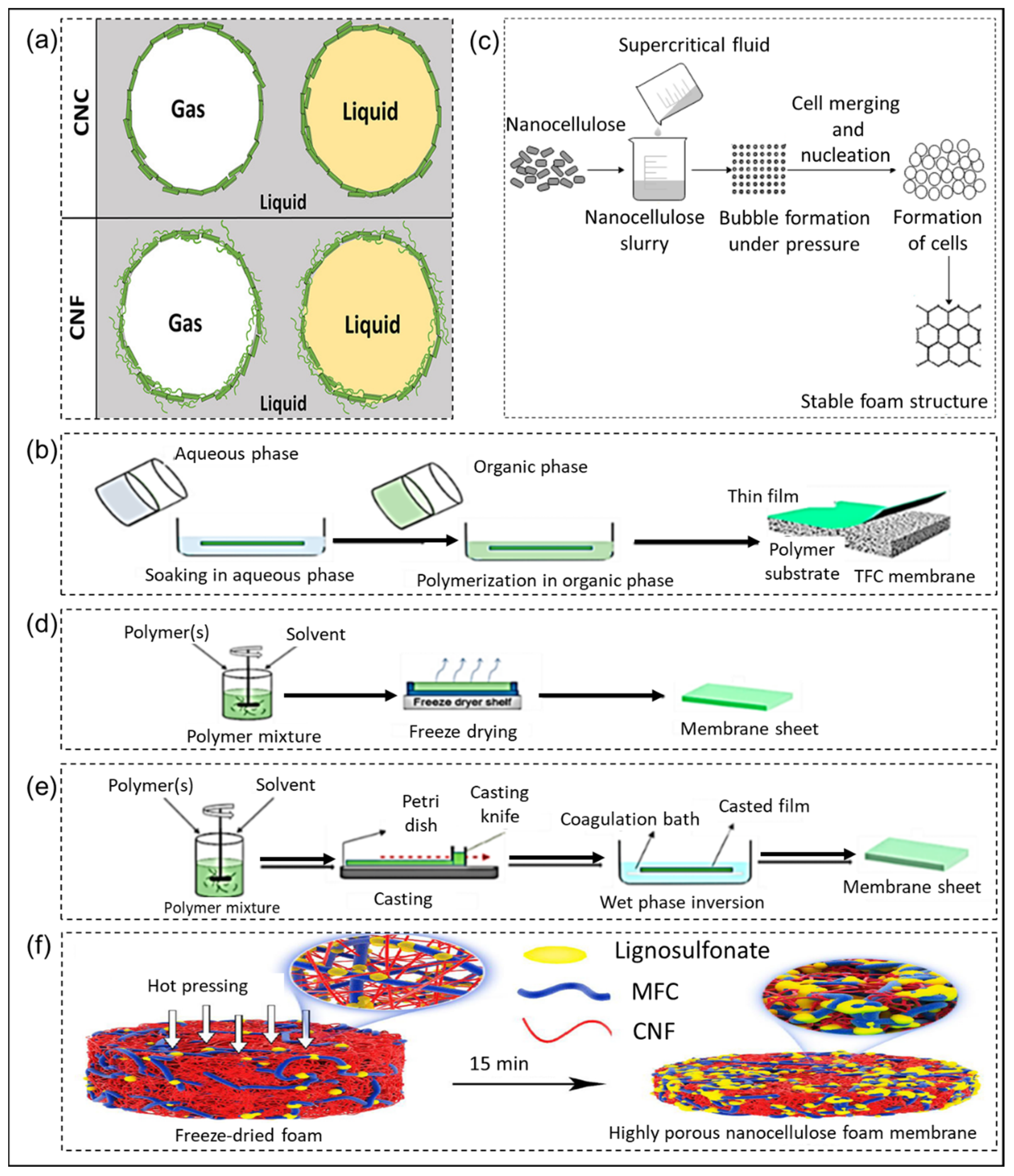
Figure 4.
Schematic representation of NC properties affecting artificial photosynthesis and factors to control these properties. The undesired properties are marked with a dotted line while the methods to achieve desired properties are mentioned in bullet points.
Figure 4.
Schematic representation of NC properties affecting artificial photosynthesis and factors to control these properties. The undesired properties are marked with a dotted line while the methods to achieve desired properties are mentioned in bullet points.
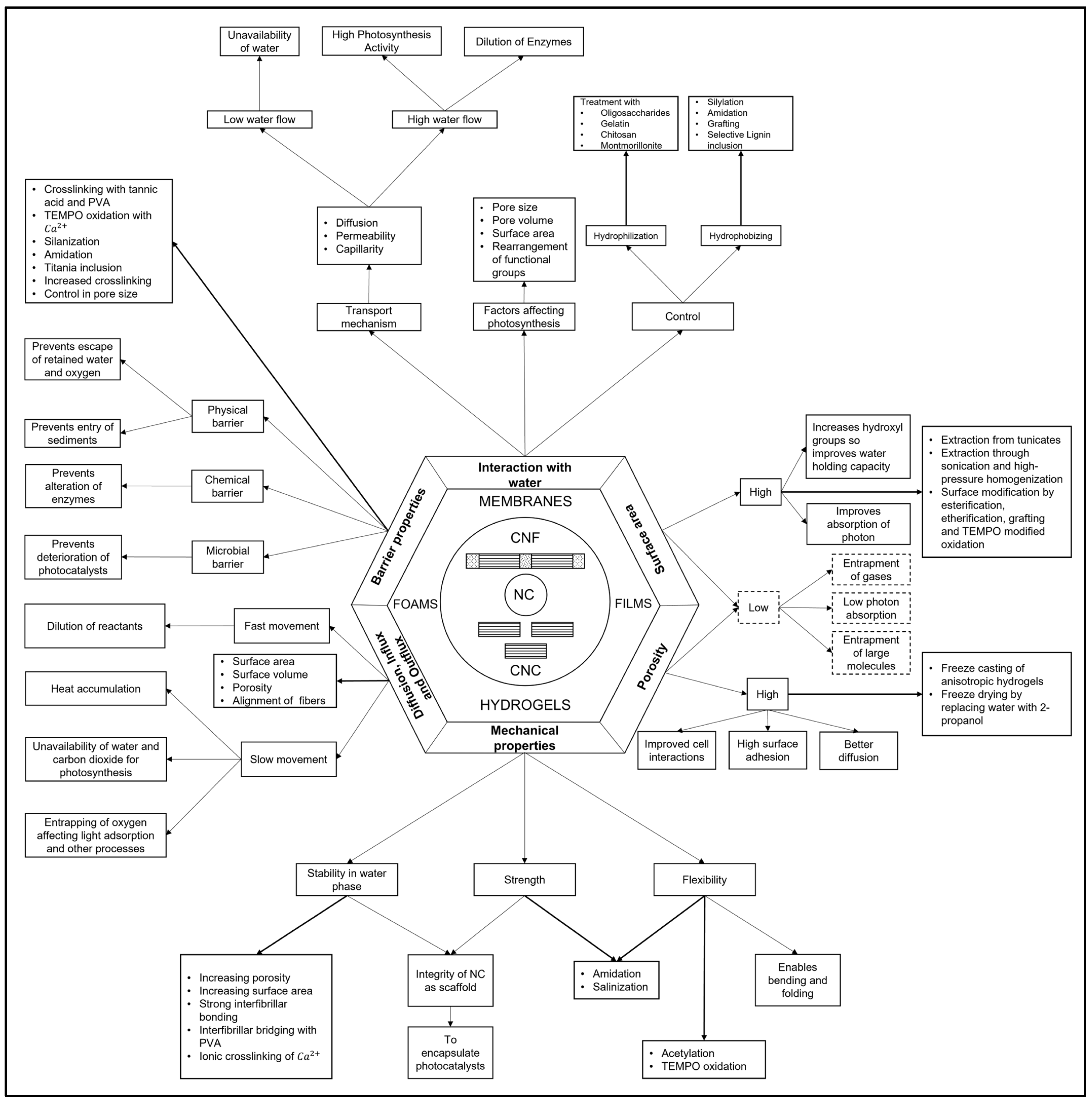
Figure 5.
Generic model of light scattering, with incident light I0, reflected light (R) and transmitted light (T). Both the reflected and the transmitted light have a perpendicular component (P) and a diffuse component (D).
Figure 5.
Generic model of light scattering, with incident light I0, reflected light (R) and transmitted light (T). Both the reflected and the transmitted light have a perpendicular component (P) and a diffuse component (D).
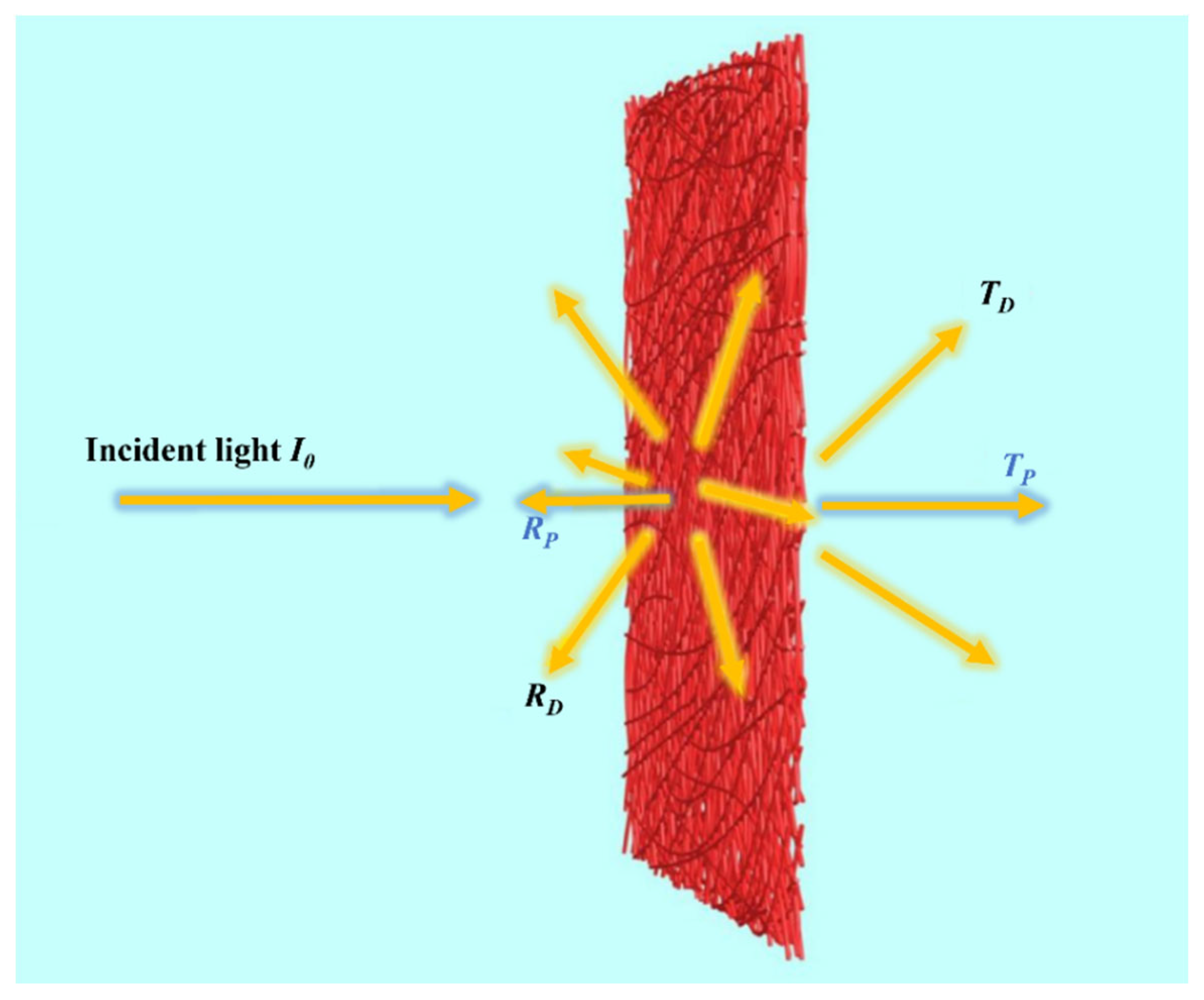
Figure 6.
Visual appearances of the transparent papers with NFC contents of 100% (a), 50% (b), and 0% (c), respectively. (d) Scattering angular distribution with arbitrary y-axis units for the various transparent papers. Maximum scattering angle for the transparent papers with a different ratio of NFC to the whole paper: (e) 0%, (f) 20%, (g) 50%, (h) 80%, and (i) 100% NFC [82] (reproduced with permission from RSC Publishing ©2014).
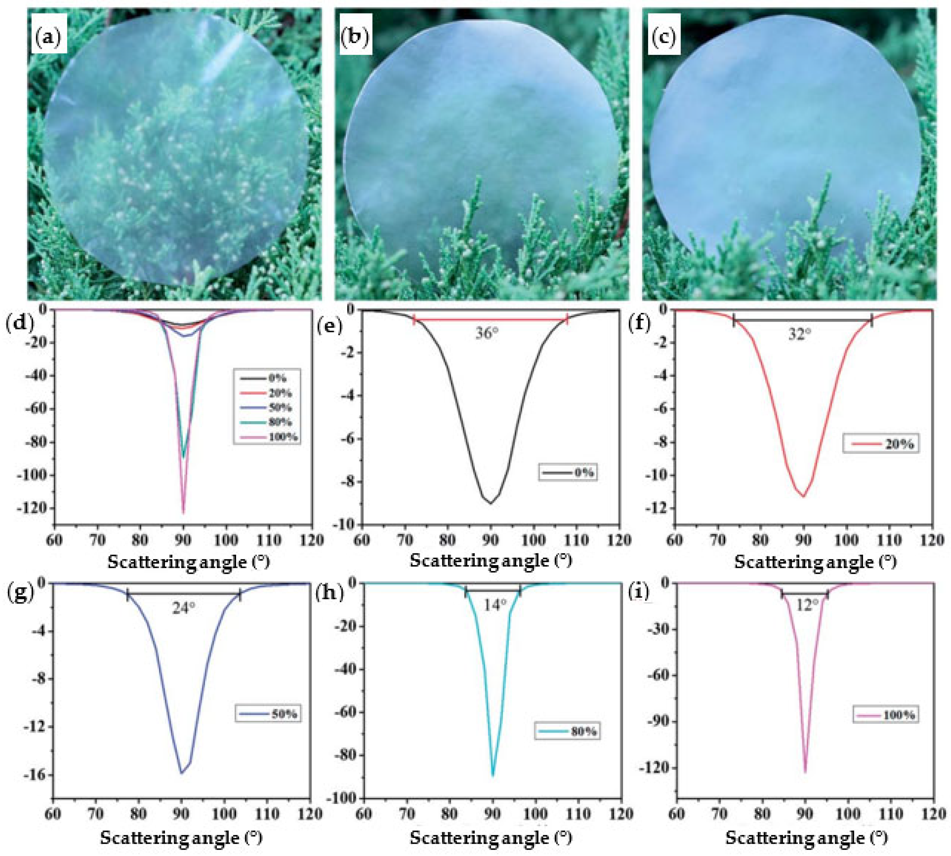
Figure 7.
Nanocellulose paper with control of haze for enhanced UV light harvesting based on lanthanide Ln3+/Eu3+ complex assembly and TEMPO-CNF: (a) schematic process for nanopaper preparation, (b) tuning of transparency and haze depending on incorporation of complexes at the surface or in the bulk of nanopapers [89] (reprinted with permission from American Chemical Society ©2019); (c) fabrication of transparent and multiluminescent TEMPO-oxidized nanofibrillated cellulose nanopaper functionalized with lanthanide complexes [90] (reproduced with permission from RSC Publishing ©2015).

Figure 8.
Transparency of nanocellulose films depending on: (a) changing effects of processing conditions, (b) transmittance and haze of nanopaper (40 µm thickness) with extracted nanocellulose from the branches of Amorpha fruticosa Linn., wheat straw, and poplar residues via combined mechanical treatments of grinding and high-pressure homogenization [103].
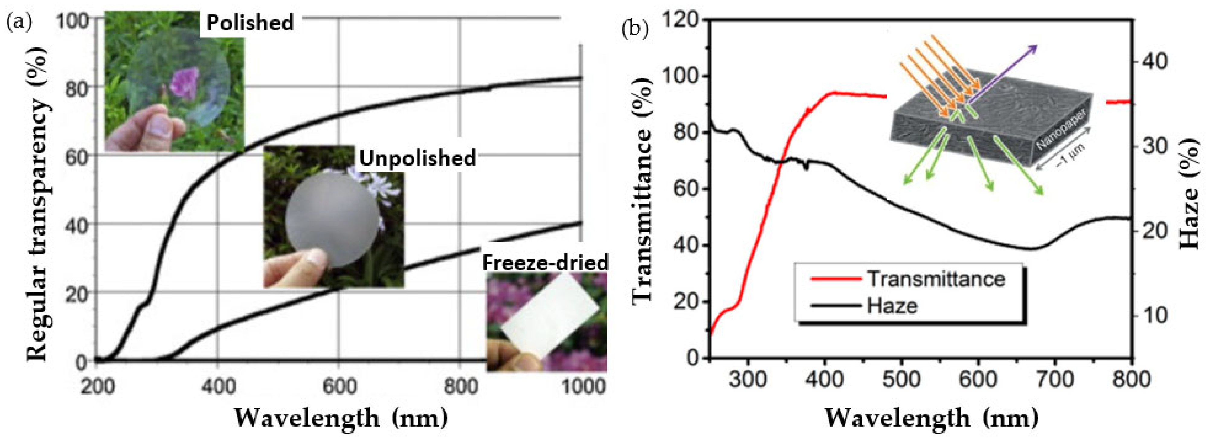
Figure 9.
Nanocellulose films with thermoresponsive change in transparency and haze, used as an optical modulation device [105] (reprinted with permission from American Chemical Society ©2021).
Figure 9.
Nanocellulose films with thermoresponsive change in transparency and haze, used as an optical modulation device [105] (reprinted with permission from American Chemical Society ©2021).
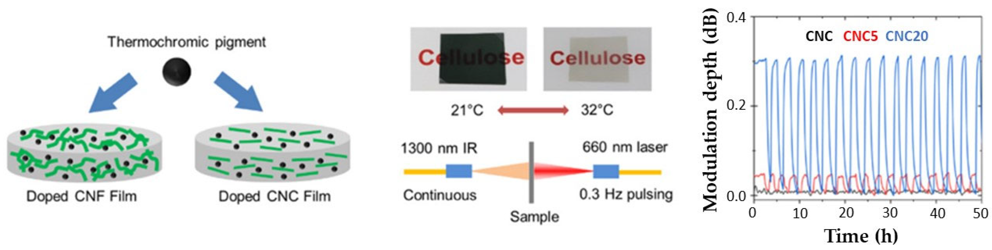
Figure 10.
Nanocellulose films as translucent diffuser for better coupling of the light in opto-electronic devices:, (a) model device of nanocellulose film on Si substrate illustrating the light-trapping effect, (b) morphology of hybrid films with nanocellulose and wood pulp, (c) total transmission and diffuse transmission spectra for NC coatings on glass, (d) total and diffuse transmission spectra of NC films under radiation intensity of 50 suns for different times [106] (reprinted with permission from American Chemical Society ©2015).
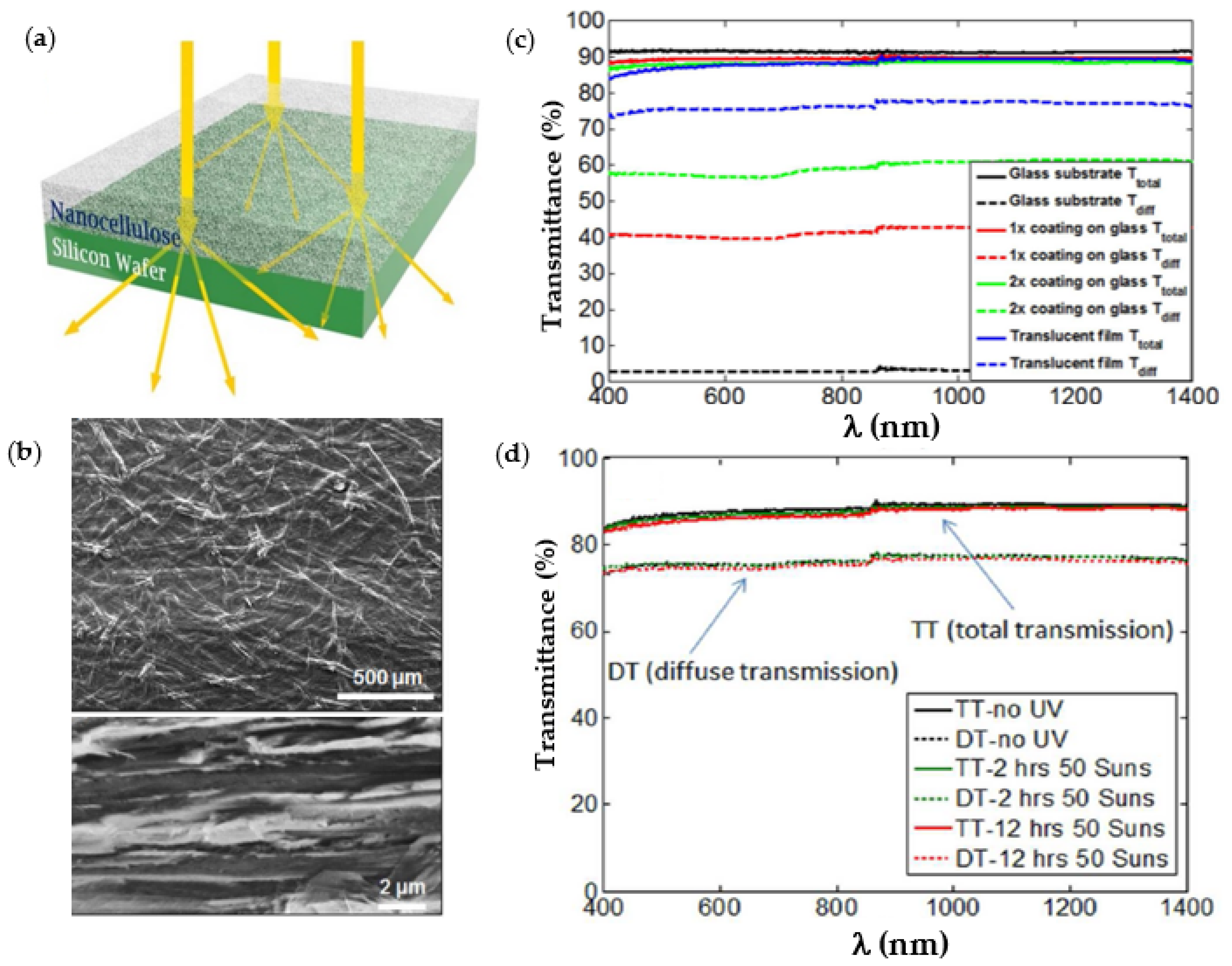
Figure 11.
Surface patterning of nanocellulose film through colloidal lithography and deposition of TiO2 nanoparticles on patterned substrates: (a) schematic drawings with subsequent processing steps for lithographic patterning (1), SEM pictures of the top views (2) and tilted views (3) of samples obtained after the different steps of the TiO2 nanostructure construction, (b) measured absorptance of the samples with a base structure, composed of a rear mirror Al and an a-Si:H absorber, coated with distinct top layers: none (Si/REF), 80 nm IZO (Si/ARC) and the TiO2 nanostructures (Si/TiO2 NS) produced by colloidal lithography [116] (reprinted with permission from RSC Publishing ©2017).
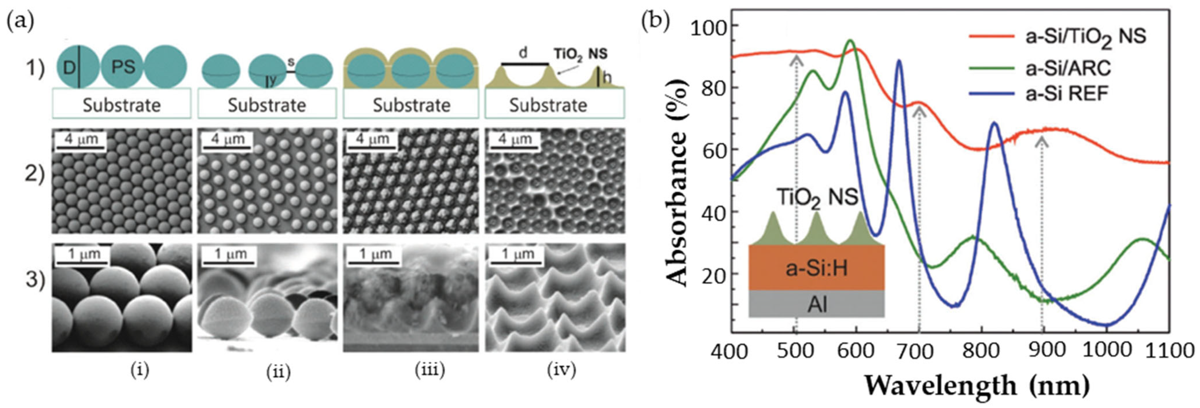
Figure 12.
The process of molecular layer-by-layer (LbL) assembly and immobilization of thylakoids in nanocellulose composite membranes through consequent dipping in a solution 1 (e.g., positively charged 0.5 wt.% PEI, or 0.5 wt.% PANI), followed by washing and dipping in the solution 2 (i.e., extracted thylakoids) [165] (reprinted with permission from Elsevier B.V. ©2016).
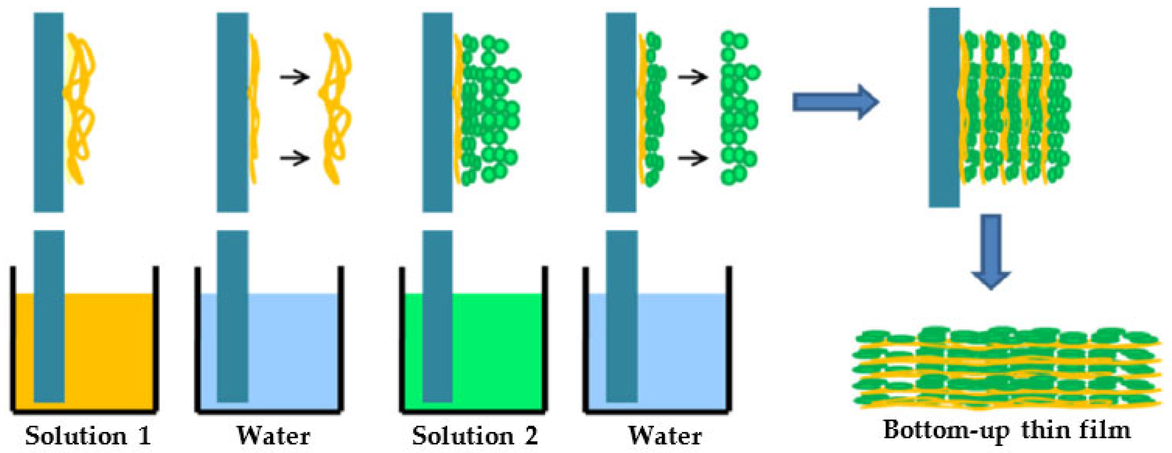
Figure 13.
Nanocellulose as template for hybrid composite materials with photoreactive inorganic nanoparticles: (a) CNF film coated with TiO2 after thermal annealing and use as photo-electrode in photoelectrochemical cell [184], (b) capillary PEC water splitting setup with cellulose-templated TiO2 photoanode; (c) schematic representation for growth of TiO2 nanorods onto CNF or CNC surfaces through seed-mediated deposition and Au nanoparticle (NP) attachment for creating a photoreactive nanocellulose composite hydrogel [187] (reproduced with permission from RCS Publishing ©2014).
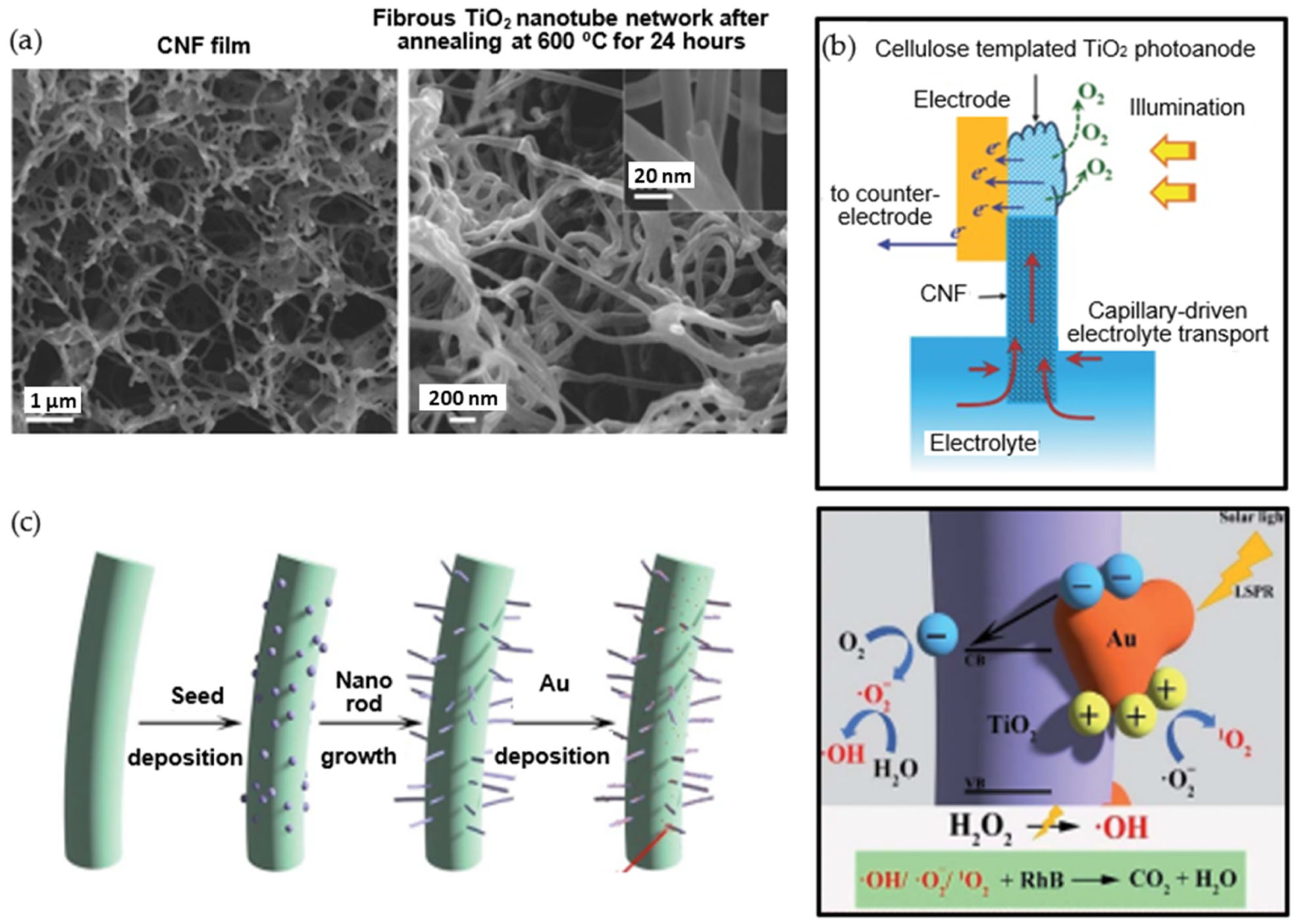
Figure 14.
Design of artificial leaf structure with encapsulated photosensitizers and emitters in microcapsules with porous wall of mixed CNF/CNC nanocelluloses: (a) natural leaf structure, (b) artificial structure with detail of porous microcapsules, (c) encapsulated photosensitizer molecules, (d) encapsulated emitter molecules [218] (reprinted with permission from American Chemical Society ©2014).
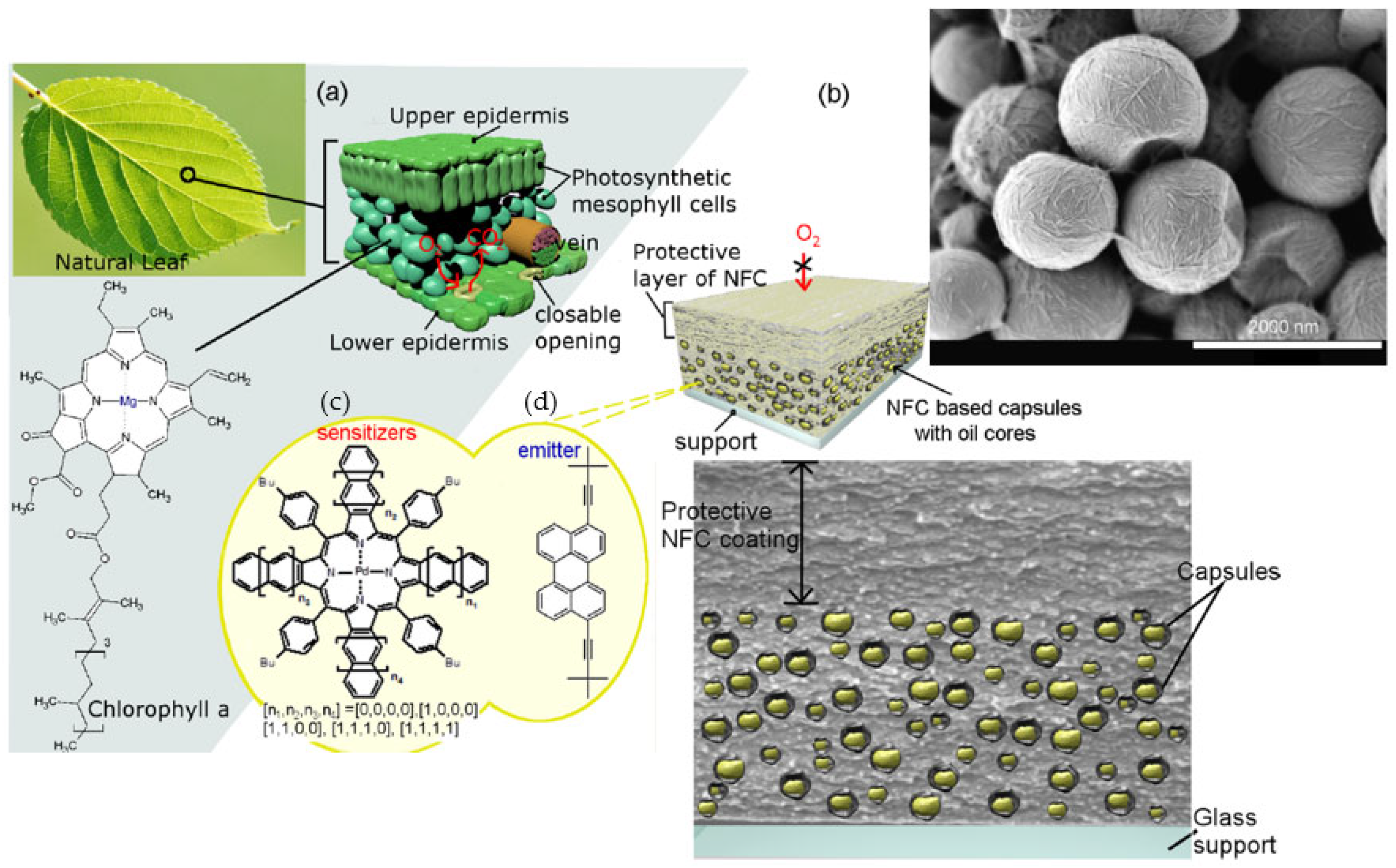
Figure 15.
Light-harvesting systems in natural green plant leaves and synthetic wood with physically encapsulated light-harvesting pigments such as chlorophyll and porphyrins: (a) biomimicking structure in redox reaction for production of photosynthetically active enzymes (NAD(P)H), (b) light-harvesting pigments, (c) synthetic wood with nanofibrous cellulose structure as host material for encapsulation [237] (reproduced with permission from WILEY-VCH Verlag GmbH & Co. KGaA ©2011).
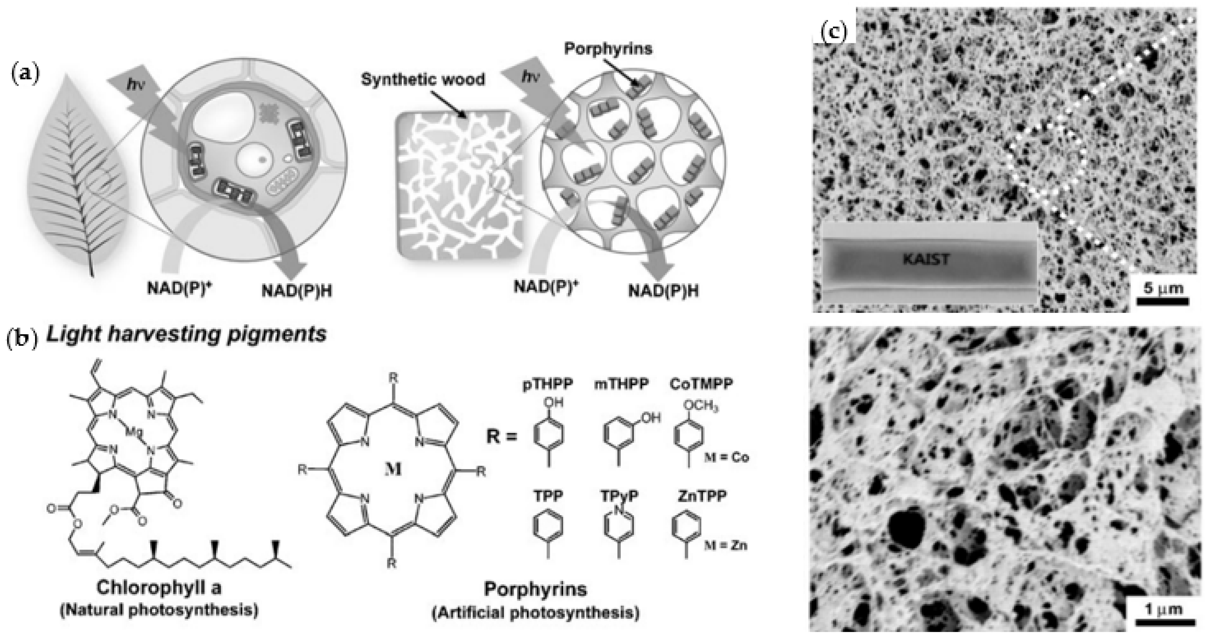
Figure 16.
Local organization of photosynthetic latex coating with incorporated cyanobacteria cells and microstructure variations on filter paper substrates: (a) pure latex coated paper, (b) formation of cell “sheets” between paper fibers, (c) cells “flowing” with latex binder between paper fibers prior to film formation, (d) “buried” cells on paper fibers, (e) “exposed” cells on paper fibers [269].
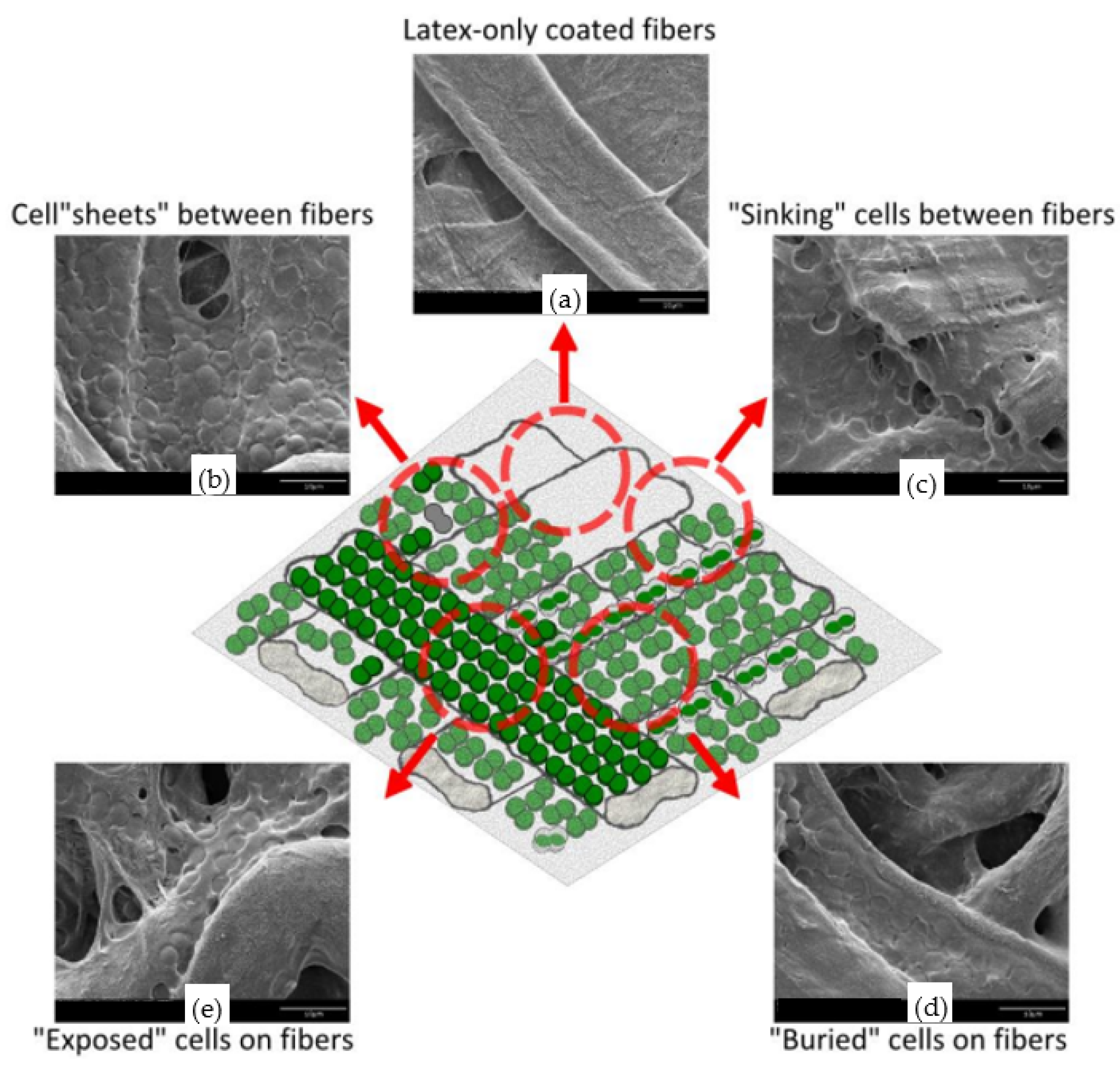
Figure 17.
Schematic representation of immobilization strategies of algae or cyanobacteria in TEMPO-CNF hydrogels utilized for photosynthetic processes: (a) hydrogel layer applied on top of the paper support and placed in the gas-tight vials above the medium on top of water-conveying sponge that provides water to the cells via capillary actions, (b) Ca2+ crosslinked hydrogel layers with entrapped algae with embedded support network submerged into the cultivation medium (c) PVA containing hydrogel layers on paper support with the drying and recovery steps. During the drying step, the solid film is formed, and PVA forms interfibrillar bridges with TEMPO CNF that enhance the film wet strength [278] (reproduced with permission from RSC Publishing ©2018).
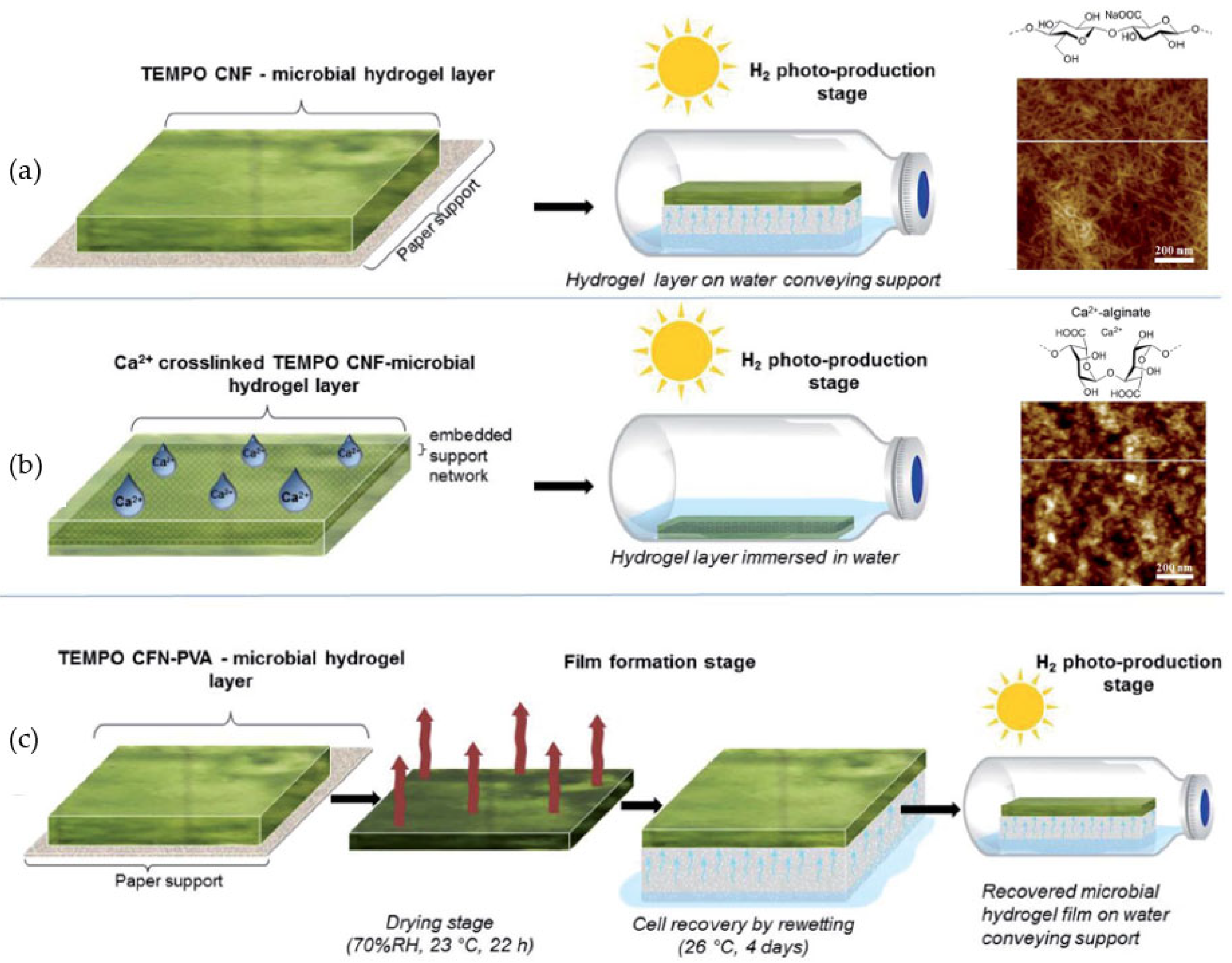
Figure 18.
Concept for nanocellulose-based mechanically stable immobilization matrix for cyanobacteria as a photosynthetic solid-state cell factory used in ethylene-production: (a) schematic representation of the experimental approach for immobilizing photosynthesis cells within TEMPO-CNF and alginate hydrogels cross-linked with polyvinyl alcohol (PVA)/Ca2+ (TEMPO-CNF) or Ca2+ (alginate); Small deformation oscillatory rheology of matrix hydrogel films for (b) PVA-Ca2+/TEMPO-CNF, and (c) Ca2+ alginate hydrogel films with (solid lines) and without (dotted lines) cyanobacterial cells, showing elastic moduli (G′, black square), viscous moduli (G″, red square) and tan (δ) (blue circle). The dashed vertical line marks the onset of critical stress, where the material starts to flow [279].

Figure 19.
Encapsulation of photoreactive bacteria in electrospun CNY fiber mats: (a) schematic electrospinning process with incorporation of bacteria through surface adhesion or matrix encapsulation [284] (reproduced with permission from Elsevier B.V. ©2021), (b) scanning electron microscopy of L. acidophilus incorporated in an electrospun BNC fiber mat with PVA co-polymers, using adsorption method [285] (reproduced with permission from Elsevier B.V. ©2020), (c) scanning electron microscopy of bacteria immobilized in a nanofibrous matrix of electrospun cellulose acetate used in photosynthesis [287].
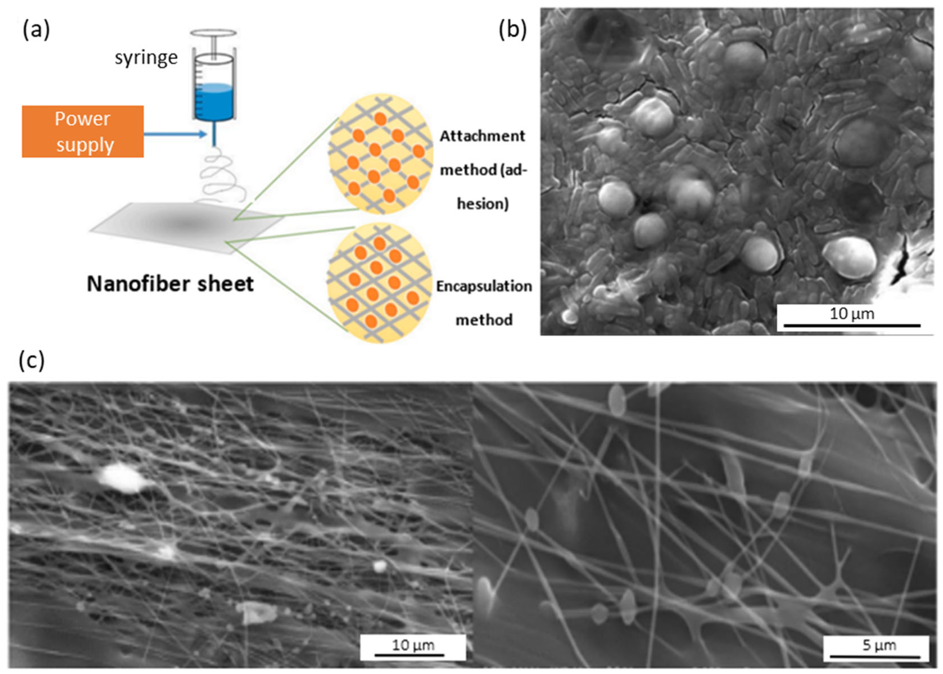
Figure 20.
Electrospinning of composite cellulose nanoyarns (CNY) with electroconductive properties in combination with PEDOT-PSS conductive polymer and PEO cross-linkers: (a) electrospinning process, (b) morphology of conductive CNF composite nanofibers with PEO crosslinker, (c) photosynthetic efficiency of encapsulated Chlamydomonas and Synechocystis cells in CNY composite nanofibrous mats [293] (reproduced with permission from American Chemical Society ©2020).
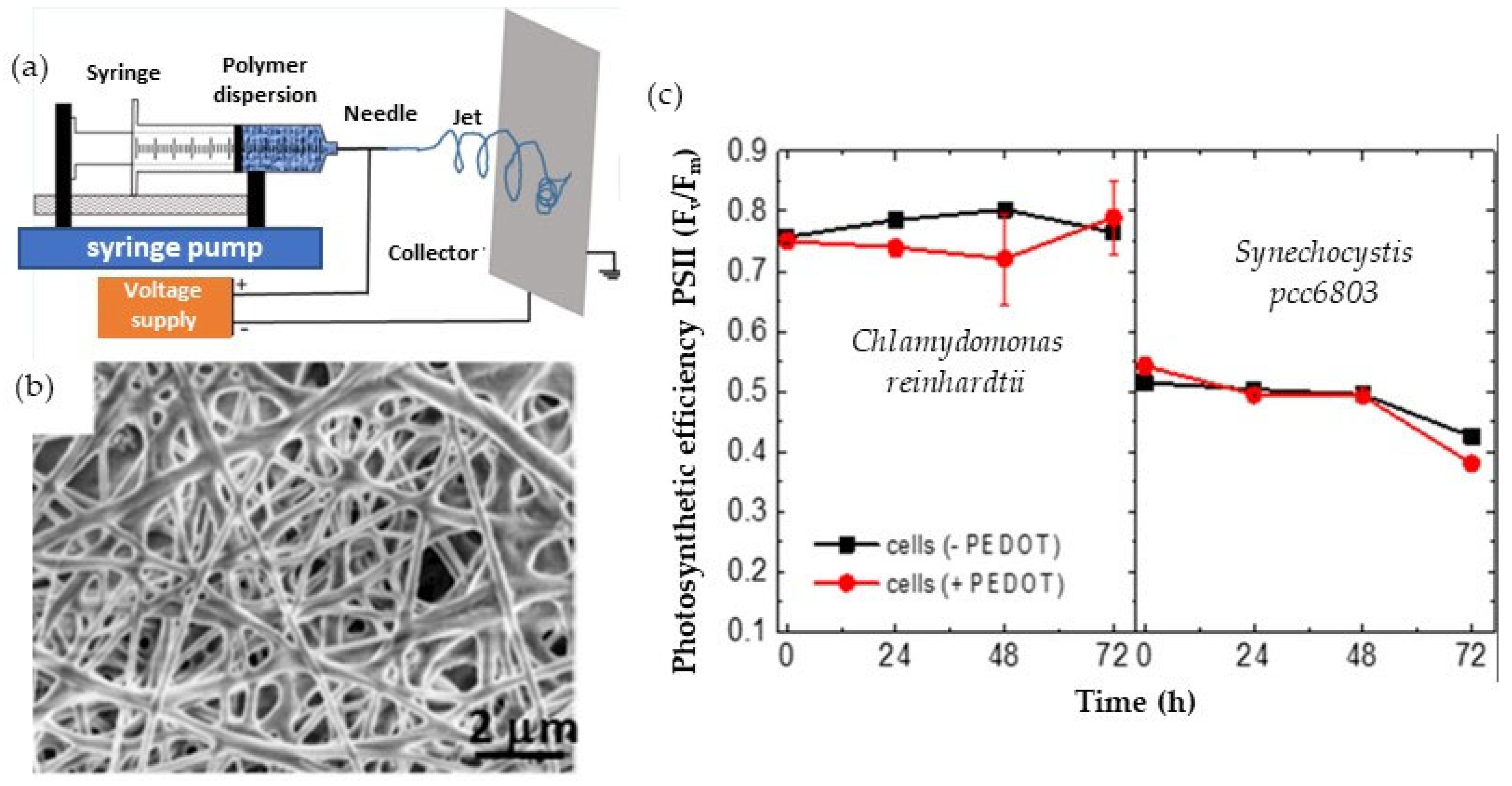
Figure 21.
Principle of a bio-photoelectrochemical cell for photosynthetic conversion: (a) cell based on thylakoid membranes and PEDOT:PSS-CNF nanocellulose electrodes and DCPIP (2,6-dichlorophenolindophenol) redox mediators: DCPIPox, deep blue at the beginning of halogen lamp illumination turns colorless upon reduction by TMs, (b) generated by TMs under halogen lamp illumination with IR long-pass filters with cut-on wavelengths of 750 nm (220–420 s) and 700 nm (800–1200 s). Complete exclusion of VIS light (220–420 s) generated no photocurrent because TMs do not absorb radiation in the NIR range of spectrum, while partial transmission around 700 nm (800–1200 s) generated low photocurrent [298] (reproduced with permission from WILEY-VCH Verlag GmbH & Co. KGaA ©2020).

Figure 22.
Advanced artificial plant leaf devices obtained through bio-mimicry using a living hydrogel sheet of alginate (Alg) and carboxymethylated cellulose nanofibers (CMCNF): (a) schematic of the Alg/CNF hydrogel with imprinted channels using a 3D printing stamp and incorporation in a device enabling capillary flow through the channels, (b) chemical structure of the Alg/CNF composite crosslinked with Ca2+, (c) optical microscopic images of the Alg/CNF hydrogel with fluidic channels of different geometry, (d) vein-imprinting of artificial maple and hydrangea leaves: the channel structure was formed by covering the groove patterns with a transparent glass [303] (reproduced with permission from Elsevier B.V. ©2022).
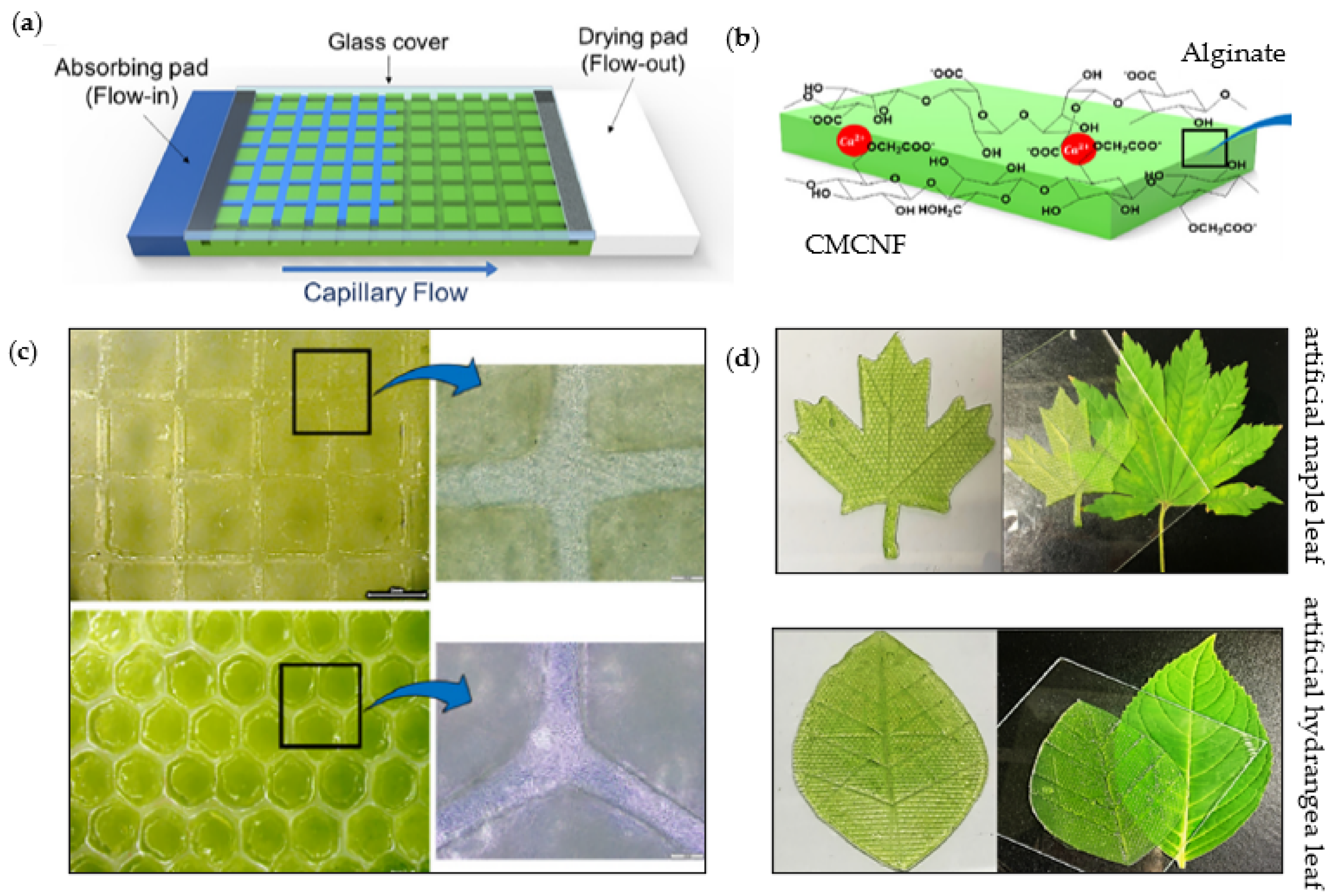

Table 1.
Selection of photoreactive metal nanoparticles coupled into nanocellulose composites.
| Photoreactive Nanoparticles | Nano-Cellulose | Processing for Incorporation within Photoreactive Nanocellulose Composites | Reference |
|---|---|---|---|
| Metals | |||
| Au | CNC | Reduction from HAuCl4 | [188] |
| Pd | CNF | Direct reduction of Pd2+ ions | [189] |
| Metal oxides | |||
| ZnO | CNC | In-situ casting from Zn(NO3)2 | [180] |
| ZnO | CNC | Co-precipitation from Zn(CH2CO2) | [190] |
| TiO2 | CNC | Hydrolysis of TiCl4 | [191] |
| TiO2 | CNC | Reduction of TiOOSO4·H2SO4·H2O | [185] |
| TiO2 | CNF | Heterocoagulation of CNC and TiO2 NP | [192] |
| Cu2O | CNC | Microwave-assisted polyol synthesis | [193] |
| Cu2O/TiO2 | CNF | Hydrothermal reaction from CuSO4 and NaOH | [194] |
| Cu2O/Ag | CNF | Reduction from CuSO4 and AgNO3 | [195] |
| CuO/Fe3O4 | MFC | In-situ co-precipitation of Cu(CH3COO)2 and Fe(NO3)3·9H2O | [196] |
| Fe-doped ZnO | CNC | In-situ precipitation from Zn(CH3CO2)2 and FeCl3 | [197] |
| Al-doped ZnO | MFC/CNF | In-situ reduction from ZnCl2 and AlCl3 | [198] |
| Metal sulphides | |||
| CdS | CNF | Quantum dots formation from CdCl2 and Na2S | [170] |
| ZnS | CNC | In-situ co-precipitation from ZnNO3 precursor | [199] |
| MoS2/CdS | CNF | Coagulation casting of Na2MoO4·2H2O and Cd(CH3CO2)2·2H2O | [172] |
| Others | |||
| Ag3PO4 | CNC | In-situ synthesis from Na2HPO4·12H2O and AgNO3 | [200] |
| Graphitic carbon nitride | CNF | Thermal polymerization of bi-component precursor | [201] |
Disclaimer/Publisher’s Note: The statements, opinions and data contained in all publications are solely those of the individual author(s) and contributor(s) and not of MDPI and/or the editor(s). MDPI and/or the editor(s) disclaim responsibility for any injury to people or property resulting from any ideas, methods, instructions or products referred to in the content. |
© 2023 by the authors. Licensee MDPI, Basel, Switzerland. This article is an open access article distributed under the terms and conditions of the Creative Commons Attribution (CC BY) license (https://creativecommons.org/licenses/by/4.0/).
Share and Cite
MDPI and ACS Style
Samyn, P.; Rastogi, V.K.; Baba, N.S.S.; Van Erps, J. Role of Nanocellulose in Light Harvesting and Artificial Photosynthesis. Catalysts 2023, 13, 986. https://doi.org/10.3390/catal13060986
AMA Style
Samyn P, Rastogi VK, Baba NSS, Van Erps J. Role of Nanocellulose in Light Harvesting and Artificial Photosynthesis. Catalysts. 2023; 13(6):986. https://doi.org/10.3390/catal13060986
Chicago/Turabian StyleSamyn, Pieter, Vibhore Kumar Rastogi, Neelisetty Sesha Sai Baba, and Jürgen Van Erps. 2023. "Role of Nanocellulose in Light Harvesting and Artificial Photosynthesis" Catalysts 13, no. 6: 986. https://doi.org/10.3390/catal13060986
Note that from the first issue of 2016, this journal uses article numbers instead of page numbers. See further details here.