Article

Synthesis and Characterization of Highly Stabilized Polymer–Trypsin Conjugates with Autolysis Resistance

1
Laboratory of Pharmaceutical Physical Chemistry, Department of Drug Delivery Technology and Sciences, Gifu Pharmaceutical University, 1-25-4, Daigaku-Nishi, Gifu 501-1196, Japan
2
Department of Pharmaceutical Physical Chemistry, College of Pharmaceutical Sciences, Matsuyama University, 4-2 Bunkyo-cho, Matsuyama, Ehime 790-8578, Japan
3
Faculty of Human Welfare, Chubu Gakuin University, 2-1 Kirigaoka, Sek-shi, Gifu 501-3993, Japan
*
Author to whom correspondence should be addressed.
Catalysts 2017, 7(1), 4; https://doi.org/10.3390/catal7010004
Submission received: 30 November 2016 / Revised: 20 December 2016 / Accepted: 22 December 2016 / Published: 26 December 2016
(This article belongs to the Special Issue Immobilized Enzymes: Strategies for Enzyme Stabilization)

Abstract

:
Protein digestion by trypsin has been widely used in many industrial and research applications. However, extensive use of trypsin is limited because of the rapid decrease in enzymatic activity caused by autolysis at optimal pH and temperature. To improve the enzymatic performance of trypsin, we synthesized highly stabilized polymer–trypsin conjugates using vinylmethylether-maleic acid copolymer (VEMAC) via multi-point attachment. The VEMAC modification significantly enhanced the thermal stability of trypsin, and the resulting conjugates showed a strong resistance to autolysis. VEMAC-modified trypsin (VEMAC-Tryp) showed maximum activity at 55 °C and at 1.4-fold higher levels than that of unmodified trypsin. Bovine serum albumin was effectively digested by VEMAC-Tryp, indicating that the modified trypsin can be used for digestion of high molecular weight substrates. VEMAC modification is a simple and cost-effective strategy to obtain fully active modified enzymes, and may be used to develop bioreactors.

Graphical Abstract

1. Introduction

The covalent modification of an enzyme with a polymer is a widely accepted strategy to design and synthesize functional enzymatic conjugates with long half-life in the blood stream, thermal stability, stimuli-responsive activity, and reusability [1,2,3].
A polymer chain can be conjugated to a protein via either a single-point or a multi-point attachment. In the former method, only one functional group of a polymer attaches to a protein. This method has often been employed to modify proteins with end-group functionalized polymers including poly(ethylene glycol) (PEG) [4,5,6]. Some PEGylated proteins (i.e., proteins modified with PEG) are approved by the U.S. Food and Drug Administration (FDA) and have been used for the treatment of some intractable diseases, such as cancer [7,8]. As demonstrated by many studies, PEGylation is effective to improve undesirable properties of proteins such as low solubility, aggregability, immunogenicity, and short half-life in the blood stream [9]. Another example of single-point attachment, the “grafting from” method (i.e., surface-initiated polymerization of a monomer from the protein surface) has been enthusiastically studied [10,11]. In multi-point attachment methods, on the other hand, several functional groups bearing a polymer backbone are used to form protein conjugates. Generally, multi-point attachment is effective in providing a higher conformational stability to proteins against changes in pH and temperature [12]. For example, Riccardi et al. reported that the long-term stability of catalase was significantly improved by conjugating with poly(acrylic acid) via a multi-point attachment method [13]. Treetharnmathurot et al. also reported that the thermal stability of trypsin was enhanced by multi-point conjugation with dextrin, and dextrin with higher molecular weight led to greater stability enhancements [14].
Trypsin—a serine protease that catalyzes the hydrolysis of peptide bonds at the C-terminal end of lysine and arginine residues—is commonly used in various industrial and research applications for protein digestion. Trypsin digestion is also an important step in the preparation of peptide fragments for protein analysis using mass spectroscopy (MS) due to its high specificity and reproducibility [15]. On the other hand, trypsin is degraded by autolysis at the working pH and temperature, and quickly loses activity. To prevent autolysis during protein digestion, trypsin with methylated lysine residues is commonly used as an MS-grade product [16]. However, the methylation of trypsin cannot completely suppress autolysis, so even modified trypsin must be used at low concentrations that lead to time-consuming digestions [17]. Moreover, the high cost of MS-grade trypsin is an additional disadvantage. Therefore, there is a strong demand for the development of trypsin with autolysis resistance.
In general, enzyme modifications with hydrophilic polymers are effective for the protection of enzymes from protease digestion, due to the shielding effects of hydrated polymers [5]. Thus, the modification of trypsin with hydrophilic polymers to prevent autolysis is promising [14,18,19,20]. The vinylmethylether-maleic acid copolymer (VEMAC) is obtained by hydrolyzing the maleic anhydride moiety from the commercially available vinylmethylether-maleic anhydride copolymer, and has been extensively tested for medical and pharmaceutical applications due to its hydrophilicity, biocompatibility, and non-toxicity [21,22,23,24,25,26]. In addition, we have reported the cell compatibility of VEMAC immobilized on polystyrene cell culture dishes [27], and abundant carboxyl groups on the VEMAC backbone can be used for multi-point conjugation of enzymes.
In this study, we synthesized VEMAC-modified trypsin (VEMAC-Tryp) via a multi-point conjugation method, and investigated the effects of VEMAC on enzymatic activity and stability using a synthetic low molecular weight substrate. In addition, considering that trypsin is frequently used for protein digestion, enzymatic activity against high molecular weight substrates (i.e., proteins) is an important issue in many applications. Thus, the ability of VEMAC-Tryp to digest protein was also evaluated using bovine serum albumin (BSA) as a model substrate.

2. Results and Discussion

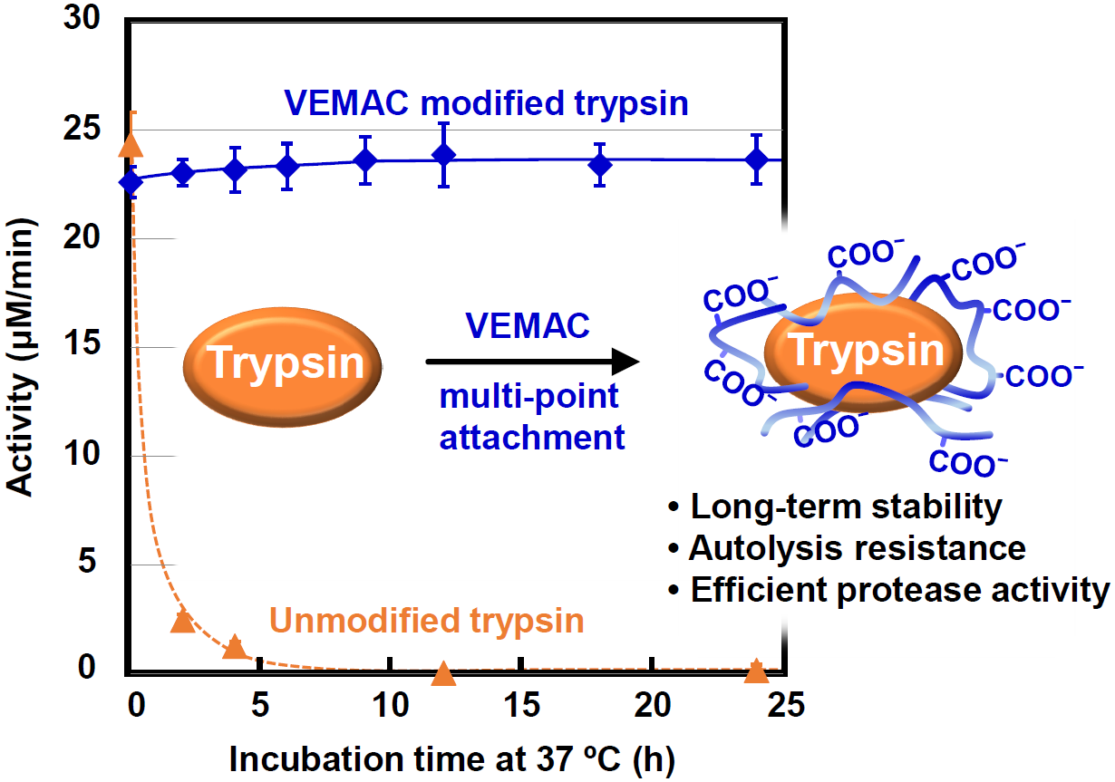
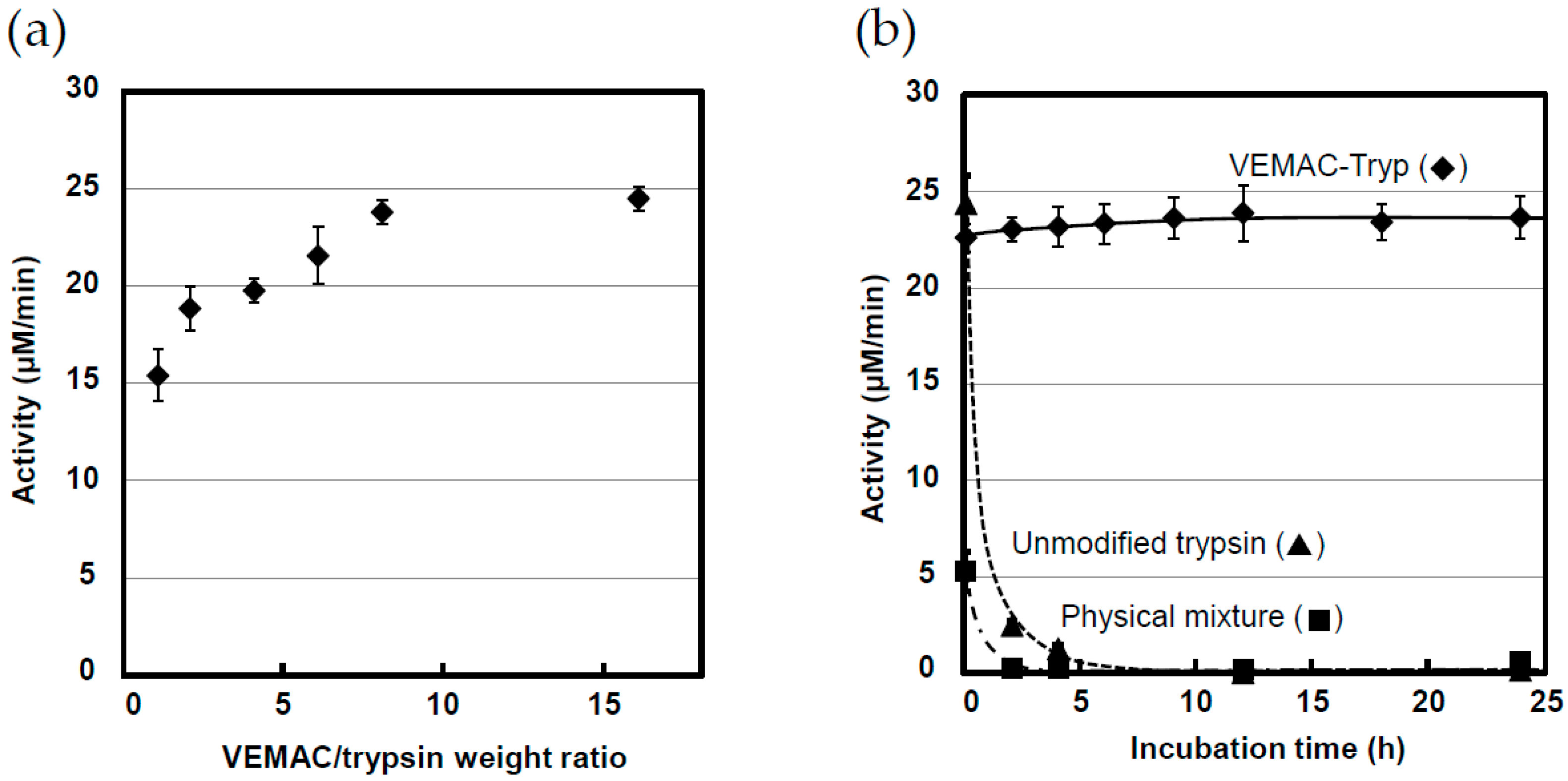
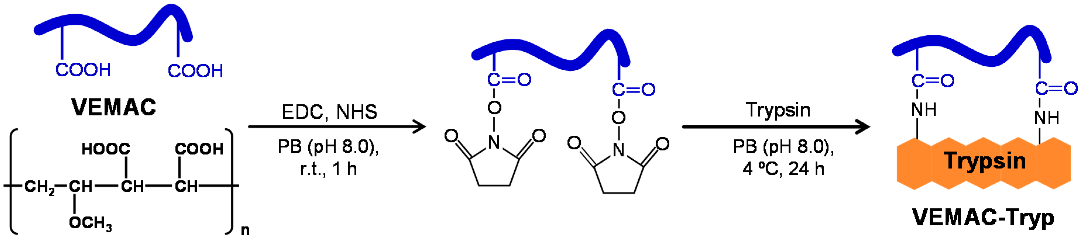
2.1. Synthesis of VEMAC-Tryp and Evaluation of Its Enzymatic Activity

VEMAC-Tryp was synthesized by coupling reactions between carboxyl groups in VEMAC and amino groups in trypsin (Scheme 1). To search for optimal conditions of trypsin modification by VEMAC, VEMAC-Tryp was synthesized using varying VEMAC/trypsin (w/w) ratios, and the reaction products were examined. As can be seen in Figure 1a, enzymatic activity—determined using benzoyl-dl-arginine p-nitroanilide hydrochloride (BAPNA) as a substrate [28]—increased with increasing VEMAC/trypsin ratios, and reached a constant value at an 8:1 ratio. Because the conjugation reaction mixture is further incubated at 37 °C for 6 h to hydrolyze unreacted hydroxysuccinimide (NHS) esters, trypsin autolysis will occur if the VEMAC modification is incomplete. Considering that the enzymatic activity of unmodified trypsin was 24.4 ± 1.5 μM/min, the results shown in Figure 1a indicate that incomplete trypsin modification due to inadequate amounts of VEMAC resulted in low activity. Thus, VEMAC-Tryp synthesized at an 8:1 VEMAC/trypsin ratio was used in this study. The number of amino groups in VEMAC-Tryp was estimated by a 2,4,6-trinitrobenzen sulfonic acid (TNBS) assay [29]. The results indicated that unmodified trypsin had ~10 accessible amino groups, which was similar to the results reported by others [19,30], and almost all of them (~95.1%) were modified by VEMAC. Figure 1b shows the comparison between enzymatic activities of VEMAC-Tryp, unmodified trypsin, and VEMAC/trypsin mixture in phosphate buffer solution (PB, 50 mM, pH 8.0) at 37 °C. Whereas the enzymatic activities of unmodified trypsin and VEMAC/trypsin mixture rapidly decreased and were not detected after a few hours due to autolysis, VEMAC-Tryp activity remained constant for more than 24 h. We monitored the activity of VEMAC-Tryp at 25 °C for a long period of time, and found that over 80% of the initial activity was still present after 12 months (Figure S1). To evaluate the effects of VEMAC modification on the catalytic efficiency of trypsin, enzyme kinetic parameters were determined using the Hanes–Woolf plot. Both Hanes–Woolf plots for unmodified trypsin and VEMAC-Tryp exhibited good linearity (R2 > 0.97) (Figure S2), indicating that both enzyme kinetics can be analyzed using the Hanes–Woolf equation [31]. The enzyme kinetic parameters obtained are summarized in Table 1. Although a slight decrease in the catalytic rate constant (Kcat) (i.e., a lower turnover number) was observed in VEMAC-Tryp, the Michaelis constant (Km) decreased, which indicates that the affinity of modified trypsin for the substrate increased. The increase in affinity of trypsin for BAPNA by VEMAC modification may be attributed to an increase in local BAPNA concentration due to electrostatic interaction between anionic VEMAC and cationic BAPNA. As a result, the Kcat/Km ratio—reflecting the catalytic efficiency of the enzyme—increased. These results indicate that the VEMAC modification of trypsin using an optimal VEMAC/trypsin ratio does not affect trypsin activity and is effective to prevent autolysis.

2.2. Physicochemical Characterization of VEMAC-Tryp

To understand the effects of VEMAC modification on the stability of trypsin, VEMAC-Tryp was characterized using various analytical methods. Table 2 shows the hydrodynamic diameter and zeta-potential of unmodified trypsin and VEMAC-Tryp. The diameter of trypsin increased from ~3.3 to ~21.6 nm, and the zeta potential decreased from −15.3 to −32.6 mV by VEMAC modification, indicating that trypsin was covered with negatively-charged VEMAC. It should be noted that the determined hydrodynamic diameter of unmodified trypsin is very close to the value reported by Chiu et al. [30]. Moreover, the observed monodispersed particle size distribution of VEMAC-Tryp suggests that the crosslinking of trypsin by NHS-activated VEMAC was negligible during the conjugation process (Figure S3). The negatively-charged VEMAC layer on trypsin would contribute to stabilize each VEMAC-Tryp molecule in the reaction system, and to avoid autolysis by electrostatic repulsion.
Circular dichroism (CD) spectroscopy was performed to examine the effects of VEMAC modification and incubation on the secondary structure of trypsin. Figure 2 shows that the CD spectrum of unmodified trypsin changed significantly after incubation in PB at 37 °C for 24 h. In contrast, the spectra of VEMAC-Tryp before and after incubation were similar, and closer to that of unmodified trypsin (Figure 2). As can be seen in Table 3, the loss of α-helix structure and the increase in random coil content in unmodified trypsin after incubation indicate that the enzyme undergoes autolysis during incubation. In contrast, the secondary structure content of trypsin in VEMAC-Tryp was roughly the same before and after incubation, and similar to that of unmodified trypsin. Thus, the results suggest that VEMAC modification does not affect enzymatic activity, and that the structure of trypsin is rigidly stabilized by multi-point conjugation with VEMAC.
The temperature and pH dependence of VEMAC-Tryp were compared to those of unmodified trypsin. The temperature at which maximum activity was observed shifted from 45 °C for unmodified trypsin to 55 °C for VEMAC-Tryp, which led to a 1.4-fold increase in maximum activity (Figure 3a). A similar increase in trypsin enzymatic activity by polymer modification has also been previously reported [18,30]. In contrast to a reaction system containing unmodified trypsin, in which autolysis will occur as well as hydrolysis of the substrate, in the reaction containing VEMAC-Tryp, autolysis was completely suppressed by VEMAC modification. The decrease in trypsin concentration during the reaction was therefore negligible, and VEMAC-Tryp can function at a higher temperature than the optimal temperature of unmodified trypsin. In fact, VEMAC-Tryp was thermally stable and retained over 50% enzymatic activity, even after incubation for 24 h at 55 °C (Figure S4). The rapid decrease in VEMAC-Tryp enzymatic activity above 60 °C shown in Figure 3a might result from thermally-induced denaturation of trypsin in VEMAC-Tryp rather than autolysis.
The pH dependence of VEMAC-Tryp enzymatic activity was also investigated. Whereas unmodified trypsin exhibited maximum activity at pH 8.0 and significantly decreased activity at higher pH, VEMAC-Tryp activity increased from pH 5.9 to 9.8, and enzymatic activities at pH 9.0 and 9.8 were almost the same as the maximum activity of unmodified trypsin (~30 µM/min) (Figure 3b). Interestingly, VEMAC-Tryp enzymatic activity below pH 8.0 was lower than that of unmodified trypsin, and no decrease in activity above pH 8.0 was observed. To investigate this unique behavior, the hydrodynamic diameter of VEMAC-Tryp was measured at different pH. The hydrodynamic diameter of VEMAC-Tryp at pH 5.9 was ~4.0 nm, which was very similar to that of unmodified trypsin (Table 2), whereas the diameter increased significantly to ~21.6 and ~20.9 nm at pH 8.0 and 9.8, respectively (Table 4). Considering that the pKa2 of the maleic acid moiety in VEMAC is 6.5 [32], many carboxyl groups in VEMAC are protonated at pH 6.0, resulting in a decrease in solubility and in shrinkage of VEMAC in aqueous media, which prevent the access of the BAPNA substrate molecule to the active site of trypsin. On the other hand, the larger hydrodynamic diameters observed above pH 8.0 suggest that the fully hydrated state of VEMAC chain on the trypsin surface with deprotonated carboxyl groups would not affect the accessibility of BAPNA to trypsin. Moreover, the results shown in Figure 3b indicate that the rigidity of trypsin modified with VEMAC led to an increase in stability under basic pH conditions. It should also be noted that the changes in VEMAC-Tryp enzymatic activity in response to changes in pH were reversible, suggesting that pH could be used as an on–off switch to modulate enzymatic activity.

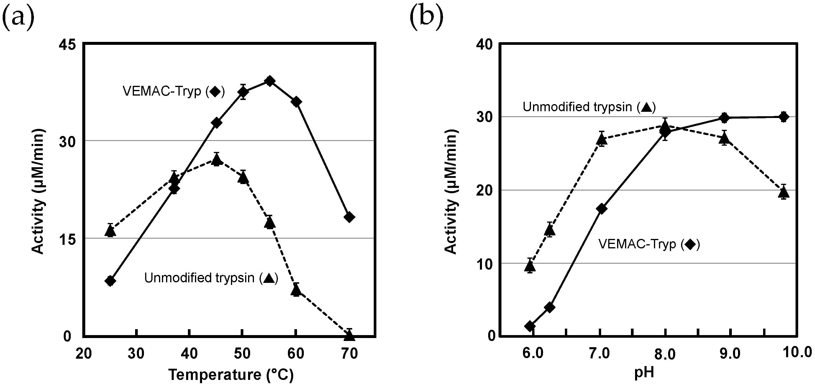
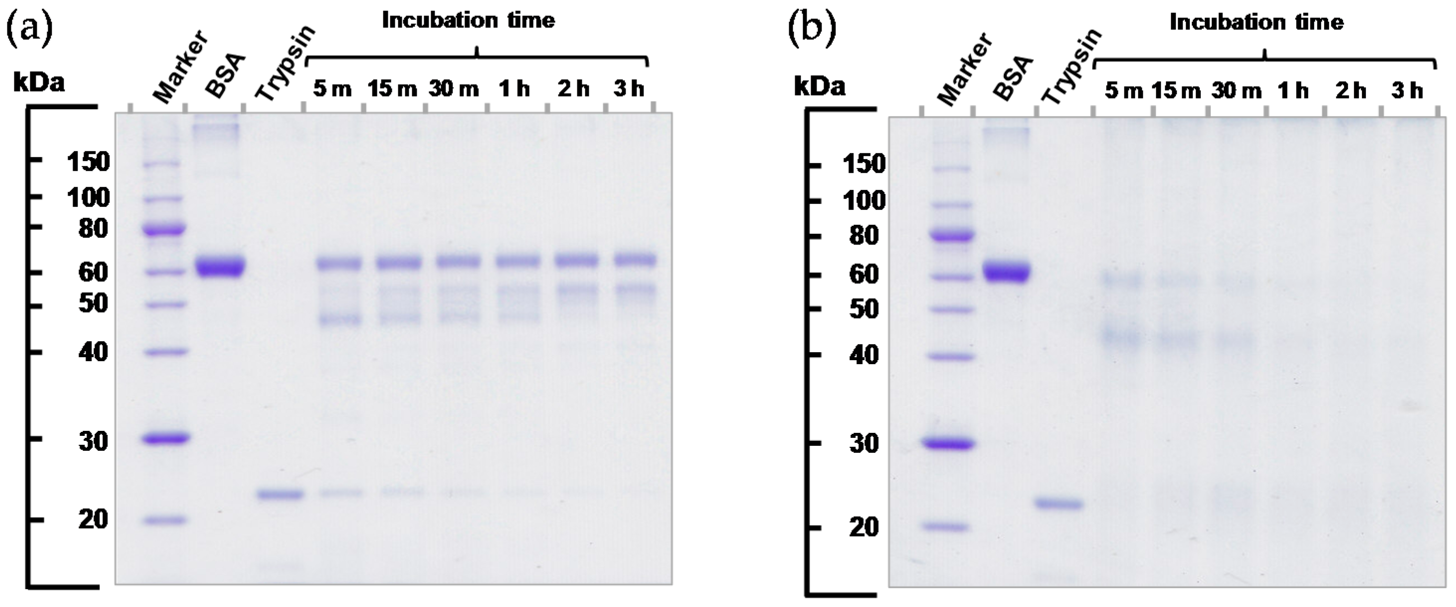
2.3. Protease Activity of VEMAC-Tryp

A low molecular weight synthetic peptide substrate is useful to evaluate the catalytic ability of trypsin. However, the enzymatic activity of trypsin with high molecular weight substrates (i.e., proteins) should also be examined, because trypsin is widely used for protein digestion. Although polymer modification can thermally stabilize the enzyme, it has a high probability of disturbing the reaction with the substrate, especially high molecular weight substrates [4,11,33]. Thus, the protease activity of VEMAC-Tryp was investigated using a BSA as a model protein substrate. The reaction with BSA was conducted at 55 °C for VEMAC-Tryp, based on the results shown in Figure 3a and Figure S4, and at 37 °C for unmodified trypsin, because it quickly lost activity at 55 °C (Figure S5). BSA digested with unmodified trypsin or VEMAC-Tryp was assayed using sodium dodecyl sulfate-polyacrylamide gel electrophoresis (SDS-PAGE), and the polypeptide patterns resulting from proteolysis were compared. The signal intensity of the polypeptide corresponding to undigested BSA (62 kDa) decreased, and polypeptides corresponding to BSA fragments appeared after incubation with unmodified trypsin for 5 min, although the digestion pattern remained roughly unchanged after further incubation (Figure 4a). Moreover, the polypeptide corresponding to unmodified trypsin (23 kDa) rapidly decreased in levels with increasing incubation time, indicating that trypsin autolysis occurred and protein digestion was interrupted at an early stage (Figure 4a). In contrast, digestion with VEMAC-Tryp led to a significantly reduced signal intensity of polypeptides corresponding to BSA after incubation for 5 min, and no polypeptides were detected after 1 h (Figure 4b). These results indicate that VEMAC-Tryp is able to digest BSA effectively, and that enough protease activity remains for at least 1 h at 55 °C. Moreover, no polypeptide corresponding to unmodified trypsin was observed (Figure 4b), suggesting that trypsin was effectively conjugated with VEMAC.

3. Materials and Methods

3.1. Materials

Trypsin (bovine pancreas) and TNBS were purchased from Wako Pure Chemical Industries, Osaka, Japan. VEMAC was prepared by hydrolyzing the maleic anhydride moiety of vinylmethylether-maleic anhydride copolymer (MW = 200 kDa) provided by ISP Japan (Tokyo, Japan). BAPNA was purchased from Peptide Institute, Osaka, Japan. All other reagents and solvents were obtained from Sigma-Aldrich (St. Louis, MO, USA). Dialysis membranes (molecular weight cut-off: 3.5 kDa) were purchased from Spectrum Laboratories (Rancho Domingo, CA, USA). PB was prepared using sodium dihydrogen phosphate, sodium hydrogen diphosphate, and deionized water, and used for VEMAC-Tryp synthesis and enzyme activity measurements.

3.2. Physicochemical Characterization

Hydrodynamic diameter and zeta-potential were measured using a Zetasizer NanoZS (Malvern Instrument, Malvern, Worcestershire, UK) at room temperature. VEMAC-Tryp was dialyzed against PB to remove low molecular weight impurities and adjusted to 0.2 mg/mL in PB. Unmodified trypsin was adjusted to 0.2 mg/mL in PB.
For CD measurements, VEMAC-Tryp was dialyzed against PB to remove low molecular weight impurities and adjusted to 0.1 mg/mL in PB. Unmodified trypsin was adjusted to 0.25 mg/mL in PB. CD spectra were recorded with a J-820 CD spectrometer (JASCO, Tokyo, Japan) in a quartz cell (light path: 1 mm) at 4 °C. Secondary structure contents were calculated by a protein secondary structure analysis program provided by JASCO.

3.3. Synthesis of VEMAC-Tryp and Preparation of a VEMAC/Trypsin Mixture

VEMAC-Tryp was synthesized at an 8:1 VEMAC/trypsin ratio as follows: 4.0 mg/mL VEMAC solution in PB was mixed with 0.3 mL of 120 mM NHS and 0.3 mL of 300 mM N-(3-dimethylaminopropyl)-N′-ethylcarbodiimide hydrochloride (EDC) in PB at 4 °C and incubated for 1 h at room temperature under shaking at 1000 rpm to activate carboxyl groups in VEMAC to NHS-ester. The resulting mixture was cooled to 4 °C and mixed with 1.2 mL of 0.5 mg/mL trypsin in PB and incubated at 4 °C for 24 h under shaking at 500 rpm. Finally, the reaction mixture was incubated at 37 °C for 6 h to hydrolyze unreacted NHS-ester and EDC. The VEMAC-Tryp was stored at 4 °C until use. To synthesize VEMAC-Tryp using other VEMAC/trypsin ratios, different amounts of EDC/NHS-activated VEMAC were used in the reaction mixture with a fixed concentration of trypsin. The number of amino groups used for VEMAC modification was estimated by TNBS assay. Briefly, a mixture of 0.25 mL of 2.8 mM TNBS solution and 0.25 mL of 10 mM of Na2SO3 solution was added to a 1.25 mL of sample dissolved in 0.1 M sodium tetraborate (pH 9.5). The solution was mixed and incubated at 30 °C for 2 h. The absorbance was measured at 420 nm using a Multiskan JX microplate reader (Thermo Fisher Scientific, Waltham, MA, USA). A blank was prepared similarly without sample.
A VEMAC/trypsin mixture was prepared using the same procedure described above for VEMAC-Tryp synthesis, but without activation of VEMAC carboxyl groups using EDC and NHS.

3.4. Evaluation of Enzymatic Activity Using a Low Molecular Weight Synthetic Substrate

To evaluate the catalytic activity of VEMAC-Tryp, the synthetic low molecular weight compound BAPNA was used as substrate. A mixture of 0.84 mL of PB and 0.06 mL of 16.7 mM BAPNA in DMSO was incubated at 37 °C for 5 min. Next, 0.1 mL of VEMAC-Tryp or unmodified trypsin containing 0.2 mg/mL of the enzyme was added to the BAPNA solution and incubated at 37 °C. After a predetermined time, a 0.1 mL aliquot was removed and mixed with 0.1 mL of 10% (w/v) acetic acid to stop the reaction. The release of p-nitroaniline from BAPNA by trypsin was determined by measuring absorbance at 405 nm using a Multiskan JX microplate reader. The release rate of p-nitroaniline versus time was plotted, and the initial slope used as enzymatic activity (μM/min). In the experiments examining temperature and pH dependence of trypsin activity, the reaction with BAPNA was conducted as described above at the indicated temperature and pH.

3.5. Evaluation of Protease Activity

To evaluate the protease activity of VEMAC-Tryp, BSA was digested with VEMAC-Tryp or unmodified trypsin and analyzed by SDS-PAGE using an AE-7300 electrophoresis system (ATTO, Tokyo, Japan) according to the manufacturer’s instructions. Briefly, the VEMAC-Tryp solution was dialyzed against deionized water to remove impurities and salts. After freeze-drying, a 10 mg/mL trypsin solution was prepared in PB. A volume of 0.1 mL of the trypsin solution was mixed with 0.1 mL of 11 mg/mL BSA in PB, and the mixture was incubated at 55 °C. After a predetermined time, a 0.01 mL aliquot was removed and mixed with 0.1 mL SDS sample loading buffer containing SDS, 2-mercaptoethanol, and glycerin. After SDS-PAGE, the gel was stained with Coomassie Brilliant Blue. The stained peptides in the gel were read using a scanner (LiDE200, Canon, Tokyo, Japan).

4. Conclusions

Highly stabilized trypsin with autolysis resistance was synthesized by nonspecific multi-point conjugation of the enzyme with VEMAC without affecting enzymatic activity. This stability extended for many months, and the modified enzyme showed highly efficient protease activity. Its activity persisted at higher temperatures and at higher pH than that of unmodified trypsin, and the effects of pH on enzymatic activity were reversible, indicating that pH could be used as an on–off switch for the modified enzyme.
The results obtained in this study indicate that the VEMAC modification of an enzyme via multi-point attachment is effective to improve thermal stability. Although nonspecific multi-point conjugation of enzyme with polymer may result in a decrease in activity as previously reported [34,35], the VEMAC modification under optimized conditions is a simple and cost-effective strategy using a commercially available polymer that can be used to obtain fully active modified enzymes. Furthermore, VEMAC-Tryp contains many unmodified carboxyl groups that can be used for immobilization of VEMAC-Tryp on solid supports like particulate and porous materials, which is relevant to the design and development of bioreactors.

Supplementary Materials

The following are available online at www.mdpi.com/2073-4344/7/1/4/s1. Figure S1: Progressive changes in enzymatic activity of vinylmethylether-maleic acid copolymer-modified trypsin (VEMAC-Tryp) in phosphate buffer (PB, 50 mM, pH 8.0) at 25 °C, Figure S2: Hanes–Woolf plots for the evaluation of the enzymatic activity of VEMAC-Tryp and unmodified trypsin, Figure S3: Particle size distribution of unmodified trypsin and VEMAC-Tryp, Figure S4: Evolution of residual activity (%) of VEMAC-Tryp upon incubation in PB (50 mM, pH 8.0) at different temperatures, Figure S5: Evolution of residual activity (%) of unmodified trypsin upon incubation in PB (50 mM, pH 8.0) at 55 °C.

Acknowledgments

This work was supported by Japan Society for the Promotion of Science (JSPS) KAKENHI (Grant No. 25860025, 15k07893).

Author Contributions

Yasushi Sasai and Hiroshi Kanno conceived, designed and performed the experiments; Yasushi Sasai, Hiroshi Kanno, Naoki Doi, Yukinori Yamauchi, Masayuki Kuzuya and Shin-ichi Kondo analyzed the data; Yasushi Sasai wrote the paper.

Conflicts of Interest

The authors declare no conflict of interest.

References

  1. Gauthier, M.A.; Klok, H.-A. Polymer–protein conjugates: An enzymatic activity perspective. Polym. Chem. 2010, 1, 1352–1373. [Google Scholar] [CrossRef]
  2. Hoffman, A.S.; Stayton, P.S. Conjugates of stimuli-responsive polymers and proteins. Prog. Polym. Sci. 2007, 32, 922–932. [Google Scholar] [CrossRef]
  3. Shakya, A.K.; Sami, H.; Srivastava, A.; Kumar, A. Stability of responsive polymer–protein bioconjugates. Prog. Polym. Sci. 2010, 35, 459–486. [Google Scholar] [CrossRef]
  4. Falatach, R.; Li, S.; Sloane, S.; McGlone, C.; Berberich, J.A.; Page, R.C.; Averick, S.; Konkolewicz, D. Why synthesize protein–polymer conjugates? The stability and activity of chymotrypsin-polymer bioconjugates synthesized by RAFT. Polymer 2015, 72, 382–386. [Google Scholar] [CrossRef]
  5. Veronese, F.M.; Pasut, G. PEGylation, successful approach to drug delivery. Drug Discov. Today 2005, 10, 1451–1458. [Google Scholar] [CrossRef]
  6. Hsieh, Y.-P.; Lin, S.-C. Effect of PEGylation on the activity and stability of horseradish peroxidase and l-N-carbamoylase in aqueous phases. Process Biochem. 2015, 50, 1372–1378. [Google Scholar] [CrossRef]
  7. Swierczewska, M.; Lee, K.C.; Lee, S. What is the future of PEGylated therapies? Expert Opin. Emerg. Drugs 2015, 20, 531–536. [Google Scholar] [CrossRef] [PubMed]
  8. Alconcel, S.N.S.; Baas, A.S.; Maynard, H.D. FDA-approved poly(ethylene glycol)–protein conjugate drugs. Polym. Chem. 2011, 2, 1442–1448. [Google Scholar] [CrossRef]
  9. Veronese, F.M.; Mero, A. The impact of PEGylation on biological therapies. BioDrugs 2008, 22, 315–329. [Google Scholar] [CrossRef] [PubMed]
  10. Murata, H.; Cummings, C.S.; Koepsel, R.R.; Russell, A.J. Polymer-based protein engineering can rationally tune enzyme activity, pH-dependence, and stability. Biomacromolecules 2013, 14, 1919–1926. [Google Scholar] [CrossRef] [PubMed]
  11. Lucius, M.; Falatach, R.; McGlone, C.; Makaroff, K.; Danielson, A.; Williams, C.; Nix, J.C.; Konkolewicz, D.; Page, R.C.; Berberich, J.A. Investigating the impact of polymer functional groups on the stability and activity of lysozyme-polymer conjugates. Biomacromolecules 2016, 17, 1123–1134. [Google Scholar] [CrossRef] [PubMed]
  12. Pedroche, J.; del Mar Yust, M.; Mateo, C.; Fernández-Lafuente, R.; Girón-Calle, J.; Alaiz, M.; Vioque, J.; Guisán, J.M.; Millán, F. Effect of the support and experimental conditions in the intensity of the multipoint covalent attachment of proteins on glyoxyl-agarose supports: Correlation between enzyme–support linkages and thermal stability. Enzyme Microb. Technol. 2007, 40, 1160–1166. [Google Scholar] [CrossRef]
  13. Riccardi, C.M.; Cole, K.S.; Benson, K.R.; Ward, J.R.; Bassett, K.M.; Zhang, Y.; Zore, O.V.; Stromer, B.; Kasi, R.M.; Kumar, C.V. Toward “stable-on-the-table” enzymes: Improving key properties of catalase by covalent conjugation with poly(acrylic acid). Bioconjugate Chem. 2014, 25, 1501–1510. [Google Scholar] [CrossRef] [PubMed]
  14. Treetharnmathurot, B.; Dieudonné, L.; Ferguson, E.L.; Schmaljohann, D.; Duncan, R.; Wiwattanapatapee, R. Dextrin–trypsin and ST-HPMA–trypsin conjugates: Enzyme activity, autolysis and thermal stability. Int. J. Pharm. 2009, 373, 68–76. [Google Scholar] [CrossRef] [PubMed]
  15. Hustoft, H.K.; Malerod, H.; Wilson, S.R.; Reubsaet, L.; Lundanes, E.; Greibrokk, T. A critical review of trypsin digestion for LC-MS based proteomics. In Integrative Proteomics; Leung, H.-C., Man, T.-K., Flores, R.J., Eds.; InTech: Rijeka, Croatia, 2012; pp. 73–92. [Google Scholar]
  16. Havliš, J.; Thomas, H.; Šebela, M.; Shevchenko, A. Fast-response proteomics by accelerated in-gel digestion of proteins. Anal. Chem. 2003, 75, 1300–1306. [Google Scholar] [CrossRef] [PubMed]
  17. Sebela, M.; Stosova, T.; Havlis, J.; Wielsch, N.; Thomas, H.; Zdrahal, Z.; Shevchenko, A. Thermostable trypsin conjugates for high-throughput proteomics: Synthesis and performance evaluation. Proteomics 2006, 6, 2959–2963. [Google Scholar] [CrossRef] [PubMed]
  18. Yan, M.; Ge, J.; Dong, W.; Liu, Z.; Ouyang, P. Preparation and characterization of a temperature-sensitive sulfobetaine polymer–trypsin conjugate. Biochem. Eng. J. 2006, 30, 48–54. [Google Scholar] [CrossRef]
  19. Treetharnmathurot, B.; Ovartlarnporn, C.; Wungsintaweekul, J.; Duncan, R.; Wiwattanapatapee, R. Effect of PEG molecular weight and linking chemistry on the biological activity and thermal stability of PEGylated trypsin. Int. J. Pharm. 2008, 357, 252–259. [Google Scholar] [CrossRef] [PubMed]
  20. Gaertner, H.F.; Puigserver, A.J. Increased activity and stability of poly(ethylene glycol)-modified trypsin. Enzyme Microb. Technol. 1992, 14, 150–155. [Google Scholar] [CrossRef]
  21. Varshosaz, J.; Hassanzadeh, F.; Aliabadi, H.S.; Khoraskani, F.R.; Mirian, M.; Behdadfar, B. Targeted delivery of doxorubicin to breast cancer cells by magnetic LHRH chitosan bioconjugated nanoparticles. Int. J. Biol. Macromol. 2016, 93, 1192–1205. [Google Scholar] [CrossRef] [PubMed]
  22. Jones, D.S.; Laverty, T.P.; Morris, C.; Andrews, G.P. Statistical modelling of the rheological and mucoadhesive properties of aqueous poly(methylvinylether-co-maleic acid) networks: Redefining biomedical applications and the relationship between viscoelasticity and mucoadhesion. Colloids Surf. B 2016, 144, 125–134. [Google Scholar] [CrossRef] [PubMed]
  23. McCrudden, M.T.C.; Torrisi, B.M.; Al-Zahrani, S.; McCrudden, C.M.; Zaric, M.; Scott, C.J.; Kissenpfennig, A.; McCarthy, H.O.; Donnelly, R.F. Laser-engineered dissolving microneedle arrays for protein delivery: Potential for enhanced intradermal vaccination. J. Pharm. Pharmacol. 2015, 67, 409–425. [Google Scholar] [CrossRef] [PubMed]
  24. Shahbazi, M.-A.; Almeida, P.V.; Mäkilä, E.; Correia, A.; Ferreira, M.P.A.; Kaasalainen, M.; Salonen, J.; Hirvonen, J.; Santos, H.A. Poly(methyl vinyl ether-alt-maleic acid)-functionalized porous silicon nanoparticles for enhanced stability and cellular internalization. Macromol. Rapid Commun. 2014, 35, 624–629. [Google Scholar] [CrossRef] [PubMed]
  25. Garland, M.J.; Singh, T.R.R.; Woolfson, A.D.; Donnelly, R.F. Electrically enhanced solute permeation across poly(ethylene glycol)–crosslinked poly(methyl vinyl ether-co-maleic acid) hydrogels: Effect of hydrogel crosslink density and ionic conductivity. Int. J. Pharm. 2011, 406, 91–98. [Google Scholar] [CrossRef] [PubMed]
  26. Finne, U.; Kyyrönen, K.; Urtti, A. Drug release from monoisopropyl ester of poly(vinyl methyl ether-maleic anhydride) can be modified by basic salts in the polymer matrix. J. Controlled Release 1989, 10, 189–194. [Google Scholar] [CrossRef]
  27. Sasai, Y.; Matsuzaki, N.; Kondo, S.; Kuzuya, M. Introduction of carboxyl group onto polystyrene surface using plasma techniques. Surf. Coat. Technol. 2008, 202, 5724–5727. [Google Scholar] [CrossRef]
  28. Erlanger, B.F.; Kokowsky, N.; Cohen, W. The preparation and properties of two new chromogenic substrates of trypsin. Arch. Biochem. Biophys. 1961, 95, 271–278. [Google Scholar] [CrossRef]
  29. Fields, R. The measurement of amino groups in proteins and peptides. Biochem. J. 1971, 124, 581–590. [Google Scholar] [CrossRef] [PubMed]
  30. Chiu, K.; Agoubi, L.L.; Lee, I.; Limpar, M.T.; Lowe, J.W., Jr.; Goh, S.L. Effects of polymer molecular weight on the size, activity, and stability of PEG-functionalized trypsin. Biomacromolecules 2010, 11, 3688–3692. [Google Scholar] [CrossRef] [PubMed]
  31. Ritchie, R.J.; Prvan, T. Current statistical methods for estimating the Km and Vmax of Michaelis-Menten kinetics. Biochem. Educ. 1996, 24, 196–206. [Google Scholar] [CrossRef]
  32. Plochocka, K.; Liu, X.; Tallon, M.A.; Musa, O.M. The quintessential alternating copolymer family: Alkyl vinyl ether co-maleic anhydride copolymers. In Handbook of Maleic Anhydride Based Materials: Syntheses, Properties and Applications; Musa, O.M., Ed.; Springer: Cham, Switzerland, 2016; pp. 211–250. [Google Scholar]
  33. Oupický, D.; Ulbrich, K.; Říhová, B. Conjugates of semitelechelic poly[N-(2-hydroxypropyl) methacrylamide] with enzymes for protein delivery. J. Bioact. Compat. Polym. 1999, 14, 213–231. [Google Scholar]
  34. Duncan, R.; Gilbert, H.R.P.; Carbajo, R.J.; Vicent, M.J. Polymer masked−unmasked protein therapy. 1. Bioresponsive dextrin−trypsin and −melanocyte stimulating hormone conjugates designed for α-amylase activation. Biomacromolecules 2008, 9, 1146–1154. [Google Scholar] [CrossRef] [PubMed]
  35. Altikatoglu, M.; Celebi, M. Enhanced stability and decolorization of Coomassie Brilliant Blue R-250 by dextran aldehyde-modified horseradish peroxidase. Artif. Cells Blood Substit. Immobil. Biotechnol. 2011, 39, 185–190. [Google Scholar] [CrossRef] [PubMed]
Scheme 1. Reaction scheme for synthesizing vinylmethylether-maleic acid copolymer-modified trypsin (VEMAC-Tryp) via multi-point conjugation. EDC: 1-ethyl-3-(3-dimethylaminopropyl) carbodiimide hydrochloride, NHS: N-hydroxysuccinimide.
Catalysts 07 00004 sch001
Figure 1. (a) Enzymatic activity of vinylmethylether-maleic acid copolymer (VEMAC)-Tryp synthesized using varying VEMAC/trypsin (w/w) ratios; (b) Enzymatic activity kinetics of VEMAC-Tryp, unmodified trypsin, and VEMAC/trypsin mixture incubated in phosphate buffer solution (50 mM, pH 8.0) at 37 °C. Enzymatic activity was determined from the hydrolysis rate of benzoyl-dl-arginine p-nitroanilide hydrochloride catalyzed by trypsin. Data are shown as mean ± standard deviation (SD) (n = 3).
Catalysts 07 00004 g001
Figure 2. Circular dichroism spectra of vinylmethylether-maleic acid copolymer (VEMAC)-Tryp and unmodified trypsin before and after incubation in phosphate buffer (50 mM, pH 8.0) at 37 °C for 24 h. Measurements were conducted at 4 °C.
Catalysts 07 00004 g002
Figure 3. (a) Temperature dependence of the enzymatic activity of unmodified trypsin and vinylmethylether-maleic acid copolymer (VEMAC)-Tryp (measured at pH 8.0); (b) pH dependence of the enzymatic activity of unmodified trypsin and VEMAC-Tryp (measured at 37 °C). Data are shown as mean ± SD (n = 3).
Catalysts 07 00004 g003
Figure 4. Sodium dodecyl sulfate-polyacrylamide gel electrophoresis (SDS-PAGE) analysis of bovine serum albumin digested with (a) unmodified trypsin at 37 °C and (b) vinylmethylether-maleic acid copolymer (VEMAC)-Tryp at 55 °C. BSA: bovine serum albumin.
Catalysts 07 00004 g004
Table 1. Enzyme kinetic parameters determined by Hanes–Woolf plots.
SampleVmax (mM/min) 1Km (mM) 1Kcat (s−1) 1Kcat/Km
Unmodified trypsin0.059 ± 0.0041.34 ± 0.121.18 ± 0.080.88
VEMAC-Tryp0.041 ± 0.0010.73 ± 0.010.80 ± 0.021.10
1 Data are shown as mean ± SD of three measurements.
Table 2. Diameter and zeta potential of trypsin before and after vinylmethylether-maleic acid copolymer (VEMAC) modification.
SampleDiameter (nm) 1Zeta-Potential (mV) 1
Unmodified trypsin3.3 ± 0.7−15.3 ± 9.4
VEMAC-Tryp21.6 ± 4.2−32.6 ± 11.3
1 Data are shown as mean ± SD of three measurements.
Table 3. Secondary structure contents of unmodified trypsin and vinylmethylether-maleic acid copolymer (VEMAC)-Tryp before and after incubation 1.
Sampleα-Helix (%)β-Sheet (%)Turns (%)Random (%)
Unmodified trypsin7.240.418.334.1
After incubation026.531.542.1
VEMAC-Tryp5.641.718.034.7
After incubation4.542.317.735.5
1 Sample was incubated in phosphate buffer (50 mM, pH 8.0) at 37 °C for 24 h.
Table 4. Hydrodynamic diameter of vinylmethylether-maleic acid copolymer (VEMAC)-Tryp at different pH.
pH ValueDiameter (nm) 1
5.94.0 ± 1.1
8.021.6 ± 4.2
9.820.9 ± 6.3
1 Data are shown as mean ± SD of three measurements.

