Total Synthesis and Biological Evaluation of Phaeosphaerides


Graduate School of Pharmaceutical Sciences, Meiji Pharmaceutical University, 2-522-1 Noshio, Kiyose, Tokyo 204-8588, Japan
*
Author to whom correspondence should be addressed.
Catalysts 2018, 8(5), 206; https://doi.org/10.3390/catal8050206
Submission received: 26 April 2018
/
Revised: 5 May 2018
/
Accepted: 7 May 2018
/
Published: 14 May 2018
(This article belongs to the Special Issue Catalyzed Synthesis of Natural Products)
Abstract
:This article reviews studies regarding the total synthesis of phaeosphaerides A and B, nitrogen-containing bicyclic natural products isolated from an endophytic fungus. Numerous synthetic efforts and an X-ray crystal structure analysis of phaeosphaeride A have enabled revision of its originally proposed structure. In addition, a successful protic acid-mediated transformation of phaeosphaeride A to phaeosphaeride B revealed the hypothetical biosynthesis of phaeosphaeride B from phaeosphaeride A. Structure–activity relationship studies of phaeosphaeride derivatives are also discussed.
1. Introduction
Signal transducer and activator of transcription 3 (STAT3), which belongs to the STAT family of proteins [1], regulates cell proliferation, differentiation, and survival [2]. Non-activated STAT3 is generally localized in the cytoplasm. Once phosphorylated at Tyr705 in the Janus kinase (JAK)/STAT signaling pathway, STAT3 dimerizes, translocates to the nucleus, and binds to a target DNA sequence to induce transcriptional activation [3,4].
Unusual activation of STAT3 is frequently found in various types of tumor cells, leading to apoptosis resistance and tumor cell proliferation via enhanced expression of gene encoding proteins such as Bcl-2, Bcl-xL, and cyclin D1 [5,6]. Therefore, STAT3 has gained considerable interest as a potential target for anticancer therapy. In fact, various STAT3 inhibitors including synthetic small molecules and natural products have been evaluated as prospective anticancer chemotherapeutic agents [7].
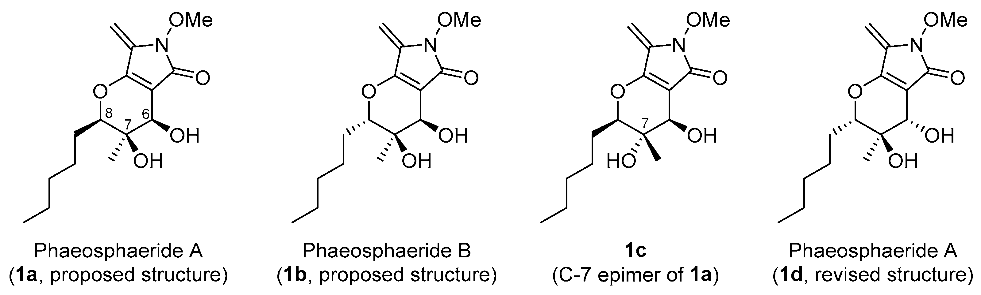
In 2006, phaeosphaerides A (proposed structure 1a) was isolated from the endophytic fungus FA39 (Phaeosphaeria avenaria) by Clardy and co-workers as an inhibitor of STAT3-DNA binding (Figure 1) [8]. Phaeosphaeride A possesses a bicyclic structure with three contiguous stereocenters in its dihydropyran ring. Phaeosphaeride A inhibits STAT3 activity with an IC50 of 0.61 mM and also inhibits cell growth in STAT3-dependent U266 multiple myeloma cells with an IC50 of 6.7 μM. Therefore, phaeosphaeride A is expected to be a potential lead compound for anticancer drug candidates. On the other hand, phaeosphaeride B (1b), the C-8 stereoisomer of phaeosphaeride A, was reported to have no STAT3 inhibitory activity.
The promising biological activity of phaeosphaeride A as well as its simple and unique molecular structure has attracted much attention from the synthetic community. In addition, structure–activity relationship (SAR) studies of STAT3 inhibitory activity should be of significant importance for potential anticancer therapy.
2. Synthetic Approach toward Phaeosphaeride A
2.1. Synthesis of the Proposed Structure of Phaeosphaeride A
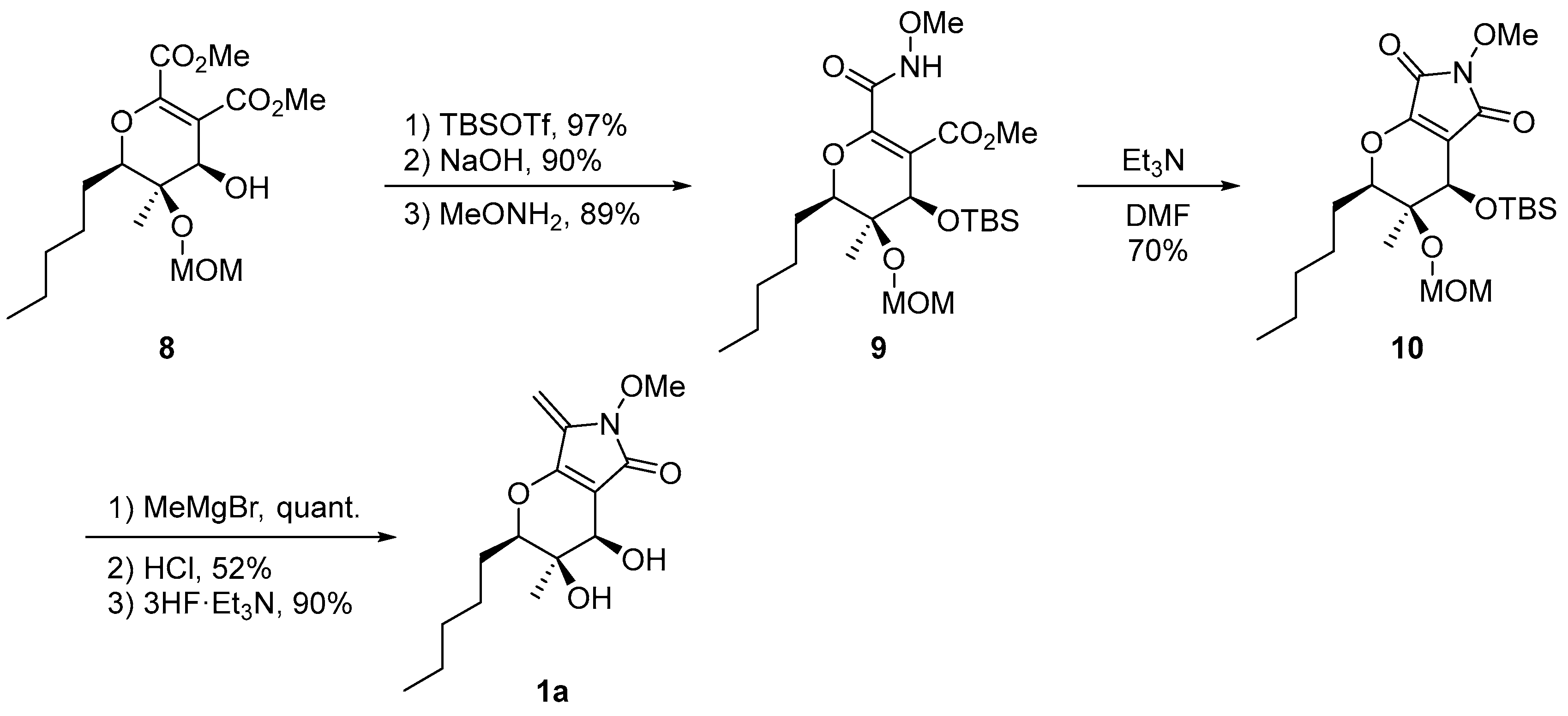
In 2011, our group accomplished the first total synthesis of the proposed structure of phaeosphaeride A (1a; Scheme 1 and Scheme 2) [9]. In this synthesis, to construct the C-7 and C-8 stereocenters, we used a typical E-selective Horner–Wadsworth–Emmons reaction followed by Sharpless asymmetric dihydroxylation using AD-mix-β to obtain the requisite diol (2S,3R)-4 in high yield. In this step, the absolute configuration and the high enantiomeric excess of diol 4 were confirmed by a modified Mosher’s method. After appropriate conversion of diol 4 to secondary alcohol 5, we found good reaction conditions for an oxy-Michael addition of alcohol 5 to dimethyl acetylenedicarboxylate: Use of a catalytic amount of n-BuLi cleanly afforded the desired Michael adduct (E)-6 along with the (Z)-isomer in 76% and 18% yields, respectively. The remaining C-6 stereogenic center was installed by a vinyl-anion aldol reaction of aldehyde 7 using sodium bis(trimethylsilyl)amide (NaHMDS), providing the desired dihydropyran derivative 8 via the plausible transition state shown in Scheme 1.
The construction of the five-membered ring in phaeosphaeride A was followed by regioselective installation of the exo-methylene group to furnish the proposed structure of phaeosphaeride A (1a; Scheme 2). However, the 1H and 13C NMR spectra of synthetic 1a did not match those reported for natural phaeosphaeride A. Therefore, this synthesis revealed that the structure of phaeosphaeride A had been incorrectly assigned.
For natural phaeosphaeride A, the Clardy group observed nuclear Overhauser effect spectroscopy (NOESY) correlations between H-6 and H-8, and between H-15 and both H-6 and H-8 (Figure 2) [8]. The correlation between H-6 and H-8 clearly indicates a pseudodiaxial relationship between these hydrogens. However, the correlations between H-15 and both H-6 and H-8 do not provide information about the configuration of the C-7 stereocenter. Hence, the correct structure of phaeosphaeride A was presumed to be the C-7 epimer 1c of the originally proposed structure, or the epimer’s enantiomer 1d.
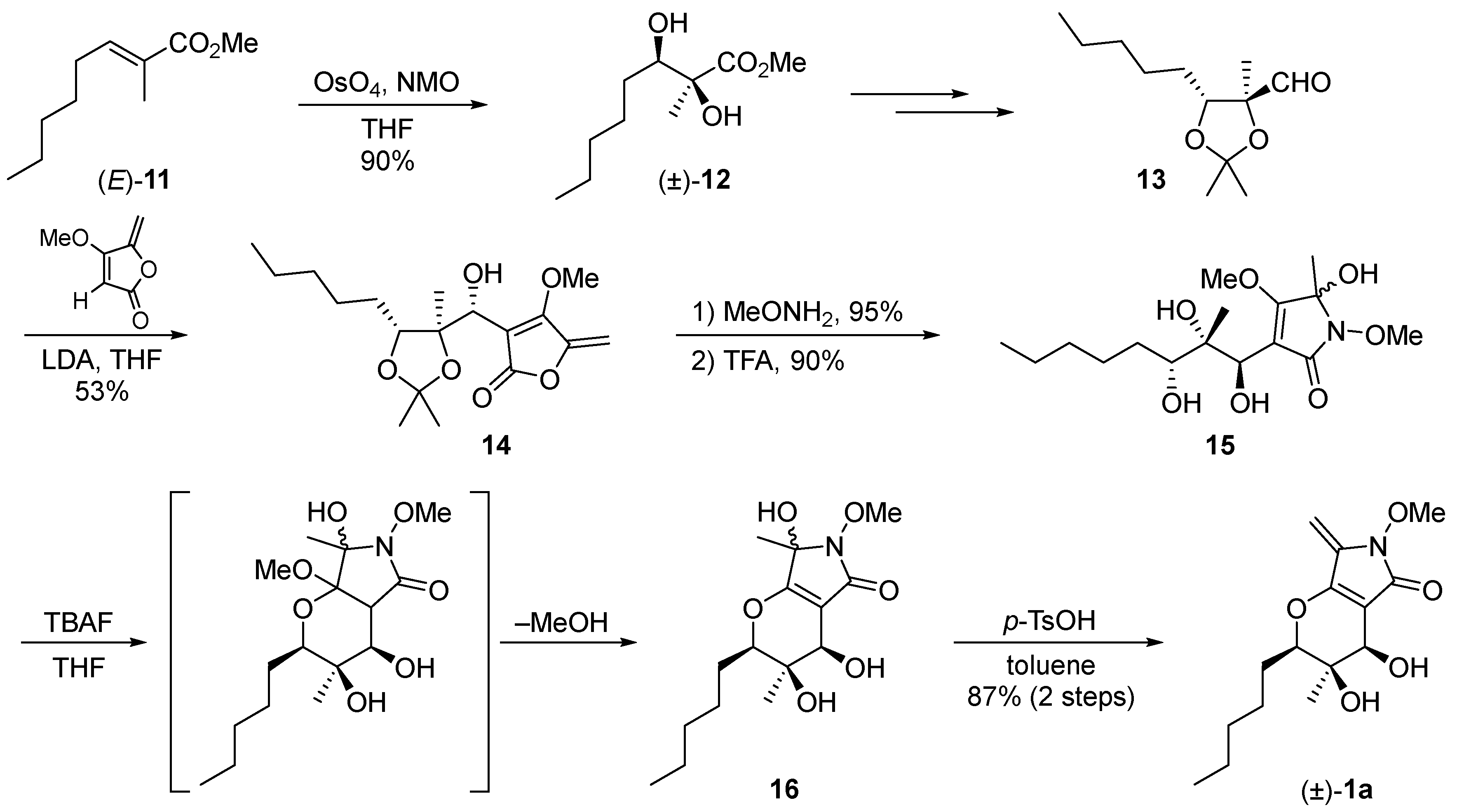
In 2012, Sarli’s group also succeeded in the total synthesis of the proposed structure of phaeosphaeride A (1a) [10]. Their synthesis involved a strategy similar to ours, in which dihydroxylation of unsaturated ester (E)-11 using catalytic OsO4 and 4-methylmorpholine N-oxide (NMO) was used to form the C-7 and C-8 stereocenters. Then, the C-6 center was stereochemically controlled by the anti-selective addition of a vinyllithium species to aldehyde 13 via the Felkin–Ahn transition state. After establishing the three contiguous stereocenters, sequential oxy-Michael addition/methanol elimination followed by selective dehydration furnished (±)-1a (Scheme 3).
They also synthesized both enantiomers of 1a, (6R,7R,8R)-1a and (6S,7S,8S)-1a, by Sharpless asymmetric dihydroxylation of (E)-11 in the first step using AD-mix-β and AD-mix-α, respectively. They obtained the crystal structure of synthetic (6R,7R,8R)-1a using synchrotron radiation, which proved their structural assignment of 1a by NMR. Their studies also pointed to the structural revision of phaeosphaeride A to 1c or its enantiomer 1d (Scheme 4).
2.2. Stereochemical Determination of Natural (−)-Phaeosphaeride A
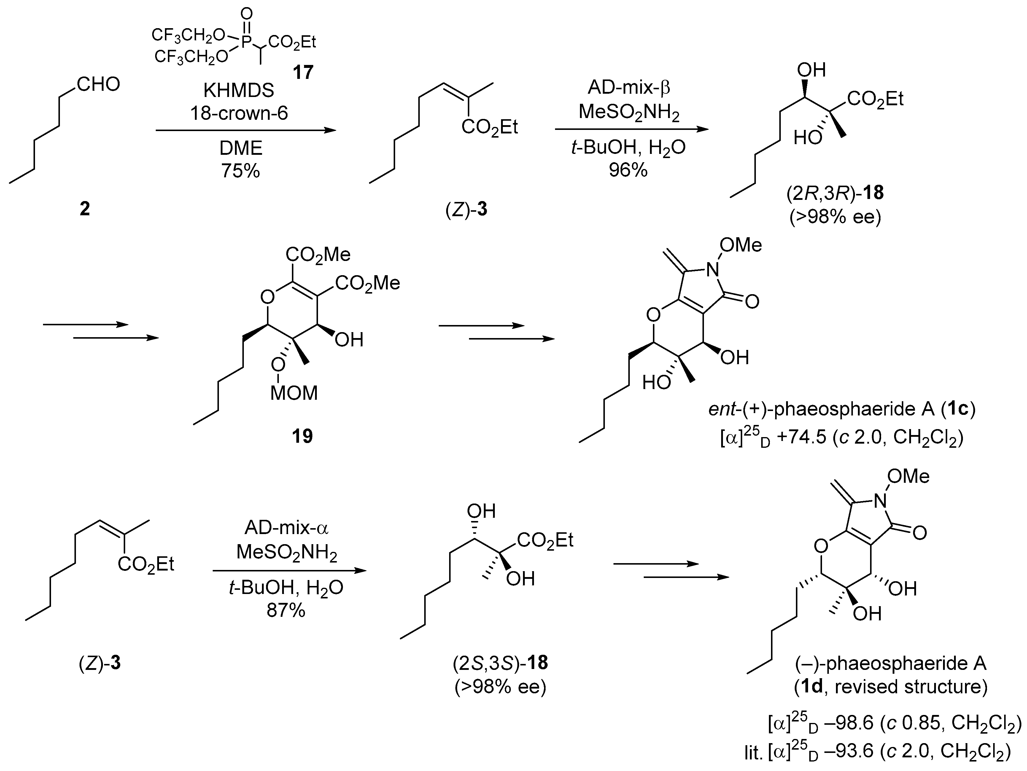
Based on the above achievements by our group and Sarli’s group, the C-7 epimer 1c of the originally proposed structure or the epimer’s enantiomer 1d needed to be synthesized to resolve the issue of the stereochemistry of natural phaeosphaeride A. To access 1c by a synthetic strategy similar to those shown in Scheme 1 and Scheme 2, (Z)-α,β-unsaturated ester (Z)-3 was first prepared instead of the (E)-ester by Still–Gennari olefination using phosphonate 17. The ester (Z)-3 was converted into the diol (2R,3R)-18 via Sharpless dihydroxylation using AD-mix-β. According to the previous route with slight modification, the intermediate diol (2R,3R)-18 was successfully converted via dihydropyran intermediate 19 into 1c, the 1H and 13C NMR spectra of which completely matched the literature data for natural phaeosphaeride A, and the optical rotation of the synthetic compound had the opposite sign to that of the natural product. The correct structure of natural (−)-phaeosphaeride A was thus shown to be the enantiomer 1d of synthetic 1c [11]. Then, natural (−)-phaeosphaeride A was synthesized by using AD-mix-α instead of AD-mix-β in the first step (Scheme 5) [12].
After the reports, Abzianidze et al. reported the crystal structure of natural phaeosphaeride A [13]. This crystal structure clearly supported the results of stereochemical revision by these synthetic approaches.
3. Synthetic Approach toward Phaeosphaeride B
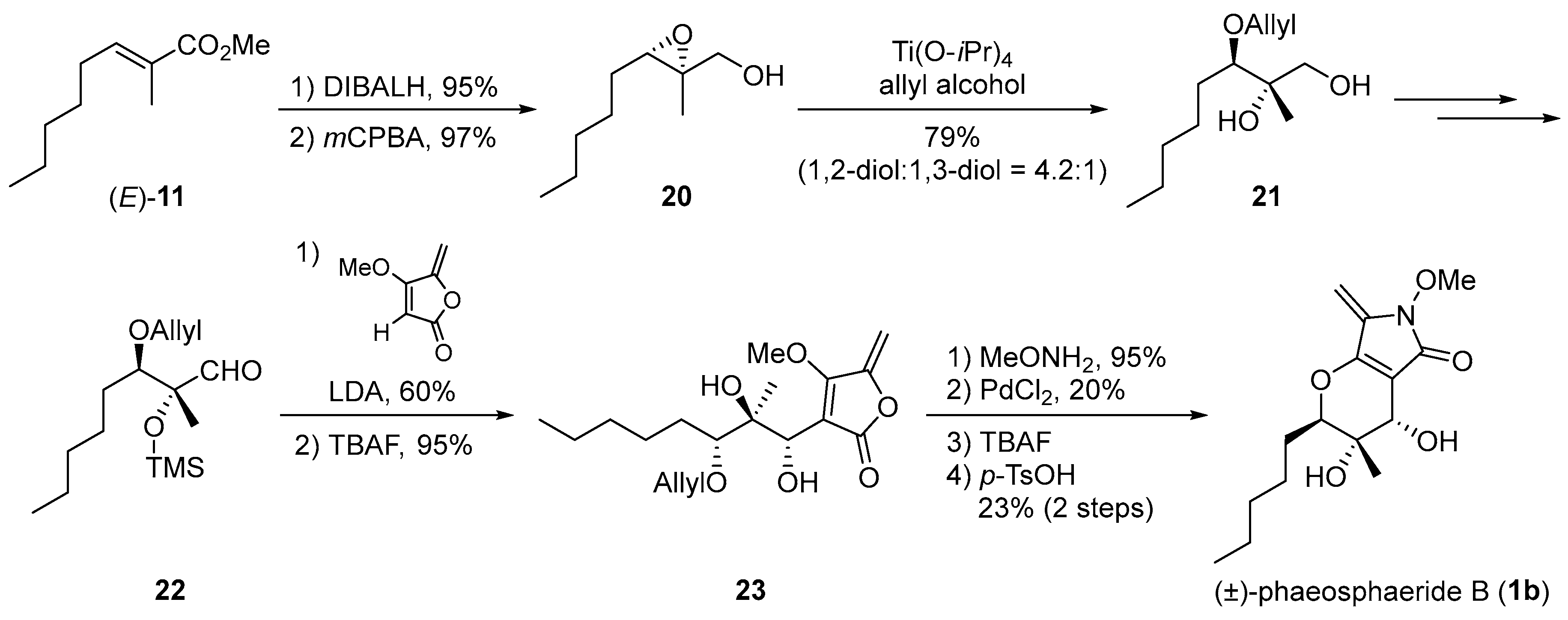
The first total synthesis of (±)-phaeosphaeride B (1b) was reported by Sarli’s group in 2014 [14]. Their synthetic strategy for 1a (Scheme 3) could be applied to the preparation of (±)-1b. In their synthesis, conversion of (E)-11 to an allylic alcohol followed by epoxidation and subsequent regioselective Ti(O-iPr)4-mediated epoxide ring-opening of 20 with allyl alcohol enabled introduction of the C-7 and C-8 stereocenters of (±)-1b. The key anti-selective nucleophilic addition of a vinyllithium species to aldehyde 22 delivered the required C-6 stereocenter through the polar Felkin–Ahn transition state. Then, the lactam and dihydropyran rings and exo-methylene group were assembled to complete the total synthesis of (±)-phaeosphaeride B (1b) (Scheme 6).
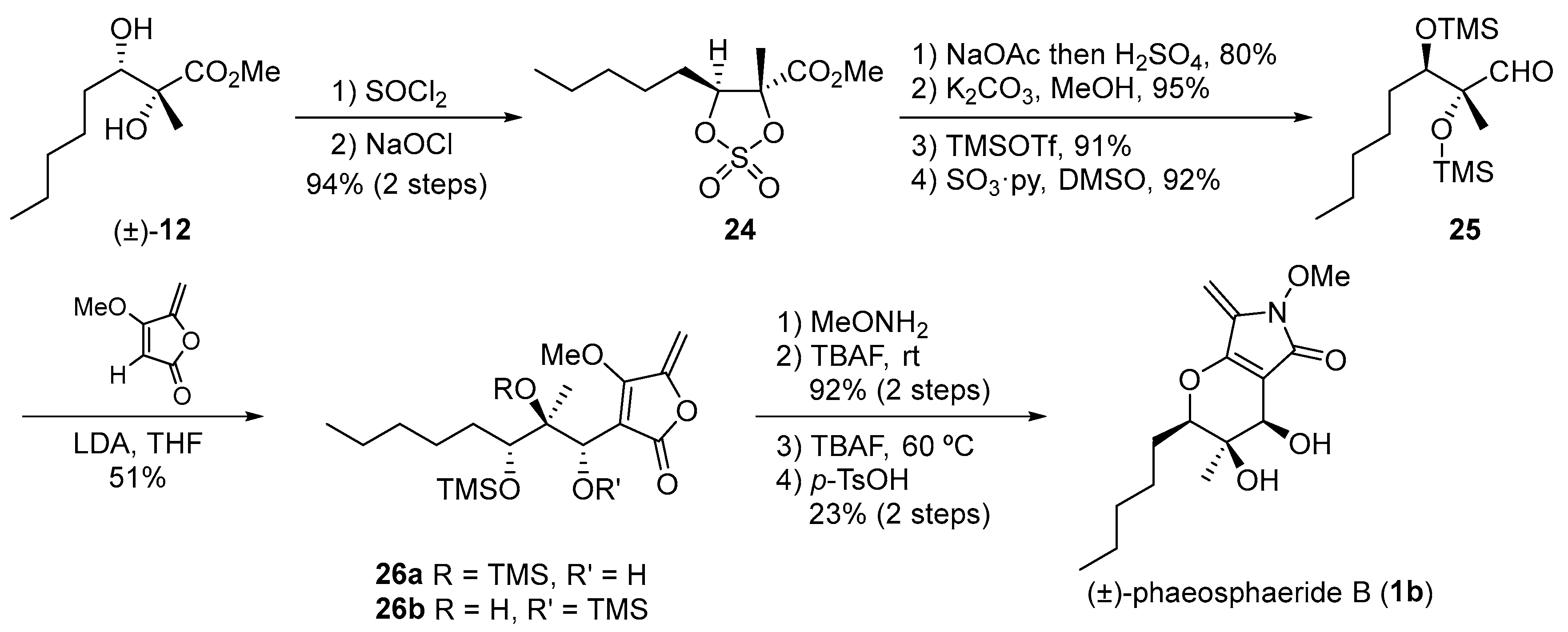
They also developed an improved synthetic scheme for (±)-1b, in which bis-TMS ether aldehyde 25, derived from diol (±)-12 via cyclic sulfate 24, was used in the reaction with α-lithio tetronate to form 26a along with the TMS-migrated product 26b. Subsequent conversion via their established route effectively yielded (±)-1b (Scheme 7).
After the successful total synthesis of (±)-phaeosphaeride B by Sarli, we demonstrated a biomimetic transformation from (−)-phaeosphaeride A to (−)-phaeosphaeride B [12]. We posited that phaeosphaerides A and B would be biosynthetically interconverted under acidic conditions. In testing this hypothesis, treatment of synthetic (−)-phaeosphaeride A (1d) with trifluoroacetic acid (TFA) as a protic acid gave the corresponding trifluoroacetate 27 with stereochemical inversion at C-6 stereocenter via dehydrative formation of the oxonium cation intermediate A. The labile trifluoroacetate 27 was immediately hydrolyzed with aqueous NaHCO3 in THF to yield (−)-phaeosphaeride B (1b) in a good yield (Scheme 8). In addition, this synthesis confirmed the absolute configuration of natural (−)-phaeosphaeride B as shown in Scheme 8.
4. Biological Evaluation of Phaeosphaerides and Their Derivatives
Considering their potential as a seed compound for anticancer treatment, Sarli’s and Abzianidze’s groups evaluated the biological activities of phaeosphaerides and their synthetic derivatives [10,14,15,16,17].
Initially, Sarli and colleagues biologically evaluated the stereoisomers of phaeosphaeride A, (6R,7R,8R)-1a and (6S,7S,8S)-1a [10]. These compounds inhibited STAT3-dependent transcriptional activity in a dose-dependent manner and decreased cell proliferation in breast (MDA-MB-231) and pancreatic (PANC-1) cancer cells in the low micromolar range. After that, they reported that the synthetic (6S,7S,8S)-1a and (6R,7S,8S)-phaeosphaeride had only very weak inhibitory activity against binding of STAT3 to its phosphotyrosine peptide ligand, suggesting that phaeosphaerides are upstream inhibitors of a tyrosine kinase in the JAK/STAT pathway [14].
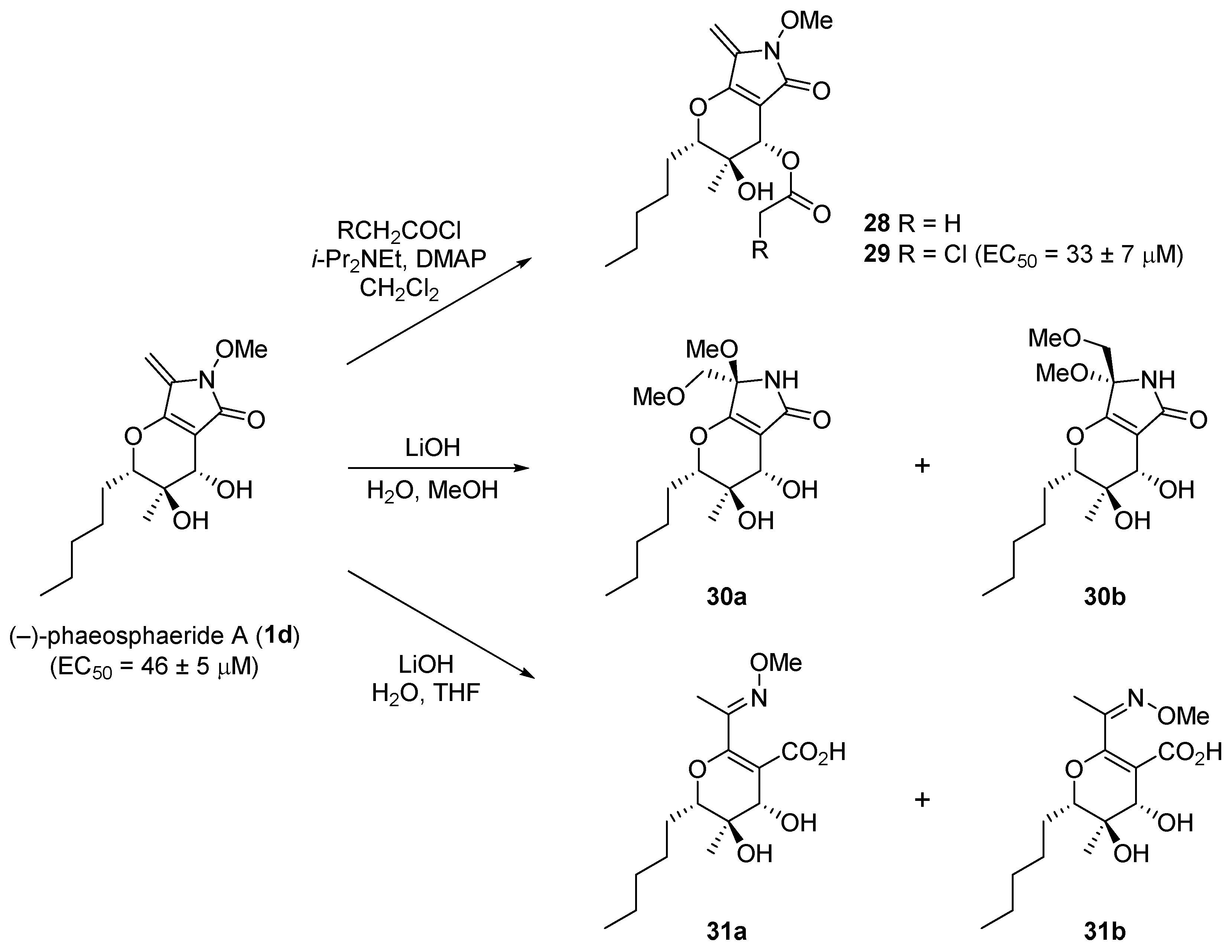
Abzianidze et al. prepared and biologically evaluated the C-6 acyl derivatives 28 and 29, bis-methanol adducts 30a and 30b without the MeO group on the nitrogen, and hydrolyzed products 31a and 31b prepared from isolated natural phaeosphaeride A [15]. Compared to natural phaeosphaeride A (EC50 = 46 ± 5 μM), chloroacetyl derivative 29 exhibited more potent cytotoxicity (EC50 = 33 ± 7 μM) against the A549 cancer cell line, while synthetic 30 and 31 had no activity (Scheme 9).
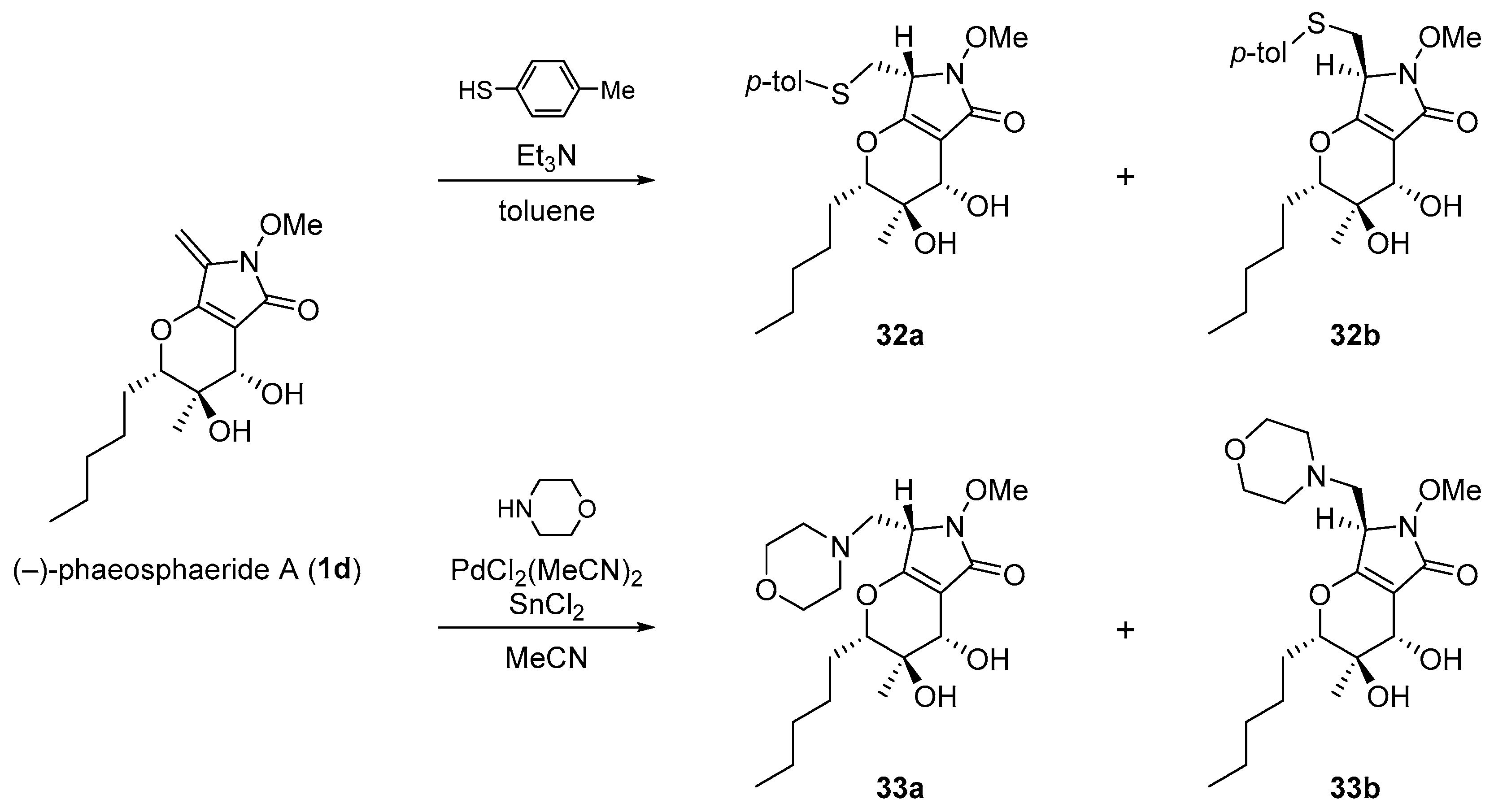
Additionally, they also synthesized 7-(4-methylphenyl)thiomethyl and 7-morpholylmethyl derivatives 32 and 33, which were less cytotoxic than the parent phaeosphaeride A (Scheme 10) [16]. These results clearly indicated that the exo-methylene and N-OMe groups were essential for potent cytotoxicity. Their studies strongly suggest that further SAR studies can provide lead compounds with greater potency for potential use as anticancer chemotherapeutic agents.
5. Conclusions
Phaeosphaerides A and B have attracted considerable attention due not only to their chemical structures but also to their biological activity, and their stereochemical structures have been unambiguously determined through the total synthesis and X-ray crystal structure analysis of phaeosphaeride A. The promising anticancer activity of phaeosphaeride A based on inhibition of STAT3-DNA binding indicates that this natural product is a promising seed compound for anticancer drug candidates. SAR studies on phaeosphaerides led to the development of more potent compounds such as chloroacetyl derivative 29, and further SAR studies are awaited for anticancer drug discovery research. In addition, structurally related natural products including paraphaeosphaerides [18,19], phyllostictines [20,21], isoaigialones [22], benesudon [23], and curvupallides [24] should also attract considerable interest as pharmaceutical targets. Further studies of phaeosphaerides are expected to make valuable contributions to synthetic and medicinal chemistry.
Author Contributions
K.K. and K.T.III prepared the manuscript. K.K. managed the project with assistance from H.K.
Acknowledgments
This work was supported by JSPS KAKENHI Grant Number 25860015 and partially by a grant from the Dementia Drug Resource Development Center Project S1511016, the Ministry of Education, Culture, Sports Science and Technology (MEXT), Japan.
Conflicts of Interest
The authors declare no conflict of interest.
References
- Levy, D.E. Physiological significance of STAT proteins: Investigations through gene disruption in vivo. Cell. Mol. Life Sci. 1999, 55, 1559–1567. [Google Scholar] [CrossRef] [PubMed]
- Darnell, J.E., Jr. STATs and gene regulation. Science 1997, 277, 1630–1635. [Google Scholar] [CrossRef] [PubMed]
- Darnell, J.E., Jr.; Kerr, I.M.; Stark, G.R. Jak-STAT pathways and transcriptional activation in response to IFNs and other extracellular signaling proteins. Science 1994, 264, 1415–1421. [Google Scholar] [CrossRef] [PubMed]
- Zhong, Z.; Darnell, J.E., Jr. Stat3: A STAT family member activated by tyrosine phosphorylation in response to epidermal growth factor and interleukin-6. Science 1994, 264, 95–98. [Google Scholar] [CrossRef] [PubMed]
- Bromberg, J.F.; Wrzeszczynska, M.H.; Devgan, G.; Zhao, Y.; Pestell, R.G.; Albanese, C.; Darnell, J.E., Jr. Stat3 as an oncogene. Cell 1999, 98, 295–303. [Google Scholar] [CrossRef]
- Subramaniam, A.; Shanmugam, M.K.; Perumal, E.; Li, F.; Nachiyappan, A.; Dai, X.; Swamy, S.N.; Ahn, K.S.; Kumar, A.P.; Tan, B.K.; et al. Potential role of signal transducer and activator of transcription (STAT)3 signaling pathway in inflammation, survival, proliferation and invasion of hepatocellular carcinoma. Biochim. Biophys. Acta Rev. Cancer 2013, 1835, 46–60. [Google Scholar] [CrossRef] [PubMed]
- Siveen, K.S.; Sikka, S.; Surana, R.; Dai, X.; Zhang, J.; Kumar, A.P.; Tan, B.K.; Sethi, G.; Bishayee, A. Targeting the STAT3 signaling pathway in cancer: Role of synthetic and natural inhibitors. Biochim. Biophys. Acta Rev. Cancer 2014, 1845, 136–154. [Google Scholar] [CrossRef] [PubMed]
- Maloney, K.N.; Hao, W.; Xu, J.; Gibbons, J.; Hucul, J.; Roll, D.; Brady, S.F.; Schroeder, F.C.; Clardy, J. Phaeosphaeride A, an Inhibitor of STAT3-dependent signaling isolated from an endophytic fungus. Org. Lett. 2006, 8, 4067–4070. [Google Scholar] [CrossRef] [PubMed]
- Kobayashi, K.; Okamoto, I.; Morita, N.; Kiyotani, T.; Tamura, O. Synthesis of the proposed structure of phaeosphaeride A. Org. Biomol. Chem. 2011, 9, 5825–5832. [Google Scholar] [CrossRef] [PubMed]
- Chatzimpaloglou, A.; Yavropoulou, M.P.; Rooij, K.E.; Biedermann, R.; Mueller, U.; Kaskel, S.; Sarli, V. Total synthesis and biological activity of the proposed structure of phaeosphaeride A. J. Org. Chem. 2012, 77, 9659–9667. [Google Scholar] [CrossRef] [PubMed]
- Kobayashi, K.; Kobayashi, Y.; Nakamura, M.; Tamura, O.; Kogen, H. Establishment of relative and absolute configurations of phaeosphaeride A: Total synthesis of ent-phaeosphaeride A. J. Org. Chem. 2015, 80, 1243–1248. [Google Scholar] [CrossRef] [PubMed]
- Kobayashi, K.; Kunimura, R.; Tanaka, K., III; Tamura, O.; Kogen, H. Total synthesis of (–)-phaeosphaeride B by a biomimetic conversion from (−)-phaeosphaeride A. Tetrahedron 2017, 73, 2382–2388. [Google Scholar] [CrossRef]
- Abzianidze, V.V.; Poluektova, E.V.; Bolshakova, K.P.; Panikorovskii, T.L.; Bogachenkov, A.S.; Berestetskiy, A.O. Crystal structure of natural phaeosphaeride A. Acta Crystallogr. 2015, E71, o625–o626. [Google Scholar] [CrossRef] [PubMed]
- Chatzimpaloglou, A.; Kolosov, M.; Eckols, T.K.; Tweardy, D.J.; Sarli, V. Synthetic and biological studies of phaeosphaerides. J. Org. Chem. 2014, 79, 4043–4054. [Google Scholar] [CrossRef] [PubMed]
- Abzianidze, V.V.; Prokofieva, D.S.; Chisty, L.A.; Bolshakova, K.P.; Berestetskiy, A.O.; Panikorovskii, T.L.; Bogachenkov, A.S.; Holder, A.A. Synthesis of natural phaeosphaeride A derivatives and an in vitro evaluation of their anti-cancer potential. Bioorg. Med. Chem. Lett. 2015, 25, 5566–5569. [Google Scholar] [CrossRef] [PubMed]
- Abzianidze, V.V.; Bolshakova, K.P.; Prokofieva, D.S.; Berestetskiy, A.O.; Kuznetsova, V.A.; Trishin, Y.G. Synthesis of 7-(4-methylphenyl)thiomethyl and 7-morpholylmethyl derivatives of natural phaeosphaeride A and their cytotoxic activity. Mendeleev Commun. 2017, 27, 82–84. [Google Scholar] [CrossRef]
- Abzianidze, V.V.; Efimova, K.P.; Poluektova, E.V.; Trishin, Y.G.; Kuznetsov, V.A. Synthesis of natural phaeosphaeride A and semi-natural phaeosphaeride B derivatives. Mendeleev Commun. 2017, 27, 490–492. [Google Scholar] [CrossRef]
- Li, C.S.; Ding, Y.; Yang, B.J.; Miklossy, G.; Yin, H.Q.; Walker, L.A.; Turkson, J.; Cao, S. A new metabolite with a unique 4-pyranone−γ-lactam−1,4-thiazine moiety from a Hawaiian-plant associated fungus. Org. Lett. 2015, 17, 3556–3559. [Google Scholar] [CrossRef] [PubMed]
- Li, C.S.; Sarotti, A.M.; Huang, P.; Dang, U.T.; Hurdle, J.G.; Kondratyuk, T.P.; Pezzuto, J.M.; Turkson, J.; Cao, S. NF-κB inhibitors, unique γ-pyranol-γ-lactams with sulfide and sulfoxide moieties from Hawaiian plant Lycopodiella cernua derived fungus Paraphaeosphaeria neglecta FT462. Sci. Rep. 2017, 7, 10424–10433. [Google Scholar] [CrossRef] [PubMed]
- Evidente, A.; Cimmino, A.; Andolfi, A.; Vurro, M.; Zonno, M.C.; Cantrell, C.L.; Motta, A. Phyllostictines A–D, oxazatricycloalkenones produced by Phyllosticta cirsii, a potential mycoherbicide for Cirsium arvense biocontrol. Tetrahedron 2008, 64, 1612–1619. [Google Scholar] [CrossRef]
- Trenti, F.; Cox, R.J. Structural revision and biosynthesis of the fungal phytotoxins phyllostictines A and B. J. Nat. Prod. 2017, 80, 1235–1240. [Google Scholar] [CrossRef] [PubMed]
- Silva, G.H.; Zeraik, M.L.; Oliveira, C.M.; Teles, H.L.; Trevisan, H.C.; Pfenning, L.H.; Nicolli, C.P.; Young, M.C.M.; Mascarenhas, Y.P.; Abreu, L.M.; et al. Lactone derivatives produced by a Phaeoacremonium sp., an endophytic fungus from Senna spectabilis. J. Nat. Prod. 2017, 80, 1674–1678. [Google Scholar] [CrossRef] [PubMed]
- Thines, E.; Arendholz, W.-R.; Anke, H. Bunesudon, a new antibiotic fungal metabolite from cultures of Mollisia benesuada (Tul.) Phill. J. Antibiot. 1997, 50, 13–17. [Google Scholar] [CrossRef] [PubMed]
- Abraham, W.-R.; Meyer, H.; Abate, D. Curvupallides, a new class of alkaloids from the fungus Curvularia pallescens. Tetrahedron 1995, 51, 4947–4952. [Google Scholar] [CrossRef]
Figure 1.
Structures of phaeosphaerides.

Scheme 1.
Synthesis of the dihydropyran intermediate 8.

Scheme 2.
Tamura’s total synthesis of the proposed structure of phaeosphaeride A (1a).

Figure 2.
Results of NOESY experiments by Clardy.

Scheme 3.
Sarli’s total synthesis of (±)-1a.

Scheme 4.
Sarli’s total synthesis of (6R,7R,8R)-1a and (6S,7S,8S)-1a.

Scheme 5.
Kobayashi and Kogen’s total synthesis of 1c and 1d.

Scheme 6.
Sarli’s total synthesis of (±)-phaeosphaeride B (1b).

Scheme 7.
Improved synthesis of (±)-1b by Sarli et al.

Scheme 8.
Biomimetic transformation from (−)-phaeosphaeride A to (−)-phaeosphaeride B.

Scheme 9.
Phaeosphaeride A derivatives 28, 29, 30, and 31 prepared by Abzianidze et al.

Scheme 10.
Phaeosphaeride A derivatives 32 and 33 by Abzianidze et al.

© 2018 by the authors. Licensee MDPI, Basel, Switzerland. This article is an open access article distributed under the terms and conditions of the Creative Commons Attribution (CC BY) license (http://creativecommons.org/licenses/by/4.0/).
Share and Cite
MDPI and ACS Style
Kobayashi, K.; Tanaka, K.; Kogen, H. Total Synthesis and Biological Evaluation of Phaeosphaerides. Catalysts 2018, 8, 206. https://doi.org/10.3390/catal8050206
AMA Style
Kobayashi K, Tanaka K, Kogen H. Total Synthesis and Biological Evaluation of Phaeosphaerides. Catalysts. 2018; 8(5):206. https://doi.org/10.3390/catal8050206
Chicago/Turabian StyleKobayashi, Kenichi, Kosaku Tanaka, and Hiroshi Kogen. 2018. "Total Synthesis and Biological Evaluation of Phaeosphaerides" Catalysts 8, no. 5: 206. https://doi.org/10.3390/catal8050206
Note that from the first issue of 2016, this journal uses article numbers instead of page numbers. See further details here.