Article

Improvement of the Photocatalytic Activity of ZnO/Burkeite Heterostructure Prepared by Combustion Method

1
Facultad de Ingeniería Química, Benemérita Universidad Autónoma de Puebla, Av. San Claudio y 18 sur, Ciudad Universitaria, C.P. 72570, Puebla, Pue., Mexico
2
Facultad de Ciencias Físico Matemáticas, Benemérita Universidad Autónoma de Puebla, Av. San Claudio y 18 sur Ciudad Universitaria, C.P. 72570, Puebla, Pue., Mexico
3
Facultad de Ciencias Químicas, Benemérita Universidad Autónoma de Puebla, Av. San Claudio y 18 sur Ciudad Universitaria, C.P. 72570, Puebla, Pue., Mexico
4
Centro de Investigación en Dispositivos Semiconductores, Instituto de Ciencias (CIDS- ICUAP), Benemérita Universidad Autónoma de Puebla, Col. Jardines de San Manuel, Av. San Claudio y 14 Sur, Cd. Universitaria, Edificios IC-5 y IC-6. Puebla, Pue., 72570, Mexico
5
Centro de Investigación en Fisicoquímica de Materiales del ICUAP, Benemérita Universidad Autónoma de Puebla, EcoCampus Valsequillo, San Pedro Zacachimalpa, Pue, 72960, Mexico
*
Author to whom correspondence should be addressed.
Catalysts 2019, 9(10), 817; https://doi.org/10.3390/catal9100817
Submission received: 6 September 2019 / Revised: 26 September 2019 / Accepted: 26 September 2019 / Published: 28 September 2019
(This article belongs to the Section Photocatalysis)

Abstract

:
In this work, a novel route is discussed to produce in one step ZnO/Burkeite powders by the modified solution combustion method. The ZnO particles enhance the photocatalytic activity in the degradation of Rhodamine B, in which Burkeite mineral acts as a support due to the pH-dependent morphology of the particle aggregates of the as-synthesized powders. The X-ray diffraction (XRD) characterization shows the presence of a heterostructure: ZnO/Burkeite. The Scanning Electron Microscopy (SEM) image shows a morphological dependence with the pH of the solution used for the synthesis. The results show that the system with the highest degradation (92.4%) corresponds to the case in which ZnO/Burkeite heterostructure was synthesized with a pH 11.

Graphical Abstract

1. Introduction

The severe environmental impact caused by climate change and pollution, as well as the dependence on non-renewable energy sources for the development of society, are part of the significant challenges for humanity [1]. Specifically, water pollution has aroused great interest in researchers; particularly heterogeneous photocatalysis has been widely used in the research about organic pollutants degradation in wastewater.
Semiconductor-based photocatalysis has already demonstrated its efficiency in terms of degradation on a wide range of organic pollutants present in water [2]; also, it can be used for air purification, CO2 reduction, H2 production, biodiesel production, and organic compounds synthesis [3,4].
Zinc oxide has been one of the most commonly used semiconductor oxides as a photocatalyst, possessing outstanding properties such as high physical and chemical stability, excellent efficiency, low-cost, high availability, and slight toxicity. Its primary disadvantage is that, due to its wide band gap energy (3.2 eV approximately), it only activates under radiation in the UV range [5]. Further, the use of a ZnO suspension hinders the recovery of this photocatalyst. For this reason, many researchers propose the use of a support with characteristics such as high surface area, excellent chemical and physical stability, and that it has a role as an anchoring media [6,7].
Burkeite (Na2CO3∙2Na2SO4) is a mineral with an orthorhombic crystalline phase, whose formation has been reported for temperatures higher than 100 ° C [8]. Shi et al. [9] report that one of the characteristics of Burkeite is that it tends to act as a binding agent, and under certain conditions, other inlays can be carried out with Burkeite crystals. Moreover, they show that compounds such as CuSO4 do not intervene in the crystallization process of Burkeite, some compounds that contain calcium, apparently are the unique that influence their crystallization [9].
Bonakdar et al. [10] carried out studies on the Burkeite breakage, which is a complex and friable porous material, and they propose as one of the uses of this material to be a carrier, since each Burkeite particle is an agglomerated structure with a surface coating. Several pasted crystals form these conglomerates, and the space between them originates the Burkeite’s porosity structure.
The ZnO has been synthetized by different methods, such as hydrothermal, sol-gel, microwave assisted-hydrothermal. In general, these methods use long periods of time in their synthesis path of up to 6 h [11,12,13,14]. Authors report the synthesis of ZnO with different morphologies which are originated due to variations of different parameters as solvents, pH and temperature [11,15,16] but do not report their influence in applications such as photocatalysis.
In particular, the solution combustion synthesis has the characteristic of being a simple method and uses relatively short synthesis times [17], and it has successfully been applied to obtain ZnO [18,19], to modify its surface with metals [20], and to obtain heterostructures [21]. The solution combustion synthesis employs an oxidizing compound (usually metal nitrate) and a fuel (urea, glycine, citric acid). A dependence on structural properties has been reported according to the fuel and oxidant ratio [22]. Cruz and Bulbulian proposed a modification of this method when using metal hydroxide instead of its nitrate [23].
This paper aims to report the synthesis in one step and photocatalytic activity of ZnO/Burkeite heterostructure powders, using the modified solution combustion method obtaining the Zn(OH)2 from ZnSO4 using NaOH as a pH modifier. The results show interesting optical, structural, and morphological properties of ZnO/Burkeite heterostructure. In addition, they show an increase in the adsorption and degradation of Rhodamine B of up to 5 times and 30%, respectively, for the ZnO/burkeite composite compared to ZnO. The heterostructure showing better photocatalytic activity was when the value of the pH corresponded to 11 in the ZnO precursor solution.

2. Results and Discussion

2.1. XRD Analysis

In Figure 1 the X-ray diffractograms (XRD) for the ZnO/Burkeite heterostructures (ZnOS) materials and the ZnO are shown. The diffractograms indicate the synthesis of ZnO in the hexagonal phase according to the datasheet PDF 05-0664. The crystal sizes for these samples were obtained with the Scherrer equation, using the reference peak at 2θ = 36.2 of the ZnO. The results reveal that as the pH increases, the crystal size of the ZnO in the powders increases too. Their corresponding values are 38.969 nm, 48.112 nm, 68.03 nm, and 44.69 nm for the samples ZnOS-9, ZnOS-10, ZnOS-11, and ZnO, respectively. The number at the end of this nomenclature indicates the pH used during the synthesis method. It has been observed that one of the main effects of the Na+ ions on the lattice parameters is the inhibition of the crystal growth of ZnO along the c-axis direction [24]. Such effect is evident in the samples under study because the a/c ratio values of the ZnO phase depend directly upon the solution pH, which corresponds to 1.0403, 1.6475, and 3.7043 for ZnOS-9, ZnOS-10, and ZnOS-11, respectively, while for ZnO the a/c ratio is 0.62 (the peaks used to calculate the a/c ratios were the corresponding to ZnO structure). Moreover, in Figure 1, it is verified that in all the samples the peaks labeled with solid circles belong to Burkeite (PDF 24-1134). Although there are reports about of Burkeite formation at low temperatures, the fact that the sharp peaks in the XRD appear well defined after the synthesis reaction during combustion process (where temperatures have been reached above 600 °C) is due to the high thermal stability of such material [25]. It is proposed that the Burkeite formation is due to the presence of urea in the synthesis of ZnO according to the following chemical reactions:
Z n S O 4 + N a O H Z n ( O H ) 2 + N a 2 S O 4
Z n ( O H ) 2 Δ Z n O + H 2 O
3 N a 2 S O 4 + C O ( N H 2 ) 2 Δ N a 6 ( C O 3 ) ( S O 4 ) 2 + b y p r o d u c t s

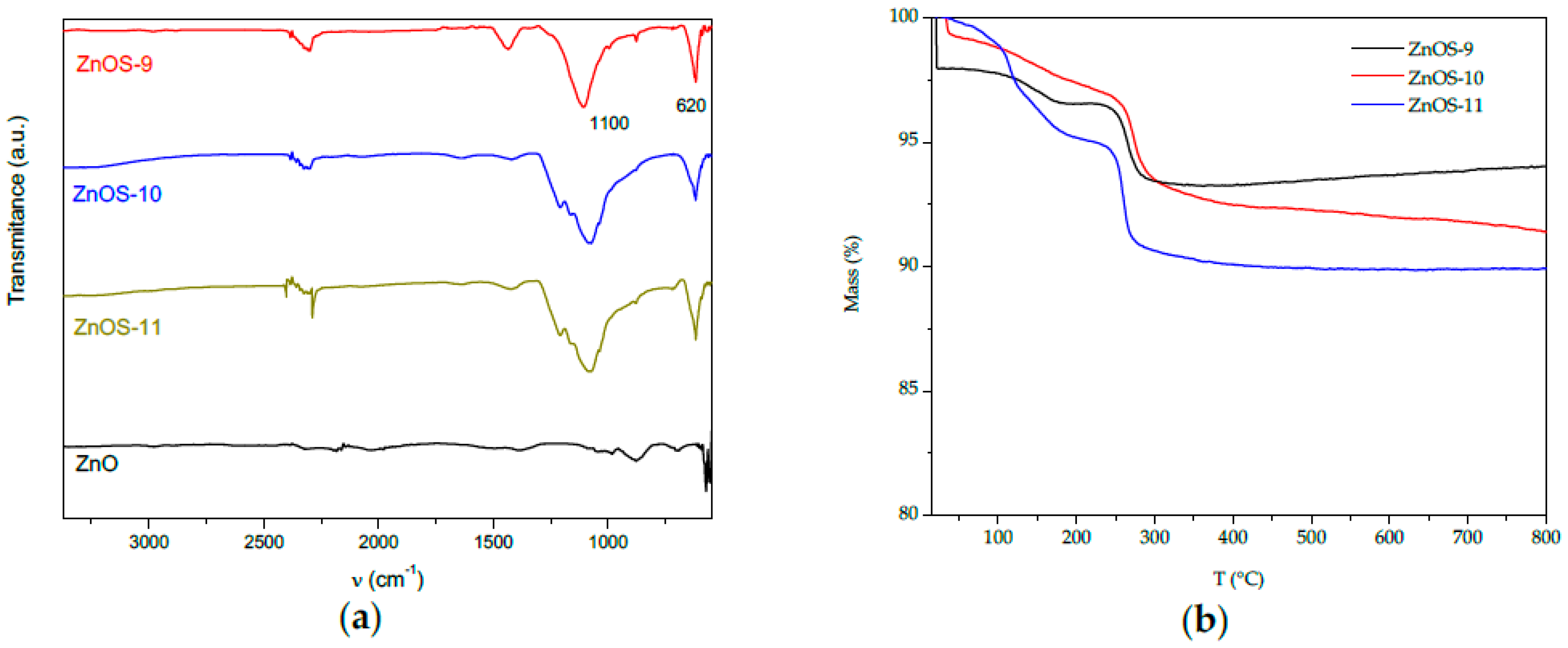
2.2. FTIR and Thermal Analysis

In Figure 2a the FTIR spectra of the as-synthesized samples are shown. In this figure, a well-defined wide and intense peak around 1100–1080 cm−1 is observed, which is associated with the stretching and bending modes of SO4− [26]. This peak is slightly distorted on the left-side as the pH is increased. The sharpest peak located around 620 cm−1 is characteristic from Zn-O [27]. Figure 2b shows the Thermogravimetric Analysis (TGA) curves from the as-synthesized samples. In all the cases are present two distinctive regions of weight reduction: the first one corresponds to the range of temperatures lower than 150 ° C in which weight lost up to 15% is observed and is associated with dehydration from the surface due to water loss adsorbed by the environment, and the second region in the range from 150 to 350 ° C is associated with the elimination of the terminal OH ions of the ZnO structure, which are preferably distributed along the c-axis of the crystalline lattice as reported before in the literature [28]. Beyond these temperatures, the weight remains practically constant.

2.3. SEM Analysis

Figure 3 shows SEM images from the as-synthesized photocatalysts where the influence of pH on the morphology of these materials is appreciated. For the case of the ZnOS-9 (Figure 3a) sample, its morphology consists of sub-micrometric particles mixed with larger particles whose sizes are of the order of several microns. The ZnOS-10 (Figure 3b) sample shows a lesser density of sub-micrometric particles being more evident the formation of sheet-like particles whose sizes are in the range of 2–10 μm. The composite ZnOS-11 (Figure 3c) sample evidences that the size of the sheet-like particles decreases giving rise to the formation of conglomerates. The Na+ ions present in the solution do not allow to obtain a material with long-range crystallinity; this means that the primary nucleus of ZnO aggregates to form larger secondary particles varying in morphology and size, which agrees with the results reported by Becker et al. [13]. The formation of Burkeite is limited by both the amount of urea present and the temperature in the aqueous solution, expecting that its formation takes place during the first stages of combustion, adhering to the surface of the ZnO conglomerates. The results of the EDS measurements are included in Table 1. The atomic weight percent of ZnO in the composites corresponded to 32.39, 32.74, and 42.20 wt% for the samples ZnOS-9, ZnOS-10, and ZnOS-11, respectively. Finally, in Figure 3d we observed a spherical morphology of the ZnO particles. Therefore, we can conclude that the anchoring process on support plays a fundamental role in the final morphology of the ZnO.

2.4. Surface Area Analysis

Table 1 summarizes the surface area values of the samples which were calculated employing Brunauer–Emmett–Teller analysis (BET). As can be seen from Table 1, pH plays a meaningful role on the aggregation mechanism of the ZnO nuclei and in the subsequent adhesion process on Burkeite, which lead to the obtaining of the ZnO/Burkeite heterostructure. The sample ZnOS-10 is the one that shows the largest surface area but the smallest pore size distribution, which suggests that the ZnO is embedded and leaves exposed the Burkeite on the surface of the heterostructure. This scenario may be due to a structural rearrangement in the crystalline lattice of Burkeite originated by the substitution between sulfate ions and carbonate ions, causing a slight disorder in its crystalline lattice [29]. Otherwise, the BET results for the ZnOS-9 and ZnOS-11 samples corresponding to adsorption area and pore sized distribution suggest that ZnO is on the surface of the ZnO/Burkeite heterostructure that gives rise to both a reduction of BET adsorption area and a more substantial pore size value. The pore size distribution was determined by analyzing the physisorption of nitrogen gas (N2), whose molecules are smaller than the molecules of Rhodamine B. Thus, although the N2 molecules were able to probe a larger surface area in the case of ZnOS-10, the heterogeneity in the form of interstitial pores likely limits the diffusion of the bulky molecules of the Rhodamine B into the interior of these and the amount of dye adsorbed. In this way, the distribution of the active phase may be another factor that limits the photocatalytic activity of the materials, since Rhodamine B must be adsorbed on the surface of the semiconductor (ZnO).

2.5. UV-Vis Analysis

In Figure 4 UV-vis absorption spectra for the ZnOS series are shown. The results present a broad absorption band in the range of 250–400 nm with two significant absorption sub-bands: a left-band with a smaller intensity centered around 265 nm, which can be associated with the Burkeite phase, and a right-band with a major intensity, which is located around 350 nm and associated with ZnO. In relation to the latter, the sample ZnOS-10 is the one with the highest absorption intensity; this means that this sample possesses a major homogeneity on the surface morphology of the sample [31], which agrees with the SEM images shown above. Finally, we observed the absorption spectra of the ZnO material. This spectrum exhibits a broad absorption band around 500 nm is associated with lattice defects formed by nitrogen-doping generated by combustion process [30]. The calculated band gap energies (Eg) for all samples are given in Table 1.

2.6. Degradation of Rhodamine B

The first step for the photocatalytic degradation of a dye is its adsorption on the surface of the catalyst. In this case, since the intensity of the absorption peak of Rhodamine B (554 nm) is directly related to the concentration of the solution, the amount of Rhodamine B adsorbed by the photocatalyst was calculated as the difference between the intensity of the initial concentration (Rhodamine 5 mg/L) and the zero-time concentration (time to which the UV lamp is on). The values of adsorption percentages for the different photocatalysts are given in Table 1. The results show that the ZnOS-9 sample exhibits the highest percentage of adsorption. The high value of the adsorption percentage for ZnOS-9 composite is also associated with its morphology and pore size; moreover, we can add that this material is the one that presents the lowest percentage of weight loss in the TG analysis, which would represent the greatest purity. The above finding agrees with the results obtained by using the XRD and the BET techniques. If the sample is considered to have both a smaller crystallite size and a larger pore size, these characteristics positively impact on the dye adsorption on the surface of the photocatalyst because they give rise to the increase in the surface area of the crystallites and the easy access of the molecules within the volume of the material.
To verify whether a de-ethylation process occurs during the degradation of Rhodamine B [32], in Figure 5 are plotted the normalized UV-vis spectra evolution during the photocatalytic degradation process of Rhodamine B by exposing of the materials as-synthetized. In the case on the ZnOS-11, the weak intensity of the absorption peak indicates that the degradation process of Rhodamine B is practically completed when the exposing time corresponds to 90 min. In Figure 5, we can see that the degradation process is carried out by two routes: cleavage and de-ethylation of Rhodamine B. Cleavage is the dominant mechanism at the beginning of the photocatalytic process (30–45 min). For photocatalysts as-synthesized, a shift towards 540 nm is observed, which indicates only the formation of the N,N,N′-triethylated Rhodamine, in accordance with results reported elsewhere [32,33]. In general, we can observe from UV-vis spectra that the longer time exposing, the weaker absorption peak.
The concentration profiles in Figure 6a represent the photocatalytic activity during the degradation of Rhodamine B using the ZnO/Burkeite heterostructures as photocatalysts. Calculation of the C/C0 ratio was carried out considering the absorbance of Rhodamine B at 554 nm. In Figure 5, the absorbance used in the calculation of the C/C0 ratio is represented by a vertical line. The photocatalyst synthesized with a pH of 11 (ZnOS-11) is the one that shows the highest photocatalytic activity with a degradation percentage of Rhodamine B of approximately 92% after 90 min of UV irradiation. The pseudo-first order kinetic constant (k) was deduced by fitting the data obtained during the photocatalytic process and is displayed in Table 1 (see also Figure 6b).
According to these results, we can establish a clear relationship between the effect of pH and the morphology characteristics of the ZnO/Burkeite heterostructure powders with the photocatalytic activity. A significant result is that for a pH of 10, the lowest photocatalytic activity is obtained. As we mentioned above, the highest photocatalytic activity was achieved by a pH of 11 for which the 92.4% of degradation of Rhodamine B is obtained, approximately 30% more than ZnO photocatalyst.
This counterintuitive behavior is well known from recent studies using another type of semiconductor material [34,35,36] and semiconductor heterostructure-based powders [37,38] where it was observed that a low surface adsorption area has a high photocatalytic activity for the Rhodamine B degradation. The origin of the high photocatalytic activity is derived from semiconductor materials that exhibit the physical property known as the photoelectric effect. The latter effect allows semiconductors to absorb photons when they are exposed to UV-light, resulting in the generation of hole-electron pairs owing to the electronic transitions from the valence to conduction band; such transitions are determined by their intrinsic band gap energy. Even more, the morphology of the particles in the semiconductor powder favors the valence electrons (photocarriers) transport to their surface, which has been demonstrated in the case of silver phosphate powders when their particles have tetrahedral and cubic morphologies [39]. Therefore, this explains the morphology role of the ZnO/Burkeite heterostructure. In the case of the ZnOS-10 sample, it inhibits the photocatalytic activity, due to the photocarriers which are inside of the same sheet morphology. Contrariwise, in the case of ZnOS-9 and ZnOS-11 samples, the photocarriers are found in the ZnO localized on the surface of semiconductor heterostructure with fine particles and sheets conglomerate morphologies.

3. Materials and Methods

3.1. Synthesis of ZnO/Burkeite Heterostructure

All reagents were used without any additional purification. The synthesis of ZnO was carried out as follows: in 5 mL of distilled water, sodium hydroxide (NaOH, Fermont, 98.5%) was added to obtain a pH of 9, 10, and 11. Afterwards, 500 mg of zinc sulfate (ZnSO4, Fermont, 99%) and 500 mg of urea (NH2CONH2, Macron, USP) were added to the solution. The solution was placed in an ultrasonic bath for 5 minutes, and then the water was evaporated at 90 °C. The resulting paste was subjected to a heat treatment at 600 °C for 15 min in a preheated muffle, and with this method, the ZnO/Burkeite heterostructure was obtained. The material synthetized was called ZnOS; therefore, the samples labeled with the nomenclature ZnOS-9, ZnOS-10, ZnOS-11 correspond to ZnO/Burkeite material obtained at pH of 9, 10, and 11, respectively. Moreover, for comparison purposes a sample of ZnO without Burkeite was prepared, following the same procedure but using 250 mg of Zn (NO3)2 6H2O (to ensure that Burkeite formation is not carried out) and 500 mg of urea without the addition of NaOH.

3.2. Characterization and Analytical Techniques

XRD analysis was performed using a Bruker X-ray Diffraction D6-Discover equipment (Karlsrhe, BW, Germany). The FTIR spectra were obtained using a Perkin Elmer Spectrum One spectrophotometer (Shelton, CT, USA) equipped with an ATR accessory. Thermal analysis was determined on a Netzsch Thermal Analyser model STA 449F3 Jupiter from 25 to 800 °C under a nitrogen atmosphere. The SEM micrographs were obtained with a JSM-6610LV JEOL electron microscope (Akishima, TYO, Japan). The nitrogen adsorption analysis was carried out to determine the pore size distribution and the isotherm curve using an Autosorb 1C Quantachrome (Quantachrome, Boynton Beach, FL, USA) equipped with a Verlab VE-5600UV photometer (Quantachrome, Boynton Beach, FL, USA) and MetaSpec Pro analysis software (Quantachrome, Boynton Beach, FL, USA). Diffuse Reflectance Spectroscopy (DRS) using a Varian Cary 400 spectrophotometer with a Harrick RD accessory was used for the optical characterization of the samples.

3.3. Degradation tests

Degradation tests were carried out following the system developed by Luna-Flores et al. [40]. The solution to be degraded was 60 mL of Rhodamine B (5 mg/L) with 250 mg/L of the photocatalyst. Once the photocatalyst was dispersed in the solution, it was kept in darkness for 15 min to establish the adsorption–desorption equilibrium. After 15 min, the suspension was irradiated with UV light (20 W, 365–465 nm). The degradation monitoring of Rhodamine B was carried out employing the UV-vis spectroscopy, taking aliquots every 15 min.

4. Conclusions

A simple methodology has been proposed for the synthesis in one step of ZnO/Burkeite heterostructures by the modified solution combustion method. This approach allows tailoring the structural and morphological properties of the photocatalyst to optimize its photocatalytic activity in the degradation of Rhodamine B. Moreover, it has been shown that the optimal synthesis condition for the synthesis of ZnO/Burkeite heterostructure with higher photocatalytic activity corresponds to a pH value in the solution of 11. Thus, the improvement in the degradation process of Rhodamine B depends on the pH-dependent morphological properties of the ZnO particles anchored on the surface of the support, which in turn, showed better structural properties (grain size) and larger pore sizes.

Author Contributions

Conceptualization, A.L.-F.; formal analysis, A.L.-F., M.A.M., R.A.-S., R.P., and G.F.P.-S.; writing—original draft preparation, A.L.-F. and M.A.M.; writing—review and editing, G.F.P-S., J.A.L.-L., A.D.H.-d.l.L., N.T.

Funding

This research received no external funding.

Conflicts of Interest

The authors declare no conflict of interest.

References

  1. Hutton, F.G.; Feulner, G.; Lund, P.D.; Henson, S.; Røttingen, J.-A.; Hoffman, S.J.; Butler, D. Global Challenges: An innovative journal for tackling humanity’s major challenges. Glob. Chall. 2017, 1, 3–4. [Google Scholar] [CrossRef]
  2. Ajmal, A.; Majeed, I.; Malik, R.N.; Idriss, H.; Nadeem, M.A. Principles and mechanisms of photocatalytic dye degradation on TiO2 based photocatalysts: A comparative overview. RSC Adv. 2014, 4, 37003–37026. [Google Scholar] [CrossRef]
  3. Yuan, J.; Liu, L.; Guo, R.-R.; Zeng, S.; Wang, H.; Lu, J.-X. Electroreduction of CO2 into Ethanol over an Active Catalyst: Copper Supported on Titania. Catalysts 2017, 7, 220. [Google Scholar] [CrossRef]
  4. Thakur, R.S.; Chaudhary, R.; Singh, C. Fundamentals and applications of the photocatalytic treatment for the removal of industrial organic pollutants and effects of operational parameters: A review. J. Renew. Sustain. Energy 2010, 2, 042701. [Google Scholar] [CrossRef] [Green Version]
  5. Kumar, S.G.; Koteswara Rao, S.R. Zinc oxide based photocatalysis: Tailoring surfacebulk structure and related interfacial charge carrier dynamics for better environmental applications. RSC Adv. 2015, 5, 3306–3351. [Google Scholar] [CrossRef]
  6. Srikanth, B.; Goutham, R.; Badri Narayan, R.; Ramprasath, A.; Gopinath, K.P.; Sankaranarayanan, A.R. Recent advancements in supporting materials for immobilized photocatalytic applications in waste water treatment. J. Environ. Manag. 2017, 200, 60–78. [Google Scholar] [CrossRef] [PubMed]
  7. Hanaor, D.A.H.; Sorrell, C.C. Sand Supported Mixed-Phase TiO2 Photocatalysts for Water Decontamination Applications. Adv. Eng. Mater. 2014, 16, 248–254. [Google Scholar] [CrossRef]
  8. Korsakov, A.V.; Golovin, A.V.; De Gussem, K.; Sharygin, I.S.; Vandenabeele, P. First finding of burkeite in melt inclusions in olivine from sheared lherzolite xenoliths. Spectrochim. Acta Part A 2009, 73, 424–427. [Google Scholar] [CrossRef]
  9. Shi, B.; Frederick, W.J., Jr.; Rousseau, R.W. Effects of Calcium and Other Ionic Impurities on the Primary Nucleation of Burkeite. Ind. Eng. Chem. Res. 2003, 42, 2861–2869. [Google Scholar] [CrossRef]
  10. Bonakdar, T.; Ghadiri, M.; Ahmadiam, H.; de Juan, M.L.; Xu, D.; Tantawy, H.; Smith, D. Impact attrition of spray-dried burkeite particles. Powder Technol. 2016, 304, 2–7. [Google Scholar] [CrossRef]
  11. Alias, S.S.; Ismail, A.B.; Mohamad, A.A. Effect of pH on ZnO nanoparticle properties synthesized by sol–gel centrifugation. J. Alloy. Compd. 2010, 499, 231–237. [Google Scholar] [CrossRef]
  12. Zhang, J.; Sun, L.; Yin, J.; Su, H.; Liao, C.; Yan, C. Control of ZnO Morphology via a Simple Solution Route. Chem. Mater. 2002, 14, 4172–4177. [Google Scholar] [CrossRef]
  13. Becker, J.; Raghupathi, K.R.; St. Pierre, J.; Zhao, D.; Koodali, R.T. Tuning of the Crystallite and Particle Sizes of ZnO Nanocrystalline Materials in Solvothermal Synthesis and Their Photocatalytic Activity for Dye Degradation. J. Phys. Chem. C 2011, 115, 13844–13850. [Google Scholar] [CrossRef]
  14. Cho, S.; Jung, S.-H.; Lee, K.-H. Morphology-Controlled Growth of ZnO Nanostructures Using Microwave Irradiation: From Basic to Complex Structures. J. Phys. Chem. C 2008, 112, 12769–12776. [Google Scholar] [CrossRef]
  15. Huang, W.; Jia, J.; Zhou, X.; Lin, Y. Morphology controllable synthesis of ZnO crystals—pH-dependent growth. Mater. Chem. Phys. 2010, 123, 104–108. [Google Scholar] [CrossRef]
  16. Amin, G.; Asif, M.H.; Zainelabdin, A.; Zaman, S.; Nur, O.; Willander, M. Influence of pH, Precursor Concentration, Growth Time, and Temperature on the Morphology of ZnO Nanostructures Grown by the Hydrothermal Method. J. Nanomater. 2011, 2011, 269692. [Google Scholar] [CrossRef]
  17. Li, F.-T.; Ran, J.; Jaroniec, M.; Qiao, S.Z. Solution combustion synthesis of metal oxide nanomaterials for energy storage and conversion. Nanoscale 2015, 7, 17590–17610. [Google Scholar] [CrossRef] [PubMed]
  18. Reddy, A.J.; Kokila, M.K.; Nagabhushana, H.; Rao, J.L.; Shivakumara, C.; Nagabhushana, B.M.; Chakradhar, R.S. Combustion synthesis, characterization and Raman studies of ZnO nanopowders. Spectrochim. Acta Part A 2011, 81, 53–58. [Google Scholar] [CrossRef]
  19. Nagaraja, R.; Kottam, N.; Girija, C.; Nagabhushana, B.M. Photocatalytic degradation of Rhodamine B dye under UV/solar light using ZnO nanopowder synthesized by solution combustion route. Powder Technol. 2012, 215, 91–97. [Google Scholar] [CrossRef]
  20. Cai, Y.; Fan, H.; Xu, M.; Li, Q. Rapid photocatalytic activity and honeycomb Ag/ZnO heterostructures via solution combustion synthesis. Colloids Surf. A 2013, 436, 787–795. [Google Scholar] [CrossRef]
  21. Schuyten, S.; Dinka, P.; Mukasyan, A.S.; Wolf, E. A Novel Combustion Synthesis Preparation of CuO/ZnO/ZrO2/Pd for Oxidative Hydrogen Production from Methanol. Catal. Lett. 2008, 121, 189–198. [Google Scholar] [CrossRef]
  22. Srinatha, N.; Kumar, V.D.; Nair, K.G.; Angadi, B. The effect of fuel and fuel-oxidizer combinations on ZnO nanoparticles synthesized by solution combustion technique. Adv. Powder Technol. 2015, 26, 1355–1363. [Google Scholar] [CrossRef] [Green Version]
  23. Cruz, D.; Bulbulian, S. Synthesis of Li4SiO4 by a Modified Combustion Method. J. Am. Ceram. Soc. 2005, 88, 1720–1724. [Google Scholar] [CrossRef]
  24. Søndergaard, M.; Bøjesen, E.D.; Christensen, M.; Iversen, B.B. Size and Morphology Dependence of ZnO Nanoparticles Synthesized by a Fast Continuous Flow Hydrothermal Method. Cryst. Growth Des. 2011, 11, 4027–4033. [Google Scholar] [CrossRef]
  25. Bayuadri, C.; Verril, C.L.; Rousseau, R.W. Stability of Sodium Sulfate Dicarbonate (∼2Na2CO3·Na2SO4) Crystals Obtained from Evaporation of Aqueous Solutions of Na2CO3 and Na2SO4. Ind. Eng. Chem. Res. 2006, 45, 7144–7150. [Google Scholar] [CrossRef]
  26. Wang, L.; Liu, G.; Zou, L.; Xue, D. Phase evolution from rod-like ZnO to plate-like zinc hydroxysulfate during electrochemical deposition. J. Alloy. Compd. 2010, 493, 471–475. [Google Scholar] [CrossRef]
  27. Senthilkumar, K.; Tokunaga, M.; Okamoto, H.; Senthilkumar, O.; Fujita, Y. Hydrogen related defect complexes in ZnO nanoparticles. Appl. Phys. Lett. 2010, 97, 091907. [Google Scholar] [CrossRef]
  28. De la Rosa, E.; Sepúlveda-Guzman, S.; Reeja-Jayan, B.; Torres, A.; Salas, P.; Elizondo, N.; Jose Yacaman, M. Controlling the Growth and Luminescense Properties of Well-Faceted ZnO Nanorods. J. Phys. Chem. C 2007, 111, 8489–8495. [Google Scholar] [CrossRef]
  29. Shi, B.; Rousseau, R.W. Structure of Burkeite and a New Crystalline Species Obtained from Solutions of Sodium Carbonate and Sodium Sulfate. J. Phys. Chem. B 2003, 107, 6932–6937. [Google Scholar] [CrossRef]
  30. Ramachandran, S.; Sivasamy, A. Synthesis and characterization of nanocrystalline N-doped semiconductor metal oxide and its visible photocatalytic activity in the degradation of an organic dye. J. Environ. Chem. Eng. 2018, 6, 3770–3779. [Google Scholar] [CrossRef]
  31. Khan, M.F.; Ansari, A.H.; Hameedullah, M.; Ahmad, E.; Husain, F.M.; Zia, Q.; Baig, U.; Zaheer, M.R.; Alam, M.M.; Khan, A.M.; et al. Sol-gel synthesis of thorn-like ZnO nanoparticles endorsing mechanical stirring effect and their antimicrobial activities: Potential role as nano-antibiotics. Sci. Rep. 2016, 6, 27689. [Google Scholar] [CrossRef] [PubMed]
  32. Pica, M.; Calzuola, S.; Donnadio, A.; Gentili, P.L.; Nocchetti, M.; Casciola, M. De-Ethylation and Cleavage of Rhodamine B by a Zirconium Phosphate/Silver Bromide Composite Photocatalyst. Catalysts 2019, 9, 3. [Google Scholar] [CrossRef]
  33. Wu, T.; Liu, G.; Zhao, J.; Hidaka, H.; Serpone, N. Photoassisted degradation of dye pollutants. V. Self-photosensitized oxidative transformation of Rhodamine B under visible light irradiation in aqueous TiO2 dispersions. J. Phys. Chem. B 1998, 102, 5845–5851. [Google Scholar] [CrossRef]
  34. Bi, Y.; Ouyang, S.; Umezawa, N.; Cao, J.; Ye, J. Facet effect of single-crystalline Ag3PO4 sub-microcrystals on photocatalytic properties. J. Am. Chem. Soc. 2011, 133, 6490–6492. [Google Scholar] [CrossRef] [PubMed]
  35. Jiao, Z.; Zhang, Y.; Yu, H.; Lu, G.; Yeb, J.; Bi, Y. Concave trisoctahedral Ag3PO4 microcrystals with high-index facets and enhanced photocatalytic properties. Chem. Commun. 2013, 49, 636–638. [Google Scholar] [CrossRef] [PubMed]
  36. Morales, M.A.; Fernández-Cervantes, I.; Agustín-Serrano, R.; Ruíz-Salgado, S.; Sampedro, M.P.; Varela-Caselis, J.L.; Portillo, R.; Rubio, E. Ag3PO4 microcrystals with complex polyhedral morphologies diversity obtained by microwave-hydrothermal synthesis for MB degradation under sunlight. Results Phys. 2019, 12, 1344–1356. [Google Scholar] [CrossRef]
  37. Bi, Y.; Hu, H.; Ouyang, S.; Jiao, Z.; Lu, G.; Ye, J. Selective growth of metallic Ag nanocrystals on Ag3PO4 submicro-cubes for photocatalytic applications. Chem. Eur. J. 2012, 18, 14272–14275. [Google Scholar] [CrossRef] [PubMed]
  38. Wu, Q.; Wang, P.; Niu, F.; Huang, C.; Li, Y.; Yao, W. A novel molecular sieve supporting material for enhancing activity and stability of Ag3PO4 photocatalyst. Appl. Surf. Sci. 2016, 378, 552–563. [Google Scholar] [CrossRef]
  39. Kim, S.; Wang, Y.; Zhu, M.; Fujitsuka, M.; Majima, T. Facet Effects of Ag3PO4 on Charge-Carrier Dynamics: Trade-Off between Photocatalytic Activity and Charge-Carrier Lifetime. Chem. Eur. J. 2018, 24, 14928–14932. [Google Scholar] [CrossRef]
  40. Luna-Flores, A.; Valenzuela, M.A.; Luna-López, J.A.; Hernández de la Luz, A.D.; Muñoz-Arenas, L.C.; Méndez-Hernández, M.; Sosa-Sánchez, J.L. Synergetic Enhancement of the Photocatalytic Activity of TiO2 with Visible Light by Sensitization Using a Novel Push-Pull Zinc Phthalocyanine. Int. J. Photoenergy 2017, 2017, 1604753. [Google Scholar] [CrossRef]
Figure 1. X-rays diffraction spectra of the as-synthesized ZnO and ZnO/Burkeite heterostructures.
Catalysts 09 00817 g001
Figure 2. (a) FTIR spectra of as-synthesized ZnOS photocatalysts and ZnO, (b) TGA for the as-synthesized ZnOS heterostructure materials.
Catalysts 09 00817 g002
Figure 3. SEM images of the ZnOS samples obtained at different pH values. (a) ZnOS-9, (b) ZnOS-10, (c) ZnOS-11, (d) ZnO.
Catalysts 09 00817 g003
Figure 4. UV-vis absorption spectra for the as-synthesized ZnOS heterostructures and ZnO.
Catalysts 09 00817 g004
Figure 5. Normalized UV-vis spectra during the degradation of Rhodamine B for the as-synthesized ZnOS heterostructures, (a) ZnOS-9, (b) ZnOS-10, (c) ZnOS-11, and (d) ZnO.
Catalysts 09 00817 g005
Figure 6. (a) Rhodamine B degradation rate for the different photocatalysts as-synthesized, (b) The pseudo-first order kinetic photocatalytic degradation of Rhodamine B for the ZnOS samples.
Catalysts 09 00817 g006
Table 1. Optical, structural, and photocatalytic characteristics of the ZnOS composite and ZnO.
SampleEg (eV)BET (m2/g) Pore Size (Å)RhB Abs. (%)% Degradation of RhBKinetic Constant k (seg−1)EDS (wt%)
NaCSZnOI
ZnOS-93.243.5442.3921.782.50.0198823.775.0710.0123.5237.490.14
ZnOS-103.2748.720.56.37.30.00099725.015.828.5520.7739.430.42
ZnOS-113.252.742.198.292.40.02776924.248.056.8122.4638.440.0
ZnO3.1121.8 a17.1 a4.363.80.009795---83.2816.72-
a Ref [30]; I- impurities.

