Article

Magnetic β-Cyclodextrin Polymer Nanoparticles for Efficient Adsorption of U(VI) from Wastewater

State Key Laboratory of Nuclear Resources and Environment, East China University of Technology, Nanchang 330013, China
*
Authors to whom correspondence should be addressed.
Crystals 2023, 13(10), 1496; https://doi.org/10.3390/cryst13101496
Submission received: 28 September 2023 / Revised: 12 October 2023 / Accepted: 12 October 2023 / Published: 14 October 2023

Abstract

:
It is a central issue to eliminate radioactive uranium (U(VI)) efficiently from water. In this manuscript, β-cyclodextrin was cross-linked with 2,3,5,6-tetrafluoro-1,4-benzenedicarbonitrile, and then a carboxylation reaction was used to prepare porous cross-linked polymers rich in carboxyl groups (CA-PCDPs). Subsequently, magnetic nanoparticles (MNPs) were loaded onto the CA-PCDPs via coprecipitation, and magnetic porous β-cyclodextrin polymer nanoparticles (CA-PCDP@MNPs) were successfully obtained, which were used for efficient elimination of U(VI) from nuclear wastewater solution. Moreover, SEM, FTIR, VSM, BET, and XRD were employed to investigate the CA-PCDP@MNP and found that it had a well-developed porous structure, high specific surface area, and abundant oxygen-containing functional groups (carboxyl, hydroxyl, C-O-C, Fe-O, etc.), providing sufficient active sites for chelating uranyl ions. Experiments illustrated that the CA-PCDP@MNP had efficient removal ability for U(VI), and the maximum theoretical adsorption amount for U(VI) reached 245.66 mg/g at pH 6.0 and 303 K. Moreover, the adsorption process was more suitable for the quasi second-order kinetic model and Langmuir adsorption isotherm model, indicating that the adsorption process was chemical adsorption. Meanwhile, the CA-PCDP@MNPs also exhibited fast response magnetic recovery ability and excellent regeneration and recycling ability. In addition, the data of the adsorption mechanism demonstrated that oxygen-containing functional groups, which were rich on the surface of CA-PCDP@MNPs, were the main binding active sites of U(VI). From the above results, it can be deduced that the CA-PCDP@MNP has a good application prospect in the practical application of nuclear wastewater treatment.

1. Introduction

As a natural radioactive heavy metal element, uranium has carcinogenicity and causes genetic variation. It will pollute water and soil once entering the environment due to uranium mining, smelting, nuclear leakage, and other reasons. In fact, uranium mainly exists in U(VI) form, which is highly soluble in water and highly toxic [1]. These pollutants will be distributed to various organs and tissues via blood circulation once entering the human body, which can cause serious damage to immune, reproductive, central nervous, and other systems. Thus, it is a research priority to handle and recycle uranium-containing wastewater [2,3]. Currently, common methods for removing radioactive nuclides from the environment include adsorption, photocatalysis, membrane separation, chemical precipitation, ion exchange, biological methods, solvent extraction, etc. Among them, the adsorption method has drawn close attention for removing U(VI) from water due to its advantages, such as low cost, simple operation, and less susceptibility to secondary pollution [4,5,6].
Various materials capable of capturing nuclide uranium have emerged. These include inorganic materials, magnetic nanoparticles (MNPs), carbon-based materials, organic frameworks, polymers, and so on [7]. Since an MNP exhibits a magnetic response, it can be easily recovered via an external magnetic field. It is noteworthy that MNPs show precious advantages in terms of efficiency and suspended solids recovery in practical applications. Many researchers have suggested that MNPs can avoid the particle filtration step [8]. Hence, they have widely attracted researchers’ attention to their ability to adsorb radioactive nuclides and heavy metal ions. Common iron-based magnetic nanomaterials are nano zero valent iron (nZVI), magnetite hematite (γ-Fe2O3) nanoparticles, and magnetite (Fe3O4) nanoparticles and their composite materials [9]. However, adsorption performance is not ideal when MNPs are used as sorbents alone, and MNPs are prone to agglomeration and corrosion in a wastewater environment, leading to adsorption performance loss. Therefore, researchers have focused their concentration on enhancing adsorption ability and selectivity for radioactive nuclides and heavy metal ions via surface functionalization and modification of MNPs. Zhao et al. synthesized Fe3O4@SiO2-AO [10], which was a functionalized core–shell magnetic microsphere with an amidoxime group and efficient removal of U(VI) due to strong chelation of amidoxime on U(VI). Meng et al. had reported a magnetic uranium adsorbent Fe3O4@C-SMA, which was acquired by loading Fe3O4 onto a porous cross-linked polymer containing carboxyl groups. Results revealed that the adsorption amount of U(VI) at pH = 6 was 178 mg/g, suggesting that the surface functionalization of C-SMA effectively solved the aggregation of exposed Fe3O4 [11].
Recently, natural biopolysaccharide sorbents (such as cyclodextrins, chitosan, starch, and alginate) have played an increasingly important role in various wastewater treatments due to their excellent characteristics, such as wide availability of sources, low cost, rich functional groups, and excellent biodegradability. β-cyclodextrin (β-CD) is a non-toxic, green, biodegradable, and inexpensive macrocyclic oligosaccharide compound with a cavity structure and a large number of hydroxyl groups that can form non-toxic complexes with heavy metal ions [12]. However, β-CD is easily soluble in water and has poor stability, making it difficult to recover when used in water and greatly limiting its application in water treatment. Hence, some scientists tried to construct it on other carriers to acquire water treatment sorbents with a long service life, excellent mechanical properties, and high dispersion performance. Yang et al. synthesized a β-CD supported by Fe3O4-eloxite nanotubes to adsorb U(VI) in an aqueous solution, revealing that the ideal pH was 7.0, and it adsorbed 92% of the U(VI) in the aqueous solution [13,14]. Zong et al. adopted coprecipitation to prepare carboxymethyl-β-CD-modified MNPs (CM-CD-MNPs), the adsorption amount of which for U(VI) at pH 5.5 and 303 K was close to 5.75 × 10⁻⁴ mol/g. It was an environmentally friendly and low-cost effective adsorbent, which would have potential application in nuclear waste treatment U(VI) [15]. Dichtel et al. found that the nucleophilic substitution process of β-CD using 2,3,5,6-tetrafluoro-1,4-benzenedicarbonitrile (TFPN) as a cross-linker produced a porous cyclodextrin polymer (PCDP) with a high specific surface area, which could remove different types of organic contaminants quickly. Xu et al. prepared porous cyclodextrin polymer materials (T-E-PCDP) through polymerization using rigid TFPN cross-linking agents and flexible EPI cross-linking agents. T-E-PCDP exhibited excellent adsorption performance for various organic micro-pollutants, and its adsorption rate was 45–581 times faster than commercial activated carbon and XAD-4 resin [16]. However, there are few reports on the application of PCDP to capture heavy metal ions, especially radionuclide U(VI).
In this work, an MNP was functionalized while introducing PCDP for composite modification. On the one hand, it utilized the excellent magnetic response characteristics of MNPs to improve the recyclable separation of adsorbents. On the other, it also took advantage of β-CD’s efficient adsorption of heavy metal ions and the porous properties of PCDP to enhance adsorption performance. Moreover, MNPs loaded onto CA-PCDPs via coprecipitation to obtain magnetic porous β-cyclodextrin polymer nanoparticles (CA-PCDP@MNP) for U(VI) removal have not been reported so far. Simultaneously, FTIR, VSM, SEM, EDXS, XPS, and BET systems were used to explore the influences on the adsorption characteristics of CA-PCDP@MNP for uranyl ions, expecting to develop high-performance CA-PCDP@MNP magnetic porous materials and provide new materials for radioactive wastewater treatment.

2. Reagents and Methods

2.1. Reagents

Uranyl nitrate hexahydrate (UO2(NO3)2·6H2O, 99%) was purchased from Macklin Reagent (Shanghai, China). Β-CD, TFPN, K2CO3, anhydrous tetrahydrofuran (THF), anhydrous N,N-dimethylformamide (DMF), dichloromethane, NaOH, FeCl3·6H2O, and FeCl2·4H2O were all analytically pure and acquired from Energy Chemical Co., Ltd. (Shanghai, China).

2.2. Preparation of PCDP and CA-PCDP

Firstly, PCDP was prepared by modifying the method of reference [17]. In a 250 mL dry round bottom flask, β-CD (1.88 g), K2CO3 (2.86 g), and TFPN (1.00 g) were weighed. Then, 72 mL dry THF and 8 mL dry DMF (in a volume ratio of 9:1) were added under an argon atmosphere. The mixture was reacted for 48 h at 85 °C. After cooling to room temperature, the mixture was filtered, and the filter cake was washed with HCl (1 mol/L). The collected yellow solids were washed twice with water, twice with THF, and once with CH2Cl2, and then the cleaned yellow solid was dried in a vacuum oven at 70 °C for 12 h.
Then, CA-PCDP was obtained by a modified preparation method based on references [18]. Technically, PCDP (0.6 g) and anhydrous ethanol (120 mL) were added to a flask, and NaOH (24 g) was dissolved in water (96 mL) and poured into a medium flask. Then, a good condensing device was assembled, and the mixture was heated to 120 °C for 48 h. After cooling to room temperature, the mixture was filtered to exclude the supernatant; the solid was taken out and dispersed in water (100 mL); and HCl (1 mol/L) was added dropwise to adjust the pH value (4.0–5.0). After that, the resulting mixture was returned to react at 120 °C for 2 h. The filtered solid was cleaned with water (three times) and dried in a vacuum oven at 70 °C for 12 h to obtain CA-PCDP. A schematic diagram is shown in Figure 1.

2.3. Preparation of CA-PCDP@MNP

Briefly, 0.40 g CA-PCDP, 0.75 g FeCl3·6H2O, and 0.27 g FeCl2·4H2O were dispersed into deoxygenated water (80 mL) in a nitrogen atmosphere and then mixed evenly by ultrasonic shaking. In addition, 20% NaOH (80 mL) solution was added at 90 °C, and the reaction was carried out for 2 h. Next, the black precipitate was collected via an applied magnetic field and repeatedly washed with pure water and ethanol. Finally, the black precipitate was dried in a vacuum drying oven for 12 h to obtain CA-PCDP@MNP.

2.4. Characterization

The measured samples were mixed with KBr powder and pressed and then scanned by Fourier-transform infrared spectrometer (FTIR, Nicolet Magana-IR Type 750, Madison, WI, USA) at a wavelength of 4000−400 cm−1. The crystal structures were identified by X-ray diffractogram (XRD, Bruker D8 Advance) with Cu Kα radiation to scan the samples from 20 to 80 degrees at a speed of 2°·min−1. The specific surface area, mean pore size, and pore volume of CA-PCDP@MNP were measured using N2 adsorption and desorption experiments at 77 K. The standard Brunauer–Emmett–Teller (BET) equation was used to calculate the specific surface area. The aperture distribution was obtained from the isothermal desorption branch by the Barrett–Joyner–Halenda (BJH) method using Quantachrome Auto sorb software (Quantachrome Instruments, Boynton Beach, FL, USA). The microstructure of CA-PCDP@MNP was examined using a scanning electron microscope (SEM, Hitachi Regulus 8100). Magnetization measurements were performed on the CA-PCDP@MNP at room temperature (298 K), using a vibrating-sample magnetometer (VSM, American Quantum Design, Santiago, MI, USA), in the range from +2 T to −2 T. X-ray photoelectron spectroscopy (XPS, Thermo ESCALAB 250XI, Waltham, MA, USA) was engaged to analyze surface elemental composition and the elemental valence state, using the comparison XPS spectra before and after U(VI) adsorption by the CA-PCDP@MNPs, and the scanning range was varied from 0 to 700 eV.

2.5. Different Factors’ Effect on U(VI) Adsorption

The adsorption experiment for U(VI) using CA-PCDP@MNP was conducted in batches. Various factors affecting the adsorption, such as pH (2.0–8.0), initial U(VI) concentration (20–160 mg/L), sorbent dosage (0.1–0.8 g/L), contact time (0–240 min), and temperature (283–313 K), were sequentially investigated. The effect of pH on adsorption performance was taken as an example to describe how to conduct adsorption experiments. Sorbent CA-PCDP@MNP (0.4 g/L) was added into a U(VI) solution (40 mg/L and 100 mg/L, 25 mL), which was adjusted to a specified pH via 0.1 or 0.01 mol/L NaOH/HCl. Then, the mixture was placed in a constant temperature oscillator, which was set at 200 r/min, 303 K. After that, the sorbent was separated via an external magnet; the supernatant (1 mL) was sucked out and filtered via a 0.22 μm microfilter membrane, and the U(VI) concentration was measured at a wavelength of 650 nm by the azo arsenic III color development method. In addition, the adsorption amount (qe, mg/g) and removal efficiency (R, %) were calculated via Equations (1) and (2) [19]:
q e = ( C 0 C e ) × V m
R   ( % ) = C 0 C e C 0 × 100
where C0 (mg/L) and Ce (mg/L) are the initial and equilibrium U(VI) concentration, respectively. Moreover, m (mg) and V (mL) are the mass of the CA-PCDP@MNP and the volume of the U(VI) solution, respectively.

2.6. Kinetic Adsorption

The quasi-first-order kinetic model, quasi-second-order kinetic model, and the intra-particle diffusion model were utilized to expound the U(VI) adsorption process, and the calculation formula of these models are exhibited as Equations (3)–(5) [20]:
ln ( q e q t ) = ln q e k 1 t
t q t = 1 k 2 q e 2 + 1 q e t
q t = k i d t 0.5 + C
where qt (mg/g) and qe (mg/g) are the adsorption amounts of U(VI) at time t and at equilibrium, respectively. Moreover, k1 (min−1), k2 (g/(mg∙min)), and kid (mg/(g∙min0.5)) are rate constants of the quasi-first-order kinetic model, quasi-second-order kinetic model, and intra-particle diffusion model, respectively, and C (mg/g) is the intercept for the in-particle diffusion model depending on the thickness of the boundary layer.

2.7. Adsorption Isotherm and Thermodynamic

The Langmuir model (Equation (6)) and Freundlich model (Equation (7)) were utilized to explore the adsorption behavior of the sorbent for U(VI) and to fit experiment data [21].
q e = q m K L C e 1 + K L C e
q e = K F C e 1 n
where qe (mg/g) and Ce (mg/L) are the adsorption amount and concentration of U(VI) at equilibrium, respectively. Moreover, qm (mg/g) is the maximum theoretical adsorption amount; KL (L/mg) is the adsorption equilibrium constant of the Langmuir model; KF (mg1 − n·Ln/g) is the constant of the Freundlich model; and n is the constant related to adsorption strength.
Adsorption thermodynamic parameters were calculated via the van’t Hoff equation, and the calculating formula is shown as follows [22]:
ln K c = Δ S 0 R Δ H 0 R T
Δ G 0 = R T l n K c
where Kc is the distribution constant, which is gained from the relation curve between   l n ( q e / C e ) and Ce. T is temperature, and R (8.314 J/(mol∙K)) is the gas constant.

2.8. Recycling and Regeneration

First, 10 mg CA-PCDP@MNP was mixed into the U(VI) solution (40 mg/L, 25 mL), and the mixture was vibrated for 4 h at 303 K, 200 r/min; then, solid–liquid separation was achieved via a magnet. Next, the obtained solid was desorbed via 25 mL 0.1 mol/L HNO3, vibrated for 4 h at 303 K, 200 r/min, and then carried out via an applied magnetic field. Finally, the desorbed adsorbent was washed via ultrapure water and dried under vacuum at 70 °C for 12 h. The regeneration experiment was performed for 5 consecutive cycles.

3. Results and Discussion

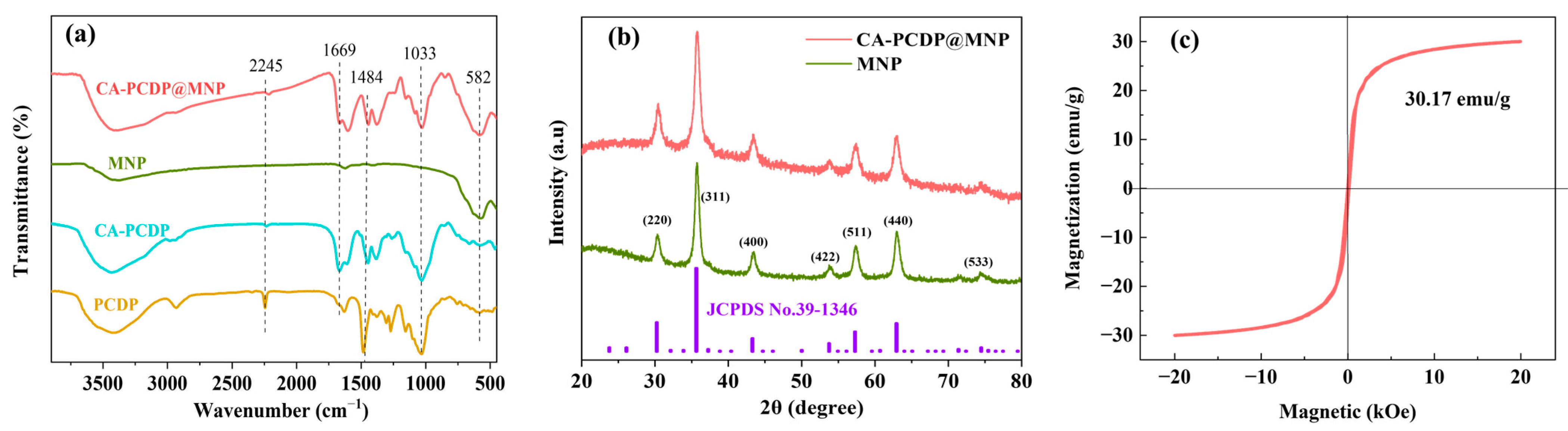
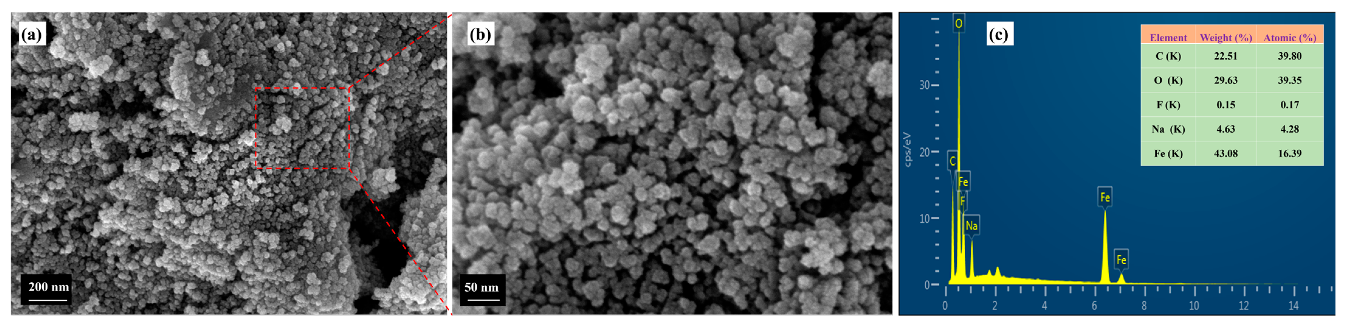
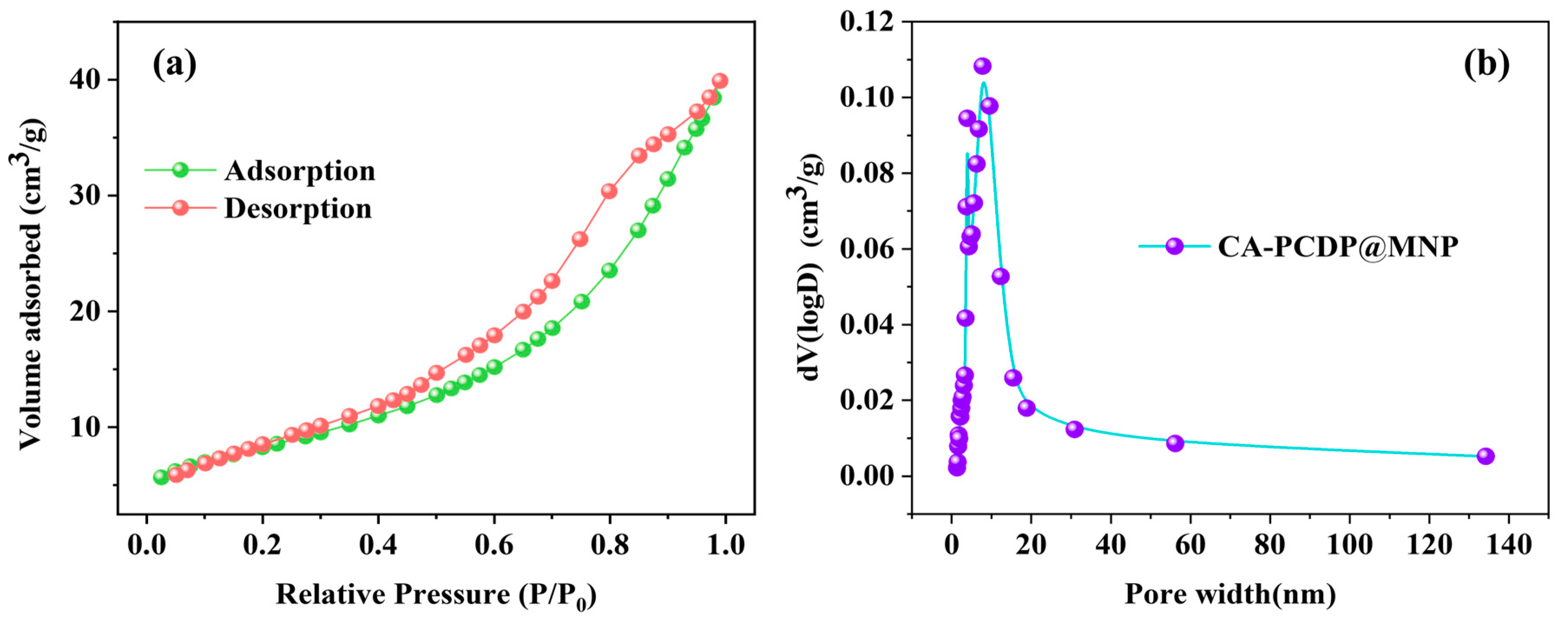
3.1. Characterization

The FTIR spectra of PCDP, CA-PCDP, MNP, and CA-PCDP@MNP are unveiled in Figure 2a. The stretching vibration of cyano C≡N, the stretching vibration of the benzene ring skeleton C=C, the stretching vibration of C-O, and the stretching vibration of C-F are displayed in the PCDP spectrum, which correspond to 2245 cm−1, 1484 cm−1, 1033 cm−1, and 1269 cm−1, respectively, demonstrating that PCDP was obtained via TFPN cross-linked β-CD [23]. Figure 2a also reveals that the disappearance of the C≡N characteristic absorption peak and the appearance of the C=O characteristic absorption peak (1669 cm−1) in the CA-PCDP spectra illustrated the success of carbonylation modification [18].
In comparison to MNP and CA-PCDP, there was an adsorption peak at 1669 cm−1 in the CA-PCDP@MNP FTIR spectrum, which was related to the C=O stretching vibration of the carboxylic acid group and provided evidence for -COOH introduction. Meanwhile, there were adsorption peaks at 1484 cm−1, 1269 cm−1, and 1033 cm−1 in the CA-PCDP@MNP FTIR spectrum, corresponding to the tensile vibration of aromatic C=C, the stretching vibration of C-F, and the stretching vibration of C-O, respectively. Moreover, the Fe-O characteristic absorption peak appeared at 582 cm−1 in CA-PCDP@MNP. These all demonstrated that MNPs were successfully loaded onto the porous polymer CA-PCDP [24].
The XRD patterns of the MNPs acquired via coprecipitation and the CA-PCDP@MNPs are unveiled in Figure 2b. There were seven obvious characteristic diffraction peaks detected in both XRD patterns at the 2θ diffraction angle, which were 30.3° (220), 35.7° (311), 43.5° (400), 53.9° (422), 57.4° (511), 63.0° (440), and 74.4° and perfectly matched crystal planes of a standard diffraction card for γ-Fe2O3 (JCPDS: No.39-1346) [25,26]. Both unmodified MNP and modified CA-PCDP@MNP had a spinel γ-Fe2O3 crystal structure, and maghemite was ascertained as the main crystal phase. Results revealed that there was no variation monitored in the MNP crystalline structure after modification, and γ-Fe2O3 was successfully loaded onto CA-PCDP.
The hysteresis loop VSM of the CA-PCDP@MNP is illustrated in Figure 2c. It was observed that the relationship between the magnetic susceptibility of a CA-PCDP@MNP and the magnetic field was S-shaped and passed through the origin, revealing that coercivity and residual magnetic field strength were close to 0, and there was almost no remanence. This demonstrated that the CA-PCDP@MNP was superparamagnetic and had good magnetic responsiveness. Meanwhile, the saturation magnetization of CA-PCDP@MNPs was 30.17 emu/g, which was lower than that of γ-Fe2O3 MNPs [27,28], illustrating that γ-Fe2O3 MNPs were deposited on a non-magnetic porous polymer. Although there was a decrease in CA-PCDP@MNP saturation magnetization, it still could be quickly separated and recovered via an external magnetic field. And CA-PCDP@MNPs were observed aggregating rapidly within 1 min under an applied magnetic field, which met the CA-PCDP@MNP requirements of rapid regeneration and reuse under a magnetic field.
Micromorphology of the CA-PCDP@MNP was measured via SEM, and the results are illustrated in Figure 3a,b. According to these pictures, there were plenty of fluffy and irregular pores inspected in the CA-PCDP@MNP, and there were abundant nanoscale spherical particles and sediments that covered its surface, which was produced via MNP depositing on a CA-PCDP surface. Moreover, the spherical particle size was about 20 nm on the surface, and agglomeration was obvious because of the magnetic force of the magnetic particle itself. However, the surface was loose and porous, resulting in a large internal and external specific surface area. These structural characteristics enhanced the contact area with U(VI), which was conducive to promoting solid–liquid contact and a reaction between the material and U(VI) and to improving the CA-PCDP@MNP adsorption amount [29]. CA-PCDP@MNPs were scanned via EDXS, and the results are displayed in Figure 3c. Strong peaks of C, O, F, Na, and Fe were observed under EDXS, and the main elements were C, O, and Fe on the surface, the mass contents of which were 22.51%, 29.63%, and 43.08%, respectively. The presence of Fe demonstrated that MNPs were successfully deposited on the porous polymer. Moreover, an Au peak appeared at 2.12 KeV under EDXS, which was due to the surface gold spray operation before SEM observation [30].
The nitrogen adsorption–desorption isotherms of CA-PCDP@MNPs at 77 K are exhibited in Figure 4a, which represent type IV in the IUPAC classification. In the high pressure range (P/P0 = 0.4–1.0), the adsorption isotherm of the CA-PCDP@MNP did not coincide with the desorption isotherm, and capillary condensation and the H3 hysteresis loop occurred in the adsorption process, revealing the existence of a mesoporous structure in the CA-PCDP@MNP [31]. The pore size distribution diagram in Figure 4b illustrates its pore size was mainly distributed in the 2–30 nm range, which was consistent with SEM observation and demonstrated that most pore channels were mesoporous in structure. Additionally, the specific surface area of the CA-PCDP@MNP was 40.619 m2/g, and the pore volume was 0.065 cm3/g. Therefore, the pore structure and the high specific surface area provided a sufficient number of active sites for the U(VI).

3.2. Effect of Sorbent Dosage and pH

Sorbent dosage is considered an important factor that can affect its economic benefits. Hence, the effect of the CA-PCDP@MNP dosage is surveyed in Figure 5a. Data revealed that the U(VI) equilibrium adsorption amount declined with an increase of sorbent dosage. When sorbent dosage rose from 0.1 g/L to 0.4 g/L, the adsorption amount decreased from 248.41 mg/g to 178.00 mg/g. This was because the adsorption active sites of the CA-PCDP@MNPs were not fully utilized by the U(VI) at high doses [32]. Furthermore, excessive adsorbent amounts would cause aggregation [33], leading to a decrease in active sites density and a drop in U(VI) adsorption performance.
Figure 5a also displays the relationship between R and CA-PCDP@MNP dosage. R increased slowly with the dosage increase and finally stabilized at 0.6 g/L. When the dosage was 0.6 g/L, R was 82.0%, and there was no obvious change in R when the sorbent amount increased too much. This could be explained as the increasing number of active sites on the surface resulting in R increasing. However, when the sorbent dosage reached a certain value, R did not increase significantly, and the equilibrium adsorption amount continued to decline. Considering R, cost, and utilization rate, the sorbent dosage was selected at 0.4–0.6 g/L.
The pH significantly affected adsorption efficiency and uranyl ion distribution [34]. As Figure 5b exhibits, the CA-PCDP@MNP adsorption amount for U(VI) increased first and then decreased with a pH increase. When the pH value was 2.0–4.0, the U(VI) adsorption amount was slowly enhanced, and when the pH value was 4.0–6.0, the U(VI) adsorption amount increased significantly. The adsorption of 100 mg/L and 40 mg/L for U(VI) attained the greatest values of 176.81 mg/g and 90.84 mg/g at pH 6.0, respectively. When the pH value was 6.0, the R achieved was the highest at 90.8% for 40 mg/L U(VI). This was because the uranium main type was UO22+ at a low pH (pH < 3), and massive H+ competed with UO22+ for active positions. Moreover, the CA-PCDP@MNP zero charge point (pHpzc) was 4.71, seen in Figure 5c. And when pH < pHpzc, the CA-PCDP@MNP was positively charged and the UO22+ and surface functional groups were protonated, which hindered UO22+ binding to the adsorbent via electrostatic repulsion, leading to a low adsorption amount [35]. When the pH rose (4.0–6.0), surface functional groups were deprotonated, but uranium still existed with a positive charge (UO22+,(UO2)3(OH)5+ and (UO2)4(OH)7+) [4]. Then, electrostatic attraction was generated between CA-PCDP@MNP and U(VI), which was conducive to the adsorption between hydroxyl and carboxyl groups on the sorbent surface and U(VI) and enhanced the U(VI) adsorption amount [36,37]. However, when the pH was unduly high, charges on the surface of the CA-PCDP@MNP turned from positive to negative. Meanwhile, U(VI) was mainly negatively charged, which was in the form of UO2(OH)3, (UO2)3(OH)7, and complexation of negatively charged groups was not prone to occur.

3.3. Adsorption Kinetics

The adsorption kinetic curve of CA-PCDP@MNP (pH = 6.0, temperature 303 K, dosage of 0.4 g/L, and C0 of 100 mg/L) is illustrated in Figure 6a. R rose fast in the first 20 min and reached about 81% of the total adsorption amount, which might be caused by carboxylic acid groups and oxygen-rich functional groups of β-CD and MNP. As adsorption proceeded, active sites were occupied by U(VI), so R slowed down. The equilibrium adsorption amount was 178.85 mg/g at about 60 min.
The quasi-first-order kinetic model and the quasi-second-order kinetic model were adopted to explore the experimental data of the CA-PCDP@MNP adsorbed U(VI) with time; the fitting curve is exhibited in Figure 6b,c, and the fitting parameters are displayed in Table 1. It was observed that the fitting results of the quasi-second-order kinetic model (R2 = 0.999) were better than that of the quasi-first-order kinetic model (R2 = 0.718), and the equilibrium amount (qe, cal) calculated by the quasi-second-order kinetic model was also closer to the actual experimental equilibrium adsorption amount (qe, exp), suggesting that the CA-PCDP@MNP adsorption process followed a quasi-second-order kinetic model and that it was chemisorption [38]. This was because the CA-PCDP@MNPs had abundant oxygen-containing groups, which provided most of the adsorption actives [15,39].
The fitting curve and the parameters of the intra-particle diffusion model are illustrated in Figure 6d and Table 1. As these exhibit, there were three stages in the adsorption process. The first stage was adsorption on the outer surface, and the second stage was diffusion within particles. The third stage was the equilibrium stage, where diffusion within particles began to slow down due to extremely low mass transfer rate in the solution. As exhibited in Table 1, the relationship between the rate constant of the three stages was kid,1 > kid,2 > kid,3. Moreover, the rate constant of the first stage, which occurred on the surface, was the largest (26.759 mg/(g·min0.5)), revealing that the transfer rate of U(VI) to the CA-PCDP@MNP surface was fastest at this stage. Additionally, the fitting lines of qt and t0.5 for CA-PCDP@MNP did not pass through the origin, demonstrating that the intra-particle diffusion was not the only rate-controlled process [40].

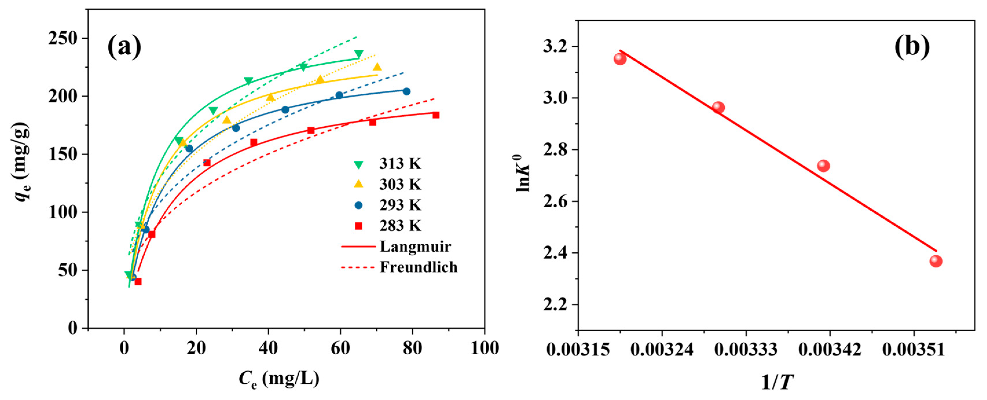
3.4. Adsorption Isotherms and Thermodynamics

The adsorption isotherm curves of CA-PCDP@MNPs for U(VI) at 283 K, 293 K, 303 K, and 313 K are displayed in Figure 7a. As U(VI) concentration rose from 20 mg/L to 160 mg/L, the adsorption amount expanded rapidly at first and then expanded slowly. To evaluate the adsorption performance, the Langmuir model and the Freundlich model were employed to fit experimental data, and the fitting parameters are represented in Table 2. Research revealed that the correlation coefficients R2 (0.992, 0.998, 0.994, and 0.992) acquired via the Langmuir model were larger than those of the Freundlich model (0.856, 0.844, 0.845, 0.875), and the maximum adsorption amount of the Langmuir model was also closer to the actual experimental data, suggesting it was mainly monolayer adsorption [41]. This might be ascribed to uniform distribution of functional groups on the CA-PCDP@MNP surface active sites. It was also observed that the adsorption amount was enhanced with a temperature increase, seen in Figure 7a and Table 2. This was because it promoted U(VI) diffusion towards sorbent active sites and enhanced the adsorption amount. In addition, a high temperature was conducive to adsorption, demonstrating that adsorption was an endothermic process.
Furthermore, the maximum theoretical adsorption (qm) of CA-PCDP@MNPs calculated via a Langmuir fitting curve was 245.66 mg/g at 303 K, which was better than that of common NMPs [11,42]. This might be related to the fact that there were abundant carboxyl and hydroxyl groups on the CA-PCDP@MNP surface, which could provide more active sites.
Adsorption thermodynamic results of CA-PCDP@MNPs at 283K, 293 K, 303 K, and 313 K are displayed in Figure 7b and Table 3. The adsorption capability of CA-PCDP@MNP was gradually enhanced with a rise in temperature. Negative values of ΔG0, which were −5.571, −6.667, −7.463, and −8.200 kJ/mol at 283 K, 293 K, 303 K, and 313 K, respectively, demonstrated that adsorption was a spontaneous process [43]. And ΔG0 values decreased with increasing temperature, suggesting that increasing temperature could promote adsorption occurrence. Moreover, the ΔS0 value was greater than 0, implying that the disorder degree on the sorbent surface was augmented with adsorption proceeding. And ΔH0 values were all positive, certifying it was an endothermic reaction.

3.5. Explanation of Adsorption Mechanism

To better appreciate the adsorption mechanism, XPS of CA-PCDP@MNPs, before and after adsorption, was conducted, and the results are illustrated in Figure 8a. The appearance of Fe, O, F, and C peaks before adsorption suggested iron-containing groups successfully loaded onto the PCDP, and there were two clear U4 f peaks observed in the total measurement scanning map after adsorption, which were observed at 392.7 eV and 381.84 eV, corresponding to U4 f5/2 and U4 f7/2, respectively, manifesting that CA-PCDP@MNP had combined with abundant U(VI) after adsorption [44].
There were four main peaks corresponding to C-O, C=O, O-H, and Fe-O, the binding energies of which were 532.8 eV, 531.5 eV, 530.4 eV, and 529.6 eV, respectively, in the CA-PCDP@MNP O1s spectrum exhibited in Figure 8b. After adsorption, changes appeared in O1s characteristic peaks, suggesting that the binding energies of C-O, C=O, O-H, and Fe-O shifted to higher values of 533.0 eV, 531.9 eV, 530.8 eV, and 529.8 eV, respectively, implying that oxygen functional groups had a huge impact on CA-PCDP@MNP binding with uranyl ions and that complexation and electrostatic interactions occurred between oxygen-containing groups (carboxyl, hydroxyl, C-O-C, Fe-O, etc.) on CA-PCDP@MNP and U(VI) [11,38].
Additionally, peaks at 723.9eV, 718.3 eV, and 710.5 eV appeared in the Fe2p spectrum before adsorption (Figure 8c), belonging to Fe 2p1/2, the Fe 2p3/2 satellite peak, and the Fe 2p3/2 characteristic peak, respectively. But there were no satellite peaks in the Fe3O4 spectrum, and it was speculated that Fe2O3 was generated [45], which was consistent with XRD. And the area of the Fe 2p3/2 satellite peak decreased after adsorption, which might be due to Fe3+ participating in the adsorption of U(VI) through an ion exchange.
Furthermore, FTIR spectrometry of the U(IV)-adsorbed products has been carried out to discuss the interaction between the CA-PCDP@MNPs and U(IV); the results are displayed in Figure 9a. The stretching vibration peak of the U=O group, which was responsible for the newly discovered absorption peak at 919 cm−1, supported the sample’s adsorption of uranyl ions. Meanwhile, the FTIR spectra were basically the same before and after adsorption, suggesting that surface functional groups were not destroyed in the adsorption process. There were weak shifts and changes in the characteristic peaks of -OH, C=O, C-O-C, and Fe-O stretching vibration, corresponding to 3388 cm−1, 1669 cm−1, 1033 cm−1, and 582 cm−1, respectively, implying that they were involved in the binding of uranyl ions. By inference, the main mechanism of U(VI) adsorption by magnetic CA-PCDP@MNPs is the complexation reaction with oxygen-containing functional groups, which is consistent with the results of XPS analysis.

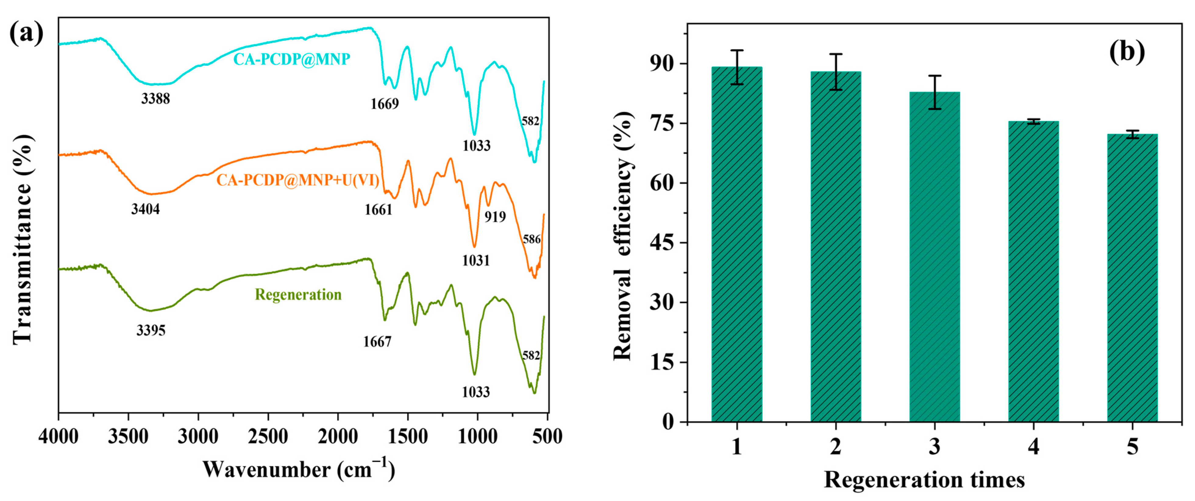
3.6. Recycling and Regeneration of CA-PCDP@MNP

Reusable adsorbent will greatly reduce U(VI) ion purification cost. Changes in the U(VI) removal rate of CA-PCDP@MNP after five adsorption–desorption cycles are displayed in detail in Figure 9b. And the R of the CA-PCDP@MNPs on U(VI) reduction with the number of cycles increasing was observed. FTIR spectra of CA-PCDP@MNPs after five cycles of adsorption and desorption were obtained, and the results are exhibited in Figure 9a. According to the data, the stretching vibration peak of U=O at 919 cm−1 disappeared completely when compared to CA-PCDP@MNP+U(VI) after adsorption, illustrating that uranyl ions were successfully removed from the adsorbent via 0.1 mol/L HNO3. The adsorption ability decreased from 90% to 70% after five cycles, which was possibly due to carboxyl and hydroxyl groups in the CA-PCDP@MNPs being protonated, and some H+ occupied the surface binding sites during strong acid elution [46]. It was also observed from Figure 9a that the C=O stretching vibration at 1667 cm−1 and the OH stretching vibration peak at 3395 cm−1 were shifted and varied. However, R was still above 70% after five adsorption–desorption cycles, illustrating that the CA-PCDP@MNPs were recyclable and had great potential for practical application in uranium-containing wastewater remediation.

4. Conclusions

In this work, magnetic γ-Fe2O3 nanoparticles were successfully loaded onto CA-PCDP to acquire CA-PCDP@MNPs, which was demonstrated via FTIR, XRD, SEM, VSM, and so on. The CA-PCDP@MNP saturation magnetization strength was 30.17 emu/g, which had superparamagnetism and good magnetic response. Results obtained via single-factor research manifested that the adsorption process for U(VI) conformed to a pseudo-second-order kinetic model and the Langmuir isotherm model, suggesting that it was a chemical monolayer adsorption. Moreover, the CA-PCDP@MNP maximum theoretical adsorption amount reached 245.66 mg/g at 303 K and pH = 6.0, and the adsorption thermodynamics followed a spontaneous endothermic process. Research on the adsorption mechanism suggested that the rich oxygen-containing groups on the CA-PCDP@MNP surface (carboxyl, hydroxyl, C-O-C, and Fe-O) might have interacted with U (VI). Furthermore, CA-PCDP@MNPs were recyclable, and R was still above 70% after five adsorption–desorption cycles, which had great potential for practical application in uranium-containing wastewater remediation.

Author Contributions

Conceptualization, writing—original draft preparation, X.Z. and C.H.; Investigation, N.L. and M.Z.; Validation, Q.Y. and Y.T.; Writing—review and editing, M.M. and Y.W.; Supervision, J.O. All authors have read and agreed to the published version of the manuscript.

Funding

This research was supported by the Jiangxi Provincial Natural Science Foundation (20202BABL203026, 20202BABL214016, 20212BAB203001) and the College Student Innovation and Enterprise Programme of Jiangxi Province (S202310405010).

Conflicts of Interest

The authors declare no conflict of interest.

References

  1. Chen, L.; Liu, J.; Zhang, W.; Zhou, J.; Luo, D.; Li, Z. Uranium (U) source, speciation, uptake, toxicity and bioremediation strategies in soil-plant system: A review. J. Hazard. Mater. 2021, 413, 125319. [Google Scholar] [CrossRef]
  2. Zhou, M.; Wang, S.; Qiu, M.; Hu, B.; Wang, G.; Lu, Y. Recent advances in the removal of U(VI) by magnetic metal oxides. J. Mol. Liq. 2023, 385, 122295. [Google Scholar] [CrossRef]
  3. Prusty, S.; Somu, P.; Sahoo, J.K.; Panda, D.; Sahoo, S.K.; Sahoo, S.K.; Lee, Y.R.; Jarin, T.; Sundar, L.S.; Rao, K.S. Adsorptive sequestration of noxious uranium (VI) from water resources: A comprehensive review. Chemosphere 2022, 308, 136278. [Google Scholar] [CrossRef] [PubMed]
  4. Zhong, X.; Yuan, Q.; Wang, Q.; Hu, C.; Guo, K.; Ouyang, J.; Chen, M. Maleic anhydride-β-cyclodextrin functionalized magnetic nanoparticles for the removal of uranium (VI) from wastewater. Crystals 2022, 12, 1731. [Google Scholar] [CrossRef]
  5. Ye, Y.; Jin, J.; Chen, F.; Dionysiou, D.D.; Feng, Y.; Liang, B.; Cheng, H.-Y.; Qin, Z.; Tang, X.; Li, H. Removal and recovery of aqueous U(VI) by heterogeneous photocatalysis: Progress and challenges. Chem. Eng. J. 2022, 450, 138317. [Google Scholar] [CrossRef]
  6. Tang, N.; Liang, J.; Niu, C.; Wang, H.; Luo, Y.; Xing, W.; Ye, S.; Liang, C.; Guo, H.; Guo, J.; et al. Amidoxime-based materials for uranium recovery and removal. J. Mater. Chem. A 2020, 8, 7588–7625. [Google Scholar] [CrossRef]
  7. Abney, C.W.; Mayes, R.T.; Saito, T.; Dai, S. Materials for the recovery of uranium from seawater. Chem. Rev. 2017, 117, 13935–14013. [Google Scholar] [CrossRef]
  8. Djellabi, R.; Yang, B.; Sharif, H.M.A.; Zhang, J.; Ali, J.; Zhao, X. Sustainable and easy recoverable magnetic TiO2-lignocellulosic biomass@Fe3O4 for solar photocatalytic water remediation. J. Clean. Prod. 2019, 233, 841–847. [Google Scholar] [CrossRef]
  9. Rasheed, T. Magnetic nanomaterials: Greener and sustainable alternatives for the adsorption of hazardous environmental contaminants. J. Clean. Prod. 2022, 362, 132338. [Google Scholar] [CrossRef]
  10. Zhao, Y.; Li, J.; Zhao, L.; Zhang, S.; Huang, Y.; Wu, X.; Wang, X. Synthesis of amidoxime-functionalized Fe3O4@SiO2 core–shell magnetic microspheres for highly efficient sorption of U(VI). Chem. Eng. J. 2014, 235, 275–283. [Google Scholar] [CrossRef]
  11. Meng, Y.; Wang, Y.; Liu, L.; Fang, Y.; Ma, F.; Zhang, C.; Dong, H. Efficient and magnetically recoverable U (VI) adsorbent: Fe3O4 loaded hypercrosslink copoly (styrene/maleic anhydride). Colloids Surf. A 2022, 632, 127644. [Google Scholar] [CrossRef]
  12. Li, N.; Yang, L.; Ji, X.; Ren, J.; Gao, B.; Deng, W.; Wang, Z. Bioinspired succinyl-β-cyclodextrin membranes for enhanced uranium extraction and reclamation. Environ. Sci. Nano. 2020, 7, 3124–3135. [Google Scholar] [CrossRef]
  13. Yang, S.; Zong, P.; Hu, J.; Sheng, G.; Wang, Q.; Wang, X. Fabrication of β-cyclodextrin conjugated magnetic HNT/iron oxide composite for high-efficient decontamination of U(VI). Chem. Eng. J. 2013, 214, 376–385. [Google Scholar] [CrossRef]
  14. Zhong, X.; Wang, R.; Xu, G.; Guo, K.; Hu, C. Adsorptive removal of uranium (VI) from wastewater using a crosslinked amidoxime-functionalized β-cyclodextrin polymer. New J. Chem. 2022, 46, 20592–20601. [Google Scholar] [CrossRef]
  15. Zong, P.; Xu, X.; Shao, M.; Cao, D.; Wang, S.; Zhang, H. Efficient elimination of uranium by carboxymethyl-β-cyclodextrin nanoparticles decorated iron oxides: Water chemistry influences and mechanism researches. Int. J. Hydrog. Energ. 2021, 46, 1370–1384. [Google Scholar] [CrossRef]
  16. Xu, G.; Xie, X.; Qin, L.; Hu, X.; Zhang, D.; Xu, J.; Li, D.; Ji, X.; Huang, Y.; Tu, Y.; et al. Simple synthesis of a swellable porous β-cyclodextrin-based polymer in the aqueous phase for the rapid removal of organic micro-pollutants from water. Green Chem. 2019, 21, 6062–6072. [Google Scholar] [CrossRef]
  17. Alsbaiee, A.; Smith, B.J.; Xiao, L.; Ling, Y.; Helbling, D.E.; Dichtel, W.R. Rapid removal of organic micropollutants from water by a porous β-cyclodextrin polymer. Nature 2016, 529, 190–194. [Google Scholar] [CrossRef]
  18. Jiang, H.; Lin, J.; Hai, W.; Tan, H.; Luo, Y.; Xie, X.; Cao, Y.; He, F. A novel crosslinked β-cyclodextrin-based polymer for removing methylene blue from water with high efficiency. Colloids Surf. A 2019, 560, 59–68. [Google Scholar] [CrossRef]
  19. Tian, Y.; Liu, L.; Ma, F.; Zhu, X.; Dong, H.; Zhang, C.; Zhao, F. Synthesis of phosphorylated hyper-cross-linked polymers and their efficient uranium adsorption in water. J. Hazard. Mater. 2021, 419, 126538. [Google Scholar] [CrossRef]
  20. Nie, X.; Zhang, Y.; Jiang, Y.; Pan, N.; Liu, C.; Wang, J.; Ma, C.; Xia, X.; Liu, M.; Zhang, H.; et al. Efficient extraction of U(VI) from uranium enrichment process wastewater by amine-aminophosphonate-modified polyacrylonitrile fibers. Sci. Total Environ. 2022, 831, 154743. [Google Scholar] [CrossRef] [PubMed]
  21. Yu, R.; Lu, Y.; Zhang, X.; Chen, W.; Chen, X.; Li, L. Amidoxime-modified ultrathin polyethylene fibrous membrane for uranium extraction from seawater. Desalination 2022, 539, 115965. [Google Scholar] [CrossRef]
  22. Liu, X.; Wu, J.; Zhang, S.; Ding, C.; Sheng, G.; Alsaedi, A.; Hayat, T.; Li, J.; Song, Y. Amidoxime-Functionalized Hollow Carbon Spheres for Efficient Removal of Uranium from Wastewater. ACS Sustain. Chem. Eng. 2019, 7, 10800–10807. [Google Scholar] [CrossRef]
  23. Xu, M.-Y.; Jiang, H.-L.; Xie, Z.-W.; Li, Z.-T.; Xu, D.; He, F.-A. Highly efficient selective adsorption of anionic dyes by modified β-cyclodextrin polymers. J. Taiwan Inst. Chem. Eng. 2020, 108, 114–128. [Google Scholar] [CrossRef]
  24. Dong, S.; Wang, S.; Wang, X.; Zhai, L. Superparamagnetic nanocomposite Fe3O4@SiO2-NH2/CQDs as fluorescent probe for copper (II) detection. Mater. Lett. 2020, 278, 128404. [Google Scholar] [CrossRef]
  25. Niu, L.; Zhang, G.; Xian, G.; Ren, Z.; Wei, T.; Li, Q.; Zhang, Y.; Zou, Z. Tetracycline degradation by persulfate activated with magnetic γ-Fe2O3/CeO2 catalyst: Performance, activation mechanism and degradation pathway. Sep. Purif. Technol. 2021, 259, 118156. [Google Scholar] [CrossRef]
  26. Chen, B.; Sun, J.; Fan, F.; Zhang, X.; Qin, Z.; Wang, P.; Li, Y.; Zhang, X.; Liu, F.; Liu, Y. Ferumoxytol of ultrahigh magnetization produced by hydrocooling and magnetically internal heating co-precipitation. Nanoscale 2018, 10, 7369–7376. [Google Scholar] [CrossRef]
  27. Le Jeune, M.; Secret, E.; Trichet, M.; Michel, A.; Ravault, D.; Illien, F.; Siaugue, J.-M.; Sagan, S.; Burlina, F.; Ménager, C. Conjugation of oligo-his peptides to magnetic γ-Fe2O3@SiO2 core–shell nanoparticles promotes their access to the cytosol. ACS Appl. Mater. Interfaces 2022, 14, 15021–15034. [Google Scholar] [CrossRef]
  28. Sobhani, S.; Khakzad, F. A novel hydrophobic copper complex supported on γ-Fe2O3 as a magnetically heterogeneous catalyst for one-pot three-component synthesis of α-aminophosphonates. Appl. Organomet. Chem. 2017, 31, 3877. [Google Scholar] [CrossRef]
  29. Ma, H.; Pu, S.; Ma, J.; Yan, C.; Zinchenko, A.; Pei, X.; Chu, W. Formation of multi-layered chitosan honeycomb spheres via breath-figure-like approach in combination with co-precipitation processing. Mater. Lett. 2018, 211, 91–95. [Google Scholar] [CrossRef]
  30. Khan, G.A.; Esentürk, E.N.; Bek, A.; Bhatti, A.S.; Ahmed, W. Fabrication of highly catalytically active gold nanostructures on filter-paper and their applications towards degradation of environmental pollutants. ChemistrySelect 2021, 6, 10655–10660. [Google Scholar] [CrossRef]
  31. Zhang, S.; Hu, C.; Ji, H.; Zhang, L.; Li, F. Facile synthesis of nitrogen-deficient mesoporous graphitic carbon nitride for highly efficient photocatalytic performance. Appl. Surf. Sci. 2019, 478, 304–312. [Google Scholar] [CrossRef]
  32. Zhang, Y.; Huang, S.; Mei, B.; Tian, X.; Jia, L.; Zhu, W. Mussel inspired synthesis of polydopamine/polyethyleneimine-grafted fly ash composite adsorbent for the effective separation of U (VI). Sci. Total Environ. 2023, 876, 162841. [Google Scholar] [CrossRef] [PubMed]
  33. Dastbaz, A.; Keshtkar, A.R. Adsorption of Th4+, U6+, Cd2+, and Ni2+ from aqueous solution by a novel modified polyacrylonitrile composite nanofiber adsorbent prepared by electrospinning. Appl. Surf. Sci. 2014, 293, 336–344. [Google Scholar] [CrossRef]
  34. Zhu, X.; Liu, L.; Wang, Y.; Ma, F.; Tian, Y.; Dong, H.; Zhang, C. Synthesis of hypercrosslinked calix [4] arenes anchored with Fe (III) via phosphate groups for excellent uranyl extraction from wastewater and seawater. J. Water Process. Eng. 2023, 53, 103615. [Google Scholar] [CrossRef]
  35. Guo, X.; Shang, Y.; Liang, X.; Diao, Z.; Song, G.; Chen, D.; Wang, S.; Kong, L. A comparison of Ni-Co layered double oxides with memory effect on recovering U(VI) from wastewater to hydroxides. Chem. Eng. J. 2022, 446, 137220. [Google Scholar] [CrossRef]
  36. Li, W.; Han, X.; Wang, X.; Wang, Y.; Wang, W.; Xu, H.; Tan, T.; Wu, W.; Zhang, H. Recovery of uranyl from aqueous solutions using amidoximated polyacrylonitrile/exfoliated Na-montmorillonite composite. Chem. Eng. J. 2015, 279, 735–746. [Google Scholar] [CrossRef]
  37. Song, S.; Huang, Q.; Cheng, G.; Wang, W.; Lu, Z.; Zhang, R.; Wen, T.; Zhang, Y.; Wang, J.; Wang, X. Immobilization of U (VI) on hierarchical NiSiO@ MgAl and NiSiO@ NiAl nanocomposites from wastewater. ACS Sustain. Chem. Eng. 2019, 7, 3475–3486. [Google Scholar] [CrossRef]
  38. Li, Y.; Li, X.-X.; Wang, Z.-Y.; Zhang, F.; Wu, Q.; Sha, L.-T.; Wang, Y.; Yan, Z.-Y. Design and synthesis of a novel bifunctional polymer with malonamide and carboxyl group for highly selective separation of uranium (VI). Sep. Purif. Technol. 2022, 302, 122115. [Google Scholar] [CrossRef]
  39. Husnain, S.M.; Um, W.; Woojin-Lee, W.-L.; Chang, Y.-S. Magnetite-based adsorbents for sequestration of radionuclides: A review. RSC Adv. 2018, 8, 2521–2540. [Google Scholar] [CrossRef]
  40. Liu, X.; Sun, J.; Xu, X.; Alsaedi, A.; Hayat, T.; Li, J. Adsorption and desorption of U (VI) on different-size graphene oxide. Chem. Eng. J. 2019, 360, 941–950. [Google Scholar] [CrossRef]
  41. Liao, J.; Xiong, T.; Zhao, Z.; Ding, L.; Zhu, W.; Zhang, Y. Synthesis of a novel environmental-friendly biocarbon composite and its highly efficient removal of uranium(VI) and thorium(IV) from aqueous solution. J. Clean. Prod. 2022, 374, 134059. [Google Scholar] [CrossRef]
  42. Fan, F.-L.; Qin, Z.; Bai, J.; Rong, W.-D.; Fan, F.-Y.; Tian, W.; Wu, X.-L.; Wang, Y.; Zhao, L. Rapid removal of uranium from aqueous solutions using magnetic Fe3O4@SiO2 composite particles. J. Environ. Radioactiv. 2012, 106, 40–46. [Google Scholar] [CrossRef] [PubMed]
  43. Cheng, Y.; He, P.; Dong, F.; Nie, X.; Ding, C.; Wang, S.; Zhang, Y.; Liu, H.; Zhou, S. Polyamine and amidoxime groups modified bifunctional polyacrylonitrile-based ion exchange fibers for highly efficient extraction of U(VI) from real uranium mine water. Chem. Eng. J. 2019, 367, 198–207. [Google Scholar] [CrossRef]
  44. He, Y.; Lin, X.; Yan, T.; Zhang, X.; Zhou, J.; Chen, Y.; Luo, X. Selective adsorption of uranium from salt lake-simulated solution by phenolic-functionalized hollow sponge-like adsorbent. J. Chem. Technol. Biot. 2019, 94, 455–467. [Google Scholar] [CrossRef]
  45. Atrei, A.; Lesiak-Orlowska, B.; Tóth, J. Magnetite nanoparticles functionalized with citrate: A surface science study by XPS and ToF-SIMS. Appl. Surf. Sci. 2022, 602, 154366. [Google Scholar] [CrossRef]
  46. Pang, Y.; Zeng, G.; Tang, L.; Zhang, Y.; Liu, Y.; Lei, X.; Li, Z.; Zhang, J.; Xie, G. PEI-grafted magnetic porous powder for highly effective adsorption of heavy metal ions. Desalination 2011, 281, 278–284. [Google Scholar] [CrossRef]
Figure 1. Schematic diagram of the preparation of CA-PCDP@MNP.
Crystals 13 01496 g001
Figure 2. (a) FTIR spectrum of PCDP, CA-PCDP, MNP, and CA-PCDP@MNP; (b) XRD spectra of MNP and CA-PCDP@MNP; (c) magnetic hysteresis loop of CA-PCDP@MNP at 298 K.
Crystals 13 01496 g002
Figure 3. SEM of CA-PCDP@MNP (a,b); EDXS spectra of CA-PCDP@MNP (c).
Crystals 13 01496 g003
Figure 4. N2 isotherms curve (a) and pore width distribution (b) of CA-PCDP@MNP.
Crystals 13 01496 g004
Figure 5. (a) Effect of CA-PCDP@MNP adsorbent dosage at pH = 6.0, C0 = 100 mg/L, 303 K. (b) Effects of initial pH on the adsorption of U(VI) by CA-PCDP@MNPs at 303 K, C0 = 100, and 40 mg/L; adsorbent dosage = 0.4 g/L. (c) Zeta potential curves of CA-PCDP@MNPs.
Crystals 13 01496 g005
Figure 6. Effect of contact times (a) for CA-PCDP@MNP. Quasi-first-order (b), quasi-second-order (c), and intra-particle diffusion (d) kinetic models of U(VI) adsorption.
Crystals 13 01496 g006
Figure 7. (a) Isothermal model fitting figure of U(VI) adsorbed by CA-PCDP@MNP. (b) Correlation between lnK0 and 1/T.
Crystals 13 01496 g007
Figure 8. (a) XPS full and U 4f spectra of CA-PCDP@MNP and CA-PCDP@MNP+U(VI); (b) fine spectra of O 1s; (c) fine spectra of Fe 2p.
Crystals 13 01496 g008
Figure 9. (a) FTIR spectra of CA-PCDP@MNP, CA-PCDP@MNP+U(VI), and after 5 adsorption–desorption cycles. (b) Reusability of CA-PCDP@MNP for U(VI) adsorption.
Crystals 13 01496 g009
Table 1. Adsorption kinetic results of U(VI) by CA-PCDP@MNP.
Pseudo-First-Order ModelPseudo-Second-Order ModelInternal Diffusion Model
k1qe,calqe,expR2k2qe,calqe,expR2kidCR2
0.012653.96178.850.7180.0012183.15178.850.999kid,1 = 26.75930.3490.991
kid,2 = 9.82099.6650.993
kid,3 = 0.246175.1840.971
Table 2. Parameters of isotherm model for U(VI) adsorption by CA-PCDP@MNP.
SampleT/KLangmuir ModelFreundlich Model
qm/(mg/g)KL/(L/mg)RL2KF/(mg/g)nRF2
CA-PCDP@MNP283213.580.077610.992206.903.4490.856
293231.250.101450.998235.573.6360.844
303245.660.113370.994280.754.1550.845
313262.520.119060.992313.324.3710.875
Table 3. The thermodynamic parameters related to the adsorption of U(VI) by CA-PCDP@MNP.
SampleT/KG0/(kJ∙mol−1)H0/(kJ∙mol−1)S0/(J∙mol−1∙K−1)
CA-PCDP@MNP283−5.57119.04887.330
293−6.667
303−7.463
313−8.200
Disclaimer/Publisher’s Note: The statements, opinions and data contained in all publications are solely those of the individual author(s) and contributor(s) and not of MDPI and/or the editor(s). MDPI and/or the editor(s) disclaim responsibility for any injury to people or property resulting from any ideas, methods, instructions or products referred to in the content.

