
Synthesis of Bimetallic Nanoparticles and Applications—An Updated Review

Department of Biotechnology, Sharda School of Engineering & Technology, Sharda University, Greater Noida 201310, India
*
Author to whom correspondence should be addressed.
Crystals 2023, 13(4), 637; https://doi.org/10.3390/cryst13040637
Submission received: 17 February 2023
/
Revised: 5 April 2023
/
Accepted: 5 April 2023
/
Published: 7 April 2023
(This article belongs to the Special Issue Advanced Nanomaterials for Photocatalytic Technologies)
Abstract
:The manipulation of matter at the atomic level (nanotechnology) has experienced an explosion in research interest in recent years. Bimetallic nanoparticles are vital due to their high biocompatibility, stability and comparatively less toxicity. The synthesis methods that include physical, chemical and biological methods are explored and explained in detail, along with their advantages. They have a wide range of applications due to their synergistic properties including biological applications (in medicine and agriculture), environmental application (in water treatment and removal of toxic contaminants), engineering application (in nanosensors, nanochips and nano-semiconductors) and chemical and physical application (in optics, catalysis and paints). The green synthesis approach is a promising method of synthesis that can give rise to more biocompatible and less toxic bimetallic nanoparticles due to increasing environmental pollution. However, despite these interesting attributes of bimetallic nanoparticle, there is still much work to be done to improve the biocompatibility of bimetallic nanoparticles because of their toxicity and potentially hazardous effects.
1. Introduction
As the name implies, nanotechnology is a technology that deals with matter at the nanoscale level. It refers to the branch of science and engineering that deals with the manipulating of atoms or molecules at the nanoscale to design systems and produce devices for various applications. The main building block of nanotechnology is the nanoparticles. These nanoparticles are tiny in size, have a high surface area and exhibit a property known as quantum effects, which means irregular or unpredictable behavior [1]. Nanoparticles are of different types, and they can be classified based on structure (metal base, carbon base, dendrimers or liposome), dimension (zero, one, two or three dimensions) or origin (natural or artificial) [2]. Among the various types of nanoparticles, metal-based nanoparticles, especially those of noble metals, have more advantages than other nanoparticles This is because metal-based nanoparticles are highly stable, biocompatible and possibly capable of large-scale production for biomedical and environmental applications [3]. However, despite having these fascinating abilities and properties, the application of metal based nanoparticles still needs to be improved in some fields of study due to their toxicity, large size, cellular uptake and chemical stability [4,5]. Therefore, there is a need to overcome those limitations. By combining any two metals to form a bimetallic nanoparticle, the fascinating and synergetic relationship between them shows new enhanced structural and physical properties, subsequently increasing their functionality and application [2]. In addition, the experimental results have shown a remarkable enhancement capability and a possible solution to overcome the limitation of monometallic nanoparticles.
Bimetallic nanoparticles are attracting more attention in recent years due to their unique physical properties (quantum effect, high surface area, mobility), chemical, mechanical, thermal, optical, catalytic and magnetic properties [6,7,8,9,10]. These properties make bimetallic distinguishable from their monometallic counterparts and give a superior performance that the individual metals do not have [11,12,13]. Both mono and bimetallic nanoparticles are suitable for applications in various fields, including biomedical, biosensors, nanomedicine, imaging, wastewater treatment, oil and gas industries, agriculture/food processing and gene/drug delivery [14,15,16,17,18,19,20]. In addition, antimicrobial and anticancer activities of bimetallic nanoparticles have also been investigated extensively [21,22,23,24,25,26].
There are three methods of synthesizing nanoparticles: physical, chemical and biological (Figure 1). Either of the two main approaches can make these methods, namely, the top-down and the bottom-up approach. The top-down approach involves the breaking down of the bulk material by external mechanical force to nanosized particles, e.g., mechanical grinding, ball milling, laser ablation, microwave irradiation, etc. On the other hand, the bottom-up approach involves building the nanoparticles right from the atomic level. Atoms build up to form molecules, then the molecules grow into a cluster and finally create the nanoscale particles. Examples of this method include the sol-gel process, pyrolysis and chemical precipitation [27]. Though the top-down approach is a faster method, there is no control of particle shape and size as compared to the bottom-up approach, where shape and size can be easily controlled by controlling the synthesis parameters such as pH, temperature and concentration. This review will focus on bimetallic nanoparticles, synthesis, characterization, biological and environmental applications and the toxicity of bimetallic nanoparticles.
2. Different Types of Bimetallic
Bimetallic nanoparticles are synthesized when two different metals are mixed together in a reaction vessel under optimized conditions, resulting in various structural and morphological changes [28]. Different combinations of metals, including noble and transition metals can produce many different types of bimetallic nanoparticles. These bimetallic nanoparticles can be in many forms, including gold-based, silver-based, copper-based, nickel-based, iron-based, platinum-based or palladium-based bimetallic nanoparticles (Table 1).
3. Synthesis of Bimetallic Nanoparticles
Generally, the synthesis of bimetallic nanoparticles involves either breaking down bulk materials to nanosized particles or building up the nanoparticles from their respective atoms. These two approaches are the top-down approach and the bottom-up approach (Figure 2). The bottom-up approach usually involves mixing two different metal precursors in a reaction vessel under the optimized condition to which a reducing and stabilizing agent is added. However, the general procedure of synthesizing bimetallic nanoparticles can be categorized into three main categories, namely, physical, chemical and biological methods (Figure 3).
3.1. Physical Method
The physical method of nanoparticle synthesis generally involves the breaking of bulk materials into fine particles. It has some advantages over the other synthesis methods, such as uniformity in the formation of nanoparticles and less contamination.
3.1.1. Laser Ablation
This is a top-down approach where the bulk material is broken down into nanosized particles. The process involves irradiating a solid or sometimes a liquid substance with a laser beam to remove particles from their surface. This process can be used to synthesize a well dispersed bimetallic nanoparticle, as reported by [64]. The mechanism of nanoparticle formation using laser ablation method is appropriately explained by [65] and [66], respectively.
3.1.2. Mechanical Grinding
In this process, bimetallic nanoparticles are formed by grinding the mixture of the metal salts with the reducing agent in a solid state. Au–Ag nanoparticles were synthesized by [67] using this method. In a simple two-step experiment, a solid-state mixture of , chitosan and NaOH was first ground using a mortar and pestle and then followed by the addition of a concentrated solution of which, subsequently, resulted in the formation of Au–Ag bimetallic nanoparticle.
3.1.3. Microwave Irradiation
Microwave irradiation can also assist in the fabrication of bimetallic nanoparticles and have certain advantages, such as excellent control of the reaction parameters, the heating source having no direct contact with the reactant/solvent and a high heating rate. Microwave heating in this method is used to replace the use of chemicals during the synthesis process. This method synthesized Ag/ZnO bimetallic nanoparticles by heating the mixture of and ZnO nanoparticles dispersed in ethanol under reflux in a microwave reactor for 20 min [68]. In addition, [69] synthesized Au–Pd and Au–Pt bimetallic nanoparticles using hydrothermal and microwave irradiation.
3.1.4. Ball Milling
Ball milling is a method that grinds materials into extremely fine powders. It consists of a hollow horizontal compartment containing balls made of steel that rotate around its axis. The subsequent rotation crushes the materials inside the compartment into fine particles with the help of the steel balls. This method was used for synthesizing Al–Fe and Ni–Fe bimetallic nanoparticles, as reported by [70] and [71], respectively.
3.1.5. Electrochemical Method
In this method, an electrolyte and an electrically conductive metal substrate are used for the synthesis of bi-metallic nanoparticles. The synthesis occurs at the interface between the electrolyte solution, containing the metal salt precursors and the conductive metal substrate [72]. A study employed this method and synthesized Co–Ni bimetallic nanoparticles that were used to detect tetracycline [73]. Similarly, Bi–Sb and Au@Ag core shells were synthesized by [74] and [75] using the same electrochemical method and used for the reduction of CO2 and detection of thiram in juice and milk, respectively.
3.2. Chemical Method
The most important factor in the chemical synthesis method is the reducing and stabilizing agent. This method generally involves using a suitable metal source, usually soluble salts or ions that can be dissolved in water. To that solution, a reducing agent in the form of chemical, polymer or ligand is added which strongly affects the nanoparticle particle formation. In some instances, only the stabilizing or reducing agent is required for the synthesis while other processes require both the reducing agent and as well as the stabilizing agent. When both metal precursors are present and simultaneously reduced during the synthesis process, it can lead to nanoparticle formation, while successive reduction can lead to core–shell formation [76]. The fabrication of bimetallic nanoparticles using the chemical method can be generally grouped into two, namely, simultaneous and successive reduction. Both are simple, fast, economic and can be used for large-scale production, and the doping of other materials during synthesis is possible. Various parameters such as pH, temperature, concentration and rate of irradiation are used to control the chemical synthesis of bimetallic nanoparticles.
3.2.1. Simultaneous Reduction
Co-Reduction
Co-reduction is the simplest method of bimetallic synthesis. It involves the mixing of two metal precursors together. To that mixture, a reducing agent or stabilizing agent is added, which can reduce the metal ion to atoms and stabilize the aggregation process [77]. In practice, polymers can be used to control the reduction and aggregation of bimetallic nanoparticles. The polymer and metal ions can interact after the reduction and vise vasa. It is believed that when the reduction of metal nanoparticles comes first, before the interactions, some properties including the size and structural properties can be determined only by the reduction process. However, when the interaction comes before the reduction, the particle size structure may be affected [78]. For example, Fe-Ni bimetallic nanoparticle supported by zeolite was prepared by sodium borohydride——for the simultaneous reduction of the corresponding mixture of the two metal salts. In the experiment, the Fe and Ni metal salts precursors were dissolved in an ethanol-water solution and then mixed with zeolite, which acts as the stabilizing agent. Adding the reducing agent——to the mixture under vigorous stirring in a dropwise manner causes a black suspension within 30 min, thereby indicating the formation of Z–Fe/Ni bimetallic nanoparticles. Subsequently, Fe–Ni bimetallic nanoparticle was also fabricated by the simultaneous reduction of the same mixture of metal salt solution using the same reducing agent but without the addition of the stabilizing agent ‘zeolite’ [79]. The same process was employed by [80] in synthesizing the same bimetallic nanoparticle supported by bentonite (B–Fe/NI), which serves as the stabilizing agent.
Sonochemical Co-Reduction
This process involves using high-intensity ultrasound radiation in a solution to create a chemical reaction at the atomic level. When the aqueous mixture is exposed to a high-intensity ultrasound wave, an acoustic cavitation effect is produced where growth occurs in the region of high temperature and pressure. Sonification also leads to the breaking down of molecules that lead to the production of radicals that act as oxidizing and reducing radicals [81]. A study reported that magnetic Ni–Ag nanoparticles supported by graphene oxide were synthesized using ultrasonication of the aqueous solution of and [82]. The graphene oxide act as the stabilizing agent while NaOH and hydrazine hydrate () act as the reducing agent. The mixture of the aqueous solution of the metal salts with stabilizing agent (graphene oxide) was first ultrasonicated for 30 min. Then, the reducing agents (NaOH and hydrazine hydrate ()) were added under atmosphere at 90 °C and the mixture was ultrasonicated again for 10 min. The mixture was refluxed at 90 °C for 1 h, then centrifuged and the solid product was removed and washed with ethanol and distilled water before drying at 50 °C in a hot air oven. In addition, a polymer-stabilized Au-Pd bimetallic nanoparticle synthesized by ultrasonic irradiation using PVP and ethylene glycol was the stabilizing agent [83].
Radiolytic Co-Reduction
Radiolytic co-reduction is the process of applying ionizing radiation (UV, X-ray, Gamma ray, electron beam, etc.) to an aqueous mixture in order to reduce the ionic solutions to atoms. This method offers some advantages due to the efficient control of radiation dose delivered to the mixture which, in return, control the structure and growing process of nanoparticles, although in terms of nanoparticles shape the radiolytic process can be difficult to control in the radiolytic co-reduction of metal ion solutions, reducing radicals produced when radiolysis occurs [84]. Moreover, the electron radicals with a high reduction potential replace the chemical reducing agent. The radiolytic co-reduction mechanism involves hydrogen-free radicals and hydrated electrons generated by the radiolysis of water. A study [85] reported a two-step synthesis of platinum-based bimetallic nanoparticles Pt–M (M: V, Mo, W) supported by graphene oxide with the help of gamma-ray irradiation at a rate of 4.5 (kGy/h) under ambient conditions. In the first step, graphene oxide was prepared, while in the second step, the metal source was dissolved in the prepared graphene oxide solution. Exposing this mixture to gamma radiation reduces the mixture and produces Pt–V, Pt–Mo and Pt–W bimetallic nanoparticles. Further, another study [86] reported that a low exposure rate can reduce gold and silver ions, thereby producing a gold core silver-coated bimetallic nanoparticle but, in contrast, a high dose rate can produce alloyed Ag–Pt bimetallic nanoparticles. Other types of bimetallic nanoparticles that were produced using gamma irradiation include Ag–Pt [87], Au–Cu [88], Pd–M (M = Ag, Au, Cu, Ni, and Pt) [89], Pt–M (M = Au, Cu, Ni) [90], Pd–Au–Ag tri-metallic nanoparticle [91] and Pt monometallic, Pt–Ru bimetallic, Pt–Ru–Sn, Pt–Ru–Mo trimetallic and Pt–Ru–Mo–Sn tetra-metallic nanoparticles [92].
Chemical Precipitation
Chemical precipitation is an easy, single step and fast method of synthesizing nanoparticles. It involves converting dissolved substances in a solution into an insoluble solid, thereby making the solution a super saturated one. The insoluble solid that is formed is known as the “precipitate”. In terms of synthesizing a bimetallic nanoparticle, it is often regarded as “co-precipitation” due to the fact that two metal salts form the precipitate. Fe–Mn bimetallic have been synthesized using co-precipitation method [51]. The metal precursors were first prepared individually in the presence of excess oxygen and then mixed in an Erlenmeyer flask at 70 °C while stirring at 50 rpm. The pH of the solution was adjusted to 9 by adding 10% while mixing continues for 4 h The solution was left in a stationary condition until the precipitate was formed. The precipitate was separated by centrifugation, washed and finally dried in hot air oven at 50 °C until constant weight was reached. Then, the final product was stored in a desiccator. hBN–Fe3O4 [93], Me–Mn (Me = Co, Ni, Sn) [94], Fe–Ti [50], CoCu–Mof [95] and Fe–Cr bimetallic nanoparticles have also been synthesized using this method.
Thermal Decomposition
Thermal decomposition is a chemical reaction that occurs when a compound breaks down at high temperature. It is known that metal nanoparticles, especially the noble metals, can be synthesized at room temperature through the action of the reducing agent that reduced the corresponding metal salts. However, some metal nanoparticles such as transition metals, require a higher temperature during synthesis because of their inability to be crystallized at room temperature [96,97]. To synthesize bimetallic nanoparticles using this method, the temperature of the reaction must be increased to or near decomposition temperature. The metal precursor with lower decomposition temperature will be decomposed (reduced) first and form a single-component monometallic nanoparticle, while the second metal precursor is still in the solution. When the decomposition temperature of the second metal precursor is reached, it will be decomposed and formed on the surface of the first metal nanoparticle, thereby forming a bimetallic nanoparticle. For example, synthesis of bimetallic titanium complexes M–Ti–O (M = nickel—Ni, Cobalt—Co, Manganese—Mn) from cobalt, manganese and nickel metal precursors by using the thermal decomposition method were reported in the literature [98]. In addition, the thermal decomposition method of synthesis has been used to synthesize Pd–Ir, [59], Co–Mn [99], [100], Ti/Ce-Sb-NFs and Pd–Ag/C bimetallic nanoparticles [101].
Sol-Gel Method
Sol-gel method is a simple, cheap and fast method of fabricating nanoparticles. It is a technique based on colloidal chemistry, where a liquid mixture is transformed into a gel. To synthesize nanoparticles, the metal precursors are first converted into a “sol” form or colloidal suspension through hydrolysis and polymerization and then subsequently converts into “gel” by gelation. The sol-gel formation process involves four stages: hydrolysis, condensation, growth and agglomeration of particles [102]. A study reported synthesis of alloyed Cu–Ni bimetallic nanoparticles using sol-gel method [39]. The metal precursors were mixed with citric acid and stirred at room temperature until transparent solution is obtained. While stirring continues, ethylene glycol was added, and the mixture was evaporated at 130 °C for 24 h to obtain dry precursor and then calcinated at 700 °C to obtain the final product. The magnetic properties were studied using SQUID magnetometry and the Cu–Ni bimetallic nanoparticle shows a ferromagnetic behavior. Other bimetallic nanoparticles synthesized using sol-gel method include Mg–Al [103], Pt–Pd [104], and Au–M (M = Ag, Pd, Pt) [105].
Micro-Emulsion Method
Micro-emulsion is a homogeneous, isotropic and thermodynamic stable solution that contains at least 3 components: a polar domain (usually water), non-polar domain (usually oil) and a surfactant (ionic or non-ionic). This solution is macroscopic in nature and the surfactant molecules usually form a tiny film that separate the polar and non-polar phase. Different types of micro-emulsion include water-in-oil (reverse micelles), oil-in-water (O/W), bicontinuous and supercritical microemulsion. Among the different types of microemulsion, water in oil microemultion is mostly used for nanoparticle synthesis while only a few nanoparticles are synthesized using the oil-in-water microemulsion method [106,107]. In a study, Ag–Au bimetallic nanoparticle was prepared using the water-in-oil microemulsion (reverse micelle) method [108]. The Ag and Au metal precursors were reduced at room temperature using sodium-bromide—as the reducing agent in water-in-oil microemulsion containing TrionX100, water and cyclohexane and 1-hexanol as the surfactant, respectively. The solution was mixed by continuous stirring and a color change was immediately noticed after the formation of the bimetallic nanoparticle. Zn–Se [109], Au–Pd [36], Au–Pt [110], Au–Ag [111], Pd–Ir [112], N–-Mo [113], Cu–Ni [114], and Pt–Ru bimetallic nanoparticles [115] were synthesized using water-in-oil microemulsion method while Ag-AgCl [116], Ag@AgCl [117], Ag@AgBr surface-sensitize [118], Mn–Zn [119], Zn-doped [120], and Cu–Ce [121] were synthesized by the oil-in-water microemulsion method, respectively.
Hydrothermal Reduction Method
The hydrothermal method is a convenient method of synthesizing nanoparticles at very high temperature and pressure. The properties of the nanoparticles can be controlled by regulating the pH, temperature and pressure of the synthesis medium. Some advantages of this method are that pure products without contamination can be produced, and thus a high yield production of high quality nanocrystal can be obtained and it offers the ability to control some properties of nanoparticles such as chemical and physical properties. However, the high cost of equipment is always a challenge while the process of crystal growth cannot be monitored. A study reported [122] synthesis of bimetallic Pd–Au alloy nanocatalysts using the hydrothermal method. In a typical experiment, the aqueous mixture of metal precursors was heated at 180 °C in a stainless-steel autoclave for 12 h. The final product was centrifuged, washed and redispersed in ethanol for further use. Additionally, microwave-assisted hydrothermal synthesis of Cu–Co embedded in nitrogen doped carbon (CuCo–N/C) was also reported [123]. In addition, other bimetallic nanoparticles were synthesized using this method, including Au–Pt and Au–Pt [69], Ni–Cu [124,125], Ce–Fe [126], Co–Ni [127], Pt–Pd [128], Pt–Ru [129], Ti–Zr [130], Pd@Au [131] and multi-walled carbon nanotube Ni–Mn–S tri-metallic nanoparticle.
3.2.2. Successive Reduction
Successive reduction (also referred to as seeded growth or seed mediated growth) is a sequential reduction approach whereby the first metal precursor is reduced to metal atoms (the seed), then the second metal precursor is added and subsequently reduced on the surface of the first metal thereby forming a bimetallic nanoparticle in form of clusters, crystals or core–shells. However, some metals are nobler than others. For example, in the synthesis of Au–Ag nanoparticles, if the aqueous solution of Au3+ is first reduced to gold nanoparticles, the subsequent addition of the aqueous solution of Ag+ in the presence of sufficient reducing agent will lead to the formation of a bimetallic that has a gold core–which is the seed and a silver shell. However, the reverse is not always the case. Assuming that Ag+ is reduced first to silver nanoparticles, the subsequent addition of Au3+ will most likely form a hollow nanoparticle where the silver core is completely dissolved. This happens because gold is nobler than silver and the cations of the noble gold oxidizes the silver core and deposited on the surface of the dissolving silver nanoparticle [76]. Galvanic replacement reaction is a technique that is mostly used to prepare hollow metallic nanostructures and it is widely used to fabricate core–shell bimetallic nanoparticles. Synthesized core–shell and seeded grown bimetallic nanoparticles include Au–Ag bimetallic nano-boxes [132], Pd–Cu [133], Pd–Au nano-cubes [134], Au–Pd [135,136], Au–Ag [137], Pd-core Au–Pt shell tri-metallic [138] and Au–Pd core Pt shell tri-metallic nanoparticles [139].
3.3. Polymer Mediated Approach
Bio-compatible polymer has also been used to synthesize bimetallic nanoparticles. Bio-compatible polymers are capable of interacting with biological systems without causing an adverse effect. Bio-compatible polymers are of two categories: natural and synthetic polymer. Natural polymers are derived from a natural source such as polysaccharides, nucleic acids and proteins. Examples include chitosan, collagen and hyaluronic acid. Synthetic polymers, on the other hand, are chemically synthesized and tailored to meet specific requirement for various biomedical applications. Some of the examples include PLGA–poly (lactic-co-glycolic acid), PEG–polyethylene glycol, and polyurethanes. Both natural and synthetic polymers have been used to synthesize various types of bimetallic nanoparticles. For instance, a study reported [140] the synthesis of Ag–Au by using poly diallyldimethyl ammonium chloride (PDADMAC) as the stabilizing agent. The Ag–Au bimetallic nanoparticle was found to be 2–20 nm in size and also possess good catalytic activity. Cu/Ag was also synthesized by using hydrazine and gelatin as the stabilizing agent [141]. Carboxymethyl cellulose coated Fe–Cu bimetallic nanoparticles was synthesized [142] to prevent aggregation and removal activity of Cr(VI). A comparative assessment of the bimetallic nanoparticles was carried out pre and post being coated with carboxymethyl cellulose to evaluate the role of stabilization method and it was found out that the removal efficiency of the coated nanoparticle was lower as compared to the uncoated particle. This was attributed to the electrostatic repulsion between the stabilized nanoparticles and Cr(VI) species. Similarly, PEG was used to stabilize Ni–Fe for the removal of Cr(VI) [143]. It was observed that more than 99% of Cr(VI) was removed from the solution in about 60 min. The excellent performance of the bimetallic nanoparticle was linked to the strong adsorption, reduction and co-precipitation of the PEG-coated Ni–Fe. Ru–Co was also stabilized by PEG and used as an efficient heterogeneous catalyst for Suzuki-Miyaura cross-couplings [144]. Bimetallic nanoparticles revealed excellent catalytic activity with a conversion rate of up to 100%. It also maintains its catalytic activity for up to 6 catalytic cycles. PLGA@Au-Ag loaded anticancer drug was synthesized by [145]. Paclitaxel (PTX) anticancer drug was loaded into PLGA and a shell of Ag formed through controlled reduction by PVP was used to coat the PLGA forming PLGA@Ag nanoparticle. The PLGA@Ag was then used to grow Ag–Au nanoshell on the PLGA, thereby forming a PLGA loaded PTX core and Ag–Au shell hybrid. Surface enhanced Raman Spectroscopy experiments showed that PTX-loaded PLGA@Ag-Au has the potential to provide a desirable SERS optical tag for biomedical imaging, controlled anti-cancer drug release and hyperthermal effect. It also has the potential to be used as a new theranotic for detecting and treating cancer effectively. Fe–Cu and Fe–Ni chitosan stabilized bimetallic nanoparticles were synthesized by [146] and [147], respectively. The former was found to be highly effective for the removal of chromium from wastewater, while the latter was found to be very effective for the removal of organic contaminants such as amoxicillin and heavy metals including Cd(II). The role of collagen concentration in stabilizing Ag@Au bimetallic nanoparticles was also investigated by [148].
3.4. Solid-Supported Bimetallic Nanoparticles
These are a class of bimetallic nanoparticles that are supported on a solid substrate such as silica, alumina or carbon. The bimetals are usually adsorbed on the surface of the substrate, thus increasing their stability and durability and preventing aggregation or detachment from the surface. Solid-supported bimetallic NPs have been widely used in various catalytic reactions, such as hydrogenation, oxidation and reduction reactions. For example, Pt-based bimetallic NPs supported on carbon have been used as efficient catalysts for the hydrogenation of aromatic compounds such as benzene and toluene [149]. Pd–Au supported by mesoporous silica have been used as high performance catalyst for the hydrogenation of cinnamaldehyde [150]. Ni–Fe supported by activated carbon was synthesized by [151] and used for the hydrogenation of biomass-derived ethyl levulinate into γ-valerolactone. Similarly, Pd-based bimetallic NPs supported on silica have been used as efficient catalysts for the selective oxidation of alcohols to aldehydes [152]. In addition to catalysis, solid-supported bimetallic NPs have also been investigated for their potential applications in other fields, such as electrocatalysis and sensing. For example, Pt-based bimetallic NPs supported on carbon have been used as efficient electrocatalysts for the oxidation of methanol in fuel cells [153]. Similarly, Au-based bimetallic NPs supported on silica have been used as efficient sensors for the detection of heavy metal ions [154].
3.5. Biological Method
Usually, synthesizing nanomaterials via physical or chemical method is expensive, tedious, time consuming, and hazardous to the ecosystem, and further, they produce highly toxic by-products, require a high energy demand and pose a potential threat to human health. Therefore, there is a need to find a more biocompatible, fast and cheap synthesis approach that can overcome these limitations. The biological method of synthesizing nanoparticles, also termed the ‘green synthesis method’, is an alternative and more biocompatible approach to fabricating nanomaterials. The green synthesis method involves producing nanoparticles without the use of hazardous or expensive chemicals. Instead, natural sources are used to produce the nanoparticles and the end product is more environmentally friendly and biocompatible. Generally, biological synthesis can be done in two ways, by using plants (i.e., leaves, stem, fruits, seeds, bark, peels, shoots, roots, etc.) as the reducing and stabilizing agent or microorganisms (bacteria, fungus, yeast etc.). Nanoparticles synthesized by this approach are often referred to as biogenic nanoparticles/nanomaterials [6].
3.5.1. Microbial Synthesis Method
This method involves the use of microbes, especially bacteria, fungi or yeast to synthesize nanoparticles by an intracellular or extracellular mechanism. For the intracellular mechanism, ions are transported into the cell of the microorganism with the help of intracellular enzymes (endo-enzymes), and thus nanoparticles can be formed. As for the extracellular mechanism, it involves metal ions binding or adsorption on the surface of the cell and subsequent reduction occurs in the presence of extracellular enzyme or exo-enzyme. Among these two mechanisms, extracellular is preferred because in intracellular mechanism, further downstream processing is required in order to recover the synthesized nanoparticles. The recovery process involves breaking the cell wall of the microorganism in order to retrieve the nanoparticles and subsequent washing, and centrifugation steps are required to purify them. Numerous research studies have reported on synthesis of monometallic nanoparticles using bacteria. However, there are very few reports on bimetallic synthesis. For example, a study reported [155] synthesis of Pd–Pt bimetallic nanoparticle by using Shewanella oneidensis MR-1 bacteria and reduction occurs via the extracellular mechanism. Bacteria were able to reduce the aqueous mixture of the metal ions after incubation for 24 h at 30 °C. The final product was washed, centrifuged and resuspended in Milli-Q water for subsequent assays. Similarly, Pd–Ag, Pd–Au and Pd–Pt bimetallic nanoparticles have been synthesized using Shewanella oneidensis MR-1 [156,157,158,159]. Core–shell Au–Pd, Au–Ag and Pd–Ru bimetallic nanoparticles have been synthesized using Escherichia coli [160,161,162]. Au–Ag alloy have been synthesized by using Lactobacillus strains and Spirulina platensis, respectively [163,164]. A study reported synthesis of Ag–Cu bimetallic nanoparticles using the fungal strain—Aspergillus terreus [164]. The fungal biomass was mixed with the aqueous solution of the metal salts and then kept under stirring in a microwave for a specific time duration. The precipitate was collected and washed multiple times before being calcinated at 450 °C to obtain the final product. In addition, Au–Ag and Au–Ag alloy nanoparticles were also synthesized using filamentous fungus Neurospora crassa [165] and Fusarium oxysporum [166], respectively. Regarding yeast mediated synthesis, most of the synthesized nanoparticles using this method are monometallic nanoparticles such as gold and silver [167], but there are limited reports on bimetallic nanoparticle synthesis. However, a study reported synthesis of Au–Ag alloy by using instant dry yeast [168]. Ag–Au was also synthesized by using algae [169].
Other microorganisms such as viruses and macromolecules such as proteins and DNA have also been used in synthesizing bimetallic nanoparticles [170]. Recently, a report suggested [171] fabrication of Au–Ag bimetallic nanoparticle using viral strain Squash leaf curl China virus (SLCCNV) isolated from a plant species. Likewise, protein and DNA mediated synthesis of Au–Ag and CuO–NiO bimetallic nanoparticle were also reported [172,173].
3.5.2. Plant Mediated Synthesis
There are many nanoparticles including mono, bi, tri and quad metallic nanoparticles that have been synthesized via plants. Plant mediated synthesis offers various advantages which makes them more suitable and a better approach than microbial synthesis. It uses plant extracts which act as a reducing and stabilizing agent. It also eliminates the need for bacterial culture medium while offering a faster synthesis route and better control of the nanoparticle size and morphology. It is believed that different phytochemicals found in the plant extract are responsible for reducing and forming nanoparticles (nano-phyto-technology). They include proteins, carbohydrates, vitamins, amino acids, flavonoids, alkaloids, terpenoids, ketones, tannins, aldehydes, amides, polysaccharides, polyphenols, carboxylic acid and phenolic acid [174,175]. The main controlling factors during synthesis of bimetallic nanoparticles using plant extract are the pH of reaction mixture, reaction time, reactant concentration, temperature, concentration of the plant extract and metal salts. These same factors also affect the size and morphology of nanoparticles during synthesis [173]. Several research articles reported the use of different types of plant parts to synthesize bimetallic nanoparticles. A study reported the synthesis of Ag–Au alloy nanoparticles using Kolanut extract [174]. The synthesized nanoparticle was 17–91 nm in size, has spherical morphology and possesses antimicrobial, larvicidal, catalytic and anticoagulant properties. Kigelia africana fruit was used to synthesize Cu–Ag by [175,176] at 120 °C under reflux. The nanoparticle was 10 nm in size and inhibits the growth of gram-positive and gram-negative bacteria more than the antibiotic used in the study. Green/red cabbage was used to synthesize Ag/Au alloy/hollow nanoparticles by [177] through a straightforward approach by simply adjusting the pH of the aqueous medium. The vegetable extract act as both reducing and stabilizing agents and the synthesized nanoparticles were 25 nm in size with spherical shape. Moringa oleifera leaf has also been used to synthesize Ni/Fe3O4. The nanoparticles were synthesized under vigorous stirring at 70 °C and they were of spherical shape with the size ranging from 16–20 nm [178]. In another study [179], Fe/Pd was synthesized using grape leaf. Equal ratio of the metal precursor and extract were mixed thoroughly for 30 min at room temperature and the nanoparticles were found to be spherical in shape between the size of 20–100 nm. Neem leaf was used to synthesize Au core–Ag shell by mixing an equal ratio of metal precursor in neem leaf broth [180]. The nanoparticles were found to be 50–100 nm in size, and spherical in shape. Mahogany was used to synthesize Au/Ag at room temperature and both metal precursors were reduced simultaneously by the mahogany leaf extract at pH 8.5 and 12.5, respectively [181]. Cashew leaf extract was used to synthesize Au–Ag nanoparticles and by effect of the quantity of the extract, pH and temperature were also observed [182]. Fe/Pd was synthesized using green tea which acts as a reducing/capping agent [183]. Date palm tree leaves are also used for synthesizing bimetallic nanoparticles. A study reported [184] Cu–Ag synthesis using aqueous extract of date palm tree by heating at 95 °C with constant stirring for 1 hr, and the nanoparticles obtained were about 26 nm in size. Synthesis of Au–Ag at room temperature using fruit juice pomegranate was also reported [33]. The bimetallic nanoparticle was synthesized via simultaneous reduction and were found to be effective in degradation of methyl orange dye. Aloe vera leaf was also used to synthesize Ag-Cu on cotton fabric that can be used for dressing wounds [185]. Fenugreek, coriander and soybean leaf were used to synthesize Au–Ag nanoparticles by mixing equal proportion of the extract and metal precursors after heating at 80 °C for 30 min [186], and chebulic myrobalan was used to synthesize Ag–Pd at room temperature under constant stirring [37]. A detailed list of bimetallic nanoparticles synthesized using different parts of plant extracts from shoots, leaves, stem, roots, buds, fruits, seeds, flowers and whole plants have been reported by [187].
3.5.3. Bio-Waste Approach for Synthesizing Bimetallic Nanoparticles
Biological waste is a rich source of phytochemicals that can also be utilized in synthesizing nanoparticles. The concept involves utilization of waste biomaterials which are sustainable sources for synthesizing nanomaterials and simultaneously remediating environmental pollution. Waste materials used in synthesizing bimetallic nanoparticles include agro waste. A study reported [29] synthesis of Au–Pt by utilizing paddy straw, which is an abundant agro waste to grow nutritionally and medically rich oyster mushroom Pleurotus florida (pf), which is further used as a reducing agent to synthesize bimetallic nanoparticle. It was found that the synthesized bimetallic nanoparticle possesses anticancer activity. Another study reported [30] synthesis of Au–Cu bimetallic nanoparticle supported on nano P zeolite modified carbon paste electrode for the detection of hydrazine. The agro waste material–stem sweep ash (SSA) was used as a starting material for silica source for the synthesis of nano P zeolite. The construction of Au–Cu/NPZ/CPE was done via Galvanic replacement technique and the sensor represented favorable analytical properties such as rapid response time, high sensitivity and wide linear range. Graphene oxide supported Au–Pd bimetallic nanoparticle with a core–shell morphology was synthesized by using water extract of pomegranate ash (WEPA) [32]. The core–shell structure of Au–Pd/rGO made a positive contribution in achieving the Suzuki coupling reaction in a very short time. Another agro waste (Amaranthus tricolor) was used as a reducing and stabilizing agent to synthesize Ag–Cu bimetallic nanoparticle [29]. The synthesized nanoparticles were found to be very useful for optical sensing of dimethoate. Lemon pomace waste was used to synthesize activated carbon Fe–Zn and the synthesized nanoparticle were utilized as catalyst for the degradation of reactive red 2 (RR2) and azo-dyestuff with a heterogeneous fenton-like reaction. The optimum decolorization condition were pH 3.0, 50 nM H2O2 concentration, dye concentration 100 mg L−1, catalyst concentration of 0.1 g L−1 and temperature of 25 °C [49]. Orange peels extract was used to synthesize Au–Pd nanoparticles through a two-step reduction process. The extract serves as a reducing and stabilizing agent. The synthesized nanoparticle showed a color change from light to dark brown in the presence of different concentrations of formaldehyde. This suggests that the synthesized nanoparticle can be used for formaldehyde sensing [188]. Pomegranate peels was used to synthesize Ni–Fe and they were tested for tetracycline removal and its efficiency was about 93% [189]. Waste tea leaves was used to synthesize Ag–Au [190]. It was found that synthesized nanoparticles can efficiently degrade congo red and 4-nitrophenol in water and in the presence of sodium borohydrate within 6 and 7 min. Avocado fruit peels has also been used to synthesize Ag–Au alloy [191] and synthesized nanoparticles were found to possess antimicrobial and antioxidant properties. Eggshell and cobalt nitrate was used to synthesize Co–Ca bimetallic nanoparticles. They were found to be efficient in the removal of norfloxacin by activating peroxymonosulfate at wide pH 3.0–9.0 in 35 min, and the degradation efficiency was over 90% [192]. NiO-ZnO was also synthesized by using eggshell membrane as template to control the size of the nanoparticle [193]. The synthesis involved a two-step process, i.e., soaking, followed by calcination method. The synthesized nanoparticles were tested for antibacterial and antifungal activity. The result indicated that cell growth was inhibited even at the lowest concentration. Water chestnut peel was used to synthesize Au–Ag bimetallic nanoparticles and synthesized nanoparticles were exposed to various cancer cell lines (HeLa, HCT116 and MDA-MB-231). It was found that bimetallic nanoparticles induced cytotoxicity effectively at 200 ug/Ml. Au-Ag bimetallic nanoparticles exposed cancer cells and exhibited apoptotic effects, including mitochondrial membrane potential loss and nuclear condensation [194]. Mahau seeds were used to synthesize Au–Ag [195]. The seed extract contains a family of seven flavonoid fraction which was used as the reducing agent for the synthesis of the bimetallic nanoparticles. The bimetallic nanoparticle was found to be effective in the enhancement of wound healing bio-efficacy. Waste silkworm cocoon has also been used to synthesize Fe/Cu alloy nanoparticles [196]. Silkworm cocoon is a by-product of the silk industry. The cocoon is rich in protein and the extract made from it serves as the reducing as well as stabilizing agent. The synthesized nanoparticles were tested for their antimicrobial activity against selected bacteria strains and fungal pathogens. The result indicated significant antimicrobial activity, which suggested that the nanoparticles can be used as protective agents in cement mortars that can inhibit the growth of pathogens of environmental concern, therefore avoiding early ageing of the mortar. Although it is a known fact that green synthesis approach has some limitations, it still offers an alternative for nanoparticle development due to the rising environmental pollution [197].
4. Characterization Techniques of Bimetallic Nanoparticles
The molecular architecture of nanoparticles such as shape, size, structure, stability, chemical composition, crystallinity and surface interaction plays an important role in figuring out the behavior, characteristics and properties of nanomaterials (Table 2). By analyzing the individual atoms, the nature and type of bond that exist between various atoms can be deduced. Similarly, when the properties of the individual atoms are known, the characteristics of the bulk counterpart can also be predicted. There are various characterization techniques available for nanoparticles depending upon the measurand.
5. Applications of Bimetallic Nanoparticles
Bimetallic nanoparticles are very crucial due to their unique and synergetic properties which are distinguishable from their individual counterparts. Several research articles have reported wide range of applications of bimetallic nanoparticles which were fabricated through physical, chemical or biological method. Bimetallic nanoparticles have application in many fields including biological applications (in medicine and agriculture), environmental application (in water treatment and removal of toxic contaminants), engineering application (in nanosensors, nanochips and nano-semiconductors), and chemical and physical application (in optics, catalysis and paints) (Figure 4).
5.1. Biological Applications
Bimetallic nanoparticles have found numerous applications in medicine, including diagnostic (bio-imaging), therapy (cancer therapy) and preventive (antimicrobial, antioxidant, antidiabetic drug delivery) medicine (Table 3). The highly magnetic properties of Au–Fe and Ni–Co bimetallic nanoparticles makes them suitable to use as contrast agent for CT and MRI diagnosis/prognosis imaging [224] and theranostic agent for tumors [225]. Similarly, Cu–Fe have been used for enhanced chemodynamic therapy [226], Pd–Pt, Au–Co, Au–Co, Ag–Cu, Au–Pt have been used for cancer therapeutics and anticancer activity [21,227,228,229,230] and Au–Bi have been used for the inhibition of tumor cells [231]. In preventive medicine, the use of bimetallic nanoparticles is quite extensive and many of them are used as antimicrobial agents, antioxidants, antidiabetic, anti-Alzheimer, anti-inflammatory and drug delivery.
5.2. Agriculture
The agricultural sector has been challenged with numerous pathogens, insufficient soil nutrients and pests that affect the growth of plants and the storage of crops, resulting in poor yield of food crops while causing food security problem. Although many approaches such as using different types of pesticides and herbicides have been applied in combating plant disease, weeds and pests. However, there is an ecological concern regarding pesticides and herbicides toxicity to the environment. Hence, there is a need for more biocompatible alternatives that are less harmful to human, plants and animals. Due to their synergetic and distinct properties, bimetallic nanoparticles have already found their applications in the agricultural sector. Bimetallic nanoparticles such as Fe–Mn and Zn–Fe have been used as nano-fertilizer to influence the growth of lettuce seedlings [251] and chickpea [252]. Synthesized Cu–Zn nano-fertilizer can also serve as a micronutrient source for plants [253]. Similarly, synthesized Cu–Zn, ZnO–TiO and Ag–Ni bimetallic nanoparticles have been used as nano-pesticide/insecticide against cotton Mealybug [254], Spodoptera fruggiperda (an important pest of several crops) [255] and Lymphatic filariasis Vector [256]. In addition, Au–Pd have also shown larvicidal activity against mosquito larvae [257]. Another application of bimetallic nanoparticles in agriculture is food packaging. In order to increase the life span of food stuffs, it is necessary that the packing material should possess the ability to block CO2, oxygen and moisture/water vapors. It is also necessary for effective transportation and preservation. In addition, the packing material should possess some properties such as biodegradability, antimicrobial activity, thermal and chemical stability, mechanical strength and recyclability [18]. Bimetallic nanoparticles used in active food packaging include Ag–TiO [258], ZnO–Ag [259] and Ag–Cu [260,261].
5.3. Environmental Application
The contamination of water bodies by heavy metal ions and dye discharge from various industries around the world have aroused concerns regarding human health. Dyes discharged from industries are known to be toxic for human organs such as liver and kidney [262]. However, there is a need to purify and remove the various contaminants from bodies of water. Bimetallic nanoparticles have been used for this purpose. A study reported that Fe–MgO is able to remove heavy metal ion and dye from water [262]. Ag–Au [263,264], Fe–TiO [265] and Pt–Ag [266,267] have been used for 4-nitrophenol reduction, Cu–Ni have been used for dye degradation and reduction of hazardous pollutants [268,269,270], Cu–Pt have been found to reduce nitrate to ammonia [270], Fe–Ni have been used for the removal of profenofos organophosphorus pesticide [271], Fe/Ni have been used for simultaneous removal of nitrate and phosphate, antibiotics and arsenic (V) from mine water [79,80,272]. Fe–Ti was used for the removal of fluoride from drinking water [50]. Fe–Mn and Co–Fe have also been used as antifouling agent to prevent corrosion or reduce the growth or speedup the detachment of the subaquatic organism that usually stick at the bottom or hull of a vessel and also affect its performance [273,274,275].
5.4. Engineering Application
The fascinating properties of bimetallic nanoparticles have made them to be suitable for various engineering applications. Bimetallic nanoparticles have been used in the fabrication of nanosensors, nanochips, nano semiconductors and nanocoating. For example, Ag–Cu, Au–Cu, Fe–Mn, Pd–Ru, Ag–Cu, Co–Ni, Au–Ag, Au–Pt, Cu–Sn and Pd–Au bimetallic nanoparticles have been used to fabricate sensors/biosensors that can sense dimethoate [34], hydrazine [30] and detect Salmonella typhimurium in milk samples [275], as well as an anti-cancer drug [276], anti-inflammatory drug 4-aminoantipyrine [277], luteolin [278], favipiravir [279], diclofenac [280], glucose in serum [281] and coal mine gases sensor[131]. For detecting pesticide contaminants in the environment, many bimetallic nanoparticles have been used including Ag–Au, Au–Pd, Au–Pt, Au–Ag for detecting organophosphorus pesticides, carbamates nerve agents, and pesticides of leachate in contaminated groundwater [282,283,284,285]. Energy storage applications of bimetallic nanoparticles include CuS nanochips [286], sheet-like Cu–Co supercapacitor [287], Ag–Cu [288], and Co–Zn [289], respectively. Ni–Mo, Ag–TiO and Cu–Ag have also been used as nanocoating for mild steel protection against corrosion [290], the self-cleaning of tellurite zinc–silicate glass [291], and as conductive nano structure coating [292].
5.5. Bimetallic Application in Physics and Chemistry
In the field of chemistry and physics, bimetallic nanoparticles have been used for various applications including catalysis and optics (Table 4). Catalysis is the process of increasing the rate of a chemical reaction by adding a substance known as a catalyst that can increase the rate of reaction by lowering the activation energy of the reaction. This allows the reaction to occur at a faster rate, without the need for additional energy input. Bimetallic nanoparticles have been used for such application. A study reported the use of Mn–Cu catalytic agent for dye degradation [238]. The degradation activity was tested against methyl red, methyl orange and eriochrome black T. The result shows good degradation efficiency of bimetallic nanoparticles. Similarly, Ag–Pt catalysts were used for the hydrolysis of NaBH4 [293]. Bimetallic nanoparticles were found to lower the activation energy and yield high hydrogen under low temperature which suggest that the bimetallic nanoparticle has a great potential for hydrogen storage application.
As for physical application, a Au–Cu bimetallic nanocrystal with orthorhombic morphology was successfully synthesized [301] and embedded in sodium borosilicate glass. The microstructural investigation of bimetallic nanoparticles suggested that Au-Cu intermetallic nanostructure was generated in the glass. Z-scan technique was applied at the wavelength of 800 nm to reveal the third-order nonlinear optical properties of the obtained glass. Similarly, bimetallic nanoparticles have been used as self-cleaning and anti-reflective coatings on glass. TiO2–SiO2 was used to construct multifunctional antireflective coating [302]. TiO2–SiO2 coating presented an average transmittance of 97.7% and a maximum transmittance of 98.4% over the visible light and near infrared region of electromagnetic spectrum. It demonstrated very high photocatalytic activity, good hydrophilicity and favorable robustness which makes them extremely attractive for application in solar photovoltaic cells and lenses. A similar application of SiO2–TiO2 double layer for solar glass anti-reflective coating was also reported [303].
6. Challenges and Future Prospective
Bimetallic nanoparticles have gained significant attention in recent years due to their unique properties and many potential applications. Looking forward, there are many outlooks and future perspective of bimetallic nanoparticles. The development of new synthesis techniques can enhance the production of bimetallic nanoparticles with controlled morphology. This will give a provision for the tuning of activity and selectivity by enabling them to be used in a wider range of applications. Additionally, bimetallic nanoparticles have the potential to play a significant role in the development of sustainable and green technologies. They can be used as a catalyst for various environmental applications such as water treatment, energy conversion/storage and air pollution control. For instance, bimetallic nanoparticles have been studied as catalysts for the conversion of biomass to biofuels, which could contribute to the reduction of greenhouse gas emissions [304]. Subsequently, the use of bimetallic nanoparticles for biomedical application could enhance the efficiency and specificity of treatments while simultaneously reducing the side effects [305]. Likewise, integrating bimetallic nanoparticles with emerging technologies such as nanosensors and nanoelectronics can lead to the development of innovative and high-performance devices, although safety and the regulation of nanoparticles is an important parameter which needs to be considered for their future use. Since nanoparticles have shown an exponential application, there is no doubt that these particles will be released into the environment intentionally or unintentionally. The impact of the release of these nanoparticles is most likely unknown and remains a major concern, although a number of toxicity assessments of bimetallic nanoparticles using different cell/animal models have been carried out [235,306]. Thus, the toxicity of nanoparticles depends on the stabilizing agent, the dose used and the ionic charge at the surface to the nanoparticles. Further research is highly needed to determine the potential toxic effects of these nanoparticles and guidelines should be established for their use and disposal. Overall, the future outlook for bimetallic nanoparticles is promising, with potential applications in various fields and the development of advanced synthesis techniques and integration with emerging technologies. Continued research and collaboration between scientists, engineers and policymakers will be necessary to fully realize the potential of bimetallic nanoparticles in the future.
7. Conclusions
The field of nanotechnology has witnessed considerable advancement over the last decade. It has been applied in many areas, including biology, medicine, engineering, environment, physics and chemistry. This is because of the fascinating and synergistic effect between the two metals. This review comprehends the overview of bimetallic nanoparticles, types, synthesis, characterization, application and toxicity. However, there is still much work needed to be done to improve the biocompatibility of bimetallic nanoparticles because of their toxicity and potentially hazardous effects. In addition, the green synthesis approach is a promising method that can give rise to more biocompatible and less toxic bimetallic nanoparticles due to increasing environmental pollution.
Author Contributions
D.S.I. and A.R. planned the overall content of the paper. D.S.I. carried out the first draft version. A.R. conducted the conceptualization and supervision of the paper. All authors have read and agreed to the published version of the manuscript.
Funding
This research received no external funding.
Data Availability Statement
Available data are presented in the manuscript.
Acknowledgments
The authors would like to thank Sharda University for providing the necessary facilities to complete this work.
Conflicts of Interest
The authors declare no conflict of interest.
References
- Rotello, V. Nanoparticles: Building Blocks for Nanotechnology; Springer Science & Business Media: Berlin, Germany, 2004; ISBN 978-0-306-48287-8. [Google Scholar]
- Mazhar, T.; Shrivastava, V.; Tomar, R.S. Green synthesis of bimetallic nanoparticles and its applications: A review. J. Pharm. Sci. Res. 2017, 9, 102–110. [Google Scholar]
- Klębowski, B.; Depciuch, J.; Parlińska-Wojtan, M.; Baran, J. Applications of Noble Metal-Based Nanoparticles in Medicine. Int. J. Mol. Sci. 2018, 19, 4031. [Google Scholar] [CrossRef] [PubMed] [Green Version]
- Venkatesh, N. Metallic Nanoparticle: A Review. BJSTR 2018, 4, 3765–3775. [Google Scholar] [CrossRef] [Green Version]
- Auffan, M.; Rose, J.; Wiesner, M.; Bottero, J.-Y.; Auffan, M.; Rose, J.; Wiesner, M.R.; Bottero, J.Y. Chemical stability of metallic nanoparticles: A parameter controlling their potential toxicity in vitro. Environ. Pollut. 2008, 157, 1127–1133. [Google Scholar] [CrossRef]
- Behera, A.; Mittu, B.; Padhi, S.; Patra, N.; Singh, J. Chapter 25—Bimetallic nanoparticles: Green synthesis, applications, and future perspectives. In Multifunctional Hybrid Nanomaterials for Sustainable Agri-Food and Ecosystems; Abd-Elsalam, K.A., Ed.; Micro and Nano Technologies; Elsevier: Amsterdam, The Netherlands, 2020; pp. 639–682. ISBN 978-0-12-821354-4. [Google Scholar]
- Saleh, T.A. Nanomaterials: Classification, properties, and environmental toxicities. Environ. Technol. Innov. 2020, 20, 101067. [Google Scholar] [CrossRef]
- Wu, Q.; Miao, W.; Zhang, Y.; Gao, H.; Hui, D. Mechanical properties of nanomaterials: A review. Nanotechnol. Rev. 2020, 9, 259–273. [Google Scholar] [CrossRef]
- Liu, X.; Wang, D.; Li, Y. Synthesis and catalytic properties of bimetallic nanomaterials with various architectures. Nano Today 2012, 7, 448–466. [Google Scholar] [CrossRef]
- Shamsiev, R.S.; Danilov, F.O.; Flid, V.R. Catalytic activity of bimetallic nanoparticles M@Pd (M = Ni, Cu, Ag, Pt, Au) in deoxygenation of carboxylic acids: A quantum chemical evaluation. Russ. Chem. Bull. 2022, 71, 220–226. [Google Scholar] [CrossRef]
- Arora, N.; Thangavelu, K.; Karanikolos, G.N. Bimetallic Nanoparticles for Antimicrobial Applications. Front. Chem. 2020, 8, 412. [Google Scholar] [CrossRef] [PubMed]
- Duan, M.; Jiang, L.; Zeng, G.; Wang, D.; Tang, W.; Liang, J.; Wang, H.; He, D.; Liu, Z.; Tang, L. Bimetallic nanoparticles/metal-organic frameworks: Synthesis, applications and challenges. Appl. Mater. Today 2020, 19, 100564. [Google Scholar] [CrossRef]
- Hu, D.; Xu, H.; Yi, Z.; Chen, Z.; Ye, C.; Wu, Z.; Garces, H.F.; Yan, K. Green CO2-assisted synthesis of mono-and bimetallic Pd/Pt nanoparticles on porous carbon fabricated from sorghum for highly selective hydrogenation of furfural. ACS Sustain. Chem. Eng. 2019, 7, 15339–15345. [Google Scholar] [CrossRef]
- Stephanie, R.; Kim, M.W.; Kim, S.H.; Kim, J.-K.; Park, C.Y.; Park, T.J. Recent advances of bimetallic nanomaterials and its nanocomposites for biosensing applications. TrAC Trends Anal. Chem. 2021, 135, 116159. [Google Scholar] [CrossRef]
- McNamara, K.; Tofail, S.A.M. Nanoparticles in biomedical applications. Adv. Phys. X 2016, 2, 54–88. [Google Scholar] [CrossRef]
- Scaria, J.; Nidheesh, P.V.; Kumar, M.S. Synthesis and applications of various bimetallic nanomaterials in water and wastewater treatment. J. Environ. Manag. 2020, 259, 110011. [Google Scholar] [CrossRef] [PubMed]
- Khalil, M.; Jan, B.M.; Tong, C.W.; Berawi, M.A. Advanced nanomaterials in oil and gas industry: Design, application and challenges. Appl. Energy 2017, 191, 287–310. [Google Scholar] [CrossRef]
- Basavegowda, N.; Mandal, T.K.; Baek, K.-H. Bimetallic and Trimetallic Nanoparticles for Active Food Packaging Applications: A Review. Food Bioprocess Technol. 2020, 13, 30–44. [Google Scholar] [CrossRef]
- Srinoi, P.; Chen, Y.-T.; Vittur, V.; Marquez, M.D.; Lee, T.R. Bimetallic Nanoparticles: Enhanced Magnetic and Optical Properties for Emerging Biological Applications. Appl. Sci. 2018, 8, 1106. [Google Scholar] [CrossRef] [Green Version]
- Pissuwan, D.; Niidome, T.; Cortie, M.B. The forthcoming applications of gold nanoparticles in drug and gene delivery systems. J. Control. Release 2011, 149, 65–71. [Google Scholar] [CrossRef]
- Aygun, A.; Gulbagca, F.; Altuner, E.E.; Bekmezci, M.; Gur, T.; Karimi-Maleh, H.; Karimi, F.; Vasseghian, Y.; Sen, F. Highly active PdPt bimetallic nanoparticles synthesized by one-step bioreduction method: Characterizations, anticancer, antibacterial activities and evaluation of their catalytic effect for hydrogen generation. Int. J. Hydrogen Energy 2022, 48, 6666–6679. [Google Scholar] [CrossRef]
- Nasrabadi, H.T.; Abbasi, E.; Davaran, S.; Kouhi, M.; Akbarzadeh, A. Bimetallic nanoparticles: Preparation, properties, and biomedical applications. Artif. Cells Nanomed. Biotechnol. 2016, 44, 376–380. [Google Scholar] [CrossRef]
- Velmurugan, P.; Mohanavel, V.; Sekar, P.; Vijayanand, S.; Chinnathambi, A.; Govindasamy, C.; Sivakumar, S. Influence of dissolved oxygen on the synthesis of Ag-Au mono and bimetallic nanostructure using Cudrania tricuspidata leaf extract and its broad-spectrum antibacterial activity. Mater. Lett. 2022, 310, 131471. [Google Scholar] [CrossRef]
- Medina-Cruz, D.; Saleh, B.; Vernet-Crua, A.; Nieto-Argüello, A.; Lomelí-Marroquín, D.; Vélez-Escamilla, L.Y.; Cholula-Díaz, J.L.; García-Martín, J.M.; Webster, T. Bimetallic Nanoparticles for Biomedical Applications: A Review. In Racing for the Surface: Antimicrobial and Interface Tissue Engineering; Li, B., Moriarty, T.F., Webster, T., Xing, M., Eds.; Springer International Publishing: Cham, Switzerland, 2020; pp. 397–434. ISBN 978-3-030-34471-9. [Google Scholar]
- Ali, S.; Sharma, A.S.; Ahmad, W.; Zareef, M.; Hassan, M.M.; Viswadevarayalu, A.; Jiao, T.; Li, H.; Chen, Q. Noble Metals Based Bimetallic and Trimetallic Nanoparticles: Controlled Synthesis, Antimicrobial and Anticancer Applications. Crit. Rev. Anal. Chem. 2021, 51, 454–481. [Google Scholar] [CrossRef] [PubMed]
- Cruces, E.; Arancibia-Miranda, N.; Manquián-Cerda, K.; Perreault, F.; Bolan, N.; Azócar, M.I.; Cubillos, V.; Montory, J.; Rubio, M.A.; Sarkar, B. Copper/Silver Bimetallic Nanoparticles Supported on Aluminosilicate Geomaterials as Antibacterial Agents. ACS Appl. Nano Mater. 2022, 5, 1472–1483. [Google Scholar] [CrossRef]
- Abid, N.; Khan, A.M.; Shujait, S.; Chaudhary, K.; Ikram, M.; Imran, M.; Haider, J.; Khan, M.; Khan, Q.; Maqbool, M. Synthesis of nanomaterials using various top-down and bottom-up approaches, influencing factors, advantages, and disadvantages: A review. Adv. Colloid Interface Sci. 2022, 300, 102597. [Google Scholar] [CrossRef] [PubMed]
- Padilla-Cruz, A.L.; Garza-Cervantes, J.A.; Vasto-Anzaldo, X.G.; García-Rivas, G.; León-Buitimea, A.; Morones-Ramírez, J.R. Synthesis and design of Ag–Fe bimetallic nanoparticles as antimicrobial synergistic combination therapies against clinically relevant pathogens. Sci. Rep. 2021, 11, 5351. [Google Scholar] [CrossRef] [PubMed]
- Chaturvedi, V.K.; Yadav, N.; Rai, N.K.; Bohara, R.A.; Rai, S.N.; Aleya, L.; Singh, M.P. Two birds with one stone: Oyster mushroom mediated bimetallic Au-Pt nanoparticles for agro-waste management and anticancer activity. Environ. Sci. Pollut. Res. 2021, 28, 13761–13775. [Google Scholar] [CrossRef] [PubMed]
- Amiripour, F.; Azizi, S.N.; Ghasemi, S. Gold-copper bimetallic nanoparticles supported on nano P zeolite modified carbon paste electrode as an efficient electrocatalyst and sensitive sensor for determination of hydrazine. Biosens. Bioelectron. 2018, 107, 111–117. [Google Scholar] [CrossRef]
- Amiripour, F.; Ghasemi, S.; Azizi, S.N. A novel non-enzymatic glucose sensor based on gold-nickel bimetallic nanoparticles doped aluminosilicate framework prepared from agro-waste material. Appl. Surf. Sci. 2021, 537, 147827. [Google Scholar] [CrossRef]
- Appa, R.M.; Raghavendra, P.; Lakshmidevi, J.; Naidu, B.R.; Sarma, L.S.; Venkateswarlu, K. Structure controlled Au@Pd NPs/rGO as robust heterogeneous catalyst for Suzuki coupling in biowaste-derived water extract of pomegranate ash. Appl. Organomet. Chem. 2021, 35, e6188. [Google Scholar] [CrossRef]
- Meena Kumari, M.; Jacob, J.; Philip, D. Green synthesis and applications of Au–Ag bimetallic nanoparticles. Spectrochim. Acta Part A Mol. Biomol. Spectrosc. 2015, 137, 185–192. [Google Scholar] [CrossRef] [PubMed]
- Ansari, Z.; Saha, A.; Singha, S.S.; Sen, K. Phytomediated generation of Ag, CuO and Ag-Cu nanoparticles for dimethoate sensing. J. Photochem. Photobiol. A: Chem. 2018, 367, 200–211. [Google Scholar] [CrossRef]
- Elemike, E.E.; Onwudiwe, D.C.; Fayemi, O.E.; Botha, T.L. Green synthesis and electrochemistry of Ag, Au, and Ag–Au bimetallic nanoparticles using golden rod (Solidago canadensis) leaf extract. Appl. Phys. A 2019, 125, 42. [Google Scholar] [CrossRef]
- Wu, M.-L.; Chen, D.-H.; Huang, T.-C. Synthesis of Au/Pd Bimetallic Nanoparticles in Reverse Micelles. Langmuir 2001, 17, 3877–3883. [Google Scholar] [CrossRef]
- Sivamaruthi, B.S.; Ramkumar, V.S.; Archunan, G.; Chaiyasut, C.; Suganthy, N. Biogenic synthesis of silver palladium bimetallic nanoparticles from fruit extract of Terminalia chebula—In vitro evaluation of anticancer and antimicrobial activity. J. Drug Deliv. Sci. Technol. 2019, 51, 139–151. [Google Scholar] [CrossRef]
- Singh, I.; Mazhar, T.; Shrivastava, V.; Tomar, R.S. Bio-assisted synthesis of bi-metallic (Ag-Zn) nanoparticles by leaf extract of Azadirachta indica and its antimicrobial properties. Int. J. Nano Dimens. 2022, 13, 168–178. [Google Scholar] [CrossRef]
- De León-Quiroz, E.L.; García-Cerda, L.A.; Obregón, D.V.; Pedraza, A.P.; Larios-Rodríguez, E.; José-Yacaman, M. Synthesis and Characterization of Alloys and Bimetallic Nanoparticles of CuNi Prepared by Sol-Gel Method. MRS Online Proc. Libr. 2012, 1479, 9–14. [Google Scholar] [CrossRef]
- Valodkar, M.; Modi, S.; Pal, A.; Thakore, S. Synthesis and anti-bacterial activity of Cu, Ag and Cu–Ag alloy nanoparticles: A green approach. Mater. Res. Bull. 2011, 46, 384–389. [Google Scholar] [CrossRef]
- Bakina, O.V.; Glazkova, E.A.; Svarovskaya, N.V.; Rodkevich, N.G.; Lerner, M.I. «Janus»-like Cu-Fe bimetallic nanoparticles with high antibacterial activity. Mater. Lett. 2019, 242, 187–190. [Google Scholar] [CrossRef]
- Bernal, M.; Bagger, A.; Scholten, F.; Sinev, I.; Bergmann, A.; Ahmadi, M.; Rossmeisl, J.; Cuenya, B.R. CO2 electroreduction on copper-cobalt nanoparticles: Size and composition effect. Nano Energy 2018, 53, 27–36. [Google Scholar] [CrossRef]
- Wu, M.; Wu, X.; Zhang, L.; Abdelhafiz, A.; Chang, I.; Qu, C.; Jiang, Y.; Zeng, J.; Alamgir, F. Cu@Pt catalysts prepared by galvanic replacement of polyhedral copper nanoparticles for polymer electrolyte membrane fuel cells. Electrochim. Acta 2019, 306, 167–174. [Google Scholar] [CrossRef]
- Hosseini, S.G.; Gholami, S.; Mahyari, M. Superb catalytic properties of nickel cobalt bimetallic nanoparticles immobilized on 3D nitrogen-doped graphene for thermal decomposition of ammonium perchlorate. Res. Chem. Intermed. 2019, 45, 1527–1543. [Google Scholar] [CrossRef]
- Foster, S.L.; Estoque, K.; Voecks, M.; Rentz, N.; Greenlee, L.F. Removal of Synthetic Azo Dye Using Bimetallic Nickel-Iron Nanoparticles. J. Nanomater. 2019, 2019, e9807605. [Google Scholar] [CrossRef]
- Pak, M.; Moshaii, A.; Nikkhah, M.; Abbasian, S.; Siampour, H. Nickel-gold bimetallic nanostructures with the improved electrochemical performance for non-enzymatic glucose determination. J. Electroanal. Chem. 2021, 900, 115729. [Google Scholar] [CrossRef]
- Sevim, M.; Kaplan, F. Ketjen Black supported monodisperse nickel–platinum alloy nanoparticles for the efficient catalyst in the hydrolytic dehydrogenation of ammonia borane. Appl. Organomet. Chem. 2021, 35, e6095. [Google Scholar] [CrossRef]
- Chen, G.; Desinan, S.; Rosei, R.; Rosei, F.; Ma, D. Synthesis of Ni–Ru Alloy Nanoparticles and Their High Catalytic Activity in Dehydrogenation of Ammonia Borane. Chem. A Eur. J. 2012, 18, 7925–7930. [Google Scholar] [CrossRef]
- Oruç, Z.; Ergüt, M.; Uzunoğlu, D.; Özer, A. Green synthesis of biomass-derived activated carbon/Fe-Zn bimetallic nanoparticles from lemon (Citrus limon (L.) Burm. f.) wastes for heterogeneous Fenton-like decolorization of Reactive Red 2. J. Environ. Chem. Eng. 2019, 7, 103231. [Google Scholar] [CrossRef]
- Chen, L.; He, B.-Y.; He, S.; Wang, T.-J.; Su, C.-L.; Jin, Y. Fe―Ti oxide nano-adsorbent synthesized by co-precipitation for fluoride removal from drinking water and its adsorption mechanism. Powder Technol. 2012, 227, 3–8. [Google Scholar] [CrossRef]
- Eslami, H.; Ehrampoush, M.H.; Esmaeili, A.; Ebrahimi, A.A.; Ghaneian, M.T.; Falahzadeh, H.; Salmani, M.H. Synthesis of mesoporous Fe-Mn bimetal oxide nanocomposite by aeration co-precipitation method: Physicochemical, structural, and optical properties. Mater. Chem. Phys. 2019, 224, 65–72. [Google Scholar] [CrossRef]
- Lozhkomoev, A.S.; Lerner, M.I.; Pervikov, A.V.; Kazantsev, S.O.; Fomenko, A.N. Development of Fe/Cu and Fe/Ag Bimetallic Nanoparticles for Promising Biodegradable Materials with Antimicrobial Effect. Nanotechnol Russ. 2018, 13, 18–25. [Google Scholar] [CrossRef]
- Younas, U.; Hassan, S.T.; Ali, F.; Hassan, F.; Saeed, Z.; Pervaiz, M.; Khan, S.; Jannat, F.T.; Bibi, S.; Sadiqa, A.; et al. Radical Scavenging and Catalytic Activity of Fe-Cu Bimetallic Nanoparticles Synthesized from Ixora finlaysoniana Extract. Coatings 2021, 11, 813. [Google Scholar] [CrossRef]
- Sofia Anuar, N.; Jeffrey Basirun, W.; Shalauddin, M.; Akhter, S. A dopamine electrochemical sensor based on a platinum–silver graphene nanocomposite modified electrode. RSC Adv. 2020, 10, 17336–17344. [Google Scholar] [CrossRef] [PubMed]
- Sravani, B.; Raghavendra, P.; Chandrasekhar, Y.; Veera Manohara Reddy, Y.; Sivasubramanian, R.; Venkateswarlu, K.; Madhavi, G.; Subramanyam Sarma, L. Immobilization of platinum-cobalt and platinum-nickel bimetallic nanoparticles on pomegranate peel extract-treated reduced graphene oxide as electrocatalysts for oxygen reduction reaction. Int. J. Hydrogen Energy 2020, 45, 7680–7690. [Google Scholar] [CrossRef]
- Rupa Kasturi, P.; Harivignesh, R.; Lee, Y.S.; Kalai Selvan, R. Polyol assisted formaldehyde reduction of bi-metallic Pt-Pd supported agro-waste derived carbon spheres as an efficient electrocatalyst for formic acid and ethylene glycol oxidation. J. Colloid Interface Sci. 2020, 561, 358–371. [Google Scholar] [CrossRef] [PubMed]
- Velidandi, A.; Sarvepalli, M.; Pabbathi, N.P.P.; Baadhe, R.R. Biogenic synthesis of novel platinum-palladium bimetallic nanoparticles from aqueous Annona muricata leaf extract for catalytic activity. 3 Biotech 2021, 11, 385. [Google Scholar] [CrossRef] [PubMed]
- Liao, M.; Li, W.; Peng, J.; Zhang, F.; Xu, W.; Huang, Z. Enhancement of anodic oxidation of formic acid on Pd–Fe bimetallic nanoparticles by thermal treatment. Int. J. Hydrogen Energy 2021, 46, 10239–10246. [Google Scholar] [CrossRef]
- Asanova, T.I.; Asanov, I.P.; Kim, M.-G.; Gerasimov, E.Y.; Zadesenets, A.V.; Plyusnin, P.E.; Korenev, S.V. On formation mechanism of Pd–Ir bimetallic nanoparticles through thermal decomposition of [Pd(NH3)4][IrCl6]. J. Nanoparticle Res. 2013, 15, 1994. [Google Scholar] [CrossRef]
- Zhang, Y.; Lv, F.; Cheng, Y.; Yuan, Z.; Yang, F.; Liu, C.; Cao, Y.; Zhang, K.; Lu, H.; Zada, S.; et al. Pd@Au Bimetallic Nanoplates Decorated Mesoporous MnO2 for Synergistic Nucleus-Targeted NIR-II Photothermal and Hypoxia-Relieved Photodynamic Therapy. Adv. Healthc. Mater. 2020, 9, 1901528. [Google Scholar] [CrossRef]
- Gulbagça, F.; Aygun, A.; Altuner, E.E.; Bekmezci, M.; Gur, T.; Sen, F.; Karimi-Maleh, H.; Zare, N.; Karimi, F.; Vasseghian, Y. Facile bio-fabrication of Pd-Ag bimetallic nanoparticles and its performance in catalytic and pharmaceutical applications: Hydrogen production and in-vitro antibacterial, anticancer activities, and model development. Chem. Eng. Res. Des. 2022, 180, 254–264. [Google Scholar] [CrossRef]
- Sultana, S.; Devi Mech, S.; Liaquat Hussain, F.; Pahari, P.; Borah, G.; Gogoi, P.K. Green synthesis of graphene oxide (GO)-anchored Pd/Cu bimetallic nanoparticles using Ocimum sanctum as bio-reductant: An efficient heterogeneous catalyst for the Sonogashira cross-coupling reaction. RSC Adv. 2020, 10, 23108–23120. [Google Scholar] [CrossRef]
- Doğan Özcan, M.; Akay, R.G.; Çelik, C.; Akın, A.N. Preparation and characterization of bimetallic Pd–Zn nanoparticles on carbon for borohydride electrooxidation. React. Kinet. Mech. Catal. 2021, 134, 163–177. [Google Scholar] [CrossRef]
- Elsayed, K.A.; Alomari, M.; Drmosh, Q.A.; Alheshibri, M.; Al Baroot, A.; Kayed, T.S.; Manda, A.A.; Al-Alotaibi, A.L. Fabrication of ZnO-Ag bimetallic nanoparticles by laser ablation for anticancer activity. Alex. Eng. J. 2022, 61, 1449–1457. [Google Scholar] [CrossRef]
- Semaltianos, N.G. Nanoparticles by Laser Ablation. Crit. Rev. Solid State Mater. Sci. 2010, 35, 105–124. [Google Scholar] [CrossRef]
- Nyabadza, A.; Vazquez, M.; Brabazon, D. A Review of Bimetallic and Monometallic Nanoparticle Synthesis via Laser Ablation in Liquid. Crystals 2023, 13, 253. [Google Scholar] [CrossRef]
- Murugadoss, A.; Kai, N.; Sakurai, H. Synthesis of bimetallic gold–silver alloy nanoclusters by simple mortar grinding. Nanoscale 2012, 4, 1280. [Google Scholar] [CrossRef]
- Motshekga, S.C.; Ray, S.S.; Onyango, M.S.; Momba, M.N.B. Microwave-assisted synthesis, characterization and antibacterial activity of Ag/ZnO nanoparticles supported bentonite clay. J. Hazard. Mater. 2013, 262, 439–446. [Google Scholar] [CrossRef]
- Belousov, O.V.; Belousova, N.V.; Sirotina, A.V.; Solovyov, L.A.; Zhyzhaev, A.M.; Zharkov, S.M.; Mikhlin, Y.L. Formation of Bimetallic Au–Pd and Au–Pt Nanoparticles under Hydrothermal Conditions and Microwave Irradiation. Langmuir 2011, 27, 11697–11703. [Google Scholar] [CrossRef]
- Li, Q.; Yang, S.; Wu, S.; Fan, D. Mechanochemically synthesized Al–Fe (oxide) composite with superior reductive performance: Solid-state kinetic processes during ball milling. Chemosphere 2022, 298, 134280. [Google Scholar] [CrossRef] [PubMed]
- Xu, F.; Deng, S.; Xu, J.; Zhang, W.; Wu, M.; Wang, B.; Huang, J.; Yu, G. Highly Active and Stable Ni–Fe Bimetal Prepared by Ball Milling for Catalytic Hydrodechlorination of 4-Chlorophenol. Environ. Sci. Technol. 2012, 46, 4576–4582. [Google Scholar] [CrossRef]
- Singaravelan, R.; Bangaru Sudarsan Alwar, S. Electrochemical synthesis, characterisation and phytogenic properties of silver nanoparticles. Appl. Nanosci. 2015, 5, 983–991. [Google Scholar] [CrossRef] [Green Version]
- Zhao, Y.; Wang, X.; Chen, Y.; Wang, Q.; Yao, Z.; Wang, L. Electrochemical synthesis of Co/Ni bimetal-organic frameworks: A high-performance SERS platform for detection of tetracycline. Spectrochim. Acta Part A Mol. Biomol. Spectrosc. 2023, 285, 121843. [Google Scholar] [CrossRef]
- Yang, C.; Hu, Y.; Li, S.; Huang, Q.; Peng, J. Self-Supporting Bi–Sb Bimetallic Nanoleaf for Electrochemical Synthesis of Formate by Highly Selective CO2 Reduction. ACS Appl. Mater. Interfaces 2023, 15, 6942–6950. [Google Scholar] [CrossRef] [PubMed]
- Wang, J.; Luo, Z.; Lin, X. An ultrafast electrochemical synthesis of Au@ Ag core-shell nanoflowers as a SERS substrate for thiram detection in milk and juice. Food Chem. 2023, 402, 134433. [Google Scholar] [CrossRef] [PubMed]
- Loza, K.; Heggen, M.; Epple, M. Synthesis, Structure, Properties, and Applications of Bimetallic Nanoparticles of Noble Metals. Adv. Funct. Mater. 2020, 30, 1909260. [Google Scholar] [CrossRef] [Green Version]
- Mi, J.-L.; Nørby, P.; Bremholm, M.; Becker, J.; Iversen, B.B. The formation mechanism of bimetallic PtRu alloy nanoparticles in solvothermal synthesis. Nanoscale 2015, 7, 16170–16174. [Google Scholar] [CrossRef]
- Thirugnanasambandan, T. Polymers-Metal Nanocomposites. In Environmental Nanotechnology: Volume 2; Dasgupta, N., Ranjan, S., Lichtfouse, E., Eds.; Environmental Chemistry for a Sustainable World; Springer International Publishing: Cham, Switzerland, 2019; pp. 213–254. ISBN 978-3-319-98708-8. [Google Scholar]
- He, Y.; Lin, H.; Dong, Y.; Li, B.; Wang, L.; Chu, S.; Luo, M.; Liu, J. Zeolite supported Fe/Ni bimetallic nanoparticles for simultaneous removal of nitrate and phosphate: Synergistic effect and mechanism. Chem. Eng. J. 2018, 347, 669–681. [Google Scholar] [CrossRef]
- Weng, X.; Cai, W.; Lan, R.; Sun, Q.; Chen, Z. Simultaneous removal of amoxicillin, ampicillin and penicillin by clay supported Fe/Ni bimetallic nanoparticles. Environ. Pollut. 2018, 236, 562–569. [Google Scholar] [CrossRef]
- Xu, H.; Zeiger, B.W.; Suslick, K.S. Sonochemical synthesis of nanomaterials. Chem. Soc. Rev. 2013, 42, 2555–2567. [Google Scholar] [CrossRef] [Green Version]
- Mirzajani, R.; Karimi, S. Ultrasonic assisted synthesis of magnetic Ni-Ag bimetallic nanoparticles supported on reduced graphene oxide for sonochemical simultaneous removal of sunset yellow and tartrazine dyes by response surface optimization: Application of derivative spectrophotometry. Ultrason. Sonochemistry 2019, 50, 239–250. [Google Scholar] [CrossRef]
- Kan, C.; Cai, W.; Li, C.; Zhang, L.; Hofmeister, H. Ultrasonic synthesis and optical properties of Au/Pd bimetallic nanoparticles in ethylene glycol. J. Phys. D Appl. Phys. 2003, 36, 1609–1614. [Google Scholar] [CrossRef]
- Redjala, T.; Remita, H.; Apostolescu, G.; Mostafavi, M.; Thomazeau, C.; Uzio, D. Bimetallic Au-Pd and Ag-Pd Clusters Synthesised by or Electron Beam Radiolysis and Study of the Reactivity/Structure Relationships in the Selective Hydrogenation of Buta-1,3-Diene. Oil Gas Sci. Technol. Rev. IFP 2006, 61, 789–797. [Google Scholar] [CrossRef] [Green Version]
- Kianfar, S.; Golikand, A.N.; ZareNezhad, B. Bimetallic-metal oxide nanoparticles of Pt-M (M: W, Mo, and V) supported on reduced graphene oxide (rGO): Radiolytic synthesis and methanol oxidation electrocatalysis. J. Nanostructure Chem. 2021, 11, 287–299. [Google Scholar] [CrossRef]
- Treguer, M.; de Cointet, C.; Remita, H.; Khatouri, J.; Mostafavi, M.; Amblard, J.; Belloni, J.; de Keyzer, R. Dose Rate Effects on Radiolytic Synthesis of Gold−Silver Bimetallic Clusters in Solution. J. Phys. Chem. B 1998, 102, 4310–4321. [Google Scholar] [CrossRef]
- Doudna, C.M.; Bertino, M.F.; Blum, F.D.; Tokuhiro, A.T.; Lahiri-Dey, D.; Chattopadhyay, S.; Terry, J. Radiolytic Synthesis of Bimetallic Ag−Pt Nanoparticles with a High Aspect Ratio. J. Phys. Chem. B 2003, 107, 2966–2970. [Google Scholar] [CrossRef]
- Hai, Z.; Kolli, N.E.; Chen, J.; Remita, H. Radiolytic synthesis of Au–Cu bimetallic nanoparticles supported on TiO2: Application in photocatalysis. New J. Chem. 2014, 38, 5279–5286. [Google Scholar] [CrossRef]
- Oh, S.-D.; Kim, M.-R.; Choi, S.-H.; Chun, J.-H.; Lee, K.-P.; Gopalan, A.; Hwang, C.-G.; Sang-Ho, K.; Hoon, O.J. Radiolytic synthesis of Pd–M (M=Ag, Au, Cu, Ni and Pt) alloy nanoparticles and their use in reduction of 4-nitrophenol. J. Ind. Eng. Chem. 2008, 14, 687–692. [Google Scholar] [CrossRef]
- Yamamoto, T.A.; Nakagawa, T.; Seino, S.; Nitani, H. Bimetallic nanoparticles of PtM (M=Au, Cu, Ni) supported on iron oxide: Radiolytic synthesis and CO oxidation catalysis. Appl. Catal. A Gen. 2010, 387, 195–202. [Google Scholar] [CrossRef]
- Henglein, A. Colloidal Palladium Nanoparticles: Reduction of Pd(II) by H2; PdCoreAuShellAgShell Particles. J. Phys. Chem. B 2000, 104, 6683–6685. [Google Scholar] [CrossRef]
- Sim, K.-S.; Jeon, J.-U.; Kwen, H.-D.; Choi, S.-H. Radiolytic Preparation of MWNT-Supported Electrocatalysts with Monometallic (Pt), Bimetallic (Pt-Ru), Trimetallic (Pt-Ru-Sn and Pt-Ru-Mo), and Tetrametallic (Pt-Ru-Mo-Sn) Nanoparticles for Direct Methanol Fuel Cells. J. Nanoelectron. Optoelectron. 2011, 6, 277–282. [Google Scholar] [CrossRef]
- Lawal Usman, U.; Kumar Allam, B.; Bahadur Singh, N.; Banerjee, S. Adsorptive removal of Cr(VI) from wastewater by hexagonal boron nitride-magnetite nanocomposites: Kinetics, mechanism and LCA analysis. J. Mol. Liq. 2022, 354, 118833. [Google Scholar] [CrossRef]
- Gao, F.; Yang, C.; Tang, X.; Yi, H.; Wang, C. One-step synthesis by redox co-precipitation method for low-dimensional Me-Mn bi-metal oxides (Me=Co, Ni, Sn) as SCR DeNOx catalysts. Environ. Sci. Pollut. Res. 2022, 29, 21210–21220. [Google Scholar] [CrossRef]
- Li, X.; Wu, Z.; Tao, X.; Li, R.; Tian, D.; Liu, X. Gentle One-Step Co-Precipitation to Synthesize Bimetallic Cocu- Mof Immobilized Laccase for Boosting Enzyme Stability and Congo Red Removal; Social Science Research Network: Rochester, NY, USA, 2022. [Google Scholar]
- Guntlin, C.P.; Kravchyk, K.V.; Erni, R.; Kovalenko, M.V. Transition metal trifluoroacetates (M = Fe, Co, Mn) as precursors for uniform colloidal metal difluoride and phosphide nanoparticles. Sci. Rep. 2019, 9, 6613. [Google Scholar] [CrossRef] [PubMed] [Green Version]
- Cao, A.; Veser, G. Exceptional high-temperature stability through distillation-like self-stabilization in bimetallic nanoparticles. Nat. Mater. 2009, 9, 75–81. [Google Scholar] [CrossRef] [PubMed]
- Absalan, Y.; Ryabov, M.; Kovalchukova, O. Thermal decomposition of bimetallic titanium complexes: A new method for synthesizing doped titanium nano-sized catalysts and photocatalytic application. Mater. Sci. Eng. C 2018, 97, 813–826. [Google Scholar] [CrossRef] [PubMed]
- Lu, Y.; Wang, R.; He, J.; Xu, P.; Li, H.; Tian, L.; Guo, C. Synthesis of bimetallic CoMn–alginate and synergistic effect on thermal decomposition of ammonium perchlorate. Mater. Res. Bull. 2019, 117, 1–8. [Google Scholar] [CrossRef]
- Ding, K.; Liu, L.; Cao, Y.; Yan, X.; Wei, H.; Guo, Z. Formic acid oxidation reaction on a PdxNiy bimetallic nanoparticle catalyst prepared by a thermal decomposition process using ionic liquids as the solvent. Int. J. Hydrogen Energy 2014, 39, 7326–7337. [Google Scholar] [CrossRef]
- Fornazier Filho, Y.; da Cruz, A.C.C.; Pedicini, R.; Salgado, J.R.C.; Rodrigues, R.V.; Luz, P.P.; Garcia-Segura, S.; Ribeiro, J. PdAg/C Electrocatalysts Synthesized by Thermal Decomposition of Polymeric Precursors Improve Catalytic Activity for Ethanol Oxidation Reaction. Catalysts 2022, 12, 96. [Google Scholar] [CrossRef]
- Rajput, N. Methods of Preparation of Nanoparticles—A Review. Int. J. Adv. Eng. Technol. 2015, 7, 1806. [Google Scholar]
- Jiang, F.; Feng, G.; Xu, C.; Qing, S.; Wu, Q.; Yu, Y.; Zhang, Q.; Jiang, W. Novel facile nonhydrolytic sol-gel synthesis of MgAl2O4 nanocrystal from bimetallic alkoxides. J. Sol-Gel Sci. Technol. 2021, 100, 555–561. [Google Scholar] [CrossRef]
- Gubanova, N.N.; Matveev, V.A.; Shilova, O.A. Bimetallic Pt/Pd nanoparticles in sol–gel-derived silica films and xerogels. J. Sol-Gel Sci. Technol. 2019, 92, 367–375. [Google Scholar] [CrossRef]
- Devarajan, S.; Bera, P.; Sampath, S. Bimetallic nanoparticles: A single step synthesis, stabilization, and characterization of Au–Ag, Au–Pd, and Au–Pt in sol–gel derived silicates. J. Colloid Interface Sci. 2005, 290, 117–129. [Google Scholar] [CrossRef] [PubMed] [Green Version]
- Malik, M.A.; Wani, M.Y.; Hashim, M.A. Microemulsion method: A novel route to synthesize organic and inorganic nanomaterials: 1st Nano Update. Arab. J. Chem. 2012, 5, 397–417. [Google Scholar] [CrossRef] [Green Version]
- Sharma, G.; Kumar, A.; Sharma, S.; Naushad, M.; Prakash Dwivedi, R.; ALOthman, Z.A.; Mola, G.T. Novel development of nanoparticles to bimetallic nanoparticles and their composites: A review. J. King Saud Univ. Sci. 2019, 31, 257–269. [Google Scholar] [CrossRef]
- Pal, A.; Shah, S.; Devi, S. Preparation of silver, gold and silver–gold bimetallic nanoparticles in w/o microemulsion containing TritonX-100. Colloids Surf. A: Physicochem. Eng. Asp. 2007, 302, 483–487. [Google Scholar] [CrossRef]
- Quinlan, F.T.; Kuther, J.; Tremel, W.; Knoll, W.; Risbud, S.; Stroeve, P. Reverse Micelle Synthesis and Characterization of ZnSe Nanoparticles. Langmuir 2000, 16, 4049–4051. [Google Scholar] [CrossRef]
- Wu, M.-L.; Chen, D.-H.; Huang, T.-C. Preparation of Au/Pt Bimetallic Nanoparticles in Water-in-Oil Microemulsions. Chem. Mater. 2001, 13, 599–606. [Google Scholar] [CrossRef]
- Chen, D.-H.; Chen, C.-J. Formation and characterization of Au–Ag bimetallic nanoparticles in water-in-oil microemulsions. J. Mater. Chem. 2002, 12, 1557–1562. [Google Scholar] [CrossRef]
- Szumełda, T.; Drelinkiewicz, A.; Kosydar, R.; Gurgul, J.; Duraczyńska, D. Synthesis of carbon-supported bimetallic palladium–iridium catalysts by microemulsion: Characterization and electrocatalytic properties. J. Mater. Sci. 2021, 56, 392–414. [Google Scholar] [CrossRef]
- Ahmadi Khoshooei, M.; Scott, C.E.; Carbognani, L.; Pereira-Almao, P. Ultrasound-assisted bimetallic NiMo nanocatalyst preparation using microemulsions for in-situ upgrading application: Impact on particle size. Catal. Today 2021, 365, 132–141. [Google Scholar] [CrossRef]
- Ahmed, J.; Ramanujachary, K.V.; Lofland, S.E.; Furiato, A.; Gupta, G.; Shivaprasad, S.M.; Ganguli, A.K. Bimetallic Cu–Ni nanoparticles of varying composition (CuNi3, CuNi, Cu3Ni). Colloids Surf. A Physicochem. Eng. Asp. 2008, 331, 206–212. [Google Scholar] [CrossRef]
- Zhang, X.; Chan, K.-Y. Water-in-Oil Microemulsion Synthesis of Platinum−Ruthenium Nanoparticles, Their Characterization and Electrocatalytic Properties. Chem. Mater. 2003, 15, 451–459. [Google Scholar] [CrossRef]
- Zhu, M.; Chen, P.; Liu, M. Sunlight-driven plasmonic photocatalysts based on Ag/AgCl nanostructures synthesized via an oil-in-water medium: Enhanced catalytic performance by morphology selection. J. Mater. Chem. 2011, 21, 16413–16419. [Google Scholar] [CrossRef]
- Liang, Y.; Lin, S.; Liu, L.; Hu, J.; Cui, W. Oil-in-water self-assembled Ag@AgCl QDs sensitized Bi2WO6: Enhanced photocatalytic degradation under visible light irradiation. Appl. Catal. B Environ. 2015, 164, 192–203. [Google Scholar] [CrossRef]
- Lin, S.; Liu, L.; Hu, J.; Liang, Y.; Cui, W. Nano Ag@AgBr surface-sensitized Bi2WO6 photocatalyst: Oil-in-water synthesis and enhanced photocatalytic degradation. Appl. Surf. Sci. 2015, 324, 20–29. [Google Scholar] [CrossRef]
- Pemartin, K.; Solans, C.; Alvarez-Quintana, J.; Sanchez-Dominguez, M. Synthesis of Mn–Zn ferrite nanoparticles by the oil-in-water microemulsion reaction method. Colloids Surf. A Physicochem. Eng. Asp. 2014, 451, 161–171. [Google Scholar] [CrossRef]
- Sanchez-Dominguez, M.; Morales-Mendoza, G.; Rodriguez-Vargas, M.J.; Ibarra-Malo, C.C.; Rodriguez-Rodriguez, A.A.; Vela-Gonzalez, A.V.; Perez-Garcia, S.A.; Gomez, R. Synthesis of Zn-doped TiO2 nanoparticles by the novel oil-in-water (O/W) microemulsion method and their use for the photocatalytic degradation of phenol. J. Environ. Chem. Eng. 2015, 3, 3037–3047. [Google Scholar] [CrossRef]
- Pemartin-Biernath, K.; Vela-González, A.V.; Moreno-Trejo, M.B.; Leyva-Porras, C.; Castañeda-Reyna, I.E.; Juárez-Ramírez, I.; Solans, C.; Sánchez-Domínguez, M. Synthesis of Mixed Cu/Ce Oxide Nanoparticles by the Oil-in-Water Microemulsion Reaction Method. Materials 2016, 9, 480. [Google Scholar] [CrossRef] [Green Version]
- Luo, L.-M.; Zhang, R.-H.; Chen, D.; Hu, Q.-Y.; Zhang, X.; Yang, C.-Y.; Zhou, X.-W. Hydrothermal synthesis of PdAu nanocatalysts with variable atom ratio for methanol oxidation. Electrochim. Acta 2018, 259, 284–292. [Google Scholar] [CrossRef]
- Kang, X.; Fu, G.; Song, Z.; Huo, G.; Si, F.; Deng, X.; Fu, X.-Z.; Luo, J.-L. Microwave-assisted hydrothermal synthesis of MOFs-derived bimetallic CuCo-N/C electrocatalyst for efficient oxygen reduction reaction. J. Alloys Compd. 2019, 795, 462–470. [Google Scholar] [CrossRef]
- Mohamed Saeed, G.H.; Radiman, S.; Gasaymeh, S.S.; Lim, H.N.; Huang, N.M. Mild Hydrothermal Synthesis of Ni–Cu Nanoparticles. J. Nanomater. 2010, 2010, e184137. [Google Scholar] [CrossRef] [Green Version]
- Seethapathy, V.; Sudarsan, P.; Pandey, A.K.; Pandiyan, A.; Kumar, T.H.V.; Sanjeevi, K.; Sundramoorthy, A.K.; Moorthy, S.B.K. Synergistic effect of bimetallic Cu:Ni nanoparticles for the efficient catalytic conversion of 4-nitrophenol. New J. Chem. 2019, 43, 3180–3187. [Google Scholar] [CrossRef]
- Wen, Z.; Ke, J.; Xu, J.; Guo, S.; Zhang, Y.; Chen, R. One-step facile hydrothermal synthesis of flowerlike Ce/Fe bimetallic oxides for efficient As(V) and Cr(VI) remediation: Performance and mechanism. Chem. Eng. J. 2018, 343, 416–426. [Google Scholar] [CrossRef]
- Wang, B.; Huang, X.; Zhu, Z.; Huang, H.; Dai, J. Hydrothermal synthesis of cobalt–nickel bimetallic phosphides. Appl. Nanosci. 2012, 2, 481–485. [Google Scholar] [CrossRef] [Green Version]
- Shi, Y.-C.; Feng, J.-J.; Lin, X.-X.; Zhang, L.; Yuan, J.; Zhang, Q.-L.; Wang, A.-J. One-step hydrothermal synthesis of three-dimensional nitrogen-doped reduced graphene oxide hydrogels anchored PtPd alloyed nanoparticles for ethylene glycol oxidation and hydrogen evolution reactions. Electrochim. Acta 2019, 293, 504–513. [Google Scholar] [CrossRef]
- Jingyu, S.; Jianshu, H.; Yanxia, C.; Xiaogang, Z. Hydrothermal Synthesis of Pt-Ru/MWCNTs and its Electrocatalytic Properties for Oxidation of Methanol. Int. J. Electrochem. Sci. 2007, 2, 8. [Google Scholar]
- Zheng, J.; Sun, L.; Jiao, C.; Shao, Q.; Lin, J.; Pan, D.; Naik, N.; Guo, Z. Hydrothermally synthesized Ti/Zr bimetallic MOFs derived N self-doped TiO2/ZrO2 composite catalysts with enhanced photocatalytic degradation of methylene blue. Colloids Surf. A Physicochem. Eng. Asp. 2021, 623, 126629. [Google Scholar] [CrossRef]
- Zhang, W.; Yuan, T.; Wang, X.; Cheng, Z.; Xu, J. Coal mine gases sensors with dual selectivity at variable temperatures based on a W18O49 ultra-fine nanowires/Pd@Au bimetallic nanoparticles composite. Sens. Actuators B Chem. 2022, 354, 131004. [Google Scholar] [CrossRef]
- Wang, Y.; Zhang, P.; Mao, X.; Fu, W.; Liu, C. Seed-mediated growth of bimetallic nanoparticles as an effective strategy for sensitive detection of vitamin C. Sens. Actuators B Chem. 2016, 231, 95–101. [Google Scholar] [CrossRef]
- Zhan, F.; Yin, J.; Zhou, J.; Jiao, T.; Zhang, L.; Xia, M.; Bai, Z.; Peng, Q. Facile Preparation and Highly Efficient Catalytic Performances of Pd-Cu Bimetallic Catalyst Synthesized via Seed-Mediated Method. Nanomaterials 2020, 10, 6. [Google Scholar] [CrossRef] [PubMed] [Green Version]
- Chen, L.; Leonardi, A.; Chen, J.; Cao, M.; Li, N.; Su, D.; Zhang, Q.; Engel, M.; Ye, X. Imaging the kinetics of anisotropic dissolution of bimetallic core–shell nanocubes using graphene liquid cells. Nat. Commun. 2020, 11, 3041. [Google Scholar] [CrossRef] [PubMed]
- Harada, M.; Asakura, K.; Toshima, N. Catalytic activity and structural analysis of polymer-protected gold/palladium bimetallic clusters prepared by the successive reduction of hydrogen tetrachloroaurate(III) and palladium dichloride. J. Phys. Chem. 1993, 97, 5103–5114. [Google Scholar] [CrossRef]
- Ferrer, D.; Torres-Castro, A.; Gao, X.; Sepúlveda-Guzmán, S.; Ortiz-Méndez, U.; José-Yacamán, M. Three-Layer Core/Shell Structure in Au−Pd Bimetallic Nanoparticles. Nano Lett. 2007, 7, 1701–1705. [Google Scholar] [CrossRef] [PubMed]
- Haldar, K.K.; Kundu, S.; Patra, A. Core-Size-Dependent Catalytic Properties of Bimetallic Au/Ag Core–Shell Nanoparticles. ACS Appl. Mater. Interfaces 2014, 6, 21946–21953. [Google Scholar] [CrossRef] [PubMed]
- Han, G.-H.; Kim, K.Y.; Nam, H.; Kim, H.; Yoon, J.; Lee, J.-H.; Kim, H.-K.; Ahn, J.-P.; Lee, S.Y.; Lee, K.-Y.; et al. Facile Direct Seed-Mediated Growth of AuPt Bimetallic Shell on the Surface of Pd Nanocubes and Application for Direct H2O2 Synthesis. Catalysts 2020, 10, 650. [Google Scholar] [CrossRef]
- Wang, L.; Yamauchi, Y. Autoprogrammed Synthesis of Triple-Layered Au@Pd@Pt Core−Shell Nanoparticles Consisting of a Au@Pd Bimetallic Core and Nanoporous Pt Shell. J. Am. Chem. Soc. 2010, 132, 13636–13638. [Google Scholar] [CrossRef] [PubMed]
- Dwivedi, C.; Chaudhary, A.; Srinivasan, S.; Nandi, C.K. Polymer Stabilized Bimetallic Alloy Nanoparticles: Synthesis and Catalytic Application. Colloid Interface Sci. Commun. 2018, 24, 62–67. [Google Scholar] [CrossRef]
- Zhang, D.; Liu, X. Synthesis of polymer-stabilized monometallic Cu and bimetallic Cu/Ag nanoparticles and their surface-enhanced Raman scattering properties. J. Mol. Struct. 2013, 1035, 471–475. [Google Scholar] [CrossRef]
- Mondal, A.; Arora, M.; Kumar Dubey, B.; Mumford, K. Comparative assessment of the characteristics and Cr(VI) removal activity of the bimetallic Fe/Cu nanoparticles pre- and post-coated with carboxymethyl cellulose. Chem. Eng. J. 2022, 444, 136343. [Google Scholar] [CrossRef]
- Wang, S.; Zhong, D.; Xu, Y.; Zhong, N. Polyethylene glycol-stabilized bimetallic nickel–zero valent iron nanoparticles for efficient removal of Cr(VI). New J. Chem. 2021, 45, 13969–13978. [Google Scholar] [CrossRef]
- Al-shaikh, H.; Lasri, J.; Knight, J.G. Bimetallic Ru:Co Mesoporous Nanoparticles Stabilized by PEG and Imidazolium Ionic Liquid Based [KIT-6] as an Efficient Heterogeneous Catalyst for Suzuki–Miyaura Cross-Couplings in H2O:EtOH Solution. Catal. Lett. 2022, 152, 3761–3771. [Google Scholar] [CrossRef]
- Li, S.-Y.; Wang, M. Novel core–shell structured Paclitaxel-loaded PLGA@Ag–Au nanoparticles. Mater. Lett. 2013, 92, 350–353. [Google Scholar] [CrossRef]
- Jiang, D.; Huang, D.; Lai, C.; Xu, P.; Zeng, G.; Wan, J.; Tang, L.; Dong, H.; Huang, B.; Hu, T. Difunctional chitosan-stabilized Fe/Cu bimetallic nanoparticles for removal of hexavalent chromium wastewater. Sci. Total Environ. 2018, 644, 1181–1189. [Google Scholar] [CrossRef]
- Weng, X.; Lin, S.; Zhong, Y.; Chen, Z. Chitosan stabilized bimetallic Fe/Ni nanoparticles used to remove mixed contaminants-amoxicillin and Cd (II) from aqueous solutions. Chem. Eng. J. 2013, 229, 27–34. [Google Scholar] [CrossRef]
- Thi, P.P.N.; Nguyen, M.T.; Hai, N.D. Role of Collagen Concentration in Stability of Star-Shaped Silver@Gold Nanoparticles. J. Nano Res. 2016, 40, 113–119. [Google Scholar] [CrossRef]
- Guo, Z.; Chen, Y.; Li, L.; Wang, X.; Haller, G.L.; Yang, Y. Carbon nanotube-supported Pt-based bimetallic catalysts prepared by a microwave-assisted polyol reduction method and their catalytic applications in the selective hydrogenation. J. Catal. 2010, 276, 314–326. [Google Scholar] [CrossRef]
- Yang, X.; Chen, D.; Liao, S.; Song, H.; Li, Y.; Fu, Z.; Su, Y. High-performance Pd–Au bimetallic catalyst with mesoporous silica nanoparticles as support and its catalysis of cinnamaldehyde hydrogenation. J. Catal. 2012, 291, 36–43. [Google Scholar] [CrossRef]
- Li, C.; Xu, G.; Zhai, Y.; Liu, X.; Ma, Y.; Zhang, Y. Hydrogenation of biomass-derived ethyl levulinate into γ-valerolactone by activated carbon supported bimetallic Ni and Fe catalysts. Fuel 2017, 203, 23–31. [Google Scholar] [CrossRef]
- Buxaderas, E.; Graziano-Mayer, M.; Volpe, M.A.; Radivoy, G. Bimetallic Cu-Pd Nanoparticles Supported on Bio-silica as an Efficient Catalyst for Selective Aerobic Oxidation of Benzylic Alcohols. Synthesis 2017, 49, 1387–1393. [Google Scholar] [CrossRef] [Green Version]
- Kaur, A.; Kaur, G.; Singh, P.P.; Kaushal, S. Supported bimetallic nanoparticles as anode catalysts for direct methanol fuel cells: A review. Int. J. Hydrogen Energy 2021, 46, 15820–15849. [Google Scholar] [CrossRef]
- Yang, M.; Guo, Z.; Li, L.-N.; Huang, Y.-Y.; Liu, J.-H.; Zhou, Q.; Chen, X.; Huang, X.-J. Electrochemical determination of arsenic(III) with ultra-high anti-interference performance using Au–Cu bimetallic nanoparticles. Sens. Actuators B Chem. 2016, 231, 70–78. [Google Scholar] [CrossRef]
- Tuo, Y.; Liu, G.; Dong, B.; Yu, H.; Zhou, J.; Wang, J.; Jin, R. Microbial synthesis of bimetallic PdPt nanoparticles for catalytic reduction of 4-nitrophenol. Environ. Sci. Pollut. Res. 2017, 24, 5249–5258. [Google Scholar] [CrossRef]
- Han, R.; Song, X.; Wang, Q.; Qi, Y.; Deng, G.; Zhang, A.; Wang, Q.; Chang, F.; Wu, C.; Cheng, Y. Microbial synthesis of graphene-supported highly-dispersed Pd-Ag bimetallic nanoparticles and its catalytic activity. J. Chem. Technol. Biotechnol. 2019, 94, 3375–3383. [Google Scholar] [CrossRef]
- Liu, J.; Zheng, Y.; Hong, Z.; Cai, K.; Zhao, F.; Han, H. Microbial synthesis of highly dispersed PdAu alloy for enhanced electrocatalysis. Sci. Adv. 2016, 2, e1600858. [Google Scholar] [CrossRef] [Green Version]
- Xu, H.; Xiao, Y.; Xu, M.; Cui, H.; Tan, L.; Feng, N.; Liu, X.; Qiu, G.; Dong, H.; Xie, J. Microbial synthesis of Pd–Pt alloy nanoparticles using Shewanella oneidensis MR-1 with enhanced catalytic activity for nitrophenol and azo dyes reduction. Nanotechnology 2018, 30, 065607. [Google Scholar] [CrossRef] [PubMed]
- Xue, K.; Xie, J.; Deng, Y.; Wu, L.; He, Z.; Van Nostrand, J.; Luo, Y.; Zhou, J. Interactive effects of clipping practice and experimental warming on soil microbial communities involved in nitrogen cycling in a tallgrass prairie. In Proceedings of the 97th ESA Annual Convention 2012, Portland, OR, USA, 5–10 August 2012. [Google Scholar]
- Deplanche, K.; Merroun, M.L.; Casadesus, M.; Tran, D.T.; Mikheenko, I.P.; Bennett, J.A.; Zhu, J.; Jones, I.P.; Attard, G.A.; Wood, J.; et al. Microbial synthesis of core/shell gold/palladium nanoparticles for applications in green chemistry. J. R. Soc. Interface 2012, 9, 1705–1712. [Google Scholar] [CrossRef]
- Jiang, X.; Fan, X.; Xu, W.; Zhang, R.; Wu, G. Biosynthesis of Bimetallic Au–Ag Nanoparticles Using Escherichia coli and its Biomedical Applications. ACS Biomater. Sci. Eng. 2020, 6, 680–689. [Google Scholar] [CrossRef]
- Gomez-Bolivar, J.; Mikheenko, I.P.; Orozco, R.L.; Sharma, S.; Banerjee, D.; Walker, M.; Hand, R.A.; Merroun, M.L.; Macaskie, L.E. Synthesis of Pd/Ru Bimetallic Nanoparticles by Escherichia coli and Potential as a Catalyst for Upgrading 5-Hydroxymethyl Furfural Into Liquid Fuel Precursors. Front. Microbiol. 2019, 10, 1276. [Google Scholar] [CrossRef] [Green Version]
- Nair, B.; Pradeep, T. Coalescence of Nanoclusters and Formation of Submicron Crystallites Assisted by Lactobacillus Strains. Cryst. Growth Des. 2002, 2, 293–298. [Google Scholar] [CrossRef]
- Ameen, F. Optimization of the Synthesis of Fungus-Mediated Bi-Metallic Ag-Cu Nanoparticles. Appl. Sci. 2022, 12, 1384. [Google Scholar] [CrossRef]
- Castro-Longoria, E.; Vilchis-Nestor, A.R.; Avalos-Borja, M. Biosynthesis of silver, gold and bimetallic nanoparticles using the filamentous fungus Neurospora crassa. Colloids Surf. B: Biointerfaces 2011, 83, 42–48. [Google Scholar] [CrossRef] [PubMed]
- Senapati, S.; Ahmad, A.; Khan, M.I.; Sastry, M.; Kumar, R. Extracellular Biosynthesis of Bimetallic Au–Ag Alloy Nanoparticles. Small 2005, 1, 517–520. [Google Scholar] [CrossRef] [PubMed]
- Boroumand Moghaddam, A.; Namvar, F.; Moniri, M.; Tahir, P.M.; Azizi, S.; Mohamad, R. Nanoparticles Biosynthesized by Fungi and Yeast: A Review of Their Preparation, Properties, and Medical Applications. Molecules 2015, 20, 16540–16565. [Google Scholar] [CrossRef]
- Zheng, D.; Hu, C.; Gan, T.; Dang, X.; Hu, S. Preparation and application of a novel vanillin sensor based on biosynthesis of Au–Ag alloy nanoparticles. Sens. Actuators B Chem. 2010, 148, 247–252. [Google Scholar] [CrossRef]
- Ramakritinan, C.M.; Kaarunya, E.; Shankar, S.; Kumaraguru, A.K. Antibacterial Effects of Ag, Au and Bimetallic (Ag-Au) Nanoparticles Synthesized from Red Algae. Solid State Phenom. 2013, 201, 211–230. [Google Scholar] [CrossRef]
- Thangavelu, R.M.; Ganapathy, R.; Ramasamy, P.; Krishnan, K. Fabrication of virus metal hybrid nanomaterials: An ideal reference for bio semiconductor. Arab. J. Chem. 2020, 13, 2750–2765. [Google Scholar] [CrossRef]
- Raju, D.; Mendapara, R.; Mehta, U.J. Protein mediated synthesis of Au–Ag bimetallic nanoparticles. Mater. Lett. 2014, 124, 271–274. [Google Scholar] [CrossRef]
- Mohamad, N.A.N.; Jai, J.; Arham, N.A.; Hadi, A. A short review on the synthesis of bimetallic nanoparticles using plant extract. In Proceedings of the 2013 IEEE International Conference on Control System, Computing and Engineering, Penang, Malaysia, 29 November–1 December 2013; pp. 334–339. [Google Scholar]
- Vijayaraghavan, K.; Ashokkumar, T. Plant-mediated biosynthesis of metallic nanoparticles: A review of literature, factors affecting synthesis, characterization techniques and applications. J. Environ. Chem. Eng. 2017, 5, 4866–4883. [Google Scholar] [CrossRef]
- Lateef, A.; Ojo, S.A.; Folarin, B.I.; Gueguim-Kana, E.B.; Beukes, L.S. Kolanut (Cola nitida) Mediated Synthesis of Silver–Gold Alloy Nanoparticles: Antifungal, Catalytic, Larvicidal and Thrombolytic Applications. J. Clust. Sci. 2016, 27, 1561–1577. [Google Scholar] [CrossRef]
- Providence, B.A.; Chinyere, A.A.; Ayi, A.A.; Charles, O.O.; Elijah, T.A.; Ayomide, H.L. Green synthesis of silver monometallic and copper-silver bimetallic nanoparticles using Kigelia africana fruit extract and evaluation of their antimicrobial activities. Int. J. Phys. Sci. 2018, 13, 24–32. [Google Scholar] [CrossRef] [Green Version]
- Mohanlall, V.; Biyela, B. Biocatalytic and biological activities of Kigelia africana mediated silver monometallic and copper-silver bimetallic nanoparticles. Indian J. Biochem. Biophys. (IJBB) 2022, 59, 94–102. [Google Scholar]
- Jacob, J.; Mukherjee, T.; Kapoor, S. A simple approach for facile synthesis of Ag, anisotropic Au and bimetallic (Ag/Au) nanoparticles using cruciferous vegetable extracts. Mater. Sci. Eng. C 2012, 32, 1827–1834. [Google Scholar] [CrossRef] [PubMed]
- Prasad, C.; Sreenivasulu, K.; Gangadhara, S.; Venkateswarlu, P. Bio inspired green synthesis of Ni/Fe3O4 magnetic nanoparticles using Moringa oleifera leaves extract: A magnetically recoverable catalyst for organic dye degradation in aqueous solution. J. Alloys Compd. 2017, 700, 252–258. [Google Scholar] [CrossRef]
- Luo, F.; Yang, D.; Chen, Z.; Megharaj, M.; Naidu, R. One-step green synthesis of bimetallic Fe/Pd nanoparticles used to degrade Orange II. J. Hazard. Mater. 2016, 303, 145–153. [Google Scholar] [CrossRef]
- Shankar, S.S.; Rai, A.; Ahmad, A.; Sastry, M. Rapid synthesis of Au, Ag, and bimetallic Au core–Ag shell nanoparticles using Neem (Azadirachta indica) leaf broth. J. Colloid Interface Sci. 2004, 275, 496–502. [Google Scholar] [CrossRef]
- Mondal, S.; Roy, N.; Laskar, R.A.; Sk, I.; Basu, S.; Mandal, D.; Begum, N.A. Biogenic synthesis of Ag, Au and bimetallic Au/Ag alloy nanoparticles using aqueous extract of mahogany (Swietenia mahogani JACQ.) leaves. Colloids Surf. B Biointerfaces 2011, 82, 497–504. [Google Scholar] [CrossRef] [PubMed]
- Sheny, D.S.; Mathew, J.; Philip, D. Phytosynthesis of Au, Ag and Au–Ag bimetallic nanoparticles using aqueous extract and dried leaf of Anacardium occidentale. Spectrochim. Acta Part A Mol. Biomol. Spectrosc. 2011, 79, 254–262. [Google Scholar] [CrossRef] [PubMed]
- Smuleac, V.; Varma, R.; Sikdar, S.; Bhattacharyya, D. Green synthesis of Fe and Fe/Pd bimetallic nanoparticles in membranes for reductive degradation of chlorinated organics. J. Membr. Sci. 2011, 379, 131–137. [Google Scholar] [CrossRef] [PubMed] [Green Version]
- Al-Haddad, J.; Alzaabi, F.; Pal, P.; Rambabu, K.; Banat, F. Green synthesis of bimetallic copper–silver nanoparticles and their application in catalytic and antibacterial activities. Clean Technol. Environ. Policy 2020, 22, 269–277. [Google Scholar] [CrossRef]
- Mamatha, G.; Varada Rajulu, A.; Madhukar, K. In Situ Generation of Bimetallic Nanoparticles in Cotton Fabric Using Aloe Vera Leaf Extract, as a Reducing Agent. J. Nat. Fibers 2020, 17, 1121–1129. [Google Scholar] [CrossRef]
- Alti, D.; Veeramohan Rao, M.; Rao, D.N.; Maurya, R.; Kalangi, S.K. Gold–Silver Bimetallic Nanoparticles Reduced with Herbal Leaf Extracts Induce ROS-Mediated Death in Both Promastigote and Amastigote Stages of Leishmania donovani. ACS Omega 2020, 5, 16238–16245. [Google Scholar] [CrossRef]
- Sasireka, K.S.; Lalitha, P. Biogenic synthesis of bimetallic nanoparticles and their applications. Rev. Inorg. Chem. 2021, 41, 223–244. [Google Scholar] [CrossRef]
- Wicaksono, W.P.; Kadja, G.T.M.; Amalia, D.; Uyun, L.; Rini, W.P.; Hidayat, A.; Fahmi, R.L.; Nasriyanti, D.; Leun, S.G.V.; Ariyanta, H.A.; et al. A green synthesis of gold–palladium core–shell nanoparticles using orange peel extract through two-step reduction method and its formaldehyde colorimetric sensing performance. Nano-Struct. Nano-Objects 2020, 24, 100535. [Google Scholar] [CrossRef]
- Ravikumar, K.V.G.; Sudakaran, S.V.; Ravichandran, K.; Pulimi, M.; Natarajan, C.; Mukherjee, A. Green synthesis of NiFe nano particles using Punica granatum peel extract for tetracycline removal. J. Clean. Prod. 2019, 210, 767–776. [Google Scholar] [CrossRef]
- Kang, C.-W.; Kolya, H. Green Synthesis of Ag-Au Bimetallic Nanocomposites Using Waste Tea Leaves Extract for Degradation Congo Red and 4-Nitrophenol. Sustainability 2021, 13, 3318. [Google Scholar] [CrossRef]
- Adebayo, A.E.; Oke, A.M.; Lateef, A.; Oyatokun, A.A.; Abisoye, O.D.; Adiji, I.P.; Fagbenro, D.O.; Amusan, T.V.; Badmus, J.A.; Asafa, T.B.; et al. Biosynthesis of silver, gold and silver–gold alloy nanoparticles using Persea americana fruit peel aqueous extract for their biomedical properties. Nanotechnol. Environ. Eng. 2019, 4, 13. [Google Scholar] [CrossRef]
- Zhang, X.; Yang, Z.; Cui, X.; Liu, W.; Zou, B.; Liao, W. Cobalt/calcium bimetallic oxides based on bio-waste eggshells for the efficient degradation of norfloxacin by peroxymonosulfate activation. J. Colloid Interface Sci. 2022, 621, 1–11. [Google Scholar] [CrossRef] [PubMed]
- Prashanna Suvaitha, S.; Sridhar, P.; Divya, T.; Palani, P.; Venkatachalam, K. Bio-waste eggshell membrane assisted synthesis of NiO/ZnO nanocomposite and its characterization: Evaluation of antibacterial and antifungal activity. Inorg. Chim. Acta 2022, 536, 120892. [Google Scholar] [CrossRef]
- Ahmad, N.; Sharma, A.K.; Sharma, S.; Khan, I.; Sharma, D.K.; Shamsi, A.; Santhosh Kumar, T.R.; Seervi, M. Biosynthesized composites of Au-Ag nanoparticles using Trapa peel extract induced ROS-mediated p53 independent apoptosis in cancer cells. Drug Chem. Toxicol. 2019, 42, 43–53. [Google Scholar] [CrossRef]
- Sharma, M.; Yadav, S.; Ganesh, N.; Srivastava, M.M.; Srivastava, S. Biofabrication and characterization of flavonoid-loaded Ag, Au, Au–Ag bimetallic nanoparticles using seed extract of the plant Madhuca longifolia for the enhancement in wound healing bio-efficacy. Prog. Biomater. 2019, 8, 51–63. [Google Scholar] [CrossRef] [PubMed] [Green Version]
- Dhruval, S.R.; Pai, N.; Dhanwant, S.S.; Hussein, B.; Nayak, S.; Rao, C.V.; Kumar, A.; Janakaraj, M. Rapid synthesis of antimicrobial Fe/Cu alloy nanoparticles using Waste Silkworm Cocoon extract for cement mortar applications. Adv. Nat. Sci. Nanosci. Nanotechnol. 2020, 11, 025006. [Google Scholar] [CrossRef]
- Ying, S.; Guan, Z.; Ofoegbu, P.C.; Clubb, P.; Rico, C.; He, F.; Hong, J. Green synthesis of nanoparticles: Current developments and limitations. Environ. Technol. Innov. 2022, 26, 102336. [Google Scholar] [CrossRef]
- Garcıa, R.; Perez, R. Dynamic atomic force microscopy methods. Surf. Sci. Rep. 2002, 47, 197–301. [Google Scholar] [CrossRef]
- Falke, S.; Betzel, C. Dynamic Light Scattering (DLS). In Radiation in Bioanalysis: Spectroscopic Techniques and Theoretical Methods; Pereira, A.S., Tavares, P., Limão-Vieira, P., Eds.; Bioanalysis; Springer International Publishing: Cham, Switzerland, 2019; pp. 173–193. ISBN 978-3-030-28247-9. [Google Scholar]
- Hoo, C.M.; Starostin, N.; West, P.; Mecartney, M.L. A comparison of atomic force microscopy (AFM) and dynamic light scattering (DLS) methods to characterize nanoparticle size distributions. J. Nanoparticle Res. 2008, 10, 89–96. [Google Scholar] [CrossRef]
- Inkson, B.J. 2—Scanning electron microscopy (SEM) and transmission electron microscopy (TEM) for materials characterization. In Materials Characterization Using Nondestructive Evaluation (NDE) Methods; Hübschen, G., Altpeter, I., Tschuncky, R., Herrmann, H.-G., Eds.; Woodhead Publishing: Sawston, UK, 2016; pp. 17–43. ISBN 978-0-08-100040-3. [Google Scholar]
- Bozzola, J.J.; Russell, L.D. Electron Microscopy: Principles and Techniques for Biologists; Jones & Bartlett Learning: Burlington, MA, USA, 1999; ISBN 978-0-7637-0192-5. [Google Scholar]
- Trache, A.; Meininger, G.A. Atomic force microscopy (AFM). Curr. Protoc. Microbiol. 2008, 8, 2C2. [Google Scholar] [CrossRef] [PubMed]
- Nasrollahzadeh, M.; Atarod, M.; Sajjadi, M.; Sajadi, S.M.; Issaabadi, Z. Chapter 6—Plant-Mediated Green Synthesis of Nanostructures: Mechanisms, Characterization, and Applications. In Interface Science and Technology; Nasrollahzadeh, M., Sajadi, S.M., Sajjadi, M., Issaabadi, Z., Atarod, M., Eds.; An Introduction to Green Nanotechnology; Elsevier: Amsterdam, The Netherlands, 2019; Volume 28, pp. 199–322. [Google Scholar]
- Kohli, R.; Mittal, K.L. (Eds.) Chapter 3—Methods for Assessing Surface Cleanliness. In Developments in Surface Contamination and Cleaning, Volume 12; Elsevier: Amsterdam, The Netherlands, 2019; pp. 23–105. ISBN 978-0-12-816081-7. [Google Scholar]
- Sima, F.; Ristoscu, C.; Duta, L.; Gallet, O.; Anselme, K.; Mihailescu, I.N. 3—Laser thin films deposition and characterization for biomedical applications. In Laser Surface Modification of Biomaterials; Vilar, R., Ed.; Woodhead Publishing: Sawston, UK, 2016; pp. 77–125. ISBN 978-0-08-100883-6. [Google Scholar]
- Patil, S.P.; Burungale, V.V. Physical and chemical properties of nanomaterials. In Nanomedicines for Breast Cancer Theranostics; Elsevier: Amsterdam, The Netherlands, 2020; pp. 17–31. ISBN 978-0-12-820016-2. [Google Scholar]
- Kumar, A.; Dixit, C.K. 3—Methods for characterization of nanoparticles. In Advances in Nanomedicine for the Delivery of Therapeutic Nucleic Acids; Nimesh, S., Chandra, R., Gupta, N., Eds.; Woodhead Publishing: Sawston, UK, 2017; pp. 43–58. ISBN 978-0-08-100557-6. [Google Scholar]
- Jayakrishnan, P.; Ramesan, M.T. Synthesis, Characterization, Electrical Conductivity and Material Properties of Magnetite/Polyindole/Poly(vinyl alcohol) Blend Nanocomposites. J. Inorg. Organomet. Polym. Mater. 2017, 27, 323–333. [Google Scholar] [CrossRef]
- Liu, J.; Wang, R.; Zhong, X.; Yan, K.; Li, Y.; Xu, Z. Li and Na storage behaviours of MgFe2O4 nanoparticles as anode materials for lithium ion and sodium ion batteries. Int. J. Electrochem. Sci. 2019, 14, 1725–1732. [Google Scholar] [CrossRef]
- Thabet Mohamed, A.; Salem, N. Optimizing Dielectric Characteristics of Electrical Materials Using Multi-Nanoparticles Technique. In Proceedings of the 2017 Nineteenth International Middle East Power Systems Conference (MEPCON), Cairo, Egypt, 19–21 December 2017. [Google Scholar]
- Mirzaei, A.; Janghorban, K.; Hashemi, B.; Bonyani, M.; Leonardi, S.G.; Neri, G. Characterization and optical studies of PVP-capped silver nanoparticles. J. Nanostructure Chem. 2017, 7, 37–46. [Google Scholar] [CrossRef] [Green Version]
- Tsoulos, T.V.; Han, L.; Weir, J.; Xin, H.L.; Fabris, L. A closer look at the physical and optical properties of gold nanostars: An experimental and computational study. Nanoscale 2017, 9, 3766–3773. [Google Scholar] [CrossRef] [PubMed]
- Anancharoenwong, E.; Chueangchayaphan, W.; Rakkapao, N.; Marthosa, S.; Chaisrikhwun, B. Thermo-mechanical and antimicrobial properties of natural rubber-based polyurethane nanocomposites for biomedical applications. Polym. Bull. 2021, 78, 833–848. [Google Scholar] [CrossRef]
- Burgaz, E. Thermomechanical Analysis of Polymer Nanocomposites. In Polymer Nanocomposites: Electrical and Thermal Properties; Huang, X., Zhi, C., Eds.; Springer International Publishing: Cham, Switzerland, 2016; pp. 191–242. ISBN 978-3-319-28238-1. [Google Scholar]
- Cuenot, S.; Frétigny, C.; Demoustier-Champagne, S.; Nysten, B. Surface tension effect on the mechanical properties of nanomaterials measured by atomic force microscopy. Phys. Rev. B 2004, 69, 165410. [Google Scholar] [CrossRef] [Green Version]
- Peymanfar, R.; Khodamoradipoor, N. Preparation and Characterization of Copper Chromium Oxide Nanoparticles Using Modified Sol-Gel Route and Evaluation of Their Microwave Absorption Properties. Phys. Status Solidi (A) 2019, 216, 1900057. [Google Scholar] [CrossRef]
- Škrátek, M.; Dvurečenskij, A.; Kluknavský, M.; Barta, A.; Bališ, P.; Mičurová, A.; Cigáň, A.; Eckstein-Andicsová, A.; Maňka, J.; Bernátová, I. Sensitive SQUID Bio-Magnetometry for Determination and Differentiation of Biogenic Iron and Iron Oxide Nanoparticles in the Biological Samples. Nanomaterials 2020, 10, 1993. [Google Scholar] [CrossRef] [PubMed]
- Kim, D.K.; Zhang, Y.; Voit, W.; Rao, K.V.; Muhammed, M. Synthesis and characterization of surfactant-coated superparamagnetic monodispersed iron oxide nanoparticles. J. Magn. Magn. Mater. 2001, 225, 30–36. [Google Scholar] [CrossRef]
- Kunc, F.; Balhara, V.; Sun, Y.; Daroszewska, M.; Jakubek, Z.J.; Hill, M.; Brinkmann, A.; Johnston, L.J. Quantification of surface functional groups on silica nanoparticles: Comparison of thermogravimetric analysis and quantitative NMR. Analyst 2019, 144, 5589–5599. [Google Scholar] [CrossRef]
- Seifi, H.; Gholami, T.; Seifi, S.; Ghoreishi, S.M.; Salavati-Niasari, M. A review on current trends in thermal analysis and hyphenated techniques in the investigation of physical, mechanical and chemical properties of nanomaterials. J. Anal. Appl. Pyrolysis 2020, 149, 104840. [Google Scholar] [CrossRef]
- Abdussalam-Mohammed, W. Comparison of Chemical and Biological Properties of Metal Nanoparticles (Au, Ag), with Metal Oxide Nanoparticles (ZnO-NPs) and their Applications. Adv. J. Chem. Sect. A 2020, 3, 192–210. [Google Scholar] [CrossRef]
- Pereira, T.M.; Polez, V.L.P.; Sousa, M.H.; Silva, L.P. Modulating physical, chemical, and biological properties of silver nanoparticles obtained by green synthesis using different parts of the tree Handroanthus heptaphyllus (Vell.) Mattos. Colloid Interface Sci. Commun. 2020, 34, 100224. [Google Scholar] [CrossRef]
- Amendola, V.; Scaramuzza, S.; Litti, L.; Meneghetti, M.; Zuccolotto, G.; Rosato, A.; Nicolato, E.; Marzola, P.; Fracasso, G.; Anselmi, C.; et al. Magneto-Plasmonic Au-Fe Alloy Nanoparticles Designed for Multimodal SERS-MRI-CT Imaging. Small 2014, 10, 2476–2486. [Google Scholar] [CrossRef] [PubMed]
- Lu, Y.; Zhang, P.; Lin, L.; Gao, X.; Zhou, Y.; Feng, J.; Zhang, H. Ultra-small bimetallic phosphides for dual-modal MRI imaging guided photothermal ablation of tumors. Dalton Trans. 2022, 51, 4423–4428. [Google Scholar] [CrossRef] [PubMed]
- Koo, S.; Park, O.K.; Kim, J.; Han, S.I.; Yoo, T.Y.; Lee, N.; Kim, Y.G.; Kim, H.; Lim, C.; Bae, J.-S.; et al. Enhanced Chemodynamic Therapy by Cu–Fe Peroxide Nanoparticles: Tumor Microenvironment-Mediated Synergistic Fenton Reaction. ACS Nano 2022, 16, 2535–2545. [Google Scholar] [CrossRef] [PubMed]
- Garfinkel, D.A.; Tang, N.; Pakeltis, G.; Emery, R.; Ivanov, I.N.; Gilbert, D.A.; Rack, P.D. Magnetic and Optical Properties of Au–Co Solid Solution and Phase-Separated Thin Films and Nanoparticles. ACS Appl. Mater. Interfaces 2022, 14, 15047–15058. [Google Scholar] [CrossRef]
- Yang, H.; Zhang, X.; Velu, P.; Liu, X.; Vijayalakshmi, A. Enhanced green mediated synthesis of optimized Ag-Cu bimetallic nanoparticles using Leucas aspera and its application in Anti-cancer activity against alveolar cancer. Mater. Lett. 2022, 313, 131645. [Google Scholar] [CrossRef]
- Koyyati, R.; Kudle, K.R.; Nagati, V.; Merugu, R.; Padigya, P.R.M. Extracellular Synthesis of Mono and Bimetallic Nanocomposites from Novel Strains of Rhodopseudomonas palustris and Evaluation of Their Biomedical Applications. Macromol. Symp. 2022, 402, 2100378. [Google Scholar] [CrossRef]
- Oladipo, A.O.; Iku, S.I.I.; Ntwasa, M.; Nkambule, T.T.I.; Mamba, B.B.; Msagati, T.A.M. Doxorubicin conjugated hydrophilic AuPt bimetallic nanoparticles fabricated from Phragmites australis: Characterization and cytotoxic activity against human cancer cells. J. Drug Deliv. Sci. Technol. 2020, 57, 101749. [Google Scholar] [CrossRef]
- He, F.; Ji, H.; Feng, L.; Wang, Z.; Sun, Q.; Zhong, C.; Yang, D.; Gai, S.; Yang, P.; Lin, J. Construction of thiol-capped ultrasmall Au–Bi bimetallic nanoparticles for X-ray CT imaging and enhanced antitumor therapy efficiency. Biomaterials 2021, 264, 120453. [Google Scholar] [CrossRef] [PubMed]
- Krishnan, S.G.; Nand, D.; Sivadas, S.; Alfarhan, A.; Muthusamy, K. CuO/NiO bimetallic nanocomposite: A facile DNA assisted synthetic approach and evaluation of bio efficacy. J. King Saud Univ. Sci. 2022, 34, 101718. [Google Scholar] [CrossRef]
- Ghosh, S.; Rana, D.; Sarkar, P.; Roy, S.; Kumar, A.; Naskar, J.; Kole, R.K. Ecological safety with multifunctional applications of biogenic mono and bimetallic (Au–Ag) alloy nanoparticles. Chemosphere 2022, 288, 132585. [Google Scholar] [CrossRef]
- Zhou, F.; Zhu, Y.; Yang, L.; Yang, D.-Q.; Sacher, E. Ag NP catalysis of Cu ions in the preparation of AgCu NPs and the mechanism of their enhanced antibacterial efficacy. Colloids Surf. A: Physicochem. Eng. Asp. 2022, 632, 127831. [Google Scholar] [CrossRef]
- Formaggio, D.M.D.; de Oliveira Neto, X.A.; Rodrigues, L.D.A.; de Andrade, V.M.; Nunes, B.C.; Lopes-Ferreira, M.; Ferreira, F.G.; Wachesk, C.C.; Camargo, E.R.; Conceição, K.; et al. In vivo toxicity and antimicrobial activity of AuPt bimetallic nanoparticles. J. Nanoparticle Res. 2019, 21, 244. [Google Scholar] [CrossRef]
- Singh, A.K. Flower extract-mediated green synthesis of bimetallic CuZn oxide nanoparticles and its antimicrobial efficacy in hydrocolloid films. Bioresour. Technol. Rep. 2022, 18, 101034. [Google Scholar] [CrossRef]
- Zhang, S.; Dong, H.; He, R.; Wang, N.; Zhao, Q.; Yang, L.; Qu, Z.; Sun, L.; Chen, S.; Ma, J.; et al. Hydro electroactive Cu/Zn coated cotton fiber nonwovens for antibacterial and antiviral applications. Int. J. Biol. Macromol. 2022, 207, 100–109. [Google Scholar] [CrossRef] [PubMed]
- Ahmad, M.M.; Kotb, H.M.; Mushtaq, S.; Waheed-Ur-Rehman, M.; Maghanga, C.M.; Alam, M.W. Green Synthesis of Mn + Cu Bimetallic Nanoparticles Using Vinca rosea Extract and Their Antioxidant, Antibacterial, and Catalytic Activities. Crystals 2022, 12, 72. [Google Scholar] [CrossRef]
- Ghosh, S.; Nitnavare, R.; Dewle, A.; Tomar, G.B.; Chippalkatti, R.; More, P.; Kitture, R.; Kale, S.; Bellare, J.; Chopade, B.A. Novel platinum–palladium bimetallic nanoparticles synthesized by Dioscorea bulbifera: Anticancer and antioxidant activities. Int. J. Nanomed. 2015, 10, 7477–7490. [Google Scholar] [CrossRef] [Green Version]
- Bhanja, S.K.; Samanta, S.K.; Mondal, B.; Jana, S.; Ray, J.; Pandey, A.; Tripathy, T. Green synthesis of Ag@Au bimetallic composite nanoparticles using a polysaccharide extracted from Ramaria botrytis mushroom and performance in catalytic reduction of 4-nitrophenol and antioxidant, antibacterial activity. Environ. Nanotechnol. Monit. Manag. 2020, 14, 100341. [Google Scholar] [CrossRef]
- Malapermal, V.; Mbatha, J.N.; Gengan, R.M.; Anand, K. Biosynthesis of bimetallic Au-Ag nanoparticles using Ocimum basilicum (L.) with antidiabetic and antimicrobial properties. Adv. Mater. Lett. 2015, 6, 1050–1057. [Google Scholar] [CrossRef] [Green Version]
- Sher, N.; Ahmed, M.; Mushtaq, N. Enhancing Antioxidant, Antidiabetic, and Antialzheimer Performance of Hippeastrum Hybridum (L.) Using Silver, Gold, and Silver/Gold Bimetallic Nanoparticles; Social Science Research Network: Rochester, NY, USA, 2022. [Google Scholar]
- D’Souza, J.N.; Nagaraja, G.K.; Prabhu, A.; Navada, K.M.; Kouser, S.; Manasa, D.J. AgVI and Ag/ZnOVI nanostructures from Vateria indica (L.) exert antioxidant, antidiabetic, anti-inflammatory and cytotoxic efficacy on triple negative breast cancer cells in vitro. Int. J. Pharm. 2022, 615, 121450. [Google Scholar] [CrossRef] [PubMed]
- Bakur, A.; Elshaarani, T.; Niu, Y.; Chen, Q. Comparative study of antidiabetic, bactericidal, and antitumor activities of MEL@AgNPs, MEL@ZnONPs, and Ag–ZnO/MEL/GA nanocomposites prepared by using MEL and gum arabic. RSC Adv. 2019, 9, 9745–9754. [Google Scholar] [CrossRef] [PubMed] [Green Version]
- Imraish, A.; Abu Thiab, T.; Al-Awaida, W.; Al-Ameer, H.J.; Bustanji, Y.; Hammad, H.; Alsharif, M.; Al-Hunaiti, A. In vitro anti-inflammatory and antioxidant activities of ZnFe2O4 and CrFe2O4 nanoparticles synthesized using Boswellia carteri resin. J. Food Biochem. 2021, 45, e13730. [Google Scholar] [CrossRef] [PubMed]
- Antonoglou, O.; Lafazanis, K.; Mourdikoudis, S.; Vourlias, G.; Lialiaris, T.; Pantazaki, A.; Dendrinou-Samara, C. Biological relevance of CuFeO2 nanoparticles: Antibacterial and anti-inflammatory activity, genotoxicity, DNA and protein interactions. Mater. Sci. Eng. C 2019, 99, 264–274. [Google Scholar] [CrossRef]
- Orlowski, P.; Zmigrodzka, M.; Tomaszewska, E.; Ranoszek-Soliwoda, K.; Pajak, B.; Slonska, A.; Cymerys, J.; Celichowski, G.; Grobelny, J.; Krzyzowska, M. Polyphenol-Conjugated Bimetallic Au@AgNPs for Improved Wound Healing. Int. J. Nanomed. 2020, 15, 4969–4990. [Google Scholar] [CrossRef]
- Oladipo, A.O.; Nkambule, T.T.I.; Mamba, B.B.; Msagati, T.A.M. The stimuli-responsive properties of doxorubicin adsorbed onto bimetallic Au@Pd nanodendrites and its potential application as drug delivery platform. Mater. Sci. Eng. C 2020, 110, 110696. [Google Scholar] [CrossRef] [PubMed]
- Stavropoulou, A.P.; Theodosiou, M.; Sakellis, E.; Boukos, N.; Papanastasiou, G.; Wang, C.; Tavares, A.; Corral, C.A.; Gournis, D.; Chalmpes, N.; et al. Bimetallic gold-platinum nanoparticles as a drug delivery system coated with a new drug to target glioblastoma. Colloids Surf. B: Biointerfaces 2022, 214, 112463. [Google Scholar] [CrossRef] [PubMed]
- Chu, C.; Bao, Z.; Sun, M.; Wang, X.; Zhang, H.; Chen, W.; Sui, Y.; Li, J.; Zhuang, Y.; Wang, D. NIR Stimulus-Responsive PdPt Bimetallic Nanoparticles for Drug Delivery and Chemo-Photothermal Therapy. Pharmaceutics 2020, 12, 675. [Google Scholar] [CrossRef]
- De França Bettencourt, G.M.; Degenhardt, J.; Zevallos Torres, L.A.; de Andrade Tanobe, V.O.; Soccol, C.R. Green biosynthesis of single and bimetallic nanoparticles of iron and manganese using bacterial auxin complex to act as plant bio-fertilizer. Biocatal. Agric. Biotechnol. 2020, 30, 101822. [Google Scholar] [CrossRef]
- Tului, V.; Janmohammadi, M.; Abbasi, A.; Vahdati-Khajeh, S.; Nouraein, M. Influence of iron, zinc and bimetallic Zn-Fe nanoparticles on growth and biochemical characteristics in chickpea (Cicer arietinum) cultivars. Poljopr. I Sumar. 2021, 67, 179–193. [Google Scholar]
- Kumar, R.; Ashfaq, M.; Verma, N. Synthesis of novel PVA–starch formulation-supported Cu–Zn nanoparticle carrying carbon nanofibers as a nanofertilizer: Controlled release of micronutrients. J. Mater. Sci. 2018, 53, 7150–7164. [Google Scholar] [CrossRef]
- Mendez-Trujillo, V.; Valdez-Salas, B.; Carrillo-Beltran, M.; Curiel-Alvarez, M.A.; Tzintzun-Camacho, O.; Ceceña-Duran, C.; Gonzalez-Mendoza, D. Green synthesis of bimetallic nanoparticles from Prosopis juliflora (Sw) DC., and its effect against cotton mealybug, Phenacoccus solenopsis (Hemiptera: Pseudococcidae). Phyton 2019, 88, 269. [Google Scholar] [CrossRef]
- Kumaravel, J.; Lalitha, K.; Arunthirumeni, M.; Shivakumar, M.S. Mycosynthesis of bimetallic zinc oxide and titanium dioxide nanoparticles for control of Spodoptera frugiperda. Pestic. Biochem. Physiol. 2021, 178, 104910. [Google Scholar] [CrossRef] [PubMed]
- Lamayi, D.W.; Abba, E.; Shehu, Z.; Adam, M.M. Nanoinsecticidal efficacy of Ag/Ni bimetallic nanoparticles (BMNPs) on lymphatic filariasis vector. Asian J. Res. Infect. Dis. 2020, 5, 14–21. [Google Scholar] [CrossRef]
- Minal, S.P.; Prakash, S. Laboratory analysis of Au–Pd bimetallic nanoparticles synthesized with Citrus limon leaf extract and its efficacy on mosquito larvae and non-target organisms. Sci. Rep. 2020, 10, 21610. [Google Scholar] [CrossRef] [PubMed]
- Metak, A.M. Effects of nanocomposite based nano-silver and nano-titanium dioxide on food packaging materials. Int. J. Appl. Sci. Technol. 2015, 5, 26–40. [Google Scholar]
- Panea, B.; Ripoll, G.; González, J.; Fernández-Cuello, Á.; Albertí, P. Effect of nanocomposite packaging containing different proportions of ZnO and Ag on chicken breast meat quality. J. Food Eng. 2014, 123, 104–112. [Google Scholar] [CrossRef]
- Ahmed, J.; Arfat, Y.A.; Bher, A.; Mulla, M.; Jacob, H.; Auras, R. Active Chicken Meat Packaging Based on Polylactide Films and Bimetallic Ag–Cu Nanoparticles and Essential Oil. J. Food Sci. 2018, 83, 1299–1310. [Google Scholar] [CrossRef]
- Ahmed, J.; Mulla, M.; Arfat, Y.A.; Bher, A.; Jacob, H.; Auras, R. Compression molded LLDPE films loaded with bimetallic (Ag-Cu) nanoparticles and cinnamon essential oil for chicken meat packaging applications. LWT 2018, 93, 329–338. [Google Scholar] [CrossRef]
- Ge, L.; Wang, W.; Peng, Z.; Tan, F.; Wang, X.; Chen, J.; Qiao, X. Facile fabrication of Fe@MgO magnetic nanocomposites for efficient removal of heavy metal ion and dye from water. Powder Technol. 2018, 326, 393–401. [Google Scholar] [CrossRef]
- Fu, J.; Wang, S.; Zhu, J.; Wang, K.; Gao, M.; Wang, X.; Xu, Q. Au-Ag bimetallic nanoparticles decorated multi-amino cyclophosphazene hybrid microspheres as enhanced activity catalysts for the reduction of 4-nitrophenol. Mater. Chem. Phys. 2018, 207, 315–324. [Google Scholar] [CrossRef]
- Tripathy, T.; Kolya, H.; Jana, S.; Senapati, M. Green synthesis of Ag-Au bimetallic nanocomposites using a biodegradable synthetic graft copolymer; hydroxyethyl starch-g-poly (acrylamide-co-acrylic acid) and evaluation of their catalytic activities. Eur. Polym. J. 2017, 87, 113–123. [Google Scholar] [CrossRef]
- Zhao, B.; Mele, G.; Pio, I.; Li, J.; Palmisano, L.; Vasapollo, G. Degradation of 4-nitrophenol (4-NP) using Fe–TiO2 as a heterogeneous photo-Fenton catalyst. J. Hazard. Mater. 2010, 176, 569–574. [Google Scholar] [CrossRef]
- Kharlamova, T.S.; Salina, M.V.; Svetlichnyi, V.A.; Salaev, M.A.; Stadnichenko, A.I.; Mamontov, G.V. CeO2-supported Pt–Ag bimetallic catalysts for 4-nitrophenol reduction. Catal. Today 2022, 384–386, 12–24. [Google Scholar] [CrossRef]
- Omiri, J.; Snoussi, Y.; Bhakta, A.K.; Truong, S.; Ammar, S.; Khalil, A.M.; Jouini, M.; Chehimi, M.M. Citric-Acid-Assisted Preparation of Biochar Loaded with Copper/Nickel Bimetallic Nanoparticles for Dye Degradation. Colloids Interfaces 2022, 6, 18. [Google Scholar] [CrossRef]
- Younas, U.; Gulzar, A.; Ali, F.; Pervaiz, M.; Ali, Z.; Khan, S.; Saeed, Z.; Ahmed, M.; Alothman, A.A. Antioxidant and Organic Dye Removal Potential of Cu-Ni Bimetallic Nanoparticles Synthesized Using Gazania rigens Extract. Water 2021, 13, 2653. [Google Scholar] [CrossRef]
- Parveen, M.F.; Ranchani, A.A.J.; Parthasarathy, V.; Anbarasan, R. Synthesis, characterization and catalytic applications of CuO–NiO bimetallic oxide nanoparticles towards the reduction of hazardous pollutants, derivative preparation and cross linking reaction. Appl. Nanosci. 2022, 12, 1643–1656. [Google Scholar] [CrossRef]
- Cerrón-Calle, G.A.; Fajardo, A.S.; Sánchez-Sánchez, C.M.; Garcia-Segura, S. Highly reactive Cu-Pt bimetallic 3D-electrocatalyst for selective nitrate reduction to ammonia. Appl. Catal. B: Environ. 2022, 302, 120844. [Google Scholar] [CrossRef]
- Mansouriieh, N.; Sohrabi, M.R.; Khosravi, M. Adsorption kinetics and thermodynamics of organophosphorus profenofos pesticide onto Fe/Ni bimetallic nanoparticles. Int. J. Environ. Sci. Technol. 2016, 13, 1393–1404. [Google Scholar] [CrossRef] [Green Version]
- Lin, Y.; Jin, X.; Khan, N.I.; Owens, G.; Chen, Z. Bimetallic Fe/Ni nanoparticles derived from green synthesis for the removal of arsenic (V) in mine wastewater. J. Environ. Manag. 2022, 301, 113838. [Google Scholar] [CrossRef] [PubMed]
- Arumugham, T.; Ouda, M.; Krishnamoorthy, R.; Hai, A.; Gnanasundaram, N.; Hasan, S.W.; Banat, F. Surface-engineered polyethersulfone membranes with inherent Fe–Mn bimetallic oxides for improved permeability and antifouling capability. Environ. Res. 2022, 204, 112390. [Google Scholar] [CrossRef] [PubMed]
- Sun, M.; Wang, T.; Wu, L.; Wang, Y. Enhancing the permeation and antifouling performance of PVDF hybrid membranes by incorporating Co–Fe hydroxide nanoparticles in reverse microemulsion. J. Environ. Chem. Eng. 2021, 9, 106556. [Google Scholar] [CrossRef]
- Sohrabi, H.; Majidi, M.R.; Asadpour-Zeynali, K.; Khataee, A.; Mokhtarzadeh, A. Bimetallic Fe/Mn MOFs/MβCD/AuNPs stabilized on MWCNTs for developing a label-free DNA-based genosensing bio-assay applied in the determination of Salmonella typhimurium in milk samples. Chemosphere 2022, 287, 132373. [Google Scholar] [CrossRef] [PubMed]
- Sravani, B.; Kiranmai, S.; Rajasekhara Reddy, G.; Park, J.P.; VeeraManohara Reddy, Y.; Madhavi, G. Highly sensitive detection of anti-cancer drug based on bimetallic reduced graphene oxide nanocomposite. Chemosphere 2022, 287, 132281. [Google Scholar] [CrossRef] [PubMed]
- Saeed, A.; Akhtar, M.; Zulfiqar, S.; Hanif, F.; Alsafari, I.A.; Agboola, P.O.; Haider, S.; Warsi, M.F.; Shakir, I. Thiamine-functionalized silver–copper bimetallic nanoparticles-based electrochemical sensor for sensitive detection of anti-inflammatory drug 4-aminoantipyrine. Chem. Pap. 2022, 76, 2721–2731. [Google Scholar] [CrossRef]
- Tang, J.; Hu, T.; Li, N.; Zhu, Y.; Li, J.; Zheng, S.; Guo, J. Ag doped Co/Ni bimetallic organic framework for determination of luteolin. Microchem. J. 2022, 179, 107461. [Google Scholar] [CrossRef]
- Mehmandoust, M.; Khoshnavaz, Y.; Tuzen, M.; Erk, N. Voltammetric sensor based on bimetallic nanocomposite for determination of favipiravir as an antiviral drug. Microchim. Acta 2021, 188, 434. [Google Scholar] [CrossRef]
- Eteya, M.M.; Rounaghi, G.H.; Deiminiat, B. Fabrication of a new electrochemical sensor based on AuPt bimetallic nanoparticles decorated multi-walled carbon nanotubes for determination of diclofenac. Microchem. J. 2019, 144, 254–260. [Google Scholar] [CrossRef]
- Huan, K.; Li, Y.; Deng, D.; Wang, H.; Wang, D.; Li, M.; Luo, L. Composite-controlled electrospinning of CuSn bimetallic nanoparticles/carbon nanofibers for electrochemical glucose sensor. Appl. Surf. Sci. 2022, 573, 151528. [Google Scholar] [CrossRef]
- Aarthi, A.; Bindhu, M.R.; Umadevi, M.; Parimaladevi, R.; Sathe, G.V.; Al-Mohaimeed, A.M.; Elshikh, M.S.; Balasubramanian, B. Evaluating the detection efficacy of advanced bimetallic plasmonic nanoparticles for heavy metals, hazardous materials and pesticides of leachate in contaminated groundwater. Environ. Res. 2021, 201, 111590. [Google Scholar] [CrossRef] [PubMed]
- Luo, Q.; Lai, J.; Qiu, P.; Wang, X. An ultrasensitive fluorescent sensor for organophosphorus pesticides detection based on RB-Ag/Au bimetallic nanoparticles. Sens. Actuators B Chem. 2018, 263, 517–523. [Google Scholar] [CrossRef]
- Zhao, F.; Yao, Y.; Jiang, C.; Shao, Y.; Barceló, D.; Ying, Y.; Ping, J. Self-reduction bimetallic nanoparticles on ultrathin MXene nanosheets as functional platform for pesticide sensing. J. Hazard. Mater. 2020, 384, 121358. [Google Scholar] [CrossRef] [PubMed]
- Upadhyay, S.; Rao, G.R.; Sharma, M.K.; Bhattacharya, B.K.; Rao, V.K.; Vijayaraghavan, R. Immobilization of acetylcholineesterase–choline oxidase on a gold–platinum bimetallic nanoparticles modified glassy carbon electrode for the sensitive detection of organophosphate pesticides, carbamates and nerve agents. Biosens. Bioelectron. 2009, 25, 832–838. [Google Scholar] [CrossRef]
- Sabeeh, H.; Aadil, M.; Zulfiqar, S.; Rasheed, A.; Al-Khalli, N.F.; Agboola, P.O.; Haider, S.; Warsi, M.F.; Shakir, I. Hydrothermal synthesis of CuS nanochips and their nanohybrids with CNTs for electrochemical energy storage applications. Ceram. Int. 2021, 47, 13613–13621. [Google Scholar] [CrossRef]
- Ramesh, S.; Karuppasamy, K.; Vikraman, D.; Santhoshkumar, P.; Bathula, C.; Palem, R.R.; Kathalingam, A.; Kim, H.-S.; Kim, J.-H.; Kim, H.S. Sheet-like morphology CuCo2O4 bimetallic nanoparticles adorned on graphene oxide composites for symmetrical energy storage applications. J. Alloys Compd. 2022, 892, 162182. [Google Scholar] [CrossRef]
- Bhiradi, I.; Hiremath, S.S. Energy storage and photosensitivity of in-situ formed silver-copper (Ag-Cu) heterogeneous nanoparticles generated using multi-tool micro electro discharge machining process. J. Alloys Compd. 2022, 897, 162950. [Google Scholar] [CrossRef]
- Lim, G.J.H.; Liu, X.; Guan, C.; Wang, J. Co/Zn bimetallic oxides derived from metal organic frameworks for high performance electrochemical energy storage. Electrochim. Acta 2018, 291, 177–187. [Google Scholar] [CrossRef]
- Zamani Meymian, M.R.; Ghaffarinejad, A.; Fazli, R.; Kosari Mehr, A. Fabrication and characterization of bimetallic nickel-molybdenum nano-coatings for mild steel corrosion protection in 3.5% NaCl solution. Colloids Surf. A Physicochem. Eng. Asp. 2020, 593, 124617. [Google Scholar] [CrossRef]
- Nazhirah, S.N.M.; Ghoshal, S.K.; Arifin, R.; Hamzah, K. Effects of bimetallic nanoparticles Ag and TiO2 embedment on tellurite zinc–silicate glass: Self-cleaning characteristics. Surf. Interfaces 2021, 25, 101236. [Google Scholar] [CrossRef]
- Sharma, M.K.; Buchner, R.D.; Scharmach, W.J.; Papavassiliou, V.; Swihart, M.T. Creating Conductive Copper–Silver Bimetallic Nanostructured Coatings Using a High Temperature Reducing Jet Aerosol Reactor. Aerosol Sci. Technol. 2013, 47, 858–866. [Google Scholar] [CrossRef] [Green Version]
- Tiri, R.N.E.; Gulbagca, F.; Aygun, A.; Cherif, A.; Sen, F. Biosynthesis of Ag–Pt bimetallic nanoparticles using propolis extract: Antibacterial effects and catalytic activity on NaBH4 hydrolysis. Environ. Res. 2022, 206, 112622. [Google Scholar] [CrossRef] [PubMed]
- Qi, Y.; Yang, Z.; Jiang, Y.; Han, H.; Wu, T.; Wu, L.; Liu, J.; Wang, Z.; Wang, F. Platinum−Copper Bimetallic Nanoparticles Supported on TiO2 as Catalysts for Photo−thermal Catalytic Toluene Combustion. ACS Appl. Nano Mater. 2022, 5, 1845–1854. [Google Scholar] [CrossRef]
- Wu, P.; Zhao, D.; Lu, G.; Cai, C. Supported Pd–Au bimetallic nanoparticles as an efficient catalyst for the hydrodeoxygenation of vanillin with formic acid at room temperature. Green Chem. 2022, 24, 1096–1102. [Google Scholar] [CrossRef]
- Karanjit, S.; Jinasan, A.; Samsook, E.; Dhital, R.N.; Motomiya, K.; Sato, Y.; Tohji, K.; Sakurai, H. Significant stabilization of palladium by gold in the bimetallic nanocatalyst leading to an enhanced activity in the hydrodechlorination of aryl chlorides. Chem. Commun. 2015, 51, 12724–12727. [Google Scholar] [CrossRef] [Green Version]
- Karanjit, S.; Tamura, A.; Kashihara, M.; Ushiyama, K.; Shrestha, L.K.; Ariga, K.; Nakayama, A.; Namba, K. Hydrotalcite-Supported Ag/Pd Bimetallic Nanoclusters Catalyzed Oxidation and One-Pot Aldol Reaction in Water. Catalysts 2020, 10, 1120. [Google Scholar] [CrossRef]
- Brett, G.L.; He, Q.; Hammond, C.; Miedziak, P.J.; Dimitratos, N.; Sankar, M.; Herzing, A.A.; Conte, M.; Lopez-Sanchez, J.A.; Kiely, C.J.; et al. Selective Oxidation of Glycerol by Highly Active Bimetallic Catalysts at Ambient Temperature under Base-Free Conditions. Angew. Chem. 2011, 123, 10318–10321. [Google Scholar] [CrossRef] [Green Version]
- García, T.; Agouram, S.; Dejoz, A.; Sánchez-Royo, J.F.; Torrente-Murciano, L.; Solsona, B. Enhanced H2O2 production over Au-rich bimetallic Au–Pd nanoparticles on ordered mesoporous carbons. Catal. Today 2015, 248, 48–57. [Google Scholar] [CrossRef] [Green Version]
- Enache, D.I.; Edwards, J.K.; Landon, P.; Solsona-Espriu, B.; Carley, A.F.; Herzing, A.A.; Watanabe, M.; Kiely, C.J.; Knight, D.W.; Hutchings, G.J. Solvent-Free Oxidation of Primary Alcohols to Aldehydes Using Au-Pd/TiO2 Catalysts. Science 2006, 311, 362–365. [Google Scholar] [CrossRef] [PubMed]
- Zhang, Y.; Jin, Y.; He, M.; Zhou, L.; Xu, T.; Yuan, R.; Lin, S.; Xiang, W.; Liang, X. Optical properties of bimetallic Au-Cu nanocrystals embedded in glass. Mater. Res. Bull. 2018, 98, 94–102. [Google Scholar] [CrossRef]
- Tao, C.; Zou, X.; Du, K.; Zhou, G.; Yan, H.; Yuan, X.; Zhang, L. Fabrication of robust, self-cleaning, broadband TiO2SiO2 double-layer antireflective coatings with closed-pore structure through a surface sol-gel process. J. Alloys Compd. 2018, 747, 43–49. [Google Scholar] [CrossRef]
- Lin, W.; Zheng, J.; Yan, L.; Zhang, X. Sol-gel preparation of self-cleaning SiO2-TiO2/SiO2-TiO2 double-layer antireflective coating for solar glass. Results Phys. 2018, 8, 532–536. [Google Scholar] [CrossRef]
- Gu, C.; Chen, L.; Liu, Y.; Zhang, X.; Liu, J.; Zhang, Q.; Wang, C.; Ma, L. One-pot conversion of biomass-derived levulinic acid to furanic biofuel 2-methyltetrahydrofuran over bimetallic NiCo/γ-Al2O3 catalysts. Mol. Catal. 2022, 524, 112317. [Google Scholar] [CrossRef]
- Singh, R.; Nawale, L.; Arkile, M.; Wadhwani, S.; Shedbalkar, U.; Chopade, S.; Sarkar, D.; Chopade, B.A. Phytogenic silver, gold, and bimetallic nanoparticles as novel antitubercular agents. Int. J. Nanomed. 2016, 11, 1889–1897. [Google Scholar] [CrossRef] [Green Version]
- Li, T.; Albee, B.; Alemayehu, M.; Diaz, R.; Ingham, L.; Kamal, S.; Rodriguez, M.; Whaley Bishnoi, S. Comparative toxicity study of Ag, Au, and Ag–Au bimetallic nanoparticles on Daphnia magna. Anal. Bioanal. Chem. 2010, 398, 689–700. [Google Scholar] [CrossRef] [PubMed]
Figure 1.
Schematic diagram of bimetallic nanoparticles.
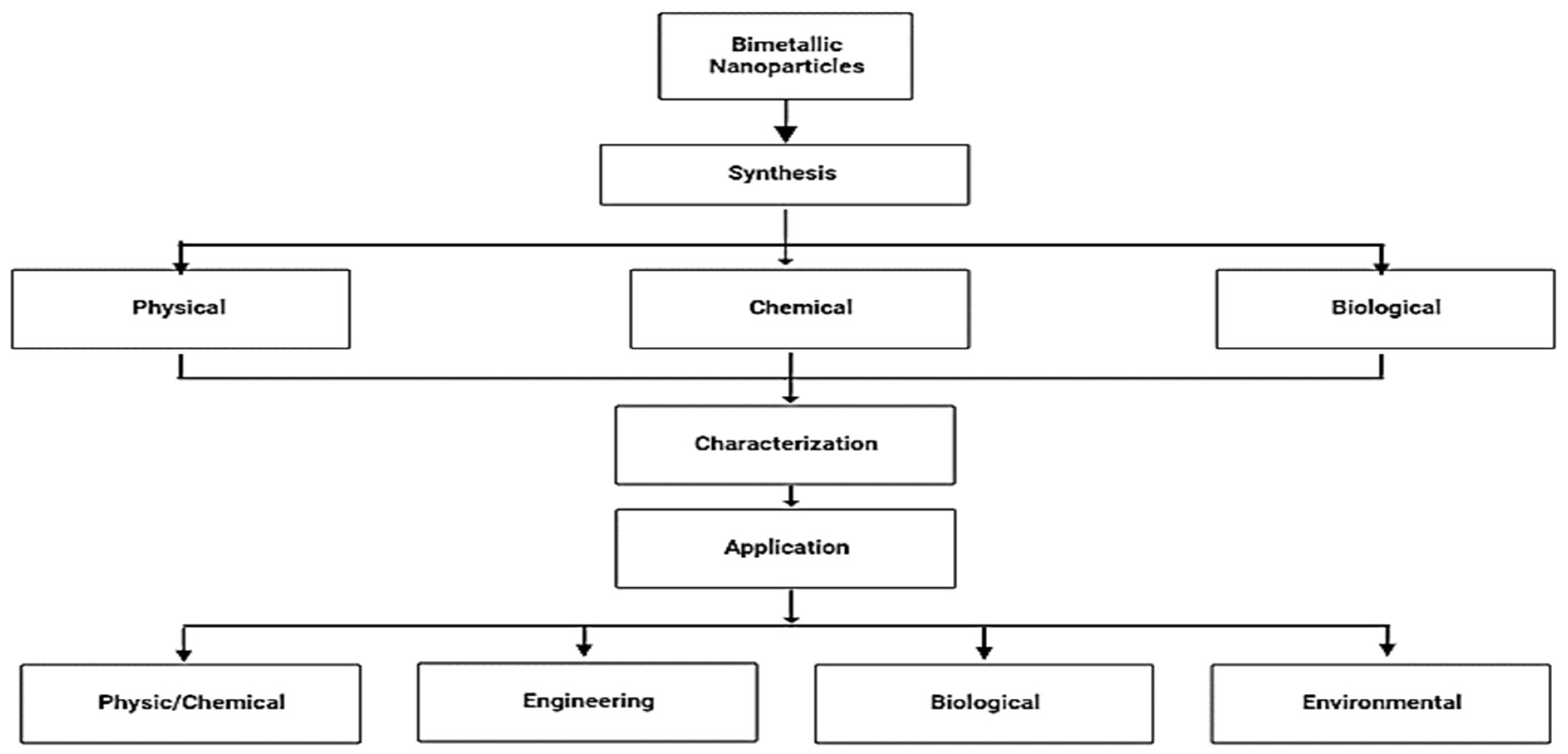
Figure 2.
Top-down and bottom-up approach.

Figure 3.
Methods of synthesizing bimetallic nanoparticles.

Figure 4.
Various applications of bimetallic nanoparticles.


Table 1.
Summary of the different combinations of bimetallic nanoparticles and the method of synthesis.
Table 1.
Summary of the different combinations of bimetallic nanoparticles and the method of synthesis.
| Forms of Bimetallic Nanoparticles | Type of Bimetallic Nanoparticle | Structure of the Bimetallic Nanoparticle | Method of Synthesis | References |
|---|---|---|---|---|
| Gold based | Au-Pt | Cubic crystal structure | Green synthesis | [29] |
| Au-Cu | Porus structure | Galvanic replacement | [30] | |
| Au-Ni | Porus structure | Chemical synthesis | [31] | |
| Au–Pd | Core-shell | Chemical Synthesis | [32] | |
| Au-Ag | Alloy & Core-shell | Green synthesis | [33] | |
| Silver based | Ag-Cu | Alloy | Green synthesis | [34] |
| Ag-Au | Alloy | Green synthesis | [35] | |
| Ag-Fe | Spherical shape | Green synthesis | [36] | |
| Ag-Pd | Cubic crystalline structure | Green synthesis | [37] | |
| Ag-Zn | Wurtzite hexagonal | Green synthesis | [38] | |
| Copper based | Cu-Ni | Alloy | Sol-gel | [39] |
| Cu-Ag | Alloy | Green synthesis | [40] | |
| Cu-Fe | Classified Janus | Electrical explosion | [41] | |
| Cu-Co | Spherical | Inverse micellar encapsulation | [42] | |
| Cu-Pt | Alloy & core | Hydrothermal method | [43] | |
| Nickel based | Ni-Co | Porous | Chemical co-reduction | [44] |
| Ni-Fe | Alloy & core shell | Chemical synthesis | [45] | |
| Ni-Au | - | Physical Synthesis | [46] | |
| Ni-Pt | Alloy | Co-reduction | [47] | |
| Ni-Ru | Alloy | Chemical synthesis | [48] | |
| Iron based | Fe-Zn | Spherical | Green synthesis | [49] |
| Fe-Ti | Nano-Sphere | Chemical synthesis | [50] | |
| Fe-Mn | Spheroid-like | Chemical synthesis | [51] | |
| Fe-Ag | Janus structure | Physical Synthesis | [52] | |
| Fe-Cu | Crystalline | Green synthesis | [53] | |
| Platinum based | Pt-Ag | face-centred cubic structure | Chemical synthesis | [54] |
| Pt-Co | face-centred cubic structure | Chemical reduction | [55] | |
| Pt-Ni | face-centred cubic structure | Chemical reduction | [56] | |
| Pt-Pd | Carbon sphere | Chemical reduction | [57] | |
| Pt-Pd | Crystalline | Green synthesis | [58] | |
| Pt-Fe | Face-cantered cubic | Chemical synthesis | [59] | |
| Palladium based | Pd-Au | Hexagonal nanoplate | DNA ligand | [60] |
| Pd-Ag | Spherical | Green synthesis | [61] | |
| Pd-Cu | Crystalline alloy | Green synthesis | [62] | |
| Pd-Zn | Spherical | Chemical synthesis | [63] |
Table 2.
Different methods of characterization of bimetallic nanoparticles.
| Properties | Features | Techniques | Reference |
|---|---|---|---|
| Morphology | shape, size, structure | DLS, TEM/SEM, STM, XPS, AFM | [198,199,200,201,202,203,204] |
| Topology | Chemical bonding/composition, physical properties, crystallographic structures | XRD, TEM, zeta potential, Brunauer Emmett Teller (BET) | [204,205,206] |
| Chemical properties | Surface energy, chemical potential, oxidation process, catalysis, functionalization of nanostructures | UV-vis, EDXS, FTIR | [207,208] |
| Electrical properties | Conductivity, resistivity, dielectric permittivity | AC/DC conductivity, electrokinetics (zeta potential/cyclic volumetry studies) | [209,210,211] |
| Optical properties | Absorption, reflectance, luminescence, phosphorescence | Microscopy, FTIR, UV-vis, DPCS, Raman spectroscopy, surface plasmon resonance. | [212,213] |
| Mechanical properties | Hardness, plasticity, young modulus | AFM, thermomechanical analysis | [214,215,216] |
| Magnetic properties | Magnetism | VSM, SQUID, MFM | [217,218,219] |
| Thermal properties | Heat flux, change of temperature, volatiles | DSC, EGA, TGA, DTA | [220,221] |
| Biological properties | Anti-microbial, biodegradability, toxicity | In vitro cell viability, in vivo microbial colony viability | [222,223] |
Table 3.
Biological applications of bimetallic nanoparticles.
| Application | Bimetallic Nanoparticle | Reference |
|---|---|---|
| Antimicrobial agents | Pd–Pt | [21] |
| Ag–Fe | [28] | |
| CuO–NiO | [232] | |
| Ag–Au | [23,169,174,233] | |
| Cu–Ag | [176] | |
| Ag–Cu | [234] | |
| Au–Pt | [235] | |
| Cu–Zn | [236,237] | |
| Cu–Ni | [237] | |
| Antioxidant | CuO/NiO | [232] |
| Ag–Cu | [228] | |
| Au–Ag | [233] | |
| Mn–Cu | [238] | |
| Pt–Pd | [239] | |
| Ag–Au | [240] | |
| Antidiabetic | Au–Ag | [241] |
| Ag–Au | [242] | |
| Ag/ZnOVI | [243] | |
| Ag–ZnO | [244] | |
| Anti-Alzheimer | Ag–Au | [242] |
| Anti-inflammatory | Ag/ZnOVI | [243] |
| Zn–Fe2O4, | [245] | |
| CuFeO2, | [246] | |
| Au@Ag | [247] | |
| Drug delivery | Au@Pd | [248] |
| Au–Pt | [249] | |
| Pd–Pt | [250] |
Table 4.
Bimetallic application in physics and chemistry.
| Types of BMNP | Light Source | Compound/Dosage | Degradation%/Activation Energy/Conversion Rate | Reference |
|---|---|---|---|---|
| Mn–Cu | visible light | 240 µg/mL of methyl red | 78.54 ± 0.16% degradation | [238] |
| 240 µg/mL of eriochrome black | 87.67 ± 0.06% degradation | |||
| 240 µg/mL of methyl orange decayed | 69.79 ± 0.36% degradation | |||
| Ag–Pt | -- | -- | 25.61 kJ/mol activation energy | [293] |
| Pd–Pt | -- | -- | 13.93 kJ/mol activation energy | [21] |
| Pd–Ag | -- | NaBH4 | 27.01 kJ/mol activation energy | [61] |
| Pt–Cu | Sun light | Toluene conversion | 55% conversion rate | [294] |
| Pd–Au | -- | HDO of vanillin at room temp. | 99% conversion rate | [295] |
| Au–Pd | -- | Hydrodechlorination of aryl chlorides | 95% conversion rate | [296] |
| Ag–Pd | -- | Alcohol oxidation | up to 97% yield | [297] |
| Au–Pd, | -- | Oxidation of glycerol | 42.9% yield | [298] |
| Au–Pt | -- | Oxidation of glycerol | 29.4% yield | [298] |
| Au–Pd | -- | H2O2 production | 99% yield | [299] |
| Au/Pd–TiO2 | -- | Alcohols oxidation to aldehydes | 83.3% conversion rate | [300] |
Disclaimer/Publisher’s Note: The statements, opinions and data contained in all publications are solely those of the individual author(s) and contributor(s) and not of MDPI and/or the editor(s). MDPI and/or the editor(s) disclaim responsibility for any injury to people or property resulting from any ideas, methods, instructions or products referred to in the content. |
© 2023 by the authors. Licensee MDPI, Basel, Switzerland. This article is an open access article distributed under the terms and conditions of the Creative Commons Attribution (CC BY) license (https://creativecommons.org/licenses/by/4.0/).
Share and Cite
MDPI and ACS Style
Idris, D.S.; Roy, A. Synthesis of Bimetallic Nanoparticles and Applications—An Updated Review. Crystals 2023, 13, 637. https://doi.org/10.3390/cryst13040637
AMA Style
Idris DS, Roy A. Synthesis of Bimetallic Nanoparticles and Applications—An Updated Review. Crystals. 2023; 13(4):637. https://doi.org/10.3390/cryst13040637
Chicago/Turabian StyleIdris, Dahir Sagir, and Arpita Roy. 2023. "Synthesis of Bimetallic Nanoparticles and Applications—An Updated Review" Crystals 13, no. 4: 637. https://doi.org/10.3390/cryst13040637
Note that from the first issue of 2016, this journal uses article numbers instead of page numbers. See further details here.