Short Note

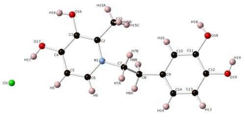
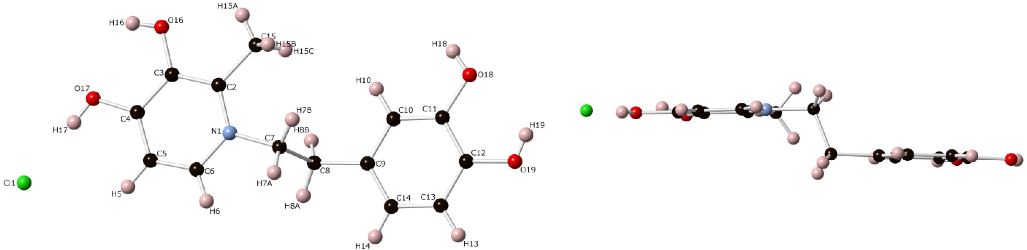
The Synthesis and Molecular Structure of 1-(3,4-Dihydroxyphenethyl)-3-hydroxy-2-methylpyridin-4(1H)-one Hydrochloride Methanol Solvate

1
Department of Chemistry and Biochemistry, Mount Allison University, Sackville, NB E4L 1G8, Canada
2
Department of Chemistry, University of New Brunswick, Fredericton, NB E3B 5A3, Canada
*
Author to whom correspondence should be addressed.
Crystals 2013, 3(2), 333-338; https://doi.org/10.3390/cryst3020333
Submission received: 14 January 2013 / Revised: 6 March 2013 / Accepted: 13 May 2013 / Published: 17 May 2013

Abstract

:
A 3-hydro-4-pyridinone compound derived from maltol and dopamine has been prepared using a microwave reactor. The molecular structure of the protonated product was confirmed by single crystal X-ray diffraction. Crystals were obtained from a saturated solution of methanol and belong to the triclinic space group P-1 with unit cell parameters a = 8.3801(11) Å; b = 9.2583(12) Å; c = 11.5671(15) Å; α = 73.566(2)°; β = 84.514(2)°; γ = 66.578(2)°. The asymmetric unit contains two molecules.

Graphical Abstract

1. Introduction

Hydroxypyridinones are an important class of heterocyclic compounds that are being examined as possible agents for sequestering and removing toxic metal ions [1,2,3]. For instance, Deferiprone (Figure 1b) belongs to a class of 3-hydroxy-4-pyridinone compounds derived from maltol (Figure 1a), a natural flavor enhancer, which is being investigated for its use as a chelating agent for the treatment of iron overload, and related disorders [1]. Although iron is an essential element for all living organisms, an excess of this metal can be extremely toxic and can lead to cancer, Parkinson’s disease or Alzheimer’s disease [1]. As such, there is pressing and recognized need to develop new non-toxic iron chelators for the treatment of these disorders. Although a plethora of N-substituted 3-hydroxy-4-pyridinones are known, the corresponding compound derived from dopamine, a monoamine neurotransmitter, has not yet been reported. As catechol groups are well known to bind to iron [4], we were interested in seeing if the catechol group of the dopamine molecule could also be used to chelate to metals (Figure 1c). In this first study, we report the synthesis and molecular structure of 1-(3,4-dihydroxyphenethyl)-3-hydroxy-2-methylpyridin-4(1H)-one (1).
Figure 1. The structures of (a) maltol; (b) Deferiprone; and (c) the pyridinone derived from dopamine (1) showing the possible binding sites to metals.
Crystals 03 00333 g001

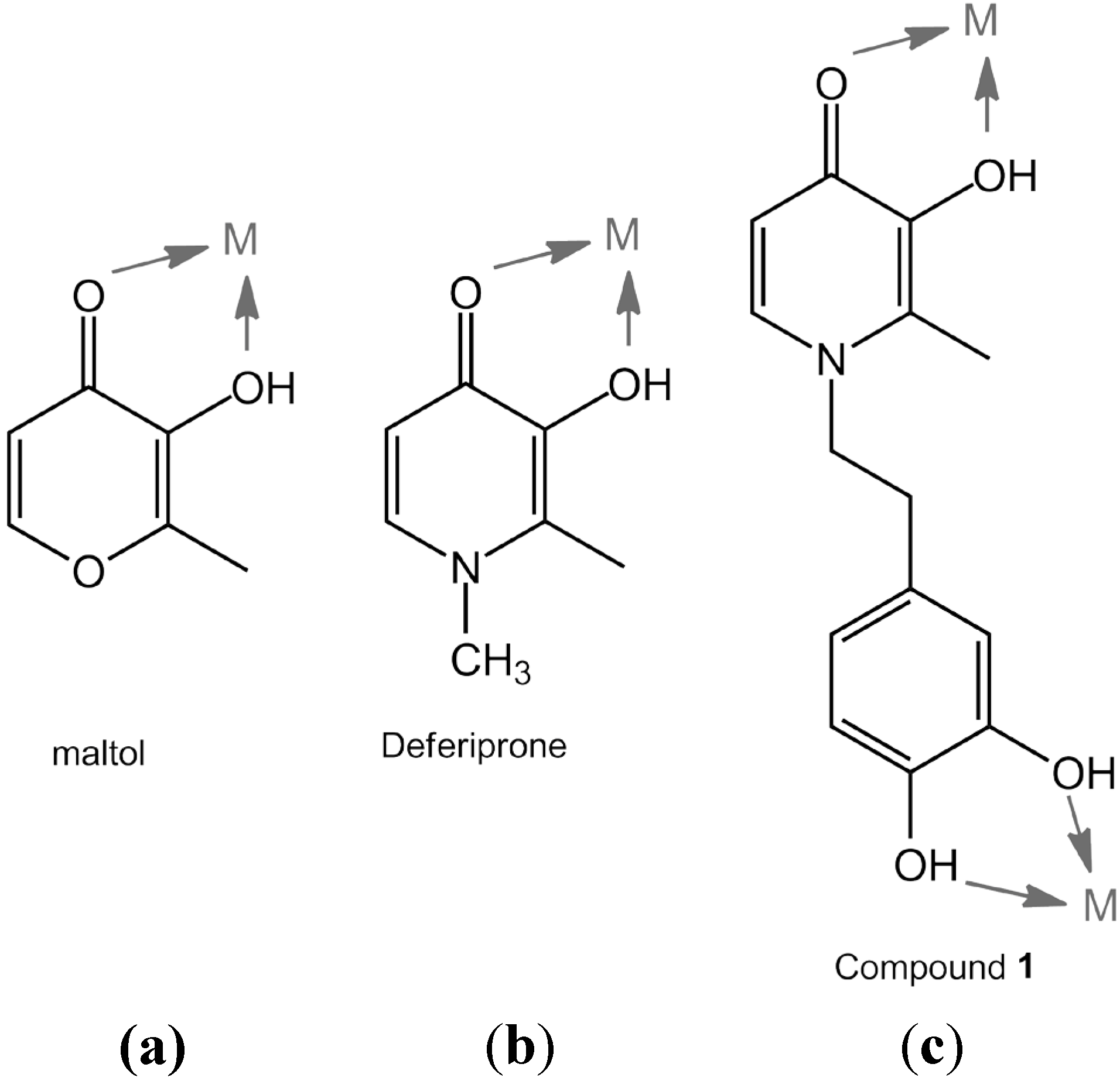
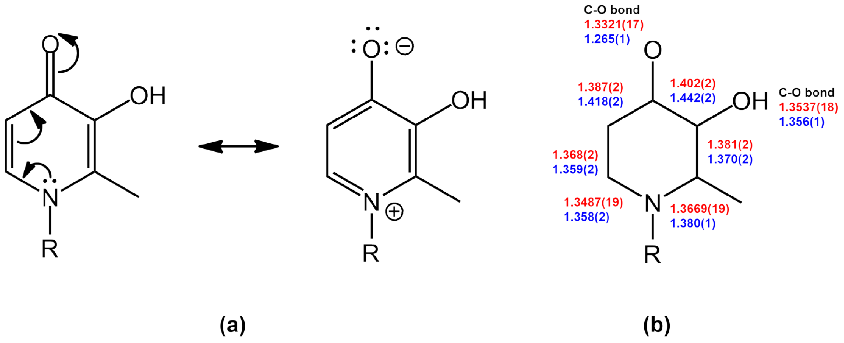
2. Results and Discussion

The most common method of generating 3-hydroxy-4-pyridinones is via aminolysis of maltol, albeit in some cases, protection of the hydroxypyrone is required to facilitate this reaction. Unfortunately, all attempts to generate 1-(3,4-dihydroxyphenethyl)-3-hydroxy-2-methylpyridin-4(1H)-one (1) from dopamine-hydrochloride and maltol under thermal conditions (water, ethanol, methanol, room temperature, reflux, acid-catalyzed, base-catalyzed, etc.) proved unsuccessful and resulted in extensive decomposition of the dopamine salt. Protection of the alcohol group prior to addition of amine also had negligible effect on product yield. However, we were able to complete the synthesis of the title compound using a microwave reactor at 150 °C for 18 h in moderate isolated yields of up to 37%. The compound has been characterized by a number of physical methods, including 1H and 13C NMR spectroscopy. The two vinylic protons on the pyridinone ring appear as doublets with JHH = 7.2 Hz at δ 7.41 and 6.04 ppm. All solution spectroscopic data are consistent with previously prepared hydroxypyridinone derivatives that have a localized alkene within the ring and elemental analysis confirms the majority of the product is the neutral species [1,2,3]. However, we were able to obtain one single crystal of the hydrochloride salt for an X-ray diffraction study, the molecular structure of which is shown in Figure 2. This structure represents, to the best of our knowledge, the first example of a hydroxypyridinone hydrochloride salt.
Figure 2. Two views of the molecular structure of the title complex with a molecule of methanol omitted for clarity.
Crystals 03 00333 g002
Although molecular structures of hydroxypyridinones are quite common [5,6], the protonated form of the title complex shows a significant contribution from the second zwitterionic resonance form for the pyridinone ring, at least in the solid state (Figure 3a). Bond distances within the ring are similar and range from 1.3487(19) to 1.402(2) Å. Most notable is the short N(1)-C(6) distance of 1.3487(19) Å. The carbon-oxygen bond of the protonated carbonyl group is somewhat elongated at 1.3321(17) Å, compared to that found in one of the examples of 1-ethyl-3-hydroxy-2-methylpyridin-4-one which has a true C=O double bond of 1.265(1) [5]. Bond distances within the catechol ring are consistent with previous structures and the hydroxyl groups have a C-O single bond distance of 1.3696(18) and 1.3709(17) Å [7]. Hydrogen atoms were found in Fourier difference maps and refined using isotropic displacement parameters and it appears that the double bond of 1 is protonated, which would be consistent with the resonance structure where the nitrogen takes on a positive charge. Indeed, the planarity of both rings in 1 is shown in the second diagram of Figure 2.
Figure 3. (a) Resonance forms of 3-hydroxypyridinones; (b) Bond distances for the title complex (red, above) and for 1-ethyl-3-hydroxy-2-methylpyridin-4-one (blue, below) [5].
Crystals 03 00333 g003

3. Experimental Section

3.1. General

Reagents and solvents were purchased from Aldrich Chemicals and used as received. NMR spectra were recorded on a Varian Mercury 200 Plus FT NMR spectrometer. Chemical shifts (δ) are reported in ppm (relative to residual solvent peaks) and coupling constants (J) in Hz. Multiplicities are reported as singlet (s), doublet (d), triplet (t), and broad (br). IR spectra were obtained with a Nicolet iS5 FT-IR spectrometer in ATR mode. Melting points were determined using a Mel-Temp apparatus and are uncorrected. Microwave reactions were performed using a CEM Discover SP system in standard closed vessels with the reaction temperature monitored by the internal IR pyrometer.

3.2. Synthesis of 1-(3,4-Dihydroxyphenethyl)-3-hydroxy-2-methylpyridin-4(1H)-one (1)

To a pale orange H2O (5 mL) solution of dopamine hydrochloride (3.00 g, 15.82 mmol) and LiOH (381 mg, 15.91 mmol) was added a MeOH (5 mL) suspension of maltol (1.00 g, 7.93 mmol). The reaction mixture was heated in a CEM microwave reactor at 150 °C for 18 h. Removal of solvent under vacuum afforded a dark oily solid which was dissolved in hot EtOAc (25 mL) and the solution stored at 5 °C. The resulting precipitate was collected by suction filtration and washed with cold EtOAc (5 mL) and Et2O (3 × 10 mL) to afford the title complex as a beige solid (776 mg, 37%). m.p. 237–240 °C. 1H NMR (200 MHz, DMSO-d6): δ 7.41 (d, J = 7.2 Hz, 1H), 6.61 (d, J = 7.8 Hz, 1H), 6.55 (d, J = 1.8 Hz, 1H), 6.40 (br s, 3H), 6.37 (dd, J = 7.8, 1.8 Hz, 1H), 6.04 (d, J = 7.2 Hz, 1H), 4.02 (t, J = 7.4 Hz, 2H), 2.73 (t, J = 7.4 Hz, 2H), 2.21 (s, 3H) ppm. 13C{1H} NMR (50 MHz, DMSO-d6): δ 169.4, 146.0, 145.9, 144.7, 138.2, 129.8, 128.7, 120.3, 117.1, 116.3, 111.4, 55.0, 36.5, 12.0 ppm. FT-IR (neat, cm-1): 3475 (br), 3071 (br), 1627 (m), 1549 (m), 1500 (s), 1456 (m), 1372 (s), 1347 (s), 1236 (s), 1203 (s), 1119 (m), 1062 (m), 1012 (m), 842 (m), 685 (m).

3.3. Crystal Structure Determination

Crystals were grown from a saturated MeOH solution stored at 5 °C. Single crystals were coated with Paratone-N oil, mounted using a polyimide MicroMount and frozen in the cold nitrogen stream of the goniometer. A hemisphere of data was collected on a Bruker AXS P4/SMART 1000 diffractometer using ω and θ scans with a scan width of 0.3 ° and 10 s exposure times. The detector distance was 5 cm. The data were reduced (SAINT) [8] and corrected for absorption (SADABS) [9]. The structure was solved by direct methods and refined by full-matrix least squares on F2(SHELXTL) [10]. All non-hydrogen atoms were refined using anisotropic displacement parameters. Hydrogen atoms were found in Fourier difference maps and refined using isotropic displacement parameters.
C15H20ClNO5, Fw = 329.77, crystal size 0.70 × 0.30 × 0.08 mm3, triclinic, space group Pī, a = 8.3801(11) Å, b = 9.2583(12) Å, c = 11.5671(15) Å, α = 73.566(2)°, β = 84.514(2)°, γ = 66.578(2)°, V = 789.70(18) ų, Z = 2, ρcalc = 1.387 mg/cm3, µ= 0.265 mm-1, F(000) = 348, T 173(1) K, MoKα radiation, 5486 reflections collected, 3429 independent collections [R(int) = 0.0147], GoF 1.078, 1.84 < Θ < 27.48°, R1 [I > 2sigma(I)] = 0.0348, wR2 (all data) = 0.1036.
Crystallographic data for the structure reported in this work have been deposited with the Cambridge Crystallographic Data Centre as supplementary publication [11]. Copies of the data can be obtained free of charge on application to The Director, CCDC, 12 Union Road, Cambridge DB2 1EZ, UK.

4. Conclusions

Although numerous attempts to generate 1-(3,4-dihydroxyphenethyl)-3-hydroxy-2-methylpyridin-4(1H)-one using standard thermal aminolysis techniques proved unsuccessful, we were able to generate this ligand derived from the neurotransmitter dopamine using a microwave reactor. Yields are moderate but the ease of isolation makes this an attractive route to synthesize this compound. The molecular structure of the hydrochloride adduct has been confirmed by a single crystal X-ray diffraction study and a considerable contribution from the zwitterionic resonance form is observed in the solid state. We are in the process of examining the binding potentials of 1 with a wide range of metals and will report our findings in due course.

Acknowledgments

Thanks are gratefully extended to the American Chemical Society, Petroleum Research Foundation (Grant # 50093-UR3), Mount Allison University, and the University of New Brunswick for financial support. We also thank Magnedan Durant for his expert technical assistance and anonymous reviewers for the very helpful suggestions.

Conflict of Interest

The authors declare no conflict of interest.

References and Notes

  1. Santos, M.A.; Marques, S.M.; Chaves, S. Hydroxypyridinones as “privileged” chelating structures for the design of medicinal drugs. Coord. Chem. Rev. 2012, 256, 240–259. [Google Scholar] [CrossRef]
  2. Thompson, K.H.; Barta, C.A.; Orvig, C. Metal complexes of maltol and close analogues in medicinal inorganic chemistry. Chem. Soc. Rev. 2006, 35, 545–556. [Google Scholar]
  3. Datta, A.; Raymond, K.N. Gd-hydroxypyridinone (HOPO)-high relaxivity magnetic resonance imagining (MRI) contrast agents. Acc. Chem. Res. 2009, 42, 938–947. [Google Scholar] [CrossRef]
  4. Philpot, C. Bioinorganic chemistry: getting a grip on iron. Nat. Chem. Biol. 2010, 6, 568–570. [Google Scholar] [CrossRef]
  5. Xiao, G.; van der Helm, D.; Hider, R.C.; Dobbin, P.S. Structure-stability relationships of 3-hydroxypyridin-4-one complexes. J. Chem. Soc. Dalton Trans. 1992, 22, 3265–3271. [Google Scholar]
  6. Nelson, W.O.; Rettig, S.J.; Orvig, C. Aluminum and gallium complexes of 1-ethyl-3-hydroxy-2-methyl-4-pyridinone: a new exoclathrate matrix. Inorg. Chem. 1989, 28, 3153–3157. [Google Scholar] [CrossRef]
  7. Giesecke, J. The structure of catecholamines. V. The crystal and molecular structure of epinine hydrobromide. Acta Cryst. 1976, B32, 2337–2340. [Google Scholar]
  8. SAINT 7.23A; Bruker AXS, Inc.: Madison, WI, USA, 2006.
  9. SADABS 2008; Bruker AXS, Inc.: Madison, WI, USA, 2008.
  10. Sheldrick, G.M. A short history of SHELX. Acta Cryst. 2008, A64, 112–122. [Google Scholar]
  11. CCDC (Cambridge Crystallographic Data Centre), SW101014.cif; No. CCDC 919059; CCDC: Cambridge, UK, 2013.

