Article

Synthesis, Crystal Structure, DFT Study of m-Methoxy-N′-(3-Methoxybenzoyl)-N-Phenylbenzohydrazide

1
Department of Chemistry, Quaid-I-Azam University, Islamabad 45320, Pakistan
2
School of Pharmacy, Hajvery University, 43-52 Industrial Area, Gulberg-III, Lahore 54000, Pakistan
3
Bio-21 Institute, School of Chemistry, University of Melbourne, Parkville-3052, Australia
4
Department of Organic Chemistry and CIBER-BBN, Networking Centre on Bioengineering, Biomaterials and Nanomedicine, Barcelona Science Park, University of Barcelona, Barcelona 08028, Spain
5
School of Chemistry and Physics, University of KwaZulu-Natal, Durban 4001, South Africa
*
Authors to whom correspondence should be addressed.
Crystals 2017, 7(1), 19; https://doi.org/10.3390/cryst7010019
Submission received: 19 August 2016 / Revised: 4 January 2017 / Accepted: 5 January 2017 / Published: 12 January 2017

Abstract

:
The crystal structure of m-methoxy-N′-(m-anisoyl)-N-phenylbenzohydrazide has been determined by means of single-crystal X-ray diffraction. The title compound crystallizes in the monoclinic space group P 21/c with unit cell parameters: a = 8.7338(1), b = 24.5602(3), c = 9.6929(1) Å, β = 113.186(2)°, V = 1911.23(4) Å3, Z = 4. The dihedral angles between the mean plane of the central benzene ring and two terminal aromatic rings are 72.44(4)° and 89.90(4)°, respectively. The two methoxyphenyl rings are orthogonal with a dihedral angle of 89.74(4)°. The crystal packing is stabilized by a combination of N–HO intermolecular hydrogen bonding and weak intermolecular C–HO interactions. The X-ray structure was compared with the optimized counterpart calculated by the B3LYP/6-311G basis set and the results showed that the optimized geometry can reproduce the crystal structure parameters well.

Graphical Abstract

1. Introduction

Insecticides are one of the major factors behind the increase in agricultural productivity in the 20th century. The insecticidal activity of diacylhydrazine derivatives has been extensively studied by many researchers; in addition, diacylhydrazines are very useful synthons for the synthesis of a variety of bioactive molecules [1,2,3,4,5].
The diacylhydrazine derivatives are a promising class of insect control agents that was serendipitously discovered at Rohm and Haas Company. The diacylhydrazine insecticide family shows efficacy against lepidopteran larvae in the field and laboratory [6]. Tebufenozide and its methoxylated derivative methoxyfenozide present intrepid activity as agonists or mimic the insect molting hormone 20-hydroxyecdynose by inducing premature, incomplete ecdysis, resulting in the death of the exposed insects [7,8,9,10,11]. Halofenozide and chromafenozide are novel members of the diacylhydrazine insecticides belonging to the class of non-steroidal ecdysone agonists that are developed as insecticides against Lepidoptera [12,13]. Tebufenozide and its analogues methoxyfenozide, halofenozide, and chromafenozide have been used extensively in crops such as apple, grape, and cruciferous vegetables in order to obtain good yields in different countries. So, it becomes important work to characterize and fully understand the properties of these insecticides in crops to ensure food safety.
We herein report an efficient one-pot synthesis and the crystal structure of m-methoxy phenyl-N′-(m-anisoyl)-N-phenylbenzohydrazide using inexpensive reagents from acid chlorides in dichloromethane as a solvent in a short time with a high yield. Also, optimized structural parameters were investigated using ab initio density functional theory (DFT) calculations and have been compared with the experimental X-ray structure.

2. Results and Discussion

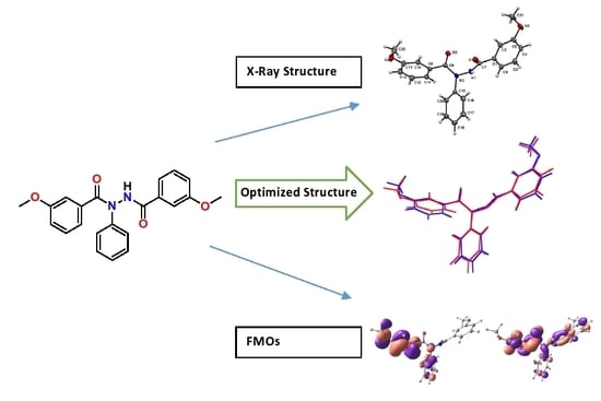
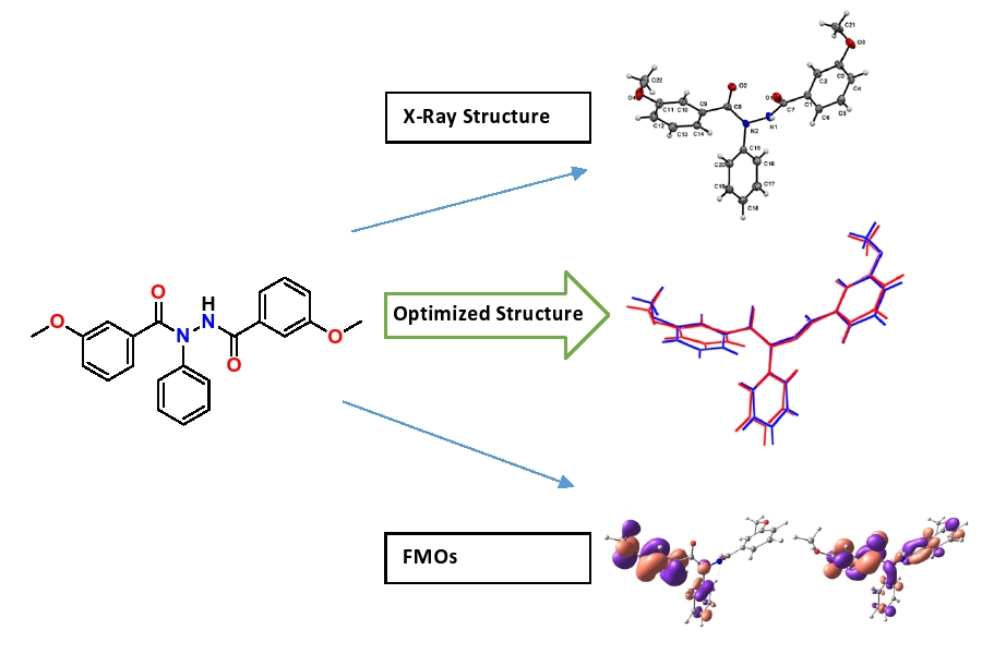
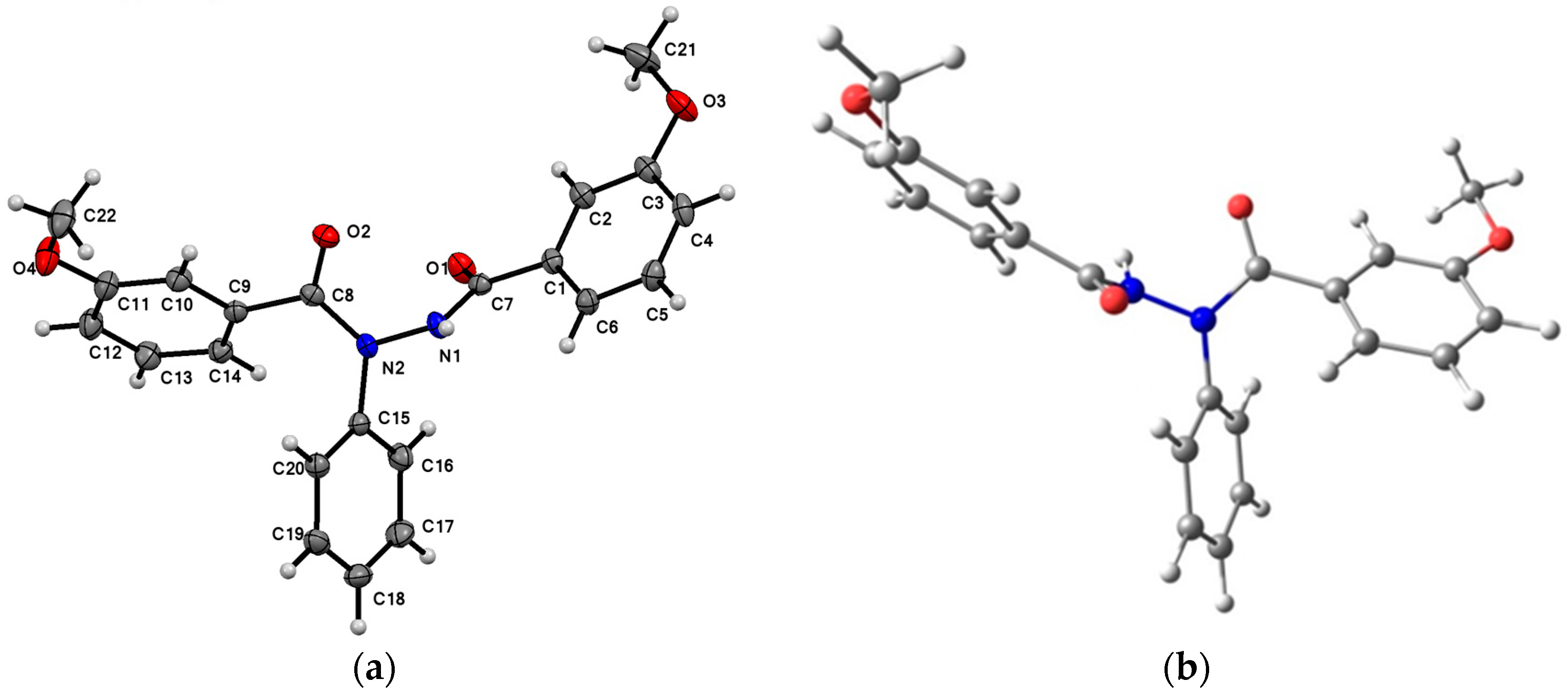
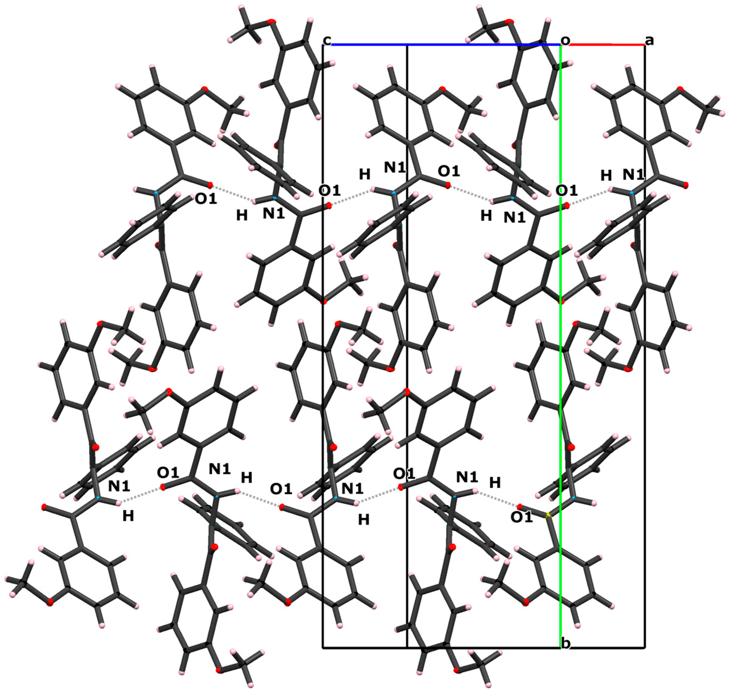
2.1. Crystal Structure

The molecular structure of the title compound, showing the atomic numbering, as determined in the crystalline phase is shown in Figure 1. Compound 1 is closely related to that of 1,2-dibenzoyl-1-phenylhydrazine [14] with two phenyl groups replaced by two m-anisoyl groups. The title compound crystallizes in the monoclinic space group P 21/c, the molecule consisting of two m-anisoyl substituents attached to the central N-phenyl hydrazine moiety. The mean plane of the central benzene ring, defined by atoms C15–C20, forms dihedral angles of 72.44(4)° and 89.90(4)° with mean planes of the m-anisoyl rings C1–C6 and C9–C14, respectively. The two m-anisoyl groups are orthogonal with regard to one another with a dihedral angle of 89.74(4)°. The conformation of the bis-hydrazide moiety as defined by the following dihedral angles, O1–C7–N1–N2 (−4.66(8)°), C7–N1–N2–C8 (−85.85(11)°) and O2–C8–N2–N1 (0.10(14)°), differs slightly with that observed in 1,2-dibenzoyl-1-phenylhydrazine for which the corresponding dihedral angles are −28.1°, −65.4° and 0.68°, respectively, except for O1–C7–N1–N2 which differ significantly with the difference of 23.44°. This difference may arise due to the differing orientation of the N-phenyl substituent in the two structures; for compound 1, the C16–C15–N2–N1 dihedral angle, which is −65.02(12)°, shows that the N–N bond is substantially out of the plane of the N-phenyl substituent, whereas for 1,2-dibenzoyl-1-phenylhydrazine, the corresponding dihedral angle is −13.2° and the N–N bond is close to coplanar with the N-phenyl substituent.
A view of the crystal packing in the unit cell is shown in Figure 2. The crystal packing of compound 1 is stabilized by several non-bonded interactions. The molecules are associated by intermolecular N–HO1 (x, 0.5 − y, 0.5 + z) hydrogen bonding interactions [15,16] in addition to weak C–HO interactions between C21–H21AO2. The details of the interactions are given in Table 1.

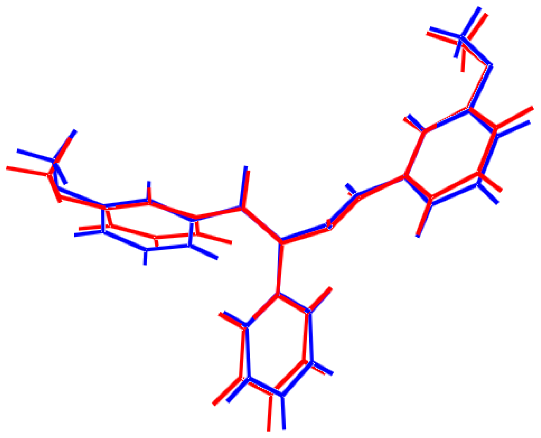
2.2. Optimized Geometry

The optimized structure of 1 shown in Figure 1b was obtained by the density functional theory calculations (DFT) using hybrid functional Becke’s three parameter nonlocal exchange functional with the Lee-Yang-Parr correlation function (B3LYP) using the 6-311G basis set. The optimized structure is closed to the molecular crystal structure. The optimized parameters, namely bond lengths, bond angles and torsion angles, given in Table 2 are in accordance with the atomic numbering scheme given in Figure 1a. The optimized structure yields the identical bond lengths for C–C bonds. The C–C bond length in the benzene ring is in the range of 1.397–1.381 Å and 1.407–1.391 Å for the experimental X-ray structure and at the B3LYP/6-311G level, respectively, which is much shorter than the characteristic C-C single bond (1.54 Å) and longer than the C=C bond (1.34 Å) [17]. For C7–O1 and C8–O2 the calculated C=O (carbonyl) bond lengths are 1.249 and 1.253 Å, respectively, which are larger than the experimental value of 1.221 Å. The N1–C7, N2–C8 and N2–C15 bond lengths are calculated as 1.386, 1.398 and 1.445 Å at the B3LYP/6-311G level and the experimental values are 1.347, 1.375 and 1.437 Å, respectively; it is noted that the N1–C7 and N2–C8 bond lengths are much shorter than the normal C–N bond length (1.47 Å) while N2–C15 is closer to that and the N1–C7 and N2–C8 bond lengths are closer to the C=N bond (1.33 Å), which results from the conjugation effect of these atoms [18].
The bond angles calculated are in good agreement with the experimental data with a maximum deviation of 2.72° for C11–O4–C22, which is calculated as 119.64° and the experimental value is 116.92°. The largest deviation between bond lengths is 0.039 Å. These deviations are because the calculations belong to the gaseous phase and the experimental results belong to the solid phase. In the solid state, the crystal field along with the intermolecular interactions has linked the molecules together, which results in the difference in bond parameters between the experimental and estimated values. When the optimized structure of the title compound is compared with its optimized counterpart (see Figure 3), minor conformational discrepancies are observed between them. A logical method for globally comparing the structures obtained with the theoretical calculation is superimposing the molecular skeleton with that obtained from the X-ray diffraction.

2.3. Frontier Molecular Orbitals

The most significant orbitals in molecules are the frontier molecular orbitals, called the Highest Occupied Molecular Orbital (HOMO) and the Lowest Unoccupied Molecular Orbital (LUMO). The HOMO represents the capability to donate an electron, and LUMO, as an electron acceptor, represents a capability to obtain an electron [19]. Figure 4 shows the distribution and energy levels of HOMO and LUMO orbitals computed at the B3LYP/6-311G level for the title compound.
As can be seen from Figure 4, LUMO is mainly localized on the benzene ring along with carbonyl groups and nitrogen atoms, and partially on the phenyl group. HOMO is mainly on one anisoyl group and the phenyl ring with the nitrogen atom. The value of the energy separation between HOMO and LUMO is 0.1769 eV. This small HOMO-LUMO energy gap could indicate that the title structure is chemically active, because it can be excited just by small energies.

3. Experimental

3.1. Synthesis m-Methoxy-N′-(3-methoxybenzoyl)-N-phenylbenzohydrazide

To a magnetically stirred solution of phenyl hydrazine (0.3 mL, 3 mmol) a solution of m-anisoyl chloride (0.85 mL, 6 mmol) in 10 mL dichloromethane was added dropwise. The resulting mixture was heated under reflux for 1 h (Scheme 1). The resulting solution was allowed to cool to room temperature. After cooling, the solution was allowed to evaporate slowly at ambient temperature. After two days colorless rods suitable for X-ray analysis were collected, and washed with a small amount of methanol. Yield: 96%. m.p.: 103–105 °C. IR (ATR): ʋ = 3286 (N–H), 1695, 1636 (C=O) cm−1; 1H NMR (300 MHz, CDCl3): δ 7.61 (s, 1H, N–H), 6.73–7.18 (m, 13H, Ph–H), 3.92 (s, 3H, OCH3), 3.86 (s, 3H, OCH3); 13C NMR (75 MHz, CDCl3): δ 166.47, 162.86, 159.55, 138.91, 136.02, 131.13, 130.98, 129.46, 124.2, 121.8, 117.92, 117.48, 114.1, 113.96, 57.54, 57.32.

3.2. Computation

Calculation of the structural parameters of the titled compound was carried out using ORCA 3.0.1 [20] program package by Gabedit [21] graphical interface using B3LYP using 6-311G basis set [22]. Geometry obtained from X-ray diffraction was used as starting point to perform all the calculations.

3.3. X-ray Crystallography

Crystallographic data is presented in Table 3. The crystal of compound 1 having dimensions of 0.59 × 0.20 × 0.06 mm was selected and the reflection data were collected on an Oxford SuperNova CCD diffractometer using Cu-Kα (λ = 1.5418 Å) X-radiation at 130 K. The structure was solved by direct methods and refined by full-matrix least squares using SHELX-97 [23].
A CIF file containing complete information on the studied structure was deposited with CCDC, deposition number 896971 and is freely available upon request from the Director, CCDC, 12 Union Road, Cambridge CB2 1EZ, UK (Fax: +44-1223-336033; e-mail: [email protected] or from the following website: www.ccdc.cam.ac.uk/data_request/cif.

4. Conclusions

In summary, we have synthesized a novel diacyl hydrazine compound 1, m-methoxy-N′-(m-anisoyl)-N-phenylbenzohydrazide, and characterized it using the X-ray diffraction technique. The molecule is stabilized by intermolecular N–HO1 hydrogen bonding interactions in addition to weak C–HO interactions between C21–H21AO2. We investigated the molecule by calculating the optimized geometry, and frontier molecular orbital analysis was also performed. All the theoretical results show good agreement with the corresponding experimental data.

Acknowledgments

Ifzan Arshad acknowledges the financial support of this research by the Higher Education Commission of Pakistan by the Indigenous Fellowship. Fernando Albericio thanks the National Research Foundation (South Africa) for financial support.

Author Contributions

Jonathan M White was responsible for the single-crystal structure determination. Ifzan Arshad and Aamer Saeed were responsible for part of the synthesis. Javeria Yameen and Ifzan Arshad were in charge of the interpretation of data and the writing process of this manuscript. Fernando Albericio supervised the work and writing of the manuscript.

Conflicts of Interest

The authors declare no conflict of interest.

References

  1. Cao, S.; Qian, X.; Song, G. N′-tert-Butyl-N′-aroyl-N-(alkoxycarbonylmethyl)-N-aroylhydrazines, a novel nonsteroidal ecdysone agonist: Syntheses, insecticidal activity, conformational, and crystal structure analysis. Can. J. Chem. 2001, 79, 272–278. [Google Scholar] [CrossRef]
  2. Ke, S.; Qian, X.; Liu, F.; Wang, N.; Fan, F.; Li, Z.; Yang, Q. Diacylhydrazine derivatives as novel potential chitin biosynthesis inhibitors: Design, synthesis, and structure–activity relationship. Eur. J. Med. Chem. 2009, 44, 2985–2993. [Google Scholar] [CrossRef] [PubMed]
  3. Mao, C.H.; Wang, Q.M.; Huang, R.Q.; Bi, F.C.; Chen, L.; Liu, Y.X.; Shang, J. Synthesis and insecticidal evaluation of novel n-oxalyl derivatives of tebufenozide. J. Agric. Food Chem. 2004, 52, 6737–6741. [Google Scholar] [CrossRef] [PubMed]
  4. Zotti, M.J.; Christiaens, O.; Rougé, P.; Grutzmacher, A.; Zimmer, P.; Smagghe, G. Structural changes under low evolutionary constraint may decrease the affinity of dibenzoylhydrazine insecticides for the ecdysone receptor in non-lepidopteran insects. Insect Mol. Biol. 2012, 21, 488–501. [Google Scholar] [CrossRef] [PubMed]
  5. Zhao, P.-L.; Li, J.; Yang, G.-F. Synthesis and insecticidal activity of chromanone and chromone analogues of diacylhydrazines. Bioorg. Med. Chem. 2007, 15, 1888–1895. [Google Scholar] [CrossRef] [PubMed]
  6. Smirle, M.J.; Lowery, D.T.; Zurowski, C.L. Chemical residues and bioactivity of tebufenozide applied to apple foliage. Pest Manag. Sci. 2004, 60, 1137–1142. [Google Scholar] [CrossRef] [PubMed]
  7. Smith, J.P.; Randall, G.E.; Castro, J.R.; Lindberg, R.D. Hypogastric artery infusion and radiation therapy for advanced squamous cell carcinoma of the cervix. Am. J. Roentgenol. Radium Ther. Nucl. Med. 1972, 114, 110–115. [Google Scholar] [PubMed]
  8. Sundaram, M.; Palli, S.; Smagghe, G.; Ishaaya, I.; Feng, Q.-L.; Primavera, M.; Tomkins, W.; Krell, P.; Retnakaran, A. Effect of RH-5992 on adult development in the spruce budworm, Choristoneura fumiferana. Insect Biochem. Mol. Biol. 2002, 32, 225–231. [Google Scholar] [CrossRef]
  9. Clarke-Harris, D.; Fleischer, S.J. Sequential sampling and biorational chemistries for management of lepidopteran pests of vegetable amaranth in the caribbean. J. Econ. Entomol. 2003, 96, 798–804. [Google Scholar] [CrossRef] [PubMed]
  10. Retnakaran, A.; Krell, P.; Feng, Q.; Arif, B. Ecdysone agonists: Mechanism and importance in controlling insect pests of agriculture and forestry. Arch. Insect Biochem. Physiol. 2003, 54, 187–199. [Google Scholar] [CrossRef] [PubMed]
  11. Rodríguez Enríquez, C.-L.; Pineda, S.; Figueroa, J.I.; Schneider, M.-I.; Martínez, A.-M. Toxicity and sublethal effects of methoxyfenozide on spodoptera exigua (lepidoptera: Noctuidae). J. Econ. Entomol. 2010, 103, 662–667. [Google Scholar] [CrossRef] [PubMed]
  12. Boudjelida, H.; Bouaziz, A.; Soin, T.; Smagghe, G.; Soltani, N. Effects of ecdysone agonist halofenozide against Culex pipiens. Pestic. Biochem. Physiol. 2005, 83, 115–123. [Google Scholar] [CrossRef]
  13. Mosallanejad, H.; Soin, T.; Swevers, L.; Iatrou, K.; Nakagawa, Y.; Smagghe, G. Non-steroidal ecdysteroid agonist chromafenozide: Gene induction activity, cell proliferation inhibition and larvicidal activity. Pestic. Biochem. Physiol. 2008, 92, 70–76. [Google Scholar] [CrossRef]
  14. Buzykin, B.; Gubaidullin, A.; Litvinov, I.; Gazetdinova, N.; Sysoeva, L. 1, 2-diacyl-1-arylhydrazines-ii. Molecular and crystalline structure of 1, 2-dibenzoyl-1-phenyl-2-acetylhydrazines and 1-salicyloyl-1-(4-nitrophenyl)-2-acetylhydrazines. Russ. J. Gen. Chem. 1998, 68, 1972–1976. [Google Scholar]
  15. Abbas, A.; Nazir, H.; Naseer, M.M.; Bolte, M.; Hussain, S.; Hafeez, N.; Hasan, A. Synthesis, spectral characterization, self-assembly and biological studies of N-acyl-2-pyrazolines bearing long alkoxy side chains. Spectrochim. Acta Part A 2014, 120, 176–184. [Google Scholar] [CrossRef] [PubMed]
  16. Saeed, A.; Arshad, M.I.; Bolte, M.; Fantoni, A.C.; Espinoza, Z.Y.D.; Erben, M.F. On the roles of close shell interactions in the structure of acyl-substituted hydrazones: An experimental and theoretical approach. Spectrochim. Acta Part A 2016, 157, 138–145. [Google Scholar] [CrossRef] [PubMed]
  17. Lide, D.R. A survey of carbon-carbon bond lengths. Tetrahedron 1962, 17, 125–134. [Google Scholar] [CrossRef]
  18. Allen, F.H.; Kennard, O.; Watson, D.G.; Brammer, L.; Orpen, A.G.; Taylor, R. Tables of bond lengths determined by X-ray and neutron diffraction. Part 1. Bond lengths in organic compounds. J. Chem. Soc. Perkin Trans. 2 1987, S1–S19. [Google Scholar] [CrossRef]
  19. Becker, H. Jan Fleming, Frontier Orbitals and Organic Chemical Reactions., John Wiley u. Sons LTD. J. Praktische Chem. 1978, 320, 879–880. [Google Scholar] [CrossRef]
  20. Neese, F. The orca program system. Wiley Interdiscip. Rev. Comput. Mol. Sci. 2012, 2, 73–78. [Google Scholar] [CrossRef]
  21. Allouche, A.R. Gabedit—A graphical user interface for computational chemistry softwares. J. Comput. Chem. 2011, 32, 174–182. [Google Scholar] [CrossRef] [PubMed]
  22. Hehre, W.J.; Ditchfield, R.; Pople, J.A. Self—Consistent molecular orbital methods. XII. Further extensions of gaussian—type basis sets for use in molecular orbital studies of organic molecules. J. Chem. Phys. 1972, 56, 2257–2261. [Google Scholar] [CrossRef]
  23. Sheldrick, G.M. A short history of shelx. Acta Crystallogr. Sect. A 2007, 64, 112–122. [Google Scholar] [CrossRef] [PubMed]
Figure 1. (a) A view of the crystal structure of the title compound 1 and (b) a view of an optimized structure from DFT calculations.
Crystals 07 00019 g001
Figure 2. Partial crystal packing of 1 viewed along the c-axis with the N–HO hydrogen bonds shown as dashed lines.
Crystals 07 00019 g002
Figure 3. Atom-by-atom superimposition of the structures calculated (blue) by B3LYP/6-311G on the X-ray structure (red) of the title compound.
Crystals 07 00019 g003
Figure 4. HOMO-LUMO surfaces of compound 1.
Crystals 07 00019 g004
Scheme 1. Synthesis of 4-methoxy-N′-(4-methoxybenzoyl)-N-phenylbenzohydrazide (1).
Crystals 07 00019 sch001
Table 1. Selected hydrogen bond parameters of (1) (Å, °).
D–HAD–H (Å)HA (Å)DA (Å)D–HA (°)
N1–HO1 i0.867 (16)1.946 (16)2.7089 (11)146.1 (14)
C21–H21AO20.9602.6923.604(2)159(1)
Symmetry code(s): i x, −y + 1/2, z + 1/2.
Table 2. Experimental and theoretical selected geometric parameters of 1 (Å, °).
Bond Lengths (Å)ExperimentalCalculated
B3LYP/6-311G
Difference
N2–C81.3751.3980.023
N2–N11.3951.4110.016
N2–C151.4371.4450.008
N1–C71.3471.3860.039
O1–C71.2211.2490.028
O2–C81.2211.2530.032
O3–C31.3691.3910.022
O3–C211.4271.4450.018
O4–C111.3641.3920.028
O4–C221.4271.4540.027
C1–C71.4941.492−0.002
C8–C91.4991.489−0.01
Angle (°)ExperimentalCalculatedDifference
C3–O3–C21117.08118.97−1.89
C11–O4–C22116.92118.92-2
C8–N2–N1115.62114.640.98
C8–N2–C15127.8126.421.38
N1–N2–C15112.92116.85−3.93
C7–N1–N2119.55119.83−0.28
O2–C8–N2120.87119.231.64
O2–C8–C9120.59121.47−0.88
N2–C8–C9118.46119.3−0.84
O3–C3–C2124.12124.24−0.12
O3–C3–C4115.69115.430.26
O1–C7–N1122.89122.220.67
O1–C7–C1122.55122.380.17
N1–C7–C1114.56115.39−0.83
O4–C11–C10124.14124.24−0.1
O4–C11–C12116115.490.51
C20–C15–N2120.75119.760.99
Table 3. Crystallographic data of titled compound (1).
Crystal Data
Empirical FormulaC22H20N2O4
Formula weight376.40
Crystal system, space group Monoclinic, P21/c
Temperature (K)130
a, b, c (Å)8.7338(1), 24.5602(3), 9.6929(1)
β (°)113.186 (2)
V (Å3)1911.23 (4)
Z4
Radiation typeCu
μ (mm−1)0.74
Crystal size (mm)0.59 × 0.20 × 0.06
Data collection
DiffractometerSuperNova, Dual, Cu at zero, Atlas diffractometer
Tmin, Tmax0.809, 1.000
No. of measured, independent and observed [I > 2s(I)] reflections 9689, 4000, 3668
Rint0.018
(sin q/l)max (Å−1)0.632
Refinement
R[F2 > 2s(F2)], wR(F2), S0.035, 0.094, 1.04
No. of reflections4000
No. of parameters259
max, Dñmin (e·Å−3)0.24, −0.24

