Article

Growth and Characterization of Ni Nano-Micro Structures in the Presence of Ethylenediamine

Institute of Industrial Catalysis, College of Chemical Engineering, Zhejiang University of Technology, Hangzhou 300014, China
*
Author to whom correspondence should be addressed.
Crystals 2018, 8(11), 397; https://doi.org/10.3390/cryst8110397
Submission received: 14 September 2018 / Revised: 8 October 2018 / Accepted: 16 October 2018 / Published: 23 October 2018

Abstract

:
Ni nano-micro structures have been synthesized via a solution reduction route in the presence of ethylenediamine (EDA) under strong alkaline conditions. The phase composition, morphology, and microstructure of the resulting products are investigated by X-ray diffraction, scanning electron microscopy, and transmission electron microscopy. The presence of EDA plays an important role in the formation of Ni nano-micro structures, and microflowers or microspheres assembled from nanosized horns can be produced by changing the amount of EDA. The size of Ni nano-micro structures is dependent on the NaOH concentration, and long chains assembled from Ni nano-micro structured microspheres can be obtained by reducing the amount of NaOH solution used. The role of both EDA and NaOH in the reduction of Ni (II) to Ni, as well as in the growth of Ni nano-micro structures, has been discussed, and a possible formation mechanism of these Ni nano-micro structures has been proposed based on the experimental results.

1. Introduction

Amines, a class of organic compounds, which contain an amino group with a pair of lone electrons on the nitrogen atom, are widely used for mediating the growth of a variety of nanostructures, including nanocrystals, nanosheets, nanorods, and nanowires [1,2,3,4,5,6,7,8,9,10,11,12,13,14]. As the simplest diamine after hydrazine, for example, ethylenediamine (EDA) can be used as a capping agent in the synthesis of ultrasmall CeO2 particles [5], PbS nanosheets [6], Mn3O4 nano-octahedrons [7], and CdS multipods [8], as a coordination template in the synthesis of CdX (X = S, Se, Te) nanorods [9] and ZnS nanowires [10], and as both capping agent and complexing agent in the growth of Cu nanowires [11,12]. In addition, EDA has also been employed in the synthesis of microsized materials [15,16,17]. For instance, EDA can serve as a morphological template for the growth of α-Bi2O3 microrods [15]. Fe2O3 microspindles have been successfully produced by an ethylenediamine ligand-controlled hydrothermal approach [16]. Although much work has been done on the EDA-mediated growth of nanostructures, there are few efforts on the use of EDA in the synthesis of nano-micro structured materials.
Nano-micro structured materials, a type of material with multi-scale structures that range from nano- to micro-length scales, have shown potential applications in many fields [18], including superhydrophobic surfaces, optics, and electrochemistry [18,19,20,21,22,23,24]. For example, Yue et al. have reported that nano-micro spherical β-Ni(OH)2 shows excellent electrochemical properties [19]. A similar result has also been observed by Zhang et al. [21]. Rajeshkhanna and Rao have found that nano-micro structured Co3O4 possesses a good performance for electro-oxidation of methanol [22]. Geng et al. observed an interesting transition from superhydrophilicity to super-hydrophobicity on a surface with Ni micro-nano cones array [23]. Dudem et al. even found that a nano-micro structured silicon surface exhibited both omnidirectional antireflection and superhydrophilicity [24]. In view of these promising applications, it is important to develop methods for the synthesis of nano-micro structures. In this work, we present a study on the EDA-mediated synthesis of Ni nano-micro structures, namely microflowers or microspheres assembled from nanosized horns, via a solution reduction route in concentrated alkaline solution. Unlike the synthesis of Ni nanostructures or nano-micro nanostructures in an alkaline solution of low concentration in the presence of polyvinyl pyrrolidone, cetyltrimethylammonium bromide, or potassium sodium tartrate [25,26,27,28], the use of EDA in high concentrated alkaline solution is inspired by our previous work on the synthesis of Cu nanowires [11] and hierarchical Cu structures [29]. The present work mainly focuses on two aspects: (i) The effect of both EDA and NaOH on the reduction of Ni(II) to Ni and the morphology of the resulting Ni products, and (ii) the growth mechanism of these Ni nano-micro structures.

2. Materials and Methods

All chemicals, purchased from Aladdin (Shanghai, China), were of analytical grade and used without further purification. In a typical protocol for the synthesis of Ni nano-micro structures, NaOH of a given amount was first dissolved into deionized water to obtain a concentrated NaOH solution. Then, 10 mL of Ni(NO3)2 solution (0.1 M) was dropped into this NaOH solution to form a mixture. Finally, a certain amount of EDA and 0.25 mL of reducing agent (namely 35 wt.% hydrazine solution) was added into the above mixture, and the obtained suspension was placed in an 80 °C waterbath and kept without stirring for a certain time. After reaction, the solid product was removed from the solution by centrifugalization at 5000 rpm for 5 min. The sediment obtained was washed with distilled water (repeated 3 times) and ethanol (repeated 3 times). The final product was dried in vacuum at a temperature of 40 °C for about 20 h.
The morphology of as-prepared materials was examined using a scanning electron microscope (SEM, Hitachi S-4700, Hitachi High-Technologies Corporation, Tokyo, Japan or JEOL/JSM-5600, JEOL Ltd., Tokyo, Japan) operating at 15 kV. Energy-dispersive X-ray spectroscopy (EDS) analysis was conducted in an Energy Dispersive Spectrometer (INAC Energy 200, Oxford Instruments, Oxfordshire, UK). The phase composition of the products was analyzed by X-ray diffraction (XRD), which was performed on a Thermo ARL XTRA X-ray diffractometer (Thermo Fisher Scientific, Waltham, MA, USA) or a Bruker D2 Phaser desktop diffractometer (Bruker AXS, Karlsruhe, Germany) using Cu Kα X-ray source. The microstructure investigations were performed with a Tecnai G2 F30 S-Twin transmission electron microscopy (Thermo Fisher Scientific, Waltham, MA, USA) (TEM) operating at 300 kV.

3. Results and Discussion

3.1. Effect of the Amount of EDA

In our early studies [11,29], we have found that the addition of EDA in concentrated alkaline solution not only changes the morphology of as-formed Cu products [11,29], but also influences the reduction of Cu(II) to Cu [11]. To determine whether similar phenomenon can be observed in the growth of Ni, we first examined the effect of the amount of EDA on the morphology and phase composition of the resulting product. 
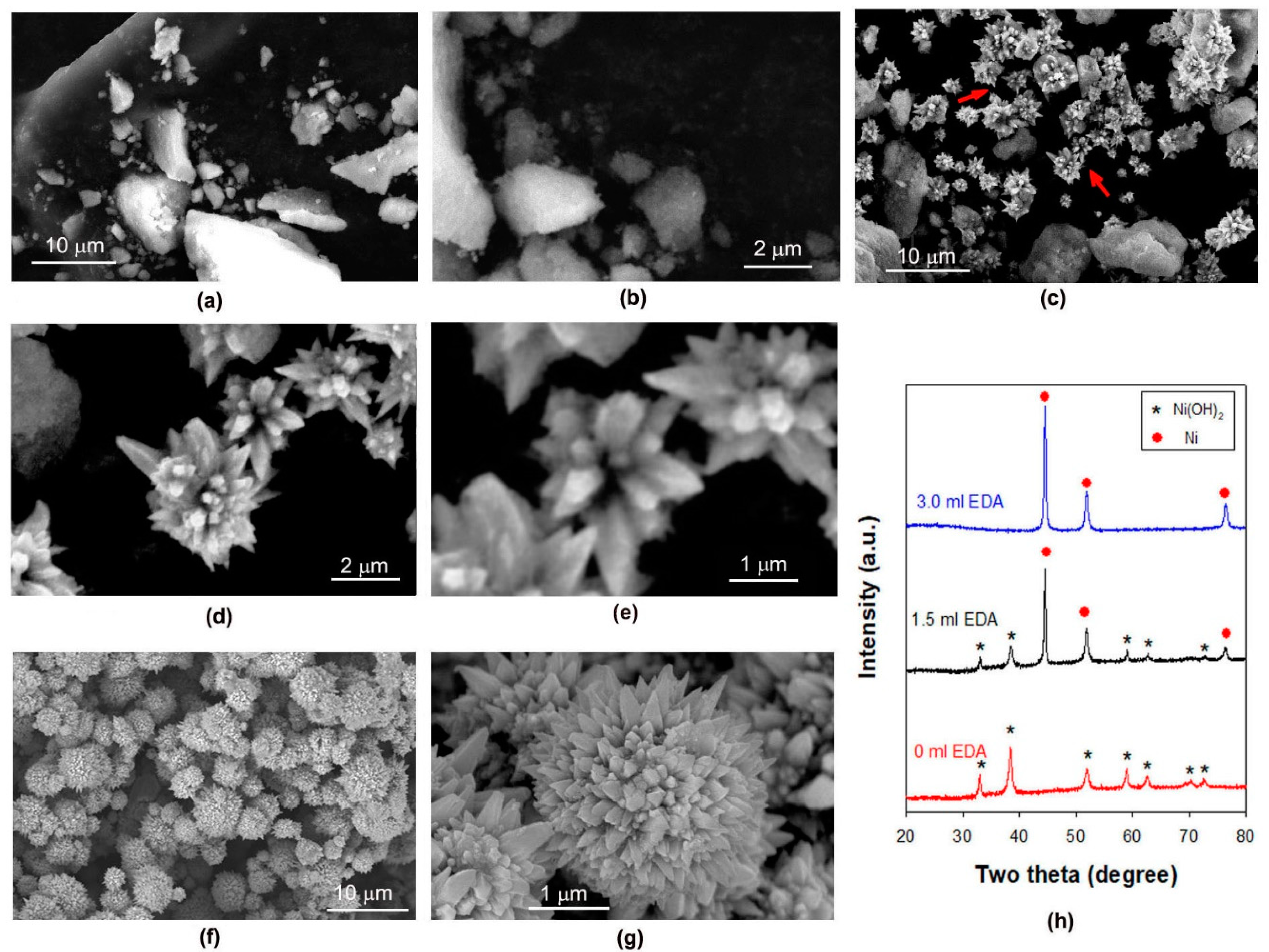
Figure 1a–g presents the SEM images of the products formed in the presence of different amounts of EDA. As shown in Figure 1a,b, the product formed in the absence of EDA exhibits a block-like morphology. When 1.5 mL of EDA is added, however, many flower-like microparticles appeared in the product (indicated by arrows in Figure 1c) and these microflowers are obviously assembled from many nanohorns of different sizes (Figure 1d,e). If the amount of EDA was raised to about 3.0 mL, the block-like materials disappeared and the product was totally composed of microspheres (Figure 1f). A close examination of these microspheres revealed that, similar to the microflowers, they are also assembled from nanosized horns (Figure 1g). The difference between the two cases lies in that the number of nanohorns in microspheres is much larger than that in microflowers. The XRD patterns of these products are illustrated in Figure 1h. In the absence of EDA, the peaks observed at 2θ of 33.2°, 38.6°, 52.1°, 59.3°, 62.9°, 70.4°, and 72.9° can all be indexed to Ni(OH)2 with a hexagonal structure (JCPDS 73-1520) and correspond to the diffractions of (100), (011), (012), (110), (111), (103), and (201) planes, respectively. Thus, the block-like materials observed in Figure 1a should be Ni(OH)2. The addition of 1.5 mL EDA leads to the appearance of new peaks at 2θ of 44.5°, 51.8°, and 76.4°, and these peaks can be indexed to Ni with a face-centered cubic (fcc) structure (JCPDS 87-0712) and correspond, respectively, to the diffractions of (111), (200), and (220) planes. When 3 mL EDA was used in the synthesis, all the peaks observed in the XRD patterns can be indexed to Ni with an fcc structure, indicating that the microspheres observed in Figure 1f are Ni microspheres. These observations indicate that under our experimental conditions, Ni(II) is difficult to be reduced to Ni in the absence of EDA and the addition of EDA favors the reduction of Ni(II) to Ni, which is different from the observation in the reduction of Cu(II) to Cu [11]. Actually, we have tried to reduce Ni(II) to Ni in the absence of EDA by raising the reaction time to 8 h. However, XRD measurement suggests that the product is still in the form of Ni(OH)2 (Figure 2a), and no microflowers or microspheres can be found in the product (Figure 2b).
The XRD analysis hints that the product formed in the presence of 1.5 mL EDA is a mixture of Ni(OH)2 and Ni. Since the morphology of block-like materials in Figure 1c is similar to that of the Ni(II) precursor in Figure 1a, we speculate that the block-like materials in 
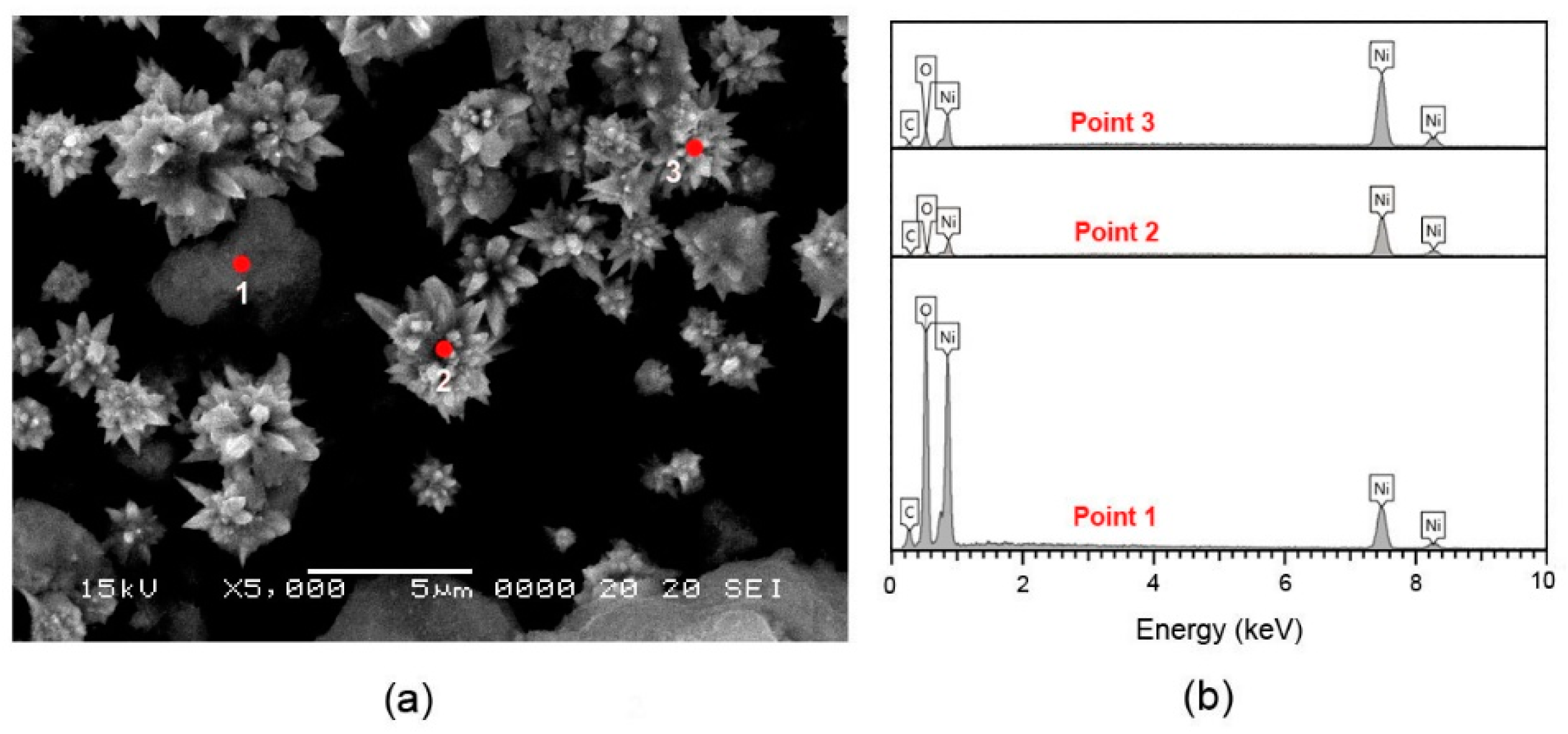
Figure 1c are Ni(II) precursors, and thus the microflowers should be Ni. To confirm this inference, the EDS analysis of the block-like materials and microflowers is provided in Figure 3. Point 1 in Figure 3a belongs to the region of a block-like material and its EDS spectrum indicates the presence of a large amount of oxygen with the weight percentages of Ni, O, and C being, respectively, about 55.1%, 34.4%, and 10.5%, suggesting that block-like material is an Ni(II) precursor. Points 2 and 3 in 
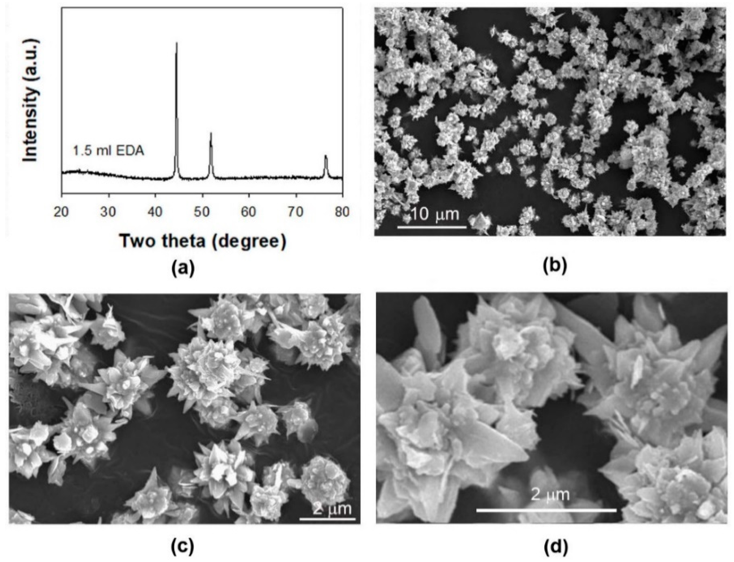
Figure 3a belong to the regions of microflowers, and their EDS spectra reveal that these microflowers are dominantly composed of Ni, with the weight percentages of Ni, O, and C being, respectively, 95.7%, 1.5%, and 2.8% on Point 2 and 93.7%, 2.8%, and 3.5% on Point 3. These observations confirm that the microflowers formed in the presence of 1.5 mL EDA are Ni microflowers. To understand whether these microflowers will evolve into microspheres after all Ni(II) precursors are reduced to Ni, we prolonged the reaction time from 2 h to 6 h. The XRD pattern of the resulting product (Figure 4a) clearly indicates that Ni(II) is completely converted into Ni. However, the SEM images of the product at different magnifications (Figure 4c,d) reveal that the grown Ni microparticles still own a microflower-like morphology, rather than the microspheres as observed in the presence of 3 mL EDA. These results suggest that the morphology of the Ni nano-micro structures can be adjusted by changing the amount of EDA.
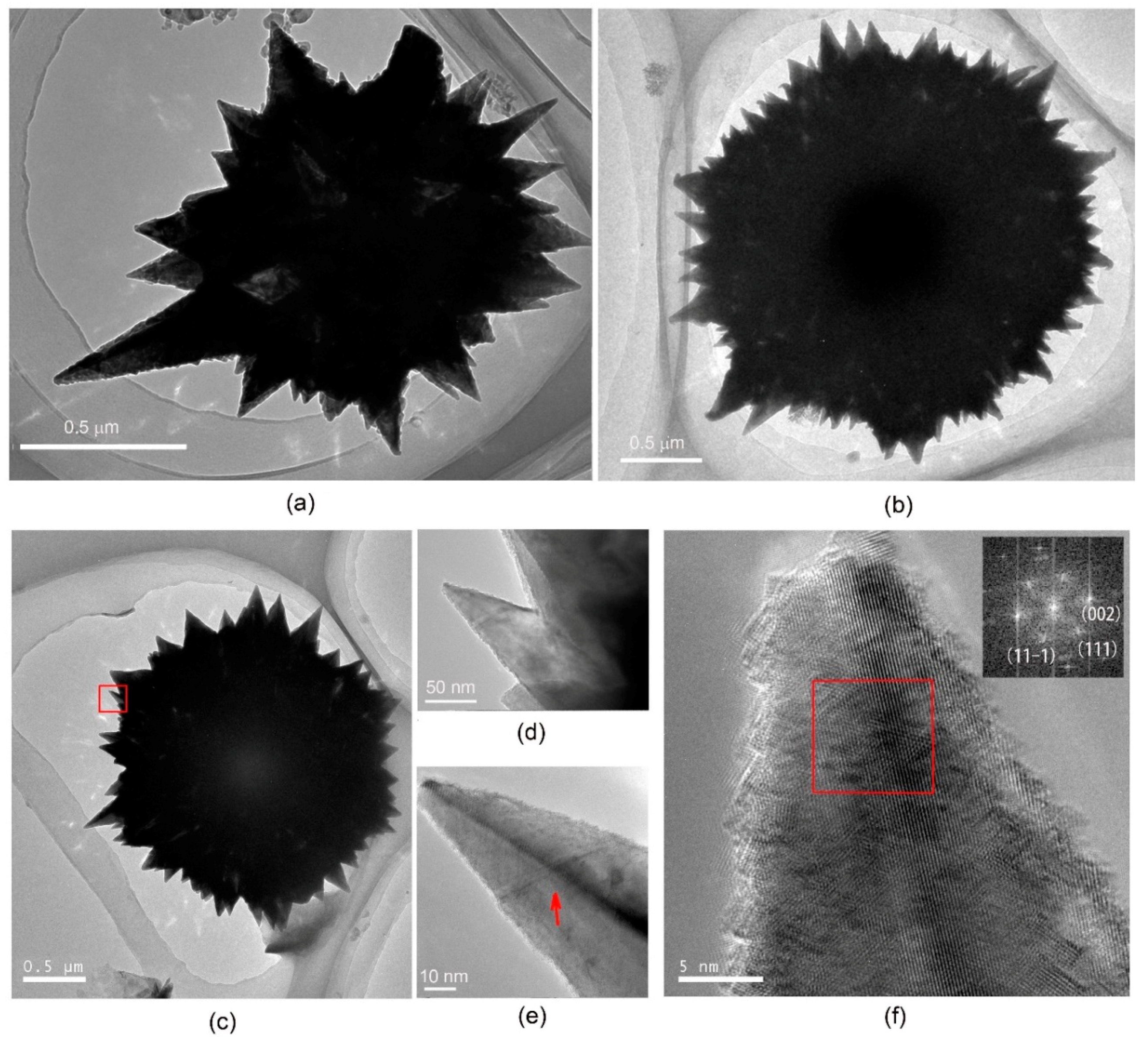
To unveil the microstructures of as-formed Ni nano-micro structures, TEM investigation of the microflowers or microspheres is depicted in Figure 5. It is obvious that the nanosized horns in both microflowers and microspheres possess a solid structure, rather than a hollow one (Figure 5a–c). However, a microsphere contains much more nanohorns than a microflower, which is consistent with the observation by SEM. A close examination of a nanohorn (see the framed region in Figure 5c) clearly indicates the existence of ridges in the nanohorn (pointed by arrow in Figure 5e), suggesting that the nanohorn is not a circular nanocone. The high resolution TEM image of the tip of this nanohorn is shown in Figure 5f. The reduced fast Fourier transform (FFT) image of the framed region (inset of Figure 5f) suggests that the nanohorn grows along the <1-10> direction, which is similar to the growth feature of Cu nanowires [11] or Cu nanocusps [29] under similar experimental conditions. In the growth of Cu nanowires [11] or Cu nanocusps [29], one of the roles of diamine is to induce an orientated growth, namely acting as capping agent. Kim et al. have found that the capping effect of EDA in the growth of Cu nanowires stems from selective surface reactivity increased by surface oxide removal, namely EDA can inhibit the formation of Cu oxide on the Cu(111) surface, and thus increase the rate of atomic addition to (111) facets at the end of a growing nanowire relative to (100) facets on the sides of a nanowire [30]. Therefore, it is expected that EDA plays a similar role in the growth of Ni nanohorns.
Our above experimental results indicate that under the experimental conditions employed, the addition of EDA not only results in the formation of Ni nano-micro structures, but also favors the reduction of Ni(II) to Ni. The reduction of Ni(II) to Ni can be simply expressed as Equation (1):
Ni2+ + 2e = Ni
The Nernst equation of this half reaction is:
φ N i 2 + / N i = φ N i 2 + / N i θ + R T 2 F l n a N i 2 +  
Equation (2) hints that the electrode potential will decrease when the concentration of free Ni2+ ions in the solution declines. Since the stability constant of [Ni(EDA)3]2+ complex ions is higher than that of Ni(OH)2, the addition of EDA will lead to the formation of complex between Ni(II) and EDA, which is clearly supported by the fact that the color of the mixture of NaOH and Ni(NO3)2 solutions changed from light green to purple after EDA is added. Obviously, the formation of complex between Ni(II) and EDA will reduce the concentration of free Ni2+ ions and thus lower the electrode potential. From the thermodynamic point of view, it seems that the addition of EDA is not in favor of the reduction of Ni(II) to Ni, just like the case of the reduction of Cu(II) to Cu [11]. However, under highly alkaline conditions, Ni(II) is dominantly in the form of solid phase Ni(OH)2, rather than free Ni2+ ions. A calculation based on φ N i 2 + / N i θ = 0.25   V , φ N i ( O H ) 2 / N i θ = 0.72   V and the stability constant of [Ni(EDA)3]2+ = 3.4 × 10¹⁸ reveals that the electrode potential for the reaction, [Ni(EDA)3]2+ + 2e→ Ni + 3 EDA, in a 200 mL 15 M NaOH solution with 3 mL EDA (about −0.74 V) is more positive than that for the reaction, Ni(OH)2 + 2e→ Ni + 2OH, in a 200 mL 15 M NaOH solution (about −0.79 V). The result suggests that, while the formation of Ni nano-micro structures of high surface area in the presence of EDA is obviously a result of kinetic reaction control, the addition of EDA also facilitates the reduction of Ni(II) thermodynamically via the formation of Ni(II)-EDA complex, which is supported by the experimental observation that in the absence of EDA, no metallic nickel forms even after extended reaction (Figure 2). The formation of Ni(II)-EDA complex makes EDA serve as a nickel transporter in the reduction of Ni(II) to Ni. In the early stages of the reaction, inert Ni(OH)2 dominates, but due to the presence of some Ni(II) dissolved by EDA, metallic Ni forms via hydrazine reduction. The reduction of the Ni(II)-EDA complex releases EDA, which then can again dissolve Ni(OH)2 until all of it is transformed to Ni. A similar strategy of solubilizing Ni(II) in alkaline hydrazine solutions has also been employed by Zhao et al. to produce thorny Ni coatings, in which an aminopolycarboxylic acid was used as the complexing agent [31].

3.2. Effects of the NaOH Concentration and the Amount of NaOH Solution

To understand the growth of Ni nano-micro structures in the presence of EDA under strong alkaline conditions, the effect of the NaOH concentration on the reduction of Ni(II) to Ni, as well as on the morphology of the resulting Ni nano-micro structures, has been investigated. We find that the reduction of Ni(II) to Ni is also heavily dependent on the concentration of NaOH when the amount of EDA is kept unchanged (Note: We can roughly determine whether or not the reaction is completed based on the color change of both the liquid phase and the formed solid phase). If the amount of EDA is fixed to be 3 mL, for example, the time needed for the complete reduction of Ni(II) to Ni is less than 2 h in the presence of 15 M NaOH (200 mL). However, when 10 M NaOH is used, we need about 3.5 h to complete this reduction. If we further reduce the concentration of NaOH to 5 M, around 5 h is required. These results imply that the reduction of Ni(II) to Ni benefits from the rise in the concentration of NaOH. As a reducing agent, hydrazine can be oxidized to N2 and the half reaction is expressed as Equation (3):
N2H4 + 4OH − 4e = N2 + 4H2O
Thus, the result that Ni(II) is more easily reduced to Ni in an NaOH solution of higher concentration when the amount of EDA is fixed can be understood based on the fact that the rise in the concentration of NaOH will reduce the electrode potential and thus enhances the reducing ability of hydrazine.
Figure 6 presents the SEM images of the products formed in the presence of 3 mL of EDA in 200 mL NaOH solutions of different concentrations. To complete this reduction, the reaction duration varies from case to case. As shown in the insets of Figure 6a,c,e, all the diffraction peaks appearing in the XRD patterns of the products formed in NaOH solutions of different concentrations can be indexed to fcc structured Ni (JCPDS 87-0712), hinting that Ni(II) is completely converted into Ni. It is clear from these SEM images that the products grown in different concentrations of NaOH have a similar morphology, namely microspheres assembled from nanohorns. However, the size of these Ni microspheres increases as the concentration of NaOH decreases, especially when 5 M NaOH is used. For example, the diameter of the nano-micro structured Ni microshperes formed in 15 M or 10 M is generally lower than 10 μm (Figure 6a–d), but many microspheres with a diameter larger than 10 μm can be observed in the product grown in 5 M NaOH (Figure 6e,f). Two factors, namely the reduction rate of Ni(II) to Ni and the reaction time, are believed to be responsible for the result observed. A low reduction rate of Ni(II) to Ni observed in NaOH of low concentration favors the formation of fewer nuclei via homogeneous nucleation in the early growth stage. Fewer nuclei, along with a longer reaction time, allow these nuclei to grow to a bigger size at the expense of remaining Ni(II) in the reacting mixture.
Besides the effect of NaOH concentration, we have also investigated the influence of the amount of NaOH solution on the morphology of the Ni nano-micro structures. 
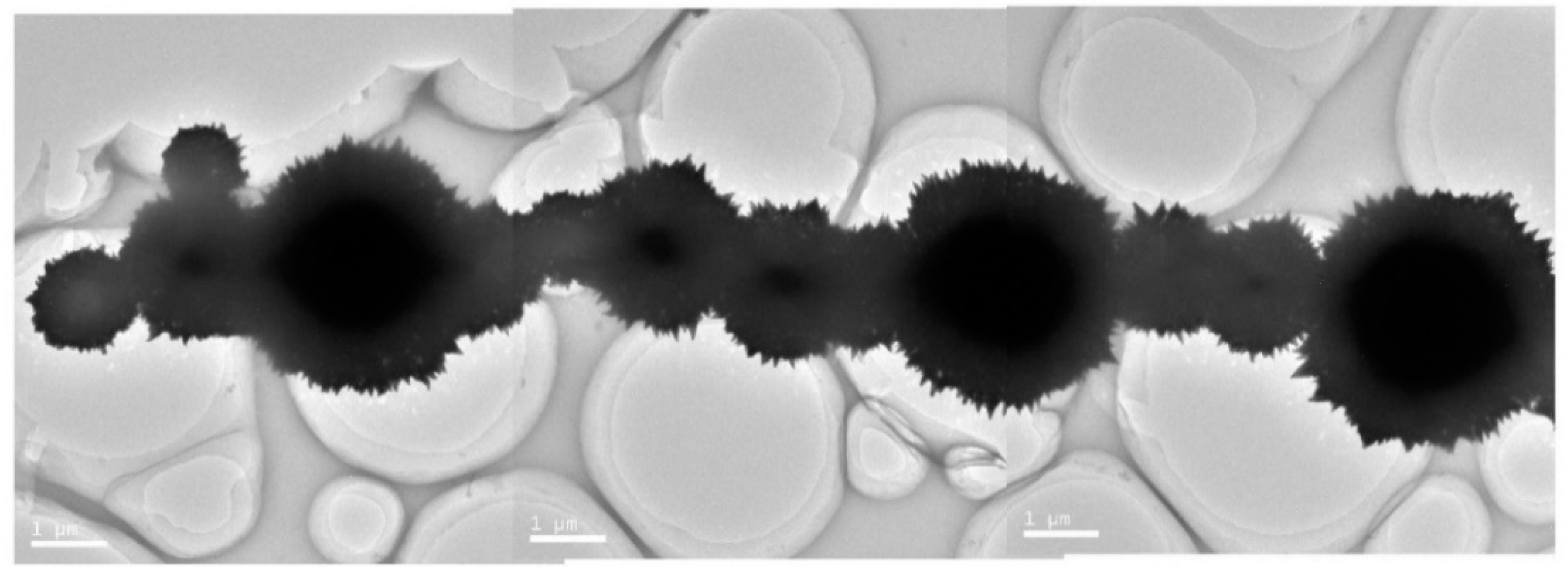
Figure 7 presents the SEM images of the products formed in the presence of 3 mL of EDA in a 15 M NaOH solution of different amounts. Interestingly, unlike the isolated nano-micro structured microspheres and their irregular aggregations observed in the case where 200 mL of 15 M NaOH solution was used (see Figure 1e), many chain-like aggregations can be found in the products when the amount of NaOH solution is reduced (Figure 7a,c,e). The high magnification SEM images (Figure 7b,d,f) indicate that these chains are composed of nano-micro structured microspheres, which is supported by the TEM image of a chain resulting from the self-assembly of the microspheres of different sizes (Figure 8). The formation of the microsphere chains is probably associated with the fact that a reduction in the amount of NaOH solution means less room for the growth of these nano-micro structured microspheres. That means there are more chances for these microspheres to connect each other during their growth, and chain-like aggregations form as a result of a reduction in the surface energy of the system. Another important factor that may promote the formation of these chain-like aggregations is the magnetism of Ni. It has been reported that the ferromagnetic nanoparticle chains, e.g., Fe3O4 [32] or Co chains [33], can be formed via dipolar assembly. Although the size of Ni particles in our case is at the micrometer scale, the similarity between the dipolar structures of the ferromagnetic nanoparticles and the Ni chains observed in our experiment hints that dipolar magnetic assembly should contribute to the growth of Ni microsphere chains.

3.3. Growth Mechanism of Ni Nano-Micro Structures

To get an insight into the formation mechanism of these Ni nano-micro structures, we have examined the products formed at an early stage in 200 mL of 15 M NaOH solution containing 3 mL of EDA. The products obtained after reaction for 45 min, 50 min, and 60 min are light green, grey, and dark grey, respectively. The XRD patterns of these three samples (Figure 9a) reveal that two phases, namely Ni(OH)2 and Ni, are present, with the 45 min sample being mainly composed of Ni(OH)2, and the 60 min sample being dominantly in the form of Ni. The morphology of these products is presented in Figure 9b–d. As indicated by arrows in Figure 9b, very few sphere-like particles can be found in the 45 min product. However, many microspheres appear after the reaction for 50 min (see arrows in Figure 9c). When the reaction time reaches 60 min, the product contains a large number of microspheres, whose sizes are obviously larger than those of the microspheres in Figure 9c. Although these observations indicate the growth of Ni microspheres during the reduction of Ni(II), the detail growth mechanism of these nano-micro structured microspheres is still unclear.
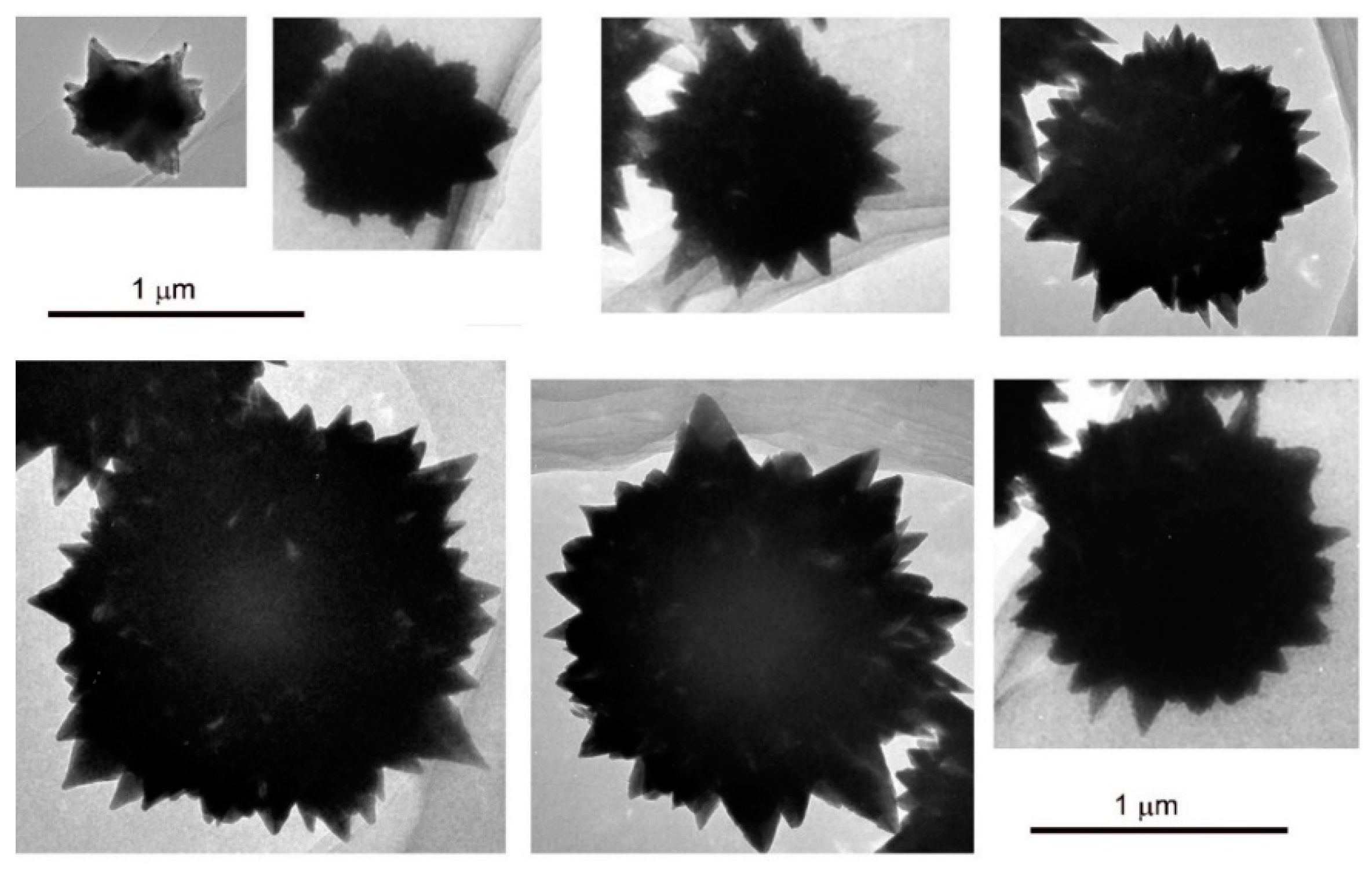
To disclose the growth process of Ni nano-micro structured microspheres, we have also investigated these products by TEM. Figure 10 presents the TEM images of the nanohorn-containing spheres of different sizes found in the products grown in a 200 mL NaOH solution (15 M) containing 3 mL of EDA. From these TEM images, we can roughly observe a growing picture of Ni microspheres in the presence of EDA under strong alkaline conditions, namely from small-sized particles with sparse nanohorns to micro-sized spheres with dense nanohorns. To unveil why more and more nanohorns form during the growth of Ni microspheres, we conducted a close examination of the nanohorn-containing spheres of different sizes by SEM. As indicated by arrows in Figure 11, the nanohorn-containing spheres, whether their sizes are big or small, contain many very small particles lying between nanohorns or on the surface of nanohorns. The finding hints that during the growth of microspheres, heterogenous nucleation, which will produce new nuclei on the surface of the growing spheres, also occurs. These new nuclei may also evolve into nanohorns as a result of orientated growth along the <1-10> direction, which not only will produce more nanohorns in the microspheres, but may also contribute to the diversity in the size of the nanohorns. As already mentioned above, a microflower contains much fewer nanohorns than a microsphere. This implies that during the growth of microflowers the heterogenous nucleation, which may produce new nuclei and thus favors the formation of new nanohorns, seldom happens. More specifically, these observations remind us that orientated growth dominates during the growth of microflowers, but both orientated growth and heterogenous nucleation coexist in the growth of nano-micro structured microspheres. The reason for this might be related to the fact that the amount of EDA used in the growth of microspheres is two times higher than that in the growth of microflowers. It is expected that in the growth of microspheres, the excessive EDA may affect the orientated growth and thus facilitates the heterogenous nucleation.
Based on the results described above, a formation mechanism of Ni nano-micro structures under our experimental condition is proposed as follows (see Figure 12). Firstly, part of Ni(II) is reduced to Ni, leading to the formation of Ni nanocrystals (Figure 12a). To reduce the surface energy of the system, the as-formed Ni nanocrystals may aggregate into large-sized particles (Figure 12b). In the presence of 3 mL of EDA, the selective adsorption of EDA on the surface of Ni nanocrystals may result in an orientated growth along the <1-10> direction and thus small-sized nanohorns will form (Figure 12c). Meanwhile, heterogenous nucleation, which may produce new nuclei (indicated by blue arrows) on the surface of the growing particle, also occurs. With the reaction proceeding, the sizes of the nanohorns increases and these new nuclei also evolve into new nanohorns (Figure 12d). The continuous orientated growth and heterogenous nucleation will finally create nano-micro structured microspheres (Figure 12e). Under suitable conditions, these Ni microspheres can form chain-like structures via dipolar magnetic assembly (Figure 12f). However, if the orientated growth dominates after the formation of Ni nanocrystal aggregation, e.g. in the case of 1.5 mL of EDA where very fewer new nuclei form via heterogenous nucleation, the morphology of the resulting Ni nano-micro structures will be different from that of nano-micro structured microspheres. As shown in Figure 12g–i, due to the lack of new nuclei for growing new nanohorns, the orientated growth will result in the formation of microflowers.

4. Conclusions

In summary, we have presented a study on the EDA-mediated growth of Ni nano-micro structures under strong alkaline conditions. Both EDA and NaOH play an important role in the reduction of Ni (II) to Ni, and Ni microflowers or microspheres assembled from nanosized horns can be grown in the presence of EDA of different amounts. The size of the nano-micro structured Ni microspheres depends on the NaOH concentration, and long microsphere chains form when the amount of NaOH solution is reduced. The nano-micro structured microflowers should be a result of the orientated growth induced by EDA, whereas the nano-micro structured microspheres are believed to be a combined result of both orientated growth and heterogenous nucleation.

Author Contributions

L.C. and Z.L. conceived and designed the experiments; L.C., H.L. and L.L. performed the experiments; Z.Z., H.T. and Z.L. analyzed the data; L.C. and Z.L. wrote the paper.

Funding

This research was supported by the National Natural Science Foundation of China (NSFC Grant No. 21776256).

Acknowledgments

Z.L. thanks Y.G. Shen at City University of Hong Kong for his help with characterization of some samples.

Conflicts of Interest

The authors declare no conflict of interest.

References

  1. Zheng, Y.F.; Chen, N.N.; Wang, C.X.; Zhang, X.P.; Liu, Z.J. Oleylamine-mediated hydrothermal growth of millimeter-long Cu nanowires and their electrocatalytic activity for reduction of nitrate. Nanomaterials 2018, 8, 192. [Google Scholar] [CrossRef] [PubMed]
  2. Liang, J.H.; Zheng, Y.F.; Liu, Z.J. Nanowire-based Cu electrode as electrochemical sensor for detection of nitrate in water. Sens. Actuators B Chem. 2016, 232, 336–344. [Google Scholar] [CrossRef]
  3. Zheng, Y.F.; Liang, J.H.; Chen, Y.Y.; Liu, Z.J. Economical and green synthesis of Cu nanowires and their use as catalyst for selective hydrogenation of cinnamaldehyde. RSC Adv. 2014, 4, 41683–41689. [Google Scholar] [CrossRef]
  4. Mourdikoudis, S.; Liz-Marzan, L.M. Oleylamine in nanoparticle synthesis. Chem. Mater. 2013, 25, 1465–1476. [Google Scholar] [CrossRef]
  5. Kar, S.; Patel, C.; Santra, S. Direct room temperature synthesis of valence state engineered ultra-small ceria nanoparticles: Investigation on the role of ethylenediamine as a capping agent. J. Phys. Chem. C 2009, 113, 4862–4867. [Google Scholar] [CrossRef]
  6. Fang, Z.; Wang, Q.; Wang, X.Q.; Zhu, B.C.; Fan, F.; Wang, C.Y.; Liu, X.W. Ethylenediamine inducing growth of {100} facets exposed PbS nanosheets. Cryst. Res. Technol. 2012, 47, 635–642. [Google Scholar] [CrossRef]
  7. Guo, C.X.; Chen, S.C.; Lu, X.M. Ethylenediamine-mediated synthesis of Mn3O4 nano-octahedrons and their performance as electrocatalysts for the oxygen evolution reaction. Nanoscale 2014, 6, 10896–10901. [Google Scholar] [CrossRef] [PubMed]
  8. Han, Q.F.; Zhao, J.; Wu, L.; Zhu, J.W.; Wang, X. Synthesis of CdS multipods from cadmium xanthate in ethylenediamine solution. Particuology 2015, 19, 45–52. [Google Scholar] [CrossRef]
  9. Deng, Z.X.; Li, L.B.; Li, Y.D. Novel inorganic−organic-layered structures:  crystallographic understanding of both phase and morphology formations of one-dimensional CdE (E = S, Se, Te) nanorods in ethylenediamine. Inorg. Chem. 2003, 42, 2331–2341. [Google Scholar] [CrossRef] [PubMed]
  10. Han, S.B.; Liu, W.; Sun, K.; Zu, X.T. Experimental evidence of ZnS precursor anisotropy activated by ethylenediamine for constructing nanowires and single-atomic layered hybrid structures. CrystEngComm 2016, 18, 2626–2631. [Google Scholar] [CrossRef]
  11. Liu, Z.J.; Chen, Y.Y.; Hu, J.G.; Zheng, Y.F. Synthesis and growth mechanism of Cu nanowires in the presence of different linear alkyl diamines. Sci. Adv. Mater. 2014, 6, 327–335. [Google Scholar] [CrossRef]
  12. Chang, Y.; Lye, M.L.; Zeng, H.C. Large-scale synthesis of high-quality ultralong copper nanowires. Langmuir 2005, 21, 3746–3748. [Google Scholar] [CrossRef] [PubMed]
  13. Wang, X.X.; Dong, L.F.; Zhang, B.Q.; Yu, M.X.; Liu, J.Q. Controlled growth of Cu–Ni nanowires and nanospheres for enhanced microwave absorption properties. Nanotechnology 2016, 27, 125602–125612. [Google Scholar] [CrossRef] [PubMed]
  14. Wang, X.X.; Zhang, B.Q.; Zhang, W.; Yu, M.X.; Cui, L.; Cao, X.Y.; Liu, J.Q. Super-light Cu@Ni nanowires/graphene oxide composites for significantly enhanced microwave absorption performance. Sci. Rep. 2017, 7, 1584. [Google Scholar] [CrossRef] [PubMed]
  15. Hernandez-Gordillo, A.; Medina, J.C.; Bizarro, M.; Zanella, R.; Monroy, B.M.; Rodil, S.E. Photocatalytic activity of enlarged microrods of alpha-Bi2O3 produced using ethylenediamine-solvent. Ceram. Int. 2016, 42, 11866–11875. [Google Scholar] [CrossRef]
  16. Zhang, M.; Chen, K.F.; Chen, X.; Peng, X.Y.; Sun, X.D.; Xue, D.F. Ethylenediamine-assisted crystallization of Fe2O3 microspindles with controllable size and their pseudocapacitance performance. CrystEngComm 2015, 17, 1521–1525. [Google Scholar] [CrossRef]
  17. Qi, X.M.; Zhu, X.Y.; Wu, J.; Wu, Q.; Li, X.; Gu, M.L. Controlled synthesis of SiVO4 with multiple morphologies via an ethylenediamine-assisted hydrothermal method. Mater. Res. Bull. 2014, 59, 435–441. [Google Scholar] [CrossRef]
  18. Zhang, C.Q.; Mcadams, D.A., II; Grunlan, J.C. Nano/micro-manufacturing of bioinspired materials: A review of methods to mimic natural structures. Adv. Mater. 2016, 28, 6292–6321. [Google Scholar] [CrossRef] [PubMed]
  19. Yue, X.H.; Pan, J.Q.; Sun, Y.Z.; Wang, Z.H. Synthesis and electrochemical properties of nano–micro spherical β-Ni(OH)2 with super high charge-discharge Speed. Ind. Eng. Chem. Res. 2012, 51, 8358–8365. [Google Scholar] [CrossRef]
  20. Brassard, J.D.; Sarkar, D.K.; Perron, J.; Audibert-Hayet, A.; Melot, D. Nano-micro structured superhydrophobic zinc coating on steel for prevention of corrosion and ice adhesion. J. Colloid Interface Sci. 2015, 447, 240–247. [Google Scholar] [CrossRef] [PubMed] [Green Version]
  21. Zhang, Y.B.; Liang, W.X.; Wang, B.; Wang, G.Y.; Guo, Z.G. Green solvothermal synthesis of micro-/nano-structured porous β-Ni(OH)2 microspheres with enhanced electrochemical performance. Mater. Res. Bull. 2015, 64, 386–394. [Google Scholar] [CrossRef]
  22. Rajeshkhanna, G.; Rao, G.R. Micro and nano-architectures of Co3O4 on Ni foam for electro-oxidation of methanol. Int. J. Hydrog. Energy 2018, 43, 4706–4715. [Google Scholar] [CrossRef]
  23. Geng, W.Y.; Hu, A.M.; Li, M. Super-hydrophilicity to super-hydrophobicity transition of a surface with Ni micro–nano cones array. Appl. Surf. Sci. 2012, 263, 821–824. [Google Scholar] [CrossRef]
  24. Dudem, B.; Leem, J.W.; Yu, J.S. A multifunctional hierarchical nano/micro-structured silicon surface with omnidirectional antireflection and superhydrophilicity via an anodic aluminum oxide etch mask. RSC Adv. 2016, 6, 3764–3773. [Google Scholar] [CrossRef]
  25. Liu, X.H.; Liang, X.D.; Zhang, N.; Qiu, G.Z.; Yi, R. Selective synthesis and characterization of sea urchin-like metallic nickel nanocrystals. Mater. Sci. Eng. B 2006, 132, 272–277. [Google Scholar] [CrossRef]
  26. Ma, F.; Huang, J.J.; Li, J.G.; Li, Q. Microwave properties of sea-urchin-like Ni nanoparticles. J. Nanosci. Nanotechnol. 2008, 9, 1–5. [Google Scholar] [CrossRef]
  27. An, Z.G.; Pan, S.L.; Zhang, J.J. Synthesis and tunable qssembly of spear-like nickel nanocrystallites: From urchin-like particles to prickly chains. J. Phys. Chem. C 2009, 113, 1346–1351. [Google Scholar] [CrossRef]
  28. Ma, F.; Li, Q.; Huang, J.J.; Li, J.G. Morphology control and characterizations of nickel sea-urchin-like andchain-like nanostructures. J. Cryst. Growth 2008, 310, 3522–3527. [Google Scholar] [CrossRef]
  29. Liu, Z.J.; Chen, Y.Y.; Zheng, Y.F. Stirring-induced growth of hierarchical Cu structures in the presence of diamine. CrystEngComm 2014, 16, 9054–9062. [Google Scholar] [CrossRef]
  30. Kim, M.J.; Flowers, P.F.; Stewart, I.E.; Ye, S.R.; Baek, S.; Kim, J.J.; Wiley, B.J. Ethylenediamine promotes Cu nanowire growth by inhibiting oxidation of Cu(111). J. Am. Chem. Soc. 2016, 139, 277–284. [Google Scholar] [CrossRef] [PubMed]
  31. Zhao, X.; Muench, F.; Schaefer, S.; Brotz, J.; Duerrschnabel, M.; Molina-Luna, L.; Kleebe, H.J.; Liu, S.X.; Tan, J.; Ensinger, W. Electroless decoration of macroscale foam with nickel nano-spikes: A scalable route toward efficient catalyst electrodes. Electrochem. Commun. 2016, 65, 39–43. [Google Scholar] [CrossRef]
  32. Klokkenburg, M.; Vonk, C.; Claesson, E.M.; Meeldijk, J.D.; Erne, B.H.; Philipse, A.P. Direct imaging of Zero-field dipolar structures in colloidal dispersions of synthetic magnetite. J. Am. Chem. Soc. 2004, 126, 16706–16707. [Google Scholar] [CrossRef] [PubMed]
  33. Benkoski, J.J.; Breidenich, J.L.; Uy, O.M.; Hayes, A.T.; Deacon, R.M.; Land, H.B.; Spicer, J.M.; Kengb, P.Y.; Pyun, J.P. Dipolar organization and magnetic actuation of flagella-like nanoparticle assemblies. J. Mater. Chem. 2011, 21, 7314–7325. [Google Scholar] [CrossRef]
Figure 1. SEM images (ag) and XRD patterns (h) of the products formed in 200 mL of 15 M NaOH solution containing different amounts of EDA: (a,b) 0 mL, (ce) 1.5 mL, and (f,g) 3 mL. Reaction duration is 2 h.
Crystals 08 00397 g001
Figure 2. XRD (a) and SEM image (b) of the product formed in 200 mL of 15 M NaOH solution in the absence of EDA. Reaction duration is 8 h.
Crystals 08 00397 g002
Figure 3. SEM image (a) and EDS spectra in different regions (b) of the product formed in 200 mL of 15 M NaOH solution containing 1.5 mL EDA. Reaction duration is 2 h.
Crystals 08 00397 g003
Figure 4. XRD patterns (a) and SEM images (bd) of the product formed in 200 mL of 15 M NaOH solution containing 1.5 mL EDA. Reaction duration is 6 h.
Crystals 08 00397 g004
Figure 5. TEM images of an Ni microflower (a), an Ni microsphere (b), and an Ni microsphere at different magnifications (cf). Inset of Figure 5f is the corresponding reduced fast Fourier transform image of the framed region.
Crystals 08 00397 g005
Figure 6. SEM images of the products formed in the presence of 3 mL EDA in 200 mL NaOH solutions of different concentrations for different reaction durations: (a,b) 15 M for 1.5 h, (c,d) 10 M for 3.5 h, and (e,f) 5 M for 5 h. Insets of Figure 6a,c,e are the XRD patterns of the corresponding products.
Crystals 08 00397 g006
Figure 7. SEM images of the products formed in the presence of 3 mL of EDA in a 15 M NaOH solution of different amounts: (a,b) 100 mL, (c,d) 50 mL, and (e,f) 25 mL. Reaction duration is 6 h.
Crystals 08 00397 g007
Figure 8. TEM image of an Ni microsphere chain formed in the presence of 3 mL of EDA in 50 mL of 15 M NaOH solution. Reaction duration is 6 h.
Crystals 08 00397 g008
Figure 9. XRD (a) and SEM images (c,d) of the products formed in 200 mL of 15 M NaOH solution containing 3 mL of EDA after different reaction times: (b) 45 min, (c) 50 min, and (d) 60 min.
Crystals 08 00397 g009
Figure 10. TEM image of the nanohorn-containing spheres of different sizes found in the products in a 200 mL NaOH solution (15 M) containing 3 mL of EDA.
Crystals 08 00397 g010
Figure 11. SEM image of the nanohorn-containing spheres of different sizes found in the products in a 200 mL NaOH solution (15 M) containing 3 mL of EDA.
Crystals 08 00397 g011
Figure 12. A scheme for the growth mechanism of Ni nano-micro structures: (a) Ni nanocrystals; (b) aggregation of Ni nanocrystals; (c) small-sized sphere with nanohorns and new nuclei; (d) middle-sized microspheres with nanohorns and new nuclei; (e) big-sized microspheres with nanohorns and new nuclei; (f) chain-like structure; (g) small-sized particle with nanohorns; (h) middle-sized microflower with nanohorns; and (i) big-sized microflower with nanohorns. New nuclei produced by heterogenous nucleation are indicated by blue arrows.
Crystals 08 00397 g012

