
Synergistic Antimicrobial Activities of Thai Household Essential Oils in Chitosan Film


1
Department of Food Science and Technology, Faculty of Agro-Industry, Kasetsart University, Bangkok 10900, Thailand
2
Faculty of Agro-Industry, School of Agro-Industry, Chiang Mai University, Chiang Mai 50100, Thailand
3
The Cluster of Agro Bio-Circular-Green Industry (Agro BCG), Chiang Mai University, Chiang Mai 50100, Thailand
4
Center of Excellence in Materials Science and Technology, Chiang Mai University, Chiang Mai 50200, Thailand
5
Center for Advanced Studied Agriculture and Food, Kasetsart University, Bangkok 10900, Thailand
*
Author to whom correspondence should be addressed.
Polymers 2021, 13(9), 1519; https://doi.org/10.3390/polym13091519
Submission received: 13 March 2021
/
Revised: 2 May 2021
/
Accepted: 2 May 2021
/
Published: 9 May 2021
(This article belongs to the Collection Antibacterial Activity of Polymeric Materials)
Abstract
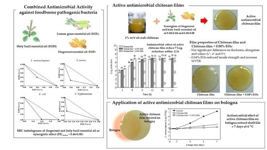
:Foodborne pathogens mostly contaminate ready-to-eat (RTE) meat products by post-process contamination and cause foodborne disease outbreaks. Preventing post-process contamination and controlling microbial growth during storage by packing the RTE meats with active antimicrobial film from chitosan combined with the synergism of Thai household essential oils was investigated. Here, we analyzed antimicrobial activity and mechanical properties of chitosan films incorporated with essential oil of fingerroot (EOF) and holy basil (EOH) based on their fractional inhibitory concentration and isobolograms. We showed that antimicrobial activities of chitosan film and chitosan films formulated with EOF:EOH displayed a dramatical reduction of Listeria monocytogenes Scott A concentration by 7 Log in 12 h. Chitosan film incorporated with EOF:EOH at ratio 0.04:0.04% v/v/w strongly retarded growth of total viable count of L. monocytogenes on vacuum-packed bologna slices during seven days of storage at 4 and 10 °C. Combined EOF and EOH added to chitosan films did not alter thickness, elongation (%) and colors (L*, a* and b*) of the chitosan film, but it increased water vapor transmission rate and decreased film tensile strength. Results suggested that chitosan film had strong antibacterial properties. Its effectiveness in inhibiting foodborne pathogenic bacteria in ready-to-eat meat products was enhanced by adding a combination of EOF:EOH.
1. Introduction
Active antimicrobial food packaging is an innovative concept that was introduced to respond to continuous changes in consumer demands and market trends. This technology illustrates its usage in food contact applications, which plays an important role in reducing the risk from pathogens and prolonging food shelf-life [1]. Regarding other biofunctional materials, chitosan has received significant interest to use as an antimicrobial film-forming agent due to its biodegradability, biocompatibility and antimicrobial activity. In our previous study, chitosan showed the strongest antilisterial activity, especially crab polymer chitosan [2]. Chitosan film is readily prepared by evaporation of its dilute acid solutions [3]. Nowadays, considerable interest has been created by the possibility of using chitosan film to retard or prevent the growth of foodborne and food spoilage microorganisms. This film serves as a carrier for a wide range of food additives including antimicrobial agents which act as a food preservative to prolong the shelf-life of products.
Antimicrobial efficacies of essential oils (EOs) were established long ago. EOs are hydrophobic liquids containing various volatile compounds extracted from several parts of plants [4]. Historically, EOs have been used in foods as flavorings, but they also have a broad spectrum against various microorganisms [5]. Several published studies have extolled the antimicrobial activity of EOs which occurs as a result of their ability to increase the permeability of bacterial cells, coagulate cytoplasm, reduce the intracellular ATP pool resulted to decrease ATP synthesis, damage cytoplasmic and membranes protein, alter the membrane of fatty acids and lower proton motive force across bacterial membrane [6]. Moreover, EOs were shown to disintegrate cell surfaces, detach the cytoplasmic membrane from the cell wall and change cytoplasmic inclusion by reducing vacuole size [7,8]. Numerous studies of the antimicrobial activity of various EOs and phytochemicals have been reviewed in great detail [9,10,11,12]. However, the requirement of a high concentration of EOs to enable sufficient inhibition of microorganisms in complex food systems is a difficult problem to tackle. This is due to their high volatility, strong odor and poor water solubility which affects food sensory quality. Thus, one of the possible ways to reduce the concentration of each EO required in the food matrix can be achieved through a combination of EOs with synergistic antimicrobial effects [13,14].
Previous studies examined synergistic antimicrobial effects; for example, carvacrol and linalool demonstrated synergistic effects against L. monocytogenes [15] and combined action of thymol/carvacrol, thymol/eugenol and carvacrol/eugenol revealed synergistic effects against Escherichia coli [16]. EOs from Thai household herbs such as holy basil, fingerroot, ginger, turmeric, lemongrass and black pepper have also been studied for their effects against pathogenic microorganisms and presented a broad spectrum against both Gram-positive and Gram-negative bacteria [17,18]. However, only a few reports have detailed the antimicrobial activity of their combinations. Moreover, incorporation of EOs by chitosan films as natural bactericides could enhance their antimicrobial effectiveness with potential development as environmentally friendly antimicrobial packaging. These films act as carriers to control the release of antimicrobials on the food surface which is the key factor in controlling microbial growth in products and thereby enhance product shelf-life.
L. monocytogenes, pathogenic E. coli, Salmonella spp. and Staphylococcus aureus are common foodborne pathogens associated with RTE meat and poultry products [19,20,21,22,23,24]. Among them, L. monocytogenes was the major concern on refrigerated RTE foods. It causes listeriosis which is widely regarded as one of the deadliest foodborne diseases and poses a serious threat to consumer health [25]. L. monocytogenes presents a high risk to processed RTE products because it can cross-contaminate from the processing environment to the food prior to packing and increase their numbers during storage in the refrigerator [26]. RTE meat products such as hot dogs, lunch meats, cold cuts and other deli meats (such as bologna) [27] are perishable and pose a higher risk for listeriosis infection in the event of post-process surface contamination such as a mechanical slicer [28,29,30]. Moreover, cold-eat RTE meat products that undergo no further control steps limited customers’ opportunities to eliminate the pathogen prior to food consumption. In the USA, an estimated 1600 people contract listeriosis annually, with about 260 deaths [25]. This problem has resulted from changes in consumer trends and legislation that prefer chemical preservatives free, safe, but minimally processed food [31,32,33]. For S. aureus, E. coli and Salmonella spp., these bacteria can cause food poisoning due to improper hygienic practices especially on the surface of equipment that is in contact with the food surface [26,34,35]. Salmonella enterica subsp. enterica serovar Typhimurium is often associated with many types of animals and has been associated with a wide range of foods including RTE meat products [36]. Moreover, E. coli and Salmonella isolated from RTE meat and poultry products were resistant to antibiotics, some isolates were multidrug resistant [19,35,37]. The prevalence of pathogenic bacteria in RTE meat can be substantially increased by post-cooking handling activities, exposure duration at points of sale and meat storage conditions [38].
In this study, we investigated the antimicrobial ability of EOs from Thai household herbs, including holy basil (EOH), fingerroot (EOF) and lemongrass (EOL) against foodborne pathogenic bacteria in a microbiological medium, both singly and in combination. Antimicrobial packaging films from crab polymer chitosan and chitosan films incorporated with selected EOs based on their fractional inhibitory concentration (FIC) index values were produced, and their antimicrobial activities were investigated on both laboratory broth and bologna as a food model. Physical and mechanical properties of the antimicrobial films were also studied.
2. Materials and Methods
2.1. Experimental Materials
Crab polymer chitosan (molecular weight of 700–1000 kDa and degree of deacetylation 97.38%) was purchased from Ta Ming Enterprises Co., Ltd. (Samut Sakhon, Thailand). Acetic acid and sodium chloride (NaCl) were obtained from Merck (Darmstadt, Germany). Sodium nitrite (NaNO2) was purchased from Lab Scan (Samut Sakhon, Thailand). For antimicrobial tests, trypticase soy agar (TSA), trypticase soy broth (TSB) and yeast extract (YE) were purchased from Merck (Darmstadt, Germany). Müeller Hinton broth (MHB), plate count agar (PCA) and peptone were bought from Difco (Maryland, USA). Modified Listeria selective agar base enrichment supplement (MOX) was sourced from Oxoid (Hampshire, England). Three types of Thai household essential oils including essential oil of fingerroot (EOF) (Boesenbergia pandurata (Roxb.) Schltr.), essential oil of holy basil (EOH) (Ocimum sanctum Linn.) and essential oil of lemongrass (EOL) (Cymbopogon citratus Stapf.) were purchased from Thai-China Flavours and Fragrances Industry Co., Ltd. (Pathum Thani, Thailand). The EOs were kept in aluminum bottles and stored at −20 °C until use [39].
2.2. Tested Bacteria
Foodborne pathogenic bacteria including Escherichia coli TISTR 780, Salmonella enterica subsp. enterica serovar Typhimurium ATCC 13,311 and Staphylococcus aureus TISTR 1466 were obtained from the Thailand Institute of Scientific and Technological Research, while Listeria monocytogenes Scott A was obtained from the Laboratory of Food Safety, Department of Food Science and Technology, Faculty of Agro-Industry, Kasetsart University, Thailand. E. coli, S. enterica and S. aureus were cultured in 10 mL of TSB, while L. monocytogenes was grown in 10 mL of TSB + 0.6% YE (TSBYE) and incubated at 37 °C for two successive 24 h and 18 h transfers before use.
2.3. Agar Dilution Assay
Minimum inhibitory concentrations (MICs) of EOs and their fractions were evaluated using the agar dilution method [40]. Antimicrobial activities of individual EOs were evaluated by stepwise-diluted concentrations. The checkerboard assay was also used to determine the combinations of tested EOs [41]. Test tubes each containing 13 mL of suitable melted agar were mixed with 2 mL of essential oil solution diluted in sterile water. The maximum concentration of tested essential oils in the dilution method was 1% v/v. After the addition of the EOs, the agar plates were poured and allowed to solidify. Tested microorganisms from Section 2.2 were diluted in 0.1% peptone water to ca. 7 Log CFU/mL. One microliter of bacterial culture was dropped on the agar using a micropipette (around 4 Log CFU/mL per spot) and allowed to dry. A control plate with only added sterile water was prepared and inoculated to ensure adequate growth of the tested microorganism. Then, the agar was incubated at 37 °C and bacterial growth was determined after 24 h. MIC is considered as the lowest concentration that completely inhibits growth [42]. All experiments were repeated in triplicate with each experiment duplicated.
2.4. Antimicrobial Interaction Assessment
MIC isobolograms: Antimicrobial interactions of mixed essential oil fractions were investigated by MIC values from the checkerboards assays which were plotted as MIC isobolograms as described by Davidson, Sofos and Branen [42]. Maximum concentrations of mixed essential oils were 1% v/v.
Fractional inhibitory concentration (FIC) index: The MICs of tested essential oils were transformed to FIC values as follows [43,44]:
FICEOA = (MIC of EOA in presence of EOB)/(MIC of EOA alone)
FICEOB = (MIC of EOB in presence of EOA)/(MIC of EOB alone)
The FIC index was calculated from the FIC values of each essential oil:
FICindex = FICEOA + FICEOB
FICindex results were interpreted as follows: FICindex < 1, synergism; FICindex = 1, additively; FICindex > 1, antagonism [43].
2.5. Preparation of Chitosan Film and Chitosan Films Incorporated with Essential Oils of Fingerroot (EOF) and Holy Basil (EOH)
Chitosan forming solution (1% w/v) was prepared as described by Tantala J. et al. [45] and 20% sorbitol (w/w of chitosan) was added to chitosan solution as a plasticizer. After mixing, the chitosan solution was sterilized at 121 °C for 15 min. EOF and EOH were added to the chitosan solution at concentrations of 0.04:0.04 and 0.08:0.08% v/v/w. These concentrations were chosen based on FICindex and 2-times FICindex against L. monocytogenes from Section 2.4. The mixture was mixed by a homogenizer at 9500 rpm for 10 min under aseptic technique conditions. The plate pour method was used to cast all the film solution (21 g) on Petri dishes to form an antimicrobial film. Chitosan film without addition of EOs was used as control film. All films were dried under aseptic conditions at 30 °C for 24 h.
2.6. Evaluation of Viable Cell Count
The bactericidal activity of antimicrobial films was examined by using the microdilution as recommended by the National Committee of Clinical Laboratory Standards [46]. A flask contains 10 mL of L. monocytogenes ca. 7 Log CFU/mL and MHB + 0.6% yeast extract (MHBYE). Then, approximately 0.3 g of antimicrobial film was put in a flask. The suspension was incubated at 37 °C with shaking at 120 rpm. Samples of the suspension were taken at 0, 6, 12, 18 and 24 h, and the number of bacteria was examined using the spread plate technique. L. monocytogenes was grown on TSAYE. Then, the agar plates were incubated at 37 °C for 24 h and the number of colonies was counted. The control was the same condition on MHBYE without adding the film specimens. All experiments were repeated in triplicate with each experiment duplicated. Inhibition of bacterial growth was calculated as Log reduction of the cell number as follows:
Log reduction = Log N0 − Log N
Note: N0 = initial bacteria population
N = surviving bacteria population at each time point
2.7. Film Applications on Bologna
Bologna slices were bought from a local grocery store and the compositions (w/w) were 72% pork, 15% chicken, 9% water and 4% of seasoning. Uniform bologna slices (each slice was ca. 0.2 cm thick and 8.5 cm diameter, 15 g weight and aw = 0.98) were stored at 4 °C until use but no longer than 1 day. L. monocytogenes Scott A from Section 2.2 was diluted in 0.85% NaCl to ca. 5 Log CFU/mL. Surface inoculation of the bologna slices was achieved by deposition of a sterile cotton swab spread on one side of each slice (final microbial concentration on bologna surface ca. 2 Log CFU/mL). Then, inoculated bologna slices were dried in a laminar flow for 30 min. Antimicrobial efficacies of films prepared from Section 2.5 were evaluated by placing either chitosan film or chitosan film + 0.04% EOs on the upper inoculated bologna surfaces. The bologna was placed in a sterile polyethylene plastic bag, then individually vacuum-sealed and finally stored at 4 or 10 °C for 1, 3, 5 and 7 days prior to enumerating bacteria using the spread plate technique. Inoculated bologna samples without application of the films served as controls. At the time of bacterial enumeration, the films were aseptically removed and bologna samples were mixed with 0.1% peptone for 2 min using stomacher. PCA was used for a total viable count (TVC), TSAYE overlaid with MOX was used for L. monocytogenes. All of the plates were incubated at 37 °C for 24 h before counting the colonies. Films recovered from the inoculated samples were flooded with 0.1% peptone water (pH = 6.3) with continuous stirring for 5 min at room temperature. Bacteria in the wash peptone water were performed by spread plate using PCA for TVC and TSAYE for L. monocytogenes. The plates were incubated at 37 °C for 24 h before counting the colonies. This experiment was done in duplicate.
2.8. Film Thickness
Film thickness was measured with average measurement from five random locations on the films by using a microcaliper (Mitutoyo, Japan). The mean thickness values were used to calculate the mechanical properties of films including tensile strength, percentage elongation and water vapor transmission rate.
2.9. Tensile Strength (TS) and Percentage Elongation (%E)
Tensile strength (TS) and percentage elongation (%E) were studied according to method ASTM-D882 [47], with some adaptation [48] using a texture analyzer (TAXT plus; Microsystem, England). Before testing, film strips (25 mm × 150 mm) were equilibrated at 65% relative humidity and 25 °C for 48 h. Initial grip separation and cross-head speed were set at 100 mm and 20 mm/min, respectively. Testing of each film’s specimen and calculation of TS and %E was conducted as describe by Rachtanapun and Tongdeesoontorn [48]. The TS value was calculated by dividing the maximum load with the initial cross-sectional area of the specimen. The %E value was calculated by dividing the elongation at the moment of rupture of the sample by the initial gauge length of a specimen (150 mm) of the sample and multiplied by 100.
2.10. Water Vapor Transmission Rate (WVTR) of Films
Water vapor transmission rate (WVTR) of the film sample was measured using the method ASTM E96 [49] at 65% relative humidity (using a saturated solution of NaNO2). The calculations of WVTR values were performed as described by Rachtanapun [50]. WVTR (g·m−2·d−1) was calculated from the slope of weight gain versus time per area of film sample.
2.11. Color Measurement
All the antimicrobial film samples were color measured using Mini Scan XE. Films were placed in glass Petri dishes and results were reported as L*, a* and b* values based on International Commission on Illumination (CIE) Lab values All samples were examined in triplicate.
2.12. Statistical Analysis
The experimental study design for antimicrobial chitosan film properties and shelf-life of bologna samples were conducted by using complete randomized design (CRD). Analysis of variance (ANOVA) was performed on all results using SPSS version 12.0 (SPSS, Inc.; IBM Corp.; Chicago, IL, USA). at a confidence interval of 95% to determine significant differences between group samples.
3. Results and Discussion
3.1. Minimum Inhibitory Concentration (MIC) of Essential Oils
Three EOs showed significant antibacterial activities depending on the type of tested bacteria as presented in Table 1. Essential oil of fingerroot (EOF) show the strongest antibacterial against S. aureus (MIC = 0.10% v/v), while essential oil of holy basil (EOH) and essential oil of lemongrass (EOL) showed the strongest against L. monocytogenes and S. aureus (MIC = 0.10% v/v). Gram-positive bacteria (L. monocytogenes and S. aureus) were more susceptible to EOs than Gram-negative bacteria (E. coli and S. enterica), in agreement with our previous studies [5,6,13,17]. A reduction in Gram-negative bacteria sensitivity to EOs is likely caused by the protection from the hydrophilic lipopolysaccharide in their outer membrane, which then prevents EOs entering and damaging the bacterial cell [51]. A small reduction in the MIC is likely caused by permeation of EOs through large hydrophilic pores protein in the outer membranes.
3.2. A Screen of Antimicrobial Combination Effect
We screened series of antimicrobial synergistic effects using MIC isobolograms studies. Using this method, we calculated an MIC for two antimicrobial agents at three different concentrations. If a combination of two antimicrobial agents yields higher inhibition than a single concentration alone, then two antimicrobial agents are acting synergistically (Figure 1). Synergistic effects were observed between EOF and EOH against all tested bacteria, EOH and EOL against E. coli and S. enterica, and EOF and EOL against S. enterica, while EOF and EOL recorded isobolograms showing antagonism against both Gram-positive bacteria and E. coli.
To simplify our calculations, we calculated the FICindex using mathematical terms as described in materials and methods. We show that the combination of EOF and EOH interacted synergistically against all tested bacteria with FICindex ranging 0.45–0.80 (Table 2). Synergistic interactions were also found in combinations between EOH and EOL against E. coli and S. enterica with FICindex at 0.53 and 0.60, respectively and a combination between EOF and EOL against S. enterica with FICindex at 0.70. However, an antagonistic effect was found in EOF and EOL against both Gram-positive bacteria and EOF and EOL against L. monocytogenes, S. aureus and E. coli with FICindex > 1. The generated FICindex was confirmed in substantial agreement with the antimicrobial combination interactions from the MIC isobolograms, which showed interaction of EOF, EOH and EOL against foodborne pathogenic bacteria.
The synergistic effect of two agents can occur in several types of situations and are closely related to the antimicrobial efficacy of the dominant compounds which comprise each EO [5]. Camphor (30.34%) and 1,8-cineole (20.25%) were found to be the main components in EOF [7]. EOs containing high amounts of 1,8-cineole showed stronger antilisterial activity than other essential oils [52]. In EOH, the strongest antimicrobial activity was recognized to the components of eugenol (41.5%) and methyl-eugenol (11.8%). This is likely due to the potent antimicrobial hydroxyl group (–OH) on phenols [53]. The main component of EOL was determined as citral (44.6%) which showed antibacterial activity against L. monocytogenes [52] and E. coli [54]. Combinations of EOF:EOH, EOH:EOL and EOF:EOL against all tested bacteria were attributed to the mode of action of their main components. Eugenol permeabilizes the cell membrane and interacts with proteins to increase potassium and ATP cellular efflux [55,56] while 1,8-cineole acted on the plasma membranes [57] and the mode of action of citral was membrane disruption and damage [54]. Therefore, the interaction of synergistic or antagonistic effects may be due to their target action on bacterial cells and the bacteria cell type and their outer membrane composition.
3.3. Antimicrobial Activity of Chitosan Films Combined with EOF and EOH in a Culture Broth Medium
Crab chitosan, as a natural biodegradable high molecular weight polymer was selected to cast the antimicrobial films based on its vigorous antilisterial activity among chitosan groups from our previous study [2]. Effectiveness of the chitosan films combined with EOF and EOH at different concentrations was assessed in a suitable broth medium against L. monocytogenes at the initial ca. 7 Log CFU/mL. Reduction of cell population from the antimicrobial film was conducted using a viability test and inhibition of bacterial growth was reported as Log reduction of the cell number. Chitosan films inactivated L. monocytogenes within 12 h by reducing initial cells at 7.0 Log CFU/mL to an undetectable level (Figure 2). Addition of EOF and EOH to the chitosan films showed a stronger antilisterial effect. The chitosan film + 0.08% EOs achieved 7 Log reduction at 6 h, reflecting the enhancement of antimicrobial film efficacies. This technology supports previous application as shown by Kanatt et al. [58] who reported that the addition of mint extract into chitosan film completely inhibited the number of tested bacteria including E. coli, Bacillus cereus and S. aureus within 4 h.
3.4. Application of Chitosan Film Combined with Essential Oils from EOF and EOH on Bologna
Based on the results of synergistic effects obtained from both MIC isobolograms and the FICindex, L. monocytogenes was chosen as a target bacterium for RTE deli meat to assess the antibacterial activity of active chitosan film. The film was added with a combination of EOF and EOH (EOF:EOH) at concentration ratios of 0.04:0.04 (chitosan film + 0.04% EOs) and 0.08:0.08 (chitosan film + 0.08% EOs)%v/v/w, respectively. Their antimicrobial efficacies on bologna against L. monocytogenes were examined using total viable counts (TVC). The microbiological criteria of bologna samples followed the Thai Industrial Standards Institute [59] and stipulated that TVC shall not be higher than 6 Log CFU/g.
Survival numbers of bacteria on bologna stored at temperatures 4 and 10 °C are shown in Figure 3. At 4 °C, the number of L. monocytogenes and TVC for all treatments showed negligible change because low temperatures retarded the growth of bacterial cells. By contrast, for storage at 10 °C, both L. monocytogenes and TVC increased rapidly in the bologna slices without antimicrobial films (control) and number of TVC was higher than the acceptable level at day 5. It can be assumed that the shelf-life of the control sample stored at 10 °C was less than 5 days. Chitosan film showed an antilisterial effect on bologna slices and retarded the growth of L. monocytogenes. However, the film did not show much inhibition effect on TVC. Bacteria present as TVC on the refrigerated bologna were possibly other spoilage bacteria such as Brochothrix thermosphacta, Pseudomonas, Moraxella and lactic acid bacteria [60]. Some of these spoilage bacteria, are Gram-negative bacteria which are more resistant to chitosan and EOs than Gram-positive bacteria.
Shelf-life of bologna slices covered with chitosan film was extended to 6 days. Interestingly, chitosan films + 0.04% EOs retarded growth of L. monocytogenes and TVC throughout the storage due to the bacteriostatic of this film. Thus, a combination of EOF:EOH in concentration ratio at 0.04:0.04% v/v/w in chitosan film was more effective at inhibiting the growth of TVC than plain chitosan films. EOF and EOH slightly improved the antimicrobial effect of chitosan film. When chitosan is solid, its antimicrobial activity is only located on the surface of the film [61]. Thus, the growth of microorganisms on bologna surface is only inhibited when the bacteria grow to make contact with the chitosan itself [33]. However, when antimicrobial agents such as EOs were added to the films, this allows EOs to diffuse from the chitosan surface. This almost allows the film to inhibit bacterial in two ways, both at the surface and at the distance away from the film [33]. This agrees with Zivanovic et al. [62], who reported that chitosan film incorporated with oregano essential oil improved the bactericidal effect of chitosan.
Chitosan films with and without EOs were analyzed for surviving bacteria consist of TVC and L. monocytogenes using the rinse test followed by the spread plate technique. Results showed that no viable cell counts of tested bacteria were detected (detection limit = 10 CFU/g) for all film samples during storage time (data not shown).
3.5. Mechanical Properties
The mechanical properties of antimicrobial films were assessed by measuring their tensile strength (TS) and percentage elongation (%E). Results showed that adding EOF and EOH to chitosan films resulted in a change in the properties and reduction of TS (Table 3). The TS of chitosan films decreased from 14.78 to 7.88 MPa when added EOF:EOH at concentrations of 0.04:0.04% v/v/w. Similar to previous reports presented that TS of the chitosan films decreased when added garlic oil, potassium sorbate, nisin or phenolic acids to chitosan films [39,63]. Lowering of TS value occurred due to a decrease in intermolecular crosslinking of chitosan matrix when incorporated with additives other [63]. Moreover, the addition of EOF:EOH at the concentration of 0.04:0.04 % v/v/w did not induce any significantly affect (p ≤ 0.05) in %E values in chitosan films. The change in mechanical properties of the films with the incorporation of EOF and EOH may occur due to the interaction between the chitosan matrix and phenolic compounds from EOs [63,64]. In phenolic acids-chitosan composited films, phenolic acids in the chitosan film interrupt the formation of ordered crystal structure, weakens intermolecular hydrogen bonds and obstructs the polymer–polymer chain interaction, which decreased the mechanical properties of chitosan film. Furthermore, the structure of phenolic acids also caused the difference in mechanical properties of chitosan film. Among gallic acid, ferulic acid, vanillic acid and salicylic acid, gallic-chitosan composite films possessed the best mechanical properties due to three phenolic hydroxyl groups in its molecule [63].
3.6. Water Vapor Transmission Rate (WVTR)
Water vapor transmission rate (WVTR) is a measure of how easily moisture penetrates and passes through a material [39]. The addition of EOF and EOH significantly increased (p ≤ 0.05) WVTR of chitosan film (Table 3). In general, the addition of antimicrobial agents increased WVTR value because they contributed to extending intermolecular interaction and loosened the compactness of the structure. This phenomenon induced segmental motions and free space, causing a more open matrix and enhanced moisture passing through the edible films, thereby increasing WVTR values [39].
3.7. Color Measurement
All films in this experiment showed very similar color characteristics. The L* and a* values of all films were not significantly different (p ≤ 0.05). Pure chitosan film was used as a color reference for chitosan film incorporated with EOF:EOH at concentration 0.04:0.04% v/v/w. The appearance of chitosan film without EOs was slightly yellow and transparent. Film transparencies reduced as amounts of EOF and EOH increased. The addition of EOF and EOH gave an intense yellow color with non-transparent characteristics as shown in Figure 4. Therefore, the incorporation of EOF and EOH into these films led to moderate changes in their appearances.
4. Conclusions
The synergistic effects of EOF and EOH successfully inhibited tested bacteria in vitro. Incorporation of essential oils from EOF and EOH based on FICindex in chitosan successfully developed active antimicrobial films. Combining EOF and EOH in chitosan film increased WVTR and %E but decreased TS and film transparencies. Enhancing the antimicrobial activity of chitosan films by adding a combination of EOF and EOH reduced the number of L. monocytogenes from 7 Log CFU/mL to an undetectable level within 12 h. Application of chitosan film incorporated with EOF and EOH at 0.04:0.04%v/v/w showed stronger inhibitory effect against TVC and L. monocytogenes than chitosan film during storage times. Therefore, chitosan films incorporated with EOF and EOH at 0.04:0.04%v/v/w had the potential to inhibit foodborne pathogens in RTE meat products. It could be applied as an edible antimicrobial film covered on the cold cuts or deli meats after slicing which can provide as a carrier distributing antimicrobials onto the food surface to control growth of post-process contaminated foodborne pathogens and extending the shelf-life of products. Further work is currently on-going to evaluate other important issues, such as sensory evaluation and water resistance of films to ensure their ability to be applied in RTE meat processors.
Author Contributions
Conceived and planned the whole experiments, C.R.; Conceptualization, and methodology, J.T., P.R. and C.R.; software, J.T.; validation, P.R. and C.R.; formal analysis, J.T.; investigation, J.T.; resources, J.T.; data curation, J.T.; writing—original draft preparation, J.T.; writing—review and editing, J.T., P.R. and C.R.; visualization, J.T. and C.R.; supervision, C.R.; project administration, C.R.; funding acquisition, C.R. All authors have read and agreed to the published version of the manuscript.
Funding
The authors gratefully acknowledge the National Research University Project under the Office of the Higher Education Commission (OHEC), Thailand and Kasetsart University Research and Development Institute (KURDI) for partial financial support. This research work was partially supported by Chiang Mai University.
Institutional Review Board Statement
Not applicable.
Informed Consent Statement
Not applicable.
Data Availability Statement
The data presented in this study are available on request from the corresponding author.
Acknowledgments
The research was supported by the Postdoctoral Fellowship from Kasetsart University and Kasetsart University Research and Development Institute; FF(KU) 25.64. This research work was also partially supported by Chiang Mai University.
Conflicts of Interest
The authors declare that there are no conflicts of interest regarding the publication of this paper.
Abbreviations
| EOH | Fingerroot essential oil |
| EOH | Holy basil essential oil |
| EOL | Lemongrass essential oil |
| Chitosan film + 0.04% EOs | Chitosan film incorporated with EOF:EOH at 0.04:0.04%v/v/w |
| Chitosan film + 0.08% EOs | Chitosan film incorporated with EOF:EOH at 0.08:0.08%v/v/w |
References
- Appendini, P.; Hotchkiss, J.H. Review of antimicrobial food packaging. Innov. Food Sci. Emerg. Technol. 2002, 3, 113–126. [Google Scholar] [CrossRef]
- Tantala, J.; Rachtanapun, C.; Rachtanapun, P. Effect of molecular sizes, sources of chitosan and plasticizer types on properties of carboxymethyl chitosan films. Adv. Mat. Res. 2012, 506, 611–614. [Google Scholar] [CrossRef]
- Park, S.Y.; Marsh, K.S.; Rhim, J.W. Characteristics of different molecular weight chitosan films affected by the type of organic solvents. J. Food Sci. 2002, 67, 194–197. [Google Scholar] [CrossRef]
- Carson, C.F.; Hammer, K.A. Chemistry and bioactivity of essential oils. In Lipids and Essential Oils as Antimicrobial Agents; John Wiley & Sons, Ltd.: Hoboken, NJ, USA, 2011; pp. 203–238. [Google Scholar]
- Burt, S. Essential oils: Their antibacterial properties and potential applications in foods—A review. Int. J. Food Microbiol. 2004, 94, 223–253. [Google Scholar] [CrossRef] [PubMed]
- Nazzaro, F.; Fratianni, F.; De Martino, L.; Coppola, R.; De Feo, V. Effect of essential oils on pathogenic bacteria. Pharmaceuticals 2013, 6, 1451–1474. [Google Scholar] [CrossRef] [PubMed]
- Thongson, C. Antimicrobial Activity of Thai Spices against Foodborne Pathogens. Ph.D. Dissertation, Kasetsart University, Bangkok, Thailand, 2005. [Google Scholar]
- Lv, F.; Liang, H.; Yuan, Q.; Li, C. In vitro antimicrobial effects and mechanism of action of selected plant essential oil combinations against four food-related microorganisms. Food Res. Int. 2011, 44, 3057–3064. [Google Scholar] [CrossRef]
- Chouhan, S.; Sharma, K.; Guleria, S. Antimicrobial activity of some essential oils—Present status and future perspectives. Medicines 2017, 4, 58. [Google Scholar] [CrossRef] [PubMed] [Green Version]
- Man, A.; Santacroce, L.; Iacob, R.; Mare, A.; Man, L. Antimicrobial activity of six essential oils against a group of human pathogens: A comparative study. Pathogens 2019, 8, 15. [Google Scholar] [CrossRef] [PubMed] [Green Version]
- Shojaee-Aliabadi, S.; Hosseini, S.M.; Mirmoghtadaie, L. Antimicrobial activity of essential oil. In Essential Oils in Food Processing; Hashemi, S.M.B., Khaneghah, A.M., Sant’Ana, A.d.S., Eds.; John Wiley & Sons Ltd.: Hoboken, NJ, USA, 2017; pp. 191–229. [Google Scholar]
- Murbach Teles Andrade, B.F.; Nunes Barbosa, L.; da Silva Probst, I.; Fernandes Júnior, A. Antimicrobial activity of essential oils. J. Essent. Oil Res. 2014, 26, 34–40. [Google Scholar] [CrossRef]
- Ma, Q.; Davidson, P.M.; Zhong, Q. Antimicrobial properties of lauric arginate alone or in combination with essential oils in tryptic soy broth and 2% reduced fat milk. Int. J. Food Microbiol. 2013, 166, 77–84. [Google Scholar] [CrossRef]
- Ma, Q.; Davidson, P.M.; Critzer, F.; Zhong, Q. Antimicrobial activities of lauric arginate and cinnamon oil combination against foodborne pathogens: Improvement by ethylenediaminetetraacetate and possible mechanisms. LWT 2016, 72, 9–18. [Google Scholar] [CrossRef] [Green Version]
- Bassole, I.H.; Lamien-Meda, A.; Bayala, B.; Tirogo, S.; Franz, C.; Novak, J.; Nebie, R.C.; Dicko, M.H. Composition and antimicrobial activities of Lippia multiflora Moldenke, Mentha x piperita L. and Ocimum basilicum L. essential oils and their major monoterpene alcohols alone and in combination. Molecules 2010, 15, 7825–7839. [Google Scholar] [CrossRef] [PubMed]
- Pei, R.S.; Zhou, F.; Ji, B.P.; Xu, J. Evaluation of combined antibacterial effects of eugenol, cinnamaldehyde, thymol, and carvacrol against E. coli with an improved method. J. Food Sci. 2009, 74, M379–M383. [Google Scholar] [CrossRef] [PubMed]
- Thongson, C.; Davidson, P.M.; Mahakarnchanakul, W.; Vibulsresth, P. Antimicrobial effect of Thai spices against Listeria monocytogenes and Salmonella Typhimurium DT104. J. Food Prot. 2005, 68, 2054–2058. [Google Scholar] [CrossRef] [PubMed]
- Jarriyawattanachaikul, W.; Chaveerach, P.; Chokesajjawatee, N. Antimicrobial activity of Thai-herbal plants against food-borne pathogens E. coli, S. aureus and C. jejuni. Agric. Agric. Sci. Procedia 2016, 11, 20–24. [Google Scholar] [CrossRef]
- Akbar, A.; Anal, A.K. Isolation of Salmonella from ready-to-eat poultry meat and evaluation of its survival at low temperature, microwaving and simulated gastric fluids. J. Food Sci. Technol. 2015, 52, 3051–3057. [Google Scholar] [CrossRef] [Green Version]
- Huang, L.; Hwang, C.A. In-package pasteurization of ready-to-eat meat and poultry products. In Advances in Meat, Poultry and Seafood Packaging; Kerry, J.P., Ed.; Woodhead Publishing: Cambridge, UK, 2012; pp. 437–450. [Google Scholar]
- Mor-Mur, M.; Yuste, J. Emerging bacterial pathogens in meat and poultry: An overview. Food Bioprocess Technol. 2009, 3, 24. [Google Scholar] [CrossRef]
- Syne, S.M.; Ramsubhag, A.; Adesiyun, A.A. Microbiological hazard analysis of ready-to-eat meats processed at a food plant in Trinidad, West Indies. Infect. Ecol. Epidemiol. 2013, 3. [Google Scholar] [CrossRef]
- Adzitey, F.; Ekli, R.; Abu, A. Prevalence and antibiotic susceptibility of Staphylococcus aureus isolated from raw and grilled beef in Nyankpala community in the Northern Region of Ghana. Cogent Food Agric. 2019, 5, 1671115. [Google Scholar] [CrossRef]
- Adzitey, F.; Ekli, R.; Aduah, M. Incidence and antibiotic susceptibility of Staphylococcus aureus isolated from ready-to-eat meats in the environs of Bolgatanga Municipality of Ghana. Cogent Environ. Sci. 2020, 6, 1791463. [Google Scholar] [CrossRef]
- CDC. Listeria (Listeriosis). Available online: https://www.cdc.gov/listeria/technical.html (accessed on 22 November 2020).
- Larsen, M.H.; Dalmasso, M.; Ingmer, H.; Langsrud, S.; Malakauskas, M.; Mader, A.; Møretrø, T.; Smole Možina, S.; Rychli, K.; Wagner, M.; et al. Persistence of foodborne pathogens and their control in primary and secondary food production chains. Food Control 2014, 44, 92–109. [Google Scholar] [CrossRef] [Green Version]
- CDC. Outbreak of Listeria Infections Linked to Pork Products. Available online: https://www.cdc.gov/listeria/outbreaks/porkproducts-11-18/index.html (accessed on 22 January 2021).
- Kurpas, M.; Wieczorek, K.; Osek, J. Ready-to-eat meat products as a source of Listeria monocytogenes. J. Vet. Res. 2018, 62, 49–55. [Google Scholar] [CrossRef] [PubMed] [Green Version]
- Ricci, A.; Allende, A.; Bolton, D.; Chemaly, M.; Davies, R.; Fernández Escámez, P.S.; Girones, R.; Herman, L.; Koutsoumanis, K.; Nørrung, B.; et al. Listeria monocytogenes contamination of ready-to-eat foods and the risk for human health in the EU. EFSA 2018, 16, e05134. [Google Scholar] [CrossRef]
- Lin, C.M.; Takeuchi, K.; Zhang, L.; Dohm, C.B.; Meyer, J.D.; Hall, P.A.; Doyle, M.P. Cross-contamination between processing equipment and deli meats by Listeria monocytogenes. J. Food Prot. 2006, 69, 71–79. [Google Scholar] [CrossRef] [PubMed]
- Begin, A.; Van Calsteren, M.R. Antimicrobial films produced from chitosan. Int. J. Biol. Macromol. 1999, 26, 63–67. [Google Scholar] [CrossRef]
- Brul, S.; Coote, P. Preservative agents in foods. Mode of action and microbial resistance mechanisms. Int. J. Food Microbiol. 1999, 50, 1–17. [Google Scholar] [CrossRef]
- Sánchez-González, L.; Cháfer, M.; Hernández, M.; Chiralt, A.; González-Martínez, C. Antimicrobial activity of polysaccharide films containing essential oils. Food Control 2011, 22, 1302–1310. [Google Scholar] [CrossRef]
- FDA. E. coli and Foodborne Illness. Available online: https://www.fda.gov/news-events/public-health-focus/e-coli-and-foodborne-illness#:~:text=Escherichia%20coli%20(E.,mild%20to%20severe%20gastrointestinal%20illness (accessed on 26 April 2020).
- Mamber, S.W.; Mohr, T.; Leathers, C.; Mbandi, E.; Bronstein, P.; Barlow, K. Occurrence of Salmonella in ready-to-eat meat and poultry product samples from U.S. Department of Agriculture-Regulated Producing Establishments. I. Results from the ALLRTE and RTE001 random and risk-based sampling projects, from 2005 to 2012. J. Food Prot. 2018, 81, 1729–1736. [Google Scholar] [CrossRef] [PubMed]
- American Meat Science Association. Salmonella Fact Sheet. Available online: https://meatscience.org/docs/default-source/publications-resources/fact-sheets/salmonella-fact-sheet-2015.pdf?sfvrsn=0 (accessed on 1 May 2021).
- Abass, A.; Adzitey, F.; Huda, N. Escherichia coli of ready-to-eat (RTE) meats origin showed resistance to antibiotics used by farmers. Antibiotics 2020, 9, 869. [Google Scholar] [CrossRef] [PubMed]
- Henriques, A.R.; Fraqueza, M.J. Listeria monocytogenes and ready-to-eat meat-based food products: Incidence and control. In Listeria monocytogenes: Incidence, Growth Behavior and Control; Vicario, T., Ed.; Nova Science Publishers, Incorporated: New York, USA, USA, 2015; pp. 71–103. [Google Scholar]
- Pranoto, Y.; Rakshit, S.K.; Salokhe, V.M. Enhancing antimicrobial activity of chitosan films by incorporating garlic oil, potassium sorbate and nisin. LWT 2005, 38, 859–865. [Google Scholar] [CrossRef]
- NCCLS. Performance standards for antimicrobial susceptibility testing. In Twelfth Informational Supplement. NCCLS Document M100-S12; National Committee for Clinical Laboratory Standards: Wayne, PA, USA, 2002. [Google Scholar]
- Davidson, P.M.; Branen, A.L. Antimicrobials in Foods, 2nd ed.; Marcel Dekker, Inc.: New York, NY, USA, 1993. [Google Scholar]
- Davidson, P.M.; Sofos, J.N.; Branen, A.L. Antimicrobials in Food; CRC Press: Boca Raton, FL, USA, 2005. [Google Scholar]
- Branen, J.K.; Davidson, P.M. Enhancement of nisin, lysozyme, and monolaurin antimicrobial activities by ethylenediaminetetraacetic acid and lactoferrin. Int. J. Food Microbiol. 2004, 90, 63–74. [Google Scholar] [CrossRef]
- Vigil, A.L.-M.; Palou, E.; Parish, M.E.; Davidson, P.M. Methods for activity assay and evaluation of results. In Antimicrobials in Food, 3rd ed.; Davidson, P.M., Sofos, J.N., Branen, A.L., Eds.; CRC Press: Boca Raton, FL, USA, 2005; pp. 659–680. [Google Scholar]
- Tantala, J.; Thongngam, M.; Rachtanapun, P.; Rachtanapun, C. Antimicrobial activity of chitosan and carboxymethyl chitosan from differences types and sources of chitosan. Ital. J. Food Sci. 2012, XXIV, 97–101. [Google Scholar]
- NCCLS. Methods for Determining Bactericidal Activity of Antimicrobial Agents: Approved Guideline; National Committee for Clinical Laboratory Standards: Wayne, PA, USA, 1999; Volume 19. [Google Scholar]
- ASTM. Standard test methods for tensile properties of thin plastic sheeting (D882-95). In Annual Book of ASTM Standard; American Society for Testing and Materials: Philadelphia, PA, USA, 2000. [Google Scholar]
- Rachtanapun, P.; Tongdeesoontorn, W. Effect of antioxidants on properties of rice flour/cassava starch film blends plasticized with sorbitol. Kasetsart J. 2009, 43, 252–258. [Google Scholar]
- ASTM. Standard test method for water vapor transmission of material (ASTM E96-80). In Annual Book of ASTM Standard; American Society for Testing and Materials: Philadelphia, PA, USA, 1980. [Google Scholar]
- Rachtanapun, P. Shelf life study of salted crackers in pouch by using computer simulation models. Chiang Mai J. Sci. 2007, 34, 1–10. [Google Scholar]
- Gao, Y.; van Belkum, M.J.; Stiles, M.E. The outer membrane of gram-negative bacteria inhibits antibacterial activity of brochocin-C. Appl. Environ. Microbiol. 1999, 65, 4329–4333. [Google Scholar] [CrossRef] [Green Version]
- Lis-Balchin, M.; Deans, S.G. Bioactivity of selected plant essential oils against Listeria monocytogenes. J. Appl. Microbiol. 1997, 82, 759–762. [Google Scholar] [CrossRef]
- Viyoch, J.; Pisutthanan, N.; Faikreua, A.; Nupangta, K.; Wangtorpol, K.; Ngokkuen, J. Evaluation of in vitro antimicrobial activity of Thai basil oils and their micro-emulsion formulas against Propionibacterium acnes. Int. J. Cosmet. Sci. 2006, 28, 125–133. [Google Scholar] [CrossRef]
- Somolinos, M.; Garcia, D.; Condon, S.; Mackey, B.; Pagan, R. Inactivation of Escherichia coli by citral. J. Appl. Microbiol. 2010, 108, 1928–1939. [Google Scholar] [CrossRef]
- Walsh, S.E.; Maillard, J.Y.; Russell, A.D.; Catrenich, C.E.; Charbonneau, D.L.; Bartolo, R.G. Activity and mechanisms of action of selected biocidal agents on Gram-positive and -negative bacteria. J. Appl. Microbiol. 2003, 94, 240–247. [Google Scholar] [CrossRef]
- Hemaiswarya, S.; Doble, M. Synergistic interaction of eugenol with antibiotics against Gram negative bacteria. Phytomedicine 2009, 16, 997–1005. [Google Scholar] [CrossRef]
- Şimşek, M.; Duman, R. Investigation of effect of 1,8-cineole on antimicrobial activity of chlorhexidine gluconate. Pharmacognosy Res. 2017, 9, 234–237. [Google Scholar] [CrossRef] [Green Version]
- Kanatt, S.R.; Chander, R.; Sharma, A. Chitosan and mint mixture: A new preservative for meat and meat products. Food Chem. 2008, 107, 845–852. [Google Scholar] [CrossRef]
- Thai Industrial Standard Institute. Standard of Ham. In TIST848-1989; Thai Industrial Standard Institute: Bangkok, Thailand, 1989. [Google Scholar]
- Sofos, J. Safety of food and beverages: Meat and meat products. Encycl. Food Saf. 2014, 3, 268–279. [Google Scholar] [CrossRef]
- Kong, M.; Chen, X.G.; Xing, K.; Park, H.J. Antimicrobial properties of chitosan and mode of action: A state of the art review. Int. J. Food Microbiol. 2010, 144, 51–63. [Google Scholar] [CrossRef]
- Zivanovic, S.; Chi, S.; Draughon, A.F. Antimicrobial activity of chitosan films enriched with essential oils. J. Food Sci. 2005, 70, M45–M51. [Google Scholar] [CrossRef]
- Liu, W.; Xie, J.; Li, L.; Xue, B.; Li, X.; Gan, J.; Shao, Z.; Sun, T. Properties of phenolic acid-chitosan composite films and preservative effect on Penaeus vannamei. J. Mol. Struct. 2021, 130531. [Google Scholar] [CrossRef]
- Rachtanapun, P.; Klunklin, W.; Jantrawut, P.; Jantanasakulwong, K.; Phimolsiripol, Y.; Seesuriyachan, P.; Leksawasdi, N.; Chaiyaso, T.; Ruksiriwanich, W.; Phongthai, S.; et al. Characterization of chitosan film incorporated with curcumin extract. Polymers 2021, 13, 963. [Google Scholar] [CrossRef] [PubMed]
Figure 1.
(A–L) Minimum inhibitory concentration (MIC) isobolograms of EOF, EOH and EOL combinations against foodborne microorganisms. Straight lines (dotted line) represent the reference additive lines and points correspond to mean MIC data from checkerboard assay. On the MIC isobolograms, additive interactions are represented as a straight line, while the variation of linearity to the right or left of the additive line is interpreted as antagonism or synergism, respectively [42].
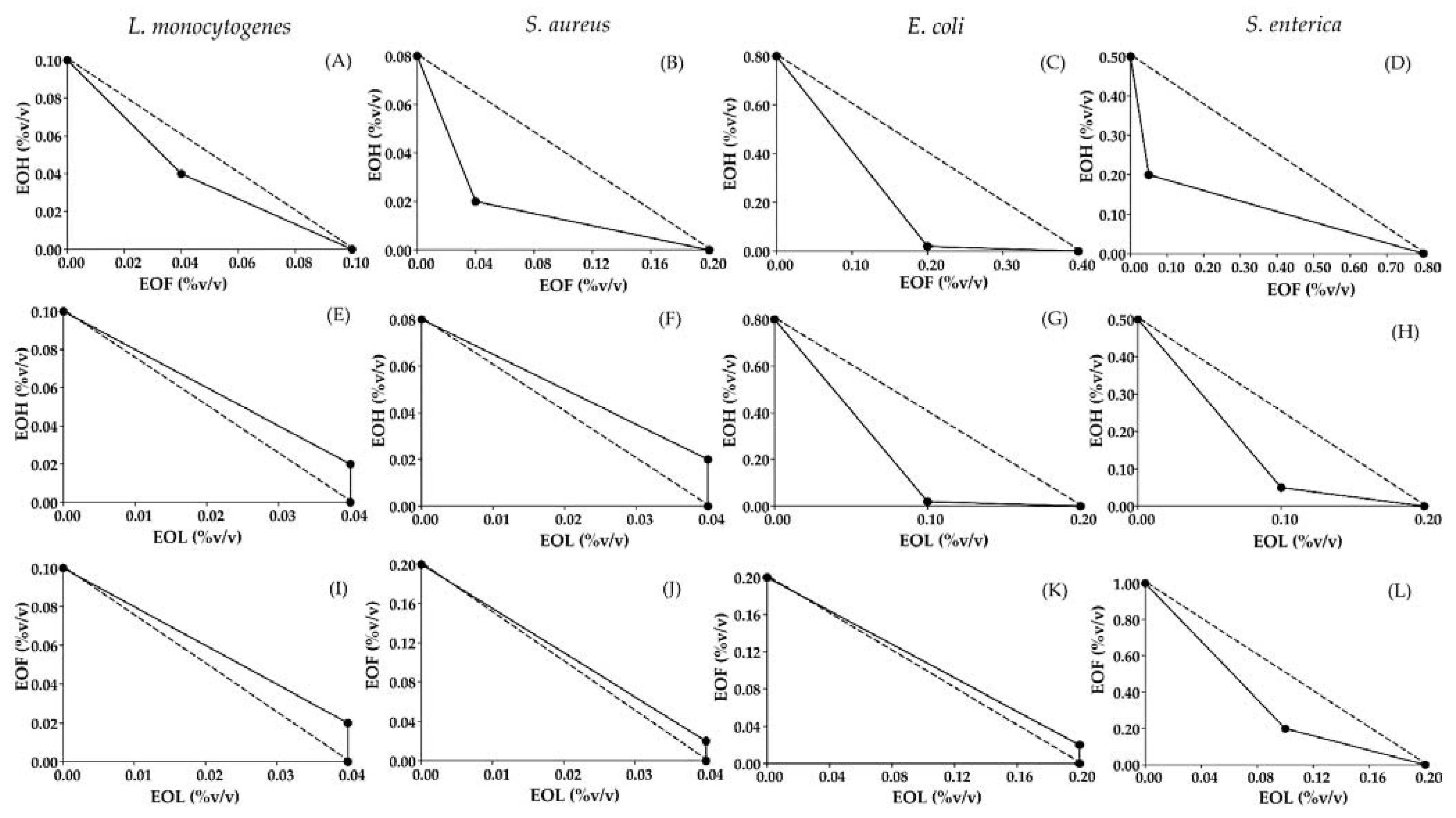
Figure 2.
Antilisterial activity of chitosan film incorporated with EOF:EOH at 0.04:0.04 (Chitosan film + 0.04% EOs) and chitosan film incorporated with EOF:EOH at 0.08:0.08 (Chitosan film + 0.08% EOs). Error bars represent ± standard deviation from three replicates. Bars of the same treatment, followed by different capital letters indicate significant differences (p ≤ 0.05). Bars of the same time followed by different small letters indicate significant differences (p ≤ 0.05).
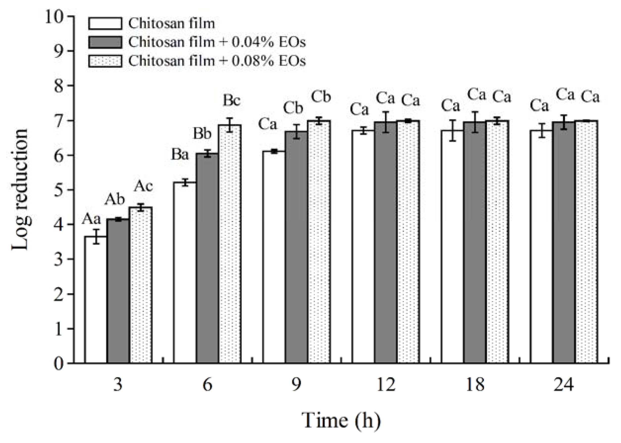
Figure 3.
Number of L. monocytogenes at 4 °C (A), 10 °C (B) and total viable counts at 4 °C (C), 10 °C (D) on bologna stored for seven days. Error bar represents standard deviation over three repeats.
Figure 3.
Number of L. monocytogenes at 4 °C (A), 10 °C (B) and total viable counts at 4 °C (C), 10 °C (D) on bologna stored for seven days. Error bar represents standard deviation over three repeats.
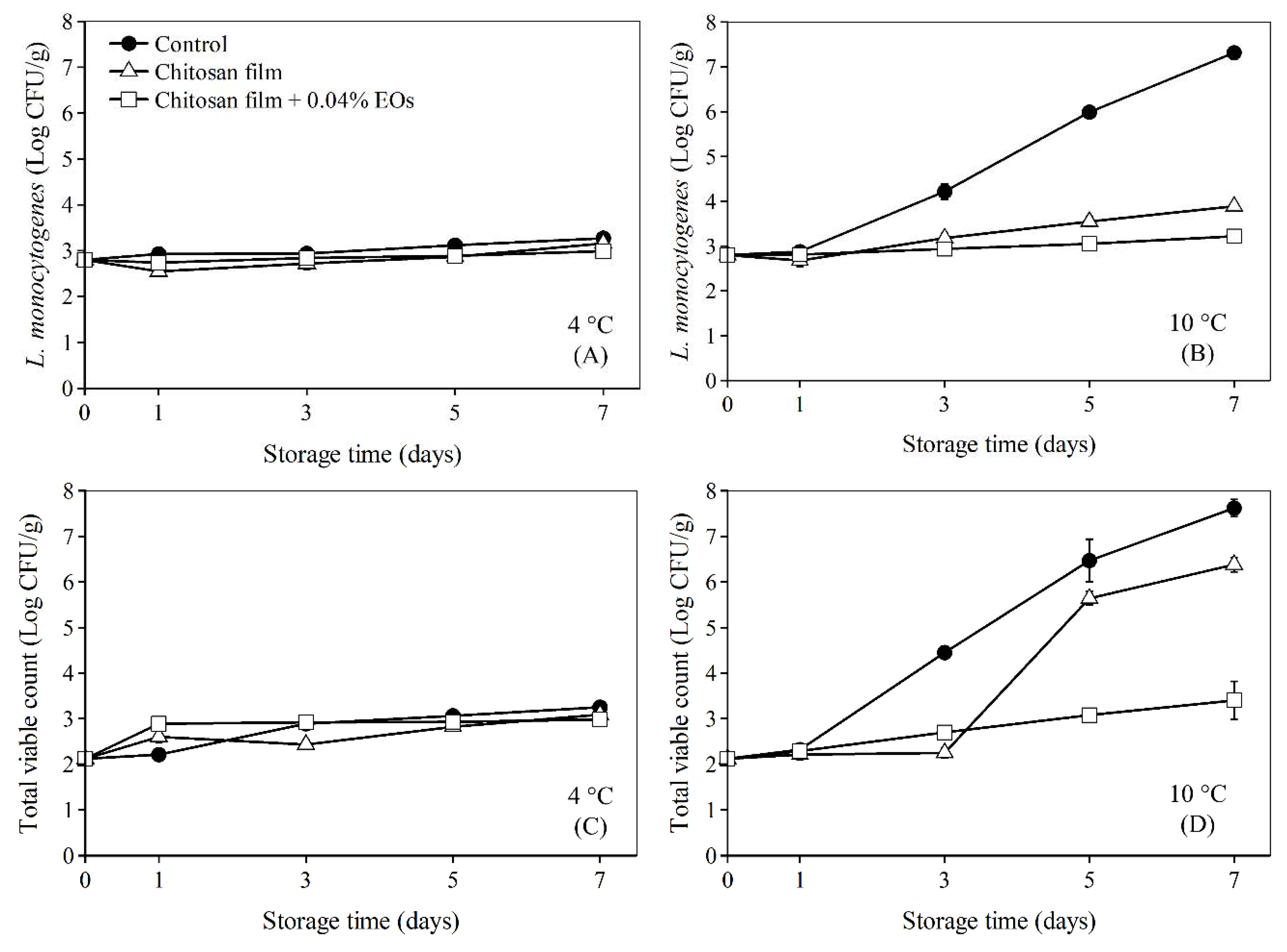
Figure 4.
Antimicrobial film from chitosan (A) and chitosan film + 0.04% EOs (B).


Table 1.
Minimum inhibitory concentrations (MICs) of various essential oils against L. monocytogenes, S. aureus, E. coli and S. enterica determined by agar dilution method.
Table 1.
Minimum inhibitory concentrations (MICs) of various essential oils against L. monocytogenes, S. aureus, E. coli and S. enterica determined by agar dilution method.
| Bacterium | MIC (%) of Essential Oils | ||
|---|---|---|---|
| Fingerroot | Holy Basil | Lemongrass | |
| L. monocytogenes | 0.30 | 0.10 | 0.10 |
| S. aureus | 0.10 | 0.10 | 0.10 |
| E. coli | 0.40 | 0.50 | 0.20 |
| S. enterica | 0.40 | 0.50 | 0.50 |
Table 2.
Fractional inhibitory concentration index (FICindex) of EOF, EOH and EOL against L. monocytogenes, S. aureus, E. coli and S. enterica a.
Table 2.
Fractional inhibitory concentration index (FICindex) of EOF, EOH and EOL against L. monocytogenes, S. aureus, E. coli and S. enterica a.
| Microorganism | Ratio of Essential Oil Combinations | FIC | FICindex | |||
|---|---|---|---|---|---|---|
| EOF | EOH | EOL | ||||
| L. monocytogenes | EOF:EOH | 0.50:0.50 | 0.04 | 0.04 | - | 0.80(S) b |
| EOH:EOL | 0.33:0.67 | - | 0.20 | 0.10 | 1.20 (AN) | |
| EOF:EOL | 0.33:0.67 | 0.20 | - | 0.10 | 1.20 (AN) | |
| S. aureus | EOF:EOH | 0.33:0.67 | 0.20 | 0.10 | - | 0.45(S) |
| EOH:EOL | 0.33:0.67 | - | 0.25 | 0.10 | 1.25 (AN) | |
| EOF:EOL | 0.33:0.67 | 0.10 | - | 0.10 | 1.10 (AN) | |
| E. coli | EOF:EOH | 0.09:0.91 | 0.50 | 0.30 | - | 0.53 (S) |
| EOH:EOL | 0.17:0.83 | - | 0.03 | 0.50 | 0.53 (S) | |
| EOF:EOL | 0.09:0.91 | 0.10 | - | 1.10 | 1.10 (AN) | |
| S. enterica | EOF:EOH | 0.80:0.20 | 0.06 | 0.40 | - | 0.46 (S) |
| EOH:EOL | 0.33:0.67 | - | 0.10 | 0.50 | 0.60 (S) | |
| EOF:EOL | 0.67:0.33 | 0.20 | - | 0.50 | 0.70 (S) | |
a FIC value was calculated by dividing MIC of an antimicrobial when used in combination with MIC of the antimicrobial when used alone. FIC was the sum of FIC values of the two antimicrobials used in combination. b The antimicrobial effect of combinations was synergistic (S, FIC < 1), additive (A, FIC = 1) or antagonistic (AN, FIC > 1).
Table 3.
Mechanical and physical properties of chitosan film and chitosan film incorporated with EOF:EOH at 0.04:0.04 (Chitosan film + 0.04% EOs). Values are mean ± standard deviation. Different letters in the same column indicate significant differences between the means using t-test (p ≤ 0.05).
Table 3.
Mechanical and physical properties of chitosan film and chitosan film incorporated with EOF:EOH at 0.04:0.04 (Chitosan film + 0.04% EOs). Values are mean ± standard deviation. Different letters in the same column indicate significant differences between the means using t-test (p ≤ 0.05).
| TYPE OF FILM | Mechanical Properties | Color | |||||
|---|---|---|---|---|---|---|---|
| Thickness (mm) | Tensile Strength (MPa) | Elongation at break (%) | WVTR (g·m−2·d−1) | L* | a* | b* | |
| Chitosan | 0.10 ± 0.01 a | 14.78 ± 2.71 b | 13.49 ± 6.59 a | 16.22 ± 0.62 a | 25.24 ± 3.52 a | −1.81 ± 0.21 a | 2.91 ± 0.75 a |
| Chitosan film + 0.04% EOs | 0.11 ± 0.01 a | 7.88 ± 1.54 a | 5.61 ± 0.55 a | 25.88 ± 5.19 b | 26.51 ± 6.38 a | −1.90 ± 0.56 a | 4.24 ± 2.01 a |
Publisher’s Note: MDPI stays neutral with regard to jurisdictional claims in published maps and institutional affiliations. |
© 2021 by the authors. Licensee MDPI, Basel, Switzerland. This article is an open access article distributed under the terms and conditions of the Creative Commons Attribution (CC BY) license (https://creativecommons.org/licenses/by/4.0/).
Share and Cite
MDPI and ACS Style
Tantala, J.; Rachtanapun, P.; Rachtanapun, C. Synergistic Antimicrobial Activities of Thai Household Essential Oils in Chitosan Film. Polymers 2021, 13, 1519. https://doi.org/10.3390/polym13091519
AMA Style
Tantala J, Rachtanapun P, Rachtanapun C. Synergistic Antimicrobial Activities of Thai Household Essential Oils in Chitosan Film. Polymers. 2021; 13(9):1519. https://doi.org/10.3390/polym13091519
Chicago/Turabian StyleTantala, Juthamas, Pornchai Rachtanapun, and Chitsiri Rachtanapun. 2021. "Synergistic Antimicrobial Activities of Thai Household Essential Oils in Chitosan Film" Polymers 13, no. 9: 1519. https://doi.org/10.3390/polym13091519
Note that from the first issue of 2016, this journal uses article numbers instead of page numbers. See further details here.