Review

Polymers for Protein Conjugation

Pharmaceutical and Pharmacological Sciences Department, University of Padova, Via F. Marzolo 5, Padova 35131, Italy
Polymers 2014, 6(1), 160-178; https://doi.org/10.3390/polym6010160
Submission received: 3 December 2013 / Revised: 30 December 2013 / Accepted: 7 January 2014 / Published: 13 January 2014
(This article belongs to the Special Issue Polymers for Drug Delivery)

Abstract

:
Polyethylene glycol (PEG) at the moment is considered the leading polymer for protein conjugation in view of its unique properties, as well as to its low toxicity in humans, qualities which have been confirmed by its extensive use in clinical practice. Other polymers that are safe, biodegradable and custom-designed have, nevertheless, also been investigated as potential candidates for protein conjugation. This review will focus on natural polymers and synthetic linear polymers that have been used for protein delivery and the results associated with their use. Genetic fusion approaches for the preparation of protein-polypeptide conjugates will be also reviewed and compared with the best known chemical conjugation ones.

1. Introduction, the Success of Polyethylene Glycol (PEG) in Protein Conjugation

The number of biologic agents used in clinical practice has increased considerably over the past few decades, and their global market is expected to reach $239 billion by 2015 [1]. Advances in the use of proteins as therapeutic agents have brought to the forefront several new advantages, such as selective and specific activity, limited toxicity and the potential for treating diseases in connection to their genetic defects. The other side of the medal is that biologics are not easy to develop or to produce and pose several challenges with regard to their stability, formulation, delivery route, optimal final dosage form and immunogenicity. These challenges are linked to the three-dimensional structure of proteins, which is the main cause of their physical instability, and to the chemical degradation profile of amino acids. Formulating a stable protein implicates, therefore, finding a balance between several parameters, and for each protein, a stable formulation is made in its own specific way. The views that proteins are delicate molecules and that chemical modifications could affect their integrity were logical assumptions that worked against efforts to conjugate proteins to polymers for their delivery. It should be remembered, nevertheless, that Mother Nature has provided the means for post-translational modifications to modify protein activity, stability and pharmacokinetics by attaching chemical groups. Pioneering studies in the field of protein modification with polymers were conducted by Davis and Abuckowski on polyethylene glycol (PEG) and bovine albumin and by Torchilin on dextran and streptokinase [2,3,4]. These studies paved the way for the conjugation of polymers to proteins, a technique that has improved the effectiveness of biologics by overcoming important shortcomings, such as instability, short half-lives and immunogenicity, which have often limited their use in clinical practice.
Among the polymers studied, PEG has emerged as the most interesting, versatile one, and nine PEG-protein conjugates are, in fact, currently being used in clinical practice [5]. The reasons for its success reside in many advantageous properties, such as its hydrophilicity, non-toxicity and non-immunogenicity. The approval of PEG use in humans by Food and Drug Administration marked the beginning of its safe use in clinical practice. Researchers have continued to consider PEG the first choice to improve the biocompatibility, half-life and safety of proteins, nanoparticles, liposomes, micelles and other drug delivery systems, a view that has been supported by a number of studies confirming that it has limited toxicity in animals and humans [6]. PEG has unique solvation properties that are due to the coordination of 2–3 water molecules per ethylene oxide unit [7]. This high solvation, together with the great flexibility of the polymeric backbone, confers biocompatibility, non-immunogenicity and non-antigenicity to the polymer. Another consequence of solvation is that PEG has an apparent molecular weight 5–10 times higher than that of a globular protein of a comparable mass, as verified by gel permeation chromatography [8]. A single PEG molecule is therefore able to cover an extended surface of a conjugated protein, thus preventing the approach of proteolytic enzymes and antibodies. The chemistry of polymer conjugation to proteins has been mainly developed with PEG, so nowadays, several PEGylating agents are available with different reactivity, molecular weight and structure.
To list the several PEGylation approaches is beyond the scope of this review, so only a brief description of site-selective chemical and enzymatic approaches will be reported. Protein amino PEGylation, as a coupling strategy, presents the drawback of yielding a mixture of several conjugate isomers with a great heterogeneity, as a consequence of the many amines present per protein molecule. The most used approach for a selective amino PEGylation is the coupling at the level of the alpha amino group at the protein N-terminus, which is feasible because of its lower pKa with respect to that of an epsilon amine in a lysine [9]. Thiol PEGylation is very selective and fast, although its feasibility is limited by the rare occurrence of an unpaired cysteine or, when this amino acid is genetically inserted in a desired position, by disulfide scrambling and protein dimerization [10]. In the literature, there are several examples of successful thiol conjugations, and remarkably, a conjugate with an anti-TNF-α, CIMZIA®, was approved for clinical use [8,11,12]. Bridging PEGylation is an innovative approach to link PEG at the level of protein disulfide bridges. The method requires a selective reduction of one disulfide bridge followed by the coupling with a tailor-made PEGylating agent, a bis-sulfone PEG, which reacts, in turn, with both thiol groups, forming a new bridge composed of three carbon atoms [13].
Enzymatic PEGylation is one of the most recent approaches to link PEG to a protein. In this case, the polymer coupling is mediated by an enzyme that offers a great selectivity and the possibility to modify amino acids that otherwise cannot be addressed by a chemical method of conjugation. In this field, the main examples of enzymatic PEGylation are: (i) transglutaminase mediated PEGylation, in which the enzyme catalyzes the transfer of an amino-PEG to the acyl group in the side chain of a glutamine [14]; (ii) glycoPEGylation, which requires two enzymes, an O-GalNAc-transferase and a sialyltransferase, to couple PEG at the level of specific serines and threonines, thus mimicking O-glycosylation [15]; (iii) sortase A mediated PEGylation, to conjugate the polymer at the level of the protein C-terminus after the addition of a specific amino acid substrate sequence (LPXTG motif) [16].
Other future directions in the development of PEGylation include: (i) releasable PEGs, which are polymers linked to the protein through cleavable linkers that allows the slow release of the conjugated protein in its full active form [17,18,19,20,21,22,23]; (ii) non-covalent PEGylation, which can be mediated through either hydrophobic interactions or the formation of coordination complexes [24,25,26]; and (iii) new PEG forms; in fact, the classic linear or branched structures of PEG can be further modified to obtain new copolymers with improved features, such as the comb shape PEGs [27,28,29].
Despite the great success of PEG, this polymer might not, however, be the perfect and optimal polymer for every application and does, in any case, present some drawbacks, listed below, which could limit its use:
(1)
As it is not biodegradable, high molecular weight PEGs should be used carefully and always at molecular weights below the kidney clearance threshold;
(2)
Specific anti-PEG antibodies have been detected in the serum of patients treated with PEG-asparaginase [30] and PEG-uricase [31], resulting in a neutralizing effect with a loss of therapeutic efficacy. Some investigators have detected the presence of pre-existing IgM and IgG anti-PEG antibodies in about 25% of patients who have never received prior treatment with PEG drugs [32,33];
(3)
While controversial kidney cell vacuolization has been observed in animals following multiple dose administration of PEG-conjugated hemoglobin [34] and TNF-R binding proteins [35], it has never been found when the polymer was administered alone or with other conjugates, and this has confirmed its safety. The finding, nonetheless, highlights another potential problem linked to the use of non-biodegradable, high molecular weight PEGs;
(4)
The large number of patents connected to the use of PEGs in drug delivery compositions might hamper or prevent the development of new conjugates.
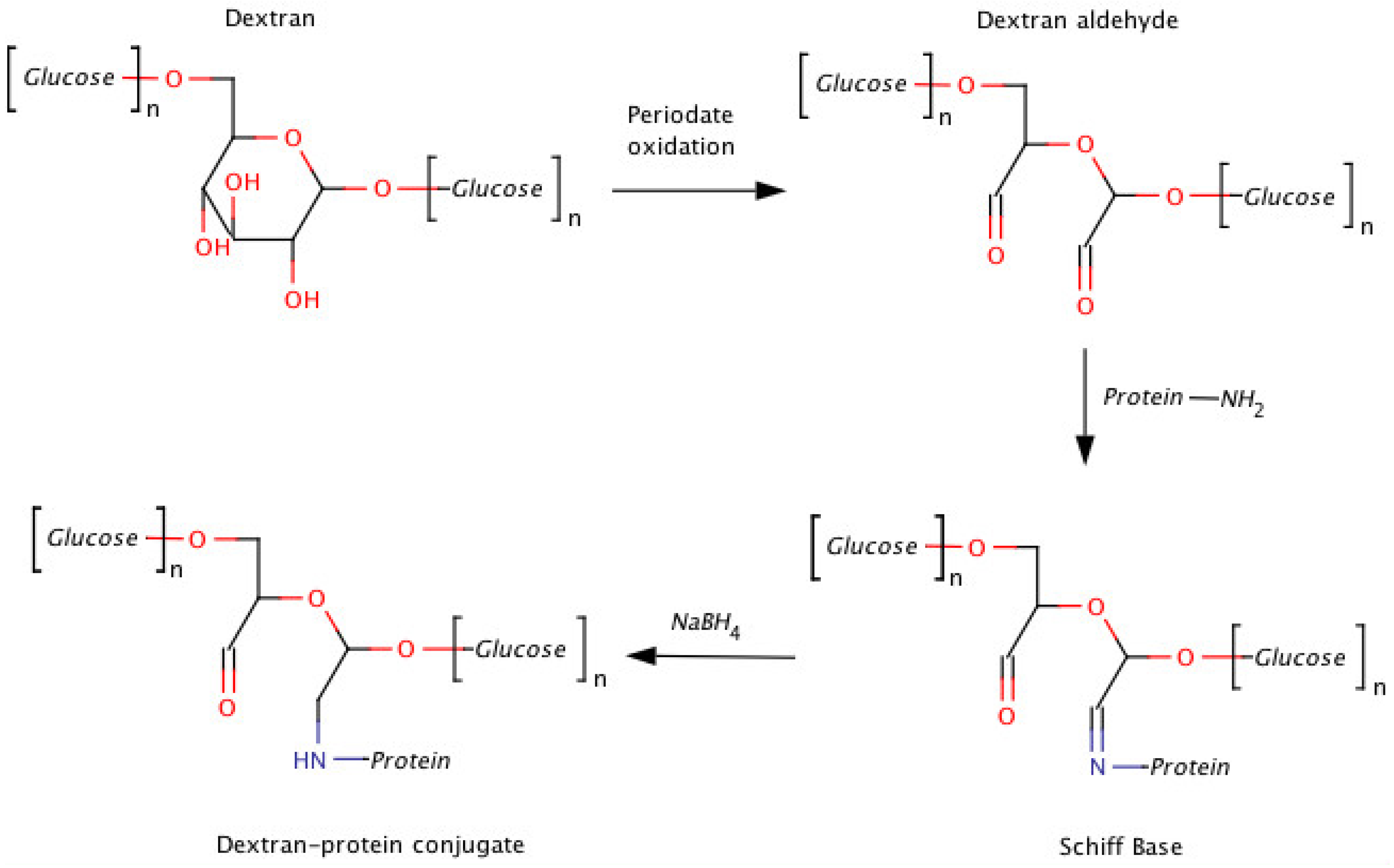
The presence of anti-PEG antibodies is probably one of the most debated concerns linked to PEG, and no clear conclusions have as yet been formulated. Although anti-PEG antibodies have indeed been described by some studies, they represent only a small percentage of the total number of cases of PEG-based therapeutics in clinical practice. Interestingly, this problem seems to be restricted to large PEG conjugates cross-linked with highly immunogenic enzymes (i.e., asparaginase, uricase, etc.) [36,37]. Some have suggested that the size of conjugates obtained by extensive PEGylation of high molecular weight proteins is similar to that of small PEGylated liposomes or nanoparticles, which subsequently lead to the so-called accelerated blood clearance (ABC) phenomenon involving complement activation by IgM [38,39]. As underlined by Schellekens and colleagues, many factors linked to immunogenicity warrant further study [40]. In fact, as that research group pointed out, most, if not all, assays for anti-PEG antibodies are flawed and lack specificity; as a consequence, the biological effects ascribed to anti-PEG antibodies are not well defined.
There are, moreover, other considerations that animate research projects aiming to develop other polymers for protein conjugation, such as the possibility of adding new features to conjugates in terms of biodegradability, targeting, combining activities, etc. The future will probably see custom-designed, personalized polymers for specific applications rather than a fit-all agent, like PEG, used indiscriminately in all sectors.
This review will focus on some of the most relevant polymers for protein modification apart from PEG, which has already been introduced here and in others’ dedicated works [39,41,42].

2. Dextran

Dextrans (initially approved as plasma expanders) were probably the first and most studied in this class of polymers [43,44]. Produced by bacteria, the polymer is composed of glucose monomers linked together mainly by α-1,6-glucosidic linkages forming a linear polymer with some degree of branching via 1,3-glucosidic linkages [45]. The degree of branching, ranging from 0.5% to 60% depending on the polymer source, affects its water solubility, as the most branched dextrans are the least soluble ones [46]. Their pharmacokinetics are directly proportional to their MW. Detectable dextran levels have been found in the serum of rats after 12 h for MW >40 kDa [47]. In vivo dextran is slowly depolymerized by dextranases mainly present in the liver and spleen [45], but this degradation pathway is only marginally relevant for conjugates, because chemical modifications of the dextran backbone further reduce the rate of such degradation [48,49].
In early studies, dextran was conjugated to streptokinase (streptodekase) and subsequently used in clinical practice in Russia, becoming the first protein conjugate to be tested in humans [4]. The polymer was oxidized by periodate yielding aldehyde groups, which, in turn, were reacted with protein amino groups (Figure 1). Although this method is still currently applied to couple proteins to dextran and polysaccharides in general, it is affected by the multiplicity of binding groups in the polymer backbone that might yield undesired cross-linked bonds if coupling conditions are not well controlled. Two aldehyde groups are, moreover, generated for each oxidized unit when polysaccharide is treated with sodium periodate. As this approach yields two aldehyde groups (e.g., RO–CH(R′)–CHO and RO–CH(OR′)–CHO) in close proximity, where R and R’ are the polysaccharide backbone (Figure 1), it might cause problems due to the differences in the reactivity of aldehydes and to the difficulty in determining which aldehyde group is involved in the protein coupling. That both aldehydes react with the proteins, moreover, is a possibility that is difficult to exclude. As a consequence, if this approach is not properly optimized, there may be constraints in the heterogeneity of the final conjugate.
This coupling strategy has, nevertheless, been utilized to prepare several dextran-protein conjugates. Antitumoral dextran (≥40 kDa) asparaginase conjugate, for example, was found to have an extended half-life with respect to asparaginase, both in non-immune and in asparaginase-immune rabbits [50,51]. The conjugated enzyme retained about 50% of the initial activity and was well tolerated, even after repeated injections. Dextran-carboxypeptidase G2 is another conjugate that has been investigated as an antitumor agent used to deplete folic acid [52], which is essential for tumor cells. In this case, protein conjugation was achieved using cyanogen bromide, an approach that is difficult to standardize, because conjugates with different structures are formed [45,53]. Investigators have demonstrated a direct correlation between the MW of the coupled dextran and the increase in the conjugate’s half-life [52]. Other examples of protein-dextran conjugates include: uricase [54], superoxide dismutase [55], insulin [56] and hemoglobin [57,58]. Readers can refer to Mehva’s review for an exhaustive overview of all dextran conjugates, including those with low MWs [43]. Briefly, then, in all cases, dextran conjugation was characterized by: (i) the good retention of protein activity even for those proteins (i.e., insulin or antibodies) requiring interaction with others (proteins) to perform their biological functions; (ii) pharmacokinetics dependent on dextran’s MW; and (iii) the ability to reduce the immunogenicity of heterologous proteins.
Figure 1. An outline of dextran oxidation by periodate and subsequent protein coupling by reductive amination.
Polymers 06 00160 g001
Although dextran is currently commercially available with a wide range of MWs and a lower polydispersity index (PI) with respect to that in the past (e.g., polymers with a PI of approximately two are accessible), it might still be less practical than synthetic polymers. The latter, in fact, can be obtained with a greater control of the PI for even high MWs, and the heterogeneity of dextrans coming from backbone branching is transferred to the final conjugate, which hinders lot-lot reproducibility/consistency and meeting regulatory requirements. Further concerns that have delayed the development of dextran conjugates are linked to the rare cases of anaphylactic reactions to high MW dextrans that have been reported.

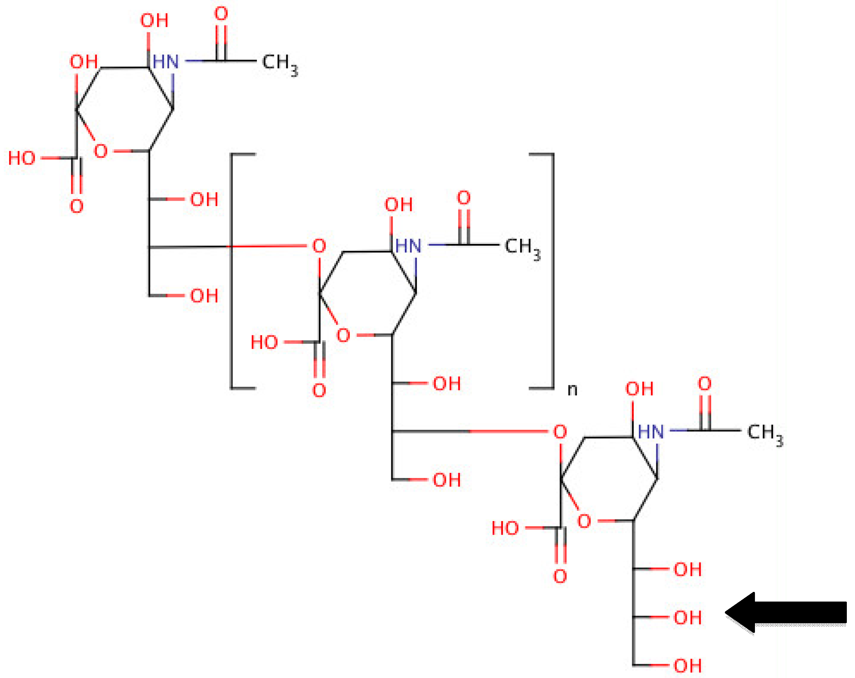
3. Polysialic Acids (PSAs)

Polysialic acids (PSAs) are natural, biodegradable polymers with a linear structure composed of N-acetylneuraminic acid (Figure 2). Several bacteria exploit its hydrophilicity to achieve stealth and protection. PSA covered bacteria can, in fact, prevent host complement activation and phagocytic activity and can thus overcome natural body defenses. Sialic acid, one of the major components of the glycans of glycosylated proteins, reduces the tendency to aggregate and, thus, prolongs drug action. In view of these properties, Gregoriadis proposed that PSA be used as a polymer to improve a drug’s pharmacokinetic profile and to reduce the immunogenicity of the chemical protein conjugation [59,60]. The protective role played by PSA in nature could presumably preserve the protein’s structure and, thus, prolong its pharmacological action. PSAs with different MWs are produced from bacteria, and the α-(2→8)-linked serogroup B capsular polysaccharide from Escherichia coli K1 is considered the most suitable for the polysialylation of drugs and proteins [61].
Figure 2. The structure of polysialic acid: the arrow indicates the site where an aldehyde group is introduced by periodate oxidation.
Polymers 06 00160 g002
Conjugated to human proteins, including interferon α-2b [59], insulin [62], IgG fab fragments [63], as well as to heterologous proteins, such as asparaginase [64,65], PSAs have been shown to reduce the immunogenicity of linked proteins. Between 10 and 60 kDa, the MW range of PSA used for protein conjugation ensures a half-life extension directly proportional to the MW of the coupled polymer. In most cases, PSA was activated by controlled periodate oxidation, making it possible to achieve a single aldehyde group at the non-reducing end of the polymer, where three vicinal hydroxyl groups are present. This approach yields an end chain functionalized PSA with a single aldehyde per polymer chain, thereby avoiding potential cross-linking problems in the subsequent conjugation with proteins [62]. The aldehyde-PSA can be exploited for amino coupling through reductive amination at the level of the lysine amino group when the reaction is performed in alkaline buffers or at the level of the protein N-terminus when the coupling is conducted in acidic buffers. Alternatively, aldehyde activated PSA can be converted into a maleimide-PSA, allowing thiol to be coupled to the protein’s free cysteine [66].
Interestingly, it has recently been demonstrated that PSA conjugates can be obtained by enzymatically transferring PSA chains to N- or O-linked glycans of a protein by means of bacterial enzymes, such as sialyltransferase Cst-II [67]. This approach further enhances the potential of polysialylation, offering a new tool for site-selective protein conjugation.
While PSAs’ polydispersity may still be higher than that of synthetic polymers, using relatively low MW (10–20 kDa) chains to augment half-life times will probably overcome this limit.

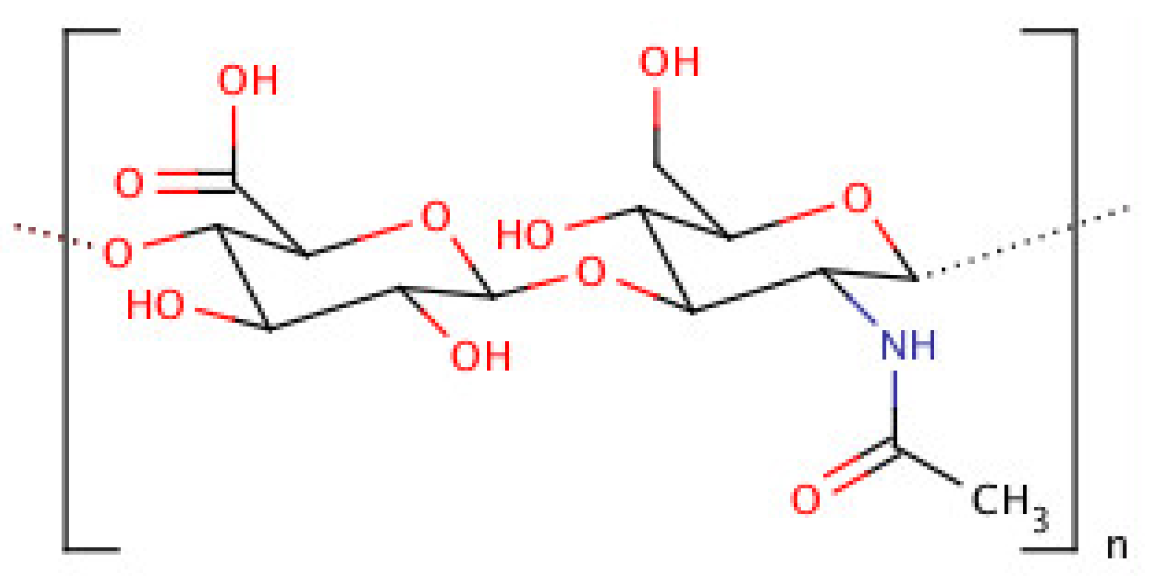
4. Hyaluronic Acid (HA)
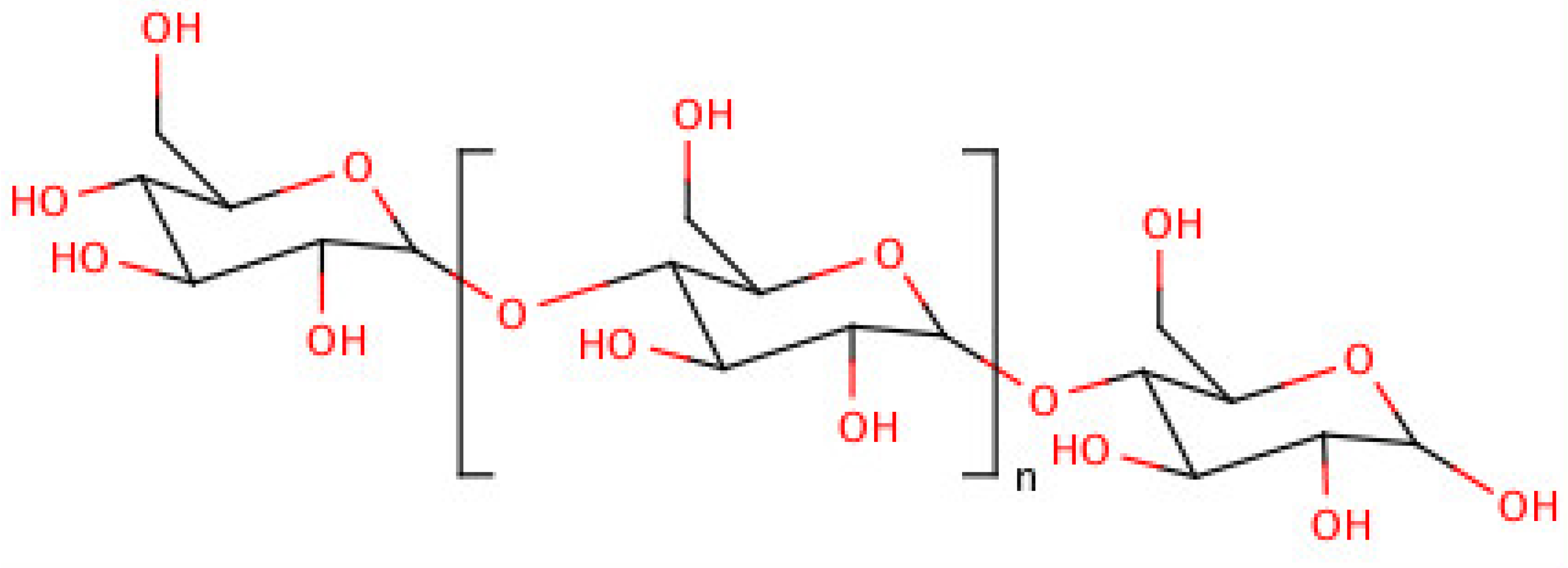
A key component of extracellular matrix, cartilage and vitreous compartments in vertebrates, hyaluronic acid is a natural polysaccharide [68]. Hyaluronic acid (HA) is unique, as it has a relatively simple linear structure of repeating disaccharide units composed of d-glucuronic acid and N-acetyl-d-glucosamine (Figure 3) [69]. The FDA has already approved its utilization for human use, and among its various therapeutic applications, the polymer is currently used as a viscosupplement to treat joints in patients affected with arthritis. Its clinical safety is well established, and it is also contained in several injectable products for cosmetic applications [70].
Figure 3. The structure of hyaluronic acid (HA).
Polymers 06 00160 g003
To date, HA’s potential for use in drug delivery systems has been investigated as a carrier of anti-tumoral and anti-inflammatory drugs [71,72]. The conjugation of biologically active substances to HA is straightforward through a direct activation of the carboxylic acid groups present along the polymer backbone. A chemical approach suitable for amino coupling might present cross-linking limits for HA conjugation to proteins. As a consequence, the periodate oxidation procedure, with the pros and cons described above for dextran, has also been applied to HA for conjugation to proteins, for example, interferon α [73]. A different HA activation has been achieved by reacting some HA carboxylic groups with a spacer bearing a protected aldehyde group. Studies on the new aldehyde-HA have produced positive results with regard to protein conjugation with several different proteins and peptides, such as hGH, insulin and salmon calcitonin [74,75]. The most commonly used HAs for protein or drug conjugation have MWs in the range between 100 and 300 kDa. HAs higher than the upper limit tend to form water solutions with an increased viscosity that is proportional to the MW and which might not be suitable for applications in the field of conjugation.
Readers can refer to the work by Mero and Campisi found in this “Polymers for drug delivery” issue for a comprehensive review of HA’s potential as a carrier of drugs and proteins [76].

5. Dextrin

Dextrin is a polymer chain composed of d-glucose units joined together by α-1,4-glucosidic linkages that form a linear polymer with some degree of branching via 1,6-glucosidic linkages (Figure 4). This biodegradable polymer produced by the controlled hydrolysis of starch is degraded in vivo by the α-amylase present in extracellular fluids.
Figure 4. The structure of dextrin.
Polymers 06 00160 g004
The polymer was succinylated to achieve conjugation to proteins. This approach links a succinic acid molecule to some hydroxyl groups of dextrin, thus obtaining a carboxyl group suitable for protein conjugation after activation, and it can thus be tuned for derivative control. Dextrin with a degree of succinylation and MWs in the ranges between 9%–32% mol and 7700–47,200 Da, respectively, were used to modify different proteins, such as phospholipase A2 [77], trypsin [78], melanocyte stimulating hormone [78] and epidermal growth factor (rhEGF) [79,80]. Dextrin conjugation proportionally reduced enzyme activity depending on the MW and the degree of succinylation.
A new concept towards protein modification, called polymer-masking-unmasking-protein therapy (PUMPT), has been tested on dextrin [78]. The hypothesis was that polymer conjugation would protect a protein and mask its activity in transit while enabling controlled recovery of activity at the target site by triggered degradation of the polymeric component. First tested with trypsin, the approach was further developed with rhEGF by preparing a dextrin-rhEGF conjugate to treat excisional wounds using topical applications in the diabetic mouse [79].
The use of dextrin described here is an example in which an intrinsic property of a polymer was exploited to achieve a conjugate with an important therapeutic effect for tissue repair. In order to avoid the risk of cross-linking connected to the carboxylic activation of a highly succinylated dextrin and to enhance its potential as a conjugating polymer applicable in systemic treatments, the chemistry of the protein conjugation should be improved by pursuing new approaches that can ensure a single point of attachment on the protein.

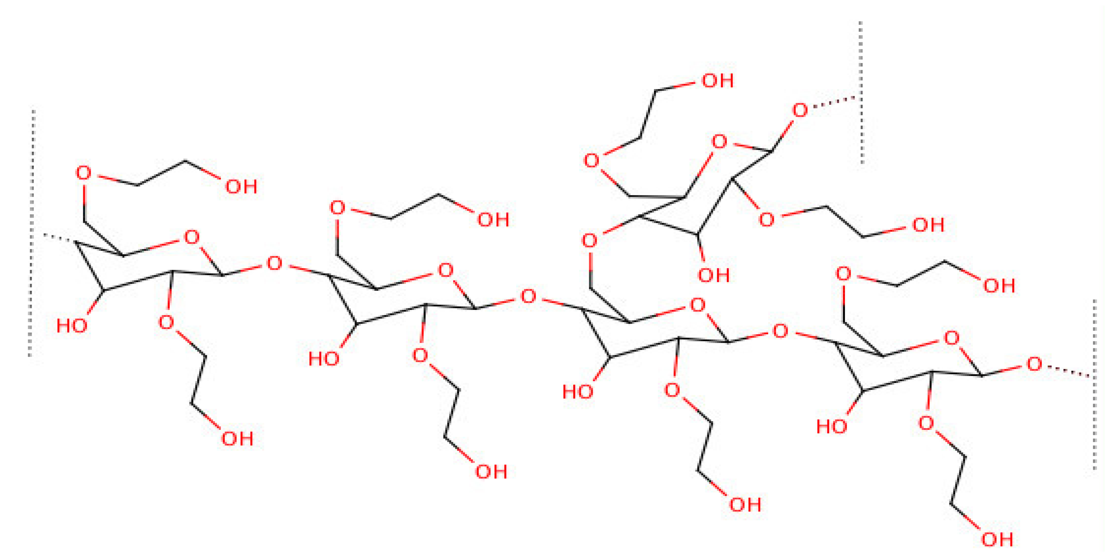
6. Hydroxyethyl-Starch (HES)

Hydroxyethyl-starch (HES) is a semi-synthetic non-ionic polysaccharide obtained by the chemical modification of the hydroxyl groups in the C2 and C6 position of starch (Figure 5). The polymer has been approved for human use as a blood plasma volume expander. In view of its high water solubility and the possibility of controlling the rate of biodegradation, some consider it a potential substitute for PEG. There are as yet few published studies on HESylation with proteins [81], nanoparticles [82] or low MW drugs [83], and most data concerning the chemical HESylation of proteins can be found in patent descriptions [84,85]. Interestingly, HES conjugation to erythropoietin (EPO) was achieved at the protein glycan position after mild oxidation with the periodate of some EPO carbohydrates followed by conjugation with a hydroxylamine HES derivative. Protein conjugation with HES was also successfully achieved by employing transglutaminase (TGase) mediated conjugation. This enzyme is able to transfer the primary amino group (amino donor) to the acyl group of glutamine (acyl donor). Enzymatic coupling has, in particular, been obtained using two strategies: the first by preparing HES derivatives bearing Z-Glu-Gly sequences, known acyl donor substrates of TGase, useful for protein conjugation to lysine; the second by synthesizing amino-HES polymers that can act as an amino donor substrate in a TGase mediated reaction directed towards the glutamines of a given protein [81]. As the presence of multi-attaching points in the HES backbone might lead to the formation of cross-linked conjugates, the reaction time must be properly optimized to avoid this risk.
Figure 5. Schematic representation of hydroxyethyl-starch (HES).
Polymers 06 00160 g005
While HES appears to be a promising polymer for protein conjugation, recent concerns about its safety have emerged in connection to an increased risk of death and kidney injury noted in critically ill patients who received the drug as a plasma expander [86,87]. In view of these reports, the European Medicines Agency’s Pharmacovigilance Risk Assessment Committee has recommended that solutions for infusions containing hydroxyethyl starch no longer be used to treat patients with sepsis (bacterial infection in the blood) or burn injuries or the critically ill, because of the increased risk of kidney injury and mortality [88].

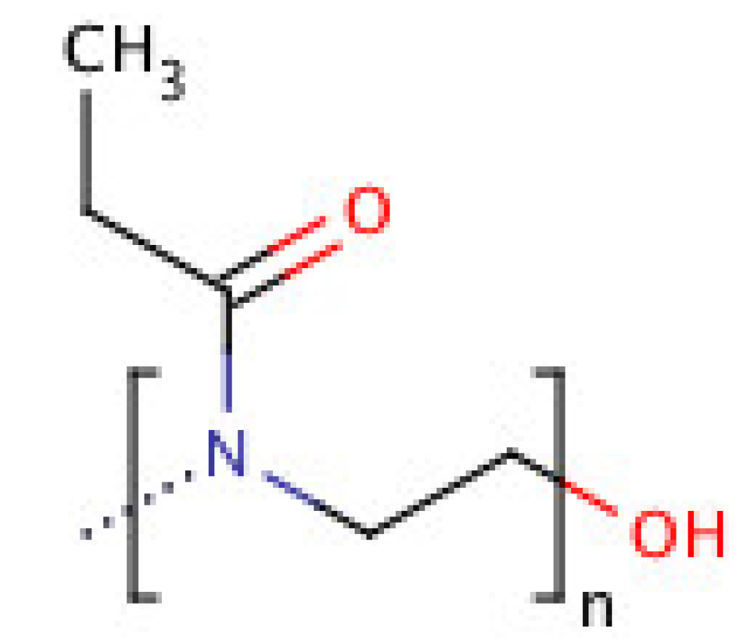
7. Poly(2-ethyl 2-oxazoline) (PEOZ)

Poly(2-ethyl 2-oxazoline) (PEOZ)(Figure 6) is a linear synthetic polymer that is considered to be a promising substitute for PEG in view of its stealth properties [89,90,91]. PEOZ is soluble in water and in many organic solvents [92]. Like PEG, PEOZ can be synthesized with low polydispersity and can have a single functional group at one extreme, useful for protein conjugation, which is obtained during the initiation [93,94,95] or termination [96,97] steps of polymerization. The pharmacokinetics of PEOZ is directly proportional to its MW and, consequently, to its hydrodynamic volume, which is slightly lower than that of a PEG having the same MW [98]. Studies in mice using radiolabelled poly(2-alkyl-oxazoline) showed that these polymers do not accumulate in the body and are mainly cleared by the kidneys and only to a small extent by the liver [99].
Figure 6. The structure of poly(2-ethyl 2-oxazoline) (PEOZ).
Polymers 06 00160 g006
It was relatively easy to transfer the chemistry expertise and know-how behind polymer activation and coupling to PEOZ in view of its similarity to PEG [91,100]. PEOZ can be produced with a terminal hydroxyl group that can be activated and, in turn, reacted with spacers bearing common functional groups and yielding carboxyl, amino, maleimide or aldehyde PEOZs. These derivatives offer a wide selection of protein coupling chemistries, and it is also possible to perform enzymatic mediated protein conjugation with PEOZ [98]. As demonstrated by the preserved enzymatic activity of PEOZylated uricase, catalase and ribonuclease and the preserved secondary structure of coupled proteins [98], PEOZ does not interact with the protein surface [100]. Its stealth properties have been confirmed, as the PEOZ-bovive serum albumin (BSA) conjugate is significantly less immunogenic with respect to BSA or PEG-BSA [100].
PEOZ-protein conjugates have, until now, been investigated only in animal models, but the polymer has produced promising results and no toxicity, even in the PEOZ form, with pendant groups used to conjugate low MW drugs [101].
The possibility of synthesizing PEOZ with pendant groups is perhaps the polymer’s main advantage with respect to PEG as far as the delivery of small drugs is concerned. PEOZ must, in any case, find its own specific application in the field of protein conjugation, or its similarities with PEG may become a disadvantage (or not a real advantage) with respect to a polymer like PEG, which boasts a long history of safe clinical use. The finding of a higher cellular uptake in Madin-Darby canine kiney (MDCK) and Caco-2 cell lines of horseradish peroxidase conjugated with polyoxazoline copolymers is interesting in view of the low membrane permeability of PEG conjugates [102]. Interested readers are referred to a recent review article for a complete overview concerning polyoxazoline polymers [103].

8. Protein Conjugates Exploiting Polypeptides as Carrying Polymers

The preparation of fusion proteins is a well-established genetic approach to achieve fused proteins with prolonged pharmacokinetics [104]. The advantage of this approach is that it is possible to create a fused protein with a precise MW increase, but the same protein expression, thereby preventing the typical polydispersity of synthetic polymers. The carrying protein can be fused at the N- or C-terminus of the target protein, and which is used depends on several variables. In most cases, albumin has been exploited as the carrying protein. A random polypeptide instead of a protein with a well-defined secondary or tertiary structure has been investigated as a carrying protein in the following two approaches.
These genetic fusion approaches are proposed as an alternative to chemical polymer to protein conjugation methods [105]. Their strengths, briefly mentioned above, are interesting, although the method may not easily be applied to all proteins. Highly immunogenic heterologous proteins or enzymes would not, for example, be suitable for this approach, because they require a strong protein surface coverage, which is better achieved with a random polymer conjugation. Some proteins are, moreover, unable to fold properly when fused to other polypeptides. Genetic fusion, nevertheless, represents one of the most important breakthroughs with regard to biologics.

8.1. XTEN Technology

XTEN technology is based on the genetic fusion of a gene encoding for an active protein with a gene encoding for long unstructured hydrophilic sequences of amino acids [106,107]. A XTEN peptide is composed of six amino acids (A, E, G, S and T) at different percentages. The sequence was selected from a large library by evaluating parameters, such as maximal expression, genetic stability, solubility, aggregation resistance, etc. [106]. The approach was tested using different peptides and proteins, such as exenatide, glucagon and hGH [106,107,108]. A novel recombinant human growth hormone (rhGH) fusion protein was designed by genetically fusing different XTEN sequences to the protein’s N- and C-terminus, obtaining a fused protein of about 119 kDa, with a five-fold MW increase with respect to the original hGH. Although the XTEN-hGH-XTEN protein showed a 12-fold reduction in hGH activity in vitro, its potency in vivo was increased, owing to its prolonged half-life (the terminal half-life was 110 h). A monthly dose of the fused protein in monkeys yielded a sustained pharmacodynamic response for one month, comparable to 0.05 mg/kg/day of rhGH, without any observed adverse effects [107].

8.2. PASylation

PASylation is an approach based on polypeptide genetic fusion similar to XTEN. In this case, an amino acid sequence is designed to achieve an unstructured polypeptide with high water solubility and the lack of charges. As preliminary studies with Gly-based polypeptides yielded unsatisfactory half-life prolongation [109], the next step was to develop polypeptides based on a combination of amino acids. The combinations of proline, alanine and serine forming polypeptides with high water solubility and properties similar to PEG were particularly interesting [110]. Different polypeptide lengths (200, 400 and 600 AA) were fused with therapeutic proteins, such as interferon, hGH and Fab fragments, and a proportional increment was demonstrated in the in vivo half-life in mice. The authors reported that fused proteins were stable in the blood circulation, and their biodegradability prevented organ accumulation. No toxicity was noted, and their unstructured nature presumably explained the absence of immunogenicity in these mice experiments [110]. In accordance with the known immunogenicity of hGH in mice, IgG antibodies reactive against the hGH moiety of PAS600-hGH were, however, detected in mice treated daily with the fused protein [110]. This finding suggests that, together with other genetic fusion approaches, PASylation may not be the delivery method of choice for highly immunogenic proteins, because it limits the polypeptide attachment by fusion at the N- and/or C-terminus, and, as a consequence, does not fully shield the protein surfaces, as does polymer conjugation.

9. Conclusions

Polymer conjugation has become one of the leading pharmaceutical technologies for the delivery of proteins. Its effectiveness in prolonging the in vivo terminal half-life and in reducing the immunogenicity of conjugated proteins has been proven by several studies and further corroborated by the profile of conjugates, until now, solely of PEG, currently in clinical use [5]. As PEG is still the gold standard for protein conjugation, new polymer candidates with ever greater advantages and fewer limits need to be designed and developed. The journey of new polymers that are quite similar to PEGylation, but without any significant improvements or personalized features or applications, will clearly encounter obstacles in view of PEG’s well established know-how and lack of toxicity. The idea of a single polymer utilized for several different applications does not, however, sit well with the increasing needs for responsive drug delivery systems and personalized medicine. It is probable that in the near future, polymers with unique tailor-made properties will be developed for specific applications despite the high development costs. It is even possible that a new polymer will be developed as a platform (backbone) to which different properties (i.e., stimuli responsive groups, imaging, different reactive groups) can be applied on demand.
Genetic fusion approaches to form specific, well-defined protein-polypeptide conjugates have also been presented in this review. Although conceptually different from chemical conjugation techniques, they are an interesting, important frontier. Together with chemical conjugation methods, their future development will no doubt contribute to modulating and enhancing the field of protein delivery.

Acknowledgments

The author thanks The Italian Ministry of Education, University and Research (MIUR; grant No. 60A04-1870/13).

Conflicts of Interest

The author declares no conflict of interest.

References

  1. Biosimilars and Follow-on Biologics Report: The Global Outlook 2010–2025. Visiongain Ltd.: London, UK, 2010. Available online: http://www.visiongain.com/Report/474/Biosimilars-and-Follow-On-Biologics-Global-Market-Outlook-2010-2025 (accessed on 9 January 2014).
  2. Abuchowski, A.; van Es, T.; Palczuk, N.; Davis, F. Alteration of immunological properties of bovine serum albumin by covalent attachment of polyethylene glycol. J. Biol. Chem. 1977, 252, 3578–3581. [Google Scholar]
  3. Abuchowski, A.; McCoy, J.R.; Palczuk, N.C.; van Es, T.; Davis, F.F. Effect of covalent attachment of polyethylene glycol on immunogenicity and circulating life of bovine liver catalase. J. Biol. Chem. 1977, 252, 3582–3586. [Google Scholar]
  4. Torchilin, V.; Voronkov, Y.I.; Mazaev, A. Use of immobilized streptokinase (Streptodekaza) for thrombosis treatment. Ter. Arkhiv 1982, 54, 21–26. [Google Scholar]
  5. Pasut, G.; Veronese, F.M. State of the art in PEGylation: The great versatility achieved after forty years of research. J. Control. Release 2012, 161, 461–472. [Google Scholar] [CrossRef]
  6. Webster, R.; Didier, E.; Harris, P.; Siegel, N.; Stadler, J.; Tilbury, L.; Smith, D. PEGylated proteins: Evaluation of their safety in the absence of definitive metabolism studies. Drug Metab. Dispos. 2007, 35, 9–16. [Google Scholar]
  7. Harris, J.M.; Chess, R.B. Effect of pegylation on pharmaceuticals. Nat. Rev. Drug Discov. 2003, 2, 214–221. [Google Scholar] [CrossRef]
  8. Manjula, B.; Tsai, A.; Upadhya, R.; Perumalsamy, K.; Smith, P.; Malavalli, A.; Vandegriff, K.; Winslow, R.; Intaglietta, M.; Prabhakaran, M. Site-specific PEGylation of hemoglobin at Cys-93(β): Correlation between the colligative properties of the PEGylated protein and the length of the conjugated PEG chain. Bioconjug. Chem. 2003, 14, 464–472. [Google Scholar] [CrossRef]
  9. Kinstler, O.; Moulinex, G.; Treheit, M.; Ladd, D.; Gegg, C. Mono-N-terminal poly(ethylene glycol)-protein conjugates. Adv. Drug Deliv. Rev. 2002, 54, 477–485. [Google Scholar] [CrossRef]
  10. Basu, A.; Yang, K.; Wang, M.; Liu, S.; Chintala, R.; Palm, T.; Zhao, H.; Peng, P.; Wu, D.; Zhang, Z. Structure-function engineering of interferon-β-1b for improving stability, solubility, potency, immunogenicity, and pharmacokinetic properties by site-selective mono-PEGylation. Bioconjug. Chem. 2006, 17, 618–630. [Google Scholar] [CrossRef]
  11. Veronese, F.M.; Mero, A.; Caboi, F.; Sergi, M.; Marongiu, C.; Pasut, G. Site-specific pegylation of G-CSF by reversible denaturation. Bioconjug. Chem. 2007, 18, 1824–1830. [Google Scholar] [CrossRef]
  12. Weir, N.; Athwal, D.; Brown, D.; Foulkes, R.; Kollias, G.; Nesbitt, A.; Popplewell, A.; Spitali, M.; Stephens, S. A new generation of high-affinity humanized PEGylated fab fragment anti-tumor necrosis factor-α monoclonal antibodies. Therapy 2006, 3, 535–545. [Google Scholar]
  13. Shaunak, S.; Godwin, A.; Choi, J.; Balan, S.; Pedone, E.; Vijayarangam, D.; Heidelberger, S.; Teo, I.; Zloh, M.; Brocchini, S. Site-specific PEGylation of native disulfide bonds in therapeutic proteins. Nat. Chem. Biol. 2006, 2, 312–313. [Google Scholar] [CrossRef]
  14. Mero, A.; Schiavon, M.; Veronese, F.M.; Pasut, G. A new method to increase selectivity of transglutaminase mediated PEGylation of salmon calcitonin and human growth hormone. J. Control. Release 2011, 154, 27–34. [Google Scholar] [CrossRef]
  15. DeFrees, S.; Wang, Z.; Xing, R.; Scott, A.E.; Wang, J.; Zopf, D.; Gouty, D.L.; Sjoberg, E.R.; Panneerselvam, K.; Brinkman-Van, der Linden; Els, C.M. GlycoPEGylation of recombinant therapeutic proteins produced in Escherichia coli. Glycobiology 2006, 16, 833–843. [Google Scholar] [CrossRef]
  16. Popp, M.W.; Dougan, S.K.; Chuang, T.; Spooner, E.; Ploegh, H.L. Sortase-catalyzed transformations that improve the properties of cytokines. Proc. Natl. Acad. Sci. USA 2011, 108, 3169–3174. [Google Scholar] [CrossRef]
  17. Zhao, H.; Yang, K.; Martinez, A.; Basu, A.; Chintala, R.; Liu, H.; Janjua, A.; Wang, M.; Filpula, D. Linear and branched bicin linkers for releasable PEGylation of macromolecules: Controlled release in vivo and in vitro from mono-and multi-PEGylated proteins. Bioconjug. Chem. 2006, 17, 341–351. [Google Scholar] [CrossRef]
  18. Greenwald, R.B.; Yang, K.; Zhao, H.; Conover, C.D.; Lee, S.; Filpula, D. Controlled release of proteins from their poly(ethylene glycol) conjugates: Drug delivery systems employing 1, 6-elimination. Bioconjug. Chem. 2003, 14, 395–403. [Google Scholar] [CrossRef]
  19. Greenwald, R.B.; Choe, Y.H.; Conover, C.D.; Shum, K.; Wu, D.; Royzen, M. Drug delivery systems based on trimethyl lock lactonization: Poly(ethylene glycol) prodrugs of amino-containing compounds. J. Med. Chem. 2000, 43, 475–487. [Google Scholar] [CrossRef]
  20. Tsubery, H.; Mironchik, M.; Fridkin, M.; Shechter, Y. Prolonging the action of protein and peptide drugs by a novel approach of reversible polyethylene glycol modification. J. Biol. Chem. 2004, 279, 38118–38124. [Google Scholar] [CrossRef]
  21. Wylie, D.C.; Voloch, M.; Lee, S.; Liu, Y.; Cannon-Carlson, S.; Cutler, C.; Pramanik, B. Carboxyalkylated histidine is a pH-dependent product of pegylation with SC-PEG. Pharm. Res. 2001, 18, 1354–1360. [Google Scholar] [CrossRef]
  22. Pasut, G.; Caboi, F.; Schrepfer, R.; Tonon, G.; Schiavon, O.; Veronese, F. New active poly(ethylene glycol) derivative for amino coupling. React. Funct. Polym. 2007, 67, 529–539. [Google Scholar] [CrossRef]
  23. Pasut, G.; Mero, A.; Caboi, F.; Scaramuzza, S.; Sollai, L.; Veronese, F.M. A new PEG-β-alanine active derivative for releasable protein conjugation. Bioconjug. Chem. 2008, 19, 2427–2431. [Google Scholar] [CrossRef]
  24. Mueller, C.; Capelle, M.A.; Arvinte, T.; Seyrek, E.; Borchard, G. Noncovalent pegylation by dansyl-poly(ethylene glycol)s as a new means against aggregation of salmon calcitonin. J. Pharm. Sci. 2011, 100, 1648–1662. [Google Scholar] [CrossRef]
  25. Mueller, C.; Capelle, M.A.; Arvinte, T.; Seyrek, E.; Borchard, G. Tryptophan-mPEGs: Novel excipients that stabilize salmon calcitonin against aggregation by non-covalent PEGylation. Eur. J. Pharm. Biopharm. 2011, 79, 646–657. [Google Scholar] [CrossRef]
  26. Mero, A.; Ishino, T.; Chaiken, I.; Veronese, F.M.; Pasut, G. Multivalent and flexible PEG-nitrilotriacetic acid derivatives for non-covalent protein pegylation. Pharm. Res. 2011, 28, 2412–2421. [Google Scholar] [CrossRef]
  27. Liu, M.; Tirino, P.; Radivojevic, M.; Phillips, D.J.; Gibson, M.I.; Leroux, J.; Gauthier, M.A. Molecular sieving on the surface of a protein provides protection without loss of activity. Adv. Funct. Mater. 2012, 23, 2007–2015. [Google Scholar]
  28. Miyaji, Y.; Kasuya, Y.; Furuta, Y.; Kurihara, A.; Takahashi, M.; Ogawara, K.; Izumi, T.; Okazaki, O.; Higaki, K. Novel comb-shaped PEG modification enhances the osteoclastic inhibitory effect and bone delivery of osteoprotegerin after intravenous administration in ovariectomized rats. Pharm. Res. 2012, 29, 3143–3155. [Google Scholar] [CrossRef]
  29. Ryan, S.M.; Frías, J.M.; Wang, X.; Sayers, C.T.; Haddleton, D.M.; Brayden, D.J. PK/PD modelling of comb-shaped PEGylated salmon calcitonin conjugates of differing molecular weights. J. Control. Release 2011, 149, 126–132. [Google Scholar] [CrossRef]
  30. Armstrong, J.K.; Hempel, G.; Koling, S.; Chan, L.S.; Fisher, T.; Meiselman, H.J.; Garratty, G. Antibody against poly(ethylene glycol) adversely affects PEG-asparaginase therapy in acute lymphoblastic leukemia patients. Cancer 2007, 110, 103–111. [Google Scholar] [CrossRef]
  31. Sherman, M.R.; Saifer, M.G.; Perez-Ruiz, F. PEG-Uricase in the management of treatment-resistant gout and hyperuricemia. Adv. Drug Deliv. Rev. 2008, 60, 59–68. [Google Scholar] [CrossRef]
  32. Leger, R.M.; Arndt, P.; Garratty, G.; Armstrong, J.K.; Meiselman, H.J.; Fisher, T.C. Normal donor sera can contain antibodies to polyethylene glycol (PEG). Transfusion 2001, 41, 29S. [Google Scholar]
  33. Fisher, T.C.; Armstrong, J.K.; Wenby, R.B.; Meiselman, H.J.; Leger, R.; Garratty, G. Isolation and identification of a human antibody to polyethylene glycol (abstract). Blood 2003, 102, 559A. [Google Scholar]
  34. Bendele, A.; Seely, J.; Richey, C.; Sennello, G.; Shopp, G. Short communication: Renal tubular vacuolation in animals treated with polyethylene-glycol-conjugated proteins. Toxicol. Sci. 1998, 42, 152–157. [Google Scholar] [CrossRef]
  35. Conover, C.; Lejeune, L.; Linberg, R.; Shum, K.; Shorr, R.G. Transitional vacuole formation following a bolus infusion of PEG-hemoglobin in the rat. Artif. Cell. Blood Sub. 1996, 24, 599–611. [Google Scholar] [CrossRef]
  36. Garay, R.P.; El-Gewely, R.; Armstrong, J.K.; Garratty, G.; Richette, P. Antibodies against polyethylene glycol in healthy subjects and in patients treated with PEG-conjugated agents. Expert Opin. Drug Del. 2012, 9, 1319–1323. [Google Scholar] [CrossRef]
  37. Ganson, N.; Kelly, S.; Scarlett, E.; Sundy, J.; Hershfield, M. Control of hyperuricemia in subjects with refractory gout, and induction of antibody against poly(ethylene glycol) (PEG), in a phase I trial of subcutaneous PEGylated urate oxidase. Arthritis Res. Ther. 2005. [Google Scholar] [CrossRef]
  38. Zhang, C.; Fan, K.; Ma, X.; Wei, D. Impact of large aggregated uricases and PEG diol on accelerated blood clearance of PEGylated canine uricase. PLoS One 2012, 7, e39659. [Google Scholar] [CrossRef]
  39. Abu Lila, A.S.; Kiwada, H.; Ishida, T. The accelerated blood clearance (ABC) phenomenon: Clinical challenge and approaches to manage. J. Control. Release 2013, 172, 38–47. [Google Scholar] [CrossRef]
  40. Schellekens, H.; Hennink, W.E.; Brinks, V. The immunogenicity of polyethylene glycol: Facts and fiction. Pharm. Res. 2013, 30, 1729–1734. [Google Scholar] [CrossRef]
  41. Jevševar, S.; Kunstelj, M.; Porekar, V.G. PEGylation of therapeutic proteins. Biotechnol. J. 2010, 5, 113–128. [Google Scholar] [CrossRef]
  42. Pasut, G.; Veronese, F.M. PEG conjugates in clinical development or use as anticancer agents: An overview. Adv. Drug Deliv. Rev. 2009, 61, 1177–1188. [Google Scholar] [CrossRef]
  43. Mehvar, R. Recent trends in the use of polysaccharides for improved delivery of therapeutic agents: Pharmacokinetic and pharmacodynamic perspectives. Curr. Pharm. Biotechnol. 2003, 4, 283–302. [Google Scholar] [CrossRef]
  44. Mehvar, R. Dextrans for targeted and sustained delivery of therapeutic and imaging agents. J. Control. Release 2000, 69, 1–25. [Google Scholar]
  45. Larsen, C. Dextran prodrugs—Structure and stability in relation to therapeutic activity. Adv. Drug Deliv. Rev. 1989, 3, 103–154. [Google Scholar] [CrossRef]
  46. Walker, G.J. Dextrans. In Biochemistry of Carbohydrates; Manners, D.J., Ed.; University Park Press: Baltimore, MD, USA, 1978; Volume 16, pp. 75–126. [Google Scholar]
  47. Mehvar, R.; Shepard, T.L. Molecular-weight-dependent pharmacokinetics of fluorescein-labeled dextrans in rats. J. Pharm. Sci. 1992, 81, 908–912. [Google Scholar] [CrossRef]
  48. Vercauteren, R.; Bruneel, D.; Schacht, E.; Duncan, R. Effect of the chemical modification of dextran on the degradation by dextranase. J. Bioact. Compat. Pol. 1990, 5, 4–15. [Google Scholar] [CrossRef]
  49. Schacht, E.; Vercauteren, R.; Vansteenkiste, S. Some aspects of the application of dextran in prodrug design. J. Bioact. Compat. Pol. 1988, 3, 72–80. [Google Scholar] [CrossRef]
  50. Wileman, T.E. Properties of asparaginase-dextran conjugates. Adv. Drug Deliv. Rev. 1991, 6, 167–180. [Google Scholar] [CrossRef]
  51. Wileman, T.E.; Foster, R.L.; Elliott, P.N. Soluble asparaginase-dextran conjugates show Increased circulatory persistence and lowered antigen reactivity. J. Pharm. Pharmacol. 1986, 38, 264–271. [Google Scholar] [CrossRef]
  52. Melton, R.G.; Wiblin, C.N.; Foster, R.L.; Sherwood, R.F. Covalent linkage of carboxypeptidase G2 to soluble dextrans: I. Properties of conjugates and effects on plasma persistence in mice. Biochem. Pharmacol. 1987, 36, 105–112. [Google Scholar] [CrossRef]
  53. Molteni, L. Dextrans as Drug Carriers. In Drug Carriers in Biology and Medicine; Gregoriadis, G., Ed.; Academic Press: New York, NY, USA, 1979; pp. 107–125. [Google Scholar]
  54. Yasuda, Y.; Fujita, T.; Takakura, Y.; Hashida, M.; Sezaki, H. Biochemical and biopharmaceutical properties of macromolecular conjugates of uricase with dextran and polyethylene glycol. Chem. Pharm. Bull. (Tokyo) 1990, 38, 2053–2056. [Google Scholar] [CrossRef]
  55. Fujita, T.; Nishikawa, M.; Tamaki, C.; Takakura, Y.; Hashida, M.; Sezaki, H. Targeted delivery of human recombinant superoxide dismutase by chemical modification with mono-and polysaccharide derivatives. J. Pharmacol. Exp. Ther. 1992, 263, 971–978. [Google Scholar]
  56. Baudyš, M.; Letourneur, D.; Liu, F.; Mix, D.; Jozefonvicz, J.; Kim, S.W. Extending insulin action in vivo by conjugation to carboxymethyl dextran. Bioconjug. Chem. 1998, 9, 176–183. [Google Scholar] [CrossRef]
  57. Caron, A.; Menu, P.; Faivre-Fiorina, B.; Labrude, P.; Vigneron, C. The effects of stroma-free and dextran-conjugated hemoglobin on hemodynamics and carotid blood flow in hemorrhaged guinea pigs. Art. Cell. Blood Sub. 1999, 27, 49–64. [Google Scholar] [CrossRef]
  58. Faivre, B.; Labaeye, V.; Menu, P.; Labrude, P.; Vigneron, C. Assessment of dextran 10-benzene-tetracarboxylate-hemoglobin, an oxygen carrier, using guinea pig isolated bowel model. Art. Cell. Blood Sub. 1995, 23, 495–504. [Google Scholar] [CrossRef]
  59. Hreczuk-Hirst, D.; Jain, S.; Genkin, D.; Laing, P.; Gregoriadis, G. Preparation and Properties of Polysialylated Interferon-α-2b. In Proceedings of the AAPS Annual Meeting, Toronto, ON, Canada, 10–14 November, 2002. M1056.
  60. Gregoriadis, G.; McCormack, B.; Wang, Z.; Lifely, R. Polysialic acids: Potential in drug delivery. FEBS Lett. 1993, 315, 271–276. [Google Scholar] [CrossRef]
  61. Gregoriadis, G.; Jain, S.; Papaioannou, I.; Laing, P. Improving the therapeutic efficacy of peptides and proteins: A role for polysialic acids. Int. J. Pharm. 2005, 300, 125–130. [Google Scholar] [CrossRef]
  62. Jain, S.; Hreczuk-Hirst, D.H.; McCormack, B.; Mital, M.; Epenetos, A.; Laing, P.; Gregoriadis, G. Polysialylated insulin: Synthesis, characterization and biological activity in vivo. Biochim. Biophys. Acta 2003, 1622, 42–49. [Google Scholar] [CrossRef]
  63. Epenetos, A.; Hreczuk-Hirst, D.; McCormack, B.; Gregoriadis, G. Polysialylated proteins: A potential role in cancer therapy. Clin. Pharm. 2002, 21, 2186. [Google Scholar]
  64. Gregoriadis, G.; Fernandes, A.; Mital, M.; McCormack, B. Polysialic acids: Potential in improving the stability and pharmacokinetics of proteins and other therapeutics. Cell. Mol. Life Sci. 2000, 57, 1964–1969. [Google Scholar] [CrossRef]
  65. Fernandes, A.I.; Gregoriadis, G. The effect of polysialylation on the immunogenicity and antigenicity of asparaginase: Implication in its pharmacokinetics. Int. J. Pharm. 2001, 217, 215–224. [Google Scholar] [CrossRef]
  66. Constantinou, A.; Epenetos, A.; Hreczuk-Hirst, D.; Jain, S.; Wright, M.; Chester, K.; Deonarain, M. Site-specific polysialylation of an antitumor single-chain Fv fragment. Bioconjug. Chem. 2009, 20, 924–931. [Google Scholar] [CrossRef]
  67. Lindhout, T.; Iqbal, U.; Willis, L.M.; Reid, A.N.; Li, J.; Liu, X.; Moreno, M.; Wakarchuk, W.W. Site-specific enzymatic polysialylation of therapeutic proteins using bacterial enzymes. Proc. Natl. Acad. Sci. USA 2011, 108, 7397–7402. [Google Scholar]
  68. Evered, D.; Whelan, J. The Biology of Hyaluronan; Wiley & Sons: Chichester, UK, 1989. [Google Scholar]
  69. Almond, A. Hyaluronan. Cell. Mol. Life Sci. 2007, 64, 1591–1596. [Google Scholar] [CrossRef]
  70. Laurent, T.C.; Fraser, J.R. Hyaluronan. FASEB J. 1992, 6, 2397–2404. [Google Scholar]
  71. Saravanakumar, G.; Choi, K.Y.; Yoon, H.Y.; Kim, K.; Park, J.H.; Kwon, I.C.; Park, K. Hydrotropic hyaluronic acid conjugates: Synthesis, characterization, and implications as a carrier of paclitaxel. Int. J. Pharm. 2010, 394, 154–161. [Google Scholar] [CrossRef]
  72. Homma, A.; Sato, H.; Okamachi, A.; Emura, T.; Ishizawa, T.; Kato, T.; Matsuura, T.; Sato, S.; Tamura, T.; Higuchi, Y. Novel hyaluronic acid-methotrexate conjugates for osteoarthritis treatment. Bioorg. Med. Chem. 2009, 17, 4647–4656. [Google Scholar] [CrossRef]
  73. Yang, J.; Park, K.; Jung, H.; Kim, H.; Hong, S.W.; Yoon, S.K.; Hahn, S.K. Target specific hyaluronic acid–interferon alpha conjugate for the treatment of hepatitis C virus infection. Biomaterials 2011, 32, 8722–8729. [Google Scholar] [CrossRef]
  74. Mero, A.; Pasqualin, M.; Campisi, M.; Renier, D.; Pasut, G. Conjugation of hyaluronan to proteins. Carbohydr. Polym. 2013, 92, 2163–2170. [Google Scholar] [CrossRef]
  75. D’Este, M.; Renier, D.; Pasut, G.; Rosato, A. Process for the Synthesis of Conjugates of Glycosaminoglycanes (GAG) with Biologically Active Molecules, Polymeric Conjugates and Relative Uses Thereof. WO2010145821 A1, 23 December 2010. [Google Scholar]
  76. Campisi, M.; Mero, A. Hyaluronic acid as polymeric carrier of drugs and proteins. Polymers 2014, in press. [Google Scholar]
  77. Ferguson, E.L.; Richardson, S.C.; Duncan, R. Studies on the mechanism of action of dextrin—Phospholipase A2 and its suitability for use in combination therapy. Mol. Pharm. 2010, 7, 510–521. [Google Scholar] [CrossRef]
  78. Duncan, R.; Gilbert, H.; Carbajo, R.; Vicent, M. Polymer masked-unmasked protein therapy (PUMPT) 1: Bioresponsive dextrin-trypsin and-MSH conjugates designed for α-amylase activation. Biomacromolecules 2008, 9, 1146–1154. [Google Scholar] [CrossRef]
  79. Hardwicke, J.T.; Hart, J.; Bell, A.; Duncan, R.; Thomas, D.W.; Moseley, R. The Effect of dextrin–rhEGF on the healing of full-thickness, excisional wounds in the (db/db) diabetic mouse. J. Control. Release 2011, 152, 411–417. [Google Scholar] [CrossRef]
  80. Hardwicke, J.; Moseley, R.; Stephens, P.; Harding, K.; Duncan, R.; Thomas, D.W. Bioresponsive dextrin−rhEGF conjugates: In vitro evaluation in models relevant to its proposed use as a treatment for chronic wounds. Mol. Pharm. 2010, 7, 699–707. [Google Scholar] [CrossRef]
  81. Besheer, A.; Hertel, T.C.; Kressler, J.; Mäder, K.; Pietzsch, M. Enzymatically catalyzed HES conjugation using microbial transglutaminase: Proof of feasibility. J. Pharm. Sci. 2009, 98, 4420–4428. [Google Scholar] [CrossRef]
  82. Besheer, A.; Hause, G.; Kressler, J.; Mäder, K. Hydrophobically modified hydroxyethyl starch: Synthesis, characterization, and aqueous self-assembly into nano-sized polymeric micelles and vesicles. Biomacromolecules 2007, 8, 359–367. [Google Scholar] [CrossRef]
  83. Schubert, S.; Autenrieth, I.B. Conjugation of hydroxyethyl starch to desferrioxamine (DFO) modulates the dual role of DFO in yersinia enterocolitica infection. Clin. Diagn. Lab. Immunol. 2000, 7, 457–462. [Google Scholar]
  84. Zander, N.; Conradt, H.; Eichner, W. Method of Producing Hydroxyalkyl Starch Derivatives. WO2004024776, 25 March 2004. [Google Scholar]
  85. Conradt, H.S.; Grabenhorst, E.; Nimtz, M.; Zander, N.; Frank, R.; Eichner, W. HASylated Polypeptides, especially HASylated Erythropoietin. WO2004024761 A1, 25 March 2004. [Google Scholar]
  86. Zarychanski, R.; Abou-Setta, A.M.; Turgeon, A.F.; Houston, B.L.; McIntyre, L.; Marshall, J.C.; Fergusson, D.A. Association of hydroxyethyl starch administration with mortality and acute kidney injury in critically Ill patients requiring volume resuscitationa systematic review and meta-analysis hydroxyethyl starch and outcomes in critically Ill. JAMA 2013, 309, 678–688. [Google Scholar] [CrossRef]
  87. Perel, P.; Roberts, I.; Pearson, M. Colloids versus crystalloids for fluid resuscitation in critically Ill patients. Cochrane Database Syst. Rev. 2007, 4, CD000567. [Google Scholar]
  88. PRAC Recommends Suspending Marketing Authorizations for Infusion Solutions Containing Hydroxyethyl Starch. Available online: http://www.ema.europa.eu/ema/index.jsp?curl=pages/ news_and_events/news/2013/06/news_detail_001814.jsp&mid=WC0b01ac058004d5c1 (accessed on 25 November 2013).
  89. Zalipsky, S.; Hansen, C.B.; Oaks, J.M.; Allen, T.M. Evaluation of blood clearance rates and biodistribution of poly(2-oxazoline)-grafted liposomes. J. Pharm. Sci. 1996, 85, 133–137. [Google Scholar] [CrossRef]
  90. Hoogenboom, R. Poly(2-oxazoline)s: A polymer class with numerous potential applications. Angew. Chem. Int. Ed. 2009, 48, 7978–7994. [Google Scholar] [CrossRef]
  91. Mero, A.; Pasut, G.; Via, L.D.; Fijten, M.W.; Schubert, U.S.; Hoogenboom, R.; Veronese, F.M. Synthesis and characterization of poly(2-ethyl 2-oxazoline)-conjugates with proteins and drugs: Suitable alternatives to PEG-conjugates? J. Control. Release 2008, 125, 87–95. [Google Scholar] [CrossRef]
  92. Goddard, P.; Hutchinson, L.E.; Brown, J.; Brookman, L.J. Soluble polymeric carriers for drug delivery: Part 2. Preparation and in vivo behaviour of N-acylethylenimine copolymers. J. Control. Release 1989, 10, 5–16. [Google Scholar] [CrossRef]
  93. Miyamoto, M.; Naka, K.; Tokumizu, M.; Saegusa, T. End capping of growing species of poly(2-oxazoline) with carboxylic acid: A novel and convenient route to prepare vinyl-and carboxy-terminated macromonomers. Macromolecules 1989, 22, 1604–1607. [Google Scholar] [CrossRef]
  94. Einzmann, M.; Binder, W.H. Novel functional initiators for oxazoline polymerization. J. Polym. Sci. A 2001, 39, 2821–2831. [Google Scholar] [CrossRef]
  95. Jordan, R.; Ulman, A. Surface initiated living cationic polymerization of 2-oxazolines. J. Am. Chem. Soc. 1998, 120, 243–247. [Google Scholar] [CrossRef]
  96. Kobayashi, S.; Masuda, E.; Shoda, S.; Shimano, Y. Synthesis of acryl-and methacryl-type macromonomers and telechelics by utilizing living polymerization of 2-oxazolines. Macromolecules 1989, 22, 2878–2884. [Google Scholar] [CrossRef]
  97. Kobayashi, S.; Iijima, S.; Igarashi, T.; Saegusa, T. Synthesis of a nonionic polymer surfactant from cyclic imino ethers by the initiator method. Macromolecules 1987, 20, 1729–1734. [Google Scholar] [CrossRef]
  98. Mero, A.; Fang, Z.; Pasut, G.; Veronese, F.M.; Viegas, T.X. Selective conjugation of poly(2-ethyl 2-oxazoline) to granulocyte colony stimulating factor. J. Control. Release 2012, 159, 353–361. [Google Scholar] [CrossRef]
  99. Gaertner, F.C.; Luxenhofer, R.; Blechert, B.; Jordan, R.; Essler, M. Synthesis, biodistribution and excretion of radiolabeled poly(2-alkyl-2-oxazoline)s. J. Control. Release 2007, 119, 291–300. [Google Scholar] [CrossRef]
  100. Viegas, T.X.; Bentley, M.D.; Harris, J.M.; Fang, Z.; Yoon, K.; Dizman, B.; Weimer, R.; Mero, A.; Pasut, G.; Veronese, F.M. Polyoxazoline: Chemistry, properties, and applications in drug delivery. Bioconjug. Chem. 2011, 22, 976–986. [Google Scholar] [CrossRef]
  101. Eskow Jaunarajs, K.L.; Standaert, D.G.; Viegas, T.X.; Bentley, M.D.; Fang, Z.; Dizman, B.; Yoon, K.; Weimer, R.; Ravenscroft, P.; Johnston, T.H. Rotigotine polyoxazoline conjugate SER-214 provides robust and sustained antiparkinsonian benefit. Mov. Disord. 2013, 28, 1675–1682. [Google Scholar] [CrossRef]
  102. Tong, J.; Luxenhofer, R.; Yi, X.; Jordan, R.; Kabanov, A.V. Protein modification with amphiphilic block copoly(2-oxazoline)s as a new platform for enhanced cellular delivery. Mol. Pharm. 2010, 7, 984–992. [Google Scholar] [CrossRef]
  103. Luxenhofer, R.; Han, Y.; Schulz, A.; Tong, J.; He, Z.; Kabanov, A.V.; Jordan, R. Poly(2-oxazoline)s as polymer therapeutics. Macromol. Rapid Commun. 2012, 33, 1613–1631. [Google Scholar] [CrossRef]
  104. Schmidt, S.R. Fusion-proteins as biopharmaceuticals—Applications and challenges. Curr. Opin. Drug Discov. Devel. 2009, 12, 284–295. [Google Scholar]
  105. Subramanian, G.M.; Fiscella, M.; Lamousé-Smith, A.; Zeuzem, S.; McHutchison, J.G. Albinterferon α-2b: A genetic fusion protein for the treatment of chronic hepatitis C. Nat. Biotechnol. 2007, 25, 1411–1419. [Google Scholar] [CrossRef]
  106. Schellenberger, V.; Wang, C.; Geething, N.C.; Spink, B.J.; Campbell, A.; To, W.; Scholle, M.D.; Yin, Y.; Yao, Y.; Bogin, O. A recombinant polypeptide extends the in vivo half-life of peptides and proteins in a tunable manner. Nat. Biotechnol. 2009, 27, 1186–1190. [Google Scholar] [CrossRef]
  107. Cleland, J.L.; Geething, N.C.; Moore, J.A.; Rogers, B.C.; Spink, B.J.; Wang, C.; Alters, S.E.; Stemmer, W.P.; Schellenberger, V. A novel long-acting human growth hormone fusion protein (vrs-317): Enhanced in vivo potency and half-life. J. Pharm. Sci. 2012, 101, 2744–2754. [Google Scholar] [CrossRef]
  108. Geething, N.C.; To, W.; Spink, B.J.; Scholle, M.D.; Wang, C.; Yin, Y.; Yao, Y.; Schellenberger, V.; Cleland, J.L.; Stemmer, W.P. Gcg-XTEN: An improved glucagon capable of preventing hypoglycemia without increasing baseline blood glucose. PLoS One 2010, 5, e10175. [Google Scholar] [CrossRef]
  109. Schlapschy, M.; Theobald, I.; Mack, H.; Schottelius, M.; Wester, H.; Skerra, A. Fusion of a recombinant antibody fragment with a homo-amino-acid polymer: Effects on biophysical properties and prolonged plasma half-life. Protein Eng. Des. Sel. 2007, 20, 273–284. [Google Scholar]
  110. Schlapschy, M.; Binder, U.; Börger, C.; Theobald, I.; Wachinger, K.; Kisling, S.; Haller, D.; Skerra, A. PASylation: A biological alternative to PEGylation for extending the plasma half-life of pharmaceutically active proteins. Protein Eng. Des. Sel. 2013, 26, 489–501. [Google Scholar]

