Review

Multifunctional Nanofibers towards Active Biomedical Therapeutics

1
Department of Chemistry and Biochemistry, Lamar University, P.O. Box 10009, Beaumont, TX 77710, USA
2
Materials Engineering And Nanosensor (MEAN) Laboratory, Dan F. Smith Department of Chemical Engineering, Lamar University, P.O. Box 10053, Beaumont, TX 77710, USA
3
Integrated Composites Laboratory (ICL), Dan F. Smith Department of Chemical Engineering, Lamar University, P.O. Box 10053, Beaumont, TX 77710, USA
*
Authors to whom correspondence should be addressed.
Polymers 2015, 7(2), 186-219; https://doi.org/10.3390/polym7020186
Submission received: 8 November 2014 / Revised: 11 December 2014 / Accepted: 23 January 2015 / Published: 3 February 2015

Abstract

:
One-dimensional (1-D) nanostructures have attracted enormous research interest due to their unique physicochemical properties and wide application potential. These 1-D nanofibers are being increasingly applied to biomedical fields owing to their high surface area-to-volume ratio, high porosity, and the ease of tuning their structures, functionalities, and properties. Many biomedical nanofiber reviews have focused on tissue engineering and drug delivery applications but have very rarely discussed their use as wound dressings. However, nanofibers have enormous potential as wound dressings and other clinical applications that could have wide impacts on the treatment of wounds. Herein, the authors review the main fabrication methods of nanofibers as well as requirements, strategies, and recent applications of nanofibers, and provide perspectives of the challenges and opportunities that face multifunctional nanofibers for active therapeutic applications.

1. Introduction

One-dimensional nanofibers have found broad applications due to their intrinsic properties and easily tunable features including high aspect ratio, flexible fabrication techniques, functionalities, and large arrays of source materials. Nanofibers have found applications especially in biomedical [1], self-cleaning [2], reinforcement composites [3,4,5,6], sensing [7,8,9], and energy devices [10]. In this review, we mainly focus on detailing the requirements, strategies, and up-to-date developments of biomedical applications of nanofibers in tissue engineering, drug delivery, and wound dressing. In general, the first and foremost requirement for nanofibers to serve as biomaterials is the biocompatibility, so that they will not cause any toxicity or inflammation to the tissue/organ. The nanofibers can be biodegradable or non-biodegradable, depending upon the kind of specific applications they are placed in. Conventionally, they are preferred to be biodegradable so as to avoid the post-removal process after application of the nanofibers into the biological system. Recently, some nanofibers have been found to be inert in the biological system, i.e., they will not cause any immune response/biological reactions or harm to the host organism [11]. Some reviews [1,11,12] have discussed the preparations and applications of nanofibers, but few have been focused on biomedical uses, especially wound dressing. Herein, most recent advances of biomedical applications of nanofibers are reviewed.
There are four major tissues in the human body: epithelial, connective, nervous, and muscle [13]. In almost every type of tissue are fibers from micro- to nano-scale, generally in bundle structures, which function to provide strength enforcement and elasticity, conduct nervous impulses, and movement of the whole body or within an organ. Therefore, nanofibers have a close connection with our body. Materials used for making nanofibers for these three aspects are centered on polymers and polymer composites, while pure inorganic sources mainly include ceramic materials derived from polymer composites by high temperature annealing to remove the organic component in the composites. As for bone tissue engineering, which involves dental, orthopedic, and maxillofacial, the major inorganic component is hydroxyapatite (HA, Ca10(PO4)6(OH)2), so HA-based composites have been the key focus to explore bone tissue engineering [14,15,16].
Nanofibers targeting tissue engineering many times carry cargos such as cells for tissue repair, drugs, and growth factors for tissue regeneration. Nanofibers as the drug delivery vehicle can offer versatility in choice of materials being biodegradable or non-biodegradable depending on the targeting treatment, and the nature of the drugs.
Drug delivery systems can be combined with implantable tissue engineering scaffolds to prevent infection while repair and regeneration occurs. Nanofibers have a very large surface-area-to-volume ratio, as large as 1000 times that of a microfiber. This property has generated a significant amount of interest in the biomedical and pharmaceutical industries, particularly for drug delivery of poorly soluble drug substances. In fact, significant efforts have already been invested in drug delivery with the aid of nanofibers, and a variety of drugs and bioactive species have been explored ranging from antibiotics [17], anticancer agents [18], proteins [19,20], DNA [21,22] and RNA [23]. Living cells have also been incorporated into electrospun fibers successfully.
Nanofibers can serve as wound dressing materials to help improve the wound healing process in many ways due to their flexible and porous structures. They can improve the permeability of gases and liquids, and their high filtration efficiency for bacteria will help reduce infections, as well as offer the great possibility of adding other functional moieties into the nanofibers. All these merits are the ideal expectations of wound dressing materials [24,25,26]. Nanofibers that have been used as wound dressing materials consist of both synthetic and natural polymers. Some nanofibers are bioresorbable so they can be applied as wound dressing and broken down by the body, thus avoiding the need of the removal process and associated surgical and functional problems to the patients. Nanofibers can also provide a barrier between organs during the wound healing process; this anti-adhesive effect is critical especially for some severe wounds like burns and chronic wounds such as ulcers [27]. All in all, nanofibers have been applied as active wound dressing materials with the aid of appropriate bioactive species, and nanofibers are being researched extensively in the arena of active tissue engineering with the addition of various cargos of bioactive species.

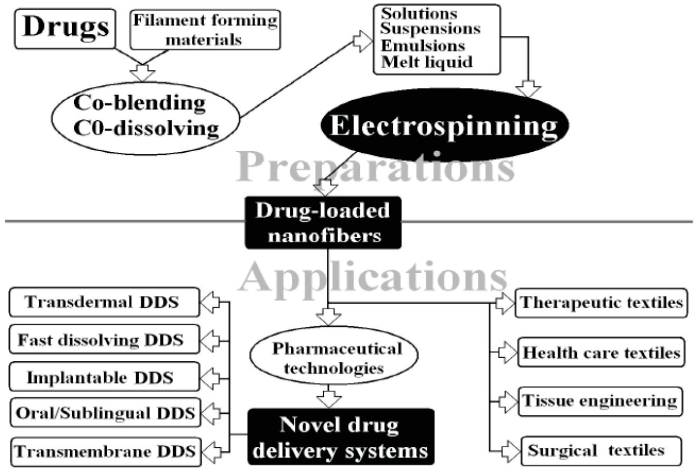
2. Methods of Preparing Nanofibers

Currently there are four major techniques available for the preparation of nanofibers: electrospinning [12,28], self-assembly [28,29], phase separation [30,31], and template synthesis [32,33]. Of these, electrospinning is the only one that can produce at a large scale continuous nanofibers for industrial applications [29], while other techniques may generate more sophisticated structures and more versatile functionalities. Nanofibers synthesized by self-assembly and phase separation have had relatively limited studies that explored their application as scaffolds for drug delivery [12].

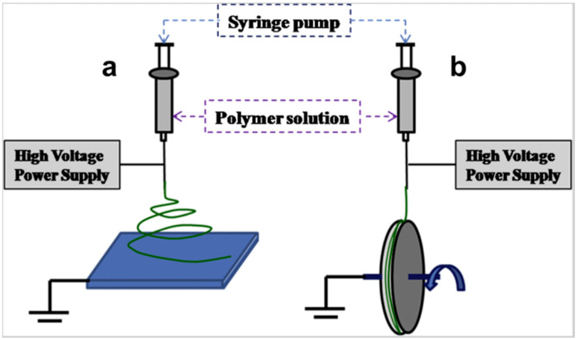
2.1. Electrospinning Method

Electrospinning is the simplest and most economical method to synthesize fibers of desirable diameters. Some of the advantages of this technique over the others are opportunity for control over thickness, composition of nanofibers, and the porosity of nanofiber meshes. Fibers ranging from 50 to 1000 nm or greater can be produced by this technique [34].
The setup consists of a syringe to hold the polymer solution, two electrodes, a collector, and a high DC voltage supply. When the voltage is applied to the polymer solution, electrostatic forces develop. These electrostatic repulsive forces oppose the surface tension of the polymer and lead to the elongation of the hemispherical surface of the solution at the tip of the capillary tube to form a conical shape known as the Taylor cone. Further application of electric potential causes the formation of a jet from this Taylor cone. The charged jet elongates and thins and finally deposits on the metal collector. During its travel from the Taylor cone to the collector, evaporation of the solvent takes place. The evaporation patters determine the porosity of the fibers. Figure 1 shows a typical electrospinning setup [28].
Figure 1. Electrospinning setup with (a) flat stationary collector; (b) rotating disc collector. Reprinted with permission from [28], Copyright© 2011 Elsevier.
Polymers 07 00186 g001
Solution parameters and processing parameters can greatly affect the diameters and morphologies of the electrospun fibers. Solution parameters usually include viscosity, surface tension, and electrical conductivity. They are strongly determined by polymer molecular weight, polymer concentration, solvent and additives [35,36]. If the solution has a very low viscosity, electrospray occurs instead of electrospinning. A very high viscosity is also detrimental for electrospinning. A hard ejection or micro-ribbon will be obtained in this situation. Surface tension is another important parameter in electrospinning. A higher surface tension will increase the bending-instability, thus producing beaded fibers. For conductivity, it can be tuned by adding ionic salts. With a higher conductivity, the electrospun fibers will have a smaller diameter. However, a very high conductivity can largely increase instability, which results in beaded fibers with a broad diameter distribution. Processing parameters usually include high voltage, flow rate, and the tip-to-collector distance. Due to the surface tension, a low applied voltage cannot initiate the Taylor-cone. The diameter of fibers will decrease as the applied voltage is increased. However, excessive high voltage can lead to beaded fibers. The fiber diameter usually increases with increasing flow rate. The high flow rate is also favorable for forming bead-on-string structures owing to short evaporating time. The effect of tip-to-collector distance is related to applied voltage. If the tip-to-collector distance is too short, there is not enough time for the polymer solution to fully elongate and the solvent to fully evaporate. However, if the distance is too long, the electrostatic force cannot overcome the surface tension, leading to electrospray or liquid drops. The parameters are important in controlling the diameter and uniformity of electrospun nanofibers. Until now, there have been no universal rules for producing uniform thin fibers. Overall, the optimal parameters depend on the choice of individual polymer and solvent, along with the purpose for the achieved nanofibers.
In addition to the most common single component non-woven mat structure, many unique fiber structures have been developed in the recent two decades. Aligned fibers can be achieved by altering the flat collector in the novel rotating drum collector [37]. The degree of alignment can be very high with the optimal rotating speed. Core-shell or core-sheath structure, first developed by Loscertales et al. [38], is another important structure in the electrospinning field. This structure is promising in biomedical applications, especially for controlled release of the drug delivery system. In the coaxial electrospinning setup, the traditional single needle is replaced by a coaxial spinneret. A lot of different compounded core-sheath nanofibers have been reported [39,40,41]. By choosing the suitable core solution, hollow structured nanofibers can be successfully obtained. For example, Li et al. choose mineral oil as the core material, which evaporates completely on electrospinning [42]. By replacing the coaxial spinneret into multi-channel microtubes, the derived nanofibers can have unique multi-channel structures. Porous nanofibers can be prepared by several methods including phase separation [43], using volatile solvent [44], post-treatment to remove one component and using non-conductive collectors [45]. The pore size and density of pores can play an important role in biomedical applications. Recently, using 3-D structured fibers as scaffold have attracted much attention in tissue engineering. The 3D structured fibers resemble a topological feature of a natural extracellular matrix. Bonino et al. [46] reported a new way to produce 3D structured nanofibers by blends of alginate and poly(ethylene oxide) (PEO) at relatively high humidity conditions. The mechanism behind this is repulsion between negative charged alginate and the high surface charges in the humid conditions.
Electrospun nanofibers show great promise for developing many types of novel drug delivery systems (DDS), as shown in Figure 2, due to their special characteristics and the simple but useful and effective top-down fabricating process, DDS is focused on drug-loaded nanofiber preparation from pharmaceutical and biodegradable polymers. All types of active pharmaceutical ingredients have been used as model drugs, such as small molecular drug, herbs, proteins, poorly water-soluble and water-soluble drugs, DNA, genes and vaccines [47].
Figure 2. Application and preparation of electrospun drug loaded nanofibers. Reprinted with permission from [47], Copyright© 2009 SciRes.
Polymers 07 00186 g002
Tissue engineering is one of the most desirable techniques for tissue regeneration. Generally, tissue repair is done using autografts or allografts. However, these two tissue repair techniques have their own disadvantages. An alternative to these two techniques is tissue engineering. The basic requirements for tissue engineering are a suitable cell source, optimal biochemical conditions and a biocompatible scaffold [48]. The scaffolds should be biodegradable, biocompatible and should serve as a framework for cell adhesion, proliferation, and differentiation. In general, scaffolds are expected to mimic the activity of extra cellular matrices (ECM). ECM is composed of several fibrous macromolecules with a length/thickness ratio greater than 100. The diameter of these macromolecules is about 500 nm [49]. Thus electrospun nanofibers can be used to prepare similar-sized scaffolds. These nanofibers have a high surface area to volume ratio in combination with microporous structure, nonwoven composite nature, and tunable physical properties, making it desirable as a scaffold. Some of the conducting polymers can be used for nerve cell regeneration as they can conduct action potentials to the nerve cell membranes [50].

2.2. Self-Assembly Method

Unlike electrospinning, which is a top-down method to break down a macroscale liquid into nanoscale fibers, self-assembly is a bottom up technique for the formation of nanofibers. In this technique, individual molecules arrange themselves in certain patterns to form macromolecular nanofibers. The shape of the nanofiber depends upon the structure of the building blocks, i.e., the smaller units of individual molecules participating in self-assembly and the intermolecular forces connecting these molecules [29]. These molecules can assemble into ordered structures like monolayers, super lattices, tubes or honey comb micro porous films, shown in Figure 3 [30].
Figure 3. Scanning electron microscope (SEM) images showing honeycomb-like structures of poly(vinyl acetate) (PVA) and poly(ethylene oxide) (PEO). Reprinted with permission from [30], Copyright© 2011, American Chemical Society.
Polymers 07 00186 g003
Patterning on a microscale may extend order in a predictable manner over large areas, expanding properties and performances. Hung et al. [51] used a sonication-assisted solution embossing self-assembly technique to prepare cylindrical 3D peptide amphiphiles (PAs). These self-assembled nanofibers had their alkyl segments in the core and the peptide sequences on the surface. Koga et al. [52] developed shape specific nanofibers with morphologically kinked structures via self-assembly of three armed peptides forming a β-pleated sheet. Rose thorn like nanofiber composites of Polyarylene ether nitriles (PEN) and iron phthalocyanine (FePc) were prepared by Meng et al. [53] using a combination of electrospinning and self-assembly techniques. The electrospun composite fibers were subjected to solvent removal and subsequent temperature treatment to form rose thorn like structures composed of FePc over the PEN nanofiber. Thus the self-assembly technique finds its application in designing novel scaffolds for drug delivery and tissue engineering.

2.3. Phase Separation Method

As to the phase separation technique, the nanofibers are formed due to the instability of the polymer within the solvent. They form two different phases because of the physical incompatibility. The general steps involved in phase separation are polymer dissolution, gelation, solvent extraction, freezing and freeze drying. Lei et al. [54] focused on a thermally induced phase separation (TIPS) for the preparation of nanofibrous gelatin-silica hybrids as tissue scaffolds. In their reported method, the freshly prepared silica sol was mixed thoroughly with gelatin solution, then the mixture was cast into a polyethylene mold and immersed in an ethanol bath at −70 °C for 4 h. The hybrid underwent a series of freezing, solvent exchange, freeze and drying processes to get the final foam like structure [31]. Li and co-workers studied nanofibrous polyhydroxyalkanoates (PHAs) matrices for cell growth support, using PHA blends polyhydroxybutyrate (PHB)/poly(3-hydroxybutyrate-co-3-hydroxyhexanoate) (PHBHHx) containing polyhydroxybutyrate (PHB) and copolyesters PHBHHx consisting of 3-hydroxybutyrate and 3-hydroxyhexanoate. The PHA hybrid nanofibers were formed through phase separation due to the solubility preference of the polymer in different solvents. Figure 4 demonstrates that PHB nanofibers only formed when dioxane (Figure 4a,b) was added to the PHB/chloroform solution, while addition of tetrahydrofuran (THF) (Figure 4c) and dimethylformamide (DMF) (Figure 4d) did not result in the nanofiber topography [32].
Figure 4. SEM studies of polyhydroxybutyrate (PHB) matrices formation under different solvent addition to the PHB/chloroform solution. Reprinted with permission from [32], Copyright© 2008 Elsevier.
Polymers 07 00186 g004

2.4. Template Method

Nanofibers can also be prepared by a template based synthesis; first a nanostructured ceramic or polymeric membrane is prepared to serve as the template, the targeting material is then added in contact with the nanostructure to form nanofibers, and finally the template is removed to leave free nanofibers. Anodized aluminum oxide (AAO) is one of the popular ceramic templates that has been used to synthesize a variety of nanostructured fibers, including conducting polymers [54,55] and carbon [56], metals [57], and other ceramics [58]. Another frequently used ceramic template is silica, which has been used widely in fabrication of polymer nanofibers [33,59].
Polymeric materials have been mostly explored for serving as templates (as seen in Figure 5), as there are many choices of polymers and also various nanopores can be designed in the polymer, thus rendering a big variety of different nanostructures including nanofibers. Wu et al. [54] recently summarized the many ways of making porous polymers, which shed light on possible routes for polymer based template synthesis. Martin’s group [60,61] extensively studied the synthesis of nanomaterials with a polymer membrane as the template.
Figure 5. (a) Transmission electron micrograph (TEM) of a microtomed section of an alumina template membrane showing 70 nm diameter Au nanofibrils within the pores; (b) Transmission electron micrograph of three polypyrrole nanotubules. The outside diameter is ~90 nm; the inside diameter is ~20–30 nm. Adapted with permission from [61], Copyright© 1996 American Chemical Society.
Polymers 07 00186 g005
A novel method for production of nanofibers/nanotubes containing living cells was reported which is efficient mechanically and has a low cost. High pressure gas was used to extrude viscous precursors through a microscale spray into air. The sprayed micro-sized droplets had a high velocity and were continuously elongated into uniform nanofibers/nanotubes. Nanofibers containing living cells produced from this method have high survival rate and can be used in bioengineering [62]. In addition, there are several other methods for production of small diameter fibers using high-volume production methods, such as fibrillation, island-in-sea, and the novel melt-blowing system. The fibrillation method is mostly used for the preparation of cellulose nanofibers from two commercial hard and soft wood pulps, which consists of initial refining and subsequent high-pressure homogenization. The process in fibrillation was studied using different microscopy techniques, mechanical testing, and fiber density measurements of cellulose films prepared after different processing stages. An increase in processing steps obviously increases the energy required for fibrillation [63]. For the island-in-sea method, fibers of diameter less than one micrometer can be made by the conjugated spinning method, in which two polymer components are extruded together from a spinning die. However even by the conjugate spinning method it is difficult to produce a nanofiber with a diameter less than 100 nm [64]. Lastly for the novel melt-blowing system, fibers are produced in a single step by extruding a polymer melt through an orifice die and drawing down the extruder with a jet of hot air (typically at the same temperature as the molten polymer). This method was first developed in the 1950s at the naval research laboratory with the goal of making sub-micron fibers to trap radioactive particles in the upper atmosphere [65]. Wente [66] first described the construction of a metal blowing die composed of a series of orifices and slots, and then researchers at Exxon extended this basic design and first demonstrated the production of melt blown microfibers on a commercial scale by modifying sheet die technology. Ward further reported an improved melt blown technique for producing polymer nanofibers such as polypropylene [10]. However the usefulness of the above methods is restricted due to narrow material ranges, low production range, high cost, and the difficulty in obtaining fibers on the nanoscale.

3. Polymer-Based Nanofibers for Drug Delivery

Nanofibers can be used to deliver drugs to attain either localized treatment or controlled release of the drugs at the target. They can also be combined with polymeric nanofiber scaffold implants to prevent infection at that particular site as the tissue regeneration occurs [67]. The principle behind the drug release mechanism of a particular drug incorporated in a nanofiber is the increase in the rate of release of the drug with an increase in the surface area of the nanofiber [68]. Desirable properties in polymers used for drug delivery are [69]:
(a)
Ability to bind the desirable drug and ability to release the same drug in the target morbid tissue;
(b)
Sustained release of drugs in the target streams over a long duration of time;
(c)
Retaining maximum drug loading capacity in order to release a drug for a long duration.
Nanofibers can be made from two major categories of polymers, these being natural and synthetic polymers. Some major natural polymers include cellulose, chitosan, gelatin, heparin, collagen, pectin, proteins, and polysaccharides. They are biocompatible and induce low antigenic response but the disadvantage with them is that they are expensive. Starch is a polysaccharide with few limitations, like poor water stability. Xu et al. [70] used an acetate derivative of starch to synthesize nanofibers, and explored its application towards drug delivery. Synthetic polymers are made from petrochemicals, and examples consist of nylon, polyethylene, polypropylene, polyester, Teflon, and epoxy. Synthetic polymers can be biodegradable or non-biodegradable in nature. Some of the biodegradable polymers used in drug delivery systems are poly(lactic-co-glycolic acid) (PLGA), poly(ethylene glycol), poly(lactic acid) [71].
Both natural and synthetic polymers can be used to prepare drug eluting membranes. Natural polymers are more capable of mimicking an extracellular matrix, whereas the synthetic polymers loaded with drugs can be easily electrospun (Figure 6) [72]. Chen et al. [73] synthesized sandwiched drug eluting membranes made up of an outer layer of PLGA/collagen and core layers of PLGA/Vancomycin, gentamicin and lidocaine. The drug release for vancomycin and gentamicin was found to last four and three weeks respectively. For lidocaine, drug release rate lasted two weeks. Meng et al. [74] worked on Fenbufen (drug) loaded PLGA/gelatin nanofibers, and found that an increase in the content of natural polymer gelatin caused an increase in the release rates of Fenbufen.
Figure 6. Different modification techniques using (A) plasma treatment (B) surface graft polymerization (C) Co-electrospinning. Reprinted with permission from [72]. Copyright© 2009 Elsevier B.V.
Polymers 07 00186 g006
Nanofibers used as a drug delivery carrier need to accomplish two steps; one is to load the drug cargo, and the other is the controlled release of the drugs. Loading drugs into the nanofibers can be achieved via various ways depending on the material choices of the nanofibers; the drug could be loaded through chemical or physical binding between the drug and the nanofiber materials, it can also use the cross-linking of the polymeric nanofiber materials to encapsulate the drugs, and nanofibers can be made into layers with drugs contained inside the layer. Release of drugs in a controlled fashion could be achieved by controlling the nanofiber membrane structure (porosity), thickness of membrane, and membrane biodegradability (time). The high surface area-volume ratio of electrospun scaffolds allows the efficient delivery of a loading drug.
Drugs and other bioactive molecules can be loaded in/on nanofibers either through physical methods or chemical methods. Depending on the nature of the polymer being used, surface modification may be needed prior to drug loading. Surface modification helps to enhance the surface properties of nanofibers by introducing new functional groups. Some of the surface modification techniques are plasma treatment, wet chemical method, and surface graft polymerization.

3.1. Physical Methods for Loading Drugs

3.1.1. Dissolution/Co-Electrospinning

In the dissolution method, the drug is mixed well with the polymer solution prior to electrospinning. It is difficult to get a uniform matrix and requires tedious and elaborate mixing. Meng et al. [74] prepared PLGA/gelatin nanofibers integrated with Fenbufen (FBF) drug using a dissolution method. Prior to electrospinning, the composite polymer-drug solution was mixed for 12 h at room temperature. There were no drug aggregates seen on the surface of fibers. Ketoprofen loaded polylactic acid (PLA) nanofibers were made by Park and colleagues in 2010 by the same method [71]. Although the co-electrospun fibers showed incorporated drugs, the drug releasing properties diminished due to harsh electrospinning processing conditions [72]. Using this method, a loading efficiency of 90% was reported for the antibiotic drug Mefoxin on PDLA electrospun nanofibers [75].

3.1.2. Sorption Method

In this method, electrospun fibers are either immersed into the drug solution or the drug solution is drop by drop loaded onto the fibers until maximum adsorption is achieved. Diclofenac dissolved in a 0.5% aqueous sodium chloride solution was loaded on starch acetate fibers by this method. Drug solution and fibers were taken in a 100:1 ratio and held in a shaking water bath at 90 °C and 120 rpm oscillation to adsorb the drug onto fibers [70].

3.1.3. Nanofiber Assembly on the Surface

Nanosized materials have high specific surface areas. Increased surface area can be obtained by using both nanofibers and nanoparticles in conjunction. Drug particles are first loaded onto the nanoparticles which are in turn loaded onto nanofibers by co-electrospinning. Zhen et al. [76] studied doxorubicin loaded electrospun composite fibers of PLGA and nano hydroxyapatite toward cancer treatment. Sustained drug release could be obtained by drug loaded inorganic–organic hybrid of n-HA particles within PLGA nanofibers which could not be seen in the individual nanoparticles/nanofibers forms.

3.2. Chemical Methods for Loading Drugs

Chemical methods include treatment of nanofibers with acid or base for a certain duration of time. Functional groups like hydroxyls and carboxyl groups can be created on the surface of nanofibers by a hydrolysis reaction. Amine groups and carboxylic groups are widely used for this purpose. The drug moiety is covalently linked to the surface of the nanofibers by chemical modification of the surface functional groups. If active sites of the drug moiety are involved in the covalent linkage, a loss in the activity of the drug could be seen. This chemical conjugation method can modulate drug release [77].
Depending on the methods of loading drugs and the nature of the loaded drugs, the morphology and distribution of drugs in the final nanofibers may be in three formats: one is that the drug formed into tiny particles and distributed on the surface of the nanofiber, another one is that the drug is electrospinnable and forms a nanofiber blend, and the third one is that the drug is uniformly distributed in the polymer matrix at the molecular level, thus forming composite. The third one is preferred as the first two situations will cause a burst release of drugs which is not favorable for drug release applications [78]. Figure 7 summarizes the major functionalities that can be incorporated into the polymer backbone, rendering potential drug delivery functions [79].
Figure 7. Ringsdorf’s model for drug-delivery systems based on synthetic polymers. Reprinted with permission from [79], Copyright© 2006 WILEY-VCH Verlag GmbH & Co. KGaA, Weinheim.
Polymers 07 00186 g007
Drug release rates can be studied through a dissolution apparatus with the desired solvent. When a solvent interacts with drug loaded nanofibers, the nanofiber matrix swells, causing the polymer chains to loosen up and release the drugs. Also, since nanofibers have large surface areas, they facilitate easy transport of the solvent to diffuse and elute the drug molecules. Factors like fiber diameter, mesh thickness, pH and temperature play important roles in drug release rates. At 37 °C a greater release rate of ketoprofen drug from PLA fibers was observed in comparison to fibers at 20 °C [71].
Poly(bis(p-methylphenoxy)phosphazene) (PNmPh) has a chemically stable phosphorus-nitrogen backbone, and thus a great potential to add functionalities onto the backbone to make a family of this type of compounds. The electrospun PNmPh nanofibers can serve as cell growth matrices and the hydrolytically stable property of the nanofibers make it a good candidate to be used as a novel drug delivery system, wound dressing, and as prosthetic devices [33]. Tetracycline hydrochloride, an antibiotic drug against bacterial infections such as periodontal diseases, was investigated on its nanofiber-facilitated delivery. Kenawy et al. [17] prepared poly(ethylene-co-vinylacetate), PEVA, poly(lactic acid), PLA, and their 50:50 blend nanofibers via electrospinning. Their results, shown in Figure 8, indicate that the release of tetracycline hydrochloride can be controlled by controlling the ratio of PLA and PEVA in the blend. It was seen that pure PEVA fibers and 50/50 PEVA/PLA fibers showed prolonged release of tetracycline drug over a period of 120 h as seen in the Figure 8.
Chew et al. [19] investigated the feasibility of encapsulating human β-nerve growth factor (NGF) which is a small secreted protein that is important for the growth, maintenance and survival of certain target neurons. The NGF was stabilized in the carrier protein, bovine serum albumin (BSA) within a copolymer of ε-caprolactone and ethyl ethylene phosphate (PCLEEP). The protein was found to be randomly dispersed throughout the electrospun fibers mesh. After analysis with pheochromocytoma 12 (PC12) neurite outgrowth assay it was confirmed that the bioactivity of electrospun NGF was retained at least throughout the sustained release (shown in Figure 9), which clearly demonstrates that the feasibility of encapsulating protein via electrospinning can be used to produce biofunctional tissue scaffolds.
Figure 8. Percentage release rate of tetracycline HCL loaded nanofibrous mats versus time (h). Reprinted with permission from [17], Copyright© 2002 Elsevier Science B.V.
Polymers 07 00186 g008
Figure 9. Bioactivity testing of the human β-nerve growth factor (NGF) encapsulated copolymer of ε-caprolactone and ethyl ethylene phosphate (PCLEEP) using PC12 cells. Adapted with permission from [19]. Copyright© 2005 American Chemical Society.
Polymers 07 00186 g009
Luu et al. [21] utilized fabricated synthetic polymer/DNA composite for therapeutic application in gene delivery, the composite was non-woven, nano-scale, membranous structured and predominantly composed of poly(lactide-co-glycolide) (PLGA) random co-polymer and a poly(d,l-lactide)–poly(ethylene glycol) (PLA–PEG) block copolymer. Release of plasmid DNA from the composite was sustained over a 20-day period, and the maximum release occurred at 2 h. Cumulative release profiles indicated that the amounts released were approximately 68%–80% of the initially loaded DNA. This proves that DNA was delivered directly from these electrospun fibers and it was capable of cellular transfection and expression of the protein β-galactosidase.

4. Polymer-Based Nanofibers for Tissue Engineering

Today, it is generally believed that nearly one hundred different polymers, mostly dissolved in solvents yet some heated into melts, have been successfully spun into ultrafine fibers including Poly(ε-caprolactone) (PCL), poly(ethylene oxide) (PEO)/Rhodamine-B (RM) [41], and Poly(N-isopropylacrylamide) (PNIPAAm)-based nanofibers [80]. These polymers can be separated into the following different categories.

4.1. Natural Polymeric Nanofibers Used for Tissue Engineering

Natural polymers used for tissue engineering can be either organic or inorganic in nature. They are identical to the macromolecules present in the ECM. Thus they show greater biocompatibility when compared to synthetic polymers. Collagen nanofibers have shown compatibility with a number of cell types like myoblasts and chondrocytes [81,82]. Type 1 collagen fibers were used for stem cell regeneration by Shih et al. [48]. Hyaluronic acid has also been used to prepare nanofiber scaffolds because it is a natural component of ECM [83]. They offer low inflammation. Min et al. [84] reported the in vitro cytocompatibility of silk nanofibers with keratinocytes and fibroblasts. Jeong et al. [85] used alginate to produce nanofibers. When the alginate nanofibers were modified by a cell adhesive peptide (GRGDSP), the results showed that the nanofibers have minimal cytotoxicity and fibroblasts can adhere to spread and proliferate on the exact nanofibers.

4.2. Synthetic Polymeric Nanofibers Used for Tissue Engineering

Several synthetic polymers are being used to prepare scaffolds like PLA [86] poly(ethylene terephthalate) (PET) [87] for blood vessel tissue engineering. Copolymeric materials are also used for this purpose such as poly(l-lactide-co-caprolactone) (PLLA-CL) for smooth muscles and endothelial cells [87]. Synthetic polymers are easy to prepare and give reproducible results which are not seen in the case of natural polymers. They are durable and have good mechanical strength. However, the biocompatibility of synthetic fibers is low. Conductive polymers like polyaniline and polypyrrole are of interest for biological tissues, because new technologies require tissue scaffolds that not only physically support tissue growth, but also are electrically conductive. Thus the tissue scaffolds are capable to stimulate specific cell functions or trigger cell responses [88,89]. Recently, Gizdavic-Nikolaidis et al. [90,91] reported that electrospun functionalized polyaniline/poly(lactic Acid) nanofibers allow mammalian cells to attach and proliferate, while killing pathogenic bacteria cells, and are novel conductive materials that are potentially well suited for use as biocompatible scaffolds for tissue engineering and as antimicrobial wound dressings that have the advantage of being able to kill microorganisms without use of an antiseptic.
Apart from classification based on natural and artificial origin of polymers used for tissue engineering, there is a classification based on degradability of the polymeric scaffolds. Scaffolds can be of two types: bioresorbable or non-bioresorbable scaffolds. The choice of polymer scaffold purely depends on the purpose of the scaffold being used.

4.3. Bioresorbable Polymeric Scaffolds

Bioresorbable polymers erode with time. Their eroded products might participate in other metabolic processes in the body. They can act as a scaffold until the ECM develops and replaces it. Such scaffolds are highly desirable because they avoid the need to surgically remove the remains of scaffolds from the body. They can be either natural or artificial or polymeric blends of both. They find their application in surgical sutures [92], controlled drug delivery, stents [93] and other orthopedic devices. Asran et al. [94] made blends of polyvinyl alcohol and polyhydroxy butyrate and focused on their application as a scaffold for skin tissue engineering. Both these polymers were shown to be biodegradable. It was seen that the rate of degradation increased with the increase in the PVA content in the blends.

4.4. Non-Bioresorbable Polymeric Scaffolds

Non bioresorbable polymers do not degrade in the body. They are strong and durable and thus do not undergo degradation easily. Such scaffolds are desired for permanent supports or encapsulations. They are frequently used for permanent or temporary prostheses in the area of biomedicine. They are not used as biological grafts. Some of the examples for nonbiodegradable polymers are polycarbonate urethane with carbon fiber [95], silk/poly(ethylene oxide) (PEO) [96], poly(dimethyl siloxane) (PDMS), and poly(tetrafluoro ethylene) (PTFE) [97].

4.5. Degradation Kinetics of Polymer Scaffolds

The rate of biodegradation of a polymer is expected to match the rate of tissue formation. During biodegradation the breakdown products should not cause any inflammation or induce any kind of toxicity to the body [97]. The rate of degradation of a polymer can be determined using several techniques. One of the simple techniques is by placing the scaffold in phosphate-buffered saline (PBS) at 37 °C in vitro or in vivo and biodegradability is measured by the amount of mass loss, strength loss and changes in morphology [68]. Zong et al. [98] electrospun various blend fibers of poly(lactide)- and poly(glycolide)-based (PLGA) at different concentrations to tune scaffold degradation rates. It was seen that 10:90 blend showed a mass loss of 20% over a period of seven days in aqueous solution.

5. Tissue Engineering with Nanofibers

Nanofibers have wide applications in tissue engineering because of their high surface area and porosity. They can be used as scaffolds for all four major tissues, i.e., epithelial, muscle, connective and nervous tissues.

5.1. Nerve Tissue

Nervous system plays a central role in the human body. Every function of the body is dependent on the nervous system. Degeneration of neurons or gal cells can lead to a lot of clinical complications. Regeneration of this tissue is a challenge because the damage done to the tissue is irreversible. Nerve tissue regeneration focuses on axonal regeneration and its functional recovery [99]. Nerve regeneration scaffolds should not only provide mechanical support for the growth of neurons but also prevents the growth of non-neural cells [50]. The growth of non-neuron cells, like gal cells, could form a fibrous scar tissue. It was shown that carbon nanotube scaffolds could discourage the scar tissue formation. Low adsorption of astrocells was seen in carbon nanotubes with small diameters. McKenzie et al. [100] showed an inverse relationship between the surface area of carbon nanotubes and the growth of fibrous scar tissue formation. Polymers like polyaniline (PANI) and polypyrrole (PPy) are also used for preparing scaffolds for nervous tissue because of their electrical conductivity. Since these conducting polymers are difficult to electrospin, they can be blended with other non-conducting polymers to produce fairly good conducting polymeric blend nanofibers. Lee et al. [101] produced conductive meshes of polypyrrole (PPy) on poly(lactic-co-glycolic acid) (PLGA) in both random and aligned fashion. It was seen that PPy–PLGA electrospun meshes supported the growth and differentiation of rat pheochromocytoma 12 (PC12) cells and hippocampal neurons comparable to non-coated PLGA control meshes. Also, aligned fibers showed the growth of longer neuritis when compared to random PPy–PLGA fibers.
Previous researchers showed that application of electrical stimulus can enhance the neural tissue growth. Shi et al. [102] support this view, by reasoning that bioelectricity present in the human body plays a cardinal role in maintaining various bodily functions like nerve cell signaling, muscle contraction, and wound healing. Various composite nanofibers have also been used for this purpose. Mobarakeh et al. [103] worked on polycaprolactone/gelatin polymeric blends; the 70:30 blend of PCL/gelatin showed enhanced neurite growth when compared to pure PCL scaffolds. Nerve tissue proteins can also be incorporated into nanofibers. Koh et al. [104] made fibrous blends of poly(l-lactic acid) and ECM protein laminin. It was shown to have regenerative properties. Axonal growth of neuritis was noticed while using these blends.

5.2. Musculoskeletal Tissue

Bone, cartilage, and ligament tissues fall under this category. Different polymeric blends and copolymers of both natural and synthetic origins have been used as scaffolds for these tissues. Commonly used polymers are PLGA [105], PCL [106], HA [107], and silk [108]. Mesenchymal stem cells (MSCs) are grown in vitro for musculoskeletal tissue cultures. MSCs have a capability to differentiate into different types of cells such as bone, cartilage, muscle, tendon, ligament, and fat [109,110]. Their differentiation in vitro can be guided by using various conditions of mechanical loading. Studies have also shown that growth factors play an important role in determining the fate of cells which develop from MSCs.
Bone infections and tumors are serious health problems. Regular techniques like autografts and allografts are not preferable due to the risk of tissue rejection and disease transmission. Bone tissue engineering is an emerging sector which could be a better option for tissue regeneration. Bone is mainly composed of hydroxyapatite (HA) and collagen (Col). Therefore collagen containing nanocomposites are more studied for this purpose. Hydroxyapatite is a mineral found in bones in the form of calcium complexes. Venugopal et al. have studied PCL/nHA/Col biocomposite nanofibers for their application in tissue engineering. It was seen that PCL provided mechanical stability, collagen supported cell proliferation and nHA helped the mineralization of oseteoblast cells [111]. Zhang et al. [112] used a hybrid nanofiber of both synthetic and natural polymers. To ensure better cell attachment and differentiation they made composite fibers containing core PCL and an external shell of cellulose. Hydrophobic PCL provides good mechanical strength, whereas hydrophilic cellulose external shell provides cell recognition signals for specific cell interaction [112]. Chiu et al. showed that migration and cell attachment increased when they used electrospun fibers of PLLA incorporated with type 1 collagen [113]. Studies have shown that cross-linkage of polymers can prevent rapid degradation of scaffolds due to hydrolysis [114]. Composite fibers of polyurethane with calcium chloride were studied by Nirmala et al. [14]. It was seen that when these composite fibers were SBF incubated, apatite like materials were formed on the surface. Such fiber scaffolds can find applications in bone tissue engineering. Sui et al. [114] investigated the properties of PLLA/Hydroxyapatite fibers for their application in bone tissue engineering. Hydroxyapatite nanoparticles were needle like in appearance as seen in Figure 10. They used a two solvent system to synthesize PLLA/HA composite fibers. These fibers had a greater surface area compared to PLLA fibers. They were biocompatible with Human Osetoblast cells (MG-63). Asli et al. [115] compared the release profiles between different structured electrospun scaffolds: single component, core-sheath, and porous nanofibers. These scaffolds were doped with tricalcium phosphate (TCP) nanoparticles. TCP can promote bone ECM production. For porous fibers and single component fibers, an initial burst release was observed. In comparison, core-sheath fibers exhibit a steady release. The seeded cells in all three scaffolds remained viable and proliferate even up to 21 days.
Figure 10. Transmission electron microscopy (TEM) image of Hydroxyapatite nanoparticles. Reprinted with permission from [15], Copyright© 2007 Wiley Periodicals, Inc.
Polymers 07 00186 g010
Like nerve cells, cartilage cells also show low rates of regeneration and this could be due to the absence of vascular networks and progenitor cells in the tissue. Non mobility of the chondrocytes could also be a reason for regeneration issues [12]. Da Silva et al. [116] used PCL nanofiber meshes to culture mesenchymal stem cells in a multichamber flow perfusion bioreactor. They showed that there was an increased rate of differentiation of MSCs into chondroitic cells. Collagen types 1 and 2 were found in the samples as well. Li et al. [117] studied the effects of growth factor on the differentiation of MSCs into chondrocytes. They found that transforming growth factor-β promotes the formation of chondrocytes in vitro on PCL scaffolds from bone marrow derived MSCs. Chondrocyte specific genes such as collagen type II, IX and XI, aggrecan, and other cartilage extracellular matrix proteins were seen as well. The above studies show that electrospun nanocomposites and nanofiber scaffolds can be used for bone and cartilage tissue engineering. In these studies there was an apparent increase in the number of chondrocytes (osteocytes differentiating from mesenchymal stem cells in the scaffold regions).

5.3. Heart Tissue and Vascular Grafts

Blood vessels and heart tissues are delicate and complex tissues. Minor internal injuries to these tissues could lead to serious health issues. According to the National Health Interview Survey 2010, for the US, the morbidity rate for adults with cardiovascular disease is 11.8% [118] and it stands at the number one position for cause of deaths due to diseases in the US according to the survey in 2010 [119]. Therefore there is a lot of focus on the fields of heart and vascular tissue engineering.
Xu et al. [120] studied poly(l-lactid-co-ε-caprolactone) (P(LLA-CL)) electrospun nanofibers and their application in blood vessel tissue engineering. It was noticed that human coronary artery smooth muscle cells (SMCs) attached and aligned along the axis of these aligned fibers and showed spindle-like contractile phenotype.
Blood vessels have three layers which are tunica intima, tunica media, and tunica adventitia. Tunica intima is the innermost layer consisting of an endothelial layer and connective tissue layer of elastic fibers. The middle layer tunica media is mostly made up of smooth muscles and the external layer, tunica adventitia, is made up of collagen [121]. The major problem with vascular grafts is the risk of rejection and clot formation [122]. If the humoral immune system detects the graft as a foreign body, antigen-antibody reactions occur leading to graft rejection by the body. Nanofiber scaffolds used for these tissues are expected to mimic the ECM. One plausible solution to reduce rejection is to develop a layer of endothelium in the core. These cells produce bioactive substances like heparin sulfate which can prevent thrombogenesis on the surface of scaffolds. In arteries with a wide diameter thrombogenesis is not much observed. Therefore vascular grafts for wide diameter blood vessels were successful. There is a real challenge to develop nanofibrous grafts for blood vessels with small diameters.
Wang et al. [123] used bilayered electrospun fibers of polyurethane (PU) and gelatin-heparin to test the biocompatibility. Polyurethane has good mechanical and elastic properties but it is hydrophobic in nature. Gelatin, which is a biodegradable polymer, was found to improve the proliferation of endothelial cells. Heparin, a highly sulfated linear glycosamine, prevents thrombosis and acts as an anticoagulant. An external layer of PU and an internal layer of gelatin-heparin fibers were made by electrospinning on a rotating mandarel-type collector. Platelet adhesion tests showed that there was a greater adhesion of platelets on the PU layer compared to the internal heparin layer, suggesting a lesser probability of clot formation within the scaffold.
Huang et al. prepared three layered nanofibers made of collagen, chitosan and poly(l-Lactide-co-caprolactone) (P(LLA-CL)). The inner and outer layers were made of chitosan and collagen blends whereas the middle layer was P(LLA-CL). They used rat endothelial cells as seeding material in scaffolds and checked for its proliferation rate at different time intervals. On day 3, it was seen that there was a dramatic increase in the proliferation value. This could be due to the presence of collagen and chitosan on the interior and exterior layers which provide bioactive sites for the endothelial cells to attach and proliferate. However, on day 7, proliferation remained constant with the other counter parts (Figure 11), i.e., blends and TCP. The limited proliferation rate on day 7 could be due to overcrowding of the cells and space issues [124].
Figure 11. (A) Cell viability after seeding measured for 1, 3 and 7 days; (B) Confocal image of endothelial cells on the composite scaffolds for day 7. Scale bar = 25 μm. Reprinted with permission from [124], Copyright© 2012 Elsevier Science B.V.
Polymers 07 00186 g011
Ishii and coworkers [125] worked exclusively on the heart tissue culture. They successfully developed five layered grafts of myocardial cells in vitro using a nanofibrous scaffold, as seen in Figure 12. They prepared PCL nanofibers using an electrospinning technique and stretched this nonwoven fibrous mesh across a wire ring. Neonatal cardiomyocytes attached well on these porous scaffolds. Grafts of these cells were placed one above the other and observed for two weeks. It was seen that the layers developed interconnections. Another interesting feature found was that these cells showed myocardial contractions, or beats, all through the period of in vitro culture.
Figure 12. SEM images of a five layered graft showing dense cell growth after two weeks of culture. Reprinted with permission from [125], Copyright© 2005 by The American Association.
Polymers 07 00186 g012

5.4. Skin

Skin is the largest organ of the body. It protects the inner organs of the body from the external environment. Although it has the capability to regenerate, low regeneration rates occur in case of major burns. Burned and damaged skin is prone to inflammation and infections. Therefore, it is necessary to cover the wound until skin regenerates. Presently autografts and allografts are used to regenerate skin. However, the risk of disease transmission and organ rejection are involved in allografts. Autograft is much more practical but the main drawback is limited availability of skin in case of major burns.
Skin has two main layers. The outer epidermis is made of keratinocytes and the inner dermis layer is made of fibroblasts. The scaffold used to regenerate skin has to support the growth of these two layers. A dermal scaffold component is expected to promote the growth, diffusion and attachment of fibroblasts whereas an epidermal scaffold component mainly has to support the growth of keratinocytes [94].
Asran and coworkers [94] developed electrospun fibers of PVA (poly(vinyl acetate)) and PHB (poly(3-hydroxy butyrate)) blends and studied their application in skin tissue engineering. Biocompatibility tests for these blends were done using human keratinocyte cell line (HaCaT) and fibroblasts. Pure PHB nanofibers promoted the adhesion and proliferation of both HaCaT cells and fibroblasts. Increasing content of PVA promoted the growth of HaCaT cells and inhibited fibroblasts. Thus by using different compositions of PVA and PHB, the bio-selectivity could be altered.
Park et al. [126] studied PGA (poly(glycolic acid))/chitin blends. Chitin is similar to glycosaminoglycan (GAG), a component of ECM. They both bear common substances like chondroitin sulfate and hyaluronic acid and PGA resembles collagen in certain properties. Cell attachment tests of Chitin/PGA nanofibers both coated and non-coated with BSA were done using normal human epidermal cells. It was seen that pure PGA nanofibers with or without any BSA coating had greater cell adhesion. Among different blends made, the blend with 25% PGA with BSA coating showed good cell adhesion. This could be attributed to the resemblance of these blend components to the natural ECM biomolecules.
Kuppan et al. [127] tried to develop growth factor loaded scaffolds. Studies have shown that growth factors help quicken the healing process. To support these studies they worked on R-Spondin 1 growth factor. Electrospun fibers of poly(3-hydroxy butyrate-co-3-hydroxyvalerate) (PHBV) were made and the effect of R-Spondin 1, an angiogenesis promoting factor was studied during the healing process. Within seven days, it was seen that R-Spondin 1 on PHBV fibers accelerated the healing process (Figure 13). It was reported that human fibroblast cells grew well on PHBV fibers which could be due to better oxygen permeability because of the porosity of fibers. Gene regulation studies conducted by them showed that there was an up regulation of collagen 1 gene whereas collagen 3 gene was down regulated, this infers that PHBV nanofibers mimics ECM.
Currently there is a lot of research going on to develop natural polymeric scaffolds for skin regeneration. The main benefit of using natural polymeric is low toxicity. Zhou et al. [128,129,130,131] developed carboxyethyl chitosan/poly(vinyl alcohol) (CECS/PVA) electrospun fibers. Chitosan is not only known for its biocompatibility and wound healing ability but also for its antibacterial properties [128,129,130]. Zhou et al. used L929 mouse fibroblasts cell line to check the biocompatibility and regenerative properties. No cytotoxicity was revealed associated with the scaffold which proved that the scaffold was biocompatible [131].
Figure 13. (A1,A2) Negative control (one week and two weeks); (E1,E2) poly(3-hydroxy butyrate-co-3-hydroxyvalerate) (PHBV) loaded with R-Spondin 1. Reprinted with permission from [127], Copyright© 2011 American Chemical Society.
Polymers 07 00186 g013
These studies were further supported by Tchemtchoua and coworkers [132]. They used chitosan sponges, films, and nanofibers to do regenerative studies using fibroblasts, keratinocytes and epithelial cells of mice. Tissue regeneration was seen when all three forms of chitosan scaffolds were used, however the physical appearance of the tissue varied. A more uniform tissue formation was seen when nanofibers were used, as seen in Figure 14. This could be due to the greater oxygen supply and surface area of the nanofibers.
Figure 14. Macroscopic appearance of (a) chitosan sponge and (b) electrospun nanofibers 12 weeks after subcutaneous implantation. Sponges appear red and thickened by fibrous deposit, whereas nanofiber scaffolds remained white and could hardly be distinguished from the neighboring mouse tissue. Reprinted with permission from [132], Copyright© 2011 American Chemical Society.
Polymers 07 00186 g014

6. Polymer-Based Nanofibers for Wound Dressing

Today wound healing has evolved from a passive to an active healing process, so the wound dressing is not merely a surface coverage and support but interacts actively with the wound by stimulating and managing cell migration. This involves incorporation of active medical agents including antibiotics, growth factors, or cell signaling agents inside the nanofiber [1]. The desirable properties of wound dressings are:
(a)
Hemostasis;
(b)
Ability to absorb cell exudates;
(c)
Enable gaseous exchange at the target area;
(d)
Maintain aseptic conditions;
(e)
Nontoxic to tissues [133];
(f)
Maintain moisture balance [78];
(g)
Be non-adherent to the wound.
Traditional wound care materials are either natural or synthetic in nature. Natural polymers are biodegradable but they have to be frequently changed as they retain dead cell debris [134]. Synthetic wound dressings are mostly non-biodegradable. Wound dressings can be classified into passive, interactive, and bioactive in nature. Passive wound dressings merely act as a cover to the wounded area. Traditional materials like gauze are passive. Interactive wound dressings are permeable in nature. Polymer dressings fall under this category. Bioactive wound dressings contain anti-microbial drugs, growth promoting factors, nano silver, and other bioactive ingredients which promote tissue growth in the wounded area [135]. To characterize wound dressing materials national test standards such as the British Standards and the American Standards for testing are followed [135].
Electrospun emodin polyvinylpyrrolidone blend membranes were used to study wound healing. Emodin exhibits anti-inflammatory and antibacterial properties [136]. Male K.M. mice were used to do the wound closure experiments. The wound excisions were made and then fixed with sterile medical infusion fixation plaster along with Polyvinylpyrrolidone (PVP)/emodin blend nanofibers. Fixation was changed on even days and fresh nanofibers were loaded. Wounds were photographed on odd days. Drug loaded or non-loaded nanofibers showed a tendency to contract and dissolve under wet conditions. The test group showed better wound healing in comparision to the control group [137]. Electrospun fibers of poly(sulfobetaine methacrylate) were shown to have promising features for wound dressing material, but the fibers are super hydrophilic and tend to dissolve in water. To make them water stable, Lelani et al. [138] synthesized cross linked zwitterionic poly(sulfobetaine methacrylate) (PSBMA) through a three-step electrospinning process, namely polymerization-electrospinning-cross linkage. Since PSBMA has anionic SO32− groups, silver ions can be incorporated. Cell adhesion studies proved that these fibers were resistant to cell or protein attachment and thus showed low biofouling.
Since nanofibers have a high surface-area-to-volume ratio, they have surface molecular chains ready to bind with desirable drugs. The formula to calculate the percentage of exposed functional groups is 110 d/D where (d is the polymer chain and D is the diameter of the nanofiber chain) [139]. Active wound dressings usually contain active agents like antibiotics, anti-inflammatory agents, growth promoting factors, vitamins, minerals, and inorganic ions like silver [140] and iodine. El-Newehy et al. [141] synthesized PVA/PEO/metronidazole (MTZ) composite nanofibers using nanospider technology, in which high applied voltage (up to 80 kV) is used to synthesize fibers. They used antimicrobial agent metronidazole in these composite fibers. It was disclosed that these fibers were effective against E. coli, P. aeruginosa, A. niger, P. notatum and A. flavus and also, the stabilized fibers had a controlled release rate in comparison to non-stabilized fibers. Phenytoin sodium (PHT-Na) loaded electrospun PVA/PCL nanofibers were synthesized and studied by Zahedi et al. [133] in vitro and in vivo for their application towards wound dressings. Phenytoin is an anti-epileptic drug with known properties of wound healing. They did a comparative study between PHT-Na loaded PVA/PCL, commercial wound dressing Comfeel® plus and 2% PHT-Na ointment over a 14 day period on male wistar rats. It was seen that the control and the 2% PHT-Na ointment wound closure levels were similar. Histological assessment showed that PVA+PHT-Na fibers showed re-epithelialization, low tissue necrosis and a high degree of wound healing.
To control the release rate of the active medical agents to the targeted wounds, different forms of wound dressing are desired. Quick release is often desired for the open wounds such as those resulting from surgical incision, which are in timely need of antibiotics for anti-inflammation purposes. In other cases when the quick release is harmful to the patient, prolonged release of the drugs is preferred, in which the drug can be entrapped inside a sandwich-typed dressing, or core-shell typed nanofibers, and therefore slows down the release process [19,142]. In this case, the biodegradation of surface nanolayers leads to the release of the core entrapped drug. Diffusion due to the concentration gradient could be one of the mechanisms for drug release [139].
A variety of polymer-based nanofibers have been applied as wound dressings, including polyvinylalcohol [143], chitosan [130], gelatine [144], hyaluronic acid [145], collagen and collagen-based composites [146,147], polyethersulfone [148], and polyurethane/polyurethane nanocomposites [149,150]. Polyethersulfone (PES), a synthetic and biocompatible polymer, was electrospun into nanofibers and applied as an epidermal wound dressing, which proved to be effective in improving the skin healing [148]. Zhang et al. [150] reported electrospun polyurethane/nano-TiO2 nanofibers as wound dressing; these dressings showed anti-bacterial efficiency as well as good water vapor transmission which is critical to prevent the wound bed from exudates accumulation. Chitosan, a natural abundant polysaccharide, has been a frequent candidate for wound dressing, either by itself or forming a hybrid with other types of biocompatible materials. Mo et al. [151] prepared composites with chitosan and silk fibroin, and tested the amenability to wound dressing by hematoxylin and eosin (H & E) staining and MTT assays in vitro which showed good cell attachment and proliferation. Jang et al. [152] fabricated dibutyryl chitin/poly(lactic acid) blends to study their effect on wound closure and wound healing in hairless mice. They kept a record on the wound healing process by measuring the content and concentration of collagen at the site of repair. It was seen that the treatment efficiency of the hairless mice with dibutyryl chitin (DBC) nanoporous non-woven mats on the fifth day was similar to the skin repair stage of control mice on the eighth day. It was also seen that the experimental group of mice showed less inflammatory cells in comparison to the control group. Exogenous Collagen dressings have shown to promote the same properties as endogenous collagen, i.e., haemostatic and chemostatic to macrophages [25]. Collagen nanofibers can also be used for wound dressings but their main drawback is that they are not stable and are degraded to gelatin. Dubsky et al. [153] synthesized gelatin nanofibers using needleless nanospider technology and concluded that gelatin nanofibers were promising wound dressing material. They did a comparative study between gelatin nanofibers and PCL nanofibers and found that gelatin showed improved wound closure by increased granular tissue formation and re-epithelialization. In vitro proliferation of adult human mesenchymal stem cells (hMSCs) on gelatin fibers was high when compared to PCL nanofibers after a seven day interval. Wang et al. [24] developed thermosensitive wound dressings. (N-isopropylacrylamide) NIPAAm was polymerized with methyl methacrylate (MMA) and 2-hydroxylethyl methacrylate (HEMA) in the presence of a surfactant using a microemulsion method. These microemulsions were then fabricated in the presence of crosslinkers and surfactants. The dressings were studied for the effect of temperature on cell detachment. It was observed that after incubation of murine neoplastic fibroblast cells with these membranes at 4 °C for 30 min 50%–70% of cells detached, when these detached cells were cultured over these membranes at 37 °C they were able to re-attach and grow (Figure 15). The membranes were mechanically and thermally stable, thermosensitive, transparent in nature, showed the ability to sustain drug release (Scopolamine drug) and could be easily detached from the wound area without disrupting the newly formed cell layers.
Figure 15. Fibroblast cells attached to the surface of transparent thermosensitive (HMN1) membrane. Reprinted with permission from [24], Copyright© 2004 Wiley-VCH Verlag GmbH & Co. KgaA, Weinheim.
Polymers 07 00186 g015

7. Summary & Outlook

In this review, we have elaborated the most up-to-date progress in nanofibers preparation, as well as requirements, strategies and achievements in nanofibers applications in all three major biomedical application fields, namely tissue engineering, drug delivery and wound dressing.
Nanofibers have proven to be a promising platform upon which biomedical functions occur, such as tissue regeneration/repair, anti-inflammation, and wound healing. In the case when more than one biomedical function is needed, multifunctional nanofibers that can generate active therapeutics are mostly desired and effective for the treatment. Therefore, integration of amenable functionalities into the biocompatible and/or biodegradable nanofibers, and meanwhile incorporation of biomedical components into the nanofiber platform are the two key interplaying factors, in the authors’ opinion, that will govern the next generation of nanofiber research and application, from the laboratory scale to the pharmaceutical industry, through in vitro, in vivo, and ultimately clinical trial. Thus interdisciplinary and concerted efforts are the key in order to tackle the challenges and promote opportunities in this field. As such, high synergies among researchers from chemistry, mechanical engineering, biomedical engineering, and life sciences are deemed necessary for a speedy success in this promising field.

Acknowledgments

The Welch Foundation (V-0004) is acknowledged.

Author Contributions

Suying Wei initiated and oversaw the writing of the review paper, wrote the introduction and summary section, and also coordinated with the polishing and revision of the paper. Evan K. Wujcik and Zhanhu Guo oversaw the writing of the review paper. Jaishri Sharma summarized and wrote up Section 4 and Section 5, Monira Lizu wrote Section 2 and Section 3. Mark Stewart and Kyle Zygula contributed to the literature survey and summary for Section 6. Yang Lu, Rajat Chauhan, and Xingru Yan contributed to the literature survey, writing, and polishing/revision of the paper.

Conflicts of Interest

The authors declare no conflict of interest.

References

  1. Leung, V.; Ko, F. Biomedical applications of nanofibers. Polym. Adv. Technol. 2011, 22, 350–365. [Google Scholar] [CrossRef]
  2. Liu, J.; Wujcik, E.K.; Qiu, B.; Rutman, D.; Salazar, E.; Qu, H.; Zhang, X.; Yan, X.; Wei, S.; Guo, Z.; et al. Hydrophobic electrospun polyimide nanofibers for self-cleaning materials. Macromol. Mater. Eng. 2015, 2015. [Google Scholar] [CrossRef]
  3. Jiang, D.; Xing, L.; Liu, L.; Yan, X.; Guo, J.; Zhang, X.; Zhang, Q.; Wu, Z.; Zhao, F.; Huang, Y.; et al. Interfacially reinforced unsaturated polyester composites by chemically grafting different functional POSS onto carbon fibers. J. Mater. Chem. A 2014, 2, 18293–18303. [Google Scholar] [CrossRef]
  4. He, Q.; Yuan, T.; Zhang, X.; Guo, S.; Liu, J.; Liu, J.; Liu, X.; Sun, L.; Wei, S.; Guo, Z.; et al. Heavy duty piezoresistivity induced strain sensing natural rubber/carbon black nanocomposites reinforced with different carbon nanofillers. Mater. Res. Express 2014, 1. [Google Scholar] [CrossRef]
  5. Zhang, X.; He, Q.; Gu, H.; Colorado, H.A.; Wei, S.; Guo, Z. Flame-retardant electrical conductive nanopolymers based on bisphenol F epoxy resin reinforced with nano polyanilines. ACS Appl. Mater. Interfaces 2013, 5, 898–910. [Google Scholar] [CrossRef] [PubMed]
  6. Zhu, J.; Wei, S.; Ryu, J.; Budhathoki, M.; Liang, G.; Guo, Z. In situ stabilized carbon nanofiber (CNF) reinforced epoxy nanocomposites. J. Mater. Chem. 2010, 20, 4937–4948. [Google Scholar] [CrossRef]
  7. Blasdel, N.J.; Wujcik, E.K.; Carletta, J.; Lee, K.-S.; Monty, C.N. Fabric nanocomposite resistance temperature detector. IEEE Sens. J. 2014, 15, 300–306. [Google Scholar] [CrossRef]
  8. Monty, C.N.; Wujcik, E.K.; Blasdel, N.J. Flexible Electrode for Detecting Changes in Temperature, Humidity, and Sodium Ion Concentration in Sweat. U.S. Patent 20,130,197,319, 28 January 2013. [Google Scholar]
  9. Wujcik, E.K.; Blasdel, N.J.; Trowbridge, D.; Monty, C.N. Ion sensor for the quantification of sodium in sweat samples. IEEE Sens. J. 2013, 13, 3430–3436. [Google Scholar] [CrossRef]
  10. Lee, Y.-S.; Im, J.S. Preparation of functionalized nanofibers and their applications. In Nanofibers; INTECH Open Access Publisher: Winchester, UK, 2010. [Google Scholar]
  11. Sill, T.J.; von Recum, H.A. Electrospinning: Applications in drug delivery and tissue engineering. Biomaterials 2008, 29, 1989–2006. [Google Scholar] [CrossRef] [PubMed]
  12. Vasita, R.; Katti, D.S. Nanofibers and their applications in tissue engineering. Int. J. Nanomed. 2006, 1, 15–30. [Google Scholar] [CrossRef]
  13. Light, D.B.; Cooley, D.A. Cells, Tissues, and Skin; Chelsea House Publishers: Philadelphia, PA, USA, 2004. [Google Scholar]
  14. Nirmala, R.; Nam, K.T.; Navamathavan, R.; Park, S.-J.; Kim, H.Y. Hydroxyapatite Mineralization on the Calcium Chloride Blended Polyurethane Nanofiber via Biomimetic Method. Nanoscale Res. Lett. 2010, 6, 1–8. [Google Scholar] [PubMed]
  15. Sui, G.; Yang, X.; Mei, F.; Hu, X.; Chen, G.; Deng, X.; Ryu, S. Poly-l-lactic acid/hydroxyapatite hybrid membrane for bone tissue regeneration. J. Biomed. Mater. Res. A 2007, 82, 445–454. [Google Scholar] [CrossRef] [PubMed]
  16. Cui, W.; Li, X.; Zhou, S.; Weng, J. In situ growth of hydroxyapatite within electrospun poly(dl-lactide) fibers. J. Biomed. Mater. Res. A 2007, 82, 831–841. [Google Scholar] [CrossRef] [PubMed]
  17. Kenawy, E.-R.; Bowlin, G.L.; Mansfield, K.; Layman, J.; Simpson, D.G.; Sanders, E.H.; Wnek, G.E. Release of tetracycline hydrochloride from electrospun poly(ethylene-co-vinylacetate), poly(lactic acid), and a blend. J. Control. Release 2002, 81, 57–64. [Google Scholar] [CrossRef] [PubMed]
  18. Xie, J.; Wang, C.-H. Electrospun micro- and nanofibers for sustained delivery of paclitaxel to treat C6 glioma in vitro. Pharm. Res. 2006, 23, 1817–1826. [Google Scholar] [CrossRef] [PubMed]
  19. Chew, S.Y.; Wen, J.; Yim, E.K.F.; Leong, K.W. Sustained release of proteins from electrospun biodegradable fibers. Biomacromolecules 2005, 6, 2017–2024. [Google Scholar] [CrossRef] [PubMed]
  20. Maretschek, S.; Greiner, A.; Kissel, T. Electrospun biodegradable nanofiber nonwovens for controlled release of proteins. J. Control. Release 2008, 127, 180–187. [Google Scholar] [CrossRef] [PubMed]
  21. Luu, Y.K.; Kim, K.; Hsiao, B.S.; Chu, B.; Hadjiargyrou, M. Development of a nanostructured DNA delivery scaffold via electrospinning of PLGA and PLA–PEG block copolymers. J. Control. Release 2003, 89, 341–353. [Google Scholar] [CrossRef] [PubMed]
  22. Nie, H.; Wang, C.-H. Fabrication and characterization of PLGA/HAp composite scaffolds for delivery of BMP-2 plasmid DNA. J. Control. Release 2007, 120, 111–121. [Google Scholar] [CrossRef] [PubMed]
  23. Chen, M.; Gao, S.; Dong, M.; Song, J.; Yang, C.; Howard, K.A.; Kjems, J.; Besenbacher, F. Chitosan/siRNA nanoparticles encapsulated in PLGA nanofibers for siRNA delivery. ACS Nano 2012, 6, 4835–4844. [Google Scholar] [CrossRef] [PubMed]
  24. Wang, L.-S.; Chow, P.-Y.; Tan, D.C.-W.; Zhang, W.-D.; Yang, Y.-Y. Nanostructured and transparent polymer membranes with thermosensitivity for wound dressing and cell grafting. Adv. Mater. 2004, 16, 1790–1794. [Google Scholar] [CrossRef]
  25. Ovington, L.G. Advances in wound dressings. Clin. Dermatol. 2007, 25, 33–38. [Google Scholar] [CrossRef] [PubMed]
  26. Menaker, G. Wound dressings in the new millennium. Semin. Cutan. Med. Surg. 2002, 21, 171–175. [Google Scholar] [CrossRef]
  27. Lee, Y.-W.; Chu, B.-Y. Multi-Layered Antiadhesion Barrier. U.S. Patent Application 12/065,713, 14 July 2006. [Google Scholar]
  28. Zhu, J.; Wei, S.; Patil, R.; Rutman, D.; Kucknoor, A.S.; Wang, A.; Guo, Z. Ionic liquid assisted electrospinning of quantum dots/elastomer composite nanofibers. Polymer 2011, 52, 1954–1962. [Google Scholar] [CrossRef]
  29. Ramakrishna, S.; Fujihara, K.; Teo, W.-E.; Lim, T.-C.; Ma, Z. An Introduction to Electrospinning and Nanofibers; World Scientific: Singapore, 2005. [Google Scholar]
  30. Yan, G.; Yu, J.; Qiu, Y.; Yi, X.; Lu, J.; Zhou, X.; Bai, X. Self-assembly of electrospun polymer nanofibers: A general phenomenon generating honeycomb-patterned nanofibrous structures. Langmuir 2011, 27, 4285–4289. [Google Scholar] [CrossRef] [PubMed]
  31. Lei, B.; Shin, K.-H.; Noh, D.-Y.; Jo, I.-H.; Koh, Y.-H.; Choi, W.-Y.; Kim, H.-E. Nanofibrous gelatin–silica hybrid scaffolds mimicking the native extracellular matrix (ECM) using thermally induced phase separation. J. Mater. Chem. 2012, 22, 14133–14140. [Google Scholar] [CrossRef]
  32. Li, X.-T.; Zhang, Y.; Chen, G.-Q. Nanofibrous polyhydroxyalkanoate matrices as cell growth supporting materials. Biomaterials 2008, 29, 3720–3728. [Google Scholar] [CrossRef] [PubMed]
  33. Wang, Y.; Angelatos, A.S.; Caruso, F. Template synthesis of nanostructured materials via layer-by-layer assembly. Chem. Mater. 2008, 20, 848–858. [Google Scholar] [CrossRef]
  34. Reneker, D.H.; Chun, I. Nanometre diameter fibres of polymer, produced by electrospinning. Nanotechnology 1996, 7. [Google Scholar] [CrossRef]
  35. Li, Z.; Wang, C. Effects of working parameters on electrospinning. In One-Dimensional Nanostructures; SpringerBriefs in Materials; Springer: Berlin, Germany, 2013; pp. 15–28. [Google Scholar]
  36. Inagaki, M.; Yang, Y.; Kang, F. Carbon nanofibers prepared via electrospinning. Adv. Mater. 2012, 24, 2547–2566. [Google Scholar] [CrossRef] [PubMed]
  37. Kameoka, J.; Craighead, H.G. Fabrication of oriented polymeric nanofibers on planar surfaces by electrospinning. Appl. Phys. Lett. 2003, 83, 371–373. [Google Scholar] [CrossRef]
  38. Loscertales, I.G.; Barrero, A.; Guerrero, I.; Cortijo, R.; Marquez, M.; Gañán-Calvo, A.M. Micro/nano encapsulation via electrified coaxial liquid jets. Science 2002, 295, 1695–1698. [Google Scholar] [CrossRef] [PubMed]
  39. Wang, C.; Yan, K.-W.; Lin, Y.-D.; Hsieh, P.C.H. Biodegradable core/shell fibers by coaxial electrospinning: Processing, fiber characterization, and its application in sustained drug release. Macromolecules 2010, 43, 6389–6397. [Google Scholar] [CrossRef]
  40. Moghe, A.K. Core-Sheath Differentially Biodegradable Nanofiber Structures for Tissue Engineering. Ph.D. Thesis, North Carolina State University, Raleigh, NC, USA, 9 October 2008. [Google Scholar]
  41. Yarin, A.L.; Zussman, E.; Wendorff, J.H.; Greiner, A. Material encapsulation and transport in core-shell micro/nanofibers, polymer and carbon nanotubes and micro/nanochannels. J. Mater. Chem. 2007, 17, 2585–2599. [Google Scholar] [CrossRef]
  42. Li, D.; Xia, Y. Direct fabrication of composite and ceramic hollow nanofibers by electrospinning. Nano Lett. 2004, 4, 933–938. [Google Scholar] [CrossRef]
  43. Laxminarayan, A.; McGuire, K.S.; Kim, S.S.; Lloyd, D.R. Effect of initial composition, phase separation temperature and polymer crystallization on the formation of microcellular structures via thermally induced phase separation. Polymer 1994, 35, 3060–3068. [Google Scholar] [CrossRef]
  44. Honarbakhsh, S.; Pourdeyhimi, B. Scaffolds for drug delivery, part I: Electrospun porous poly(lactic acid) and poly(lactic acid)/poly(ethylene oxide) hybrid scaffolds. J. Mater. Sci. 2011, 46, 2874–2881. [Google Scholar] [CrossRef]
  45. Liu, H.; Hsieh, Y.-L. Ultrafine fibrous cellulose membranes from electrospinning of cellulose acetate. J. Polym. Sci. B Polym. Phys. 2002, 40, 2119–2129. [Google Scholar] [CrossRef]
  46. Bonino, C.A.; Efimenko, K.; Jeong, S.I.; Krebs, M.D.; Alsberg, E.; Khan, S.A. Three-dimensional electrospun alginate nanofiber mats via tailored charge repulsions. Small 2012, 8, 1928–1936. [Google Scholar] [CrossRef] [PubMed]
  47. Yu, D.-G. Electrospun nanofiber-based drug delivery systems. Health 2009, 1, 67–75. [Google Scholar] [CrossRef]
  48. Shih, Y.-R.V.; Chen, C.-N.; Tsai, S.-W.; Wang, Y.J.; Lee, O.K. Growth of mesenchymal stem cells on electrospun type I collagen nanofibers. Stem Cells 2006, 24, 2391–2397. [Google Scholar] [CrossRef] [PubMed]
  49. McCullen, S.D.; Ramaswamy, S.; Clarke, L.I.; Gorga, R.E. Nanofibrous composites for tissue engineering applications. Wiley Interdiscip. Rev. Nanomed. Nanobiotechnol. 2009, 1, 369–390. [Google Scholar] [CrossRef] [PubMed]
  50. Huang, Y.-C.; Huang, Y.-Y. Tissue engineering for nerve repair. Biomed. Eng. Appl. Basis Commun. 2006, 18, 100–110. [Google Scholar] [CrossRef]
  51. Hung, A.M.; Stupp, S.I. Simultaneous self-assembly, orientation, and patterning of peptide-amphiphile nanofibers by soft lithography. Nano Lett. 2007, 7, 1165–1171. [Google Scholar] [CrossRef] [PubMed]
  52. Koga, T.; Matsui, H.; Matsumoto, T.; Higashi, N. Shape-specific nanofibers via self-assembly of three-branched peptide. J. Colloid Interface Sci. 2011, 358, 81–85. [Google Scholar] [CrossRef] [PubMed]
  53. Meng, F.; Zhan, Y.; Lei, Y.; Zhao, R.; Xu, M.; Liu, X. Rose thorns-like polymer micro/nanofibers via electrospinning and controlled temperature-induced self-assembly. Eur. Polym. J. 2011, 47, 1563–1568. [Google Scholar] [CrossRef]
  54. Wu, D.; Xu, F.; Sun, B.; Fu, R.; He, H.; Matyjaszewski, K. Design and preparation of porous polymers. Chem. Rev. 2012, 112, 3959–4015. [Google Scholar] [CrossRef] [PubMed]
  55. Liu, R.; Cho, S.I.; Lee, S.B. Poly(3,4-ethylenedioxythiophene) nanotubes as electrode materials for a high-powered supercapacitor. Nanotechnology 2008, 19. [Google Scholar] [CrossRef]
  56. Zhi, L.; Wu, J.; Li, J.; Stepputat, M.; Kolb, U.; Müllen, K. Diels-Alder reactions of tetraphenylcyclopentadienones in nanochannels: Fabrication of nanotubes from hyperbranched polyphenylenes. Adv. Mater. 2005, 17, 1492–1496. [Google Scholar] [CrossRef]
  57. Kartopu, G.; Yal, O. Fabrication and applications of metal nanowire arrays electrodeposited in ordered porous templates. In Electrodeposited Nanowires and Their Applications; Lupu, N., Ed.; InTech: Winchester, UK, 2010. [Google Scholar]
  58. Huang, J.; Chiam, S.Y.; Tan, H.H.; Wang, S.; Chim, W.K. Fabrication of silicon nanowires with precise diameter control using metal nanodot arrays as a hard mask blocking material in chemical etching. Chem. Mater. 2010, 22, 4111–4116. [Google Scholar] [CrossRef]
  59. Zhang, X.; Yan, W.; Yang, H.; Liu, B.; Li, H. Gaseous infiltration method for preparation of three-dimensionally ordered macroporous polyethylene. Polymer 2008, 49, 5446–5451. [Google Scholar] [CrossRef]
  60. Martin, C.R. Nanomaterials: A membrane-based synthetic approach. Science 1994, 266, 1961–1966. [Google Scholar] [CrossRef] [PubMed]
  61. Martin, C.R. Membrane-based synthesis of nanomaterials. Chem. Mater. 1996, 8, 1739–1746. [Google Scholar] [CrossRef]
  62. Lu, B.; He, Y.; Duan, H.; Zhang, Y.; Li, X.; Zhu, C.; Xie, E. A new ultrahigh-speed method for the preparation of nanofibers containing living cells: A bridge towards industrial bioengineering applications. Nanoscale 2012, 4, 1003–1009. [Google Scholar] [CrossRef] [PubMed]
  63. Stelte, W.; Sanadi, A.R. Preparation and characterization of cellulose nanofibers from two commercial hardwood and softwood pulps. Ind. Eng. Chem. Res. 2009, 48, 11211–11219. [Google Scholar] [CrossRef]
  64. Nakata, K.; Fujii, K.; Ohkoshi, Y.; Gotoh, Y.; Nagura, M.; Numata, M.; Kamiyama, M. Poly(ethylene terephthalate) nanofibers made by sea-island-type conjugated melt spinning and laser-heated flow drawing. Macromol. Rapid Commun. 2007, 28, 792–795. [Google Scholar] [CrossRef]
  65. McCulloch, J. The history of the development of melt blowing technology. Int. Nonwovens J. 1998, 8, 139–149. [Google Scholar]
  66. Ellison, C.J.; Phatak, A.; Giles, D.W.; Macosko, C.W.; Bates, F.S. Melt blown nanofibers: Fiber diameter distributions and onset of fiber breakup. Polymer 2007, 48, 3306–3316. [Google Scholar] [CrossRef]
  67. Pham, Q.P.; Sharma, U.; Mikos, A.G. Electrospinning of polymeric nanofibers for tissue engineering applications: A review. Tissue Eng. 2006, 12, 1197–1211. [Google Scholar] [CrossRef] [PubMed]
  68. Saw, S.H.; Wang, K.; Yong, T.; Ramakrishna, S. Polymeric nanofibers in tissue engineering. In Nanotechnologies for the Life Sciences; Wiley-VCH Verlag GmbH & Co. KGaA: Weinheim, Germany, 2007. [Google Scholar]
  69. Tarun Garg, O.S. Scaffold: A novel carrier for cell and drug delivery. Crit. Rev. Ther. Drug Carr. Syst. 2012, 29, 1–63. [Google Scholar] [CrossRef]
  70. Xu, W.; Yang, W.; Yang, Y. Electrospun starch acetate nanofibers: Development, properties, and potential application in drug delivery. Biotechnol. Prog. 2009, 25, 1788–1795. [Google Scholar] [CrossRef] [PubMed]
  71. Park, J.-Y.; Lee, I.-H. Controlled release of ketoprofen from electrospun porous polylactic acid (PLA) nanofibers. J. Polym. Res. 2011, 18, 1287–1291. [Google Scholar] [CrossRef]
  72. Yoo, H.S.; Kim, T.G.; Park, T.G. Surface-functionalized electrospun nanofibers for tissue engineering and drug delivery. Adv. Drug Deliv. Rev. 2009, 61, 1033–1042. [Google Scholar] [CrossRef] [PubMed]
  73. Chen, D.W.; Hsu, Y.-H.; Liao, J.-Y.; Liu, S.-J.; Chen, J.-K.; Ueng, S.W.-N. Sustainable release of vancomycin, gentamicin and lidocaine from novel electrospun sandwich-structured PLGA/collagen nanofibrous membranes. Int. J. Pharm. 2012, 430, 335–341. [Google Scholar] [CrossRef] [PubMed]
  74. Meng, Z.X.; Xu, X.X.; Zheng, W.; Zhou, H.M.; Li, L.; Zheng, Y.F.; Lou, X. Preparation and characterization of electrospun PLGA/gelatin nanofibers as a potential drug delivery system. Colloids Surf. B Biointerfaces 2011, 84, 97–102. [Google Scholar] [CrossRef] [PubMed]
  75. Zong, X.; Kim, K.; Fang, D.; Ran, S.; Hsiao, B.S.; Chu, B. Structure and process relationship of electrospun bioabsorbable nanofiber membranes. Polymer 2002, 43, 4403–4412. [Google Scholar] [CrossRef]
  76. Zheng, F.; Wang, S.; Shen, M.; Zhu, M.; Shi, X. Antitumor efficacy of doxorubicin-loaded electrospun nano-hydroxyapatite-poly(lactic-co-glycolic acid) composite nanofibers. Polym. Chem. 2013, 4, 933–941. [Google Scholar] [CrossRef]
  77. Ki, C.S.; Baek, D.H.; Gang, K.D.; Lee, K.H.; Um, I.C.; Park, Y.H. Characterization of gelatin nanofiber prepared from gelatin–formic acid solution. Polymer 2005, 46, 5094–5102. [Google Scholar] [CrossRef]
  78. Zhang, Y.; Lim, C.T.; Ramakrishna, S.; Huang, Z.-M. Recent development of polymer nanofibers for biomedical and biotechnological applications. J. Mater. Sci. Mater. Med. 2005, 16, 933–946. [Google Scholar] [CrossRef] [PubMed]
  79. Haag, R.; Kratz, F. Polymer therapeutics: Concepts and applications. Angew. Chem. Int. Ed. 2006, 45, 1198–1215. [Google Scholar] [CrossRef]
  80. Gersbach, C.A.; Byers, B.A.; Pavlath, G.K.; Guldberg, R.E.; García, A.J. Runx2/Cbfa1-genetically engineered skeletal myoblasts mineralize collagen scaffolds in vitro. Biotechnol. Bioeng. 2004, 88, 369–378. [Google Scholar] [CrossRef] [PubMed]
  81. Shields, K.J.; Beckman, M.J.; Bowlin, G.L.; Wayne, J.S. Mechanical properties and cellular proliferation of electrospun collagen type II. Tissue Eng. 2004, 10, 1510–1517. [Google Scholar] [CrossRef] [PubMed]
  82. Um, I.C.; Fang, D.; Hsiao, B.S.; Okamoto, A.; Chu, B. Electro-spinning and electro-blowing of hyaluronic acid. Biomacromolecules 2004, 5, 1428–1436. [Google Scholar] [CrossRef] [PubMed]
  83. Min, B.-M.; Lee, G.; Kim, S.H.; Nam, Y.S.; Lee, T.S.; Park, W.H. Electrospinning of silk fibroin nanofibers and its effect on the adhesion and spreading of normal human keratinocytes and fibroblasts in vitro. Biomaterials 2004, 25, 1289–1297. [Google Scholar] [CrossRef] [PubMed]
  84. Tu, C.; Cai, Q.; Yang, J.; Wan, Y.; Bei, J.; Wang, S. The fabrication and characterization of poly(lactic acid) scaffolds for tissue engineering by improved solid-liquid phase separation. Polym. Adv. Technol. 2003, 14, 565–573. [Google Scholar] [CrossRef]
  85. Jeong, S.I.; Krebs, M.D.; Bonino, C.A.; Khan, S.A.; Alsberg, E. Electrospun alginate nanofibers with controlled cell adhesion for tissue Engineering. Macromol. Biosci. 2010, 10, 934–943. [Google Scholar] [CrossRef] [PubMed]
  86. Ma, Z.; Kotaki, M.; Yong, T.; He, W.; Ramakrishna, S. Surface engineering of electrospun polyethylene terephthalate (PET) nanofibers towards development of a new material for blood vessel engineering. Biomaterials 2005, 26, 2527–2536. [Google Scholar] [CrossRef] [PubMed]
  87. Mo, X.M.; Xu, C.Y.; Kotaki, M.; Ramakrishna, S. Electrospun P(LLA-CL) nanofiber: A biomimetic extracellular matrix for smooth muscle cell and endothelial cell proliferation. Biomaterials 2004, 25, 1883–1890. [Google Scholar] [CrossRef] [PubMed]
  88. Bidez, P.R.; Li, S.; MacDiarmid, A.G.; Venancio, E.C.; Wei, Y.; Lelkes, P.I. Polyaniline, an electroactive polymer, supports adhesion and proliferation of cardiac myoblasts. J. Biomater. Sci. Polym. Ed. 2006, 17, 199–212. [Google Scholar] [CrossRef] [PubMed]
  89. Li, M.; Guo, Y.; Wei, Y.; MacDiarmid, A.G.; Lelkes, P.I. Electrospinning polyaniline-contained gelatin nanofibers for tissue engineering applications. Biomaterials 2006, 27, 2705–2715. [Google Scholar] [CrossRef] [PubMed]
  90. Gizdavic-Nikolaidis, M.; Ray, S.; Bennett, J.R.; Easteal, A.J.; Cooney, R.P. Electrospun functionalized polyaniline copolymer-based nanofibers with potential application in tissue engineering. Macromol. Biosci. 2010, 10, 1424–1431. [Google Scholar] [CrossRef] [PubMed]
  91. Gizdavic-Nikolaidis, M.; Ray, S.; Bennett, J.; Swift, S.; Bowmaker, G.; Easteal, A. Electrospun poly(aniline-co-ethyl 3-aminobenzoate)/poly(lactic acid) nanofibers and their potential in biomedical applications. J. Polym. Sci. A Polym. Chem. 2011, 49, 4902–4910. [Google Scholar] [CrossRef]
  92. Kulkarni, R.K.; Pani, K.C.; Neuman, C.; Leonard, F. Polylactic acid for surgical implants. Arch. Surg. 1966, 93, 839–843. [Google Scholar] [CrossRef] [PubMed]
  93. Yamawaki, T.; Shimokawa, H.; Kozai, T.; Miyata, K.; Higo, T.; Tanaka, E.; Egashira, K.; Shiraishi, T.; Tamai, H.; Igaki, K.; et al. Intramural delivery of a specific tyrosine kinase inhibitor with biodegradable stent suppresses the restenotic changes of the coronary artery in pigs in vivo. J. Am. Coll. Cardiol. 1998, 32, 780–786. [Google Scholar] [CrossRef] [PubMed]
  94. Asran, A.S.; Razghandi, K.; Aggarwal, N.; Michler, G.H.; Groth, T. Nanofibers from blends of polyvinyl alcohol and polyhydroxy butyrate as potential scaffold material for tissue engineering of skin. Biomacromolecules 2010, 11, 3413–3421. [Google Scholar] [CrossRef] [PubMed]
  95. Price, R.L.; Waid, M.C.; Haberstroh, K.M.; Webster, T.J. Selective bone cell adhesion on formulations containing carbon nanofibers. Biomaterials 2003, 24, 1877–1887. [Google Scholar] [CrossRef] [PubMed]
  96. Li, C.; Vepari, C.; Jin, H.-J.; Kim, H.J.; Kaplan, D.L. Electrospun silk-BMP-2 scaffolds for bone tissue engineering. Biomaterials 2006, 27, 3115–3124. [Google Scholar] [CrossRef] [PubMed]
  97. Ramakrishna, S. Textile scaffolds in tissue engineering. In Smart Fibers, Fabrics and Clothing: Fundamentals and Applications; Woodhead Publishing Limite: Cambride, UK, 2001. [Google Scholar]
  98. Zong, X.; Bien, H.; Chung, C.-Y.; Yin, L.; Fang, D.; Hsiao, B.S.; Chu, B.; Entcheva, E. Electrospun fine-textured scaffolds for heart tissue constructs. Biomaterials 2005, 26, 5330–5338. [Google Scholar] [CrossRef] [PubMed]
  99. Schmidt, C.E.; Shastri, V.R.; Vacanti, J.P.; Langer, R. Stimulation of neurite outgrowth using an electrically conducting polymer. Proc. Natl. Acad. Sci. USA 1997, 94, 8948–8953. [Google Scholar] [CrossRef] [PubMed]
  100. McKenzie, J.L.; Waid, M.C.; Shi, R.; Webster, T.J. Decreased functions of astrocytes on carbon nanofiber materials. Biomaterials 2004, 25, 1309–1317. [Google Scholar] [CrossRef] [PubMed]
  101. Lee, J.Y.; Bashur, C.A.; Goldstein, A.S.; Schmidt, C.E. Polypyrrole-coated electrospun PLGA nanofibers for neural tissue applications. Biomaterials 2009, 30, 4325–4335. [Google Scholar] [CrossRef] [PubMed]
  102. Shi, G.; Zhang, Z.; Rouabhia, M. The regulation of cell functions electrically using biodegradable polypyrrole-polylactide conductors. Biomaterials 2008, 29, 3792–3798. [Google Scholar] [CrossRef] [PubMed]
  103. Ghasemi-Mobarakeh, L.; Prabhakaran, M.P.; Morshed, M.; Nasr-Esfahani, M.H.; Baharvand, H.; Kiani, S.; Al-Deyab, S.S.; Ramakrishna, S. Application of conductive polymers, scaffolds and electrical stimulation for nerve tissue engineering. J. Tissue Eng. Regen. Med. 2011, 5, e17–e35. [Google Scholar] [CrossRef] [PubMed]
  104. Koh, H.S.; Yong, T.; Chan, C.K.; Ramakrishna, S. Enhancement of neurite outgrowth using nano-structured scaffolds coupled with laminin. Biomaterials 2008, 29, 3574–3582. [Google Scholar] [CrossRef] [PubMed]
  105. Terai, H.; Hannouche, D.; Ochoa, E.; Yamano, Y.; Vacanti, J.P. In vitro engineering of bone using a rotational oxygen-permeable bioreactor system. Mater. Sci. Eng. C 2002, 20, 3–8. [Google Scholar] [CrossRef]
  106. Shin, M.; Yoshimoto, H.; Vacanti, J.P. In Vivo bone tissue engineering using mesenchymal stem cells on a novel electrospun nanofibrous scaffold. Tissue Eng. 2004, 10, 33–41. [Google Scholar] [CrossRef] [PubMed]
  107. Ramay, H.R.; Zhang, M. Preparation of porous hydroxyapatite scaffolds by combination of the gel-casting and polymer sponge methods. Biomaterials 2003, 24, 3293–3302. [Google Scholar] [CrossRef] [PubMed]
  108. Jin, H.-J.; Chen, J.; Karageorgiou, V.; Altman, G.H.; Kaplan, D.L. Human bone marrow stromal cell responses on electrospun silk fibroin mats. Biomaterials 2004, 25, 1039–1047. [Google Scholar] [CrossRef] [PubMed]
  109. Caplan, A.I. Review: Mesenchymal stem cells: Cell-based reconstructive therapy in orthopedics. Tissue Eng. 2005, 11, 1198–1211. [Google Scholar] [CrossRef] [PubMed]
  110. Pittenger, M.F.; Mackay, A.M.; Beck, S.C.; Jaiswal, R.K.; Douglas, R.; Mosca, J.D.; Moorman, M.A.; Simonetti, D.W.; Craig, S.; Marshak, D.R.; et al. Multilineage potential of adult human mesenchymal stem cells. Science 1999, 284, 143–147. [Google Scholar] [CrossRef] [PubMed]
  111. Venugopal, J.; Vadgama, P.; Kumar, T.S.S.; Ramakrishna, S. Biocomposite nanofibres and osteoblasts for bone tissue engineering. Nanotechnology 2007, 18. [Google Scholar] [CrossRef] [PubMed]
  112. Zhang, Y.Z.; Venugopal, J.; Huang, Z.-M.; Lim, C.T.; Ramakrishna, S. Characterization of the surface biocompatibility of the electrospun PCL-collagen nanofibers using fibroblasts. Biomacromolecules 2005, 6, 2583–2589. [Google Scholar] [CrossRef] [PubMed]
  113. Chiu, J.B.; Liu, C.; Hsiao, B.S.; Chu, B.; Hadjiargyrou, M. Functionalization of poly(l-lactide) nanofibrous scaffolds with bioactive collagen molecules. J. Biomed. Mater. Res. A 2007, 83A, 1117–1127. [Google Scholar] [CrossRef] [PubMed]
  114. Sell, S.A.; Wolfe, P.S.; Garg, K.; McCool, J.M.; Rodriguez, I.A.; Bowlin, G.L. The use of natural polymers in tissue engineering: A focus on electrospun extracellular matrix analogues. Polymers 2010, 2, 522–553. [Google Scholar] [CrossRef]
  115. Asli, M.M.; Pourdeyhimi, B.; Loboa, E.G. Release profiles of tricalcium phosphate nanoparticles from poly(l-lactic acid) electrospun scaffolds with single component, core-sheath, or porous fiber morphologies: Effects on hASC viability and osteogenic differentiation. Macromol. Biosci. 2012, 12, 893–900. [Google Scholar] [CrossRef] [PubMed]
  116. Alves da Silva, M.L.; Martins, A.; Costa-Pinto, A.R.; Costa, P.; Faria, S.; Gomes, M.; Reis, R.L.; Neves, N.M. Cartilage tissue engineering using electrospun PCL nanofiber meshes and MSCs. Biomacromolecules 2010, 11, 3228–3236. [Google Scholar] [CrossRef] [PubMed] [Green Version]
  117. Li, W.-J.; Tuli, R.; Okafor, C.; Derfoul, A.; Danielson, K.G.; Hall, D.J.; Tuan, R.S. A three-dimensional nanofibrous scaffold for cartilage tissue engineering using human mesenchymal stem cells. Biomaterials 2005, 26, 599–609. [Google Scholar] [CrossRef] [PubMed]
  118. Schiller, J.; Lucas, J.; Ward, B.; Peregoy, J. Summary Health Statistics for U.S. Adults: National Health Interview Survey; Series 10; U.S. Department of Health and Human Services, Centers for Disease Control and Prevention, National Center for Health Statistics, Division of Health Interview Statistics: Hyattsville, MD, USA, 2010. [Google Scholar]
  119. Kochanek, K.D.; Xu, J.; Murphy, S.L.; Miniño, A.M.; Kung, H.-C. National vital statistics reports. Natl. Vital Stat. Rep. 2011, 59, 1–10. [Google Scholar] [PubMed]
  120. Xu, C.Y.; Inai, R.; Kotaki, M.; Ramakrishna, S. Aligned biodegradable nanofibrous structure: A potential scaffold for blood vessel engineering. Biomaterials 2004, 25, 877–886. [Google Scholar] [CrossRef] [PubMed]
  121. Edward, A.; Krumhardt, B. Anatomy and Physiology the Easy Way; Barrons: New York, NY, USA, 2006. [Google Scholar]
  122. Dahlin, R.L.; Kasper, F.K.; Mikos, A.G. Polymeric nanofibers in tissue engineering. Tissue Eng. B Rev. 2011, 17, 349–364. [Google Scholar] [CrossRef]
  123. Wang, H.; Feng, Y.; Zhao, H.; Xiao, R.; Lu, J.; Zhang, L.; Guo, J. Electrospun hemocompatible PU/gelatin-heparin nanofibrous bilayer scaffolds as potential artificial blood vessels. Macromol. Res. 2012, 20, 347–350. [Google Scholar] [CrossRef]
  124. Huang, C.; Geng, X.; Qinfei, K.; Xiumei, M.; Al-Deyab, S.S.; El-Newehy, M. Preparation of composite tubular grafts for vascular repair via electrospinning. Prog. Nat. Sci. Mater. Int. 2012, 22, 108–114. [Google Scholar] [CrossRef]
  125. Ishii, O.; Shin, M.; Sueda, T.; Vacanti, J.P. In vitro tissue engineering of a cardiac graft using a degradable scaffold with an extracellular matrix-like topography. J. Thorac. Cardiovasc. Surg. 2005, 130, 1358–1363. [Google Scholar] [CrossRef] [PubMed]
  126. Park, K.E.; Kang, H.K.; Lee, S.J.; Min, B.-M.; Park, W.H. Biomimetic nanofibrous scaffolds: Preparation and characterization of PGA/chitin blend nanofibers. Biomacromolecules 2006, 7, 635–643. [Google Scholar] [CrossRef] [PubMed]
  127. Kuppan, P.; Vasanthan, K.S.; Sundaramurthi, D.; Krishnan, U.M.; Sethuraman, S. Development of poly(3-hydroxybutyrate-co-3-hydroxyvalerate) fibers for skin tissue engineering: Effects of topography, mechanical, and chemical stimuli. Biomacromolecules 2011, 12, 3156–3165. [Google Scholar] [CrossRef] [PubMed]
  128. Khor, E.; Lim, L.Y. Implantable applications of chitin and chitosan. Biomaterials 2003, 24, 2339–2349. [Google Scholar] [CrossRef] [PubMed]
  129. No, H.K.; Young Park, N.; Ho Lee, S.; Meyers, S.P. Antibacterial activity of chitosans and chitosan oligomers with different molecular weights. Int. J. Food Microbiol. 2002, 74, 65–72. [Google Scholar] [CrossRef] [PubMed]
  130. Ueno, H.; Mori, T.; Fujinaga, T. Topical formulations and wound healing applications of chitosan. Adv. Drug Deliv. Rev. 2001, 52, 105–115. [Google Scholar] [CrossRef] [PubMed]
  131. Zhou, Y.; Yang, D.; Chen, X.; Xu, Q.; Lu, F.; Nie, J. Electrospun water-soluble carboxyethyl chitosan/poly(vinyl alcohol) nanofibrous membrane as potential wound dressing for skin regeneration. Biomacromolecules 2008, 9, 349–354. [Google Scholar] [CrossRef] [PubMed]
  132. Tchemtchoua, V.T.; Atanasova, G.; Aqil, A.; Filée, P.; Garbacki, N.; Vanhooteghem, O.; Deroanne, C.; Noël, A.; Jérome, C.; Nusgens, B.; et al. Development of a chitosan nanofibrillar scaffold for skin repair and regeneration. Biomacromolecules 2011, 12, 3194–3204. [Google Scholar] [CrossRef] [PubMed]
  133. Zahedi, P.; Rezaeian, I.; Jafari, S.H. In vitro and in vivo evaluations of phenytoin sodium-loaded electrospun PVA, PCL, and their hybrid nanofibrous mats for use as active wound dressings. J. Mater. Sci. 2013, 48, 3147–3159. [Google Scholar] [CrossRef]
  134. Singh, A.V.; Andti, A.S.; Gade, W.N.; Vats, T.; Lenardi, C.; Milani, P. Nanomaterials: New generation therapeutics in wound healing and tissue repair. Curr. Nanosci. 2010, 6, 577–586. [Google Scholar] [CrossRef]
  135. Zahedi, P.; Rezaeian, I.; Ranaei-Siadat, S.-O.; Jafari, S.-H.; Supaphol, P. A review on wound dressings with an emphasis on electrospun nanofibrous polymeric bandages. Polym. Adv. Technol. 2010, 21, 77–95. [Google Scholar] [CrossRef]
  136. Lu, H.-M.; Ni, W.-D.; Liang, Y.-Z.; Man, R.-L. Supercritical CO2 extraction of emodin and physcion from Polygonum cuspidatum and subsequent isolation by semipreparative chromatography. J. Sep. Sci. 2006, 29, 2136–2142. [Google Scholar] [CrossRef] [PubMed]
  137. Dai, X.-Y.; Nie, W.; Wang, Y.-C.; Shen, Y.; Li, Y.; Gan, S.-J. Electrospun emodin polyvinylpyrrolidone blended nanofibrous membrane: A novel medicated biomaterial for drug delivery and accelerated wound healing. J. Mater. Sci. Mater. Med. 2012, 23, 2709–2716. [Google Scholar] [CrossRef] [PubMed]
  138. Lalani, R.; Liu, L. Electrospun zwitterionic poly(sulfobetaine methacrylate) for nonadherent, superabsorbent, and antimicrobial wound dressing applications. Biomacromolecules 2012, 13, 1853–1863. [Google Scholar] [CrossRef] [PubMed]
  139. Reed, C.; Han, L.; Kolappa, K.K.; Cairns, B.A.; Andrady, T.; van Aalst, J.A.; Hromadka, M.; Collins, J.B. Nanofiber applications for burn care. J. Burn Care Res. 2008, 29, 695–703. [Google Scholar] [CrossRef] [PubMed]
  140. Lakshman, L.R.; Shalumon, K.T.; Nair, S.V.; Jayakumar, R.; Nair, S.V. Preparation of silver nanoparticles incorporated electrospun polyurethane nano-fibrous mat for wound dressing. J. Macromol. Sci. A 2010, 47, 1012–1018. [Google Scholar] [CrossRef]
  141. El-Newehy, M.H.; Al-Deyab, S.S.; Kenawy, E.-R.; Abdel-Megeed, A. Fabrication of electrospun antimicrobial nanofibers containing metronidazole using nanospider technology. Fibers Polym. 2012, 13, 709–717. [Google Scholar] [CrossRef]
  142. Saraf, A.; Baggett, L.S.; Raphael, R.M.; Kasper, F.K.; Mikos, A.G. Regulated non-viral gene delivery from coaxial electrospun fiber mesh scaffolds. J. Control. Release 2010, 143, 95–103. [Google Scholar] [CrossRef] [PubMed]
  143. Ko, F.; Leung, V.; Hartwell, R.; Yang, H.; Ghahary, A. Nanofibre based biomaterials—Bioactive nanofibres for wound healing applications. In Proceedings of the 2012 International Conference on Biomedical Engineering and Biotechnology (iCBEB), Macau, Macao, China, 28–30 May 2012; pp. 389–392.
  144. Chong, E.J.; Phan, T.T.; Lim, I.J.; Zhang, Y.Z.; Bay, B.H.; Ramakrishna, S.; Lim, C.T. Evaluation of electrospun PCL/gelatin nanofibrous scaffold for wound healing and layered dermal reconstitution. Acta Biomater. 2007, 3, 321–330. [Google Scholar] [CrossRef] [PubMed]
  145. Uppal, R.; Ramaswamy, G.N.; Arnold, C.; Goodband, R.; Wang, Y. Hyaluronic acid nanofiber wound dressing—Production, characterization, and in vivo behavior. J. Biomed. Mater. Res. B Appl. Biomater. 2011, 97B, 20–29. [Google Scholar] [CrossRef] [PubMed]
  146. Rho, K.S.; Jeong, L.; Lee, G.; Seo, B.-M.; Park, Y.J.; Hong, S.-D.; Roh, S.; Cho, J.J.; Park, W.H.; Min, B.-M.; et al. Electrospinning of collagen nanofibers: Effects on the behavior of normal human keratinocytes and early-stage wound healing. Biomaterials 2006, 27, 1452–1461. [Google Scholar] [CrossRef] [PubMed]
  147. Liu, S.-J.; Kau, Y.-C.; Chou, C.-Y.; Chen, J.-K.; Wu, R.-C.; Yeh, W.-L. Electrospun PLGA/collagen nanofibrous membrane as early-stage wound dressing. J. Membr. Sci. 2010, 355, 53–59. [Google Scholar] [CrossRef]
  148. Babaeijandaghi, F.; Shabani, I.; Seyedjafari, E.; Naraghi, Z.S.; Vasei, M.; Haddadi-Asl, V.; Hesari, K.K.; Soleimani, M. Accelerated epidermal regeneration and improved dermal reconstruction achieved by polyethersulfone nanofibers. Tissue Eng. A 2010, 16, 3527–3536. [Google Scholar] [CrossRef]
  149. Khil, M.-S.; Cha, D.-I.; Kim, H.-Y.; Kim, I.-S.; Bhattarai, N. Electrospun nanofibrous polyurethane membrane as wound dressing. J. Biomed. Mater. Res. B Appl. Biomater. 2003, 67B, 675–679. [Google Scholar] [CrossRef] [PubMed]
  150. Yan, L.; Si, S.; Chen, Y.; Yuan, T.; Fan, H.; Yao, Y.; Zhang, Q. Electrospun in-situ hybrid polyurethane/nano-TiO2 as wound dressings. Fibers Polym. 2011, 12, 207–213. [Google Scholar] [CrossRef]
  151. Cai, Z.; Mo, X.; Zhang, K.; Fan, L.; Yin, A.; He, C.; Wang, H. Fabrication of chitosan/silk fibroin composite nanofibers for wound-dressing applications. Int. J. Mol. Sci. 2010, 11, 3529–3539. [Google Scholar] [CrossRef] [PubMed]
  152. Jang, S.I.; Mok, J.Y.; Jeon, I.H.; Park, K.-H.; Nguyen, T.T.T.; Park, J.S.; Hwang, H.M.; Song, M.-S.; Lee, D.; Chai, K.Y.; et al. Effect of electrospun non-woven mats of dibutyryl chitin/poly(lactic acid) blends on wound healing in hairless mice. Molecules 2012, 17, 2992–3007. [Google Scholar] [CrossRef] [PubMed]
  153. Dubský, M.; Kubinová, Š.; Širc, J.; Voska, L.; Zajíček, R.; Zajícová, A.; Lesný, P.; Jirkovská, A.; Michálek, J.; Munzarová, M.; et al. Nanofibers prepared by needleless electrospinning technology as scaffolds for wound healing. J. Mater. Sci. Mater. Med. 2012, 23, 931–941. [Google Scholar] [CrossRef]

