Evaluation of Chitosan and Cellulosic Polymers as Binding Adsorbent Materials to Prevent Aflatoxin B1, Fumonisin B1, Ochratoxin, Trichothecene, Deoxynivalenol, and Zearalenone Mycotoxicoses Through an In Vitro Gastrointestinal Model for Poultry

 , , , , and
Abstract
:1. Introduction
2. Materials and Methods
2.1. Mycotoxins and Adsorbents
2.2. Mycotoxin Solutions
2.3. Inoculation of Feed Material
2.4. In Vitro Digestive Model
2.5. Quantitation of the Percentage of Adsorbed Mycotoxins
2.6. Analysis and Quantification of Mycotoxins
2.7. Statistical Analysis
3. Results
4. Discussion
5. Conclusions
Acknowledgments
Author Contributions
Conflicts of Interest
References
- Hussein, H.S.; Brasel, J.M. Toxicity, metabolism, and impact of mycotoxins on humans and animals. Toxicology 2001, 167, 101–134. [Google Scholar] [CrossRef]
- Zain, M.E. Impact of mycotoxins on humans and animals. J. Saudi Chem. Soc. 2011, 15, 129–144. [Google Scholar] [CrossRef]
- Armando, M.R.; Pizzolitto, R.P.; Dogi, C.A.; Cristofolini, A.; Merkis, C.; Poloni, V.; Dalcero, A.M.; Cavaglieri, L.R. Adsorption of ochratoxin A and zearalenone by potential probiotic Saccharomyces cerevisiae strains and its relation with cell wall thickness. J. Appl. Microbiol. 2012, 113, 256–264. [Google Scholar] [CrossRef] [PubMed]
- Streit, E.; Schatzmayr, G.; Tassis, P.; Tzika, E.; Marin, D.; Taranu, I.; Tabuc, C.; Nicolau, A.; Aprodu, I.; Puel, O.; et al. Current situation of mycotoxin contamination and co-occurrence in animal feed—Focus on Europe. Toxins 2012, 4, 788–809. [Google Scholar] [CrossRef] [PubMed]
- Smith, L.E.; Stoltzfus, R.J.; Prendergast, A. Food chain mycotoxin exposure, gut health, and impaired growth: A conceptual framework. Adv. Nutr. 2012, 3, 526–531. [Google Scholar] [CrossRef] [PubMed]
- Greco, M.V.; Franchi, M.L.; Rico Golba, S.L.; Pardo, A.G.; Pose, G.N. Mycotoxins and mycotoxigenic fungi in poultry feed for food-producing animals. Sci. World J. 2014, 2014, 968215. [Google Scholar] [CrossRef] [PubMed]
- Andrade, P.D.; da Silva, J.L.G.; Caldas, E.D. Simultaneous analysis of aflatoxins B1, B2, G1, G2, M1 and ochratoxin A in breast milk by high-performance liquid chromatography/fluorescence after liquid-liquid extraction with low temperature purification (LLE-LTP). J. Chromatogr. A. 2013, 1304, 61–68. [Google Scholar] [CrossRef] [PubMed]
- Galarza-Seeber, R.; Latorre, J.D.; Bielke, L.R.; Kuttappan, V.A.; Wolfenden, A.D.; Hernandez-Velasco, X.; Merino-Guzman, R.; Vicente, J.L.; Donoghue, A.; Cross, D.; et al. Leaky gut and mycotoxins: Aflatoxin B1 does not increase gut permeability in broiler chickens. Front. Vet. Sci. 2016, 3, 10. [Google Scholar] [CrossRef] [PubMed]
- Jouany, J.P. Methods for preventing, decontaminating and minimizing the toxicity of mycotoxins in feeds. Anim. Feed Sci. Technol. 2007, 137, 342–362. [Google Scholar] [CrossRef]
- Méndez-Albores, A.; Arambula-Villa, G.; Loarca-Piña, M.G.; Castano-Tostado, E.; Moreno-Martínez, E. Safety and efficacy evaluation of aqueous citric acid to degrade B-aflatoxins in maize. Food Chem. Toxicol. 2005, 43, 233–238. [Google Scholar] [CrossRef] [PubMed]
- Kolosova, A.; Stroka, J. Evaluation of the effect of mycotoxin binders in animal feed on the analytical performance of standardised methods for the determination of mycotoxins in feed. Food Addit. Contam. Part A Chem. Anal. Control Expo. Risk Assess. 2012, 29, 1959–1971. [Google Scholar] [CrossRef] [PubMed]
- Avantaggiato, G.; Solfrizzo, M.; Visconti, A. Recent advances on the use of adsorbent materials for detoxification of Fusarium mycotoxins. Food Addit. Contam. 2005, 22, 379–388. [Google Scholar] [CrossRef] [PubMed]
- Piotrowska, M.; Masek, A. Saccharomyces cerevisiae cell wall components as tools for ochratoxin a decontamination. Toxins 2015, 7, 1151–1162. [Google Scholar] [CrossRef] [PubMed]
- Di Natale, F.; Gallo, M.; Nigro, R. Adsorbents selection for aflatoxins removal in bovine milks. J. Food Eng. 2009, 95, 186–191. [Google Scholar] [CrossRef]
- Hokkanen, S.; Bhatnagar, A.; Sillanpää, M. A review on modification methods to cellulose-based adsorbents to improve adsorption capacity. Water Res. 2016, 91, 156–173. [Google Scholar] [CrossRef] [PubMed]
- Silva, F.C.; Lima, L.C.; Bezerra, R.D.; Osajima, J.A.; Silva Filho, E.C. Use of Cellulosic Materials as Dye Adsorbents—A Prospective Study. In Cellulose-Fundamental Aspects and Current Trends; InTech: Rijeka, Croatia, 2015; Available online: https://www.intechopen.com/books/cellulose-fundamental-aspects-and-current-trends/use-of-cellulosic-materials-as-dye-adsorbents-a-prospective-study (accessed on 27 June 2017).
- Tan, K.B.; Abdullah, A.Z.; Horri, B.A.; Salamatinia, B. Adsorption mechanism of microcrystalline cellulose as green adsorbent for the removal of cationic methylene blue dye. J. Chem. Soc. Pak. 2016, 38, 651–664. [Google Scholar]
- Zhao, Z.; Liu, N.; Yang, L.; Wang, J.; Song, S.; Nie, D.; Yang, X.; Hou, J.; Wu, A. Cross-linked chitosan polymers as generic adsorbents for simultaneous adsorption of multiple mycotoxins. Food Control 2015, 57, 362–369. [Google Scholar] [CrossRef]
- Bornet, A.; Teissedre, P. Chitosan, chitin-glucan and chitin effects on minerals (iron, lead, cadmium) and organic (ochratoxin A) contaminants in wines. Eur. Food Res. Technol. 2008, 226, 681–689. [Google Scholar] [CrossRef]
- Filipkowska, U.; Józwiak, T.; Szymczyk, P. Application of cross-linked chitosan for phosphate removal from aqueous solutions. Prog. Chem. Appl. Chitin Deriv. 2014, 19, 5–14. [Google Scholar]
- Wysokowski, M.; Klapiszewski, Ł.; Moszynski, D.; Bartczak, P.; Szatkowski, T.; Majchrzak, I.; Siwinska-Stefanska, K.; Bazhenov, V.V.; Jesionowski, T. Modification of Chitin with Kraft Lignin and Development of New Biosorbents for Removal of Cadmium(II) and Nickel(II) Ions. Mar. Drugs 2014, 12, 2245–2268. [Google Scholar] [CrossRef] [PubMed]
- Szymczyk, P.; Filipkowska, U.; Józwiak, T.; Kuczajowska-Zadrozna, M. Phosphate removal from aqueous solutions by chitin and chitosan in flakes. Prog. Chem. Appl. Chitin Deriv. 2016, 21, 192–202. [Google Scholar]
- Ledoux, D.R.; Rottinghaus, G.E. In vitro and in vivo testing of adsorbents for detoxifying mycotoxins in contaminated feedstuffs. In Biotechnology in the Feed Industry; Nottingham University Press: Nottingham, UK, 1999; pp. 369–379. [Google Scholar]
- Avantaggiato, G.; Havenaar, R.; Visconti, A. Assessing the zearalenone-binding activity of adsorbent materials during passage through a dynamic in vitro gastrointestinal model. Food Chem. Toxicol. 2003, 41, 1283–1290. [Google Scholar] [CrossRef]
- Avantaggiato, G.; Havenaar, R.; Visconti, A. Evaluation of the intestinal absorption of deoxynivalenol and nivalenol by an in vitro gastrointestinal model, and the binding efficacy of activated carbon and other adsorbent materials. Food Chem. Toxicol. 2004, 42, 817–824. [Google Scholar] [CrossRef] [PubMed]
- Kong, C.; Shin, S.Y.; Kim, B.G. Evaluation of mycotoxin sequestering agents for aflatoxin and deoxynivalenol: An in vitro approach. Springerplus 2014, 3, 346. [Google Scholar] [CrossRef] [PubMed]
- National Research Council. Nutrient Requirements of Poultry, 9th ed.; National Academy Press: Washington, DC, USA, 1994; pp. 26–34. [Google Scholar]
- Cobb-Vantress Inc. Cobb 500 Broiler Performance and Nutrition Supplement. 2013. Available online: http://www.cobb-vantress.com/products/guide-library/cobbsasso/broiler-performance-and-nutrition-supplement (accessed on 12 June 2016).
- Annett, C.; Viste, J.; Chirino-Trejo, M.; Classen, H.; Middleton, D.; Simko, E. Necrotic enteritis: Effect of barley, wheat and corn diets on proliferation of Clostridium perfringens type A. Avian Pathol. 2002, 31, 598–601. [Google Scholar] [CrossRef] [PubMed]
- Latorre, J.D.; Hernandez-Velasco, X.; Kuttappan, V.A.; Wolfenden, R.E.; Vicente, J.L.; Wolfenden, A.D.; Bielke, L.R.; Prado-Rebolledo, O.F.; Morales, E.; Hargis, B.M.; et al. Selection of Bacillus spp. for cellulase and xylanase production as direct-fed microbials to reduce digesta viscosity and Clostridium perfringens proliferation using an in vitro digestive model in different poultry diets. Front. Vet. Sci. 2015, 2, 25. [Google Scholar] [CrossRef] [PubMed]
- Wang, R.; Fui, S.; Miao, C.; Feng, D. Effects of different mycotoxin adsorbents on performance, meat characteristics and blood profiles of avian broilers fed mold contaminated corn. Asian-Aust. J. Anim. Sci. 2006, 19, 72–79. [Google Scholar] [CrossRef]
- Kana, J.R.; Gnonlonfin, B.G.; Harvey, J.; Wainaina, J.; Wanjuki, I.; Skilton, R.A.; Teguia, A. Assessment of aflatoxin contamination of maize, peanut meal and poultry feed mixtures from different agroecological zones in Cameroon. Toxins 2013, 5, 884–894. [Google Scholar] [CrossRef] [PubMed]
- Kubena, L.F.; Harvey, R.B.; Huff, W.E.; Elissalde, M.H.; Yersin, A.G.; Phillips, T.D.; Rottinghaus, G. Efficacy of a hydrated sodium calcium aluminosilicate to reduce the toxicity of aflatoxin and diacetoxyscirpenol. Poult. Sci. 1993, 72, 51–59. [Google Scholar] [CrossRef] [PubMed]
- Watts, C.; Chen, Y.; Ledoux, D.; Broomhead, J.; Bermudez, A.; Rottinghaus, G. Effects of multiple mycotoxins and a hydrated sodium calcium aluminosilicate in poultry. Int. J. Poult. Sci. 2003, 2, 372–378. [Google Scholar]
- Rawal, S.; Kim, J.E.; Coulombe, R. Aflatoxin B1 in poultry: Toxicology, metabolism and prevention. Res. Vet. Sci. 2010, 89, 325–331. [Google Scholar] [CrossRef] [PubMed]
- Khan, F.A.; Zahoor, M. In vivo detoxification of aflatoxinB1 by magnetic carbon nanostructures prepared from bagasse. BMC Vet. Res. 2014, 10, 255. [Google Scholar] [CrossRef] [PubMed]
- Siró, I.; Plackett, D. Microfibrillated cellulose and new nanocomposite materials: A review. Cellulose 2010, 17, 459–494. [Google Scholar] [CrossRef]
- LaCount, W.; An, G.; Lee, J.M. The effect of polyvinylpyrrolidone (PVP) on the heavy chain monoclonal antibody production from plant suspension cultures. Biotechnol. Lett. 1997, 19, 93–96. [Google Scholar] [CrossRef]
- Yu, L.; Dean, K.; Li, L. Polymer blends and composites from renewable resources. Prog. Polym. Sci. 2006, 31, 576–602. [Google Scholar] [CrossRef]
- Sawyer, C.B.; Reed, J.S. Adsorption of hydroxypropyl methyl cellulose in an aqueous system containing multicomponent oxide particles. J. Am. Ceram. Soc. 2001, 84, 1241–1249. [Google Scholar] [CrossRef]
- Suteu, D.; Biliuta, G.; Rusu, L.; Coseri, S.; Nacu, G. Cellulose cellets as new type of adsorbent for the removal of dyes from aqueous media. EEMJ 2015, 14, 525–532. [Google Scholar]
- Tritt-Goc, J.; Kowalczuk, J.; Pislewski, N. Hydration of hydroxypropylmethyl cellulose: Effects of pH and molecular mass. PACS A 2005, 108, 197–206. [Google Scholar] [CrossRef]
- Wang, J.; Somasundaran, P. Adsorption and conformation of carboxymethyl cellulose at solid-liquid interfaces using spectroscopic, AFM and allied techniques. J. Colloid Interface Sci. 2005, 291, 75–83. [Google Scholar] [CrossRef] [PubMed]
- Pensini, E.; Yip, C.M.; O’Carroll, D.; Sleep, B.E. Carboxymethyl cellulose binding to mineral substrates: Characterization by atomic force microscopy-based force spectroscopy and quartz-crystal microbalance with dissipation monitoring. J. Colloid Interface Sci. 2013, 402, 58–67. [Google Scholar] [CrossRef] [PubMed]
- Kurtbay, H.M.; Bekçi, Z.; Merdivan, M.; Yurdakoç, K. Reduction of ochratoxin a levels in red wine by bentonite, modified bentonites, and chitosan. J. Agric. Food Chem. 2008, 56, 2541–2545. [Google Scholar] [CrossRef] [PubMed]


| Item | Diet based on corn-soybean |
|---|---|
| Ingredients | |
| Corn | 54.64 |
| Soybean meal | 36.94 |
| Vegetable oil | 3.32 |
| Dicalcium phosphate | 1.58 |
| Calcium carbonate | 1.44 |
| Salt | 0.35 |
| DL-Methionine | 0.25 |
| Vitamin premix 1 | 0.30 |
| L-Lysine HCl | 0.10 |
| Choline chloride 60% | 0.10 |
| Mineral premix 2 | 0.30 |
| Antioxidant 3 | 0.15 |
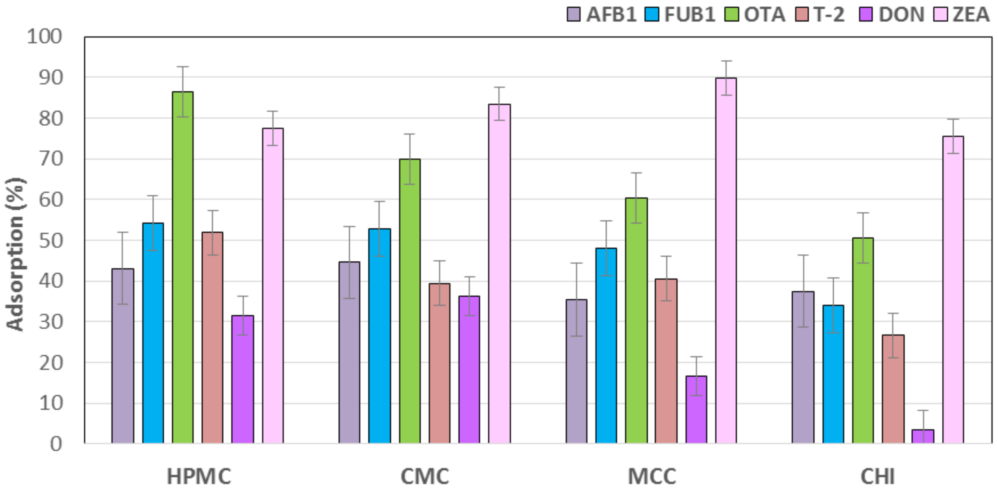
| Adsorbent | Mycotoxin | ||||||||
| AFB1 (ng/mL) | Adsorption 2 (%) | FUB1 (ng/mL) | Adsorption 2 (%) | OTA (ng/mL) | Adsorption 2 (%) | ||||
| Initial | Unbound | Initial | Unbound | Initial | Unbound | ||||
| Control | 12.00 | 12.00 ± 0.43 a | 0.00 b | 1832.33 | 1832.33 ± 43.21 a | 0.00 c | 28.32 | 28.32 ± 2.52 a | 0.00 d |
| HPMC | 6.833 ± 1.12 b | 43.06 ± 9.33 a | 840.67 ± 67.98 c | 54.12 ± 3.71 a | 3.87 ± 0.48 d | 86.35 ± 1.70 a | |||
| CMC | 6.65 ± 0.85 b | 44.58 ± 7.12 a | 863.00 ± 54.51 c | 52.90 ± 2.97 a | 8.55 ± 0.33 c,d | 69.81 ± 1.15 b | |||
| MCC | 7.75 ± 0.45 b | 35.42 ± 3.78 a | 951.00 ± 44.75 c | 48.10 ± 2.44 a | 11.18 ± 2.24 b,c | 60.51 ± 7.90 b,c | |||
| CHI | 7.50 ± 0.31 b | 37.50 ± 2.55 a | 1208.67 ± 144.15 b | 34.04 ± 7.87 b | 13.98 ± 0.90 b | 50.63 ± 3.16 c | |||
| SEM 3 | - | 0.70 | 5.63 | - | 80.30 | 4.25 | - | 1.58 | 3.92 |
| p-value | - | 0.0017 | 0.0013 | - | 0.0000 | 0.0000 | - | 0.0000 | 0.0000 |
| Adsorbent | Mycotoxin | ||||||||
| T-2 (ng/mL) | Adsorption 2 (%) | DON (ng/mL) | Adsorption 2 (%) | ZEA (ng/mL) | Adsorption 2 (%) | ||||
| Initial | Unbound | Initial | Unbound | Initial | Unbound | ||||
| Control | 174.83 | 174.83 ± 7.20 a | 0.00 d | 99.35 | 99.35 ± 1.86 a | 0.00 c | 115.33 | 115.33 ± 7.17 a | 0.00 c |
| HPMC | 84.17 ± 6.27 c | 51.86 ± 3.59 a | 68.12 ± 4.09 c | 31.43 ± 4.12 a | 25.83 ± 2.83 b | 77.60 ± 2.46 b | |||
| CMC | 105.83 ± 4.60 c | 39.47 ± 2.63 b | 63.31 ± 3.92 c | 36.27 ± 3.94 a | 19.00 ± 5.25 b,c | 83.53 ± 4.55 a,b | |||
| MCC | 103.83 ± 3.98 c | 40.61 ± 2.28 b | 82.77 ± 2.96 b | 16.69 ± 2.98 b | 11.83 ± 3.06 c | 89.74 ± 2.65 a | |||
| CHI | 128.17 ± 10.54 b | 26.69 ± 6.03 c | 95.82 ± 1.92 a | 3.55 ± 1.93 c | 28.17 ± 1.20 b | 75.58 ± 1.04 b | |||
| SEM 3 | - | 6.92 | 3.50 | - | 3.10 | 3.00 | - | 4.42 | 2.64 |
| p-value | - | 0.0000 | 0.0000 | - | 0.0000 | 0.0000 | - | 0.0000 | 0.0000 |
© 2017 by the authors. Licensee MDPI, Basel, Switzerland. This article is an open access article distributed under the terms and conditions of the Creative Commons Attribution (CC BY) license (http://creativecommons.org/licenses/by/4.0/).
Share and Cite
Solís-Cruz, B.; Hernández-Patlán, D.; Beyssac, E.; Latorre, J.D.; Hernandez-Velasco, X.; Merino-Guzman, R.; Tellez, G.; López-Arellano, R. Evaluation of Chitosan and Cellulosic Polymers as Binding Adsorbent Materials to Prevent Aflatoxin B1, Fumonisin B1, Ochratoxin, Trichothecene, Deoxynivalenol, and Zearalenone Mycotoxicoses Through an In Vitro Gastrointestinal Model for Poultry. Polymers 2017, 9, 529. https://doi.org/10.3390/polym9100529
Solís-Cruz B, Hernández-Patlán D, Beyssac E, Latorre JD, Hernandez-Velasco X, Merino-Guzman R, Tellez G, López-Arellano R. Evaluation of Chitosan and Cellulosic Polymers as Binding Adsorbent Materials to Prevent Aflatoxin B1, Fumonisin B1, Ochratoxin, Trichothecene, Deoxynivalenol, and Zearalenone Mycotoxicoses Through an In Vitro Gastrointestinal Model for Poultry. Polymers. 2017; 9(10):529. https://doi.org/10.3390/polym9100529
Chicago/Turabian StyleSolís-Cruz, Bruno, Daniel Hernández-Patlán, Eric Beyssac, Juan D. Latorre, Xochitl Hernandez-Velasco, Ruben Merino-Guzman, Guillermo Tellez, and Raquel López-Arellano. 2017. "Evaluation of Chitosan and Cellulosic Polymers as Binding Adsorbent Materials to Prevent Aflatoxin B1, Fumonisin B1, Ochratoxin, Trichothecene, Deoxynivalenol, and Zearalenone Mycotoxicoses Through an In Vitro Gastrointestinal Model for Poultry" Polymers 9, no. 10: 529. https://doi.org/10.3390/polym9100529