Article

A CRISPR/Cas9-Based Study of CgloRPCYG, a Gene That Regulates Pathogenicity, Conidial Yield, and Germination in Colletotrichum gloeosporioides

1
Key Laboratory of Integrated Pest Management on Tropical Grops, Ministry of Agriculture and Rural Affairs, Environment and Plant Protection Institute, Chinese Academy of Tropical Agricultural Sciences, Haikou 571101, China
2
College of Plant Protection, Hainan University, Haikou 570228, China
3
Jiangsu Lixiahe District Institute of Agricultural Sciences, National Agricultural Experimental Station for Agricultural Microbiology, Yangzhou 225007, China
4
Institute of Tropical Bioscience and Biotechnology, Chinese Academy of Tropical Agricultural Sciences, Haikou 571101, China
*
Author to whom correspondence should be addressed.
These authors contributed equally to this work.
Agronomy 2023, 13(7), 1681; https://doi.org/10.3390/agronomy13071681
Submission received: 22 May 2023 / Revised: 17 June 2023 / Accepted: 18 June 2023 / Published: 22 June 2023
(This article belongs to the Special Issue Plant Anthracnose: Etiology and Current Management Options)

Abstract

:
The filamentous fungus Colletotrichum gloeosporioides is the causative agent of one of the most serious diseases that damage plant fruit. In this study, we discovered and experimentally characterized a new gene in Colletotrichum gloeosporioides named CgloRPCYG. The CRISPR/Cas9 knockout mutant and complementary strain of CgloRPCYG were then obtained by polyethylene glycol (PEG)-mediated protoplast transformation to determine the related phenotypes. Compared with the wild-type strain and complementary mutant, the pathogenicity of the CRISPR/Cas9 knockout mutant was significantly decreased, the conidial yield was significantly reduced, and conidial germination was significantly delayed. These data indicate that CgloRPCYG contributes to pathogenicity, conidial yield, and germination in C. gloeosporioides. The successful application of the CRISPR/Cas9 system in C. gloeosporioides also confirms its utility in filamentous fungi for fundamental research and practical application. Furthermore, CgloRPCYG is a potential target gene for use in the development of plant protection technologies, such as spray-induced gene silencing, with the aim of controlling plant anthracnose disease caused by C. gloeosporioides.

1. Introduction

Filamentous fungi have long been known to play a significant role in agricultural production, which is severely disrupted due to disease occurrence and mycotoxin production. With the advancement of sequencing technology and fungal genome databases, a more extensive range of opportunities for genetic manipulation of filamentous fungi is available [1]. Colletotrichum spp. is a model system for fundamental, biochemical, physiological, and genetic studies [1]. Most Colletotrichum species are hemibiotrophic, with features including conidial yield, conidial germination, germ tubes development, melanized appressorium formation, infection pegs that penetrate host plant cell walls [1], biotrophic intracellular hyphae, and eventual progression into a necrotrophic phase in dead host plant cells [1]. Currently, the Colletotrichum genus includes more than 600 species. C. gloeosporioides complex species is ubiquitous and can infect over 470 plant species; it can infect leaves, flowers, and fruit, with symptoms including irregularly shaped lesions, mainly black in color, and typical reddish-pink conidia piles. The resulting disease is commonly referred to as anthracnose. C. gloeosporioides complex species is a globally distributed pathogenic fungus that causes fruit anthracnose in a vast range of hosts [2], such as avocado (Persea americana) [3], strawberry (Fragaria × ananassa) [4], mango (Mangifera indica) [5], citrus (Citrus sinensis) [6], apple (Malus pumila) [7], pepper (Capsicum annuum) [8,9], pear (Pyrus bretschneideri), and tomato (Solanum lycopersicum) [10,11]. C. gloeosporioides complex species breaches the fruit cuticle of tropical and subtropical fruits but remains quiescent until fruit ripening signals a switch to necrotrophy, culminating in devastating fruit anthracnose disease [10,12]. Previous research has confirmed that C. gloeosporioides species has evolved a sophisticated mechanism to infect hosts via various types of pathogenicity factors such as secondary metabolites, effectors, plant cell wall hydrolase and other enzymes, and transcription factors, among others. Discovering new pathogenicity genes is still the key to explaining the pathogenic mechanisms.
The clustered regularly interspaced short palindromic repeat/CRISPR-related nuclease 9 (CRISPR/Cas9) system evolved in bacterial and archaeal systems as an innate component of their adaptive immune response, and represents a new, efficient genome-editing tool for gene insertion, knockout, and replacement [13]. The system contains two crucial modules, endonuclease Cas9 and a chimeric single-guide RNA (sgRNA) [14]. The Cas9–sgRNA ribonucleoprotein complex can bind to a specific DNA sequence upstream of the protospacer adjacent motif (PAM) (5′-NGG-3′) via complementary base pairing, inducing DNA double-strand breaks and simultaneously editing multiple genes [15,16,17,18]. The CRISPR/Cas9 system is now used for specific targeted genome modification in a wide range of eukaryotes. With its high efficiency and easy operation, this technology has been successfully applied in a variety of organisms, including animals (zebrafish (Danio rerio) [19], mice [20], pigs [21], plants (Arabidopsis [22,23,24,25], tobacco [22], sorghum [22], rice [22,26], and papaya [27]), and fungi and oomycetes (Aspergillus oryzae [28], Trichoderma reese [29], Magnaporthe oryzae [30], Phytophthora capsici [31], and P. sojae [32]). The CRISPR/Cas9 system used Phytophthora to demonstrate that PcAvh1 [33] and PlAvh142 [34] are essential for pathogenicity. However, while the CRISPR/Cas9 system has been used for different plant pathogens, there has been limited application in the hemibiotrophic fungus C. gloeosporioides. The construction of a CRISPR/Cas9 genome-editing toolkit for C. gloeosporioides could be an important supplement to the molecular technology of functional genomics awaiting construction.
In this study, the CRISPR/Cas9 system was used to functionally characterize a gene potentially important for pathogenicity in C. gloeosporioides. For this purpose, we obtained knockout mutants through the CRISPR/Cas9 system, which can delete long fragments of DNA. Then, knockout mutants with long fragment deletion were complemented, and complementary mutants were finally obtained. The new gene was named CgloRPCYG based on its function in regulating pathogenicity, conidial yield, and germination in C. gloeosporioides. This preliminary exploration is expected to provide a foundation for research on functional genes in the filamentous fungus C. gloeosporioides.

2. Materials and Methods

2.1. Fungal Strain, Growth Conditions, and Plant Fruits

C. gloeosporioides wild-type (WT) strain 171-1 and backbone vectors pHS-Cas9-Backbone and pOE-Backbone used in this study were provided by the Institute of Environment and Plant Protection, Chinese Academy of Tropical Agricultural Sciences. The C. gloeosporioides strain was routinely grown on potato dextrose agar (PDA) and maintained at 28 °C [11]. Escherichia coli DH5α competent cells were purchased from Shanghai Weidi Biotechnology Co., Ltd. (Cat# DL1001) (Shanghai, China). Conidiation was assayed in 3-day-old PD broth cultures. Fruits used in the pathogenicity assay included apple (M. pumila, cultivar Hongfushi), pear (P. bretschneideri, cultivar Xuehua), and mango (M. indica, cultivar Guifei).

2.2. Sequence Alignment and Phylogenetic Analysis

The genomic sequence was obtained from the sequence database of C. gloeosporioides (https://www.ncbi.nlm.nih.gov/; GenBank assembly accession no. GCA_011800055.1, 30 September 2018). Physicochemical properties, including the amino acid sequence length, isoelectric point, and stability, were predicted using the ProtParam tool in the ExPASY server [35]. The amino acid sequences of CgloRPCYG were analyzed using the Protein Basic Local Alignment Search Tool (BlastP) of the National Biotechnology Information Center (NCBI) for multiple sequence alignment. According to the similarity with the sequence of CgloPRCYG, 54 amino acid sequences were screened, including 34 of the same genus and 20 of different genera. The 54 amino acid sequences were derived from Colletotrichum spp. (XP 031887974.1, KAF4899211.1, KAF0317795.1, KAF4820371.1, XP 037173807.1, Cg-14 EQB55550.1, XP 045256771.1, KAF4868737.1, KAH0431835.1, KAH9227001.1, XP 038738854.1, XP 036575154.1, TDZ39120.1, TEA19528.1, TDZ44972.1, MAFF 240422 TDZ16536.1, KAF6790751.1, XP 022480039.1, KXH66003.1, PJ7 EXF76443.1, XP 049151650.1, KXH32929.1, XP 035326615.1, M1.001 XP 008097816.1, KDN71047.1, XP 049133866.1, TKW59130.1, CCF38965.1, TQN70612.1, IMI 349063 XP 018161448.1, KZL86094.1, GJC78716.1, KZL77832.1, OLN87683.1), Lasiodiplodia theobromae (KAF9631773.1, KAB2573554.1), Diplodia corticola (XP 020134537.1), Neofusicoccum parvum (UCRNP2 EOD47268.1), Macrophomina phaseolina (MS6 EKG15908.1), Saccharata proteae (CBS 121410 KAF2087595.1), Xylariales sp. (PMI 506 KAH8661846.1), Dactylonectria estremocensis (KAH7139677.1), Clonostachys solani (CAH0056040.1), Jackrogersella minutella (KAI1098867.1), Hypoxylon rubiginosum (KAI6085517.1), Daldinia sp. (XP 047787477.1, XP 049095194.1, EC12 OTB20813.1, KAI1477780.1), Ophiobolus disseminans (KAF2833591.1), Pyrenochaeta sp. (MPI-SDFR-AT-0127 KAH7357626.1), Plenodomus lindquistii (KAI8934303.1), and Leptosphaeria maculans (KAH9881555.1, JN3 XP 003835460.1). CgloRPCYG was also compared with other amino acid sequences, and the phylogenetic tree was constructed based on 1000 bootstrap replicates and using the neighbor-joining method in DNAMAN v5.2.2 software (Lynnon Biosoft, San Ramon, CA, USA) and MEGA v6.06 software [36]. The Sanger sequencing results were analyzed with DNAMAN v5.2.2 and Chromas v2.22 software (Technelysium Pty Ltd., South Brisbane, QLD, Australia).

2.3. Plasmid Construction

In this work, we used a Cas9 nuclease and the usual PAM sequence (NGG) [35]. Using the online Eukaryotic Pathogen gRNA Design Tool (EuPaGDT, available at http://grna.ctegd.uga.edu, 30 September 2018) [37], a 20 nt sgRNA that can guide the Cas9 nuclease to the target for genomic DNA cleavage was designed according to the sequence of CgloRPCYG. The sequence of the sgRNA CgloRPCYG sgRNA02 is 5′-AACCACCTGACGATGCTTGT-3′. The fusion vector pHS-Cas9-CgloRPCYG was obtained by integrating the sgRNA into the backbone vector pHS-Cas9-Backbone (with hygromycin resistance cassette (Hygr), ampicillin resistance cassette (Ampr), Cas9 nuclease cassette (hCas9), and origin of replication (Ori)). The fusion plasmid was verified via Sanger sequencing analysis, and the pHS-Cas9-Backbone was used as the control plasmid (EV). The pHS-Cas9-Backbone plasmid was purchased from Beijing Syngentech Company Limited. For the complementary construct, a fragment consisting of pgpdA promoter, CgloRPCYG open reading frame, and TtrpC terminator sequences were cloned into complementary vector pEO (containing the bialaphos resistance gene, Bar). EcoRI and XbaI restriction endonuclease sequences and protective bases were added to the 5′ and 3′ ends, respectively, of the CgloRPCYG gene. Then, the pEO vector was constructed by restriction endonuclease and T4 ligase. The successful complementary vector was named pEO-CgloRPCYG. All successfully fused plasmids were propagated in E. coli DH5α competent cells. PCR was used for the identification and sequencing of E. coli-positive clones.

2.4. Polyethylene Glycol-Mediated Protoplast Transformation

Protoplasts were subjected to polyethylene glycol (PEG)-mediated transformation according to a previously published protocol [38]. For transformation, at least 1.0 × 10⁷ protoplasts and 20 μg of pHS-Cas9-RPCYG plasmid were mixed with up to 2 mL PEG buffer [38]. The protoplast mixture (3 mL) was added to potato dextrose broth (PDB) containing 0.8 M sucrose and cultured at 28 °C for 24 h with light for regeneration [38]. The culture was plated onto PDA containing 0.8 M sucrose and 150 μg/mL hygromycin B (VWR (Shanghai), Co. Ltd., Shanghai, China) and cultured at 28 °C for 24 h. A second cladding layer of PDA containing 100 μg/mL hygromycin B was added to the plates and cultured at 28 °C until mycelia grew out of the medium [39]. Then, the complementary vector was introduced into the CgloRPCYG knockout mutant protoplasts, and transformants were screened by PDS medium with glufosinate and verified by PCR, which resulted in the complementary mutant strain CΔCgloRPCYG-1.

2.5. Extraction of DNA and Verification of Transformants

Monoclonal colonies were selected for further verification. The genomic DNA of the transformants was isolated using cetyltrimethylammonium bromide (CTAB) and phenol–chloroform extraction, as previously described [40], and the DNA templates were used for the verification of transformants [40]. Phusion High-Fidelity DNA polymerase (Thermo Fisher Scientific, Waltham, MA, USA) was used for PCR amplification. The primers used are listed in Text S1 and Table S1. Agarose gel electrophoresis and Sanger sequencing were performed for verification of the transformants.

2.6. Characterization of C. gloeosporioides Mutants

To study the fungal growth, the wild-type (WT) strain, CgloRPCYG knockout mutants (ΔCgloRPCYG-1, KO), WT strain transformed with the empty vector (EV), CgloRPCYG complementary mutant (CΔCgloRPCYG-1, CKO), and WT strain transformed with the complementary empty vector (CEV) were cultured on PDA plates at 28 °C, and the diameter of colonies was measured daily. For the stress tolerance assay, strains were grown on minimal medium (MM; 50 g/L dextrose, 1.0 g/L ammonium nitrate, 0.5 g/L potassium dihydrogen phosphate, 1.5 g/L disodium hydrogen phosphate, 1.0 g/L sodium chloride, and 0.2 g/L magnesium sulfate heptahydrate, pH 6.5, control) containing 1 M NaCl, 1 M KCl, 5 mM H2O2, 0.01% sodium dodecyl sulfate (SDS), and 300 μg/mL Congo red (CR) [41]. To determine the conidial yield, mature conidia of the strains were harvested from 3-day-old PDB cultures. A hemocytometer was used to count the conidia. For the conidial germination assay, a 25 μL conidial suspension (1 × 10⁵ conidium/mL in ddH2O) of each strain was dripped onto hydrophobic coverslips and cultured in a moistened incubator at 28 °C [39]. Conidial germination was observed and recorded at 10 h [39].

2.7. Pathogenicity Assay

The pathogenicity assay was performed as described in [42], including assessment of pathogenicity, conidial yield, and germination. Physiologically ripe apples, pears, and mangoes without diseased spots were used for pathogen inoculation. The fruits were inoculated with 5 mm diameter plugs of 7-day-old cultures of WT strain and mutants grown on PDA at 28 °C. Before inoculation, fruits were wounded with a sterilized microneedle holder to facilitate penetration of the fungus into fruit tissue. Inoculated fruits were kept at 25 °C and >90% relative humidity for 4 days. The diameters of the fruit anthracnose were measured by cross intersection method and the symptoms were photographed. Each treatment contained 3 replicates of 6 fruits, and the entire experiment was repeated 3 times.

2.8. Statistical Analysis

In this study, all quantitative data were derived from at least 3 replicates. The significance of the data was analyzed using SAS 9.2 software (SAS Institute Inc., Cary, NC, USA) and WPS 2022 (Kingsoft, Beijing, China). We used the One-Way ANOVA test for analysis and Duncan’s multiple range test. In this study, different lowercase letters above bars indicate significant differences between means (p < 0.05).

3. Results

3.1. Bioinformatics and Phylogenetic Analyses of the CgloRPCYG Gene

The CgloRPCYG gene was identified in the C. gloeosporioides genomic sequence (GenBank assembly accession no. GCA_011800055.1). CgloRPCYG contains a 684 bp open reading frame containing two introns (intron 1, 57 bp; intron 2, 61 bp). The CgloRPCYG gene encodes 246 amino acid residues. The prediction results show that the molecular formula of the CgloRPCYG protein is C1136H1763N291O366S14, with a molecular weight of 25,802 Dalton. The isoelectric point and instability index are 4.67 and 42.96, respectively, indicating that it is an unstable protein. A phylogenetic tree of 55 amino acid sequences from 16 genera shows that CgloRPCYG is most similar to sequences in Colletotrichum spp. followed by Lasiodiplodia (Figure 1). In Colletotrichum spp., CgloRPCYG and homologous proteins have little difference and cluster into a large group. Other genera of filamentous fungi cluster in different groups. Similar results were found for the Lasiodiplodia, Daldinia, and Leptosphaeria genera. The BLAST searches suggest that the CgloRPCYG protein has a similar sequence in C. tropicale strain CgS9275 (KAF4820371.1). The phylogenetic analysis shows that homologous proteins of CgloRPCYG are widely present in filamentous fungi and that the sequence is conserved (Figure 1).

3.2. Successful Creation of Fusion Plasmid, Knockout, and Complementary Mutants

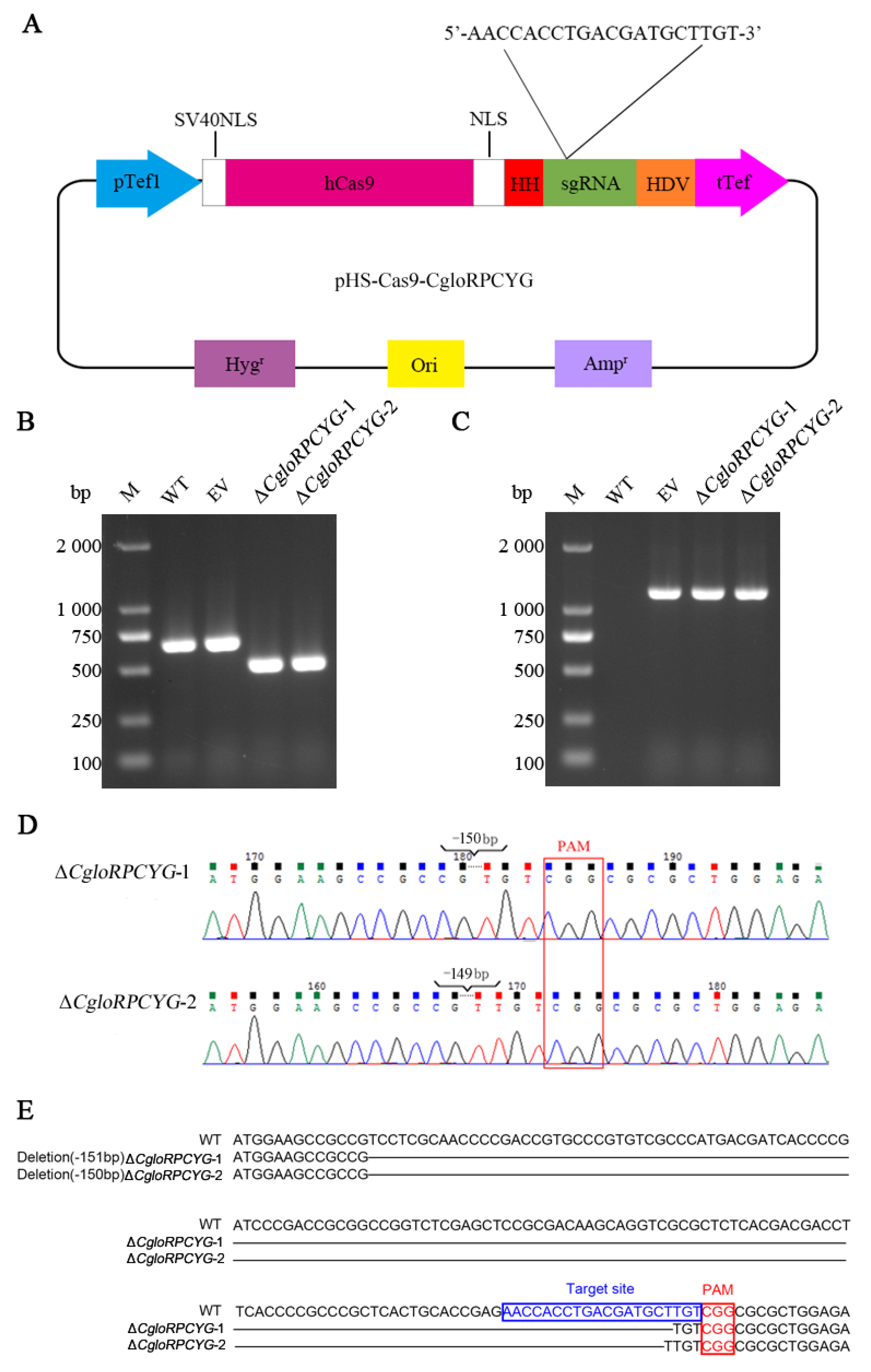
The fusion vector pHS-Cas9-CgloRPCYG was obtained by successfully integrating the sgRNA into the backbone vector pHS-Cas9-Backbone, which was confirmed via sequencing analysis (Figure 2A). Transformants were obtained by PEG-mediated protoplast transformation and selection on PDA plates containing 100 μg/mL hygromycin B. PCR amplification with primers CgloRPCYG-D-F/CgloRPCYG-D-R and Hyg-F/Hyg-R and agarose gel electrophoresis showed that two deletion mutants, ΔCgloRPCYG-1 and ΔCgloRPCYG-2, were obtained (Figure 2B,C), and Sanger sequencing also confirmed that the two mutants are deletion mutants (Figure 2D,E). As shown in Figure 2E, compared with the sequence of WT, a 150 bp deletion occurred in ΔCgloRPCYG-1, while a 149 bp deletion was detected in ΔCgloRPCYG-2. The mutated sequences in the ΔCgloRPCYG-1 and ΔCgloRPCYG-2 mutants were found to be respectively located 3 and 4 bp upstream of the PAM sequence (NGG). The efficiency of >100 bp deletion in the CRISPR/Cas9 knockout is 8.33%. In this study, CgloRPCYG knockout mutants were successfully obtained, but the deletion efficiency was low.
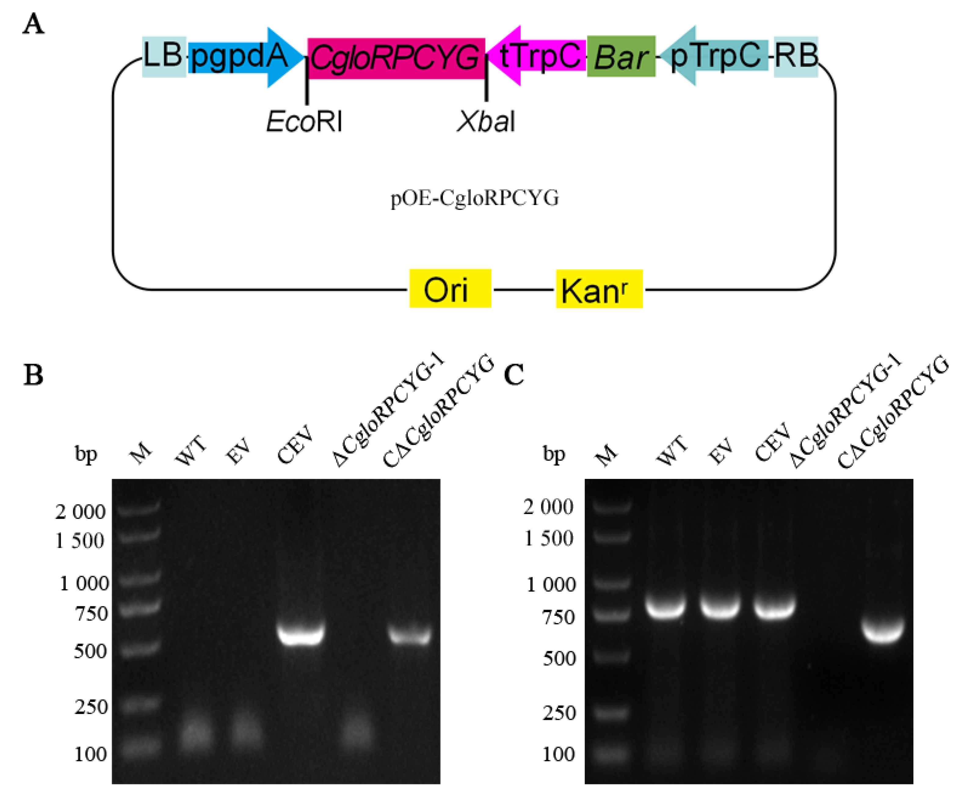
The complementary vector pOE-CgloRPCYG was obtained by successfully integrating the CgloRPCYG ORF into the backbone vector pOE-Backbone, which was confirmed via sequencing analysis (Figure 3A). PCR amplification of transformant DNA was performed using primers Bar-F/Bar-R with CgloRPCYG-F/CgloRPCYG-R and agarose gel electrophoresis. The PCR products were sequenced using the Sanger method. The DNA of the wild-type strain was used as a control. The Bar-F/Bar-R primer pair amplified specific 552 bp bands for complementary mutant and complementary backbone vector pOE-Backbone (Figure 3B). At the same time, a specific bright band of 684 bp for complementary mutant was found in the amplified transformants with the CgloRPCYG-F/CgloRPCYG-R primer pair, indicating that CgloRPCYG ORF was successfully transferred into the transformants (Figure 3C). The efficiency of positive transformation was 34.28%. To sum up, the knockout mutants and complementary strains of CgloRPCYG were successfully obtained and named ∆CgloRPCYG-1, ∆CgloRPCYG-2, and C-∆CgloRPCYG, respectively.

3.3. Role of CgloRPCYG in Mycelial Growth and Stress Tolerance

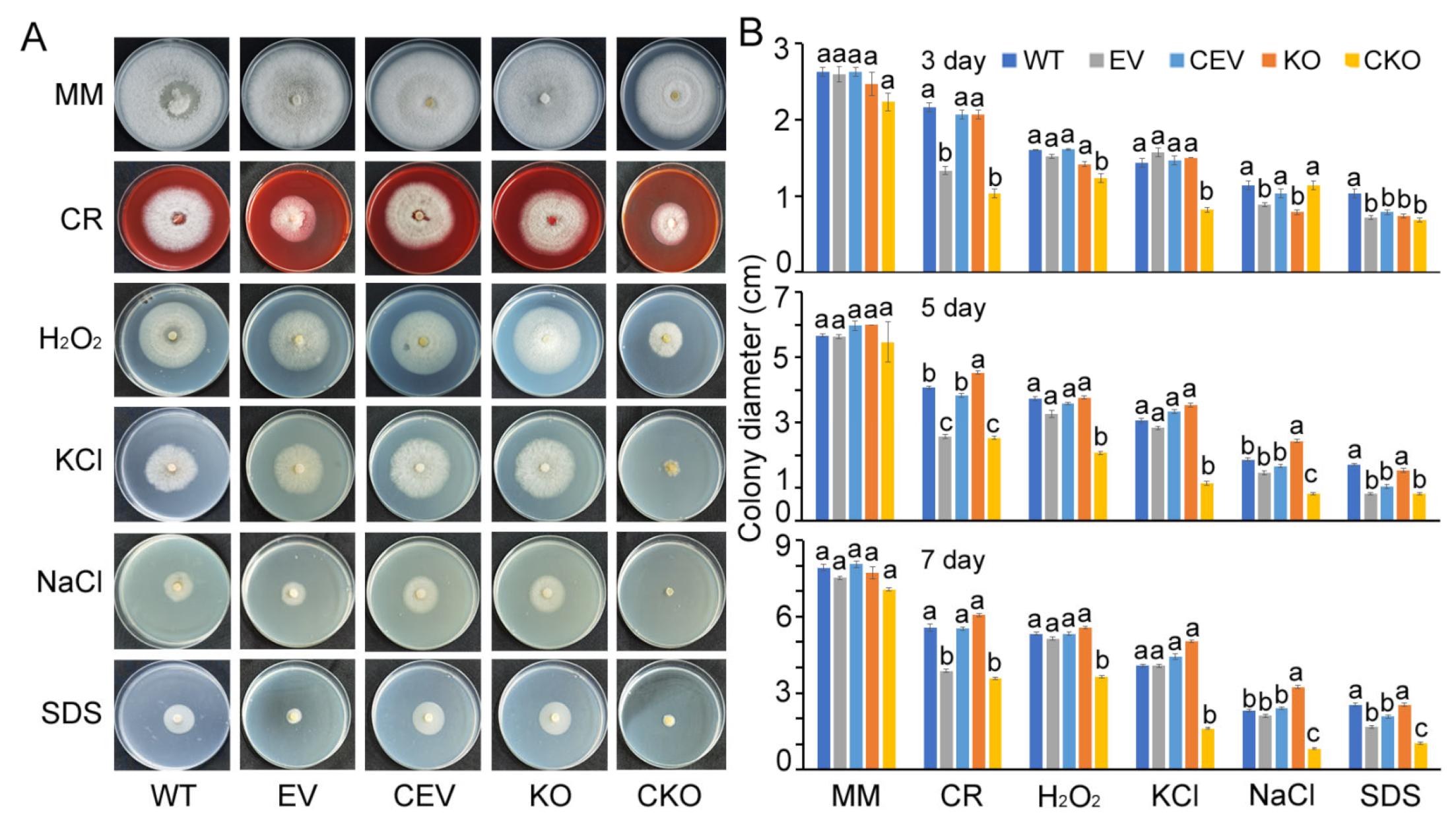
To address the role of CgloRPCYG in environmental adaptation, the WT, EV, CEV, KO, and CKO strains were inoculated on MM agar plates containing 5 mM H2O2, 0.01% SDS, 1 M NaCl, 1 M KCl, or 300 mg/L Congo red. Compared with the WT strain (Figure 4A,B), the ΔCgloRPCYG-1 mutant (KO) showed similar sensitivity to the various abiotic stresses above (Figure 4B). The data show that there are no significant differences in the colony diameter of the WT, EV, CEV, KO, and CKO strains after 3, 5, and 7 days of culture on MM plates. Additionally, the colony colors and mycelial sparsity of ΔCgloRPCYG-1 show differences. For a pathogen to successfully infect a host, it must overcome host-derived reactive oxygen species (ROS). Thus, we used H2O2 to mimic host-derived ROS. We found that deletion of the CgloRPCYG gene did not alter the sensitivity of the pathogen to ROS. This result indicates that CgloRPCYG is not involved in mediating responses to oxidative stress. Salt stress is an important basis for assessing fungal adaptation to the environment. We used KCl and NaCl to mimic environmental salt stress and found that CgloRPCYG gene deletion has different effects on the salt stress of the pathogen. The results showed that in the MM media containing NaCl for 3 days, the colony diameters of KO strain were reduced by 30.88%, compared with WT strain. In addition, after 5 and 7 days of mycelial growth in MM medium containing NaCl, the colony diameter of KO strains increased by 29.95% and 40.43% compared to WT strains, respectively.

3.4. CgloRPCYG Plays a Crucial Role in Pathogenicity, Conidial Yield, and Germination

To investigate whether deletion of CgloRPCYG would affect the pathogenicity of C. gloeosporioides, mycelial plugs were inoculated by microneedle wounded ripe apple, pear, and mango fruits. After 4 d of infection, the size of lesions on fruits caused by the indicated strains was significantly reduced compared with the lesions induced by WT and CKO (Figure 4B and Figure 5A). Compared with the WT strain, the diameter of lesions caused by knockout mutant on wounded apple, pear, and mango decreased by 86.06, 44.70, and 39.47%, respectively (Figure 4B and Figure 5A). On wounded apple fruit, the diameter of lesions caused by the ΔCgloRPCYG-1 mutant was 0.23 cm in contrast to 1.65 cm for the WT strain and 1.57 cm for the complementary mutant CΔCgloRPCYG-1. On wounded pear fruit, the diameter of lesions caused by the ΔCgloRPCYG-1 mutant was 0.73 cm in contrast to 1.32 cm for the WT strain and 1.17 cm for the complementary mutant CΔCgloRPCYG-1. On wounded mango fruit, the diameter of lesions caused the ΔCgloRPCYG-1 mutant was 0.46 cm in contrast to 0.76 cm for the WT strain and 0.66 cm for the complementary mutant CΔCgloRPCYG-1. These data suggest that CgloRPCYG plays a vital role in determining the pathogenicity of C. gloeosporioides.
Next, to investigate whether CgloRPCYG plays a role in C. gloeosporioides conidial yield, mature conidia harvested from 3-day-old PDB cultures were counted with a hemocytometer. The conidial yield and germination of strains were observed under an optical microscope. The relative conidiation of the KO mutant was significantly lower than that of the other four mutants (Figure 5C). In addition, to identify whether the deletion of CgloRPCYG had an effect on conidial germination, we recorded and compared conidial germination among the different strains within 8, 12, and 24 h. The results show that the conidia germination rate of ΔCgloRPCYG-1 was delayed in comparison to WT, EV, CV, and CΔCgloRPCYG-1 on the glass slides (Figure 5D). These results indicate that CgloRPCYG is essential for conidial yield and germination in C. gloeosporioides.

4. Discussion

The CRISPR/Cas systems can be divided into six types and more than 20 subtypes based on the differences in Cas9, Cas12, and Cas13 effectors [43,44,45,46]. The most common of these is CRISPR/Cas9 system, which is efficient, easy to operate, and has many successful cases [46]. There is also an online software designed with sgRNA. In this study, we analyzed the genomic sequence of C. gloeosporioides and designed an sgRNA target using EuPaGDT online software [37]. EuPaGDT allows users to tune the design of gRNA and characterization of on/off targets, including different CRISPR/Cas9 orthologs and PAM sequences, such as NGG, NAG, and NGA [37]. We referred to the tables of on-target/off-target scores to visually check them and corresponding PAMs, as well as the alignment of gRNA with the CgloRPCYG gene sequence, selecting the one with the highest score and finally obtaining sgRNA02: 5′-AACCACCTGACGATGCTTGT-3′. Next, we successfully constructed a CgloRPCYG gene editing vector using the CRISPR/Cas9 system, resulting in the deletion of the CgloRPCYG fragment, which provides a reference for the application of CRISPR/Cas9 in the filamentous fungus C. gloeosporioides. The results of this study confirm the applicability of EuPaGDT in C. gloeosporioides. This system can be used in the long term to study the function of other genes by only changing the sequence of sgRNA.
In Claviceps purpurea, CRISPR/Cas9 pyr4 mutations are mostly located 3 bp upstream of the PAM sequence, with insertions and deletions of larger DNA fragments in addition [47]. This shows that the effect of applying CRISPR/Cas9 in C. purpurea is similar to that in C. gloeosporioides. Meanwhile, the gene editing efficiency of the CgloRPCYG gene is higher than that of pyr4 gene, TrpE gene in C. purpurea [47]. Recently, several studies have reported the successful development of this technology in filamentous fungi, including the industrial filamentous fungi A. fumigatus [48], T. reesei [29], and Myceliophthora thermophila [49], as well as plant-pathogenic fungi Ustilago maydis [50], M. oryzae [30], and Ustilaginoidea virens [41]. For instance, Liu et al. [49] successfully developed a CRISPR/Cas9 system for efficient multiplexed genome engineering in Myceliophthora species. However, few studies on C. gloeosporioides have used this system. Hence, the novel CRISPR/Cas9 technique has been shown to have great potential in the study of functional genes in filamentous fungi. On this basis, a CRISPR/Cas9-mediated in situ complementation method for C. gloeosporioides could also be developed, with reference to P. sojae [51].
The research reports suggest that CgloRPCYG is responsible for the pathogenicity of filamentous fungi. Certainly, the deletion of CgloRPCYG led to lower pathogenicity in apple, pear, and mango fruits in this study, indicating that CgloRPCYG functions as a pathogenicity factor. In C. gloeosporioides, knockout of the CgHOS2 [52], CgHSF1 [53], CgGa1 [54], CgCPS1 [55], CgOPT2 [56], and CgEnd3 [57] genes showed the same result. C. gloeosporioides produce conidia, exhibit conidial germination and appressorium, form penetration pegs, and invade hosts, thus causing plant disease. We analyzed the effects of gene deletion on conidial yield and germination rate. Deletion of CgloRPCYG caused an obvious decrease in conidial yield and delayed conidial germination. Compared with the wild-type strain, the conidial yield was decreased by 60.60% in the knockout mutant. Similarly, conidial germination rates at 8, 12, and 24 h decreased by 36.11, 31.71, and 60.24%, respectively. A study reported that the deletion of BcCFEM1 in B. cinerea is responsible for a decrease in conidia production [58]. In C. gloeosporioides, CgHOS2 gene regulates the length of the germ tube [52]. CgloRPCYG is a potential target gene for use in the development of both plant protection technologies, such as spray-induced gene silencing, as well as new fungicides targeted to control plant anthracnose disease caused by C. gloeosporioides.

5. Conclusions

In conclusion, we experimentally characterized a new gene in C. gloeosporioides named CgloRPCYG. We successfully developed a CRISPR/Cas9 system for gene editing in C. gloeosporioides, a filamentous fungus with a wide range of plant hosts. The length of the knockout fragment is about 150 bp. Through this deletion system and complementary mutants, we demonstrated that the CgloRPCYG protein is involved in conidial yield and germination of C. gloeosporioides, as well as pathogenicity in three hosts (apple, pear, and mango). We provided a new pathogenic gene from C. gloeosporioides. It can be used to develop plant protection technologies and develop new fungicide targets. At the same time, our study extends upon the knowledge regarding the pathogenic molecular mechanisms of C. gloeosporioides.

Supplementary Materials

The following supporting information can be downloaded at https://www.mdpi.com/article/10.3390/agronomy13071681/s1: Text S1. Primer sequences of PCR and CgloRPCYG, Hyg, and Bar gene sequences. Table S1. Primers used for PCR and PCR products’ expected length.

Author Contributions

Conceptualization, H.Z., X.-M.L. and J.-J.P.; methodology, H.Z. and Y.-Q.X.; validation, Y.X., M.-T.Z., Z.Y. and R.-Q.S.; formal analysis, H.Z.; investigation, Y.X.; resources, H.Z.; data curation, Y.X.; writing—original draft preparation, H.Z. and Y.X.; writing—review and editing, H.Z.; supervision, X.-M.L. and J.-J.P.; project administration, H.Z.; funding acquisition, H.Z. All authors have read and agreed to the published version of the manuscript.

Funding

This research was funded by the Hainan Province Science and Technology Special Fund (grant number 322RC755), the Major Science and Technology Plan of Hainan Province (grant number ZDKJ2021014), the National Key R&D Program of China (grant number 2019YFD1000504), the Central Public-Interest Scientific Institution Basal Research Fund for Chinese Academy of Tropical Agricultural Sciences (grant numbers 1630042022009, 16300320220007), and the China Agriculture Research System of MOF and MARA (grant number CARS-31).

Institutional Review Board Statement

Not applicable.

Informed Consent Statement

Not applicable.

Data Availability Statement

The data that support the findings of this study are available from the corresponding author upon reasonable request.

Conflicts of Interest

The authors declare no conflict of interest.

References

  1. Dean, R.; Van Kan, J.A.L.; Pretorius, Z.A.; Hammond-Kosack, K.E.; Di Pietro, A.; Spanu, P.D.; Rudd, J.J.; Dickman, M.; Kahmann, R.; Ellis, J.; et al. The top 10 fungal pathogens in molecular plant pathology. Mol. Plant Pathol. 2012, 13, 414–430. [Google Scholar] [CrossRef] [PubMed] [Green Version]
  2. Bernstein, B.; Zehr, E.I.; Dean, R.A.; Shabi, E. Characteristics of Colletotrichum from peach, apple, pecan, and other hosts. Plant Dis. 1995, 79, 478–482. [Google Scholar] [CrossRef]
  3. Yakoby, N.; Beno-Moualem, D.; Keen, N.T.; Dinoor, A.; Pines, O.; Prusky, D. Colletotrichum gloeosporioides pelB is an important virulence factor in avocado fruit-fungus interaction. Mol. Plant Microbe Interact. 2001, 14, 988–995. [Google Scholar] [CrossRef] [PubMed] [Green Version]
  4. Xiao, C.L.; MacKenzie, S.J.; Legard, D.E. Genetic and pathogenic analyses of Colletotrichum gloeosporioides isolates from strawberry and noncultivated hosts. Phytopathology 2004, 94, 446–453. [Google Scholar] [CrossRef] [Green Version]
  5. Kefialew, Y.; Ayalew, A. Postharvest biological control of anthracnose (Colletotrichum gloeosporioides) on mango (Mangifera indica). Postharvest Biol. Technol. 2008, 50, 8–11. [Google Scholar] [CrossRef]
  6. Lima, W.G.; Spósito, M.B.; Amorim, L.; Gonçalves, F.P.; de Filho, P. Colletotrichum gloeosporioides, a new causal agent of citrus post-bloom fruit drop. Eur. J. Plant Pathol. 2011, 131, 157–165. [Google Scholar] [CrossRef]
  7. González, E.; Sutton, T.B.; Correll, J.C. Clarification of the etiology of glomerella leaf spot and bitter rot of apple caused by Colletotrichum spp. based on morphology and genetic, molecular, and pathogenicity tests. Phytopathology 2006, 96, 982–992. [Google Scholar] [CrossRef] [Green Version]
  8. Manandhar, J.B.; Hartman, G.L.; Wang, T.C. Anthracnose development on pepper fruits inoculated with Colletotrichum gloeosporioides. Plant Dis. 1995, 79, 380–383. [Google Scholar] [CrossRef]
  9. Liu, F.; Tang, G.; Zheng, X.; Li, Y.; Sun, X.; Qi, X.; Zhou, Y.; Xu, J.; Chen, H.; Chang, X.; et al. Molecular and phenotypic characterization of Colletotrichum species associated with anthracnose disease in peppers from Sichuan Province, China. Sci. Rep. 2016, 6, 32761. [Google Scholar] [CrossRef] [Green Version]
  10. Alkan, N.; Friedlander, G.; Ment, D.; Prusky, D.; Fluhr, R. Simultaneous transcriptome analysis of Colletotrichum gloeosporioides and tomato fruit pathosystem reveals novel fungal pathogenicity and fruit defense strategies. New Phytol. 2015, 205, 801–815. [Google Scholar] [CrossRef]
  11. Huang, Y.; Li, B.; Yin, J.; Yang, Q.; Sheng, O.; Deng, G.; Li, C.; Hu, C.; Dong, T.; Dou, T.; et al. CgGCS, encoding a glucosylceramide synthase, is required for growth conidiation and pathofenicity in Colletotrichum gloeosporioides. Front. Microbiol. 2019, 10, 1016. [Google Scholar] [CrossRef] [Green Version]
  12. Hubballi, M.; Nakkeeran, S.; Raguchander, T. First report of anthracnose on noni caused by Colletotrichum gloeosporioides in India. Arch. Phytopathol. Plant Prot. 2012, 45, 276–279. [Google Scholar] [CrossRef]
  13. Song, R.; Zhai, Q.; Sun, L.; Huang, E.; Zhang, Y.; Zhu, Y.; Guo, Q.; Tian, Y.; Zhao, B.; Lu, H. CRISPR/Cas9 genome editing technology in filamentous fungi: Progress and perspective. Appl. Microbiol. Biotechnol. 2019, 103, 6919–6932. [Google Scholar] [CrossRef] [Green Version]
  14. Peng, C.; Lu, M.; Yang, D. CRISPR/Cas9-based tools for targeted genome editing and replication control of HBV. Virol. Sin. 2015, 30, 317–325. [Google Scholar] [CrossRef]
  15. Cobb, R.E.; Wang, Y.; Zhao, H. High-efficiency multiplex genome editing of Streptomyces species using an engineered CRISPR/Cas system. ACS Synth. Biol. 2015, 4, 723–728. [Google Scholar] [CrossRef] [Green Version]
  16. Yan, Q.; Zhang, Q.; Yang, H.; Zou, Q.; Tang, C.; Fan, N.; Lai, L. Generation of multi-gene knockout rabbits using the Cas9/gRNA system. Cell Regener. 2014, 3, 12. [Google Scholar] [CrossRef] [Green Version]
  17. Jiang, Y.; Chen, B.; Duan, C.; Sun, B.; Yang, J.; Yang, S. Multigene editing in the Escherichia coli genome via the CRISPR-Cas9 system. Appl. Environ. Microbiol. 2015, 81, 2506–2514. [Google Scholar] [CrossRef] [Green Version]
  18. Gao, S.; Tong, Y.; Wen, Z.; Zhu, L.; Ge, M.; Chen, D.; Jiang, Y.; Yang, S. Multiplex gene editing of the Yarrowia lipolytica genome using the CRISPR-Cas9 system. J. Ind. Microbiol. Biotechnol. 2016, 43, 1085–1093. [Google Scholar] [CrossRef]
  19. Hruscha, A.; Krawitz, P.; Rechenberg, A.; Heinrich, V.; Hecht, J.; Haass, C.; Schmid, B. Efficient CRISPR/Cas9 genome editing with low off-target effects in zebrafish. Development 2013, 140, 4982–4987. [Google Scholar] [CrossRef] [Green Version]
  20. Platt, R.J.; Chen, S.; Zhou, Y.; Yim, M.J.; Swiech, L.; Kempton, H.R.; Dahlman, J.E.; Parnas, O.; Eisenhaure, T.M.; Jovanovic, M.; et al. CRISPR-Cas9 knockin mice for genome editing and cancer modeling. Cell 2014, 159, 440–455. [Google Scholar] [CrossRef] [Green Version]
  21. Wang, X.; Cao, C.; Huang, J.; Yao, J.; Hai, T.; Zheng, Q.; Wang, X.; Zhang, H.; Qin, G.; Cheng, J.; et al. One-step generation of triple gene-targeted pigs using CRISPR/Cas9 system. Sci. Rep. 2016, 6, 20620. [Google Scholar] [CrossRef] [PubMed] [Green Version]
  22. Jiang, W.; Zhou, H.; Bi, H.; Fromm, M.; Yang, B.; Weeks, D.P. Demonstration of CRISPR/Cas9/sgRNA-mediated targeted gene modification in Arabidopsis, tobacco, sorghum and rice. Nucleic Acids Res. 2013, 41, e188. [Google Scholar] [CrossRef] [PubMed]
  23. Belhaj, K.; Chaparro-Garcia, A.; Kamoun, S.; Patron, N.J.; Nekrasov, V. Editing plant genomes with CRISPR/Cas9. Curr. Opin. Biotechnol. 2015, 32, 76–84. [Google Scholar] [CrossRef]
  24. Yu, Z.; Chen, Q.; Chen, W.; Zhang, X.; Mei, F.; Zhang, P.; Zhao, M.; Wang, X.; Shi, N.; Jackson, S.; et al. Multigene editing via CRISPR/Cas9 guided by a single-sgRNA seed in Arabidopsis. J. Integr. Plant Biol. 2018, 60, 376–381. [Google Scholar] [CrossRef] [Green Version]
  25. Wang, Y.; Li, Y.; Rosas-Diaz, T.; Caceres-Moreno, C.; Lozano-Duran, R.; Macho, A.P. The IMMUNE-ASSOCIATED NUCLEOTIDE-BINDING 9 protein is a regulator of basal immunity in Arabidopsis thaliana. Mol. Plant Microbe In. 2019, 32, 65–75. [Google Scholar] [CrossRef] [PubMed] [Green Version]
  26. Wang, Y.; Geng, L.; Yuan, M.; Wei, J.; Jin, C.; Li, M.; Yu, K.; Zhang, Y.; Jin, H.; Wang, E.; et al. Deletion of a target gene in Indica rice via CRISPR/Cas9. Plant Cell Rep. 2017, 36, 1333–1343. [Google Scholar] [CrossRef]
  27. Gumtow, R.; Wu, D.; Uchida, J.; Tian, M. A Phytophthora palmivora extracellular cystatin-like protease inhibitor targets papain to contribute to virulence on papaya. Mol. Plant. Microbe In. 2018, 31, 363–373. [Google Scholar] [CrossRef] [Green Version]
  28. Katayama, T.; Tanaka, Y.; Okabe, T.; Nakamura, H.; Fujii, W.; Kitamoto, K.; Maruyama, J. Development of a genome editing technique using the CRISPR/Cas9 system in the industrial filamentous fungus Aspergillus oryzae. Biotechnol. Lett. 2015, 38, 637–642. [Google Scholar] [CrossRef]
  29. Liu, R.; Chen, L.; Jiang, Y.; Zhou, Z.; Zou, G. Efficient genome editing in filamentous fungus Trichoderma reesei using the CRISPR/Cas9 system. Cell Discov. 2015, 1, 15077. [Google Scholar] [CrossRef] [Green Version]
  30. Foster, A.J.; Martin-Urdiroz, M.; Yan, X.; Wright, H.S.; Soanes, D.M.; Talbot, N.J. CRISPR-Cas9 ribonucleoprotein-mediated co-editing and counterselection in the rice blast fungus. Sci. Rep. 2018, 8, 14355. [Google Scholar] [CrossRef] [Green Version]
  31. Miao, J.; Chi, Y.; Lin, D.; Tyler, B.M.; Liu, X. Mutations in ORP1 conferring oxathiapiprolin resistance confirmed by genome editing using CRISPR/Cas9 in Phytophthora capsici and P. sojae. Phytopathology 2018, 108, 1412–1419. [Google Scholar] [CrossRef] [Green Version]
  32. Peng, Q.; Wang, Z.; Fang, Y.; Wang, W.; Cheng, X.; Liu, X. Point mutations in the β-tubulin of Phytophthora sojae confer resistance to ethaboxam. Phytopathology 2019, 109, 2096–2106. [Google Scholar] [CrossRef]
  33. Chen, X.; Zhang, Y.; Li, H.; Zhang, Z.; Sheng, G.; Li, Y.; Xing, Y.; Huang, S.; Tao, H.; Kuan, T.; et al. The RXLR effector PcAvh1 is required for full virulence of Phytophthora capsici. Mol. Plant Microbe Interact. 2019, 32, 986–1000. [Google Scholar] [CrossRef]
  34. Situ, J.; Jiang, L.; Fan, X.; Yang, W.; Li, W.; Xi, P.; Deng, Y.; Kong, G.; Jiang, Z. An RXLR effector PlAvh142 from Peronophythora litchii triggers plant cell death and contributes to virulence. Mol. Plant Pathol. 2020, 21, 415–428. [Google Scholar] [CrossRef] [Green Version]
  35. Gasteiger, E.; Hoogland, C.; Gattiker, A.; Duvaud, S.; Wilkins, M.R.; Appel, R.D.; Bairoch, A. Protein identification and analysis tools on the ExPASy Server. In The Proteomics Protocols Handbook; Walker, J.M., Ed.; Humana Press: Totowa, NJ, USA, 2015; pp. 571–607. [Google Scholar]
  36. Tamura, K.; Stecher, G.; Peterson, D.; Filipski, A.; Kumar, S. MEGA6: Molecular evolutionary genetics analysis version 6.0. Mol. Biol. Evol. 2013, 30, 2725–2729. [Google Scholar] [CrossRef] [Green Version]
  37. Peng, D.; Tarleton, R. EuPaGDT: A web tool tailored to design CRISPR guide RNAs for eukaryotic pathogens. Microb. Genom. 2015, 1, e000033. [Google Scholar] [CrossRef] [Green Version]
  38. Turgeon, B.G.; Garber, R.C.; Yoder, O.C. Transformation of the fungal maize pathogen Cochliobolus heterostrophus using the Aspergillus nidulans amdS gene. Mol. Gen. Genet. 1985, 201, 450–453. [Google Scholar] [CrossRef]
  39. Wei, Y.; Pu, J.; Zhang, H.; Liu, Y.; Zhou, F.; Zhang, K.; Liu, X. The laccase gene (LAC1) is essential for Colletotrichum gloeosporioides development and virulence on mango leaves and fruits. Physiol. Mol. Plant Pathol. 2017, 99, 55–64. [Google Scholar] [CrossRef]
  40. Liu, J.K.; Chang, H.W.; Liu, Y.; Qin, Y.H.; Ding, Y.H.; Wang, L.; Zhao, Y.; Zhang, M.Z.; Cao, S.N.; Li, L.T.; et al. The key gluconeogenic gene PCK1 is crucial for virulence of Botrytis cinerea via initiating its conidial germination and host penetration. Environ. Microbiol. 2018, 20, 1794–1814. [Google Scholar] [CrossRef]
  41. Liang, Y.; Han, Y.; Wang, C.; Jiang, C.; Xu, J.R. Targeted deletion of the USTA and UvSLT2 genes efficiently in Ustilaginoidea virens with the CRISPR-Cas9 system. Front. Plant Sci. 2018, 9, 699. [Google Scholar] [CrossRef]
  42. Yin, Y.; Wu, S.; Chui, C.; Ma, T.; Jiang, H.; Hahn, M.; Ma, Z. The MAPK kinase BcMkk1 suppresses oxalic acid biosynthesis via impeding phosphorylation of BcRim15 by BcSch9 in Botrytis cinerea. PLoS Pathog. 2018, 14, e1007285. [Google Scholar] [CrossRef] [PubMed]
  43. Makarova, K.S.; Wolf, Y.I.; Koonin, E.V. Classification and nomenclature of CRISPR-Cas systems: Where from here? CRISPR J. 2018, 1, 325–336. [Google Scholar] [CrossRef] [PubMed] [Green Version]
  44. Li, Y.; Li, S.; Wang, J.; Liu, G. CRISPR/Cas systems towards nextgeneration biosensing. Trends Biotechnol. 2019, 37, 730–743. [Google Scholar] [CrossRef]
  45. Jiang, C.; Lv, G.; Tu, Y.; Cheng, X.; Duan, Y.; Zeng, B.; He, B. Applications of CRISPR/Cas9 in the synthesis of secondary metabolites in filamentous fungi. Front. Microbiol. 2021, 12, 638096. [Google Scholar] [CrossRef] [PubMed]
  46. Verma, V.; Batta, A.; Singh, H.; Srivastava, A.; Garg, S.K.; Singh, V.P.; Arora, P.K. Bioengineering of fungal endophytes through the CRISPR/Cas9 system. Front. Microbiol. 2023, 14, 1146650. [Google Scholar] [CrossRef] [PubMed]
  47. Králová, M.; Bergougnoux, V.; Frébort, I. CRISPR/Cas9 genome editing in ergot fungus Claviceps purpurea. J. Biotechnol. 2021, 325, 341–354. [Google Scholar] [CrossRef]
  48. Fuller, K.K.; Chen, S.; Loros, J.J.; Dunlap, J.C. Development of the CRISPR/Cas9 system for targeted gene disruption in Aspergillus fumigatus. Eukaryotic Cell 2015, 14, 1073–1080. [Google Scholar] [CrossRef] [Green Version]
  49. Liu, Q.; Gao, R.; Li, J.; Lin, L.; Zhao, J.; Sun, W.; Tian, C. Development of a genome-editing CRISPR/Cas9 system in thermophilic fungal Myceliophthora species and its application to hyper-cellulase production strain engineering. Biotechnol. Biofuels 2017, 10, 1. [Google Scholar] [CrossRef] [Green Version]
  50. Schuster, M.; Schweizer, G.; Reissmann, S.; Kahmann, R. Genome editing in Ustilago maydis using the CRISPR–Cas system. Fungal Genet. Biol. 2016, 89, 3–9. [Google Scholar] [CrossRef]
  51. Qiu, M.; Li, Y.; Ye, W.; Zheng, X.; Wang, Y. A CRISPR/Cas9-mediated in situ complementation method for Phytophthora sojae mutants. Mol. Plant Pathol. 2021, 22, 373–381. [Google Scholar] [CrossRef]
  52. Liu, S.; Wang, Q.; Liu, N.; Luo, H.; He, C.; An, B. The histone deacetylase HOS2 controls pathogenicity through regulation of melanin biosynthesis and appressorium formation in Colletotrichum gloeosporioides. Phytopathol. Res. 2022, 4, 21. [Google Scholar] [CrossRef]
  53. Gao, X.; Wang, Q.; Feng, Q.; Zhang, B.; He, C.; Luo, H.; An, B. Heat shock transcription factor CgHSF1 is required for melanin biosynthesis, appressorium formation, and pathogenicity in Colletotrichum gloeosporioides. J. Fungi 2022, 8, 175. [Google Scholar] [CrossRef]
  54. Li, X.; Ke, Z.; Xu, S.; Tang, W.; Liu, Z. The G-protein alpha subunit CgGa1 mediates growth, sporulation, penetration and pathogenicity in Colletotrichum gloeosporioides. Microb. Pathog. 2021, 161, 105254. [Google Scholar] [CrossRef]
  55. Mushtaq, A.; Tariq, M.; Ahmed, M.; Zhou, Z.; Ali, I.; Mahmood, R.T. Carbamoyl phosphate synthase subunit CgCPS1 is necessary for virulence and to regulate stress tolerance in Colletotrichum gloeosporioides. Plant Pathol. J. 2021, 37, 232–242. [Google Scholar] [CrossRef]
  56. Li, X.; Liu, S.; Zhang, N.; Liu, Z. Function and transcriptome analysis of an oligopeptide transporter CgOPT2 in the rubber anthracnose fungus Colletotrichum gloeosporioides. Physiol. Mol. Plant Pathol. 2021, 115, 101661. [Google Scholar] [CrossRef]
  57. Wang, X.; Lu, D.; Tian, C. CgEnd3 regulates endocytosis, appressorium formation, and virulence in the poplar anthracnose fungus Colletotrichum gloeosporioides. Int. J. Mol. Sci. 2021, 22, 4029. [Google Scholar] [CrossRef]
  58. Zhu, W.; Wei, W.; Wu, Y.; Zhou, Y.; Peng, F.; Zhang, S.; Chen, P.; Xu, X. BcCFEM1, a CFEM domain-containing protein with putative GPI-anchored site, is involved in pathogenicity, conidial production, and stress tolerance in Botrytis cinerea. Front. Microbiol. 2017, 8, 1807. [Google Scholar] [CrossRef]
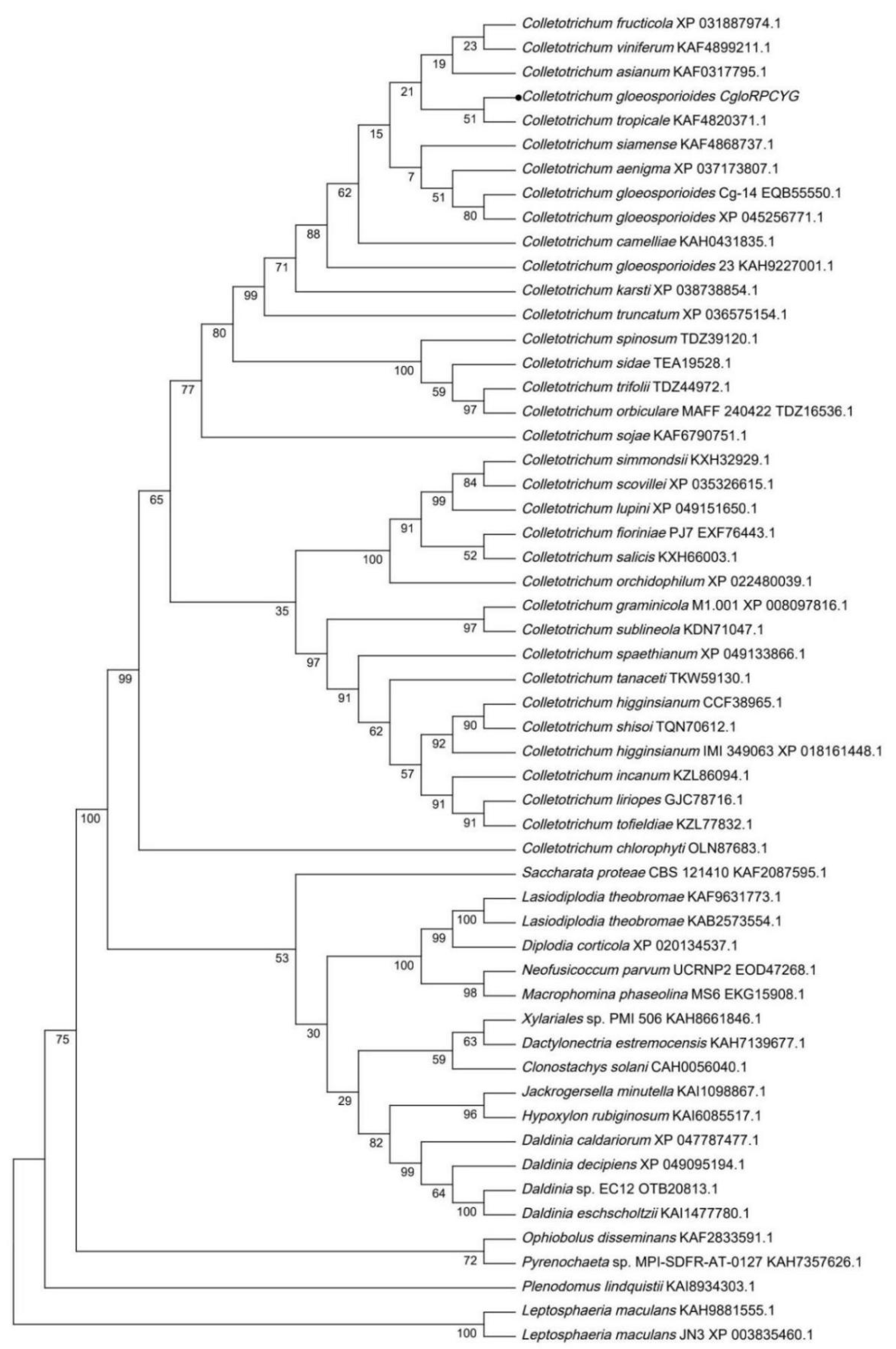
Figure 1. Phylogenetic analysis of similar sequences of CgloRPCYG protein from different fungi. Protein sequences were analyzed using DNAMAN v5.2.2 and MEGA v6.06 software with neighbor-joining method and 1000 bootstrap replicates. Solid black circle represents CgloRPCYG.
Agronomy 13 01681 g001
Figure 2. CRISPR/Cas9-assisted genome editing in C. gloeosporioides. (A) Map of fusion vector pHS-Cas9-CgloRPCYG. DNA elements not drawn to scale. (B) PCR assays to verify gene deletion events in mutants with primers CgloRPCYG-D-F/CgloRPCYG-D-R. M, DL 2000 DNA marker; WT, wild-type C. gloeosporioides; EV, mutant transformed with empty vector. ΔCgloRPCYG-1, ΔCgloRPCYG-2, CgloRPCYG: deletion mutants. (C) PCR assays to verify hygromycin B phosphotransferase gene in mutants with primers Hyg-F/Hyg-R. (D) DNA peak map of ΔCgloRPCYG-1 and ΔCgloRPCYG-2. Red frame indicates protospacer adjacent motif (PAM). (E) Sequence alignment between WT and CgloRPCYG deletion mutants. The blue frame indicates the target site, and the red frame indicates the PAM.
Agronomy 13 01681 g002
Figure 3. CgloRPCYG gene complementation of the C. gloeosporioides knockout mutant. (A) Map of the fusion vector pOE-CgloRPCYG. (B) PCR assays to verify Bar gene in the mutants with primers Bar-F/Bar-R. (C) PCR assays to verify complementary mutants using primers CgloRPCYG-F/CgloRPCYG-R. M, DL 2000 DNA marker; WT, wild-type strain; EV, mutant transformed with empty vector; CEV, wild-type strain transformed with complementary empty vector. ΔCgloRPCYG-1, CgloRPCYG: knockout mutants. CΔCgloRPCYG, CgloRPCYG-1: complementary mutants.
Agronomy 13 01681 g003
Figure 4. Mycelial growth and colony morphology of CgloRPCYG mutants in response to different stresses. (A) Mycelial radial growth of WT, EV, CEV, KO, and CKO mutants in response to stress. Typical photographs were taken after 7 days of cultivation. (B) Colony diameter of strains cultured on MM containing 300 mg/L Congo red (CR), 5 mM H2O2, 1 M KCl, 1 M NaCl, or 0.01% SDS. Colony diameter was measured after 3, 5, and 7 days of cultivation. WT, wild-type strain; EV, mutant transformed with empty vector; CEV, wild-type strain transformed with empty complementation vector; KO, CgloRPCYG knockout mutant ΔCgloRPCYG-1; CKO, CgloRPCYG complementary mutant CΔCgloRPCYG-1. Data represent mean ± SD from three separate experiments, each with three replications. In the same group, different lowercase letters above the bar indicate significant differences between the averages, while the same lowercase letters indicate insignificant differences between the averages (p < 0.05, Duncan’s multiple range test).
Agronomy 13 01681 g004
Figure 5. CgloRPCYG plays a crucial role in pathogenicity, conidial yield, and germination of C. gloeosporioides. (A) Deletion of CgloRPCYG in C. gloeosporioides results in attenuated pathogenicity on apple, pear, and mango fruits. Typical photographs were taken after 4 days of cultivation. (B) Diameter of lesions caused by strains shown in (A). (C) Relative conidiation of strains after 3 days of cultivation in PDB. (D) Conidial germination rate of strains at 8, 12, and 24 h. Data represent mean ± SD from three separate experiments, each with three replications. In the same group, different lowercase letters above the bar indicate significant differences between the averages, while the same lowercase letters indicate insignificant differences between the averages (p < 0.05, Duncan’s multiple range test).
Agronomy 13 01681 g005
Disclaimer/Publisher’s Note: The statements, opinions and data contained in all publications are solely those of the individual author(s) and contributor(s) and not of MDPI and/or the editor(s). MDPI and/or the editor(s) disclaim responsibility for any injury to people or property resulting from any ideas, methods, instructions or products referred to in the content.