Article

Halophilic Plant Growth-Promoting Rhizobacteria as Producers of Antifungal Metabolites under Salt Stress

1
Laboratoire de Biochimie Analytique et Biotechnologies, Université Mouloud Mammeri de Tizi-Ouzou, Tizi Ouzou 15000, Algeria
2
Laboratoire de Microbiologie Appliquée, Université Ferhat Abbas, Sétif 1, Setif 19000, Algeria
3
cE3c—Center for Ecology, Evolution and Environmental Changes & CHANGE—Global Change and Sustainability Institute, Faculdade de Ciências da Universidade de Lisboa Campo Grande, 1749-016 Lisboa, Portugal
4
Laboratoire de Maitrise des Energies Renouvelables, Faculté des Sciences de la Nature et de le Vie, Université de Bejaïa, Bejaïa 06000, Algeria
*
Author to whom correspondence should be addressed.
Agronomy 2024, 14(4), 845; https://doi.org/10.3390/agronomy14040845
Submission received: 4 February 2024 / Revised: 30 March 2024 / Accepted: 4 April 2024 / Published: 18 April 2024
(This article belongs to the Section Soil and Plant Nutrition)

Abstract

:
Salinity is one of the main factors causing soil deterioration, making it unsuitable for agriculture. It is well documented that the application of halotolerant and halophilic plant growth-promoting bacteria (PGPR: plant growth-promoting rhizobacteria) with biological control activities as an inoculant of cultivated plants offers a biological alternative to the use of agrochemicals, particularly when subjected to salt stress. From this perspective, 70 bacterial strains were isolated from saline soils (sebkha) in arid and semi-arid areas of Eastern Algeria. Three isolates were selected based on their ability to produce bioactive molecules allowing them to promote plant growth, such as hydrolytic enzymes, indole acetic acid (auxin-phytohormone), HCN, NH3, etc. Two of these isolates belonged to the genus Serratia and the third was a halophilic Halomonas bacteria. These bacteria were identified based on their 16S rDNA sequences. Antagonism tests against phytopathogenic fungi were carried out. The identification of the antifungal molecules produced by these bacteria was determined using high-performance liquid chromatography. These bacteria can inhibit mycelial development against phytopathogenic fungi with rates reaching 80.67% against Botrytis cinerea, 76.22% against Aspergillus niger, and 66.67% against Fusarium culmorum for Serratia sp. The strain Halomonas sp. inhibited mycelial growth through the production of volatile substances of Aspergillus niger at 71.29%, Aspergillus flavus at 75.49%, and Penicillium glabrum at a rate of 72.22%. The identification of the antifungal molecules produced by these three bacteria using HPLC revealed that they were polyphenols, which makes these strains the first rhizobacteria capable of producing phenolic compounds. Finally, pot tests to determine the effectiveness of these strains in promoting wheat growth under salinity stress (125 mM, 150 mM, and 200 mM) was carried out. The results revealed that a consortium of two isolates (Serratia sp. and Halomonas sp.) performed best at 125 mM. However, at higher concentrations, it was the halophilic bacteria Halomonas sp. that gave the best result. In all cases, there was a significant improvement in the growth of wheat seedlings inoculated with the bacteria compared to non-inoculated controls.

1. Introduction

Soil salinization is a major challenge in arid and semi-arid regions, which seriously affects agricultural production worldwide [1,2]. According to the Food and Agriculture Organization (FAO), approximately 1 billion hectares of the world’s land surface were affected by salinization, which represents 8.7% of the planet’s land surface; this causes an estimated 27.3 billion dollars in economic losses [3]. Each year, approximately 10 million hectares of the world’s agricultural land is destroyed by salt accumulation [4].
Salinity has negative repercussions on soil properties, plant physiology, and development [5]. Saline soils have a high electrical conductivity (EC) and may impose ionic toxicity, osmotic and oxidative stress, and nutrient deficiency on plants and thus limit plant water absorption from the soil [2].
Many strategies, such as plant breeding, genetic engineering of plants, and agricultural techniques have been developed to fight against the effects of salinity on crop productivity. Using plant growth-promoting rhizobacteria (PGPR) is one of the most recent developments [6] and is the one with the highest potential [7,8]. Many studies have shown that PGPR can promote plant growth directly or indirectly. Direct mechanisms refer to the ability of PGPR to fix nitrogen, dissolve organic or inorganic phosphorus, produce phytohormones such as auxin or indole-3-acetic acid (IAA), and produce other bioactive molecules that stimulate plant growth and development [9,10,11,12,13]. Indirect mechanisms refer to the potential of PGPR to prevent the development of phytopathogens, either through competition for space occupation and nutrient use or through the production of metabolites such as antibiotics or compounds inducing plant resistance [14,15,16].
Halotolerant and halophilic PGPRs may promote plant growth under salt stress through complex mechanisms, such as enhancing antioxidant enzymes to regulate reactive oxygen (ROS), enhancing the accumulation of intracellular osmolytes to ensure ionic balance, and through the accumulation of secondary metabolites [17,18,19].
Therefore, PGPR are able to increase plant tolerance to multiple and simultaneous stressors (biotic and abiotic) can be instrumental to developing more sustainable agriculture under the expected climate changes to hotter and dryer environments [20].
In this study, three bacterial strains were isolated from halophytic plant rhizospheres in arid and semi-arid regions of Eastern Algeria. Bacterial screening was performed based on the ability of the strains to produce hydrolytic enzymes, hydrogen cyanide, ammonia, indole acetic acid (IAA), and other PGPR traits. After molecular identification, the PGPR isolates tested for their in vitro ability to inhibit fungal growth and to enhance the growth of wheat plants under salt stress.

2. Materials and Methods

2.1. Soil Sampling

Sampling was performed at two sites: Sebkhet Bazer (36°03′16″ N 5°40′35″ E) and Chott El Hodna (35°25′34″ N 4°44′39″ E) in Eastern Algeria. In each site, ten soil samples were collected from the rhizospheres of wild halophilic plants.
The samples were transported to the laboratory at 4 °C in sterile plastic bags. From each soil sample, an aliquot of 10 g was homogenized in 10 mL of sterile physiological water. A volume of one hundred microliters was taken from each suspension and inoculated into liquid culture media for halophiles containing (in g/L): 2 KCl; 1 MgSO4,7H2O; 0.36 CaCl2,2H2O; 0.23 NaBr; 0.06 NaHCO3; traces of FeCl3; 5 peptone; 10 yeast extract; and 1 glucose at a pH of 7.2 ± 0.02. The NaCl concentration ranged from 0.5 M to 3 M. All tests were performed in triplicate. After 72 h of incubation at 28 °C, 10 µL of each culture was inoculated in the same medium supplemented with agar (15 g/L). Bacterial isolates (24 h old) were obtained from 70 colonies, and each originated from a single cell. The isolates were preserved in culture and/or cold conditions for the subsequent studies.

2.2. Production of Extracellular Lytic Enzymes

2.2.1. Cellulase

The extracellular cellulase determination was carried out using agar disks that were 6 mm in diameter containing young bacterial cultures. The disks were placed inside Petri dishes with Carder’s medium (1986) containing (in g/L): 6 Na2HPO4; 3 KH2PO4; 0.5 NaCl; 1 NH4Cl; 3 yeast extract; 7 CMC (carboxymethylcellulose); and 15 gar at a pH of 7.2 ± 0.02. The plates were subsequently incubated at 28 °C for 8 days [21]. After the incubation period, a Congo red solution (0.1%) was poured onto the medium’s surface. After 20 min, the Petri dish’s surface was flooded again with 1 M NaCl solution and left to stand overnight. The appearance of a clear halo around the colonies indicated the degradation of CMC, reflecting the presence of extracellular cellulase activity.

2.2.2. Amylase

Discs of young bacterial cultures were placed on starch-based culture medium in order to demonstrate their ability to degrade starch. The medium was prepared according to Vinoth et al. [22] and contained (in g/L): 0.5 KNO3; 1.0 K2HPO4; 0.2 MgSO4; 0.1 CaCl2; 0.001 FeCl3; 10.0 soluble starch; 15.0 agar; 7.2 ± 0.02 pH. The Petri dishes incubated for 48–72 h at 28 °C. After the incubation period, a lugol solution (1%) was used to flood the culture medium inside the Petri dishes. After 5 min of contact, the plates washed with distilled water. The appearance of clear zones around the agar discs indicated the presence of extracellular amylase in the medium. Non-degraded starch appeared blackish blue.

2.2.3. Lipase

Discs of young bacterial cultures were placed on culture medium containing (in g/L): 10 peptone; 5.0 NaCl; 0.1 CaCl2 2H2O; (1%, v/v) Tween 20; and 18 agar. The pH was adjusted to 7.4 ± 0.02. After 48 h of incubation at 28 °C, a clear halo appearance around the colonies was considered as a positive result [21,23].

2.2.4. Protease

The proteolytic activity was determined as described above, but using a culture medium containing (in g/L) 5 pancreatic casein; 2 yeast extract; 1 glucose; 15 agar. The Ph was adjusted to 7 ± 0.02. The medium was autoclaved at 120 °C for 20 min. In parallel, a 10% skimmed milk solution was prepared and autoclaved at 120 °C for 10 min. 100 mL of the skimmed milk solution was aseptically added to the culture medium. After incubation at 28 °C for 48 h, 2 mL of HCl (0.1 mol/L) are added to the surface of the Petri dishes. A positive result was indicated by the appearance of a clear halo around the colonies [21,24].

2.2.5. Urease

Based on Christensen’s methods [25], urease was revealed using the agar disk method using a medium with following preparation (pH 6.8) in g per 950 mL of distilled water: 1 peptone; 1 glucose; 5 NaCl; 1.2 Na2HPO4; 0.8 KH2PO4; 0.012 phenol red; 15 agar. After autoclaving for 20 min at 120 °C, a sterile urea solution (40% w/v) was added to the medium. The solution spread out through the plates. Urease activity was detected as a pink halo around the colonies.

2.2.6. Chitinase

The following culture medium was used to detect the chitinase activity (in g/L): 0.8 to 0.6 colloidal chitin; 2.7 K2HPO4; 0.3 KH2PO4; 0.7 MgSO47H2O; 0.5 NaCl; 0.5 KCl; 0.13 yeast extract; 15 agar. The medium was inoculated with young culture disks and incubated at 28 °C for a minimum of 7 days. Chitinase activity was identified by a transparent halo appearance around each disc [26,27].

2.3. PGPR Traits

2.3.1. Nitrogen Fixation

The bacterial isolates were streaked on nitrogen-free “Burk’s N-free” culture medium containing (in g/L): 10 glucose; 30 NaCl; 0.4 KH2PO4; 0.5 K2HPO4; 0.05 Na2SO4; 0.2 CaCl2; 0.005 MgSO4.7H2O; 0.003 Na2MoO4; 15 agar. The development of bacterial colonies confirmed the capacity of the strain to fix atmospheric nitrogen.

2.3.2. Phosphate Solubilization

The bacterial isolates were incubated at 28 °C for 7 days on modified Pikovskaya medium containing (in g/L): 10 glucose; 5 Ca3(PO4)2; 0.5 NH4)2SO4; 0.1 MgSO4-7H2O; 0.2 KCl; 0.5 yeast extract; 0.002 MnSO4-H2O; 0.002 FeSO4-7H2O; 50 NaCl; 15 agar.

2.3.3. Production of Ammonia (NH3)

The isolates were inoculated in tubes containing 5 mL of peptone water (peptone 20 g/L and NaCl 30 g/L) and incubated at 28 °C for 48 h. After the incubation period, 0.25 mL of Nessler reagent was added to each tube. A color change from brown to yellow revealed the production of ammonium.

2.3.4. Production of Hydrogen Cyanide (HCN)

The HCN production was determined according to Lorck [28]. The bacterial isolates were streaked on nutrient agar supplemented with glycine (4.4 g/L). A sterile disk of Whatman paper was placed on the lid of the Petri dish and impregnated with a solution of sodium picrate (2% anhydrous sodium carbonate in 5% picric acid). Each plate was sealed with parafilm and incubated at 28 °C for 96 h. A color change from orange to brown indicated the production of HCN.

2.3.5. Production of Indole 3-Acetic Acid (IAA)

To determine the capacity of the strains to produce IAA-auxin-phytohormone, the Salkowski colorimetric method was used [29]. The bacterial isolate was inoculated in modified HM culture broth containing 1 mg/mL of tryptophan and 5% glucose. The cultures were incubated under shaking conditions (100 rpm) at 28 °C for 3 days. After incubation, 5 mL of each culture was centrifuged. A sample of 2 mL of the supernatant was added to 2 mL of Salkowski’s reagent (2% FeCl3 0.5 M in 35% HClO4) and incubated in the dark at room temperature for 25 min. The optical density of each culture was measured at 530 nm, and the IAA concentration was determined against an IAA calibration curve.

2.4. Molecular Identification of the Bacterial Isolates

The selected strains were identified based on 16S rDNA sequencing. DNA extraction was followed by amplification of this gene using the forward primer (356F) 5’ACWCCTACGGGWGGCWGC and the reverse primer (1064R) 5’AYCTCACGRCACGAGCTGAC. Polymerase chain reactions (PCR) were carried out in a final volume of 20 μL. The reaction mixture contained 10 µL of Xpert Fast Hotstar Mastermix 2× (Grisp), 1 µL of DNA sample, and 7 µL of ultra-pure water. The mixture was placed in the T100 PCR thermocycler (BIO-RAD, Hercules, CA, USA), using an initial denaturation at 95 °C for 3 min followed by 35 cycles, each composed of three steps: denaturation at 95 °C for 15 s, annealing at 55 °C for 15 s, elongation at 72 °C for 15 s, then a final elongation at 72 °C for 3 min. Aliquots of the PCR reactions were resolved on 1% agarose gels stained with ethidium bromide. The purified PCR products were sequenced using StabVida (Caparica, Portugal) using the previously mentioned primers.

2.5. Antifungal Activity

The antifungal activity of the selected strains was tested against five fungi: Aspergillus niger, Aspergillus flavus, Penicillium glabrum, Fusarium culmurum, and Botrytis cinerea.

2.5.1. In Vitro Tests

Directed inhibition of mycelial growth (direct confrontation test):
The inhibition of fungal growth under direct confrontation with the isolates was realized on agar [30,31]. A disc of young mycelium (5 days old) from each mushroom was placed in the center of a Petri dish containing PDA (potato–dextrose–agar) medium. Three disks of a young culture of each bacterial strain were sited 2.5 cm from the fungus. Plates with sterile agar disks served as controls. After 7 days of incubation at 25 °C, the percentage of inhibition was calculated according to the following formula: PGI% = (KR − R1)/KR × 100, where KR is the average diameter between the point of deposition of the fungus and the margin of the colony contained in the control (mm), and R1 is the average diameter between the point of deposition of the fungus and the margin of the colony contained in the treated plates (mm) [32,33].
Indirect inhibition of mycelial growth (indirect confrontation test):
This test was applied against A. niger. Bacterial filtrates were prepared using the modified method of Li et al. [34]. The bacterial strains were inoculated on HM agar at 28 °C for 24 h then subcultured in the same liquid medium. Three categories of cultures were prepared. The first was pure, containing the bacterial strain alone. The second was mixed, where disks of the pathogen (A. niger) were added. In the third, the bacterial strains were cultivated in HM (halophilic medium) culture medium, free of glucose. After incubation with shaking (100 rpm) at 28 °C for 72 h, the media was centrifuged at 6000× g rpm for 30 min and the supernatant was filtered through a sterile 0.22 µm multi-pore membrane.
Petri dishes containing PDA medium were inoculated in tight streaks with the sporal suspension of the phytopathogen, and then sterile paper disks were soaked in the filtrate obtained above and placed aseptically to test the antifungal activity. Sterile discs soaked in sterile HM medium served as controls. The boxes were incubated at 25 °C for 2 days. This test was performed following the modified method of Aghighi et al. [35] and Mahdieh et al. [36].
Indirect inhibition of mycelial growth through the production of volatile substances:
The test for inhibition of mycelial growth through the production of volatile substances was carried out following the protocol described by Dennis and Webster [37]. A Petri dish containing the HM medium was inoculated in tight streaks with the bacterial antagonist. Another Petri dish containing the PDA medium was inoculated with a 5 mm diameter disk of the pathogenic fungus (A. niger, A. flavus, P. glabrum, or F. culmorum). The bottoms of the two plates containing the seeded culture media were placed face to face and sealed with parafilm. Plates without bacteria were used as a control. The radial growth of the mushrooms was measured after 7 days of incubation and compared to the control. Only filtrates with positive results (inhibition zone) were used for identification using HPLC.

2.5.2. Characterization of Bacterial Antifungal Metabolites

The filtrates were lyophilized at 40 °C and 200 KPa [38] followed by the dissolution in 50 mL of methanol of the residue obtained, which was then adjusted to 100 mL with distilled water. The obtained mixture was centrifuged (1535× g) for 20 min then completed to 100 mL with a methanol–water solution (50:50 v/v) following the modified method of Kim et al. [39] and N’Guessan et al. [40].
A Hypersil BDS-C18 reversed-phase column (250 × 4.6 mm, 5 μm) was used for chromatographic separations. The mobile phase was composed of 0.2% acetic acid/acetonitrile (ACN) in a gradient, starting with 95% 0.2% acetic acid and ending with 100% ACN. The flow rate was 1.5 mL min−1 for 30 min.
The injection volume of the SPCs (standard polyphenol compounds) was 20 μL, while that of the samples and their standard solutions was 10 μL. The column temperature was maintained at 30 °C. The UV detection wavelengths were 230-255-280-300 and 355 nm, consistent with the UV scanning results [41]. Antifungal molecules were produced by bacterial strains SB6, CH11, and SB39, which were identified using RP-HPLC by matching their retention times to that of the corresponding standard. The quantification of the compounds was determined by comparing their surface area to that of the corresponding standard of a known concentration.
The standards used in this study were gallic acid, hydroxyquinone, resorcinol, catechin, 1-2dihydroxybenzene, syringic acid, naringenin-7-glucoside, 3,4,5-trimethoxybenzoic acid, m-anisic acid, and hesperidine.

2.5.3. In Vivo Test

Biological test on fruits (apples):
This test was carried out according to the modified protocol of Xiao and Kim [42]. Fresh apples of the “Golden Delicious” variety of approximately the same size and at the same level of maturity were sterilized with bleach (2%) for 2 min and then rinsed three times with sterile distilled water. Four wounds (3 mm wide × 3 mm deep) were made in the equatorial zone of each fruit. Each well was inoculated with 30 µL of the bacterial antagonist (108 CFU/mL for each strain tested) and 30 µL of sterile distilled water for the control [43]. After 2 h at room temperature, 15 µL of the spore suspension of the pathogen (2 × 10⁴ spores/mL for Botrytis cinerea, and 105 spores/mL for Aspergillus niger) was added. The fruits incubation was performed at 25 °C at 90 to 95% relative humidity for 6 days [44]. The experiment was carried out in triplicate, with 6 fruits in each repetition. The injuries assessed daily. The disease severity calculation was performed based on the incidence of lesions and the size around the wounds. After 3 and 6 days of inoculation, we measured the wound diameters [45].

2.6. Plant Growth Stimulation Tests

2.6.1. Potting

For durum wheat seeds (Triticum durum, Simeto variety), surface sterilization was accomplished using 70% ethanol and 3% sodium hypochlorite followed by rinsing 5 times in sterile distilled water according to the Orhan method [46].
Seed inoculation: CH11, SB6, and SB39 bacteria were cultured in HM medium at 28 °C for 18 h. The bacterial suspensions were centrifuged (6000× g rpm/10 min), and the supernatant was discarded. The cells were washed 3 times with 20 mL of sterile phosphate-buffered saline (PBS, pH 7.2), then re-suspended in sterile PBS (108 CFU/mL). The sterile seeds were soaked in the three isolates of bacterial suspensions, selected either alone or in combinations of two by two or all three together for 1 h. Sterile seeds soaked in sterile PBS served as controls [47].
Growing wheat: wheat seeds were sown in plastic pots filled with sterilized soil at a rate of three seeds per pot. The pots were divided into batches (9 pots per batch), and each batch was irrigated with the same volume of saline solution (125, 150, and 200 Mm NaCl concentration) on the 1st day and the 8th day. Other watering was carried out with the same volume of sterile tap water (10 mL/pot) to avoid dehydration. The experiments were performed under semi-controlled conditions with temperatures of 20/25 °C night/day; a photoperiod of 16/8 h light/dark, and a relative humidity of 60% for two weeks. A block randomization plan was used.

2.6.2. Evaluation of Plant Growth Factors under Salt Stress

After 14 days of growth, the seedlings were harvested and the plant growth was observed. Hence, the morphological properties of the seedlings were evaluated based on different experimental growth conditions: stem height, roots length, fresh and dry weight of roots, and aerial part. The fresh weight was obtained just after harvest. The dry weight was measured after drying in a hot oven at 45 °C until the weight stabilized.

2.7. Statistical Analysis

The effects of inoculation on the plant parameters (germination, biomass, etc.) were tested separately using a one-way analysis of variance (ANOVA) with the strain and salinity level used as fixed factors using the Statistical Package for the Social Sciences (IBM SPSS V.26, Chicago, IL, USA). The Tukey test was used to compare the mean values between the treatments at a significance level of p = 0.05.

3. Results

3.1. Bacterial Traits Are Able to Promote Plant Growth

In this study, 70 halotolerant bacteria were isolated from saline soils. These bacteria were studied for their ability to increase soil fertility and promote plant growth. The potential functions of the isolates based on their enzyme activities (Table 1) allowed for the selection of six bacterial strains (SB6, CH11, CH12, SB29, SB39, CH42) that were able to produce cellulases, amylases, lipases, proteases, ureases, and chitinases. All these bacteria were able to fix molecular nitrogen and to produce ammonia (NH3). Three isolates (SB6, CH11, and SB29) were able to solubilize inorganic phosphate, and two (SB6 and SB39) were able to produce HCN. The isolates SB39 and CH42 had the potential to produce IAA, although in small quantities (2.031 ± 0.01 and 1.027 μg/mL ± 0.1, respectively).

3.2. Molecular Identification

The isolates with the highest scores for the secretion of certain enzymes such as cellulases, proteases, and chitinases, and the production of ammonia and hydrogen cyanide, which have an inhibitory effect on fungal growth while being capable of fixing atmospheric nitrogen and solubilizing inorganic phosphate, were selected (SB6, CH11, and SB39) for molecular identification.
Bacterial identification based on the 16S rDNA allowed for genus identification. The SB6 and CH11 strains were closely related to Serratia, with a similarity of 98.84% for SB6 and 98.98% for CH11. The SB39 strain was related to the halophilic bacterium Halomonas sp. with a 98.98% similarity.

3.3. Bacterial Antifungal Activity

3.3.1. Direct Inhibition of Mycelial Growth

Figure 1a shows the results of the direct inhibition of fungal growth due to the presence of the bacterial strains. CH11 was more efficient at inhibiting phytopathogenic fungal growth, with rates of inhibition ranging from 51 to 80%. The SB6 strain gave an inhibition rate of 71.28% against A. niger. On the other hand, isolate SB39 recorded a maximum rate of 57.44% against A. flavus. The results demonstrate that the fungi were inhibited by isolates SB6, CH11, and SB39, but to different degrees (Figure 1b).

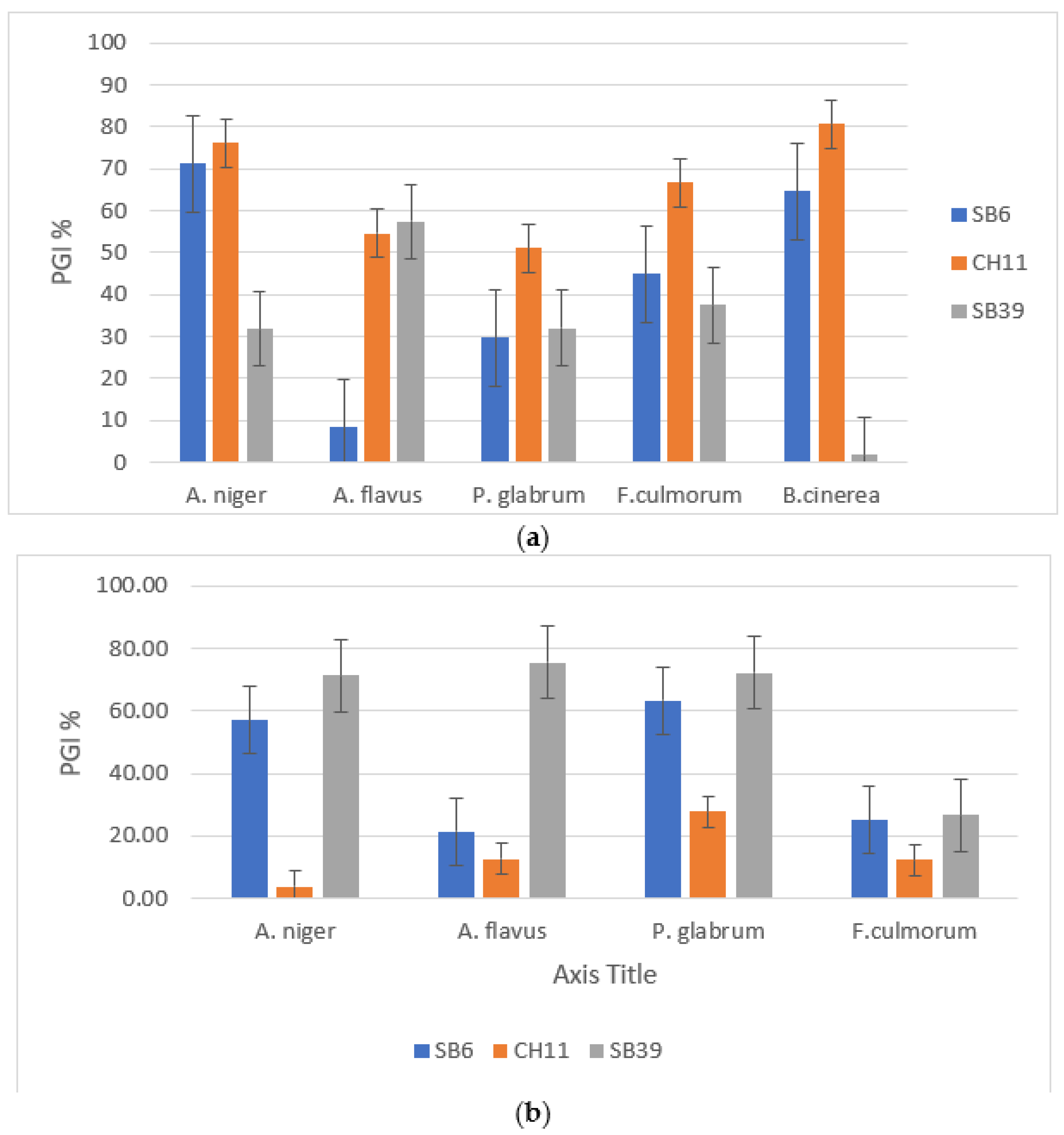
3.3.2. Indirect Inhibition of Mycelial Growth

The bacterial cultures’ media filtrates showed the presence of antifungal activity against A. niger for the strain CH11 in three environments (with and without biotic and abiotic stress). This means that the isolate produces antifungal molecules even in the absence of the fungus. SB39 is able to produce antifungal compounds only when in presence of the fungus or when the medium is free of glucose. The capacity of the isolate SB6 to inhibit fungal growth was negligible in the three environments.

3.3.3. Inhibition of Mycelial Growth by Volatile Substances

The results obtained revealed that the volatile substances were reduced the phytopathogenic fungi’s mycelial growth. For A. niger and P. glabrum, they were decreased by 71.29% and 72.22%, respectively. For the strain SB39, we noted a 56.93% decrease. The same was noted for SB6 (63.33%), but only a 3.96% to 27.78% decrease was shown for CH11. Concerning F. culmorum, the reduction varied from 12.5% to 26.67% for the three isolates (Figure 2).

3.3.4. Characterization of Bacterial Antifungal Metabolites

The analysis of the filtrates of the culture broths, which showed positive antifungal activities, allowed for the identification of polyphenols produced by the bacterial isolates (Table 2). Thus, the three bacterial isolates produced gallic acid, catechins, and syringic acid. In the presence of the pathogen, the SB39 strain produced gallic acid and syringic acid in concentrations exceeding 20 mg/mL. In addition, it was the only one producing hydroxyquinone.

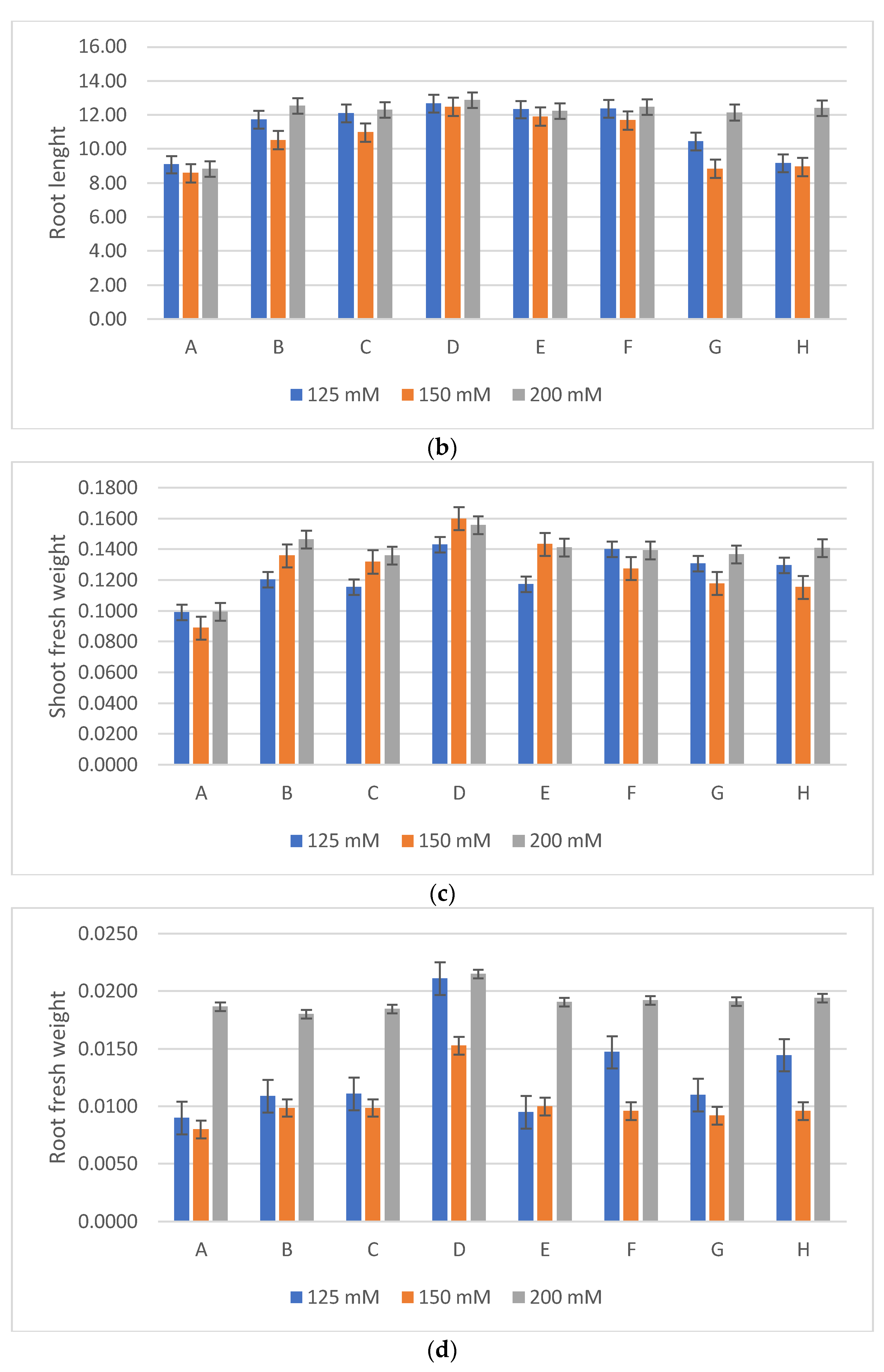
3.4. Tests to Stimulate the Growth of Wheat Plants Subjected to Salt Stress

The results of the treatment of wheat seeds with the three bacterial isolates and their different combinations showed a significant improvement in the growth parameters considered compared to those obtained for the control under salt stress (Figure 3 and Figure 4). To evaluate the harmful effects of salt stress, different concentrations of NaCl were used (125, 150, and 200 mM). The results obtained indicated that the increase in the NaCl concentration cause a reduction in the length of the stems and roots, and consequently in their fresh and dry weight in the control plants. However, the inoculation of the three bacterial isolates showed a significant attenuation of the harmful effects of salinity (Figure 4). Thus, the inoculation of the bacterial strains caused an increase of 30% in the length of the stems of the plants grown under the 125 mM treatment. The two strains SB6 and SB39 (Serratia sp. and Halomonas sp.) were associated with increases of 34% and 24% at 150 and 200 mM, respectively. Concerning the length of the stems, the best result was obtained for the Halomonas sp. strain, with increases of 39%, 45%, and 53% for NaCl concentrations of 125 mM, 150 mM, and 200 mM, respectively.

Bioassay on Apples

In this experiment, the apples that were inoculated with the pathogens (A. niger or B. cinerea) with the antagonist developed much smaller areas of infection compared to the fruits that were inoculated with the pathogens alone (Figure 5a). Thus, the severity of the disease was reduced by more than 50% in the case of A. niger and by more than 70% for B. cinerea (Figure 5b).

4. Discussion

It should be remembered that agricultural soil salinization constitutes one of the major problems in agriculture, which limits plant growth and agricultural productivity by up to 50% [48,49,50]. Approximately 1 billion hectares of the world’s land surface are affected by salinization, which represents approximately 7% of the planet’s land surface [31]. On the other hand, the use of fungicidal chemicals has a negative effect on health and the environment. Therefore, the application of plant growth-promoting rhizobacteria (PGPR) as growth-promoting and plant disease-fighting agents is a better alternative [20,51,52].
Recently, most research has focused on the isolation and study of halotolerant and halophilic bacteria with plant growth potential [53,54,55]. In our work, all the isolated bacteria could fix molecular nitrogen and solubilize mineral phosphate. These traits are relevant to improve plant growth, since they increase the availability of two essential nutrients for plant development, root growth, ripening of flowers and fruits, and mobilization of resources to the fruits. The studied strains were also able to produce hydrolytic enzymes (cellulase, amylase, lipase, proteases, urease, and chitinase) (Table 1). These microbial enzyme activities intervene with soil functionality by improving the availability of essential nutrients for plant growth [56]. Of the six bacterial isolates, SB39 and CH42 were the only ones that produced IAA in detectable amounts. IAA is a phytohormone involved in root initiation, cell division, and enlargement. IAA-producing microorganisms stimulate root growth, allowing for increased nutrient and water use efficiency [57,58,59,60]. The simultaneous expression of different PGP traits promotes plant growth [53] and increases the potential of the isolates to be used as plant biostimulants, especially under stress conditions such as saline soils.
The aim was to understand if the isolates with the highest PGP potential based on their PGP traits were also able to antagonize phytopathogenic fungi, known for their capacity to attack plants, especially those under stress. To answer this question, the three strains with the highest PGP potential, namely nitrogen fixation, phosphate solubilization, plant hormone production (IAA), and secretion of phytopathogen-limiting enzymes, were investigated for their potential to inhibit the growth of five phytopatogenic fungi (Aspergillus niger, Aspergillus flavus, Penicillium glabrum, Fusarium culmurum, and Botrytis cinerea). Antifungal activity was observed in all of the three bacterial strains. SB6 and CH11 showed better antagonistic potential when in direct confrontation with the fungi, while SB39 was the most efficient in fungal growth inhibition due to the production of volatile substances. Several studies suggest the involvement of biomolecules (HCN, NH3) and lytic enzymes such as cellulase, proteases, and chitinases (Table 1) in the antifungal activities of several bacteria. These enzymes may play an important role in degrading the fungal cell walls [48,49,50]. The production of HCN by PGPRs is a trait that is frequently reported in PGP bacteria, which may contribute to the promotion of plant defenses [56,57,58,59,60,61].
A comparison of the compounds detected in the mixed cultures of bacteria and the phytopathogen A. niger or the bacteria alone revealed that SB39 only produced antifungal compounds when in the presence of A. niger. The same was reported for the interaction between Psudomonas and Serratia strains with A. niger. On the contrary, the strain CH11 produced antifungal compounds even in the absence of the fungi. This may be a very interesting train for the design of disease-preventing products.
The HPLC characterization of some of the compounds produced by the bacterial strains revealed their capacity to produce polyphenols. According to Skorokhod et al. [62], only plants and certain microorganisms can synthesize the precursors of phenolic compounds. Most of the bacteria described as able to produce phenolic compounds are endophytic, possibly because of their need to synthesize secondary metabolites identical or like those of their host [63]. Based on the molecular identification of SB6 and CH11 as belonging to the genus Serratia and SB39 as belonging to the genus Halomonas, the genera for which endophitic bacteria are common may explain the production of polyphenols by the three isolates studied. However, this is the first study to demonstrate the capacity of bacteria from Serratia and Halomonas genera to produce polyphenols.
Plant inoculation with each of the isolated bacteria improved plant performance under the salinity conditions. It is interesting to point out that the wheat inoculation stimulated more growth at 200 mM NaCl than at lower salinity levels in comparison to the control. These results may be mediated through the synthesis of phytohormones by these bacteria as well as to the improvement in nutrient bioavailability [64]. These results agree with those of previous studies [46,47,54,65,66,67,68].
Apart from stimulating plant growth, the three bacterial strains can also contribute to increased plant defense against fungal infections in the post-harvest stage. One of the problems of harvests is the contamination of products during storage, which leads to significant losses [42]. In our study, inoculation of apples previously contaminated by phytopathogenic fungi with one of the three bacterial strains limited the expansion of the root zone induced by the fungi. Furthermore, after the application of the three bacterial antagonists, the rot in the apples contaminated with A. niger and B. cinerea was reduced. Similar results were reported by Sun et al. [59] and Jimtha et al. [69,70,71,72,73,74] using bacteria of the genus Serratia.

5. Conclusions

Ultimately, the strains selected in this work proved to be halophilic and to have the capacity to restore the growth of wheat under saline stress conditions. Additionally, their production of enzymes involved in soil fertilization, nitrogen fixation, and phosphate solubilization was increased. This also highlights the production of antifungal metabolites.
The three halotolerant and halophilic strains isolated belonging to Serratia sp. (SB6 and CH11) and Halomonas sp. (SB39) were revealed to be able to stimulate the growth of wheat under salinity conditions. These isolates were also able to inhibit the development of phytopathogenic fungi, most likely due to the production of polyphenols. Based on the results, these strains have a high potential to be used as bio-stimulants. Field trials should be performed to demonstrate their potential in the field.

Author Contributions

Conceptualization, K.H. and E.-H.N.; methodology, K.O.O., K.H. and Z.M.; validation, E.-H.N.; formal analysis, J.M.; investigation, C.C.; data curation, Y.B.; writing—original draft, L.O.; writing—review and editing, K.O.O.; supervision, C.C. and E.-H.N. All authors have read and agreed to the published version of the manuscript.

Funding

This research was funded by Portuguese funds from Fundação para a Ciência e a Tecnologia through the project UIDB/00329/2020 (DOI 10.54499/UIDB/00329/2020).

Data Availability Statement

The data are contained within this article.

Conflicts of Interest

The authors declare no conflicts of interest.

References

  1. El Hasini, S.; Iben Halima, O.; El Azzouzi, M.; Douaik, A.; Azim, K.; Zouahri, A. Organic and inorganic remediation of soils affected by salinity in the Sebkha of Sed El Mesjoune–Marrakech (Morocco). Soil Tillage Res. 2019, 193, 153–160. [Google Scholar] [CrossRef]
  2. Shrivastava, P.; Kumar, R. Soil salinity: A serious environmental issue and plant growth promoting bacteria as one of the tools for its alleviation. Saudi J. Biol. Sci. 2015, 22, 123–131. [Google Scholar] [CrossRef] [PubMed]
  3. Ha-Tran, D.M.; Nguyen, T.T.M.; Hung, S.-H.; Huang, E.; Huang, C.-C. Roles of Plant Growth-Promoting Rhizobacteria (PGPR) in stimulating salinity stress defense in plants: A review. Int. J. Mol. Sci. 2021, 22, 3154. [Google Scholar] [CrossRef] [PubMed]
  4. Pimentel, D.; Berger, B.; Filiberto, D.; Newton, M.; Wolfe, B.; Karabinakis, E.; Clark, S.; Poon, E.; Abbett, E.; Nandagopal, S. Water Resources: Agricultural and Environmental Issues. BioScience 2004, 54, 909–918. [Google Scholar] [CrossRef]
  5. Machado, R.M.A.; Serralheiro, R.P. Soil Salinity: Effect on Vegetable Crop Growth. Management Practices to Prevent and Mitigate Soil Salinization. Horticulturae 2017, 3, 30. [Google Scholar] [CrossRef]
  6. Roy, S.; Chowdhury, N. Salt Stress in Plants and Amelioration Strategies: A Critical Review. In Abiotic Stress in Plants; IntechOpen: London, UK, 2020; Chapter 19. [Google Scholar]
  7. Egamberdieva, D.; Wirth, S.; Bellingrath-Kimura, S.D.; Mishra, J.; Arora, N.K. Salttolerant plant growth promoting rhizobacteria for enhancing crop productivity of saline soils. Front. Microbiol. 2019, 10, 2791. [Google Scholar] [CrossRef] [PubMed]
  8. Bhat, M.A.; Kumar, V.; Bhat, M.A.; Wani, I.A.; Dar, F.L.; Farooq, I.; Bhatti, F.; Koser, R.; Rahman, S.; Jan, A.T. Mechanistic insights of the interaction of plant growthpromoting rhizobacteria (PGPR) with plant roots toward enhancing plant productivity by alleviating salinity stress. Front. Microbiol. 2020, 11, 1952. [Google Scholar] [CrossRef] [PubMed]
  9. Qin, S.; Miao, Q.; Feng, W.W.; Wang, Y.; Zhu, X.; Xing, K.; Jiang, J.H. Biodiversity and plant growth promoting traits of culturable endophytic actinobacteria associated with Jatropha curcas L. growing in Panxi dry-hot valley soil. Appl. Soil Ecol. 2015, 93, 47–55. [Google Scholar] [CrossRef]
  10. Backer, R.; Rokem, J.S.; Ilangumaran, G.; Lamont, J.; Praslickova, D.; Ricci, E.; Subramanian, S.; Smith, D.L. Plant growth-promoting rhizobacteria: Context, mechanisms of action, and roadmap to commercialization of biostimulants for sustainable agriculture. Front. Plant Sci. 2018, 9, 1473. [Google Scholar] [CrossRef] [PubMed]
  11. Wang, W.; Wu, Z.; He, Y.; Huang, Y.; Li, X.; Ye, B.C. Plant growth promotion and alleviation of salinity stress in Capsicum annuum L. by Bacillus isolated from saline soil in Xinjiang. Ecotoxicol. Environ. Saf. 2018, 164, 520–529. [Google Scholar] [CrossRef]
  12. Lobo, C.B.; Tomás, M.S.J.; Viruel, E.; Ferrero, M.A.; Lucca, M.E. Development of low-cost formulations of plant growth-promoting bacteria to be used as inoculants in beneficial agricultural technologies. Microbiol. Res. 2019, 219, 12–25. [Google Scholar] [CrossRef] [PubMed]
  13. Vaishnav, A.; Singh, J.; Singh, P.; Rajput, R.S.; Singh, H.B.; Sarma, B.K. Sphingobacterium sp. BHU-AV3 induces salt tolerance in tomato by enhancing antioxidant activities and energy metabolism. Front. Microbiol. 2020, 11, 443. [Google Scholar] [CrossRef] [PubMed]
  14. Li, X.; Li, B.; Cai, S.; Zhang, Y.; Xu, M.; Zhang, C.; Yuan, B.; Xing, K.; Qin, S. Identification of rhizospheric actinomycete Streptomyces lavendulae SPS-33 and the inhibitory effect of its volatile organic compounds against Ceratocystis Fimbriata in postharvest sweet potato (Ipomoea Batatas (L.) Lam.). Microorganisms 2020, 8, 319. [Google Scholar] [CrossRef]
  15. Zainab, N.; Amna, D.B.U.; Javed, M.T.; Afridi, M.S.; Mukhtar, T.; Kamran, M.A.; Ul Ain, Q.; Khan, A.A.; Ali, J.; Jatoi, W.N.; et al. Deciphering metal toxicity responses of flax (Linum usitatissimum L.) with exopolysaccharide and ACC-deaminase producing bacteria in industrially contaminated soils. Plant Physiol. Biochem. 2020, 152, 90–99. [Google Scholar] [CrossRef] [PubMed]
  16. Orozco-Mosqueda, M.D.C.; Glick, B.R.; Santoyo, G. ACC deaminase in plant growth-promoting bacteria (PGPB): An efficient mechanism to counter salt stress in crops. Microbiol. Res. 2020, 235, 126439. [Google Scholar] [CrossRef] [PubMed]
  17. El-Esawi, M.; Glascoe, A.; Engle, D.; Ritz, T.; Link, J.; Ahmad, M. Cellular metabolites modulate in vivo signaling of Arabidopsis cryptochrome-1. Plant Signal. Behav. 2015, 10, e1063758. [Google Scholar] [CrossRef] [PubMed]
  18. Qin, S.; Feng, W.W.; Zhang, Y.J.; Wang, T.T.; Xiong, Y.W.; Xing, K. Diversity of bacterial microbiota of coastal halophyte Limonium sinense and amelioration of salinity stress damage by symbiotic plant growth-promoting actinobacterium Glutamicibacter halophytocola KLBMP 5180. Appl. Environ. Microbiol. 2018, 84, e01533-18. [Google Scholar] [CrossRef] [PubMed]
  19. Xiong, Y.W.; Gong, Y.; Li, X.W.; Chen, P.; Ju, X.Y.; Zhang, C.M.; Yuan, B.; Lv, Z.P.; Xing, K.; Qin, S. Enhancement of growth and salt tolerance of tomato seedlings by a natural halotolerant actinobacterium Glutamicibacter halophytocola KLBMP 5180 isolated from a coastal halophyte. Plant Soil. 2019, 445, 307–322. [Google Scholar] [CrossRef]
  20. Tabli, N.; Nabti, E.H.; Dahel, D.; Mokrane, N.; Manyani, H.; Dary, M.; Megias, M.G. Impact of Diazotrophic Bacteria on Germination and Growth of Tomato, with Bio-Control Effect, Isolated from Algerian Soil. J. Ecol. Health Environ. 2014, 2, 1–7. [Google Scholar] [CrossRef]
  21. Carrim, A.J.I.; Barbosa, E.C.; Vieira, J.D.G. Enzymatic activity of endophytic bacterial isolates of Jacaranda decurrens Cham.(Carobinha-do-campo). Braz. Arch. Biol. Technol. 2006, 49, 353–359. [Google Scholar] [CrossRef]
  22. Vinoth, R.S.; Kanikkai, R.A.; Babu, V.A.; Manoj, G.T.; Naman, H.S.; Johnson, A.J.; Infant, S.B.; Sathiyaseelan, K. Study of starch degrading bacteria from kitchen waste soil in the production of amylase by using paddy straw. Recent. Res. Sci. Technol. 2009, 1, 008–013. [Google Scholar]
  23. Sierra, G.A. A simple method for the detection of lypolytic activity of microorganisms and some observations on the influence of the contact between cells and fatty substrates. Antonie Leeuwenhoek 1957, 28, 15–22. [Google Scholar] [CrossRef] [PubMed]
  24. Kumar, S.; Karan, R.; Kapoor, S.; Singh, S.P.; Khare, S.K. Screening and isolation of halophilic bacteria producing industrially important enzymes. Braz. J. Microbiol. 2012, 43, 1595–1603. [Google Scholar] [CrossRef] [PubMed]
  25. Christensen, W.B. Urea decomposition as a means of differentiating Proteus and paracolon cultures from each other and from Salmonella and Shigella types. J. Bacteriol. 1946, 52, 461–466. [Google Scholar] [CrossRef] [PubMed]
  26. Hsu, S.C.; Lockwood, J. Powdered chitin agar as a selective medium for enumeration of actinomycetes in water and soil. Appl. Microbiol. 1975, 29, 422–426. [Google Scholar] [CrossRef] [PubMed]
  27. Kopečný, J.; Hodrová, B.; Stewart, C.S. The isolation and characterization of a rumen chitinolytic bacterium. Lett. Appl. Microbiol. 1996, 23, 195–198. [Google Scholar] [CrossRef] [PubMed]
  28. Lorck, H. Production of hydrocyanic acid by bacteria. Physiol. Plant 1948, 1, 142–146. [Google Scholar] [CrossRef]
  29. Bric, J.M.; Bostock, R.M.; Silverston, S.E. Rapid in situ assay for indole acetic acid production by bacteria immobilization on a nitrocellulose membrane. Appl. Environ. Microbiol. 1991, 57, 535–538. [Google Scholar] [CrossRef] [PubMed]
  30. Fokkema, N.J. Fungal antagonism in the phylosphere. Ann. Appl. Biol. 1978, 89, 115–117. [Google Scholar] [CrossRef]
  31. Sagahón, I.P.; Reyes, M.A.A.; Rojas, H.V.S.; Cuenca, A.A.; Jurado, A.T.; Alvarez, I.O.C.; Flores, Y.M. Isolation of bacteria with antifungal activity against the phytopathogenic fungi Stenocarpella maydis and Stenocarpella macrospora. Int. J. Mol. Sci. 2011, 12, 5522–5537. [Google Scholar] [CrossRef] [PubMed]
  32. Korsten, L.; De Jager, E.S. Mode of action of Bacillus subtilis for control of avocado postharvest pathogens. S. Afr. Avocado Grow. Assoc. Yearb. 1995, 18, 124–130. [Google Scholar]
  33. Lee, J.Y.; Hwang, B.K. Diversity of antifungal Actinomycetes in various vegetative soils of Korea. Can. J. Microbiol. 2002, 48, 407–417. [Google Scholar] [CrossRef] [PubMed]
  34. Li, Q.; Ning, P.; Zheng, L.; Huang, J.; Li, G.; Hsiang, T. Fumigant activity of volatiles of Streptomyces globisporus JK-1 against Penicillium italicum on Citrus microcarpa. Postharvest Biol. Technol. 2010, 58, 157–165. [Google Scholar] [CrossRef]
  35. Aghighi, S.; Shahidi Bonjar, G.H.; Saadoun, I.; Rawashdeh, R.; Batayneh, S. First report of antifungal spectra of activity of Iranian actinomycetes strains against Alternaria solani, Alternaria alternata, Fusarium solani, Phytophthora megasperma, Verticillium dahliae and Saccharomyces cerevisiae. Asian J. Plant Sci. 2004, 3, 463–471. [Google Scholar] [CrossRef]
  36. Mahdieh, S.; Gholam, H.S.B.; Gholam, R.S.S. Post-harvest biological control of apple bitter rot by soil-borne Actinomycetes and molecular identification of the active antagonist. Postharvest Biol. Technol. 2016, 112, 46–54. [Google Scholar]
  37. Dennis, C.; Webster, J. Antagonistic properties of species groups of Trichoderma, II. Production of volatile antibiotics. Trans. Br. Mycol. Soc. 1971, 57, 363–369. [Google Scholar] [CrossRef]
  38. Jorjandi, M.; Shahidi Bonjar, G.H.; Baghizadeh, A.; Sharifi Sirchi, G.R.; Massumi, H.; Baniasadi, F.; Aghighi, S.; Rashid Farokhi, P. Biocontrol of Botrytis allii Munn the causal agent of neck rot, the post-harvest disease in onion, by use of a new Iranian Iisolate of Streptomyces. Am. J. Agric. Biol. Sci. 2009, 4, 72–78. [Google Scholar] [CrossRef]
  39. Kim, D.O.; Jeong, S.W.; Lee, C.Y. Antioxidant capacity of phenolic phytochemicals from various cultivars from pulms. Food Chem. 2002, 81, 321–326. [Google Scholar] [CrossRef]
  40. N’Guessan, A.H.O.; Dago Déliko, C.E.; Akhanovna, J.; Békro, M.; Békro, Y.A. Teneurs en composés phénoliques de 10 plantes médicinales employées dansla tradithérapie de l’hypertension artérielle, une pathologie émergente en Côted’Ivoire. Génie Ind. 2011, 6, 55–61. [Google Scholar]
  41. Zhao, Y.; Wang, Y.; Jiang, Z.-T.; Li, R. Screening and evaluation of active compounds in polyphenol mixtures by HPLC coupled with chemical methodology and its application. Food Chem. 2017, 227, 187–193. [Google Scholar] [CrossRef]
  42. Xiao, C.L.; Kim, Y.K. Postharvest fruit rots in apples caused by Botrytis cinerea, Phacidiopycnis washingtonensis, and Sphaeropsis pyriputrescens. Online Plant Health Prog. 2008, 9, 34. [Google Scholar] [CrossRef]
  43. Lu, L.; Lu, H.; Wu, C.; Fang, W.; Yu, C.; Ye, C.; Shi, Y.; Yu, T.; Zheng, X. Rhodosporidium paludigenum induces resistance and defense-related responses against Penicillium digitatum in citrus fruit. Postharvest Biol. Technol. 2013, 85, 196–202. [Google Scholar] [CrossRef]
  44. Huang, Y.; Sun, C.C.; Guan, X.N.; Lian, S.; Li, B.; Wang, C. Butylated hydroxytoluene induced resistance against Botryosphaeria dothidea in apple fruit. Front. Microbiol. 2021, 11, 599062. [Google Scholar] [CrossRef] [PubMed]
  45. Zihao, S.; Baihui, H.; Cuicui, W.; Shiyu, L.; Yuxin, X.; Baohua, L.; Caixia, W. Biocontrol features of Pseudomonas syringae B-1 against Botryosphaeria dothidea in apple fruit. Front. Microbiol. 2023, 11, 599062. [Google Scholar] [CrossRef]
  46. Orhan, F. Alleviation of salt stress by halotolerant and halophilic plant growth-promoting bacteria in wheat (Triticum aestivum). Braz. J. Microbiol. 2016, 47, 621–627. [Google Scholar]
  47. Bensidhoum, L.; Nabti, E.H.; Tabli, N.; Kupferschmied, P.; Weiss, A.; Rothballer, M.; Schmid, M.; Keel, C.; Hartmann, A. Heavy metal tolerant Pseudomonas protegens isolates from agricultural well water in northeastern Algeria with plant growth promoting, insecticidal and antifungal activities. Eur. J. Soil Biol. 2016, 75, 38–46. [Google Scholar] [CrossRef]
  48. Vimal, S.R.; Singh, J.S.; Arora, N.K.; Singh, D.P. PGPR-an effective bioagent in stress agriculture management. In Microbial Empowerment in Agriculture: A Key to Sustainability and Crop Productivity; Sarma, B.K., Jain, A., Eds.; Biotech Books: New Delhi, India, 2016; pp. 1–108. [Google Scholar]
  49. Farahmand, N.; Sadeghi, V. Estimating soil salinity in the dried lake bed of Urmia Lake using optical sentinel-2 images and nonlinear regression models. J. Indian Soc. Rem. Sens. 2020, 48, 675–687. [Google Scholar] [CrossRef]
  50. Li, H.; La, S.; Zhang, X.; Gao, L.; Tian, Y. Salt-induced recruitment of specific root-associated bacterial consortium capable of enhancing plant adaptability to salt stress. ISME J. 2021, 15, 2865–2882. [Google Scholar] [CrossRef] [PubMed]
  51. Noumavo, P.A.; Agbodjato, N.A.; Baba-Moussa, F.; Adjanohoun, A.; Baba-Moussa, L. Plant growth promoting rhizobacteria: Beneficial effects for healthy and sustainable agriculture. Afr. J. Biotechnol. 2016, 15, 1452–1463. [Google Scholar]
  52. Djebaili, R.; Marika, P.; Claudia, E.; Beatrice, F.; Mahmoud, K.; Maddalena, D.G. Biocontrol of Soil-Borne Pathogens of Solanum lycopersicum L. and Daucus carota L. by Plant Growth-Promoting Actinomycetes: In Vitro and In Planta Antagonistic Activity. Pathogens 2021, 10, 1305. [Google Scholar] [CrossRef] [PubMed]
  53. Oliva, G.; Di Stasio, L.; Vigliotta, G.; Guarino, F.; Cicatelli, A.; Castiglione, S. Exploring the potential of four novel halotolerant bacterial strains as plant-growth-promoting rhizobacteria (PGPR) under saline conditions. Appl. Sci. 2023, 13, 4320. [Google Scholar] [CrossRef]
  54. Cherif, H.; Sghaier, I.; Hassen, W.H.; Amara, C.; Masmoudi, A.S.; Cherif, A.; Neifar, M. Halomonas desertis G11, Pseudomonas rhizophila S211 and Oceanobacillus iheyensis E9 as biological control agents against wheat fungal pathogens: PGPB consorcia optimization through mixture design and response surface analysis. Int. Clin. Pathol. J. 2022, 9, 20–28. [Google Scholar]
  55. Alijani, Z.; Amini, J.; Ashengroph, M.; Bahramnejad, B. Antifungal Activity of Serratia rubidaea Mar61-01 Purified Prodigiosin against Colletotrichum nymphaeae, the Causal Agent of Strawberry Anthracnose. J. Plant Growth Regul. 2022, 41, 585–595. [Google Scholar] [CrossRef]
  56. Essghaier, B.; Dhieb, C.; Rebib, H.; Ayari, S.; Boudabous, A.R.A.; Sadfi-Zouaoui, N. Antimicrobial behavior of intracellularproteins from two moderately halophilic bacteria: Strain J31 of Terribacillus halophilus and strain M3-23 of Virgibacillusmarismortui. J. Plant Pathol. Microb. 2014, 5, 214. [Google Scholar] [CrossRef]
  57. Masmoudi, F.; Abdelmaleka, N.; Tounsia, S.; Dunlapb, C.A.; Triguia, M. Abiotic stress resistance, plant growth promotion and antifungal potential of halotolerant bacteria from a Tunisian solar saltern. Microbiol. Res. 2019, 229, 126331. [Google Scholar] [CrossRef] [PubMed]
  58. Boiero, L.; Perrig, D.; Masciarelli, O.; Penna, C.; Cassan, F.; Luna, V. Phytohormone production by three strains of Bradyrhizobium japonicum and possible physiological and technologicalimplications. Appl. Microbiol. Biotechnol. 2007, 74, 874–880. [Google Scholar] [CrossRef] [PubMed]
  59. Singh, R.P.; Jha, P.N. The multifarious PGPR Serratia marcescens CDP-13 augments induced systemic resistance and enhanced salinity tolerance of wheat (Triticum aestivum L.). PLoS ONE 2016, 11, e0155026. [Google Scholar] [CrossRef] [PubMed]
  60. Etesami, H.; Maheshwari, D.K. Use of plant growth promoting rhizobacteria (PGPRs) with multiple plant growth promoting traits in stress agriculture: Action mechanisms and future prospects. Ecotoxicol. Environ. Saf. 2018, 156, 225–246. [Google Scholar] [CrossRef]
  61. Tabli, N.; Rai, A.; Bensidhouma, L.; Palmierib, G.; Gogliettinob, M.; Coccab, E.; Consigliob, C.; Cilloc, F.; Bubicic, G.; Nabti, E.-H. Plant growth promoting and inducible antifungal activities of irrigation well water-bacteria. J. Biocontrol. 2017, 117, 78–86. [Google Scholar] [CrossRef]
  62. Sun, M.; Liu, J.; Li, J.; Huang, Y. Endophytic Bacterium Serratia plymuthica from Chinese Leek Suppressed Apple Ring Rot on Postharvest Apple Fruit. Front. Microbiol. 2022, 12, 802887. [Google Scholar] [CrossRef] [PubMed]
  63. Xun, F.; Xie, B.; Liu, S.; Guo, C. Effect of plant growth-promoting bacteria (PGPR) and arbuscular mycorrhizal fungi (AMF) inoculation on oats in saline-alkali soil contaminated by petroleum to enhance phytoremediation. Environ. Sci. Pollut. Res. 2015, 22, 598–608. [Google Scholar] [CrossRef] [PubMed]
  64. Vacheron, J.; Desbrosses, G.; Bouffaud, M.L.; Touraine, B.; Moënne-Loccoz, Y.; Muller, D.; Prigent-Combaret, C. Plant growth-promoting rhizobacteria and root system functioning. Front. Plant Sci. 2013, 4, 356. [Google Scholar] [CrossRef] [PubMed]
  65. Nawaz, A.; Shahbaz, M.; Imran, A.; Marghoob, M.U.; Imtiaz, M.; Mubeen, F. Potential of Salt Tolerant PGPR in Growth and Yield Augmentation of Wheat (Triticum aestivum L) under Saline Conditions. Front. Microbiol. 2020, 11, 2019. [Google Scholar]
  66. Desoky, E.S.M.; Saad, A.M.; El-Saadony, M.T.; Merwad, A.R.M.; Rady, M.M. Plant growth-promoting rhizobacteria: Potential improvement in antioxidant defense system and suppression of oxidative stress for alleviating salinity stress in Triticum aestivum (L.) plants. Front. Plant Sci. 2020, 30, 1878–8181. [Google Scholar] [CrossRef]
  67. Mukherjee, P.; Mitra, A.; Roy, M. Halomonas rhizobacteria of avicennia marina of indian sundarbans promote rice growth under saline and heavy metal stresses through exopolysaccharide production. Front. Microbiol. 2019, 10, 1207. [Google Scholar] [CrossRef] [PubMed]
  68. Kamou, N.N.; Dubey, M.; Tzelepis, G.; Menexes, G.; Papadakis, E.N.; Karlsson, M.; Agopodi, A.L.; Jensen, D.F. Investigating the compatibility of the biocontrol agent Clonostachys rosea IK726 with prodigiosin-producing Serratia rubidaea S55 and phenazine producing Pseudomonas chlororaphis ToZa7. Arch. Microbiol. 2016, 198, 369–377. [Google Scholar] [CrossRef] [PubMed]
  69. Fillinger, S.; Elad, Y. Botrytis–The Fungus, the Pathogen and Its Management in Agricultural Systems; Springer International Publishing: Cham, Switzerland, 2016; p. 488. [Google Scholar]
  70. Trejo-López, J.A.; Rangel-Vargas, E.; Gómez-Aldapa, C.A.; Villagómez-Ibarra, J.R.; Falfán-Cortes, R.N.; Acevedo-Sandoval, O.A.; Castro-Rosas, J. Isolation and Molecular Identification of Serratia Strains Producing Chitinases, Glucanases, Cellulases, and Prodigiosin and Determination of Their Antifungal Effect against Colletotrichum siamense and Alternaria alternata In Vitro and on Mango Fruit. Int. J. Plant Biol. 2022, 13, 281–297. [Google Scholar] [CrossRef]
  71. Azeem, S.; Agha, S.I.; Jamil, N.; Tabassum, B.; Ahmed, S.; Raheem, A.; Jahan, N.; Ali, N.; Khan, A. Characterization and survival of broad-spectrum biocontrol agents against phytopathogenic fungi. Rev. Argent. Microbiol. 2022, 54, 233–242. [Google Scholar] [CrossRef] [PubMed]
  72. Jimtha, J.C.; Jishma, P.; Sreelekha, S.; Chithra, S.; Radhakrishnan, E.K. Antifungal properties of prodigiosin producing rhizospheric Serratia sp. Rhizosphere 2017, 3, 105–108. [Google Scholar] [CrossRef]
  73. Alijani, Z.; Amini, J.; Karimi, K.; Pertot, I. Characterization of the Mechanism of action of Serratia rubidaea Mar61-01 against Botrytis cinerea in Strawberries. Plants 2023, 12, 154. [Google Scholar] [CrossRef] [PubMed]
  74. Skorokhod, I.; Kurdysh, I. The low-molecular weight antioxidants of microorganisms. Mikrobiology 2014, 76, 48–59. [Google Scholar]
Figure 1. (a) Percentage growth inhibition (PGI%) of phyopathogenic fungi by isolates (plate confrontation tests). (b) Effect of volatiles substances produced by the three strains on growth inhibition (PGI%). Bars are the mean ± SE. No significant differences are shown.
Agronomy 14 00845 g001
Figure 2. Efficiency of bacterial isolates (SB6, CH11, and SB39) against A. niger and B. cinerea in apples. Bars show the mean ± SE. No significant differences are shown.
Agronomy 14 00845 g002
Figure 3. Effects of bacterial strain treatment on the growth of wheat seedlings under the 125 mM salt stress condition. (A) Control; (B) SB6; (C) CH11; (D) SB39; (E) SB6-SB39; (F) SB6-CH11; (G) CH11-SB39; (H) SB6-CH11-SB39.
Agronomy 14 00845 g003
Figure 4. The effects of inoculation with halotolerant rhizobacteria (SB6: Serratia sp.; CH11: Serratia sp.; SB39: Halomonas sp.) on the growth ((a) shoot length; (b) root length; (c) shoot fresh weight; (d) root fresh weight; (e) shoot dry weight; (f) root dry weight) of wheat plants grown under salt stress conditions of 125 mM, 150 mM, or 200 mM of NaCl. (A) Control; (B) SB6; (C) CH11; (D) SB39; (E) SB6-SB39; (F) SB6-CH11; (G) CH11-SB39; (H) SB6-CH11-SB39. Different letters indicate significant differences between the control and different treatments (with bacteria): B, C, D, E, F, G, H. Bars indicate the mean ±1SE. Significance level: p = 0.05. (a) At 125 mM NaCl: significant differences between A and different treatments (B, C, D, E, F, and G). No significant difference between A and H. For 150 mM NaCl, there was a significant difference between the treatments. For 200 mM, NaCl there was a significant difference between A, B, and D. No significant differences were shown with the other treatments. (b) For 125 mM, all the treatments were significantly different (A, B, C, D, E, F, and G) except for H. For 150 mM, there was significant difference between all the letters except for G and H. For 200 mM, all the treatments were significantly different. (c) For 125 mM, there were significant differences between A, D, F, G, and H. For 150 mM, there were significant differences between all the treatments except for G and H. For 200 mM, there were significant differences between A and all the other treatments. (d) For 125 mM, there were significant differences between A, D, F, and H. At 150 mM, there was a significant difference between only A and D. For 200 mM, there was no significant difference shown. (e) For 125 mM, there were significant differences between A, D, F, G, and H. For 125 mM, there were significant differences between A, D, and F. For 200 mM, there was a significant difference between A and D only. (f) For 125 mM, no significant differences are shown. For 150 mM, there was a significant difference between only A and D. For 200 mM, there were significant differences between A, D, and H.
Agronomy 14 00845 g004aAgronomy 14 00845 g004bAgronomy 14 00845 g004c
Figure 5. Effects of fungal mold on apples treated (b) or not treated (a) with the bacterial antagonist after 6 days of incubation.
Agronomy 14 00845 g005
Table 1. Plant growth-promoting traits of the selected isolates.
SB6CH11CH12SB29SB39CH42
Cellulase++++nd++
Amylase+++ndndnd+++nd
Lipasend++nd++nd+
Protease+++++++++++++++nd
Ureasendnd+nd++++
Chitinase++++++++++++++++
Nitrogen fixation+++++++++
Phosphate solubilization++++nd+ndnd
IAA (µg/mL)ndndndnd++
NH3+++++++++++
HCN+ndndnd+++nd
+++—high activity; ++—intermediate activity; +—low activity; nd—activity not detected. For enzymes, +++—∅ > 20   m m ; ++— 10 < 20   m m ; +— 10   m m ; nd—activity not detected. For NH3, +++—orange–red; ++— o r a n g e ; +— y e l l o w ; nd—activity not detected. For HCN, +++—orange–red; ++—orange; +—light orange; and nd—activity not detected.
Table 2. Phenolic compounds identified in the filtrate culture broth and their concentrations.
RRT (min)Compound NameBroth Filtrate (mg/mL)
SB6 −CH11 −CH11 +CH11 depSB39 +SB39 dep
3.15 ± 0.05Gallic acid13.7720.7912.7613.4321.569.53
3.61 ± 0.00Hydroxy quinonendndndnd6.31nd
5.47 ± 0.23Resorcinol2.242.822.292.885.38nd
6.10 ± 0.17Catechin3.814.844.271.295.173.5
6.23 ± 0.411,2-hydroxybenzenend8.163.133.55ndnd
7.21 ± 0.05Syringic acid16.6513.2418.5411.7222.8812.23
10.63 ± 0.00Naringenin-7-glucosidendnd4.56ndndnd
11.08 ± 0.003,4,5-Trimethoxy benzoic acid2.51ndndndndnd
11.36 ± 0.01m-anisic acidnd1.821.012.39ndnd
15.07 ± 0.01Hesperidine9.691.91ndndndnd
(−): in the absence of the phytopathogen; (+): in the presence of the phytopathogen; (dep): in a depleted culture medium; nd—not detected. The purification of molecules using HPLC was performed only for bacteria showing inhibition zones (positive results only).
Disclaimer/Publisher’s Note: The statements, opinions and data contained in all publications are solely those of the individual author(s) and contributor(s) and not of MDPI and/or the editor(s). MDPI and/or the editor(s) disclaim responsibility for any injury to people or property resulting from any ideas, methods, instructions or products referred to in the content.

