1. Introduction
Plastics having a particle size of less than 5 mm are typically classified as microplastics. The term “microplastics” was initially introduced by British researchers Thompson et al. in 2004 [
1]. Microplastics are a relatively new class of environmental pollutants that are difficult to break down, chemically stable, and long-lasting in the environment—studies have shown that they will linger there for up to 1000 years [
2]. Long-term environmental exposure to microplastics will cause them to absorb organic pollutants, heavy metals, antibiotics, and other pollutants. The small particles’ density allows them to be carried by air, water, the food chain, and other pathways, while the slightly larger particles’ density may deposit in the water column and cause soil retention [
3]. The influence of microplastics on marine ecosystems is currently the subject of numerous studies; these particles are known as the “PM2.5” of marine ecosystems, and the primary research objectives are seawater, fish, shellfish, algae, etc. [
4]. The majority of microplastics found in aquatic environments come from terrestrial systems [
5], and these systems have a higher abundance of microplastics than marine systems do. In fact, it has been reported that the abundance of microplastics in terrestrial systems is 4–23 times higher than that in oceans [
6]. When animals ingest microplastics in terrestrial systems, the microplastics desorb in their bodies, releasing pollutants that have been adsorbed on the microplastics together with plastic additives. This can result in alterations in the behavior of the animals, as well as in cells, tissues, and molecular levels, and death [
7]. Plant growth may be impacted by the physical and chemical changes brought about by the buildup of microplastics in the soil, which may also have an impact on soil function and biodiversity [
8]. Microplastics have the ability to stick to plant root surfaces, preventing the plants from growing. Once ingested, the microplastics can cause oxidative stress and genetic and cellular toxicity in the body when they reach the stems, leaves, and fruits of the plant [
9]. Furthermore, microplastics raise the risk of cancer and chronic illnesses by entering the body through food and common items.
According to Hu Hanwen et al. (2020), microplastics made of polystyrene and polyvinyl chloride have the ability to enter the lymphatic and circulatory systems of humans through the intestinal tract, endangering their health [
10]. Large amounts of plastic particles have found their way into agricultural soils as a result of the widespread use of sewage sludge as a way to increase soil productivity and encourage plant growth [
11]. According to research conducted over the past five years, the amount of microplastics found in soils worldwide is expected to reach 1.5–6.6 million tons, which is one to two orders of magnitude more than the amount found on the ocean’s surface [
12]. In a survey conducted on flood basins in Switzerland, Scheurer et al. (2018) discovered microplastics in 90% of the soil samples [
13]. According to some research, the annual amount of microplastics in Australian agroecosystems ranges from 2800 to 19,000 tons [
14]. China is among the most microplastic-polluted countries in the world, with the greatest annual discharge of 1.32–3.53 million tons of plastic garbage in coastal areas alone [
15]. Microplastics have a substantial impact on biological growth and biomass accumulation, environmental microbial activity and diversity distribution, and the geochemical cycling of soil nutrients, all of which have an impact on the health of soil ecosystems. They can also directly affect the physicochemical properties and material cycles of soil [
16].
Microplastics can be stored in soil for extended periods of time due to the extreme difficulty of natural degradation. This can result in biotoxic effects on a variety of organisms that depend on the soil environment, posing serious ecological risks, as well as the accumulation of microplastics in organisms that could eventually pass through the food chain and endanger the health and safety of humans and other higher animals [
17]. Investigations into the possible negative effects of microplastics on terrestrial ecosystems are desperately needed, given the majority of current research on the distribution and effects of microplastics is concentrated on aquatic habitats. The impacts of microplastics on terrestrial creatures have actually only been the subject of a small amount of research. Liuxiaohong et al. (2022) found that during the seedling growth stage, besides the 0.1% mass fraction of 13 μm sized PE microplastics significantly increasing
Zea mays L. plant height, the 0.1% and 0.5% mass fractions of 13 μm sized PE microplastics also significantly increased
Zea mays L. root volume and root surface area, while all sizes of PE microplastics inhibited the growth of both
Zea mays L. and
Cucumis sativus L. seedlings to varying degrees [
18]. In a similar vein,
Candida berkhout’s gut flora was changed when it was exposed to 80–250 μm polyvinyl chloride (PVC) microplastic particles [
19]. According to these findings, animals living in soil are exposed to certain harmful impacts of microplastics. Microplastics can be absorbed by plants and accumulate in leaves, stems, and other plant tissues, according to a study on
Lactuca sativa var. ramosa by Li Lianzhen et al. [
20]. A different study discovered that nanosized plastic beads can enter
Nicotiana tabacum L cells through endocytosis [
21].
Triticum aestivum L plants’ elongation, development, and synthesis of photosynthetic pigments are all inhibited by microplastics, which can have an impact on
Triticum aestivum L seed germination [
22]. According to research by Angel Liu et al. (2017), microplastics may have an impact on
Vigna radiata germination and growth by preventing these processes [
23]. Bosker et al. (2019) discovered that the germination rate of
Oenanthe javanica was dramatically decreased by microplastics of varying particle sizes [
24] and that the detrimental effect grew as particle size increased. Giorgetti et al. (2020) discovered that
Allium cepa L’s root meristematic zone cells were capable of internalizing polystyrene nanoplastics [
25]. Additionally, Abbasi et al. (2020) discovered that microplastics have a detrimental influence on root growth when they are utilized as a carrier to deliver heavy metals (Cd, Pb, and Zn) to the root zone of
Triticum aestivum L. [
26]. On the other hand, it was also discovered that microplastics might aid in the growth of plants. According to research by Li et al. (2020), the two PVC particle sizes 100 nm–18 m and 18 m–150 m had no discernible impact on the root activity of lettuce [
27]; but, the total length, surface area, volume, and diameter of the
Lactuca sativa var. ramosa root system were dramatically increased by 0.5% and 1% (
w/
w) of PVC (100 nm–18 m). Consequently, it is important to consider how soil-borne microplastics affect crop growth.
Common thermoplastic polystyrene has good physicochemical qualities and is used extensively in plastic films, food packaging, and building insulation [
28]. To investigate the mechanism of microplastics’ effects on plant growth, three types of polystyrene plastic microspheres with varying particle sizes (5 μm, 1 μm, and 80 nm) were chosen as the tested pollutants in this experiment.
P. sativum is a plant belonging to the Leguminosae family. The experiment’s goals were to examine the effects of various microplastic particle sizes and concentrations on
P. sativum germination and growth, investigate the effects of microplastics on plant growth, and offer fundamental information and a scientific foundation for assessing the ecotoxicity of microplastics on terrestrial crops.
3. Results
3.1. Effects of PS-MPs on P. sativum Germination and Growth
3.1.1. Germination Rate
Different PS-MP particle sizes and concentrations have varying impacts on
P. sativum germination.
P. sativum seed germination was clearly inhibited by particle sizes of 0.1 μm and 0.08 μm. At concentrations below 1000 mg/L, the inhibition was essentially the same in all groups under the condition of 0.08 μm; when the concentration increased to 2000 mg/L, the inhibition was further strengthened, and the inhibition rate reached 57.50% under the conditions of 0.1 μm and 0.08 μm (
Table 1).
In contrast to the 0.1 μm and 0.08 μm conditions, the
P. sativum germination was somewhat promoted by the 5 μm microplastics. PS-MPs promoted
P. sativum germination at concentrations below 500 mg/L; at 500 mg/L,
P. sativum germination reached its maximum; as concentrations rose,
P. sativum germination was gradually inhibited; at 2000 mg/L, the strongest inhibitory effect was observed, and, at this point, the germination rate was only 55.00% (
Table 1). Thus, it is evident that the impact of small-sized microplastics on
P. sativum germination was not statistically significant, but the impact of microplastics with 5 μm particle size on
P. sativum germination was not significant. This indicates that microplastics with small particle sizes have a more substantial impact on germination. The combination of small particle size (0.1 µm, 0.08 µm) and high concentration (>500 mg/L) had a stronger inhibitory effect than the combination of large particle size (5 μm) and low to medium concentration (<500 mg/L) based on a comparison of the results of different particle size and concentration combinations.
3.1.2. Germination Potential
When compared to the control group, 5 μm PS-MPs did not significantly affect
P. sativum germination potential at concentrations below 100 mg/L. However, when the concentration exceeded 100 mg/L, 0.1 μm, a considerable promotion impact was seen. Only at low concentrations (50 mg/L) did PS-MPs significantly promote
P. sativum germination potential; at low concentrations (<100 mg/L), 0.08 μm PS-MPs significantly promoted
P. sativum germination potential. The germination potential of
P. sativum was not affected by concentrations of 100 mg/L; however, concentrations greater than 100 mg/L resulted in a significant inhibitory effect. At 500 mg/L, the germination potential of
P. sativum was only 40.00%, which was 33.00% lower than that of the control group (
Table 1), demonstrating a significant inhibitory effect.
3.1.3. Germination Index
In this experiment, the germination index of
P. sativum was not significantly affected by 5 μm PS-MPs; however, under specific concentration conditions, the germination index was significantly affected by 0.1 μm and 0.08 μm PS-MPs. The germination index of
P. sativum seeds was enhanced by 0.1 μm PS-MPs, and they demonstrated greater promotion when the concentration of PS-MPs exceeded 500 mg/L. In contrast, 0.08 μm PS-MPs demonstrated a significant inhibition in this condition, and the germination index fell by 28.64% when exposed to 2000 mg/L PS-MPs in comparison to the control group.
P. sativum seed germination index was significantly inhibited by three different types of particle sizes of microplastics; the lowest concentration size was 5 µm (not found in the test) > 0.1 µm (500 mg/L) > 0.08 µm (200 mg/L), small particle size (0.08 µm).
Table 1 shows that PS-MPs were more likely to exert inhibitory effects and alter the germination index of
P. sativum seeds.
3.1.4. Average Germination Time
In comparison to the control group, the mean germination time of seeds under exposure conditions exceeding 200 mg/L could be significantly shortened by 5 μm PS-MPs, whereas the mean germination time of
P. sativums under concentration conditions less than 100 mg/L could be significantly shortened by 0.08 μm PS-MPs, and the mean germination time of
P. sativums was not significantly affected by 0.1 μm PS-MPs (
Table 1). When compared to the other experimental groups, 0.08 μm PS-MPs had a significant effect under the 200 mg/L exposure condition under the same concentration conditions. According to the aforementioned findings, small particle size PS-MPs had a more notable negative impact on the average germination time of
P. sativum seeds and were more detrimental to the seeds’ quick germination under these circumstances.
The germination index and vigor index did not exhibit a significant interaction with the germination properties of P. sativum seeds, according to the two-way ANOVA results. On the germination characteristics of P. sativum seeds, however, germination potential, germination rate, and germination time demonstrated noteworthy interaction effects.
The absence of any discernible interaction between the germination index and vigor index suggests that the effects of microplastic particle size and concentration on P. sativum seed germination may be independent of one another or may not be correlated at all. This could be due to the fact that the size and concentration of microplastic particles exhibit varying physiological processes or biological mechanisms that influence germination traits, resulting in inconsistent effects on germination.
This might point to a more intricate relationship when there is an interaction between germination potential, germination rate, and germination time. One explanation for this could be that during the germination process of P. sativum seeds, germination potential, germination rate, and germination time interact or are regulated. This could be due to variations in the physiological conditions of P. sativum seeds at various germination stages, or interactions between these indicators as a result of interactions with the environment.
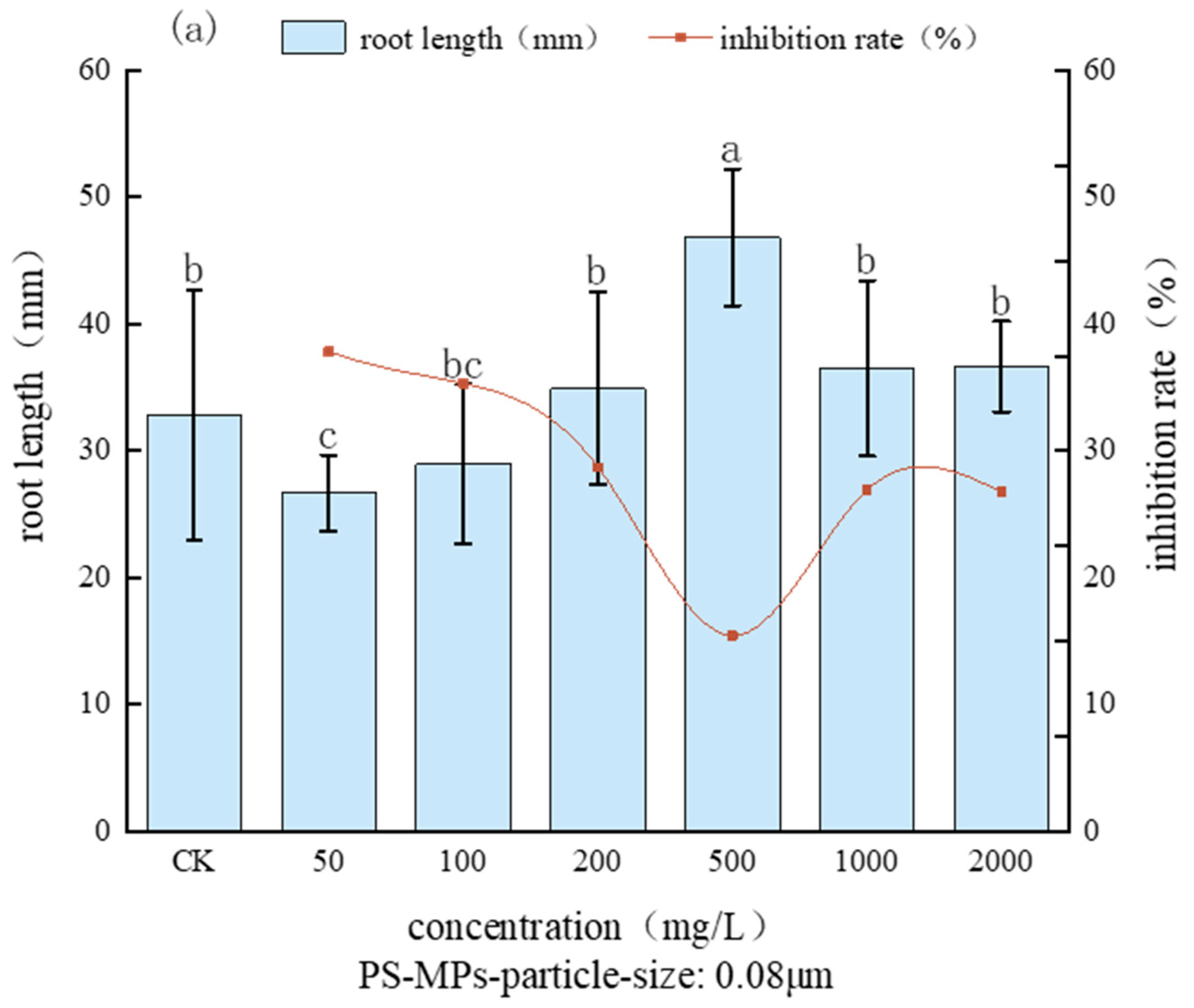
3.2. Effects on P. sativum Root Length and Stem Length
Figure 4 shows a plot showing the impact of microplastics on
P. sativum sprouts’ root length. The figure illustrates that while the root elongation of
P. sativum sprouts treated with 5 μm microplastics did not approach statistical differences (
p > 0.05), the root growth was stimulated. It is evident that when exposed to 5 μm microplastics,
P. sativum sprouts’ root growth was encouraged and their root system grew longer, albeit this difference was not statistically significant (
p > 0.05). The exposure to 100 mg/L and 500 mg/L microplastics corresponded to the length of the
P. sativum roots, respectively.
P. sativum sprouts exposed to microplastics at doses of 100 mg/L and 500 mg/L had root lengths of 46.24 mm and 45.36 mm, respectively; at higher exposure concentrations, the root length of the sprouts was significantly affected by the application of 0.1 μm microplastic treatment.
P. sativum root length was considerably increased by the 0.1 μm microplastic treatment at higher exposure doses; the corresponding root lengths for the 500, 1000, and 2000 mg/L treatments were 50.13 mm, 53.45 mm, 55.16 mm, and 55.16 mm, respectively; 53.45 mm and 55.16 mm, respectively, increased by 48.53%, 58.37%, and 63.44% in comparison to the control. The promotion effect grew steadily as the concentration of microplastic increased.
The promoting effect of microplastic increased gradually as its concentration increased; when treated with 0.08 μm microplastic, the root length of each treatment group changed with concentration before declining. Each treatment group’s root length under the 0.08 μm microplastic treatment showed a trend of decreasing with concentration, reaching a maximum when the concentration reached 500 mg/L. Under these conditions, the root length was 46.85 mm, which differed significantly from the control group’s root length. The root length under these conditions was 46.85 mm, which was considerably different from the control’s value (p < 0.05), but there was no statistically significant difference between the other treatments and the control (p > 0.05). The maximum value was attained at 500 mg/L.
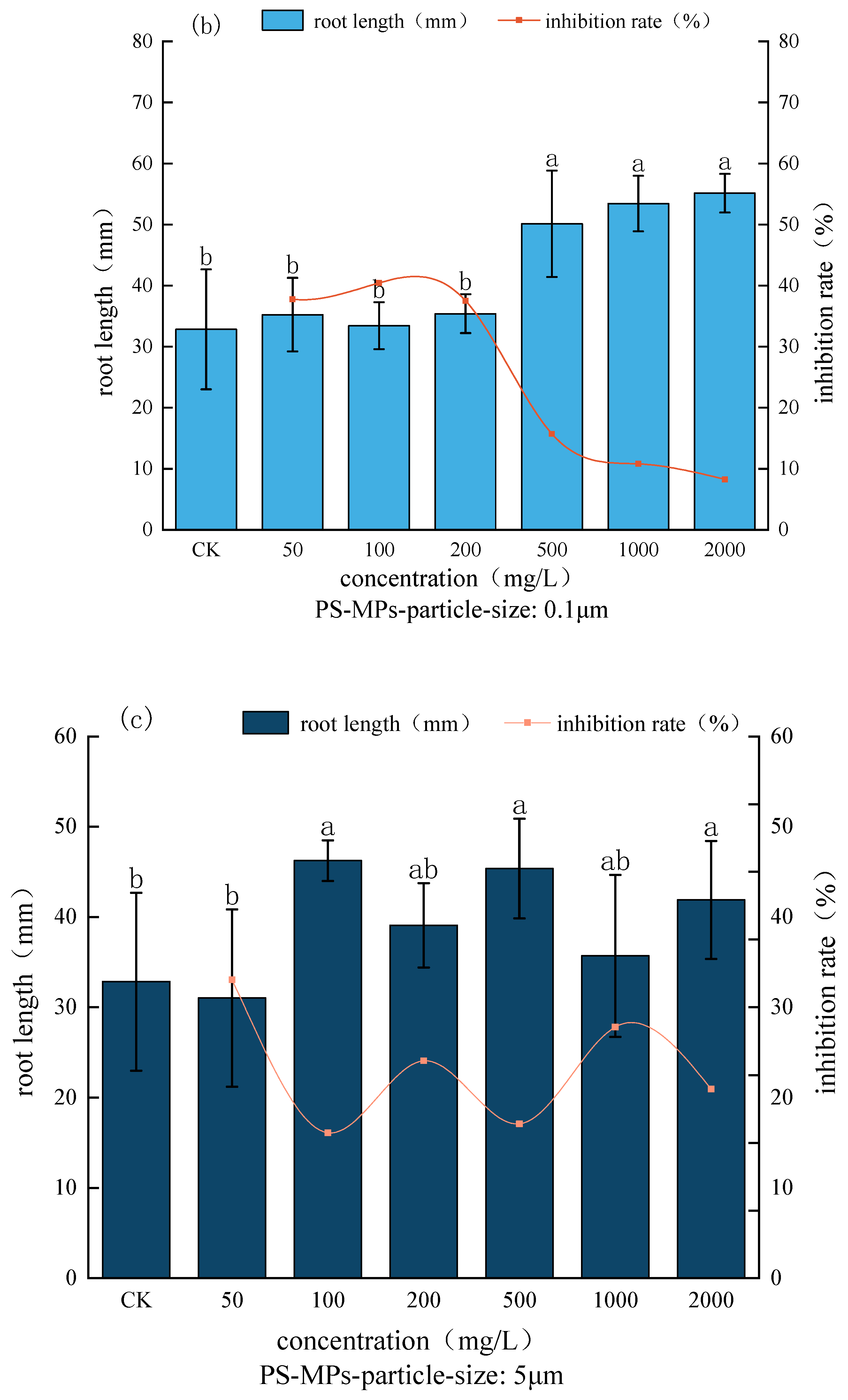
Figure 5 illustrates how microplastics affect the growth of
P. sativum seedlings. The image illustrates how exposure to microplastics with bigger particle sizes favorably influenced
P. sativum sprout growth. When exposed to 5 μm microplastic, the length of the
P. sativum sprouts increased gradually as the concentration increased, but under 50 mg/L exposure, there was a modest decrease in the length of the sprouts. The length of the
P. sativum sprout seedlings increased gradually under 5 μm microplastic treatment as the concentration increased. However, under 50 mg/L exposure, the length of the seedlings decreased slightly, and this difference did not reach a statistically significant level when compared to the control group (
p > 0.05). The promotion impact became stronger when the concentration surpassed 50 mg/L, and the exposed
P. sativum sprouts’ seedling length ranged from 35.08 to 44.86 mm, exceeding that of the control.
When compared to the control, the seedling length of P. sativum sprouts exposed to microplastics rose by 3.94% to 42.31%, measuring between 35.08 and 44.86 mm. The 0.1 μm microplastic treatment resulted in a significant difference (p < 0.05) in the promotion effect on P. sativum seedling length when compared to the control at concentrations of 200–2000 mg/L. This effect increased as the concentration changed. (p < 0.05), the length of the P. sativum seedlings under 200, 500, 1000, and 2000 mg/L concentration, reached 45.16 mm, 51.5 mm, and 51.5 mm, respectively, on the seventh day of the experiment. Upon reaching the seventh day of the experiment, the P. sativum seedlings under 200, 500, 1000, and 2000 mg/L concentrations measured 45.16 mm, 51.40 mm, 51.75 mm, and 57.70 mm, respectively. These values showed significant increases (p < 0.05) of 33.81%, 52.30%, 53.33%, and 70.96% when compared to the control group. There was a significant difference (p < 0.05) between the 0.08 μm microplastic treatment and the control treatment, with the concentration of microplastic exposure. The length of P. sativum shoot seedlings was progressively inhibited as the concentration of 0.08 μm microplastics was exposed to higher levels.
All things considered, the smaller particle size microplastics had an inhibitory effect on the normal growth of P. sativum sprouts. The length of the P. sativum sprouts in each group of 200–2000 mg/L treatment was 32.95 mm, 28.74 mm, 25.43 mm, and 24.33 mm; these decreased by 2.37%, 14.84%, 24.65%, 27.91%, and 27.91%, respectively, compared with the control, which was 27.91%.
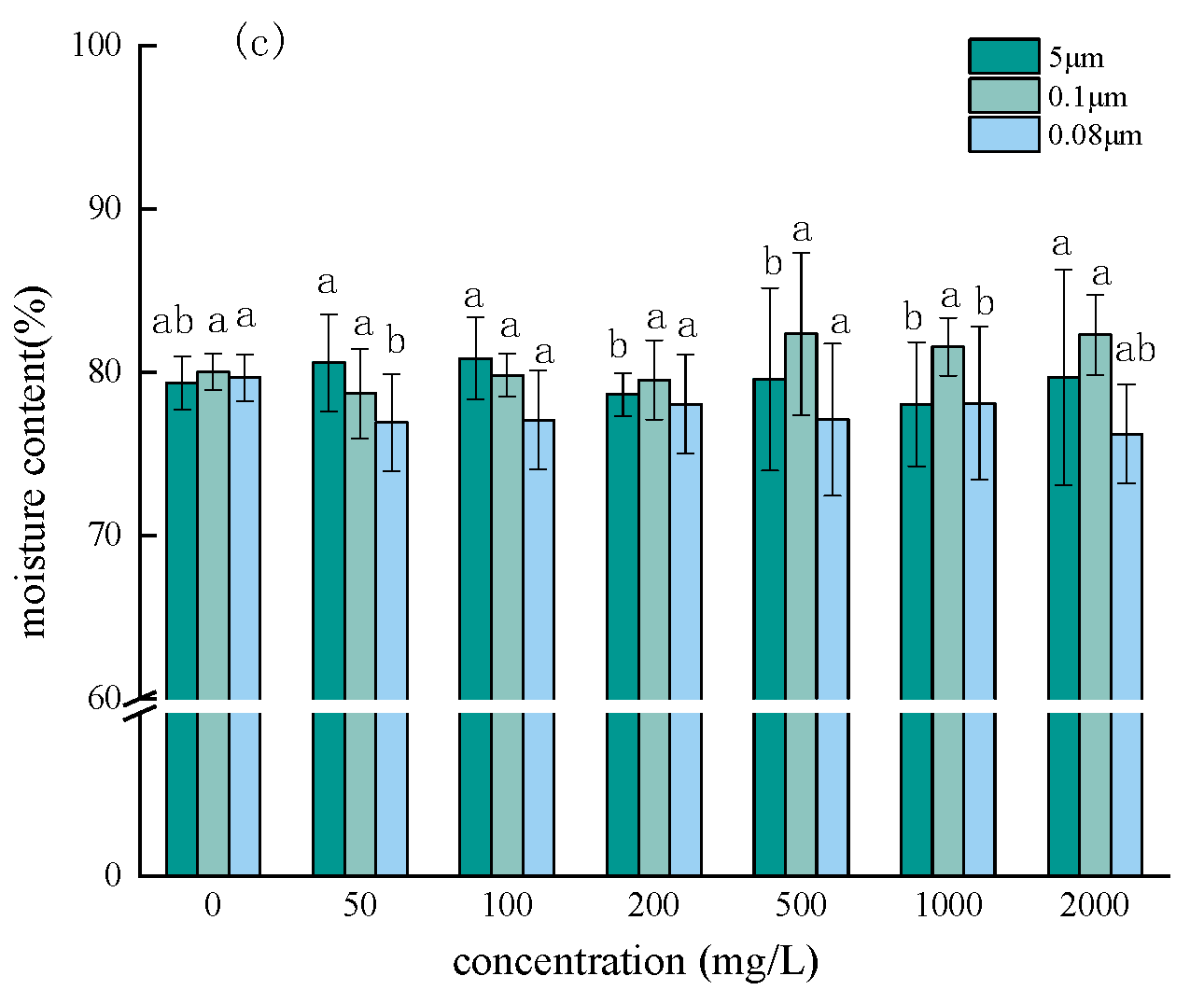
3.3. Effects on Substance Accumulation
The effects of varying microplastic particle sizes and concentrations on material accumulation during
P. sativum germination are depicted in
Figure 6. When comparing the fresh weight of
P. sativum seedlings in each experimental group to the other two particle size treatments, the 0.08 μm particle size revealed a decrease. The water content of
P. sativum seedlings in each concentration group under the 0.08 μm particle size condition showed a slight decrease compared with the treatment of unadulterated PS-MPs, indicating that small-sized microplastics might have a negative effect on the water accumulation of
P. sativum seedlings (
Figure 6b). However, the remaining two groups of microplastic particle sizes had no significant effect on dry weight (
Figure 6a). On the other hand, there was no discernible variation in water content across the groups with varying PS-MP concentrations (
Figure 6c).
3.4. Effects on Photosynthetic Pigments
By taking part in photosynthesis, which creates the elements required for plant growth, chlorophyll is essential to the healthy development of plants. The content of
P. sativum carotenoids a and b showed an overall trend of reducing and subsequently increasing with the increase in microplastic concentration, as shown in
Figure 7, under the same particle size conditions. Following seven days of germination, the experimental groups’ levels of a and B exhibited an overall trend of declining and then increasing in response to concentration increases, with a notable increase noted under high concentration (1000 mg/L, 2000 mg/L) circumstances.
While the concentration conditions in the range of 50–500 mg/L demonstrated a significant inhibitory effect, the high concentration conditions significantly promoted the production of a, which was increased by 61.67% (5 μm, 2000 mg/L), 38.75% (0.1 μm, 1000 mg/L), and 29.35% (0.08 μm, 2000 mg/L) compared with the control group. Even at a concentration of 1000 mg/L, microplastics with a particle size of 0.08 μm demonstrated a noteworthy suppression of
P. sativum a production (
Figure 7a). At high concentrations, microplastics had a considerable impact on b synthesis; at lower quantities, the effect was not significant (
Figure 7b). The range of the ratio in each experimental group was between 2.5 and 3.0 (
Figure 7c), and the effect was not significant across the groups, according to the results of the ratio of a to b content in each experimental group. At 500 mg/L, the measured carotenoid contents of each particle size test group increased by 16.72% (5 μm), 24.84% (0.1 μm), and 26.13% (0.08 μm), relative to the control group. The low concentration gradient had no significant effect. Medium and high concentrations of microplastics significantly promoted the production of carotenoids when compared with the control group (
Figure 7d).
4. Discussion
Using RDA analysis, the effects of PS content and particle size on
P. sativum seed germination, seedling growth, and root morphology were examined in this study (
Figure 8). The RDA sorting results for
P. sativum revealed that the correlation coefficients between the PS features in the first and second axes and the indicators of corn growth and development were 0.902 and 0.712, respectively, and that the eigenvalues of the first two sorting axes were 0.387 and 0.266. Comparing treatments with and without the addition of polystyrene microplastics (PS-MPs), it was discovered in this study that the inclusion of PS-MPs could have a noteworthy impact.
Microplastics have been found to possess attributes such as hydrophobicity and low density. The specific surface area of microplastics increases with decreasing particle size, making it simpler for them to adhere to surfaces or seed roots. This prevents water absorption while also impairing plant respiration, which in turn influences the growth and development of roots and shoots [
29,
30].
The germination of
P. sativum seedlings was significantly inhibited by smaller particle sizes (0.1 μm, 0.08 μm) of polystyrene microplastics; however, this inhibitory effect was reduced when the particle size increased to 5 μm, and a more significant inhibitory effect was only observed at high concentrations (>1000 mg/L). In the study of wheat seed germination conducted by Lian Gaphan et al., it was discovered that all three types of microplastics that were inhibited at low to medium concentrations (<500 mg/L) had an inhibitory effect on wheat seed germination with a range of 2.86% to 20% [
31], which is comparable to the findings of the present study, which showed that
P. sativum has different sensitivities to the toxicity of microplastics of different particle sizes.
The germination rate demonstrated that the lowest inhibitory concentrations of various particle size conditions on
P. sativum seeds were in the range of 0.08 μm < 0.1 μm < 5 μm. The small-sized microplastics were more likely to enter the root cortex or even the mid-column through the plasma-exosome barrier and the free space of the root cell interstitial space, and under the same relatively low concentration conditions, they could therefore have obvious toxic effects. Simultaneously, microplastics with small particle sizes have a tendency to aggregate. When the aggregated particle size of PS-MPs is greater than the root system’s pore size, PS-MPs adsorb and accumulate on the surface of plant root cells, causing a physical blockage that hinders the plant’s ability to absorb water and nutrients, thereby impeding the normal growth of
P. sativum seeds [
32].
Plant growth is a direct indicator of whether or not they are contaminated by toxins. Exogenously supplied PS-MPs have a direct impact on
P. sativum seedling growth, as evidenced by characteristics like dry matter buildup and water uptake. Seed germination is attenuated and biomass is reduced by PS-MPs [
33]. By comparing the fresh weight and dry weight, it was discovered that the behavior of water uptake is where PS-MPs have the greatest effect on
P. sativum seedlings’ material accumulation. Particle size had a greater impact on the fresh weight of the experimental groups than on the dry weight, which the PS-MPs had less of an impact on. According to van Weert et al. [
34], plants with more developed root systems will come into direct contact with the microplastics in the sediment in the surrounding environment. This will affect how well the root system functions, hence, reducing the plant’s ability to absorb water. The plant’s fresh weight is influenced by the root system’s intake of water. Plant fresh weight declines with decreasing particle size, and
P. sativum seedlings exhibit a reduction in water content, indicating that plant water content is impacted. By preventing plant roots from absorbing water and nutrients, microplastics have the potential to drastically change the biomass of plants. Smaller particle sizes of microplastics have a greater specific surface area and are more likely to adhere to the surfaces of roots or seeds. This causes a decrease in water content by obstructing cellular pores and other pathways, which inhibits water absorption and respiration [
35,
36]. Owing to its small particle size, it can go through the xylem conduit to the above-ground portion, where it can impact stem development. Microplastics can go up the plant through transpiration after entering the mid-column. They can go from the roots to the stems and leaves through their pores, and then they can travel through the exoplasmic body pathway to the veins in the leaves. Microplastics may be able to penetrate cell walls and membranes for extended periods of time and enter the interior of the cell, where they may change nutrient and water absorption and transport, induce oxidative stress, and interfere with normal plant growth and development [
37]. This study demonstrated how exposure to microplastics can have a major impact on a plant’s ability to absorb water through its roots.
The experiment’s findings demonstrate that PS-MPs have a notable inhibitory effect on P. sativum seed germination and seedling growth, which may be connected to toxicity consequences. Particle size and concentration both affect P. sativum seed germination and seedling growth in response to PS-MPs. More study is required to understand the intrinsic mechanism underlying this growth suppression, the toxicity consequences, and the plant’s adsorption and uptake of PS-MPs. The mechanism of PS-MP growth suppression on P. sativum, the toxicity effect, the mechanism of PS-MP adsorption and uptake by plants, and the high concentration of microplastics in the simulated environment in the current experiment require more investigation. The findings of this study offer fundamental information and a solid scientific foundation for researching the biological impacts of microplastics in various conditions and comprehending how they affect crop germination and seedling growth.
5. Conclusions
In this study, hydroponics was used to study the effects of microplastics on the physiological and biochemical characteristics of P. sativum seed germination, morphology, biomass, photosynthetic pigment content, and other characteristics at different particle sizes and concentrations, to reveal the toxic effects of microplastics on P. sativum, and to provide basic data and theoretical references for the ecological risk assessment of microplastics. The main conclusions are as follows:
The combination of small particle size and high concentration conditions had the most significant inhibitory effect on P. sativum germination. A trend of 0.08 μm < 5 μm < 0.1 μm was seen in the threshold values of microplastics at different particle sizes during seed germination. Among them, 0.08 μm could significantly reduce the vigor and seed germination index, whereas 5 μm and 0.1 μm could only significantly reduce these variables at high concentrations, indicating that only 0.1 μm and 5 μm could significantly reduce the vigor and seed germination index. The vigor index is only at high concentrations, exhibiting a trend of 5 μm = 0.1 μm.
While seedling length was restricted at 0.08 μm particle size and increased biomass accumulation of P. sativum seedlings, high concentrations of microplastics considerably enhanced the root development behavior of P. sativum. Concurrently, the impact of microplastics on photosynthetic pigment synthesis has shown a tendency to decline at low concentrations, recover at higher concentrations, and then promote at high concentrations.
Both microplastic particle size and concentration affected the growth of P. sativum seedlings; however, at low concentrations of microplastic exposure, the effect of microplastic particle size on P. sativum seed germination was more significant, affecting the germination rate, root length, and other aspects of P. sativum seeds. When P. sativum seed germination, root length, and other indices were exposed to low concentrations of microplastics, the impact of microplastic particle size on these indicators was more pronounced. Microplastics at low concentrations demonstrated a tendency of 0.08 μm < 5 μm < 0.1 μm for their inhibitory influence on the germination rate and root length of P. sativum seeds, while their effect on photosynthetic pigments, biomass accumulation, and other associated.


