Article

Differences in Gene Expression Profiles of Seven Target Proteins in Third-Stage Larvae of Anisakis simplex (Sensu Stricto) by Sites of Infection in Blue Whiting (Micromesistius poutassou)

by
Marialetizia Palomba
1,
Paolo Cipriani
2,
Lucilla Giulietti
2,
Arne Levsen
2,
Giuseppe Nascetti
3 and
Simonetta Mattiucci
1,*,†
1
Department of Public Health and Infectious Diseases, Section of Parasitology, Sapienza University of Rome, 00185 Rome, Italy
2
Section of Contaminants and Biohazards, Institute of Marine Research (IMR), P.O. Box 1870 Nordnes, 5817 Bergen, Norway
3
Department of Biological and Ecological Sciences, Tuscia University, 01100 Viterbo, Italy
*
Author to whom correspondence should be addressed.
Laboratory affiliated to Istituto Pasteur Italia—Fondazione Cenci Bolognetti.
Genes 2020, 11(5), 559; https://doi.org/10.3390/genes11050559
Submission received: 2 April 2020 / Revised: 29 April 2020 / Accepted: 14 May 2020 / Published: 17 May 2020
(This article belongs to the Special Issue Novel Omics Studies on Anisakid Nematodes)

Abstract

:
The third-stage larvae of the parasitic nematode genus Anisakis tend to encapsulate in different tissues including the musculature of fish. Host tissue penetration and degradation involve both mechanic processes and the production of proteins encoded by an array of genes. Investigating larval gene profiles during the fish infection has relevance in understanding biological traits in the parasite’s adaptive ability to cope with the fish hosts’ defense responses. The present study aimed to investigate the gene expression levels of some proteins in L3 of A. simplex (s.s.) infecting different tissues of blue whiting Micromesistius poutassou, a common fish host of the parasite in the NE Atlantic. The following genes encoding for Anisakis spp. proteins were studied: Kunitz-type trypsin inhibitor (TI), hemoglobin (hb), glycoprotein (GP), trehalase (treh), zinc metallopeptidase 13 (nas 13), ubiquitin-protein ligase (hyd) and sideroflexin 2 (sfxn 2). Significant differences in gene transcripts (by quantitative real-time PCR, qPCR) were observed in larvae located in various tissues of the fish host, with respect to the control. ANOVA analysis showed that relative gene expression levels of the seven target genes in the larvae are linked to the infection site in the fish host. Genes encoding some of the target proteins seem to be involved in the host tissue migration and survival of the parasite in the hostile target tissues of the fish host.

1. Introduction

Third-stage nematode larvae (L3) of the genus Anisakis, mainly Anisakis simplex (s.s.), infect several marine species of the Arctic–Boreal Region [1]. Planktonic or semi-planktonic crustaceans are intermediate hosts; fish and squids act as paratenic/intermediate hosts, while cetaceans are definitive hosts in the life cycle of these parasites [1,2]. Fishes acquire Anisakis spp. larvae through the diet, preying upon infected crustaceans and/or other fish species. Once infected prey is digested, the larvae are able to penetrate the stomach wall of the fish [3] and migrate into the visceral cavity. After crossing the stomach wall, Anisakis spp. larvae usually settle on the external surface of internal organs, such as liver, gonads, and mesentery, eventually followed by host-generated encapsulation. However, some larvae may migrate intra-vitam or post-mortem into the body musculature of the fish host [4,5,6,7,8,9].
Anisakis spp. third-stage larvae are considered parasites of generally low pathogenicity and virulence in fish [3]. However, during tissue migration, the larvae can modify the structure and function of the host tissues, causing hemorrhages and focal immune reactions [10]. The severity of the pathological effects differs widely, depending on the intensity of infection and the parasitized tissue [10,11,12,13]. Moreover, various fish host species may show differential susceptibility to the infection [10], and considerable differences seem to exist between fish species with respect to their ability to respond, immunologically, against the larvae [3,10]. Micromesistius poutassou is apparently not associated with any significant tissue damage, unlike the Atlantic mackerel, which appears to be capable of reducing the A. simplex (s.l.) infection by immunological means [3]. Additionally, the infection-site selection of the L3 would differ with both fish species and species of Anisakis [1,10].
In an experimental study, Bahlool et al. [13] elucidated the influence of larval excretory/secretory (ES) products on the fish immune system by measuring the immune gene expression in spleen and liver of rainbow trout (Oncorhynchus mykiss) in response to an intraperitoneal injection of excretory/secretory (ES) products isolated from L3 of A. simplex (s.l.). The overall gene expression profile of the injected hosts showed a down-regulation of certain immune genes, suggesting that ES products from the nematode larva can dampen the immune reactions of fish [13,14]. Recently, Marnis et al. [15] also showed a worm-induced immune suppression (i.e., downregulation of genes encoding cytokines) locally in the infected liver of the Baltic cod (Gadus morhua) with Contracaecum osculatum. However, some ES products seem also to be able to elicit immune responses, and their immunogenicity can provoke antibody production [16,17,18,19,20].
The analysis of transcribed mRNAs of Anisakis spp. larvae can provide useful information on the genes associated with the parasite–host interactions [21]. In particular, gene expression profiles of adaptive molecules may provide clues related to their role(s) in the biological pathways and pathogenesis of L3 Anisakis spp. in naturally and accidentally infected hosts (human), and it remains an aspect of the host–parasite interaction to be further investigated [22,23,24,25]. According to Palomba et al. [25], temperature can play an active role in modulating the gene expression profiles of immunogenic and adaptive proteins (i.e., the Kunitz-type trypsin inhibitor and the hemoglobin) in L3 of A. pegreffii. Likewise, a down-regulation of the glycoprotein (GP) gene expression was found under different temperature conditions, suggesting a possible role of other factors, such as the host immune responses [25]. In this respect, one may assume that during the penetration process as well as after having settled in or on a target organ, the L3-larva transcribes mRNAs related to different biological processes (e.g., invasion, migration, and survival).
Thus, the present study aimed to evaluate the gene expression profiles of some proteins of the parasite species A. simplex (s.s.) in different tissues of a naturally infected fish host, the blue whiting (Micromesistius poutassou). Target genes were selected among those coding for proteins which have previously found by other authors to be highly expressed in L3 of Anisakis sp. [21], either up- or down-regulated under the effect of various abiotic conditions [25], as well as found as up-regulated in the transcripts of the larval pharyngeal tissues of A. simplex (s.s.) [23]. In addition, the herein investigated genes on the one hand are known to be involved in facilitating parasite survival and adaptation to the host, and on the other, they are able to trigger the host immune response [21,24,26].

2. Materials and Methods

2.1. Anisakis spp. Larvae Sampling

A total of N = 60 blue whiting (total mean length 280 ± 35 mm) were sampled in April 2018 off St. Kilda (N 58°04′ W 09°40′), in the NE Atlantic Ocean (FAO 27 area, Division VI a, Northwest Coast of Scotland and North Ireland or West of Scotland) onboard the commercial fishery and research vessel MS Kings Bay (Institute of Marine Research cruise no. “Kings Bay” 2018843). Fish were freshly analyzed onboard, and Anisakis spp. L3 were collected within one hour post-catch. The infection site of each larva was recorded. A total of N = 27 larvae, nine from each site of infection (i.e., from viscera (mesenteries), liver and muscle), were rinsed several times in sterile saline solution (0.9% NaCl) and stored in RNAlaterTM (Sigma-Aldrich, St. Louis, MO, USA) (24 h at 4 °C, then at −20 °C) for further molecular identification and gene expression analysis. A set of Anisakis spp. larvae (N = 9) recovered from the fish stomach were used as control in the quantitative real-time PCR (qPCR) assay. Larvae collected from the stomach were considered as just acquired by the fish during predation upon other infected intermediate/paratenic hosts and mainly from amphipods, appendicularians, and euphausiids [27] (i.e., larvae not migrated).

2.2. Total DNA/RNA Isolation and cDNA Synthesis

DNA and RNA were simultaneously extracted from each Anisakis larva using TRIzol reagent (Invitrogen, Carlsbad, CA, USA) according to the manufacturer’s instructions, with some modifications, as described in [25]. Briefly, each larva was homogenized in 1 mL of TRIzol. After the sample was homogenized, 0.2 mL of chloroform was added to separate the aqueous phase containing RNA from the interphase/organic phase containing DNA.
DNA was precipitated from the interphase/organic layer with 300 μL of ethanol, washed several times to remove impurities and then resuspended in 40 uL of nuclease and RNase-free water. DNA was stored at −20 °C for the larval identification at species level, based on the direct sequence analysis of mitochondrial (mtDNA cox2) and nuclear (elongation factor EF1 α-1 of nDNA) gene loci.
RNA was precipitated from the aqueous layer with 0.5 mL of isopropanol, washed and then resuspended in 40 μL of nuclease and RNase-free water. Total RNA was subsequently treated with DNase (DNase I, Invitrogen) to remove any genomic DNA contamination, according to the manufacturer’s protocol. RNA concentration and purity were determined by spectrometry using a NanoDrop®TC1-E20 spectrophotometer (BioTek SynergyTM HT, BioTek Instruments, Winoosky, VT, USA) and by 1.5% agarose gel electrophoresis. RNA was reverse-transcribed to cDNA using High-Capacity cDNA Reverse Transcription (RT) kit (Applied Biosystems, Foster City, CA, USA) from an RNA pool of three larvae of Anisakis simplex (s.s.) previously identified, to increase the efficiency of normalization and maximize the number for which quantitative data were obtained.
The RT reaction was carried out at 37 °C for 2 h. A final volume of 20 μL of cDNA was stored at –20 °C for further Real-Time qPCR analysis.

2.3. DNA Sequencing for Larval Identification

The Anisakis spp. larvae used in the gene expression experiments (N = 36) were identified at species level by means of genetic/molecular markers. For this purpose, a multi-locus approach based on the sequences’ analysis of mitochondrial (mtDNA cox2) (629 bp) and nuclear (EF1 α-1 nDNA) genes was applied.
For sequencing the mitochondrial cytochrome C oxidase subunit II (cox2) gene, PCR amplification was performed using the primers 211F (5′-TTTTCTAGTTATATAGATTGRTTTYAT-3′) and 210R (5′-CACCAACTCTTAAAATTATC-3′) [28]. Polymerase chain reaction (PCR) was carried out according to the procedures provided by Mattiucci et al. [28]. The sequences obtained at the mtDNA, for the Anisakis spp. larvae, were compared with those previously deposited in the GenBank database.
All the Anisakis spp. larvae, identified by the mtDNA cox2 gene, were sequenced at the elongation factor (EF1 α−1 nDNA) nuclear gene. The EF1 α−1 nDNA was amplified using the primers EF-F (5′-TCCTCAAGCGTTGTTATCTGTT-3′) and EF-R (5′-AGTTTTGCCACTAGCGGTTCC-3′) [29]. The PCR conditions and procedures followed those previously reported in [29]. The sequences obtained for the EF1 α−1 nDNA gene for the larval specimens were compared with those previously deposited in GenBank, at the diagnostic positions (i.e., 186 and 286) as previously detailed [29].

2.4. Gene Expression by Quantitative Real-Time PCR

Quantitative real-time PCR (qPCR) was carried out using the StepOnePlusTM system (Applied Biosystems) to detect and quantify the transcript amounts of TI (Kunitz-type trypsin inhibitor), hb (hemoglobin), GP (glycoprotein), treh (trehalase), nas 13 (zinc metallopeptidase 13), hyd (ubiquitin-protein ligase), and sfxn 2 (sideroflexin 2), in A. simplex (s.s.) larvae recovered from different sites of infection of the blue whiting.
Reactions were performed in triplicates, and the protocol used was optimized for a total reaction volume of 20 µL: 0.5 µL forward and reverse primers (10 µM each) (Table 1), 10 µL of SYBRTM Green PCR Master Mix (Applied Biosystems) containing SYBR Green I dye, AmpliTaq Gold DNA polymerase, dUTP and buffer, 1.0 µL of 10-fold diluted cDNA and 8.0 µL of dH2O.
Primers used for the TI, GP, and hb were previously designed by Palomba et al. [25], while those for sfxn 2, hyd, treh, and nas 13 genes were designed by Kim et al. [21]. Gene expression results were normalized to the geometric mean of gpd (Glyceraldehyde-3-phosphate dehydrogenase), used as reference gene [30] (Table 1). The qPCR assays conditions were the following: one cycle at 95 °C for 10 min, 40 cycles of a denaturation step at 95 °C for 15 s, and annealing step at 57–60 °C for 1 s (Table 1). Melting curve data were collected at 55–95 °C.
Gene expression data were analyzed according to the ΔΔCt method [31]. Change in threshold cycle (ΔCt) was calculated as the difference between the target gene and the reference gene for each sample. ΔΔCt was calculated as the difference between the ΔCt of the control group and the ΔCt of the experimental groups.

2.5. Statistical Analyses

Statistical analyses were performed by using GraphPad Prism version 6.0 (GraphPad Software, La Jolla, CA, USA, www.graphpad.com). Normality was analyzed using the Shapiro–Wilk test. Statistical differences between the gene expression levels of Anisakis spp. larvae by fish host tissues were assessed by applying one-way ANOVA followed by Dunnett’s multiple comparison test to compare the mean of each site of infection with the mean of the control group and between the sites of infection.
Significant differences were considered at p < 0.05 for all statistical tests performed. Data are presented as mean (M) ± standard deviation (SD). Significant differences are indicated with different asterisks.

3. Results

3.1. Identification of Anisakis spp.

According to the sequences of 629 bp in length of the mtDNA cox2 gene locus [29], all the Anisakis spp. larvae used in this study were assigned to the species A. simplex (s.s.). The sequences obtained in the present study matched 99%–100% with the sequences previously deposited in GenBank for the species. In addition, the identity of the specimens recognized by mtDNA cox2 seq. analysis as belonging to A. simplex (s.s.) was confirmed by analysis of the two diagnostic nucleotide positions observed at the locus EF1 α-1 nDNA [29].

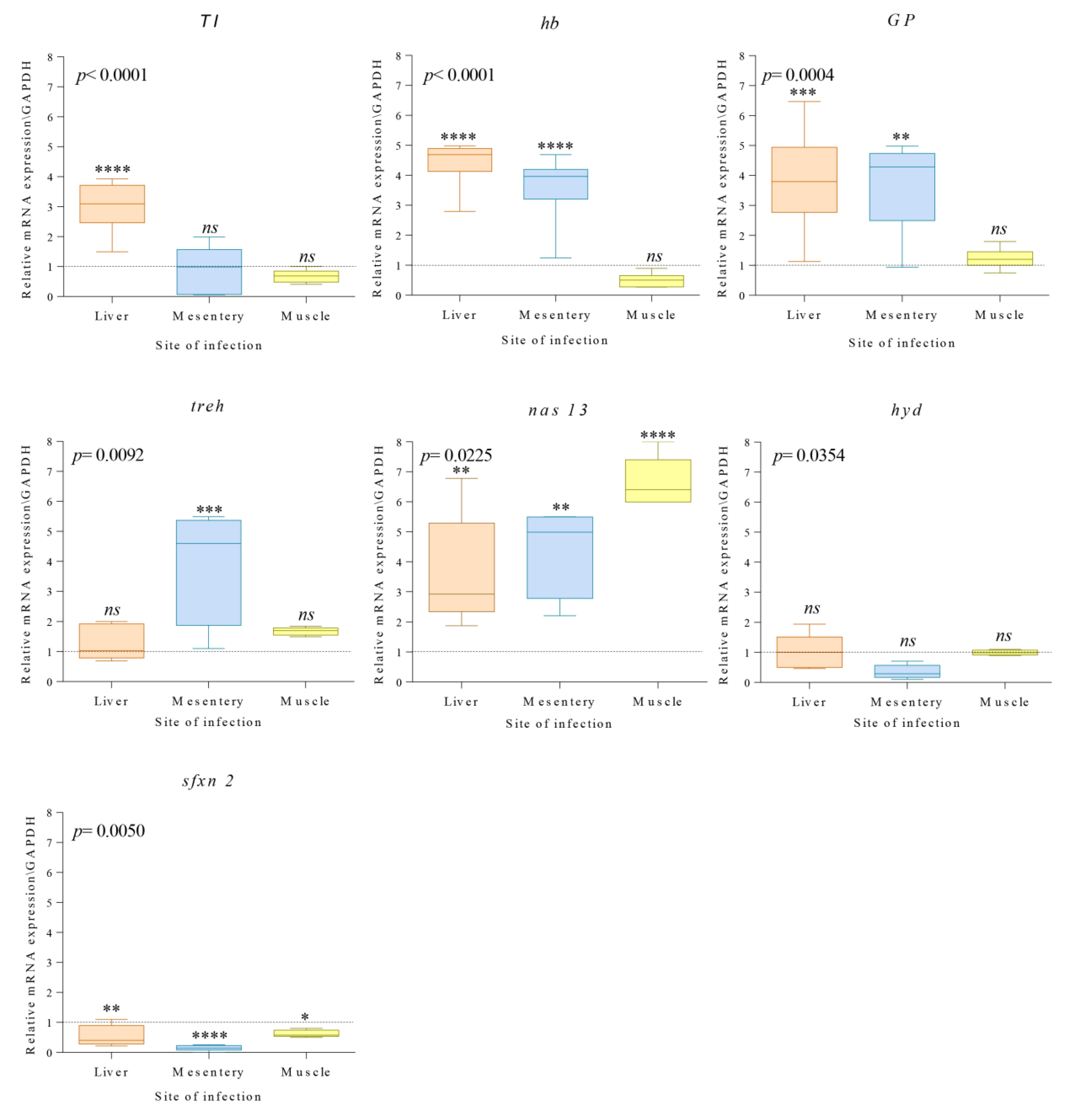
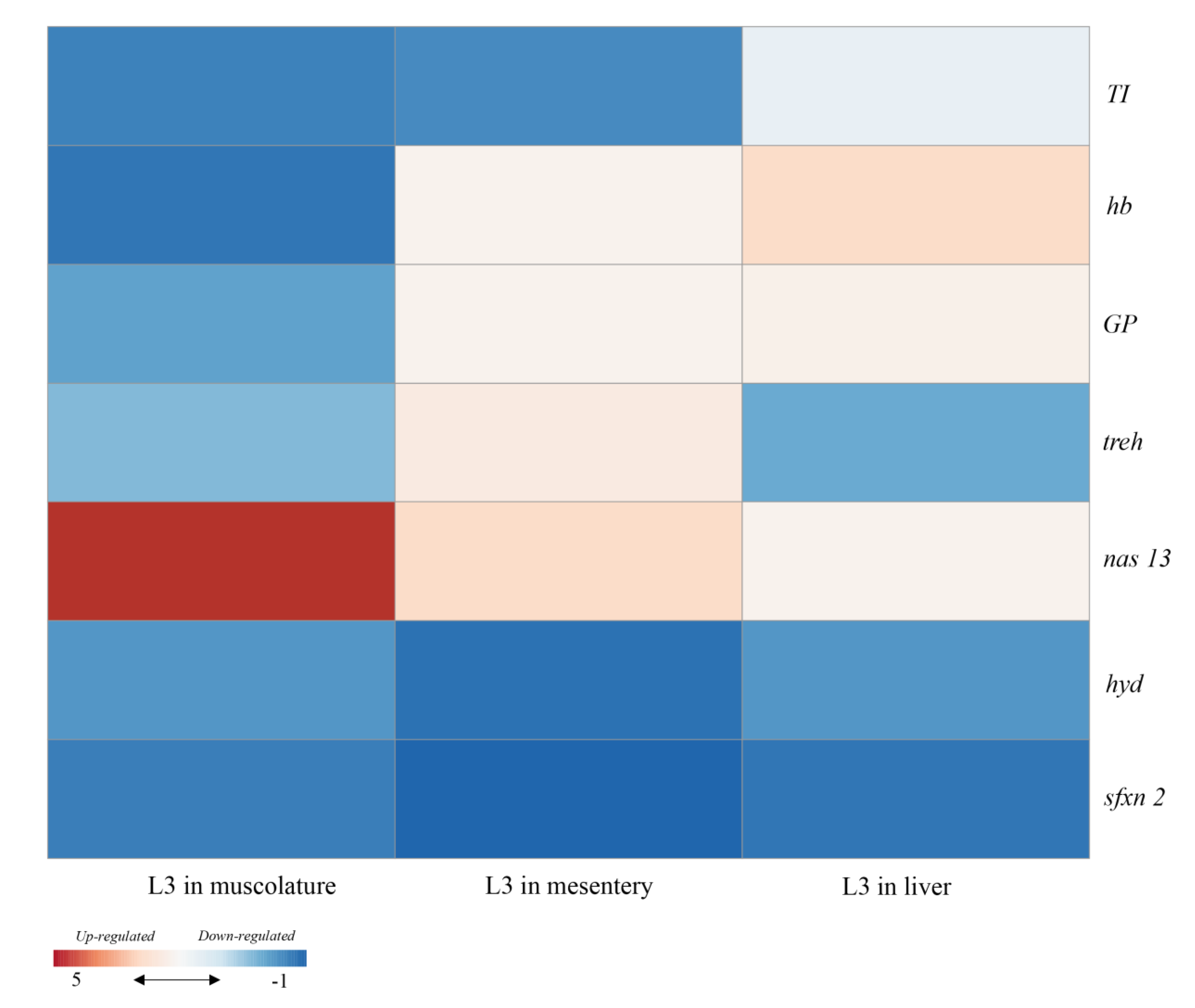
3.2. qPCR Analysis of Gene Coding for Seven Target Proteins

The analysis of gene expression profiles of seven adaptive proteins of the L3 of A. simplex (s.s.) recovered from different sites of the parasite infection (liver, mesentery, muscle) in M. poutassou are shown in Figure 1.
Quantitative real-time PCR (qPCR) analysis revealed differential regulation of the target genes in response to the infection site of the A. simplex (s.s.) larvae. Shapiro–Wilk test supported a normal distribution of the data obtained from the qPCR assay tests (all tests p < 0.05). Comparison in the expression levels of genes coding for Kunitz-type trypsin inhibitor, hemoglobin, glycoprotein, trehalase, zinc metallopeptidase 13, ubiquitin-protein ligase, and sideroflexin 2 was performed between A. simplex (s.s.) larvae collected from different sites of infection and the control (Figure 1).
The gene coding for the Kunitz-type trypsin inhibitor in the A. simplex (s.s.) larvae located in the liver showed statistically significant higher levels of expression (p < 0.0001) (Figure 1) compared to larvae of the control (stomach lumen). No significant differences were recovered for the same gene in A. simplex (s.s.) larvae located in the mesentery and muscle (p = 0.571, p = 0.301, respectively) (Figure 1).
A statistically significant up-regulation of the hb gene, coding for the hemoglobin, was observed in A. simplex (s.s.) larvae recovered from the liver (p < 0.0001) and mesentery (p < 0.0001) (Figure 1). Standard levels of the hb transcripts (p = 0.208) were measured in the larvae located in the muscle with respect to the control.
The GP gene was found to be significantly up-regulated in A. simplex (s.s.) larvae collected from the liver (p < 0.0001) and mesentery (p = 0.0015), while the expression values of this gene in larvae from the muscle were at a level similar to the control (p = 0.760).
No significant difference was recorded in the expression levels of the treh gene coding for the trehalase between Anisakis larvae collected from the fish liver (p = 0.596) and muscle (p = 0.230) and the control. Conversely, significant (p = 0.0002) were the differences in the transcripts of this gene estimated in the parasites recovered from mesenteries.
The nas 13 gene of A. simplex (s.s.) showed a generally significant (p < 0.05) up-regulation of the larvae located in all of the infection sites compared to the larvae of the control group (Figure 1).
A low gene expression level was recorded for the hyd gene coding for the ubiquitin-protein ligase, studied in the larvae from liver, mesenteries, and muscle of the fish host; nevertheless, non-significant differences (p > 0.05) were recorded in comparison with the larvae of the control.
Finally, the transcripts levels of the sfxn 2 gene coding for the sideroflexin 2 were significantly down-regulated (p < 0.05) in A. simplex (s.s.) larvae located in all fish tissues.
Dunnett’s multiple comparison test for differences in expression level per target gene between larvae from different fish host tissues are shown in Table 2. The ANOVA analysis showed that the overall transcripts values of the seven selected genes in A. simplex (s.s.) were significantly (p < 0.05) affected by the infection site of the parasite species in the fish host (Figure 1).

4. Discussion

In the present study, target gene transcripts of L3 of A. simplex (s.s.), correlated with their tissue localization in a commonly infected fish host, have been investigated for the first time.
The selected fish host species, blue whiting, is an intermediate/paratenic host in the life cycle of A. simplex (s.s.), which acquires the parasite through predation on other infected hosts (i.e., crustaceans, small fish, and cephalopods) [27,32]. The fish host populations caught in the NE Atlantic Ocean from off St. Kilda island (FAO VIa) are known to have high infection levels of this parasite both in the liver, mesenteries, and flesh [1,33,34]. Moreover, blue whiting from the present fishing area are usually not infected with other anisakid nematode species [34,35,36], which, in this respect, makes this fish host a suitable model since interference from other closely related anisakid species is not likely to occur.
Anisakis simplex (s.s.) larvae usually penetrate the gastric wall of the fish host to reach the visceral cavity. There, some L3 settle on the surface of the visceral organs (i.e., mesentery, liver, gonads), followed by host-induced encapsulation, where the capsule thickness seems largely to depend on infection site, i.e., the host target organ [3,37]. However, some larvae may migrate into the musculature of fish or squid [1,4,8,34,38,39], but so far, only L3 of A. simplex (s.s.) and A. pegreffii have been recorded in the muscular tissue of various fish and squid hosts, with A. simplex (s.s.) having apparently a higher capability to invade the flesh of fish than its sibling species A. pegreffii [7,33,40,41]. Once established and encapsulated, the larvae go through a period of latency, remaining infective until the next host (i.e., another transport host or a definitive marine mammalian host) [2]. To date, few studies have investigated the interactions between Anisakis spp. and their intermediate/paratenic hosts. Levsen and Berland (personal observation, 2012) observed that different fish hosts might show different degrees of immunological reactions to larvae. In some fish hosts, including the target species of this study, M. poutassou, evident melanomacrophage aggregates around larval infections sites on the liver have been observed [3]. The role of these aggregates is not yet completely understood, but Agius and Roberts [42] suggested that the melanomacrophage aggregates might develop in association with late-stage chronic inflammations induced by parasites. Recently, a cellular immune response against A. simplex (s.s.) larvae was observed in the liver of blue whiting specimens of the same samples examined in the present study; conversely, no host reaction was present around larvae lodged in the musculature of fish (Dezfuli pers. obs.).
Larsen et al. [43] assumed that the parasite-secreted molecules could be the cause of fish host cellular reactions.
In other studies, the gene expression of target proteins of L3 A. simplex (s.l.) was compared with L4 stage larvae [21]. Additionally, the A. pegreffii L3 gene expression at different temperature conditions was tested and compared [25,30]. However, no study concerning the differential gene expression related to the Anisakis spp. fish–host interaction has so far been carried out.
In this study, amongst the selected target genes, a significant differential gene expression of the TI gene coding for a Kunitz-type trypsin inhibitor protein was detected in A. simplex (s.s.) larvae removed from the fish liver (Figure 2). The Kunitz-type trypsin inhibitor protein seems to have important functions in the parasite’s development [44,45] and in the host proteases’ inhibition [20,46]. Indeed, a wide range of diverse nematode species has been found to express proteins with trypsin-inhibitor-like domains, including Ascaris suum [47], Ancylostoma caninum [48], and Trichuris suis [44]. Many of these nematode proteins have been shown to inhibit serine proteases, e.g., chymotrypsin, trypsin, elastase, and, in the case of the hookworms, host’ thrombin and coagulation factor [49,50,51]. Hawley and Peanasky [52] assumed that, in order to parasitize a host, a nematode requires those protease inhibitors that interact strongly with host proteases located in different sites of infection. The Kunitz-type trypsin inhibitor protein would aid A. simplex (s.s.) to counteract the host proteolytic enzyme and coagulation factors. Indeed, proteins with Kunitz domains have previously been detected in expressed transcripts sets obtained from pharyngeal tissues of A. pegreffii and A. simplex (s.s.) larvae [22,23]. In this study, an up-regulation of transcripts of TI in A. simplex (s.s.) located in the liver could be correlated to the fish host’s proteolytic enzymes, i.e., serine proteases. Fish organs are a source of those enzymes, especially digestive proteases, such as aspartic protease pepsin and serine proteases (i.e., trypsin, chymotrypsin, and elastase) [53,54]. In addition, some serine proteases, such as the tryptases, are produced and stored in the mast cell granules located in different host immune tissues [55]. Those host serine proteases would be implicated in the response to invading parasite larva which again could react with increasing production of inhibitor proteins, e.g., the Kunitz-type trypsin.
Nematode hemoglobins are molecules with an unusually high oxygen affinity. Several studies indicate that they may represent an important component of parasite adaptation to the co-existence with the host [25,56,57,58]. Thus, hemoglobin, sequestering oxygen, has been suggested to aid the parasite in maintaining a locally anaerobic environment, while their ability to break down nitric oxide (NO) and hydrogen peroxide produced by innate immune cells would also aid the parasite survival. Hemoglobin’s catalase and nitric oxide dioxygenase activities are important in protection against host immune responses to infection [59]. Results obtained in this study seem to support the possible function of the parasite’s hemoglobin in increasing its protection against the host immune response. Cavallero et al. [23] reported the presence of transcripts of hb even in pharyngeal tissues of Hysterothylacium aduncum larvae infecting viscera of the fish host. An up-regulation of the gene hb is reported as well in the present study for the larvae located in both host viscera (i.e., mesentery) and liver (Figure 2).
In previous investigations [25], we found that since the transcript levels of the GP in A. pegreffii remained stable under certain temperature conditions, its gene expression level would be modulated by the host immune response. Indeed, we observed in the present study that the transcripts of a GP gene were significantly up-regulated in A. simplex (s.s.) located in the liver and mesentery, where the immune reactivity of the host seems to be stronger [60]. Conversely, a standard GP expression similar to the control was detected in larvae recovered in the host skeletal muscle, where the host’s immune reactivity seems to be weaker (Figure 2). Indeed, it is generally recognized that the parasite glycoproteins which occur abundantly on worms’ surface and in excretory/secretory (ES) products [61] are able to down-modulate the host immune response [61,62]. For instance, in the case of Schistosoma mansoni eggs, an omega-1 glycoprotein was found to promote Th-2 skewing of dendritic cells (DCs) and T cells during infection [63]. Previous studies have suggested that Anisakis spp. larvae secrete ES products, whose glycosylated components regulate the fundamental processes of antigen recognition, processing, and presentation [13]. The Anisakis glycoprotein would be able to interact with both innate and adaptive branches of the immune system in intermediate and accidental human hosts. Indeed, these molecules are involved in the regulation of the IgM response of the intermediate/paratenic fish host [64].
Lopienska-Biernat et al. [24] have recently described a pathway of genes coding proteins of trehalose and glycogen metabolism in A. simplex (s.l.). The trehalose has an important role in the energy storage, sugar circulation, glucose uptake in the absence of food, and protection during adverse environmental conditions [65,66]. Indeed, the trehalose is essential for survival of parasitic nematodes, e.g., A. simplex (s.s.) [67,68].
Third-stage larvae of A. simplex (s.l.) do not use external sources of glucose [68,69]. Indeed, even if the alimentary system is developed, there is no intake and assimilation of food, so the trehalose is necessary for the glucose storage and their survival without directly feeding [70,71,72]. In this study, no significantly high levels of treh gene in A. simplex (s.s.) larvae recovered from the liver and muscle, compared with control, were observed. However, significantly higher treh gene expression levels were detected in larvae recovered from the mesenteries (Figure 2). These results would reflect the necessity of the parasite L3 to transcribe the treh gene since trehalose appears to be indispensable to accumulate endogenous saccharide reserve compounds [72]. The Anisakis spp. larvae are typically surrounded by fibrous connective tissues generated by the fish host [3]; the capsule’s thickness formed around the larva seems to be related to the abundance of connective tissue in different infection sites in the host. Thus, it would occur that, where the connective tissue layers become more compact (i.e., mesenteries), the host’s immune system produces a thicker capsule around the larva, which remains “imprisoned” [73]. Capsules formation would lead to a major need of energy for the larva to contrast the host immune response, with a consequent increase of the treh gene transcripts.
In parasitic nematodes, peptidase proteins represent a conspicuous percentage of expressed genes [74]. They are involved in host–parasite interactions [75] and, in many cases, constitute the main component of antigens, known for triggering immune responses of the host [76,77]. The metallopeptidases family is involved in the host tissue invasion by degrading the extracellular matrix. It has been also suggested that they conserve their proteolytic activity even after the action of the host antibodies [78,79]. Kim et al. [21] found that some metallopeptidases genes are differentially regulated in the A. simplex (s.l.) larvae transcriptome. In particular, the authors observed that the nas 13 was up-regulated in L3 larvae in comparison with the L4 stage of the parasite species. Cavallero et al. [23] have found high transcripts of the metallopeptidases in the pharyngeal tissues of Hysterothylacium aduncum as well. These enzymes, in Anisakis spp. larvae, were considered to be involved in host tissue penetration [21]. In this study, a generalized up-regulation of nas 13 was recorded in A. simplex (s.s.) larvae located in all the fish-host tissues, compared to the control (Figure 2). This finding highly supports a major role of the parasite’s peptidases in the fish host tissue invasion and migration.
The role of ubiquitin-protein in A. simplex (s.l.) is not yet fully elucidated. It was hypothesized [80] that the ubiquitin–proteasome system could be involved in protein degradation and apoptosis in parasitic nematodes, as well as in the withstanding to the oxidative-stress. The ubiquitin pathway is also known to be involved in the survival of parasitic nematode Trichinella spiralis and in the development of the parasitic trematode Schistosoma mansoni [81]. Nematodes rely on proteomic plasticity to remodel themselves during periods of developmental change, to endure varying environmental conditions, and to respond to biotic and abiotic stressors. Considering A. simplex (s.s.) from the different tissues of M. poutassou, the gene hyd was apparently not differentially regulated compared to the control (Figure 2). This would indicate that the larvae lay in a stable developmental stage; i.e., they are not going to molt in the paratenic fish host. As a consequence, the parasite specimen at this stage does not need to regulate the internal state, usually mediated by the up-regulation of stress genes, such as the hyd [80].
Since a significantly higher sfxn 2 expression in A. simplex (s.s.) L3 larvae compared to L4 was previously observed [21], the gene expression of the sideroflexin 2 in different sites of fish infection was here investigated. To date, it is known that the sideroflexins are mitochondrial multiple transmembrane proteins associated with the iron accumulation in the mitochondria [82]. In the present study, a down-regulation of sfxn 2 was recorded in A. simplex (s.s.) larvae removed from different fish tissues (Figure 2). However, because no information on the function of sideroflexin in parasitic nematodes is so far available, further studies are needed to elucidate the possible roles of the sideroflexin 2 in A. simplex (s.s.) larvae.

5. Conclusions

Anisakis is an endoparasite exposed to changing life conditions, considering that its hosts vary between the poikilothermic crustaceans, heterothermic fish, and homeothermic marine mammals. However, the L4 stage and adults of A. simplex (s.l.) parasitize a more stable environment, such as the stomach of the cetacean definitive hosts, with consequent levels of transcripts more stable. Instead, the third-stage larvae differentially transcribe mRNAs, depending on different fish host tissue environments. Survival in an array of such different habitats must require costly molecular and biochemical adaptations. Natural selection should remove the behaviors that are not compensating benefits [84]. For example, during migration through the tissues, the larvae can become lost or killed by the fish host; migration seems to be not necessary for their development.
Migration in different sites of infections of fish is probably an advantageous life-history strategy; however, the evolutionary factors responsible for the larval migration in the fish are poorly understood.
The findings acquired during this study show that the relative gene expression levels of the seven target genes in the A. simplex (s.s.) larvae are linked to the infection site in the fish host, blue whiting. Indeed, different sites of infection influence the gene expression levels of A. simplex (s.s.) larvae. The genes encoding some of the present adaptive proteins seem to have protective properties for the parasite, facilitating host tissue migration (i.e., nas 13) and its survival (i.e., TI, GL, hb, treh) in the basically hostile target tissues of the fish host. Tissue migration is known from many different nematode orders and even other parasite phyla. Therefore, tissue migration has evolved many times and independently in the parasitic nematodes.
An interesting question would be: why tissue migration has evolved at all? Why do larvae migrate over relatively long distances through a hostile environment instead of staying and developing in just one organ—the stomach or intestine, where the parasites first arrived? It could be assumed that tissue migration of A. simplex (s.s.) and A. pegreffii [8] is an adaptive and purpose-directed character, e.g., facilitating escape from host reactions (especially those in the flesh), and, similarly, that the host-induced encapsulation “protects”, in terms of isolating, the larvae, which remain in a “dormant” state until the larval transmission to the next host.

Author Contributions

Conceptualization, S.M., M.P., P.C., L.G., G.N., and A.L.; methodology, S.M. and M.P.; writing—original draft preparation, S.M. and M.P.; writing—review and editing, all authors; supervision, S.M. and G.N. All authors have read and agreed to the published version of the manuscript.

Funding

This research was funded by Istituto Pasteur Italia—Fondazione Cenci Bolognetti.

Acknowledgments

We thank the anonymous referees for their comments on the manuscript.

Conflicts of Interest

The authors declare no conflict of interest.

References

  1. Mattiucci, S.; Cipriani, P.; Levsen, A.; Paoletti, M.; Nascetti, G. Molecular epidemiology of Anisakis and anisakiasis: An ecological and evolutionary road map. Adv. Parasitol. 2018, 99, 93–263. [Google Scholar] [PubMed]
  2. Mattiucci, S.; Nascetti, G. Advances and trends in the molecular systematics of anisakids nematodes, with implications for their evolutionary ecology and host-parasite co-evolutionary processes. Adv. Parasitol. 2008, 66, 47–148. [Google Scholar] [PubMed]
  3. Levsen, A.; Berland, B. Anisakis species. In Fish Parasites: Pathobiology and Protection; Patrick, T.K., Buchmann, K., Eds.; CABI Press: Wallingford, UK, 2012; p. 298. [Google Scholar]
  4. Smith, J.W. The abundance of Anisakis simplex L3 in the body-cavity and flesh of marine teleosts. Int. J. Parasitol. 1984, 14, 491–495. [Google Scholar] [CrossRef]
  5. Smith, J.W.; Wootten, R. Experimental studies on the migration of Anisakis sp. larvae (Nematoda: Ascaridida) into the flesh of herring, Clupea harengus L. Int. J. Parasitol. 1975, 5, 133–136. [Google Scholar] [PubMed]
  6. Karl, H.; Meyer, C.; Banneke, S.; Jark, U.; Feldhusen, F. The abundance of nematode larvae Anisakis sp. in the flesh of fishes and possible post-mortem migration. Archiv. für Lebensmittelhygiene 2002, 53, 118–120. [Google Scholar]
  7. Quiazon, K.M.; Yoshinaga, T.; Ogawa, K. Experimental challenge of Anisakis simplex sensu stricto and Anisakis pegreffii (Nematoda: Anisakidae) in rainbow trout and olive flounder. Parasitol. Int. 2011, 60, 126–131. [Google Scholar] [CrossRef]
  8. Cipriani, P.; Acerra, V.; Bellisario, B.; Sbaraglia, G.L.; Cheleschi, R.; Nascetti, G.; Mattiucci, S. Larval migration of the zoonotic parasite Anisakis pegreffii (Nematoda: Anisakidae) in European anchovy, Engraulis encrasicolus: Implications to seafood safety. Food Control 2016, 59, 148–157. [Google Scholar] [CrossRef] [Green Version]
  9. Cipriani, P.; Sbaraglia, G.; Palomba, M.; Giulietti, L.; Bellisario, B.; Buselic, I.; Mladineo, I.; Cheleschi, R.; Nascetti, G.; Mattiucci, S. Anisakis pegreffii (Nematoda: Anisakidae) in European anchovy Engraulis encrasicolus from the Mediterranean Sea: Considerations in relation to fishing ground as a driver for parasite distribution. Fish. Res. 2017, 202, 59–68. [Google Scholar] [CrossRef]
  10. Bahlool, Q.Z.M.; Skovgaard, A.; Kania, P.; Haarder, S.; Buchmann, K. Microhabitat preference of Anisakis simplex in three salmonid species: Immunological implication. Vet. Parasitol. 2012, 190, 489–495. [Google Scholar] [CrossRef]
  11. Abollo, E.; Gestal, C.; Pascal, S. Anisakis infestation in marine fish and cephalopods from Galician waters: And updated perspective. Parasitol. Res. 2001, 87, 492–499. [Google Scholar]
  12. Dezfuli, B.S.; Giovinazzo, G.; Lui, A.; Giari, L. Inflammatory response to Dentitruncus truttae (Acanthocephala) in the intestine of brown trout. Fish Shellfish Immunol. 2008, 24, 726–733. [Google Scholar] [CrossRef] [PubMed]
  13. Bahlool, Q.Z.; Skovgaard, A.; Kania, P.W.; Buchmann, K. Effects of excretory/secretory products from Anisakis simplex (Nematoda) on immune gene expression in rainbow trout (Oncorhynchus mykiss). Fish Shellfish Immunol. 2013, 35, 734–739. [Google Scholar] [CrossRef] [PubMed] [Green Version]
  14. Mehrdana, F.; Kania, P.W.; Nazemi, S.; Buchmann, K. Immunomodulatory effects of excretory/secretory compounds from Contracaecum osculatum larvae in a zebrafish inflammation model. PLoS ONE 2017, 12, e0181277. [Google Scholar] [CrossRef] [PubMed] [Green Version]
  15. Marnis, H.; Kania, P.W.; Syahputra, K.; Zuo, S.; Dirks, R.P.; Buchmann, K. Transcriptomic analysis of Baltic cod (Gadus morhua) liver infected with Contracaecum osculatum third stage larvae indicated parasitic effects on growth and immune response. Fish Shellfish Immunol. 2019, 93, 965–976. [Google Scholar] [CrossRef] [PubMed]
  16. Priebe, K.; Huber, C.; Martlbauer, E.; Terplan, G. Detection of antibodies against the larva of Anisakis simplex in the pollock Pollachius virens using ELISA. J. Vet. Med. 1991, 38, 209–214. [Google Scholar] [CrossRef]
  17. Kim, J.S.; Kim, K.H.; Cho, S.; Park, H.Y.; Cho, S.W.; Kim, Y.T.; Joo, K.H.; Lee, J.S. Immunochemical and biological analysis of allergenicity with excretory-secretory products of Anisakis simplex third stage larva. Int. Arch. Allergy Immunol. 2005, 136, 320–328. [Google Scholar] [CrossRef]
  18. Kuhn, T.; Hailer, F.; Palm, H.W.; Klimpel, S. Global assessment of molecularly identified Anisakis Dujardin, 1845 (Nematoda: Anisakidae) in their teleost intermediate hosts. Folia Parasitol. 2013, 60, 123–134. [Google Scholar] [CrossRef] [Green Version]
  19. Skrzypczak, M.; Rokicki, J.; Pawliczka, I.; Najda, K.; Dzido, J. Anisakids of seals found on the Southern coast of Baltic Sea. Acta Parasitol. 2014, 59, 165–172. [Google Scholar] [CrossRef]
  20. Mehrdana, F.; Buchmann, K. Excretory/secretory products of anisakid nematodes: Biological and pathological roles. Acta Vet. Scand. 2017, 59, 42. [Google Scholar] [CrossRef] [Green Version]
  21. Kim, J.H.; Kim, J.O.; Jeon, C.H.; Nam, U.H.; Subramaniyan, S.; Yoo, S.I.; Park, J.H. Comparative transcriptome analyses of the third and fourth stage larvae of Anisakis simplex (Nematoda: Anisakidae). Mol. Biochem. Parasitol. 2018, 226, 24–33. [Google Scholar] [CrossRef]
  22. Cavallero, S.; Lombardo, F.; Su, X.; Salvemini, M.; Cantacessi, C.; D’Amelio, S. Tissue-specific transcriptomes of Anisakis simplex (sensu stricto) and Anisakis pegreffii reveal potential molecular mechanisms involved in pathogenicity. Parasites Vectors 2018, 11, 31. [Google Scholar] [CrossRef] [PubMed] [Green Version]
  23. Cavallero, S.; Lombardo, F.; Salvemini, M.; Pizzarelli, A.; Cantacessi, C.; D’Amelio, S. Comparative transcriptomics reveals clues for differences in pathogenicity between Hysterothylacium aduncum, Anisakis simplex sensu stricto and Anisakis pegreffii. Genes 2020, 11, 321. [Google Scholar] [CrossRef] [PubMed] [Green Version]
  24. Lopienska-Biernat, E.; Paukszto, L.; Jastrzebski, J.P.; Myszczynski, K.; Polak, I.; Stryinski, R. Genome-wide analysis of Anisakis simplex sensu latu: The role of carbohydrate metabolism genes in the parasite’s development. Int. J. Parasitol. 2019, 49, 933–943. [Google Scholar] [CrossRef] [PubMed]
  25. Palomba, M.; Paoletti, M.; Colantoni, A.; Rughetti, A.; Nascetti, G.; Mattiucci, S. Gene expression profiles of antigenic proteins of third stage larvae of the zoonotic nematode Anisakis pegreffii in response to temperature conditions. Parasite 2019, 26, 52. [Google Scholar] [CrossRef] [Green Version]
  26. Baird, F.J.; Su, X.; Aibinu, I.; Nolan, M.J.; Suglyama, H.; Otranto, D.; Lopata, A.L.; Cantacessi, C. The Anisakis transcriptome provides a resource for fundamental and applied studies on allergy-causing parasites PLOS Neglect. Trop. Dis. 2016, 10, e0004845. [Google Scholar] [CrossRef] [PubMed] [Green Version]
  27. Prokopchuk, I.; Sentyabov, E. Diets of herring, mackerel and blue whiting in the Norwegian Sea in relation to Calanus finmarchius distribution and temperature conditions. ICES J. Mar. Sci. 2006, 63, 117–127. [Google Scholar] [CrossRef] [Green Version]
  28. Mattiucci, S.; Cipriani, P.; Webb, S.C.; Paoletti, M.; Marcer, F.; Bellisario, B.; Gibson, D.I.; Nascetti, G. Genetic and morphological approaches distinguishing the three sibling species of the Anisakis simplex species complex, with a species designation as Anisakis berlandi n. sp. for A. simplex sp. C (Nematoda: Anisakidae). J. Parasitol. 2014, 15, 12–15. [Google Scholar]
  29. Mattiucci, S.; Acerra, V.; Paoletti, M.; Cipriani, P.; Levsen, A.; Webb, S.C.; Canestrelli, D.; Nascetti, G. No more time to stay ‘single’ in the detection of Anisakis pegreffii, A. simplex (s.s.) and hybridization events between them: A multi-marker nuclear genotyping approach. Parasitology 2016, 143, 998–1011. [Google Scholar] [CrossRef] [Green Version]
  30. Chen, H.Y.; Cheng, Y.S.; Grabner, D.S.; Chang, S.H.; Shih, H.H. Effect of different temperatures on the expression of the newly characterized heat shock protein 90 (Hsp90) in L3 of Anisakis spp. isolated from Scomber australasicus. Vet. Parasitol. 2014, 205, 540–550. [Google Scholar] [CrossRef]
  31. Livak, K.J.; Schmittgen, T.D. Analysis of relative gene expression data using Real-Time quantitative PCR and the 2 (Delta CT) method. Methods 2001, 25, 402–408. [Google Scholar] [CrossRef]
  32. Molina-Fernandez, D.; Rubio-Calvo, D.; Adroher, F.J.; Benitez, R. Molecular epidemiology of Anisakis spp. in blue whiting Micromesistius poutassou in eastern waters of Spain, western Mediterranean Sea. Int. J. Food Microbiol. 2018, 282, 49–56. [Google Scholar] [PubMed]
  33. Gomez-Mateos, M.; Valero, A.; Morales-Yuste, M.; Martin-Sanchez, J. Molecular epidemiology and risk factors for Anisakis simplex s. l. infection in blue whiting (Micromesistius poutassou) in a confluence zone of the Atlantic and Mediterranean: Differences between A. simplex s.s. and A. pegreffii. Int. J. Food Microbiol. 2016, 232, 111–116. [Google Scholar] [CrossRef] [PubMed]
  34. Levsen, A.; Svanevik, C.S.; Cipriani, P.; Mattiucci, S.; Gay, M.; Hastiee, L.C.; Pierce, G.J.; Bušelić, I.; Mladineo, I.; Karl, O.; et al. A survey of zoonotic nematodes of commercial key fish species from major European fishing grounds introducing the FP7 PARASITE exposure assessment study. Fish. Res. 2018, 202, 4–21. [Google Scholar] [CrossRef]
  35. McKenzie, K. Some parasites and diseases of blue whiting, Micromesistius poutassou (Risso), to the North and West of Scotland and at the Faroe Islands. Scottish Fish. Res. Rep. 1979, 17, 1–14. [Google Scholar]
  36. Højgaard, D.P. Sníkar í svartkjafti (Parasites of Blue Whiting). Fiskirannsóknir 1998, 8, 114–126, (In Faroese, with English summary). [Google Scholar]
  37. Berland, B. Musings on onematode parasites. Fisken Oghavet 2006, 11, 1–26. [Google Scholar]
  38. Karl, H. Nematode larvae in fish on the German market—20 years of consumer related research. Arch. Lebensmittelhyg 2008, 59, 107–116. [Google Scholar]
  39. Cipriani, P.; Giulietti, L.; Palomba, M.; Mattiucci, S.; Bao, M.; Levesen, A. Occurrence of larval ascaridoid nematodes in the Argentinean short-finned squid Illex argentinus from the Southwest Atlantic Ocean (off Falkland Islands). Int. J. Food. Microbiol. 2019, 297, 27–31. [Google Scholar] [CrossRef]
  40. Suzuki, J.; Murata, R.; Hosaka, M.; Araki, J. Risk factors for human Anisakis infection and association between the geographic origins of Scomber japonicus and anisakid nematodes. Int. J. Food Microbiol. 2010, 137, 88–93. [Google Scholar] [CrossRef]
  41. Cipriani, P.; Smaldone, G.; Acerra, V.; D’Angelo, L.; Anastasio, A.; Bellisario, B.; Palma, G.; Nascetti, G.; Mattiucci, S. Genetic identification and distribution of the parasitic larvae of Anisakis pegreffii and A. simplex (s.s.) in European hake Merluccius merluccius from the Tyrrhenian Sea and Spanish Atlantic coast: Implications for food safety. Int. J. Food Microbiol. 2014, 198, 1–8. [Google Scholar] [CrossRef]
  42. Agius, C.; Roberts, R.J. Melano-macrophage centres and their role in fish pathology. J. Fish Dis. 2003, 26, 499–509. [Google Scholar] [CrossRef] [PubMed]
  43. Larsen, A.H.; Bresciani, J.; Buchmann, K. Interactions between ecto- and endoparasites in trout Salmo trutta. Vet. Parasitol. 2002, 103, 167–173. [Google Scholar] [CrossRef]
  44. Rhoads, M.L.; Fetterer, R.H.; Hill, D.E. Trichuris suis: A secretory serine protease inhibitor. Exp. Parasitol. 2000, 94, 1–7. [Google Scholar] [CrossRef]
  45. Stepek, G.; McCormack, G.; Page, A.P. Collagen processing and cuticle formation is catalysed by the astacin metalloprotease DPY-31 in free-living and parasitic nematodes. Int. J. Parasitol. 2010, 40, 533–542. [Google Scholar] [CrossRef] [PubMed] [Green Version]
  46. Moneo, I.; Caballero, M.L.; Gómez, F.; Ortega, E.; Alonso, M.J. Isolation and characterization of a major allergen from the fish parasite Anisakis simplex. J. Allergy Clin. Immunol. 2000, 106, 177–182. [Google Scholar] [CrossRef] [PubMed]
  47. Bernard, V.D.; Peanasky, R.J. The serine protease inhibitor family from Ascaris suum: Chemical determination of the five disulphide bridges. Arch. Biochem. Biophys. 1993, 303, 367–376. [Google Scholar] [CrossRef] [PubMed]
  48. Stanssens, P.; Bergum, P.W.; Gansemans, Y.; Jespers, L.; Laroche, Y.; Huang, S.; Maki, S.; Messens, J.; Lauwereys, M.; Cappello, M.; et al. Anticoagulant repertoire of the hookworm Ancylostoma caninum. Proc. Natl. Acad. Sci. USA 1996, 93, 2149–2154. [Google Scholar] [CrossRef] [Green Version]
  49. Matthews, B.E. Behaviour and enzyme release by Anisakis sp. larvae (Nematoda: Ascaridida). J. Helminthol. 1982, 56, 177–183. [Google Scholar] [CrossRef]
  50. Matthews, B.E. The source, release and specificity of proteolytic enzyme activity produced by Anisakis simplex larvae (Nematoda: Ascaridida) in vitro. J. Helminthol. 1984, 58, 175–185. [Google Scholar] [CrossRef]
  51. Quiazon, K.M.; Santos, M.D.; Yoshinaga, T. Anisakis species (Nematoda: Anisakidae) of Dwarf Sperm Whale Kogia sima (Owen, 1866) stranded off the Pacific coast of southern Philippine archipelago. Vet. Parasitol. 2013, 197, 221–230. [Google Scholar] [CrossRef]
  52. Hawley, J.; Peanasky, R.J. Ascaris suum: Are trypsin inhibitors involved in species specificity of Ascarid nematodes? Exp. Parasitol. 1992, 75, 112–128. [Google Scholar] [CrossRef]
  53. Khangembam, B.K.; Sharma, K.; Chakrabarti, R. Purification and characterization of trypsin from the digestive system of carp Catla catla (Hamilton). Int. Aquat. Res. 2012, 4, 9. [Google Scholar] [CrossRef] [Green Version]
  54. Klomklao, S.; Benjakul, S. Two trypsin isoforms from albacore tuna (Thunnus alalonga) liver: Purification and physicochemical and biochemical characterization. Int. J. Biol. Macro. 2018, 107, 1864–1870. [Google Scholar] [CrossRef] [PubMed]
  55. McNeil, H.P. Mast cell-restricted tryptases: Structure and function in inflammation and pathogen defense. J. Biol. Chem. 2007, 282, 20785–20789. [Google Scholar] [CrossRef] [PubMed] [Green Version]
  56. Gonzalez-Fernandez, J.; Daschner, A.; Nieuwenhuizen, N.E.; Lopata, A.L.; Frutos, C.D.; Valls, A.; Cuellar, C. Haemoglobin, a new major allergen of Anisakis simplex. Int. J. Parasitol. 2015, 45, 399–407. [Google Scholar] [CrossRef] [PubMed]
  57. Abe, N.; Teramoto, I. Anisakis haemoglobin is a main antigen indicung strong and prolonged immunoreactions in rats. Parasitol. Res. 2017, 116, 2035–2039. [Google Scholar] [CrossRef]
  58. Gonzalez-Fernandez, J.; Rivas, L.; Luque-Ortega, J.R.; Nunez-Ramirez, R.; Campioli, P.; Garate, T.; Perteguer, M.J.; Daschner, A.; Cuellar, C. Recombinant vs native Anisakis haemoglobin (Ani s 13): Its appraisal as a new gold standard for the diagnosis of allergy. Exp. Parasitol. 2017, 181, 119–129. [Google Scholar] [CrossRef]
  59. Nieuwenhuizen, N.E.; Meter, J.M.; Horsnell, W.G.; Hoving, J.C.; Fick, L.; Sharp, M.F.; Darby, M.G.; Parihar, S.P.; Brombacher, F.; Lopata, A.L. A cross-reactive monoclonal antibody to nematode haemoglobin enhances protective immune responses to Nippostrongylus brasiliensis. PLoS Negl. Trop. Dis. 2013, 7, e2395. [Google Scholar] [CrossRef] [Green Version]
  60. Secombes, C.J.; Wang, T. The Innate and Adaptive Immune System of Fish. In Infectious Disease in Aquaculture: Prevention and Control; Austin, B., Ed.; Woodhead Publishing Limited: Aberdeen, UK, 2012; pp. 1–67. [Google Scholar]
  61. Prasanphanich, N.S.; Mickum, M.L.; Molinaro, J.H.; Cummings, R.D. Glycoconjugates in host-helminth interactions. Front. Immunol. 2013, 4, 240. [Google Scholar] [CrossRef] [Green Version]
  62. White, R.R.; Artavanis-Tsakonas, K. How helminths use excretory secretory fractions to modulate dendritic cells. Virulence 2012, 3, 668–677. [Google Scholar] [CrossRef] [Green Version]
  63. Steinfelder, S. The major component in schistosome eggs responsible for conditioning dendritic cells for Th2 polarization is a T2 ribonuclease (omega-1). J. Exp. Med. 2009, 206, 1681–1690. [Google Scholar] [CrossRef] [PubMed]
  64. Hrabar, J.; Trumbic, Z.; Bocina, I.; Buselic, I.; Vrbatovic, A.; Mladineo, I. Interplay between proinfiammatory cytokines, miRNA, and tissue lesions in Anisakis-infected Sprague-Dawley rats. PLoS Neglect. Trop. Dis. 2019, 13, e0007397. [Google Scholar] [CrossRef] [PubMed]
  65. Elbein, A.D.; Pan, Y.T.; Pastuszak, I.; Carroll, D. New insights on trehalose: A multifunctional molecule. Glycobiology 2003 13, 17R–27R. [CrossRef]
  66. Tang, B.; Wang, S.; Wang, S.G.; Wang, H.J.; Zhang, J.Y.; Cui, S.Y. Invertebrate trehalose-6-phosphate synthase gene: Genetic architecture, biochemistry, physiological function, and potential applications. Front. Physiol. 2018, 9, 30. [Google Scholar] [CrossRef] [PubMed] [Green Version]
  67. Łopienska-Biernat, E.; Zaobidna, E.A.; Dmitryjuk, M. Expression of genes encoding the enzymes for glycogen and trehalose metabolism in L3 and L4 larvae of Anisakis simplex. J. Parasitol. Res. 2015, 2015, 438145. [Google Scholar] [CrossRef] [Green Version]
  68. Łopienska-Biernat, E.; Stryinski, R.; Dmitryjuk, M.; Wasilewska, B. Infective larvae of Anisakis simplex (Nematoda) accumulate trehalose and glycogen in response to starvation and temperature stress. Biol. Open 2019, 8, bio040014. [Google Scholar] [CrossRef] [Green Version]
  69. Smith, J.W.; Wootten, R. Anisakis and anisakiasis. Adv. Parasitol. 1978, 16, 93–163. [Google Scholar]
  70. Grabda, J. Studies on the life cycle and morphogenesis of Anisakis simplex (Rudolphi, 1809) (Nematoda: Anisakidae) cultured in vitro. Acta Ichthyol. Piscat. 1976, 6, 119–141. [Google Scholar] [CrossRef] [Green Version]
  71. Iglesias, L.; Valero, A.; Benítez, R.; Adroher, F.J. In vitro cultivation of Anisakis simplex: Pepsin increases survival and moulting from fourth larval to adult stage. Parasitology 2001, 1123, 285–291. [Google Scholar] [CrossRef] [Green Version]
  72. Łopienska-Biernat, E.; Zółtowska, K.; Rokicki, J. The content of carbohydrates in larval stages of Anisakis simplex (Nematoda, Anisakidae). Helmintologia 2006, 43, 125–129. [Google Scholar] [CrossRef] [Green Version]
  73. Levsen, A.; Lunestad, B.T.; Berland, B. Parasites in farmed fish and fishery products. In Improving Farmed Fish Quality and Safety; Oyvind, L., Ed.; WP: New York, NY, USA, 2008; pp. 428–445. [Google Scholar]
  74. Sajid, M.; McKerrow, J.H. Cysteine proteases of parasitic organism. Mol. Biochem. Parasit. 2002, 120, 1–21. [Google Scholar] [CrossRef]
  75. Malagon, D.; Benitez, R.; Kasny, M.; Adroher, F.J. Peptidases in parasitic nematodes: A review. In Parasites: Ecology, Diseases and Management; Erzinger, G.S., Ed.; Nova Science Publishers Inc.: New York, NY, USA, 2013; pp. 61–102. [Google Scholar]
  76. Hotez, P.J.; Pritchard, D.I. Hookworm infection. Sci. Am. 1995, 272, 68–74. [Google Scholar] [CrossRef] [PubMed]
  77. Todorova, V.K.; Stoyanov, D.I. Partial characterization of serine proteinases secreted by adult Trichinella spiralis. Parasitol. Res. 2000, 86, 684–687. [Google Scholar] [CrossRef] [PubMed]
  78. Malagón, D.; Benítez, R.; Adroher, F.J.; Díaz-López, M. Proteolytic activity in Hysterothylacium aduncum (Nematoda: Anisakidae), a fish gastrointestinal parasite of worldwide distribution. Vet. Parasitol. 2011, 183, 95–102. [Google Scholar] [CrossRef]
  79. Bruschi, F.; Pinto, B. Metalloproteinases in Parasitic Infections. In Pathophysiological Aspects of Proteases; Chakraborti, S., Dhalla, N., Eds.; Springer: Singapore, 2017; pp. 61–102. [Google Scholar]
  80. Adhikari, B.N.; Adams, B.J. Molecular Analyses of Desiccation Survival in Antarctic Nematodes. In Molecular and Physiological Basis of Nematode Survival; Perry, R.N., Wharton, D.A., Eds.; CAB International: Oxford, UK, 2011; pp. 205–232. [Google Scholar]
  81. Shang, F.; Gong, X.; Taylor, A. Activity of ubiquitin-dependent pathway in response to oxidative stress: Ubiquitin-activating enzye (E1) is transiently upregulated. J. Biol. Chem. 1997, 276, 23086–23093. [Google Scholar] [CrossRef] [Green Version]
  82. Miotto, G.; Tessaro, S.; Rotta, G.A.; Bonatto, D. In silico analyses of Fsf1 sequences, a new group of fungal proteins orthologous to the metazoan sideroblastic anemia-related sideroflexin family. Fungal. Genet. Biol. 2007, 44, 740–753. [Google Scholar] [CrossRef]
  83. Metsalu, T.; Vilo, J. ClustVis: A web tool for visualizing clustering of multivariate data using Principal Component Analysis and heatmap. Nucleic Acids Res. 2015, 43, w566–w570. [Google Scholar] [CrossRef]
  84. Read, A.; Sharping, A. The evolution of tissue migration by parasitic nematode larvae. Parasitology 1995, 111, 359–371. [Google Scholar] [CrossRef]
Figure 1. Box-plot representation of relative expression profiles of 7 genes coding for proteins, i.e., TI, hb, GP, treh, nas 13, hyd, sfxn 2, normalized to the geometric mean of gpd in Anisakis simplex (s.s.) larvae infecting different host-fish tissues (liver, mesentery, muscle). The control, represented by larvae recovered in the stomach lumen of the fish host, is given as normalized to a value of 1 (dashed line). Each box shows the minimum, first quartile, median, third quartile, and maximum gene expression values of three biological replicates. Asterisks indicate the significance level of the differences between each group of A. simplex (s.s.) larvae from different sites of infection and the control group. Significance was fixed at p < 0.05 (* p < 0.05, ** p < 0.01, *** p < 0.001, **** p < 0.0001); ns = not significant. One-way ANOVA analysis on the relative gene expression is shown in the top left of each box.
Genes 11 00559 g001
Figure 2. Heat map presenting the changes in the gene expression of Anisakis simplex (s.s.) larvae collected from different fish tissues. Gene expression was first calculated relative to the geometric mean of gpd and then was standardized following a standard score normalization against the corresponding control (larvae recovered in the internal lumen of the stomach) samples. Shades of red and blue, respectively, indicate the highest and lowest expression levels, as specified in the scale bar at the bottom of the figure. This heatmap was produced with the ClustVis web tool [83].
Genes 11 00559 g002
Table 1. Primers sets of target genes used in quantitative real-time PCR (qPCR) analysis.
Gene LocusSequence (5′ to 3′)Ta/°CProduct Size (bp) Reference
TICATGTGCCGATAAATGCGGG57 °C130[25]
CCCTGTGAGCATGCATCCTT
hbAAACATTCGACGCCTACACC60 °C108[25]
CATCGTGGTCTTCTCTGCGA
GPTATCGGAATGCGTGACTGCA57 °C130[25]
AGGCAGTTTCCATGGTGTATG
trehTCAGCAAGCATTTGAGTGAAGAGT60 °C117[21]
TAACATGATTGAAAACTTCGCAACA
nas 13AGCAATAGCAGCACGATGA60 °C131[21]
CTGCGGTAGCCAATGCTTTT
hydCCATCCAGTGAAGAAGGATTCC60 °C116[21]
GAATAGAGCGGTAAGTAGAGCCTTGA
sfxn 2TTAGAATGGCGTTGAAGCAGTAGTAG60 °C130[21]
AGTATCGGTTCTGACCAGTTTTTTG
gpdCCCCTTCATCAACATCGACT60 °C152[30]
TCAGCTCCCCATTTGATTTC
Table 2. Dunnett’s multiple comparison test for differences in gene expression level per target gene between larvae from different fish host tissues. Significance was fixed at p < 0.05 (* p < 0.05, ** p < 0.01, *** p < 0.001, **** p < 0.0001); ns: not significant.
Gene LocusLiver vs. MesenteryLiver vs. MuscleMesentery vs. Muscle
TI********ns
hbns********
GPns*****
treh*ns*
nas 13ns*ns
hyd*nsns
sfxn 2*ns****

