Review

Immune-Mediated Therapies for Liver Cancer

by
Rajagopal N. Aravalli
1,* and
Clifford J. Steer
2
1
Department of Electrical and Computer Engineering, University of Minnesota, 200 Union Street S.E., Minneapolis, MN 55455, USA
2
Departments of Medicine and Genetics, Cell Biology and Development, University of Minnesota, 420 Delaware Street S.E., Minneapolis, MN 55455, USA
*
Author to whom correspondence should be addressed.
Genes 2017, 8(2), 76; https://doi.org/10.3390/genes8020076
Submission received: 27 November 2016 / Revised: 6 February 2017 / Accepted: 13 February 2017 / Published: 17 February 2017
(This article belongs to the Section Human Genomics and Genetic Diseases)

Abstract

:
In recent years, immunotherapy has gained renewed interest as an alternative therapeutic approach for solid tumors. Its premise is based on harnessing the power of the host immune system to destroy tumor cells. Development of immune-mediated therapies, such as vaccines, adoptive transfer of autologous immune cells, and stimulation of host immunity by targeting tumor-evasive mechanisms have advanced cancer immunotherapy. In addition, studies on innate immunity and mechanisms of immune evasion have enhanced our understanding on the immunology of liver cancer. Preclinical and clinical studies with immune-mediated therapies have shown potential benefits in patients with liver cancer. In this review, we summarize current knowledge and recent developments in tumor immunology by focusing on two main primary liver cancers: hepatocellular carcinoma and cholangiocarcinoma.

1. Introduction

The major primary liver cancers can be classified as hepatocellular carcinoma (HCC), cholangiocarcinoma (CCA), hepatoblastoma, and angiosarcoma. Among these, HCC is the most common form accounting for almost 80% of all primary liver cancers. It is the third leading cause of cancer-related deaths worldwide [1]. HCC commonly develops as a consequence of end-stage liver disease, and is associated with cirrhosis in >90% of cases. A variety of risk factors are known to be causative for HCC. These include infection with hepatitis viruses, aflatoxin B, tobacco, vinyl chloride, heavy alcohol intake, non-alcoholic fatty liver disease (NAFLD), hemochromatosis, and diabetes [2]. In prototypic tumors, when unresolved, low-grade inflammation associated with hepatotropic viruses (HBV, hepatitis B virus; HCV, hepatitis C virus), or intoxications such as alcohol promotes liver cell death. In this pathogenic process, repetitive cycles of cell death and compensatory hepatocellular proliferation cause DNA damage in the presence of mutagenic factors and oncogenic mutations. This toxic environment favors neoplastic transformation and the emergence of HCC. Since it is frequently diagnosed at an advanced stage, HCC has a very poor prognosis rendering current treatment modalities rather ineffective. Tumor resection, chemo- and radiation therapy, percutaneous ethanol injection, radiofrequency ablation (RFA), various embolization procedures, and sorafenib are being used in the clinic to treat patients with liver cancer [2]. However, recurrence is quite common in patients who have had a resection and survival rates range from 30% to 40% at five-year post-surgery.
Cholangiocarcinoma (CCA) is a rare tumor that arises from the neoplastic transformation of cholangiocytes, the epithelial cells that line the intra- and extra-hepatic bile ducts [3]. It is the most common biliary tract cancer and accounts for 10%–20% of all hepatobiliary malignancies [4]. Infection with liver flukes Clonorchis sinensis and Opistorchis viverrini, hepatitis viruses (HBV and HCV), and primary sclerosing cholangitis (PSC) are major risk factors for CCA [3,5]. Chronic inflammation of biliary ducts, hepatitis, thorotrast are also other known risk factors [6]. Based on their anatomic location within the biliary tree, CCAs are classified as intrahepatic, extrahepatic and perihilar. CCA is difficult to diagnose at an early stage, has poor prognosis, and its incidence is rising rapidly [7]. Current treatment options for CCA include radiotherapy and surgical resection, but the rate of recurrence even after such aggressive therapy is very high. While surgery is used as a curative treatment in the early stages, radiotherapy is more commonly employed to treat CCA. Chemotherapy has not yet been established as a therapeutic option due to very limited effects, and is used only as a last resort treatment option. Administration of gemcitabine either alone or in combination with cisplatin, capecitabine, and oxaliplatin has shown limited efficacy [8]. Overall, liver transplantation remains the best viable treatment option for both HCC and CCA, but is limited due to the severe shortage of available donor livers [9,10].

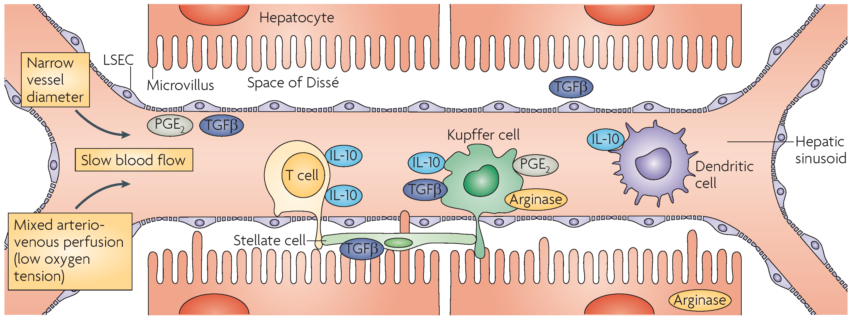
2. Immunobiology of the Liver

The liver is a lymphoid organ with important metabolic, biosynthetic, digestive, and detoxifying functions [11]. It receives blood from both the systemic circulation via the hepatic artery and the intestine via the portal vein (Figure 1) [11,12,13]. Its major focus is metabolic activity, where the products of digestion are processed and dangerous foreign chemicals are detoxified [11]. Since the liver is exposed continuously to viruses, bacteria, and gut flora, the hepatic environment establishes a balance between immunological tolerance to maintain homeostasis and hepatic immunity against invading pathogens. Hepatocytes and cholangiocytes (bile duct cells) are the main cell types that comprise ~80% of the all liver cells. Non-parenchymal cells in the liver include hepatic stellate cells (HSCs), liver sinusoidal endothelial cells (LSECs), Kupffer cells (KCs), and dendritic cells (DCs). The liver contains a diverse population of both innate and adaptive immune cells [14]. In the normal liver, natural killer (NK) and NK-T (NKT) cells, DCs, LSECs, and KCs present the antigen in the context of inhibitory cell surface ligands and immunosuppressive cytokines, thereby maintaining the liver’s tolerance to various antigens [11,13]. Pathogens such as hepatitis viruses (HBV and HCV) are able to subvert this immunity and establish persistent infection. NK and NKT cells present a first line of immune defense against invading pathogens. Circulating lymphocytes contact the antigen presenting cells (APCs) such as KCs and DCs to modulate immune responses and contribute to the antigen-specific tolerance of the liver [12].

2.1. Hepatocytes

Immunological functions of hepatocytes have been evaluated in a number of studies. The combination of allogeneic hepatocytes with T cells, resulted in their activation, followed by the apoptosis of T cells in vitro [15]. Similar results were obtained when CD8+ T cells from T-cell receptor transgenic mice were exposed to hepatocytes that expressed H2-Kb alloantigen [16]. Analogous studies in vivo using alloantigens also showed that hepatocytes induce T cell activation and apoptosis [17]. When a non-self-antigen was expressed from an adeno-associated viral (AAV) vector in hepatocytes, T-cell activation and cytotoxic effector functions were observed suggesting full functional differentiation of T cells [18]. CD8+ T cells can be activated while they are in the intravascular space to perform effector functions against hepatocyte-specific antigens [19], and interact with hepatocytes through fenestrations in LSECs [20].
Hepatocyte expression of major histocompatibility class (MHC) I antigens is generally considered very low, and they do not normally express MHC class II antigens. However, it was recently shown that hepatocytes express MHC class I molecules at levels comparable to that of splenocytes, and efficiently use transporters associated with antigen processing, tapasin (TPN), and low molecular weight polypeptide proteasome subunit components of antigen processing and presentation pathways [21]. They do not express MHC class I loading complex factors LMP7, LMP10, PA28-α, and PA38-β, while TAP1 and TAP2 are expressed in low abundance. In chronic liver diseases, however, both class I and class II antigens can be detected on hepatocytes. Similarly, hepatocytes express MHC class I complexes when exposed to IFN-γ and Listeria monocytogenes with high levels of stable class I trimers displayed on their cell surface [21]. Recently it was shown that the expression of MHC class I-related chain A (MICA) was highly elevated after 27 days of infection in human hepatocytes, suggesting that they can act as APCs [22]. Overexpression of class II transactivator molecule, a key transcriptional regulator of MHC class II gene expression, in hepatocytes resulted in the surface expression of MHC class II molecules and activation of CD4+ T cells [23]. In this case, MHC II-expressing hepatocytes were able to process antigens and stimulate Th1 or Th2 cell lines, and there was no sign of hepatic inflammation and autoimmune liver disease in transgenic mice expressing class II molecules. Collectively, these findings demonstrated that hepatocytes can also act as APCs.

2.2. Cholangiocytes

Even though cholangiocytes express many molecules that are often linked to APC function, there is no evidence to support the prospect that they activate T cells [24]. However, bile duct cells were shown to express MHC I and II, CD40, CD80, and CD86 molecules [25], and secrete CXCL16 that promote T-cell adhesion to epithelial cells [26].

2.3. Kupffer Cells

KCs are the resident macrophages of the liver. They are a unique cell type that are typically radio-resistant and difficult to isolate from tissue, even after collagenase digestion [24]. KCs induce immunotolerance under physiological conditions. For instance, they secrete immunosuppressive prostaglandin E2 (PGE2) under metabolic stress [27], and interleukin-10 (IL-10) when stimulated with lipopolysaccharide (LPS) [28]. They also express MHC classes I and II, and co-stimulatory molecules at low density [29]. Continuous exposure to endotoxin (LPS) limits the ability of KCs to activate T cells [29], and PGE2 released by KCs abrogates CD4+ T-cell activation [30]. In response to reactive oxygen species, however, KCs produce MHC class II molecules and act as APCs [31]. Thus, KCs are able to switch their immunological role from tolerance-inducing APCs to immunogenic APCs, and from inactivators to activators of NK cells when exposed to certain bacteria, such as Borrelia burgdorferi [24,32].

2.4. Dendritic Cells

The liver contains multiple populations of DCs, including plasmactyoid (pDC), myeloid (mDC), and lymphoid-derived (CD8α+) DCs. Both pDCs and mDCs are weak APCs because they are immature cells, whereas CD8α+ DCs are powerful APCs [24]. mDCs are characterized by their expression of CD11b and CD11c and lack CD8α and B220 expression. On the other hand, mouse liver pDCs are B220+ and express CD11c at lower levels than mDCs, whereas human pDCs lack CD11c but express blood DC antigen-2 (BDCA-2) [24]. A comparison of liver mDCs with skin-derived mDCs showed that liver cells secrete greater amounts of interleukins IL-10 and IL-4, whereas skin DCs were potent stimulators of interferon-γ (IFN-γ) and IL-4 [33]. Moreover, liver mDCs were found to be less effective at stimulating T-cell proliferation suggesting that hepatic mDCs predispose T cells towards tolerance [24]. Even though pDCs are not effective at stimulating T-cell activation [34], growth factors and Toll-like receptor (TLR) signaling can induce maturation of these cells into APCs and stimulate T cells [35].

2.5. Liver Sinusoidal Endothelial Cells

LSECs, which account for almost half of the non-parenchymal cells, induce immune tolerance via their expression of MHC I and II, as well as costimulatory molecules CD40, CD80, and CD86 [36]. They are able to eliminate viruses, colloids, and macromolecular waste from circulation through the expression of acetylated low density lipoprotein and mannosylated protein receptors [37]. Antigen presentation to T cells by LSECs via MHCs results in the up-regulation of specific molecules like the programmed death ligand 1 (PDL-1) which binds to its cognate receptor PD-1 causing T-cell tolerance [24]. However, the exposure to endotoxin reduces the ability of LSECs to activate antigen-specific CD4+ T cells [38]. In addition, IL-10 secreted by KCs can enhance the antigen presentation capacity of LSECs [30].

2.6. Hepatic Stellate Cells

HSCs, also termed Ito cells, reside in the Space of Dissé and regulate blood flow through the sinusoids [39]. In the murine liver, HSCs express CD1d, and low levels of CD11c and MHC class II molecules but not MHC class I [40]. However, a tiny population of CD11c-high cells do exist and they activate NKT cells and T cells in the presence of α-glactosyl ceramide [40]. Additional data from this study showed that HSCs presented antigenic peptides to CD8+ and CD4+ T cells and mediated cross-priming of CD8+ cells. Further, rat HSCs were reported to stimulate antigen-specific CD8+ responses [41], whereas human HSCs induced very little proliferation of CD8+ T cells [42]. Additional studies are needed to ascertain the function of HSCs as APCs.

3. Tumor Microenvironment

Carcinogenesis is a multistage process that appears to involve the transformation of a normal differentiated or stem-like cell into a preneoplastic lesion that develops into a malignant tumor [14]. Most of the gene mutations are somatic and occur as sporadic events; and some cancers could arise from epigenetic changes, such as DNA methylation and histone modification [43]. The tumor microenvironment is a complex interplay of T cells, B cells, macrophages, and hepatic cells resulting in the activation of multiple cellular signaling cascades [2].

3.1. Tumor-Infiltrating Lymphocytes

There is growing evidence to suggest that the gradual accrual of genetic changes and mutations in preneoplastic hepatocytes causes a cellular transformation resulting in the development of HCC [44]. Interactions of immune cells in the tumor microenvironment also play a critical role in cancer development, and the presence of intratumoral T and B cells, termed tumor infiltrating lymphocytes (TILs), correlates with improved clinical outcomes [45,46,47,48,49]. TILs in HCC include subpopulations of CD3+, CD4+, and CD8+ T cells [48]. The presence of CD8+ T cells in tumors is associated with better outcomes, whereas the presence of regulatory T cells (Tregs) is associated with poor prognosis [50]. Although antitumor effects of T cells seem to be impaired in HCC, the underlying mechanism(s) for this phenomenon is unclear [48]. Both in vitro and in vivo studies have shown that Tregs are involved in the deterioration of antitumor effects of T-lymphocytes. In HCC, TILs interact with a number of tumor-associated antigens (TAAs), such as glypican-3 (GPC3), α-fetoprotein (AFP), telomerase reverse transcriptase (TERT), synovial sarcoma X breakpoint 2 (SSX-2), melanoma antigen gene-A (MAGE-A), and New York esophageal squamous cell carcinoma 1 (NY-ESO-1) [51].

3.2. Tumor-Associated Macrophages

In the tumor microenvironment, tumor-associated macrophages (TAMs) are “polarized” into M2 mononuclear phagocyte-like cells by various cytokines (IL-4, IL-10, and transforming growth factor β (TGF-β)) [52]. These M2-like TAMs, in turn, express cytokines (IL-10 and TGF-β), chemokines (CCL17, CCL22, and CCL24), vascular endothelial growth factor (VEGF), and epidermal growth factor (EGF) to recruit Tregs and promote angiogenesis [53,54]. KCs are liver-specific TAMs, and are able to impair CD8+ cytotoxic T lymphocyte (CTL)-mediated immune responses through PD-L1, which interacts with programmed death 1 (PD-1), a cell surface protein of CD8+ T cells [55]. Moreover, when stimulated with pro-inflammatory cytokines (IL-1β, TNF-α, and PDGF), KCs and HSCs produce osteopontin, which plays a pivotal role in various cell signaling pathways, promotes inflammation, tumor progression, and metastasis [56,57]. Similar transitions in bile duct cells were shown to also result in the development of CCA [58].

3.3. Innate Immunity in the Liver

The innate immune system provides the first line of defense to limit infection in the liver by recognizing conserved structural motifs, termed pathogen-associated molecular patterns (PAMPs), on the surface of various pathogens. TLRs are key players in this process [34] and also recognize damage-associated molecular patterns (DAMPs) on dying host cells [59]. Although hepatocytes express all TLRs at low levels, they only respond to TLR2 and TLR4 ligands [60]. Under inflammatory conditions, however, hepatocyte response to TLR2 ligands is significantly enhanced but interestingly not so against TLR4 ligands [61]. LSECs express mRNAs of TLRs 1–9, and respond to various TLR ligands by producing TNF-α, IL-6, and IFN-β [62,63]. KCs express all TLRs and respond to a variety of TLR ligands by producing TNF-α and IL-6 [64,65]. KCs activated in response to TLR2 and TLR4 produced the immunosuppressive IL-10, and in turn, were able to suppress IL-18-dependent NK cell activation [60]. When stimulated with ligands for TLRs 1, 2, 4, and 6, KCs also produced IFN-γ and promoted the proliferation of T cells [62]. In response to TLR3 and TLR4 ligands, they secreted IFN-β, and with ligands against TLR1 and TLR8, displayed a high level of MHC class II expression. HSCs express low levels of TLR4 and TLR9, but activation of TLR4 has been shown to induce the expression of TLR2 [62]. In human HSCs, TLR4 activation resulted in the production of CCL2, CCL3, and CCL4 [66], and their expression of TGF-β was implicated in the promotion of hepatic fibrosis [67]. Activation of TLR9 by DAMPs induced the differentiation of HSCs and increased the production of collagen [68]. The potential of innate immune mechanisms must and can be harnessed to target infections, such as HBV and HCV that cause HCC [69].

4. Immunotherapeutic Approaches to Treat Liver Cancer

In the past decade, advances in our understanding of the immunobiology of the liver has led to the development of immunotherapeutic strategies, including vaccination, antibody-based treatments, adoptive cell therapy, immune checkpoint blockade, and cytokine targeting. Some of these have produced positive results and paved the way for their use in the clinical setting (Table 1).

4.1. Vaccines

Several vaccines targeting TAAs that were identified in HCC are being used for cancer therapy [51]. Among them, GPC-3, a member of the glypican family of heparin sulfate proteoglycans, was found to be overexpressed in HCC patients and was associated with poor prognosis [70]. In a phase I clinical trial, when GPC3298-306 peptide was used as a TAA in HLA-A*24-positive patients and GPC3144-152 peptide in HLA-A*02-positive patients, only one out of 33 patients manifested a partial response and all patients had marked intratumoral infiltration of CD8+ T cells [71]. In a phase II clinical trial with GPC-3 vaccine, recurrence rates were lower in 35 out of 64 patients that had undergone surgery or RFA prior to receiving the vaccine, and the GPC3 peptide vaccine improved one-year recurrence rate [72]. On the other hand, a similar phase II trial using hTERT peptide did not demonstrate any CTL activity [73].
Recently the development of an in situ cancer vaccine InCVAX for the treatment of HCC has been reported [74,75]. InCVAX works by stimulating robust antitumor immune reactions via a two-pronged approach. This includes a thermal treatment of tumors, such as with a laser, followed by administration of N-dihydro-galacto-chitosan (NDGC). Thermal treatment releases antigens and increases immunogenicity, while NDGC acts as a potent immune activator [74]. In that study, the strategy of local immunological cell death with DC activation with NDGC was found to be effective in a murine HCC model. Tumor infiltration of CD3+, CD4+, and CD8+ cells was observed after the injection of InCVAX, which indicated the in situ elimination of the tumor [74]. A vaccine containing the epitope of α-fetoprotein (AFP) linked with a heat shock protein 70 epitope also elicited anti-tumoral immunity by activating AFP-specific CD8+ T cells and was effective in reducing tumors in mice [76]. These vaccines need further improvement in determining therapeutic efficacy on tumor regression and recurrence.
In contrast to HCC, a limited number of protocols have been conducted to treat CCA. In one study, 36 patients with intrahepatic CCA were vaccinated with autologous tumor lysate pulsed DCs, together with the transfer of activated T cells [77]. A five-year progression-free survival and overall survival in these patients were substantially higher than in 26 patients who received curative resection alone (progression-free survival: 18.3 months vs. 7.7 months and overall survival: 31.9 months vs. 17.4 months). These findings suggested that a combination of DC vaccine and T-cell transfer might prevent recurrence and achieve long(er)-term survival in CCA patients [77].
Another potential candidate for immunization is the mucin protein 1 (MUC1), a glycoprotein that is overexpressed in 59%–77% of CCA, and its expression correlated with the patients′ overall survival rate [78,79,80]. In a non-randomized trial, DC vaccine targeting MUC1 showed a modest increase in the median survival time and had no adverse effects on patients, suggesting that it was safe for administration in humans [81]. However, further studies are needed to evaluate its effect, in particular in the early disease stage and in the absence of immunosuppressive therapy.

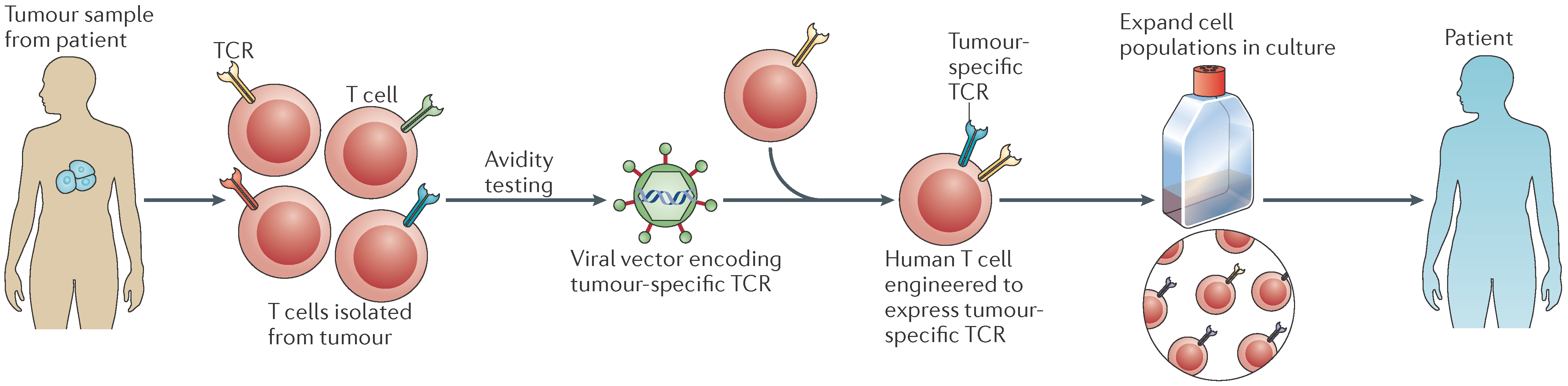
4.2. Adoptive Cell Therapy

Adoptive cell therapy (ACT) involves engineering patients′ own immune cells to recognize and destroy their tumors. In this approach, T cells from a healthy patient′s blood are collected and are genetically modified to express an artificial receptor consisting of the variable fragment of an antibody specific for a cell surface molecule involved in T cell signaling. Such engineered receptors (termed chimeric antigen receptors (CARs)) allow T cells to recognize specific antigens on tumor cells (Figure 2). CAR-T cells are then propagated in cell culture until they reach several billions in number, and injected back into the patient′s blood stream. After infusion, CAR-T cells are designed to recognize tumor cells and kill them. CAR-T cells have advantages over T-cell receptor-modified T (TCR-T) cells in that they recognize tumor cells without MHC restrictions [82], which will allow improved patient targeting and overcoming tumor escape mechanisms of MHC loss. This ACT approach has shown promise in recent clinical trials of patients with acute lymphoblastic leukemia [83], acute myeloid leukemia [84,85], and gastrointestinal cancer [86]. A clear distinction between autologous transfer using endogenous T cells and an allogeneic approach should be noted. The former approach involves modified T cell receptors that recognize peptides with HLA restrictions, whereas in the latter there is no HLA restriction and it involves the transfer of cells from an immunized individual to a non-immune recipient. This difference is critical for assessing the extent of toxicities associated with off-tumor and on-target effects.
To date, very few studies have been conducted on the application of CAR-T cells to HCC and CCA immunotherapy. In one study, T cells expressing CAR targeted to GPC-3 have successfully eliminated GPC3-positive cells in vitro and in orthotopic Huh-7 xenografts in mice [87]. However, substantial risks and toxicities are associated with the use of CAR-T cells, as they release massive amounts of cytokines into the patient’s blood stream. These include, but are not limited, to cytokine release syndrome, B-cell aplasia, and tumor lysis syndrome [85]. Clinical studies of solid tumors using CAR-T cells have also shown considerable toxicity towards normal tissues [88,89]. In this regard, studying the efficacy and safety of CAR-T cells in animal models is necessary for the validation of CAR constructs under therapeutic settings in the treatment of solid tumors [82]. Distinguishing therapeutic efficacy from off-tumor toxicity would be of the utmost importance for ACT, as are further modifications of CAR-T cells, identifying better targets, and improving preconditioning regimens.
Cytokine-induced cells, that include activated T cells and NK cells, have shown clinical benefits in adoptive immunotherapy [90,91,92]. Randomized clinical trials of adjuvant immunotherapy using cytokine-induced killer (CIK) cells showed mixed results for HCC. In one study, no improvement in patient survival was observed even though there was an increase in the recurrence-free survival [93]. Others showed that lymphocyte infusions (CD3+, CD3+/HLA-DR+, CD4+, and CD8+) lowered recurrence and improved recurrence-free outcomes after surgery in HCC patients [94]; CIK therapy improved overall survival in HCC patients [95]; and infusion of CD3+/CD56+ and CD3+/CD56 T cells, as well as CD3/CD56+ NKT cells increased recurrence-free and overall survival in HCC patients who underwent surgical resection or RFA or percutaneous ethanol injection [96]. Two other studies showed that CIK therapy also reduced 1-year recurrence rates in patients who received a combination treatment with RFA and TACE [97], and in those HCC patients unsuitable for surgery [98]. Collectively, these clinical trials suggested that CIK therapy might be more suitable for patients who have early stage HCC that those with advanced disease.
There have been a limited number of studies conducted with CIK cells for immunotherapy. In one study, human CIK cells comprising of CD3+ T cells and CD3+/CD56+ T cells were able to reduce the growth of inoculated CCA cells in SCID mice [100]. In another study, CIK cells expressing inducible co-stimulator had profound cytotoxic effects on CCA cells in vitro and in vivo [101]. CIK cells in combination with cetuximab, an antibody that targets epidermal growth factor receptor, significantly enhanced cytotoxicity of human CCA cells in vitro than when used alone [102]. Further studies are needed to evaluate the clinical use of CIK cells for the treatment of CCA.
Another interesting line of research is the combination of peptide vaccine together with ACT. A case report using a personalized peptide-vaccine that elicited strong immune response was recently shown to be effective in a patient with metastatic CCA [103]. Immunotherapy using peptide vaccines together with adoptive transfer of T cells is also emerging as an exciting option [86,104].

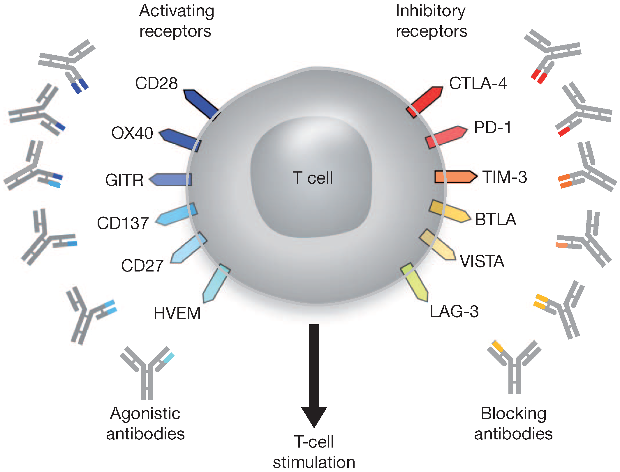
4.3. Immune Checkpoint Blockade

In recent years, a blockade of checkpoint molecules that block immune responses against tumor cells, such as PD-1, PD-L1 and CTLA-4, has emerged as a novel therapeutic approach in oncology [105]. Antibodies that target these molecules have been developed, and they have shown significant efficacy (Figure 3). Among these, pembrolizumab and nivolumab target PD-1, and tremelimumab and ipilimumab target CTLA-4 [106]. Pembrolizumab has been approved by the Food and Drug Administration (FDA) in the United States for the treatment of metastatic melanoma (MM) and non-small cell lung cancer (NSCLC); nivolumab for MM, NSCLC and renal cell carcinoma, and ipilimumab for MM, either as single agents or in combination [107]. These were also approved by the European Medicines Agency [108].

4.3.1. Programmed Death-1

PD-1, a negative co-stimulatory molecule of the CD28 immunoglobulin superfamily of transmembrane receptors, is a strong inhibitor of T cell response. Therefore, blocking its function is a strategy for immunotherapy. PD-L1 and PD-L2, members of the B7 costimulatory molecule family, are ligands for PD-1. Engagement of PD-L1 and PD-L2 with PD-1 inhibits T cell receptor-mediated lymphocyte proliferation and cytokine production by CD4+ T cells [110,111]. A clinical trial using tremelimumab in 17 HCC patients with liver cirrhosis and HCV showed that three patients developed a partial response with a median survival of 8.2 months, and HCV-specific T cell responses did not correlate with tumor regression [112]. Therefore, TAA-specific CD8+ T cell responses could reduce the recurrence of HCC suggesting that immunotherapy to induce TAA-specific CTLs by such means as peptide vaccines might be an effective clinical application in HCC patients after local therapy [113]. In another randomized trial, a single dose of the antibody nivolumab (BMS-936558) was administered to patients with chronic HCV infection. There were no significant side effects, and one-third of the recipients had reduced viral loads [114]. Blocking PD-1 function might also improve disease outcomes in CCA as shown recently [115]. Patients with high levels of soluble PD-L1 had worse overall survival than those with low levels of the ligand. In another study, PD-L1 and MHC class I expression were elevated in eight and 11 out of 27 intrahepatic CCA tumors samples, respectively; whereas all tumor samples had infiltration of CD8+ T cells [116]. Results from this study also showed that the defects in MHC class I antigen expression and high PD-L1 expression by tumor cells could potentially provide them with an immune escape mechanism. Therefore, immunotherapy with antibodies against PD-1 in patients with normal MHC class I expression might be an effective strategy to treat intrahepatic CCA [116].

4.3.2. CD160

CD160, a negative co-stimulatory molecule, was found to be associated with T cell exhaustion in patients with chronic HCV infection where it was overexpressed on HCV-specific CD8+ T cells in the peripheral blood [117]. However, it is not overexpressed in intrahepatic HCV-specific CD8+ T cells [118]. Additional studies are needed to understand the importance of CD160/CD160L blockade.

4.3.3. Natural Killer Cell Receptor 2B4

NK cell receptor 2B4 was also found to be overexpressed in the blood of patients with chronic HCV [117]. Its ligand CD48 has a 6 to 8-fold greater affinity to 2B4 than CD2, a molecule implicated in the regulation of NK and T cell activation [119]. Expression of 2B4 on HCV- specific CD8+ T cells also correlated with the overexpression of PD-1 [117], which was higher in hepatic cells than in blood cells [118]. Collectively, these findings indicated that the up-regulation of 2B4 is potentially linked to PD-1 expression.

4.3.4. Lymphocyte Activation Gene-3

LAG-3 was another receptor overexpressed on HCV-specific CD8+ cells, where it negatively regulated the function of these cells in chronic HCV patients [120]. Blockade of LAG-3 restored T cell effector functions in this cohort. Similar results were obtained in patients with HBV-related HCC [121].

4.3.5. T-Cell Immunoglobulin and Mucin-Domain Containing-3

Tim-3 was identified as a negative regulator of T-helper type 1 immunity through the binding of its ligand galectin-9 [122]. It is up-regulated in CD8+ cells of patients with chronic HCV, and its blockade restored the effector function of these cells [123]. Thus, Tim-3 is a prime candidate for immunotherapy.

4.3.6. Glycoprotein-2

In patients with PSC, antibodies against GP-2 were found to be elevated, and was associated with poor patient survival [124]. High levels of anti-GP2 IgA also correlated with the development of CCA in patients with PSC in this study. Therefore, anti-GP2 IgA might be a valuable tool for risk stratification in patients with PSC.

4.4. Antiplatelet Therapy

Platelets (also known as thrombocytes) are small enucleated cells that are present in the bloodstream. When vascular injury and damage occurs, platelets are activated resulting the secretion of growth factors such as hepatocyte growth factor (HGF), fibroblast growth factor (FGF), insulin-like growth factor (IGF)-1, VEGF, EGF, and platelet-derived growth factor (PDGF), during a process called thrombosis [125]. Platelets improve liver fibrosis by inactivating HSCs to decrease collagen production and accelerate liver regeneration [126]. Thrombocytosis occurs when platelet numbers are elevated in the blood, and a strong association has been found between HCC and thrombocytosis. In a retrospective study of 1154 HCC patients, it was found that patients with thrombocytosis had higher AFP levels, large tumor volume and high platelet count with short survival rates [127]. A recent study has shown that high platelet count could be a reliable marker for extrahepatic metastasis in early stage HCC following curative treatment [128]. High platelet levels were also reported more recently in CCA [129]. On the other hand, thrombocytopenia, a condition with low platelet count, was also shown to be associated with HCC in a cirrhotic background [130], and correlated with a reduced overall survival in patients with HCC [131]. Collectively, these results demonstrated that platelets play an important role in predicting the overall survival of patients with liver cancer.
Within the liver sinusoids, circulating CD8+ T cells induce the arrest of platelet aggregates that are bound to sinusoidal hyaluronan via CD44 [19]. In the mouse model of HBV, CD8+ T cells with effector functions control hepatotropic pathogens by crawling along liver sinusoids. Activated platelets mediate CTL-induced liver damage in mouse model of acute viral hepatitis [132]. Administration of aspirin and clopidogrel (Asp/Clo) that block platelet activation prevents HCC and improves survival in a mouse model of chronic HBV infection [133]. In this study, the authors also observed a significant reduction in the progression of liver fibrosis. Similarly, HBV-associated HCC patients who received aspirin and clopidogrel had a better recurrence-free survival and OS than those without the antiplatelet therapy [134]. The mechanism by which platelets interact with T cells is unknown, and antiplatelet therapy could diminish the release of TGF-β as platelets are the main repository for this profibrogenic cytokine [135]. Due to the protective role of platelets against various other infections, such as malaria [136], blocking platelet activation could be risky as antiplatelet therapy could increase the bleeding risk in patients with impaired liver function [137]. Further studies are needed to assess the impact of antiplatelet drugs on liver inflammation and HCC.

5. Future Directions

The incidence of liver cancer is rapidly increasing, and the annual health care costs of HCC treatment are skyrocketing [138]. In the United States, the overall median cost to care for a patient with HCV-associated HCC was recently estimated to be almost $180,000/year [139]. Therefore, there is a substantial burden of medical expense of illness associated with liver cancer, and a greater need exists for the development of novel therapies. A number of clinical trials are currently underway for both HCC [140] and CCA [141], which could produce very effective immunotherapies.
A critical avenue to pursue further is the identification of new targets, which requires extensive validation across species, although caution must be exercised in their discovery. For instance, mesothelin was reported as a potential immunotherapeutic target in various cancers. When HCC and CCA specimens were investigated for the expression of mesothelin, none was found in HCC specimens, but one-third of CCA tissues overexpressed mesothelin, as did three distinct CCA cell lines [142]. Addition of sulfatase-1 that targets mesothelin showed very high and specific growth inhibition of these cell lines, suggesting that it could be a potential therapeutic agent for CCA [142]. Even though this finding suggested that sulfatase-1 could act as a tumor suppressor in HCC, caution should be exercised in interpreting it as no mesothelin expression was detected in human HCC specimen.
TGN1412 was developed in the late 1990s as an antagonistic monoclonal antibody against CD28, a key co-stimulator for T-cell responses [143]. Experimental ground work conducted in rats and mice using this antibody has revealed significant promise to treat autoimmune and inflammatory diseases. However, when a single dose of TGN1412 was administered to six healthy volunteers, they all experienced severe cytokine release syndrome with multi-organ failure [144]. The study results demonstrated the potential danger involved in extrapolation of results from animal studies to humans in preclinical studies. Similarly, extrapolation of data from one species to another may not be feasible in case of CAR-T cells, particularly in HLA-restricted peptides, due to the differences between species.
Recent advances in immunotherapy have paved the way towards the goal of not only treating tumor recurrence, but also preventing the development of cancer in cirrhotic livers. Immune tolerance of the liver is still a major problem. However, efforts are being made to circumvent this issue by using immune checkpoint inhibitors. Preclinical studies and clinical trials have demonstrated promising results in this direction. At present, it appears that a combination therapy of vaccines and/or immune checkpoint inhibitors with local ablative therapies is an attractive approach in treating liver cancer. However, safety, toxicity, and efficacy in clinical trials must be addressed before bringing these immunotherapeutic interventions to the bedside.

Acknowledgments

The authors acknowledge the financial support of the Institute of Engineering in Medicine, University of Minnesota, Minneapolis, MN, USA.

Author Contributions

Rajagopal N. Aravalli wrote the initial draft of the review; and Clifford J. Steer revised, commented upon and edited at least 5 drafts of the manuscript with Rajagopal N. Aravalli.

Conflicts of Interest

The authors declare no conflict of interest.

Abbreviations

The following abbreviations are used in this manuscript:
AFPα-fetoprotein
APCAntigen-presenting cell
BDCA-2Blood dendritic cell antigen-2
CARChimeric antigen receptor
CCACholangiocarcinoma
CIKCytokine-induced killer cell
CTLCytotoxic T-lymphocyte
DCDendritic cell
GPC3Glypican-3
GP-2Glycoprotein-2
HBVHepatitis B virus
HCCHepatocellular carcinoma
HCVHepatitis C virus
HSCHepatic stellate cell
IFN-γInterferon-γ
IL-10Interleukin-10
KCKupffer cell
LAG-3Lymphocyte activation gene-3
LPSLipopolysaccharide
LSECLiver sinusoidal endothelial cell
MAGE-AMelanoma antigen gene-A
MHCMajor histocompatibility complex
NAFLDNon-alcoholic fatty liver disease
NKNatural killer
NY-ESO-1New York esophageal squamous cell carcinoma 1
NKTNK T cell
PD-1Programmed death 1
PD-L1Programmed death ligand 1
PGE2Prostaglandin E2
PSCPrimary sclerosing cholangitis
RFARadiofrequency ablation
SSX-2Synovial sarcoma X breakpoint 2
TAATumor-associated antigen
TAMTumor-associated macrophages
TAP-1Tapasin-1
TCRT-cell receptor
TERTTelomerase reverse transcriptase
TGF-βTransforming growth factor-β
TILTumor-infiltrating lymphocyte
Tim-3T-cell immunoglobulin and mucin-domain containing-3
TLRToll-like receptor
TregRegulatory T-cell

References

  1. Nordenstedt, H.; White, D.L.; El-Serag, H.B. The changing pattern of epidemiology in hepatocellular carcinoma. Dig. Liver Dis. 2010, 42, S206–S214. [Google Scholar] [CrossRef]
  2. Aravalli, R.N.; Steer, C.J.; Cressman, E.N. Molecular mechanisms of hepatocellular carcinoma. Hepatology 2008, 48, 1049–1053. [Google Scholar] [CrossRef] [PubMed]
  3. Lazaridis, K.N.; Gores, G.J. Cholangiocarcinoma. Gastroenterology 2005, 128, 1655–1667. [Google Scholar] [CrossRef] [PubMed]
  4. Bergquist, A.; Von Seth, E. Epidemiology of cholangiocarcinoma. Best Pract. Res. Clin. Gastroenterol. 2015, 29, 221–232. [Google Scholar] [CrossRef] [PubMed]
  5. Shin, H.R.; Oh, J.K.; Masuyer, E.; Curado, M.P.; Bouvard, V.; Fang, Y.Y.; Wiangnon, S.; Sripa, B.; Hong, S.T. Epidemiology of cholangiocarcinoma: An update focusing on risk factors. Cancer Sci. 2010, 101, 579–585. [Google Scholar] [CrossRef] [PubMed]
  6. De Martel, C.; Plummer, M.; Franceschi, S. Cholangiocarcinoma: Descriptive epidemiology and risk factors. Gastroenterol. Clin. Biol. 2010, 34, 173–180. [Google Scholar] [CrossRef]
  7. Shaib, Y.; El-Serag, H.B. The epidemiology of cholangiocarcinoma. Semin. Liver Dis. 2004, 24, 115–125. [Google Scholar] [CrossRef] [PubMed]
  8. Eckmann, K.R.; Patel, D.K.; Landgraf, A.; Slade, J.H.; Lin, E.; Kaur, H.; Loyer, E.; Weatherly, J.M.; Javie, M. Chemotherapy outcomes for the treatment of unresectable intrahepatic and hilar cholangiocarcinoma: A retrospective analysis. Gastrointest. Cancer Res. 2011, 4, 155–160. [Google Scholar] [PubMed]
  9. Mancuso, A.; Perricone, G. Hepatocellular carcinoma and liver transplantation: State of the art. J. Clin. Transl. Hepatol. 2014, 2, 175–181. [Google Scholar]
  10. Sapisochín, G.; Fernández De Sevilla, E.; Echeverri, J.; Charco, R. Liver transplantation for cholangiocarcinoma: Current status and new insights. World J. Hepatol. 2015, 7, 2396–2403. [Google Scholar] [CrossRef]
  11. Crispe, I.N. The liver as a lymphoid organ. Ann. Rev. Immunol. 2009, 27, 147–163. [Google Scholar] [CrossRef] [PubMed]
  12. Racanelli, V.; Rehermann, B. The liver as an immunological organ. Hepatology 2006, 43, S54–S62. [Google Scholar] [CrossRef] [PubMed]
  13. Thomson, A.W.; Knolle, P.A. Antigen-presenting cell function in the tolerogenic liver environment. Nat. Rev. Immunol. 2010, 10, 753–766. [Google Scholar] [CrossRef] [PubMed]
  14. Severi, T.; Van Malenstein, H.; Verslype, C.; Van Pelt, J.F. Tumor initiation and progression in hepatocellular carcinoma: Risk factors, classification, and therapeutic targets. Acta Pharmacol. Sin. 2010, 31, 1409–1420. [Google Scholar] [CrossRef] [PubMed]
  15. Qian, S.; Wang, Z.; Lee, Y.; Chiang, Y.; Bonham, C.; Fung, J.; Lu, L. Hepatocyte-induced apoptosis of activated T cells, a mechanism of liver transplant tolerance, is related to the expression of ICAM-1 and hepatic lectin. Transp. Proc. 2001, 33, 226. [Google Scholar] [CrossRef]
  16. Bertolino, P.; Trescol-Biemont, M.C.; Rabourdin-Combe, C. Hepatocytes induce functional activation of naive CD8+ T lymphocytes but fail to promote survival. Eur. J. Immunol. 1998, 28, 221–236. [Google Scholar] [CrossRef]
  17. Bertolino, P.; McCaughan, G.W.; Bowen, D.G. Role of primary intrahepatic T-cell activation in the “liver tolerance effect”. Immunol. Cell Biol. 2002, 80, 84–92. [Google Scholar] [CrossRef] [PubMed]
  18. Wuensch, S.A.; Spahn, J.; Crispe, I.N. Direct, help-independent priming of CD8+ T cells by adeno-associated virus-transduced hepatocytes. Hepatology 2010, 52, 1068–1077. [Google Scholar] [CrossRef] [PubMed]
  19. Guidotti, L.G.; Inverso, D.; Sironi, L.; Di Lucia, P.; Fioravanti, J.; Ganzer, L.; Fiocchi, A.; Vacca, M.; Aiolfi, R.; Sammicheli, S.; et al. Immunosurveillance of the liver by intravascular effector CD8+ T cells. Cell 2015, 161, 489–500. [Google Scholar] [CrossRef] [PubMed]
  20. Warren, A.; Le Couteur, D.G.; Fraser, R.; Bowen, D.G.; McCaughan, G.W.; Bertolino, P. T lymphocytes interact with hepatocytes through fenestrations in murine liver sinusoidal endothelial cells. Hepatology 2006, 44, 1182–1190. [Google Scholar] [CrossRef] [PubMed]
  21. Chen, M.; Tabaczewski, P.; Truscott, S.M.; Van Kaer, L.; Stroynowski, I. Hepatocytes express abundant surface class I MHC and efficiently use transporter associated with antigen processing, tapasin, and low molecular weight polypeptide proteasome subunit components of antigen processing and presentation pathway. J. Immunol. 2005, 175, 1047–1055. [Google Scholar] [CrossRef] [PubMed]
  22. Sasaki, R.; Kanda, T.; Wu, S.; Nakamoto, S.; Haga, Y.; Jiang, X.; Nakamura, M.; Shirasawa, H.; Yokosuka, O. Association between hepatitis B virus and MHC class I polypeptide-related chain A in human hepatocytes derived from human-mouse chimeric mouse liver. Biochem. Biophys. Res. Commun. 2015, 464, 1192–1195. [Google Scholar] [CrossRef] [PubMed]
  23. Herkel, J.; Jagemann, B.; Wiegard, C.; Lazaro, J.F.; Lueth, S.; Kanzler, S.; Blessing, M.; Schmitt, E.; Lohse, A.W. MHC class II-expressing hepatocytes function as antigen-presenting cells and activate specific CD4 T lymphocytes. Hepatology 2003, 37, 1079–1085. [Google Scholar] [CrossRef] [PubMed]
  24. Crispe, I.N. Liver antigen-presenting cells. J. Hepatol. 2011, 54, 357–365. [Google Scholar] [CrossRef] [PubMed]
  25. Barnes, B.H.; Tucker, R.M.; Wehrmann, F.; Mack, D.G.; Ueno, Y.; Mack, C.L. Cholangiocytes as immune modulators in rotavirus-induced murine biliary atresia. Liver Int. 2009, 29, 1253–1261. [Google Scholar] [CrossRef] [PubMed]
  26. Heydtmann, M.; Lalor, P.F.; Eksteen, J.A.; Hübscher, S.G.; Briskin, M.; Adams, D.H. CXC chemokine ligand 16 promotes integrin-mediated adhesion of liver-infiltrating lymphocytes to cholangiocytes and hepatocytes within the inflamed human liver. J. Immunol. 2005, 174, 1055–1062. [Google Scholar] [CrossRef] [PubMed]
  27. Callery, M.P.; Mangino, M.J.; Flye, M.W. Arginine-specific suppression of mixed lymphocyte culture reactivity by Kupffer cells—A basis of portal venous tolerance. Transplantation 1991, 51, 1076–1080. [Google Scholar] [CrossRef] [PubMed]
  28. Knolle, P.; Schlaak, J.; Uhrig, A.; Kempf, P.; Meyer zum Büschenfelde, K.H.; Gerken, G. Human Kupffer cells secrete IL-10 in response to lipopolysaccharide (LPS) challenge. J. Hepatol. 1995, 22, 226–229. [Google Scholar] [CrossRef]
  29. You, Q.; Cheng, L.; Kedl, R.M.; Ju, C. Mechanism of T cell tolerance induction by murine hepatic Kupffer cells. Hepatology 2008, 48, 978–990. [Google Scholar] [CrossRef] [PubMed]
  30. Knolle, P.A.; Gerken, G. Local control of the immune response in the liver. Immunol. Rev. 2000, 174, 21–34. [Google Scholar] [CrossRef] [PubMed]
  31. Maemura, K.; Zheng, Q.; Wada, T.; Ozaki, M.; Takao, S.; Aikou, T.; Bulkley, G.B.; Klein, A.S.; Sun, Z. Reactive oxygen species are essential mediators in antigen presentation by Kupffer cells. Immunol. Cell Biol. 2005, 83, 336–343. [Google Scholar] [CrossRef] [PubMed]
  32. Lee, W.-Y.; Moriarty, T.J.; Wong, C.H.Y.; Zhou, H.; Strieter, R.M.; Van Rooijen, N.; Chaconas, G.; Kubes, P. An intravascular immune response to Borrelia burgdorferi involves Kupffer cells and iNKT cells. Nat. Immunol. 2010, 11, 295–302. [Google Scholar] [CrossRef] [PubMed]
  33. Goddard, S.; Youster, J.; Morgan, E.; Adams, D.H. Interleukin-10 secretion differentiates dendritic cells from human liver and skin. Am. J. Pathol. 2004, 164, 511–519. [Google Scholar] [CrossRef]
  34. Tokita, D.; Sumpter, T.L.; Raimondi, G.; Zahorchak, A.F.; Wang, Z.; Nakao, A.; Mazariegos, G.V.; Abe, M.; Thomson, A.W. Poor allostimulatory function of liver plasmacytoid DC is associated with pro-apoptotic activity, dependent on regulatory T cells. J. Hepatol. 2008, 49, 1008–1018. [Google Scholar]
  35. Kingham, T.P.; Chaudhry, U.I.; Plitas, G.; Katz, S.C.; Raab, J.; DeMatteo, R.P. Murine liver plasmacytoid dendritic cells become potent immunostimulatory cells after Flt-3 ligand expansion. Hepatology 2007, 45, 445–454. [Google Scholar] [CrossRef] [PubMed]
  36. Connolly, M.K.; Bedrosian, A.S.; Malhotra, A.; Henning, J.R.; Ibrahim, J.; Vera, V.; Cieza-Rubio, N.E.; Hassan, B.U.; Pachter, H.L.; Cohen, S.; et al. In hepatic fibrosis, liver sinusoidal endothelial cells acquire enhanced immunogenicity. J. Immunol. 2010, 185, 2200–2208. [Google Scholar] [CrossRef] [PubMed]
  37. Elvevold, K.; Smedsrød, B.; Martinez, I. The liver sinusoidal endothelial cell: A cell type of controversial and confusing identity. Am. J. Physiol. Gastrointest. Liver Physiol. 2008, 294, G391–G400. [Google Scholar] [CrossRef] [PubMed]
  38. Knolle, P.A.; Germann, T.; Treichel, U.; Uhrig, A.; Schmitt, E.; Hegenbarth, S.; Lohse, A.W.; Gerken, G. Endotoxin down-regulates T cell activation by antigen-presenting liver sinusoidal endothelial cells. J. Immunol. 1999, 162, 1401–1407. [Google Scholar]
  39. Friedman, S.L. Hepatic stellate cells: Protean, multifunctional, and enigmatic cells of the liver. Physiol. Rev. 2008, 88, 125–172. [Google Scholar] [CrossRef] [PubMed]
  40. Winau, F.; Hegasy, G.; Weiskirchen, R.; Weber, S.; Cassan, C.; Sieling, P.A.; Modlin, R.L.; Liblau, R.S.; Gressner, A.M.; Kaufmann, S.H. Ito cells are liver-resident antigen-presenting cells for activating T cell responses. Immunity 2007, 26, 117–129. [Google Scholar] [CrossRef] [PubMed]
  41. Bomble, M.; Tacke, F.; Rink, L.; Kovalenko, E.; Weiskirchen, R. Analysis of antigen-presenting functionality of cultured rat hepatic stellate cells and transdifferentiated myofibroblasts. Biochem. Biophys. Res. Commun. 2010, 396, 342–347. [Google Scholar] [CrossRef] [PubMed]
  42. Eksteen, B.; Mora, J.R.; Haughton, E.L.; Henderson, N.C.; Lee-Turner, L.; Villablanca, E.J.; Curbishley, S.M.; Aspinall, A.I.; Von Andrian, U.H.; Adams, D.H. Gut homing receptors on CD8 T cells are retinoic acid dependent and not maintained by liver dendritic or stellate cells. Gastroenterology 2009, 137, 320–329. [Google Scholar] [CrossRef] [PubMed] [Green Version]
  43. Esteller, M. Epigenetics in cancer. N. Engl. J. Med. 2008, 358, 1148–1159. [Google Scholar] [CrossRef] [PubMed]
  44. Farazi, P.A.; DePinho, R.A. Hepatocellular carcinoma pathogenesis: From genes to environment. Nat. Rev. Cancer 2006, 6, 674–687. [Google Scholar] [CrossRef] [PubMed]
  45. Zhang, L.; Conejo-Garcia, J.R.; Katsaros, D.; Gimotty, P.A.; Massobrio, M.; Regnani, G.; Makrigiannakis, A.; Gray, H.; Schlienger, K.; Liebman, M.N.; et al. Intratumoral T cells, recurrence, and survival in epithelial ovarian cancer. N. Engl. J. Med. 2003, 348, 203–213. [Google Scholar] [CrossRef] [PubMed]
  46. Galon, J.; Costes, A.; Sanchez-Cabo, F.; Kirilovsky, A.; Mlecnik, B.; Lagorce-Pagès, C.; Tosolini, M.; Camus, M.; Berger, A.; Wind, P.; et al. Type, density, and location of immune cells within human colorectal tumors predict clinical outcome. Science 2006, 313, 1960–1964. [Google Scholar] [CrossRef] [PubMed]
  47. Criscitiello, C.; Esposito, A.; Trapani, D.; Curigliano, G. Prognostic and predictive value of tumor infiltrating lymphocytes in early breast cancer. Cancer Treat. Rev. 2016, 50, 205–207. [Google Scholar] [CrossRef] [PubMed]
  48. Shirabe, K.; Motomura, T.; Muto, J.; Toshima, T.; Matono, R.; Mano, Y.; Takeishi, K.; Ijichi, H.; Harada, N.; Uchiyama, H.; et al. Tumor-infiltrating lymphocytes and hepatocellular carcinoma: Pathology and clinical management. Int. J. Clin. Oncol. 2010, 15, 552–558. [Google Scholar] [CrossRef] [PubMed]
  49. Knief, J.; Reddemann, K.; Petrova, E.; Herhahn, T.; Wellner, U.; Thorns, C. High density of tumor-infiltrating B-lymphocytes and plasma cells signifies prolonged overall survival in adenocarcinoma of the esophagogastric junction. Anticancer Res. 2016, 36, 5339–5345. [Google Scholar] [CrossRef] [PubMed]
  50. Ormandy, L.A.; Hillemann, T.; Wedemeyer, H.; Manns, M.P.; Greten, T.F.; Korangy, F. Increased populations of regulatory T cells in peripheral blood of patients with hepatocellular carcinoma. Cancer Res. 2005, 65, 2457–2464. [Google Scholar] [CrossRef] [PubMed]
  51. Breous, E.; Thimme, R. Potential of immunotherapy for hepatocellular carcinoma. J. Immunol. 2011, 54, 830–834. [Google Scholar] [CrossRef] [PubMed]
  52. Mantovani, A.; Sica, A.; Allavena, P.; Garlanda, C.; Locati, M. Tumor-associated macrophages and the related myeloid-derived suppressor cells as a paradigm of the diversity of macrophage activation. Hum. Immunol. 2009, 70, 325–330. [Google Scholar] [CrossRef] [PubMed]
  53. Mantovani, A.; Germano, G.; Marchesi, F.; Locatelli, M.; Biswas, S.K. Cancer-promoting tumor-associated macrophages: New vistas and open questions. Eur. J. Immunol. 2011, 41, 2522–2525. [Google Scholar] [CrossRef] [PubMed]
  54. Movahedi, K.; Laoui, D.; Gysemans, C.; Baeten, M.; Stangé, G.; Van den Bossche, J.; Mack, M.; Pipeleers, D.; In’t Veld, P.; De Baetselier, P.; et al. Different tumor microenvironments contain functionally distinct subsets of macrophages derived from Ly6C (high) monocytes. Cancer Res. 2010, 71, 5728–5739. [Google Scholar] [CrossRef] [PubMed]
  55. Wu, K.; Kryczek, I.; Chen, L.; Zou, W.; Welling, T.H. Kupffer cell suppression of CD8+ T cells in human hepatocellular carcinoma is mediated by B7-H1/programmed death-1 interactions. Cancer Res. 2009, 69, 8067–8075. [Google Scholar] [CrossRef] [PubMed]
  56. Leonardi, G.C.; Candido, S.; Cervello, M.; Nicolosi, D.; Raiti, F.; Travali, S.; Spandidos, D.A.; Libra, M. The tumor microenvironment in hepatocellular carcinoma (review). Int. J. Oncol. 2012, 40, 1733–1747. [Google Scholar] [PubMed]
  57. Ramaiah, S.K.; Rittling, S. Pathophysiological role of osteopontin in hepatic inflammation, toxicity, and cancer. Toxicol. Sci. 2008, 103, 4–13. [Google Scholar] [CrossRef] [PubMed]
  58. Maroni, L.; Pierantenelli, I.; Banales, J.M.; Benedetti, A.; Marzioni, M. The significance of genetics for cholangiocarcinoma development. Ann. Transl. Med. 2013, 1, 3. [Google Scholar]
  59. Szabo, G.; Dolganiuc, A.; Mandrekar, P. Pattern recognition receptors: A contemporary view on liver diseases. Hepatology 2006, 44, 287–298. [Google Scholar] [CrossRef] [PubMed]
  60. Liu, S.; Gallo, D.J.; Green, A.M.; Williams, D.L.; Gong, X.; Shapiro, R.A.; Gambotto, A.A.; Humphris, E.L.; Vodovotz, Y.; Billiar, T.R. Role of Toll-like receptors in changes in gene expression and NF-κB activation in mouse hepatocytes stimulated with lipopolysaccharide. Infect. Immun. 2002, 70, 3433–3442. [Google Scholar] [CrossRef] [PubMed]
  61. Matsumura, T.; Ito, A.; Takii, T.; Hayashi, H.; Onozaki, K. Endotoxin and cytokine regulation of Toll-like receptor (TLR) 2 and TLR4 gene expression in murine liver and hepatocytes. J. Interferon Cytokine Res. 2000, 201, 915–921. [Google Scholar] [CrossRef] [PubMed]
  62. Wu, J.; Meng, Z.; Jiang, M.; Zhang, E.; Trippler, M.; Broering, R.; Bucchi, A.; Krux, F.; Dittmer, U.; Yang, D.; et al. Toll-like receptor-induced innate immune responses in non-parenchymal liver cells are cell type-specific. Immunology 2010, 129, 363–374. [Google Scholar] [CrossRef] [PubMed]
  63. Martin-Armas, M.; Simon-Santamaria, J.; Pettersen, I.; Moens, U.; Smedsrød, B.; Sveinbjørnsson, B. Toll-like receptor 9 (TLR9) is present in murine liver sinusoidal endothelial cells (LSECs) and mediates the effect of CpG-oligonucleotides. J. Hepatol. 2006, 44, 939–946. [Google Scholar] [CrossRef] [PubMed] [Green Version]
  64. Su, G.L.; Klein, R.D.; Aminlari, A.; Zhang, H.Y.; Steinstraesser, L.; Alarcon, W.H.; Remick, D.G.; Wang, S.C. Kupffer cell activation by lipopolysaccharide in rats: Role for lipopolysaccharide binding protein and Toll-like receptor 4. Hepatology 2000, 31, 932–936. [Google Scholar] [CrossRef] [PubMed]
  65. Thobe, B.M.; Frink, M.; Hildebrand, F.; Schwacha, M.G.; Hubbard, W.J.; Choudhry, M.A.; Chaudry, I.H. The role of MAPK in Kupffer cell Toll-like receptor (TLR) 2-, TLR4-, and TLR9-mediated signaling following trauma-hemorrhage. J. Cell Physiol. 2007, 210, 667–675. [Google Scholar] [CrossRef] [PubMed]
  66. Paik, Y.H.; Schwabe, R.F.; Bataller, R.; Russo, M.P.; Jobin, C.; Brenner, D.A. Toll-like receptor 4 mediates inflammatory signaling by bacterial lipopolysaccharide in human hepatic stellate cells. Hepatology 2003, 37, 1043–1055. [Google Scholar] [CrossRef] [PubMed]
  67. Seki, E.; De Minicis, S.; Osterreicher, C.H.; Kluwe, J.; Osawa, Y.; Brenner, D.A.; Schwabe, R.F. TLR4 enhances TGF-β-signaling and hepatic fibrosis. Nat. Med. 2007, 13, 1324–1332. [Google Scholar] [CrossRef] [PubMed]
  68. Watanabe, A.; Hashmi, A.; Gomes, D.A.; Town, T.; Badou, A.; Flavell, R.A.; Mehal, W.Z. Apoptotic hepatocyte DNA inhibits hepatic stellate cell chemotaxis via toll-like receptor 9. Hepatology 2007, 46, 1509–1518. [Google Scholar] [CrossRef] [PubMed]
  69. Aravalli, R.N. Role of innate immunity in the development of hepatocellular carcinoma. World J. Gastroenterol. 2013, 19, 7500–7514. [Google Scholar] [CrossRef] [PubMed]
  70. Shirakawa, H.; Suzuki, H.; Shimomura, M.; Kojima, M.; Gotohda, N.; Takahashi, S.; Nakagohri, T.; Konishi, M.; Kobayashi, N.; Kinoshita, T.; et al. Glypican-3 expression is correlated with poor prognosis in hepatocellular carcinoma. Cancer Sci. 2009, 100, 1403–1407. [Google Scholar] [CrossRef] [PubMed]
  71. Sawada, Y.; Yoshikawa, T.; Nobuoka, D.; Shirakawa, H.; Kuronuma, T.; Motomura, Y.; Mizuno, S.; Ishii, H.; Nakachi, K.; Konishi, M.; et al. Phase I trial of a glypican-3-derived peptide vaccine for advanced hepatocellular carcinoma: Immunologic evidence and potential for improving overall survival. Clin. Cancer Res. 2012, 18, 3686–3696. [Google Scholar] [CrossRef] [PubMed]
  72. Sawada, Y.; Yoshikawa, T.; Ofuji, K.; Yoshimura, M.; Tsuchiya, N.; Takahashi, M.; Nobuoka, D.; Gotohda, N.; Takahashi, S.; Kato, Y.; et al. Phase II study of the GPC3-derived peptide vaccine as an adjuvant therapy for hepatocellular carcinoma patients. Oncoimmunology 2016, 5, e1129483. [Google Scholar] [CrossRef] [PubMed]
  73. Greten, T.F.; Forner, A.; Korangy, F.; N′Kontchou, G.; Barget, N.; Ayuso, C.; Ormandy, L.A.; Manns, M.P.; Beaugrand, M.; Bruix, J. A phase II open label trial evaluating safety and efficacy of a telomerase peptide vaccination in patients with advanced hepatocellular carcinoma. BMC Cancer 2010, 10, 209. [Google Scholar] [CrossRef] [PubMed]
  74. Qi, X.; Lam, S.S.; Liu, D.; Kim, D.Y.; Ma, L.; Alleruzzo, L.; Chen, W.; Hode, T.; Henry, C.J.; Kaifi, J.; et al. Development of InCVAX, in situ cancer vaccine, and its immune response in mice with hepatocellular cancer. J. Clin. Cell Immunol. 2016, 7, 438. [Google Scholar] [CrossRef] [PubMed]
  75. Zhou, F.; Li, X.; Naylor, M.F.; Hode, T.; Nordquist, R.E.; Alleruzzo, L.; Raker, J.; Lam, S.S.; Du, N.; Shi, L.; et al. InCVAX—A novel strategy for treatment of late-stage, metastatic cancers through photoimmunotherapy induced tumor-specific immunity. Cancer Lett. 2015, 359, 169–177. [Google Scholar] [CrossRef] [PubMed]
  76. Wang, X.-P.; Wang, Q.-X.; Lin, H.-P.; Xu, B.; Zhao, Q.; Chen, K. Recombinant heat shock protein 70 functional peptide and alpha-fetoprotein epitope peptide vaccine elicits specific anti-tumor immunity. Oncotarget 2016, 7, 71274–71284. [Google Scholar] [CrossRef] [PubMed]
  77. Shimizu, K.; Kotera, Y.; Aruga, A.; Takeshita, N.; Takasaki, K.; Yamamoto, M. Clinical utilization of postoperative dendritic cell vaccine plus activated T-cell transfer in patients with intrahepatic cholangiocarcinoma. J. Hepatobiliary Pancreat Sci. 2012, 19, 171–178. [Google Scholar] [CrossRef] [PubMed]
  78. Matsumura, N.; Yamamoto, M.; Aruga, A.; Takasaki, K.; Nakano, M. Correlation between expression of MUC1 core protein and outcome after surgery in mass-forming intrahepatic cholangiocarcinoma. Cancer 2002, 94, 1770–1776. [Google Scholar] [CrossRef] [PubMed]
  79. Mall, A.S.; Tyler, M.G.; Ho, S.B.; Krige, J.E.; Kahn, D.; Spearman, W.; Myer, L.; Govender, D. The expression of MUC mucin in cholangiocarcinoma. Pathol. Res. Pract. 2010, 206, 805–809. [Google Scholar] [CrossRef] [PubMed]
  80. Boonla, C.; Sripa, B.; Thuwajit, P.; Cha-On, U.; Puapairoj, A.; Miwa, M.; Wongkham, S. MUC1 and MUC5AC mucin expression in liver fluke-associated intrahepatic cholangiocarcinoma. World J. Gastroenterol. 2005, 11, 4939–4946. [Google Scholar] [CrossRef] [PubMed]
  81. Kobayashi, M.; Sakabe, T.; Abe, H.; Tanii, M.; Takahashi, H.; Chiba, A.; Yanagida, E.; Shibamoto, Y.; Ogasawara, M.; Tsujitani, S.; et al. Dendritic cell-based immunotherapy targeting synthesized peptides for advanced biliary tract cancer. J. Gastrointest. Surg. 2013, 17, 1609–1617. [Google Scholar] [CrossRef] [PubMed]
  82. Wang, L.; Ma, N.; Okamoto, S.; Amaishi, Y.; Sato, E.; Seo, N.; Mineno, J.; Takesako, K.; Kato, T.; Shiku, H. Efficient tumor regression by adoptively transferred CEA-specific CAR-T cells associated with symptoms of mild cytokine release syndrome. Oncoimmunology 2016, 5, e1211218. [Google Scholar] [CrossRef] [PubMed]
  83. Lee, D.W.; Kochenderfer, J.N.; Stetler-Stevenson, M.; Cui, Y.K.; Delbrook, C.; Feldman, S.A.; Fry, T.J.; Orentas, R.; Sabatino, M.; Shah, N.N.; et al. T cells expressing CD19 chimeric antigen receptors for acute lymphoblastic leukaemia in children and young adults: A phase 1 dose-escalation trial. Lancet 2015, 385, 517–528. [Google Scholar] [CrossRef]
  84. Gill, S.; Tasian, S.K.; Ruella, M.; Shestova, O.; Li, Y.; Porter, D.L.; Carroll, M.; Danet-Desnoyers, G.; Scholler, J.; Grupp, S.A.; et al. Preclinical targeting of human acute myeloid leukemia and myeloablation using chimeric antigen receptor-modified T cells. Blood 2014, 123, 2343–2354. [Google Scholar] [CrossRef] [PubMed]
  85. Grupp, S.A.; Kalos, M.; Barrett, D.; Aplenc, R.; Porter, D.L.; Rheingold, S.R.; Teachey, D.T.; Chew, A.; Hauck, B.; Wright, J.F.; et al. Chimeric antigen receptor-modified T cells for acute lymphoid leukemia. N. Engl. J. Med. 2013, 368, 1509–1518. [Google Scholar] [CrossRef] [PubMed]
  86. Tran, E.; Turcotte, S.; Gros, A.; Robbins, P.F.; Lu, Y.C.; Dudley, M.E.; Wunderlich, J.R.; Somerville, R.P.; Hogan, K.; Hinrichs, C.S.; et al. Cancer immunotherapy based on mutation-specific CD4+ T cells in a patient with epithelial cancer. Science 2014, 344, 641–645. [Google Scholar] [CrossRef] [PubMed]
  87. Gao, H.; Li, K.; Tu, H.; Pan, X.; Jiang, H.; Shi, B.; Kong, J.; Wang, H.; Yang, S.; Gu, J.; et al. Development of T cells redirected to glypican-3 for the treatment of hepatocellular carcinoma. Clin. Cancer Res. 2014, 20, 6418–6428. [Google Scholar] [CrossRef] [PubMed]
  88. Lamers, C.H.; Sleijfer, S.; Van Steenbergen, S.; Van Elzakker, P.; Van Krimpen, B.; Groot, C.; Vulto, A.; Den Bakker, M.; Oosterwijk, E.; Debets, R.; et al. Treatment of metastatic renal cell carcinoma with CAIX CAR-engineered T cells: Clinical evaluation and management of on-target toxicity. Mol. Ther. 2013, 21, 904–912. [Google Scholar] [CrossRef] [PubMed]
  89. Morgan, R.A.; Yang, J.C.; Kitano, M.; Dudley, M.E.; Laurencot, C.M.; Rosenberg, S.A. Case report of a serious adverse event following the administration of T cells transduced with a chimeric antigen receptor recognizing ERBB2. Mol. Ther. 2010, 18, 843–851. [Google Scholar] [CrossRef] [PubMed]
  90. Parkhurst, M.R.; Riley, J.; Dudley, M.E.; Rosenberg, S.A. Adoptive transfer of autologous natural killer cells leads to high levels of circulating natural killer cells but dose not mediate tumor regression. Clin. Cancer Res. 2011, 17, 6287–6297. [Google Scholar] [CrossRef] [PubMed]
  91. Zhang, T.; Herlyn, D. Combination of active specific immunotherapy or adoptive antibody or lymphocyte immunotherapy with chemotherapy in the treatment of cancer. Cancer Immunol. Immunother. 2009, 58, 475–492. [Google Scholar] [CrossRef] [PubMed]
  92. Franceschetti, M.; Pievani, A.; Borleri, G.; Vago, L.; Fleischhauer, K.; Golay, J.; Introna, M. Cytokine-induced killer cells are terminally differentiated activated CD8 cytotoxic T-EMRA lymphocytes. Exp. Hematol. 2009, 37, 616–628. [Google Scholar] [CrossRef] [PubMed]
  93. Hui, D.; Qiang, L.; Jian, W.; Ti, Z.; Da-Lu, K. A randomized, controlled trial of postoperative adjuvant cytokine-induced killer cells immunotherapy after radical resection of hepatocellular carcinoma. Dig. Liver Dis. 2009, 41, 36–41. [Google Scholar] [CrossRef] [PubMed]
  94. Takayama, T.; Sekine, T.; Makuuchi, M.; Yamasaki, S.; Kosuge, T.; Yamamoto, J.; Shimada, K.; Sakamoto, M.; Hirohashi, S.; Ohashi, Y.; et al. Adoptive immunotherapy to lower postsurgical recurrence rates of hepatocellular carcinoma: A randomised trial. Lancet 2000, 356, 802–807. [Google Scholar] [CrossRef]
  95. Ma, Y.; Xu, Y.C.; Tang, L.; Zhang, Z.; Wang, J.; Wang, H.X. Cytokine-induced killer (CIK) cell therapy for patients with hepatocellular carcinoma: Efficacy and safety. Exp. Hematol. Oncol. 2012, 1, 11. [Google Scholar] [CrossRef] [PubMed]
  96. Lee, J.H.; Lee, J.H.; Lim, Y.S.; Yeon, J.E.; Song, T.J.; Yu, S.J.; Gwak, G.Y.; Kim, K.M.; Kim, Y.J.; Lee, J.W.; et al. Adjuvant immunotherapy with autologous cytokine-induced killer cells for hepatocellular carcinoma. Gastroenterology 2015, 148, 1383–1391. [Google Scholar] [CrossRef] [PubMed]
  97. Weng, D.S.; Zhou, J.; Zhou, Q.M.; Zhao, M.; Wang, Q.J.; Huang, L.X.; Li, Y.Q.; Chen, S.P.; Wu, P.H.; Xia, J.C. Minimally invasive treatment combined with cytokine-induced killer cells therapy lower the short-term recurrence rates of hepatocellular carcinomas. J. Immunother. 2008, 31, 63–71. [Google Scholar] [CrossRef] [PubMed]
  98. Yu, X.; Zhao, H.; Liu, L.; Cao, S.; Ren, B.; Zhang, N.; An, X.; Yu, J.; Li, H.; Ren, X. A randomized phase II study of autologous cytokine-induced killer cells in treatment of hepatocellular carcinoma. J. Clin. Immunol. 2014, 34, 194–203. [Google Scholar] [CrossRef] [PubMed]
  99. Restifo, N.P.; Dudley, M.E.; Rosenberg, S.A. Adoptive immunotherapy for cancer: Harnessing the T cell response. Nat. Rev. Immunol. 2012, 12, 269–281. [Google Scholar] [CrossRef] [PubMed]
  100. Wongkajornsilp, A.; Somchitprasert, T.; Butraporn, R.; Wamanuttajinda, V.; Kasetsinsombat, K.; Huabprasert, S.; Maneechotesuwan, K.; Hongeng, S. Human cytokine-induced killer cells specifically infiltrated and retarded the growth of the inoculated human cholangiocarcinoma cells in SCID mice. Cancer Investig. 2009, 27, 140–148. [Google Scholar] [CrossRef] [PubMed]
  101. He, M.; Wang, Y.; Shi, W.J.; Wang, S.J.; Sha, H.F.; Feng, J.X.; Wang, J. Immunomodulation of inducible co-stimulator (ICOS) in human cytokine-induced killer cells against cholangiocarcinoma through ICOS/ICOS ligand interaction. J. Dig. Dis. 2011, 12, 393–400. [Google Scholar] [CrossRef] [PubMed]
  102. Morisaki, T.; Umebayashi, M.; Kiyota, A.; Koya, N.; Tanaka, H.; Onishi, H.; Katano, M. Combining cetuximab with killer lymphocytes synergistically inhibits human cholangiocarcinoma cells in vitro. Anticancer Res. 2012, 32, 2249–2256. [Google Scholar] [PubMed]
  103. Löffler, M.W.; Chandran, P.A.; Laske, K.; Schroeder, C.; Bonzheim, I.; Walzer, M.; Hilke, F.J.; Trautwein, N.; Kowalewski, D.J.; Schuster, H.; et al. Personalized peptide vaccine-induced immune response associated with long-term survival of a metastatic cholangiocarcinoma patient. J. Hepatol. 2016, 65, 849–855. [Google Scholar] [CrossRef] [PubMed]
  104. Tran, E.; Ahmadzadeh, M.; Lu, Y.C.; Gros, A.; Turcotte, S.; Robbins, P.F.; Gartner, J.J.; Zheng, Z.; Li, Y.F.; Ray, S.; et al. Immunogenicity of somatic mutations in human gastrointestinal cancers. Science 2015, 350, 1387–1390. [Google Scholar] [CrossRef] [PubMed]
  105. Mahoney, K.M.; Freeman, G.J.; McDermott, D.F. The next immune-checkpoint inhibitors: PD-1/PD-L1 blockade in melanoma. Clin. Ther. 2015, 37, 764–782. [Google Scholar] [CrossRef] [PubMed]
  106. Lee, J.Y.; Lee, H.T.; Shin, W.; Chae, J.; Choi, J.; Kim, S.H.; Lim, H.; Won Heo, T.; Park, K.Y.; Lee, Y.J.; et al. Structural basis of checkpoint blockade by monoclonal antibodies in cancer immunotherapy. Nat. Commun. 2016, 7, 13354. [Google Scholar] [CrossRef] [PubMed]
  107. Lee, J.; Kefford, R.; Carlino, M. PD-1 and PD-L1 inhibitors in melanoma treatment: Past success, present application and future challenges. Immunotherapy 2016, 8, 733–746. [Google Scholar] [CrossRef] [PubMed]
  108. Specenier, P. Ipilimumab in melanoma. Expert Rev. Anticancer Ther. 2016, 16, 811–826. [Google Scholar] [CrossRef] [PubMed]
  109. Mellman, I.; Coukos, G.; Dranoff, G. Cancer immunotherapy comes of age. Nature 2011, 480, 480–489. [Google Scholar] [CrossRef] [PubMed]
  110. Latchman, Y.; Wood, C.R.; Chernova, T.; Chaudhary, D.; Borde, M.; Chernova, I.; Iwai, Y.; Long, A.J.; Brown, J.A.; Nunes, R.; et al. PD-L2 is a second ligand for PD-1 and inhibits T cell activation. Nat. Immunol. 2001, 2, 261–268. [Google Scholar] [CrossRef] [PubMed]
  111. Freeman, G.J.; Long, A.J.; Iwai, Y.; Bourque, K.; Chernova, T.; Nishimura, H.; Fitz, L.J.; Malenkovich, N.; Okazaki, T.; Byrne, M.C.; et al. Engagement of the PD-1 immunoinhibitory receptor by a novel B7 family member leads to negative regulation of lymphocyte activation. J. Exp. Med. 2000, 192, 1027–1034. [Google Scholar] [CrossRef] [PubMed]
  112. Sangro, B.; Gomez-Martin, C.; De la Mata, M.; Iñarrairaegui, M.; Garralda, E.; Barrera, P.; Riezu-Boj, J.I.; Larrea, E.; Alfaro, C.; Sarobe, P.; et al. A clinical trial of CTLA-4 blockade with tremelimumab in patients with hepatocellular carcinoma and chronic hepatitis C. J. Hepatol. 2013, 59, 81–88. [Google Scholar] [CrossRef] [PubMed]
  113. Hiroishi, K.; Eguchi, J.; Baba, T.; Shimazaki, T.; Ishii, S.; Hiraide, A.; Sakaki, M.; Doi, H.; Uozumi, S.; Omori, R.; et al. Strong CD8+ T-cell responses against tumor-associated antigens prolong the recurrence-free interval after tumor treatment in patients with hepatocellular carcinoma. J. Gastroenterol. 2010, 45, 451–458. [Google Scholar] [CrossRef] [PubMed]
  114. Gardiner, D.; Lalezari, J.; Lawitz, E.; DiMicco, M.; Ghalib, R.; Reddy, K.R.; Chang, K.M.; Sulkowski, M.; Marro, S.O.; Anderson, J.; et al. A randomized, double-blind, placebo-controlled assessment of BMS-936558, a fully human monoclonal antibody to programmed death-1 (PD-1), in patients with chronic hepatitis C virus infection. PLoS ONE 2013, 8, e63818. [Google Scholar] [CrossRef] [PubMed]
  115. Ha, H.; Nam, A.R.; Bang, J.H.; Park, J.E.; Kim, T.Y.; Lee, K.H.; Han, S.W.; Im, S.A.; Kim, T.Y.; Bang, Y.J.; et al. Soluble programmed death-ligand 1 (sPDL1) and neutrophil-to-lymphocyte ratio (NLR) predicts survival in advanced biliary tract cancer patients treated with palliative chemotherapy. Oncotarget 2016, 7, 76604–76612. [Google Scholar] [CrossRef] [PubMed]
  116. Sabbatino, F.; Villani, V.; Yearley, J.H.; Deshpande, V.; Cai, L.; Konstantinidis, I.T.; Moon, C.; Nota, S.; Wang, Y.; Al-Sukaini, A.; et al. PD-L1 and HLA class 1 antigen expression and clinical course of the disease in intrahepatic cholangiocarcinoma. Clin. Cancer Res. 2016, 22, 470–478. [Google Scholar] [CrossRef] [PubMed]
  117. Schlaphoff, V.; Lunemann, S.; Suneetha, P.V.; Jaroszewicz, J.; Grabowski, J.; Dietz, J.; Helfritz, F.; Bektas, H.; Sarrazin, C.; Manns, M.P.; et al. Dual function of the NK cell receptor 2B4 (CD244) in the regulation of HCV-specific CD8+ T cells. PLoS Pathog. 2011, 7, e1002045. [Google Scholar] [CrossRef] [PubMed]
  118. Kroy, D.C.; Ciuffreda, D.; Cooperrider, J.H.; Tomlinson, M.; Hauck, G.D.; Aneja, J.; Berger, C.; Wolski, D.; Carrington, M.; Wherry, E.J.; et al. Liver environment and HCV replication affect human T-cell phenotype and expression of inhibitory receptors. Gastroenterology 2014, 146, 550–561. [Google Scholar] [CrossRef] [PubMed]
  119. Brown, M.H.; Boles, K.; Van der Merwe, P.A.; Kumar, V.; Mathew, P.A.; Barclay, A.N. 2B4, the natural killer and T cell immunoglobulin superfamily surface protein, is a ligand for CD48. J. Exp. Med. 1998, 188, 2083–2090. [Google Scholar] [CrossRef] [PubMed]
  120. Chen, N.; Liu, Y.; Guo, Y.; Chen, Y.; Liu, X.; Liu, M. Lymphocyte activation gene 3 negatively regulates the function of intrahepatic hepatitis C virus-specific CD8+ T cells. J. Gastroenterol. Hepatol. 2015, 30, 1788–1795. [Google Scholar] [CrossRef] [PubMed]
  121. Li, F.J.; Zhang, Y.; Jin, G.X.; Yao, L.; Wu, D.Q. Expression of LAG-3 is coincident with the impaired effector function of HBV-specific CD8+ T cell in HCC patients. Immunol. Lett. 2013, 150, 116–122. [Google Scholar] [CrossRef]
  122. Zhu, C.; Anderson, A.C.; Schubart, A.; Xiong, H.; Imitola, J.; Khoury, S.J.; Zheng, X.X.; Strom, T.B.; Kuchroo, V.K. The Tim-3 ligand galectin-9 negatively regulates T helper type 1 immunity. Nat. Immunol. 2005, 6, 1245–1252. [Google Scholar] [CrossRef] [PubMed]
  123. McMahan, R.H.; Golden-Mason, L.; Nishimura, M.I.; McMahon, B.J.; Kemper, M.; Allen, T.M.; Gretch, D.R.; Rosen, H.R. Tim-3 expression on PD-1+ HCV-specific human CTLs is associated with viral persistence, and its blockade restores hepatocyte-directed in vitro cytotoxicity. J. Clin. Investig. 2010, 120, 4546–4557. [Google Scholar] [CrossRef] [PubMed]
  124. Jendrek, S.T.; Gotthardt, D.; Nitzsche, T.; Widmann, L.; Korf, T.; Michaels, M.A.; Weiss, K.H.; Liaskou, E.; Vesterhus, M.; Karlsen, T.H.; et al. Anti-GP2 IgA autoantibodies are associated with poor survival and cholangiocarcinoma in primary sclerosing cholangitis. Gut 2016, 66, 137–144. [Google Scholar] [CrossRef] [PubMed]
  125. Yilmaz, Y.; Erdal, E.; Atabey, N.; Carr, B.I. Platelets, microenvironment and hepatocellular carcinoma. Biochem. Anal. Biochem. 2016, 5, 281. [Google Scholar] [CrossRef]
  126. Kurokawa, T.; Zheng, Y.W.; Ohkohchi, N. Novel functions of platelets in the liver. J. Gastroenterol. Hepatol. 2016, 31, 745–751. [Google Scholar] [CrossRef] [PubMed]
  127. Hwang, S.J.; Luo, J.C.; Li, C.P.; Chu, C.W.; Wu, J.C.; Lai, C.R.; Chiang, J.H.; Chau, G.Y.; Lui, W.Y.; Lee, C.C.; et al. Thrombocytosis: A paraneoplastic syndrome in patients with hepatocellular carcinoma. World J. Gastroenterol. 2004, 10, 2472–2477. [Google Scholar] [CrossRef] [PubMed]
  128. Lee, C.H.; Lin, Y.J.; Lin, C.C.; Yen, C.L.; Shen, C.H.; Chang, C.J.; Hsieh, S.Y. Pretreatment platelet count early predicts extrahepatic metastasis of human hepatoma. Liver Int. 2015, 35, 2327–2336. [Google Scholar] [CrossRef] [PubMed]
  129. Watanabe, A.; Araki, K.; Hirai, K.; Kubo, N.; Igarashi, T.; Tsukagoshi, M.; Ishii, N.; Hoshino, K.; Kuwano, H.; Shirabe, K. A novel clinical factor, D-dimer platelet multiplication, may predict postoperative recurrence and prognosis for patients with cholangiocarcinoma. Ann. Surg. Oncol. 2016, 23, 886–891. [Google Scholar] [CrossRef] [PubMed]
  130. Lu, S.N.; Wang, J.H.; Liu, S.L.; Hung, C.H.; Chen, C.H.; Tung, H.D.; Chen, T.M.; Huang, W.S.; Lee, C.M.; Chen, C.C.; et al. Thrombocytopenia as a surrogate for cirrhosis and a marker for the identification of patients at high-risk for hepatocellular carcinoma. Cancer 2006, 107, 2212–2222. [Google Scholar] [CrossRef] [PubMed]
  131. Pang, Q.; Qu, K.; Zhang, J.Y.; Song, S.D.; Liu, S.S.; Tai, M.H.; Liu, H.C.; Liu, C. The prognostic value of platelet count in patients with hepatocellular carcinoma: A systematic review and meta-analysis. Medicine 2015, 94, e1431. [Google Scholar] [CrossRef] [PubMed]
  132. Iannacone, M.; Sitia, G.; Isogawa, M.; Marchese, P.; Castro, M.G.; Lowenstein, P.R.; Chisari, F.V.; Ruggeri, Z.M.; Guidotti, L.G. Platelets mediate cytotoxic T lymphocyte-induced liver damage. Nat. Med. 2005, 11, 1167–1169. [Google Scholar] [CrossRef] [PubMed]
  133. Sitia, G.; Aiolfi, R.; Di Lucia, P.; Mainetti, M.; Fiocchi, A.; Mingozzi, F.; Esposito, A.; Ruggeri, Z.M.; Chisari, F.V.; Iannacone, M.; et al. Antiplatelet therapy prevents hepatocellular carcinoma and improves survival in a mouse model of chronic hepatitis B. Proc. Natl. Acad. Sci. USA 2012, 109, E2165–E2172. [Google Scholar] [CrossRef] [PubMed]
  134. Lee, P.C.; Yeh, C.M.; Hu, Y.W.; Chen, C.C.; Liu, C.J.; Su, C.W.; Huo, T.I.; Huang, Y.H.; Chao, Y.; Chen, T.J.; et al. Antiplatelet therapy is associated with a better prognosis for patients with hepatitis B virus-related hepatocellular carcinoma after liver resection. Ann. Surg. Oncol. 2016, 23, 874–883. [Google Scholar] [CrossRef] [PubMed]
  135. Maini, M.K.; Schurich, A. Platelets harness the immune response to drive liver cancer. Proc. Natl. Acad. Sci. USA 2012, 109, 12840–12841. [Google Scholar] [CrossRef] [PubMed]
  136. McMorran, B.J.; Marshall, V.M.; De Graaf, C.; Drysdale, K.E.; Shabbar, M.; Smyth, G.K.; Corbin, J.E.; Alexander, W.S.; Foote, S.J. Platelets kill intraerythrocytic malarial parasites and mediate survival to infection. Science 2009, 323, 797–800. [Google Scholar] [CrossRef] [PubMed]
  137. Sitia, G.; Iannacone, M.; Guidotti, L.G. Anti-platelet therapy in the prevention of hepatitis b virus-associated hepatocellular carcinoma. J. Hepatol. 2013, 59, 1135–1138. [Google Scholar] [CrossRef] [PubMed]
  138. Thein, H.H.; Isaranuwatchai, W.; Campitelli, M.A.; Feld, J.J.; Yoshida, E.; Sherman, M.; Hoch, J.S.; Peacock, S.; Krahn, M.D.; Earle, C.C. Health care costs associated with hepatocellular carcinoma: A population-based study. Hepatology 2013, 58, 1375–1384. [Google Scholar] [CrossRef] [PubMed]
  139. Tapper, E.B.; Catana, A.M.; Sethi, N.; Mansuri, D.; Sethi, S.; Vong, A.; Afdhal, N.H. Direct costs of care for hepatocellular carcinoma in patients with hepatitis C cirrhosis. Cancer 2016, 122, 852–858. [Google Scholar] [CrossRef] [PubMed]
  140. Tsuchiya, N.; Sawada, Y.; Endo, I.; Uemura, Y.; Nakatsura, T. Potentiality of immunotherapy against hepatocellular carcinoma. World J. Gastroenterol. 2015, 21, 10314–10326. [Google Scholar] [CrossRef] [PubMed]
  141. Tampellini, M.; La Salvia, A.; Scagliotti, G.V. Novel investigational therapies for treating biliary tract carcinoma. Expert Opin. Investig. Drugs 2016, 25, 1423–1436. [Google Scholar] [CrossRef] [PubMed]
  142. Yu, L.; Feng, M.; Kim, H.; Phung, Y.; Kleiner, D.E.; Gores, G.J.; Qian, M.; Wang, X.W.; Ho, M. Mesothelin as a potential therapeutic target in human cholangiocarcinoma. J. Cancer 2010, 1, 141–149. [Google Scholar] [CrossRef] [PubMed]
  143. Hünig, T. The rise and fall of the CD28 superagonist TGN1412 and its return as TAB08: A personal account. FASEB J. 2016, 283, 3325–3334. [Google Scholar] [CrossRef] [PubMed]
  144. Suntharalingam, G.; Perry, M.R.; Ward, S.; Brett, S.J.; Castello-Cortes, A.; Brunner, M.D.; Panoskaltsis, N. Cytokine storm in a phase 1 trial of the anti-CD28 monoclonal antibody TGN1412. N. Engl. J. Med. 2006, 355, 1018–1028. [Google Scholar] [CrossRef] [PubMed]
Figure 1. Anatomical location of hepatic antigen-presenting cells (APCs) and the factors that regulate their function. Branches of the hepatic artery merge with sinusoidal vessels carrying blood from the portal vein in the liver, resulting in a mixed arterio-venous perfusion of the liver with low oxygen tension. Owing to extensive branching of portal vessels into liver sinusoids, and the accompanying increase in cumulative vessel diameter, the hepatic microcirculation is characterized by low pressure and slow, sometimes irregular, blood flow. Together with the narrow diameter of hepatic sinusoids, this facilitates the interaction of circulating leukocytes with hepatic sinusoidal cell populations. The hepatic sinusoids are lined by a population of microvascular liver sinusoidal endothelial cells (LSECs) that separate hepatocytes and hepatic stellate cells (HSCs) (all of which function as APCs) from leukocytes circulating through the liver in the blood. Fenestrations in the LSEC lining allow the passive exchange of molecules between the Space of Dissé and the blood, as well as direct contact of lymphocyte filopodia with hepatocyte microvilli. The liver interstitium is highly enriched in cells of the innate immune system (such as antigen-presenting DCs, KCs, NK and NKT cells, and in T cells, which participate in adaptive immune responses. Mediators produced by both parenchymal and non-parenchymal cells, including interleukin-10 (IL-10), transforming growth factor-β (TGFβ), arginase, and prostaglandin E2 (PGE2), regulate immune function within the liver. Reprinted with permission from NPG [13].
Genes 08 00076 g001
Figure 2. Adoptive cell transfer. T cells can be genetically engineered to recognize tumor-associated antigens in various ways. If a patient expresses a tumor-associated antigen (TAA) that is recognized by an available receptor structure, autologous T cells can be genetically engineered to express the desired receptor. New receptors can be generated in a variety of ways. T cells can be identified and cloned from patients with particularly good antitumor responses. Their T cell receptors (TCRs) can be cloned and inserted into retroviruses or lentiviruses, which are then used to infect autologous T cells from the patient to be treated. Reproduced with permission from NPG with modifications [99].
Genes 08 00076 g002
Figure 3. T cell targets for immunoregulatory therapy using antibodies. In addition to specific antigen recognition through the TCR, T-cell activation is regulated through a balance of positive and negative signals provided by co-stimulatory receptors. These surface proteins are typically members of either the TNF receptor or B7 superfamilies. Agonistic antibodies directed against activating co-stimulatory molecules and blocking antibodies against negative co-stimulatory molecules might enhance T-cell stimulation to promote tumor destruction. Reprinted with permission from NPG [109].
Genes 08 00076 g003
Table 1. Some of the immunotherapeutic strategies currently used in clinical trials to treat liver cancer.
Immunotherapeutic StrategyMode of ApplicationMechanism of Action
VaccinesAntigenic peptides/proteins (GPC-3, AFP, NDGC, MUC1) DC-based vaccines, APCs from tumor lysates, InCVAXTargeting TAAs to overcome immune tolerance by expressing antigenic proteins/peptides or co-stimulatory molecules in DCs or tumor cells
Adoptive cell therapyCIK infusion, CTL transfer TCR-T cellsTransfer of tumor-specific T cells from a healthy individual into the bloodstream of the patient to be treated after propagating them ex vivo to enhance immune responses
CAR-T cells, TCR-T cellsPatient-derived T cells are modified to express artificial receptors, propagated in vitro and administered back into the same patient
Immune checkpoint blockadeAntibody (Pembrolizumab Ipilimumab, Nivolumab Tremelimumab, AMP-224 Lambrolizumab, CT-011, and others)Targeting specific cellular receptors and their ligands (PD-1, PD-L1, CTLA-1, CD160, 2B4, LAG-3, Tim-3, GP-2), and to enhance antigen presentation

