Article

Developing a Biotechnological Tool for Monitoring Water Quality: In Vitro Clone Culture of the Aquatic Moss Fontinalis Antipyretica

Ecology Unit, Department of Functional Biology, Universidade de Santiago de Compostela, Fac. Bioloxía, Lope Gómez de Marzoa s/n, Santiago de Compostela, 15702 A Coruña, Spain
*
Author to whom correspondence should be addressed.
Water 2019, 11(1), 145; https://doi.org/10.3390/w11010145
Submission received: 18 December 2018 / Revised: 10 January 2019 / Accepted: 12 January 2019 / Published: 15 January 2019
(This article belongs to the Special Issue Biomonitoring of Water Quality)

Abstract

:
One of the main factors limiting active biomonitoring with aquatic mosses is the lack of sufficient material. A laboratory culture of the moss would solve this problem and thus convert the technique into a valuable biotechnological tool for monitoring water quality. With this aim, we first established small and large scale axenic in vitro culture systems for the aquatic moss Fontinalis antipyretica. We then attempted to enhance the growth rate of the cultures by modifying temperature, photoperiod and medium composition (N:P ratio, P concentration, CO2 supply, NH4NO3 supply and sucrose supply). None of these modifications greatly increased the in vitro growth rate. However, the growth rates were sufficiently high (relative to the initial weight of the cultures) in both systems (45 and 6 mg·day−1·g−1 for flasks and bioreactors respectively) to enable the production of large amounts of material. The ability to culture the material will substantially improve the applicability of the moss bag technique.

1. Introduction

It is beyond debate that managing water is essential for sustainable development worldwide, and monitoring water quality by means of traditional methods or by biomonitoring should be mandatory. The aquatic moss Fontinalis antipyrectica Hewd. is the most commonly used bryophyte for biomonitoring inland water quality [1]. The wide range of distribution of this species in temperate regions and its ease of identification and handling in the laboratory make it ideal for this type of studies [2]. It can be used both as a passive (native specimens) and an active biomonitoring agent (transplanted specimens). The advantages of active biomonitoring have recently been described, and are summarised as follows: (i) the elimination of phenotypic and genotypic adaptations; (ii) improved temporal interpretation of the results as the duration of the exposure period is known; (iii) it enables the assessment of the magnitude of the pollution as the initial concentrations of elements in the transplants are also known; and (iv) it removes the need for the selected species to be present in the sampling sites [1,3]. The method most commonly used to expose the mosses in active biomonitoring surveys (ca. 80% of the published papers [1]) is the moss bag technique, in which mosses are transplanted inside mesh bags from uncontaminated sites to the study sites. The technique has been used to determine numerous types of aquatic pollutants, including heavy metals and metalloids [1], persistent organic pollutants (such as PCBs (polychlorinated biphenyls) and PAHs (Polycyclic aromatic hydrocarbons); e.g., [4]), and even some radioactive isotopes (such as Cs137; e.g., [5]). However, the method is limited by several factors: (i) the availability of unpolluted sites where material can be collected; (ii) the year-round presence of sufficient numbers of individuals of the selected biomonitoring species at unpolluted sites to enable collection without threatening conservation of the native population; and (iii) the availability of homogeneous material, as the natural variability in the elemental composition of the moss may differ depending on anthropogenic and natural factors (i.e., genetic, phenotypic, micro-sites, etc. [1]), which change over time.
A paradigm shift has taken place in recent years, and, instead of moss being collected in the field, selected species of moss can be cultured in the laboratory, thus helping to minimize the above-mentioned limitations. The ability to culture the moss material should convert the technique into a biotechnological tool which could be widely used by local, national and international bodies to monitor water quality. Unlimited availability of the material would remove the need for the specialized knowledge required for species identification and designing sampling strategies, etc. Culturing the moss would also ensure a constant supply of the material for biomonitoring purposes by producing large amounts of high-quality material with the same initial (very low) concentrations of elements [6]. This would also minimize the variability in the results [7], as demonstrated for terrestrial mosses [8]. Furthermore, it would prevent damage to the environment associated with frequent collection of mosses at unpolluted sites.
The first attempt to culture an aquatic moss (F. antipyretica) was carried out by Rausch de Traubenberg and Ah-Peng [7]; however, the material was not cultured under axenic conditions and the clone was not maintained for very long. Unfortunately, the clone produced by these authors has been lost and was never used as a biomonitoring agent. More recently, a clone of Sphagnum pallustre was successfully cultured under axenic conditions for use in atmospheric active biomonitoring surveys [9]. The cloning and use of this moss have been successfully standardized (e.g., in [8]). We are aware of two axenic cultures of aquatic moss clones that are currently available: Rinchostegium riparioides (Ralf Reski, pers. comm. (personal communication)) and the clone of F. antipyretica grown in our laboratory, obtained from the gametophytes sterilized by Ares et al. [10].
To our knowledge, no previous attempts have been made to culture those aquatic mosses under axenic conditions in bioreactors with the aim of producing material for use in active biomonitoring surveys. On the other hand, the growth rate of the clone must be sufficiently high to make the biomonitoring technique viable. However, the growth conditions have not yet been optimized for this species. Thus, with the aim of culturing an aquatic moss clone for the purpose of biomonitoring water quality, the study objectives were as follows: (i) to culture F. antipyretica under in vitro axenic conditions in liquid medium, and (ii) to increase the growth rate of the culture by modifying the temperature, photoperiod and medium composition (N:P ratio, P concentration, CO2 supply, NH4NO3 supply and sucrose supply).

2. Material and Methods

2.1. Propagation of Fontinalis Antipyretica Gametophytes for In Vitro Culture

The F. antipyretica gametophyte used to initiate the cultures was originally collected, sterilised and preserved on solid medium by Ares et al. [10]. A single gametophyte from this collection was isolated, fragmented and placed in Petri dishes with modified Knop medium. The medium was prepared as described by Hohe and Reski [11] (4.24 mM Ca(NO3)2·4H2O, 3.36 mM KCl, 1.84 mM KH2PO4, 1.02 mM MgSO4·7H2O, 45 µM FeSO4·7H2O) and the pH was adjusted to 6.5 with KOH and HCl (before autoclaving). The medium was also supplemented with 0.3% sucrose and the microelements recommended by Schween et al. [12] (i.e., 50 µM H3BO4, 15 µM ZnSO4·7H2O, 50 µM MnSO4·1H2O, 2.5 µM KI, 500 nM Na2MoO4·2H2O, 50 nM Co(NO3)2·6H2O, 50 nM CuSO4·5H2O) and was solidified with 1% (w/v) Phytagel (Sigma-Aldrich, St. Louis, MO, USA). The Petri dishes were sealed with Parafilm® and maintained at 15 °C with a day/light regime of 16/8 h and irradiated by fluorescent tubes (OSRAM L36W/865 cool daylight, OSRAM, Munich, Germany). Once the asexual propagules had formed and the apices had elongated, the elongated shoots were fragmented as before and the pieces were transferred to new plates with fresh medium. This process was repeated until sufficient material was available to initiate the cultures in liquid medium. The sterility conditions were maintained during transfer of the cultures and controlled with B medium (20 g/L LB broth low salt—Duchefa Biochemie, Haarlem, The Netherlands; and 1.2% BactoTM Agar—Becton, Dickinson and Company, Franklin Lakes, NJ, USA) and with PDA medium (39 g·L−1 Potato Dextrose Agar—Panreac AppliChem, Darmstadt, Germany), which were held at room temperature for at least 4 weeks.

2.2. In Vitro Cultivation Techniques

The F. antipyretica clones were cultured at small and large scales in liquid medium to study the effects of different culture conditions. For the small-scale culture, Erlenmeyer flasks containing 500 mL of medium were held inside a growth chamber at 15 °C. For the large-scale culture, photobioreactors (Bio Bundle 15M Applikon, Delft, The Netherlands) containing 12 L of medium were used; the temperature of the bioreactors was maintained by a double jacket system connected to a cooling bath (Isotemp—Fisher 9.5-14.5 L Ra12, Fisher Scientific SL, Madrid, Spain) and controlled by software in the bioreactor software system (ezControl, Delft, The Netherlands). Although the culture pH can also be controlled in these bioreactors, this option was disabled for all the experiments carried out (see below) and the pH was allowed to vary depending on the culture conditions.
The culture medium used in both Erlenmeyer and bioreactor cultures was the same as that used in the propagation step, except that it was not solidified and was only modified when the aim of the experiment required it (see below and Table 1). Before transfer to the flasks or bioreactors, the moss samples were placed on a glass dish and weighed (fresh) on a balance Mettler Toledo B502-S (Mettler Toledo, Columbus, OH, USA). Weighing and transfer were carried out in a laminar flow cabinet Telstar AV-100 (Azbil Telstar Technologies Slu, Terrassa, Barcelona, Spain) to prevent the contamination of the cultures.
Different experiments were conducted (see Table 1 for details) with the aim of increasing the growth rate of the moss by optimizing the following variables: (i) the photoperiod to which the cultures are subjected (Experiment 1); (ii) the sucrose content of the culture medium (Experiment 2); (iii) the N:P ratio of the culture medium, and consequently the N and P content (Experiment 3); (iv) the temperature in continuous culture (Experiment 4); (v) the culture temperature and renovation of nutrients by simulating spring conditions (i.e., culture during 1 month in 4 L of medium at 15 °C followed by an increase in temperature to 20 °C and the addition of 8 L of fresh culture medium) (Experiment 5); (vi) the intermittent addition of CO2 (i.e., addition of 0.1 L per day) (Experiment 6); and (vii) the addition of NH4NO3 (0.1 g·L−1) (Experiment 7).

2.3. Statistical Analysis

The normality of the data was checked with the Kolmogórov–Smirnov tests for the data of the flask cultures. As the data fulfilled the requirements of parametric tests, the differences in growth between different experiments (1 to 3) were therefore compared by one-way ANOVA (the Analysis of the Variance). The analysis was carried out using IBM SPSS statistical software, version 24.0 (IBM, New York, NY, USA).

3. Results

The clone cultured in the original culture medium (control in all experiments) grew successfully in both flasks and bioreactors (Figure 1 and Figure 2). The growth rate (mg·day−1), expressed relative to the initial weight of the cultures (g−1), was 45 and 6 mg·day−1·g−1 for flasks and bioreactors, respectively, whereas the daily growth rate was, respectively, 184 and 557 mg·day−1. The proportion of growth relative to the total weight (initial and growth) for the flask cultures varied between 70% and 87% (mean = 76%; n = 13). The growth of the culture (relative to the control) was not significantly affected (p < 0.05) by the variations in either photoperiod (E.1) or sucrose concentration (E.2). The modifications to the culture medium (E.3—N:P ratio) negatively affected the cultures, leading to significantly lower growth than in controls. This difference increased as the concentration of P increased, independently of the N:P ratio.
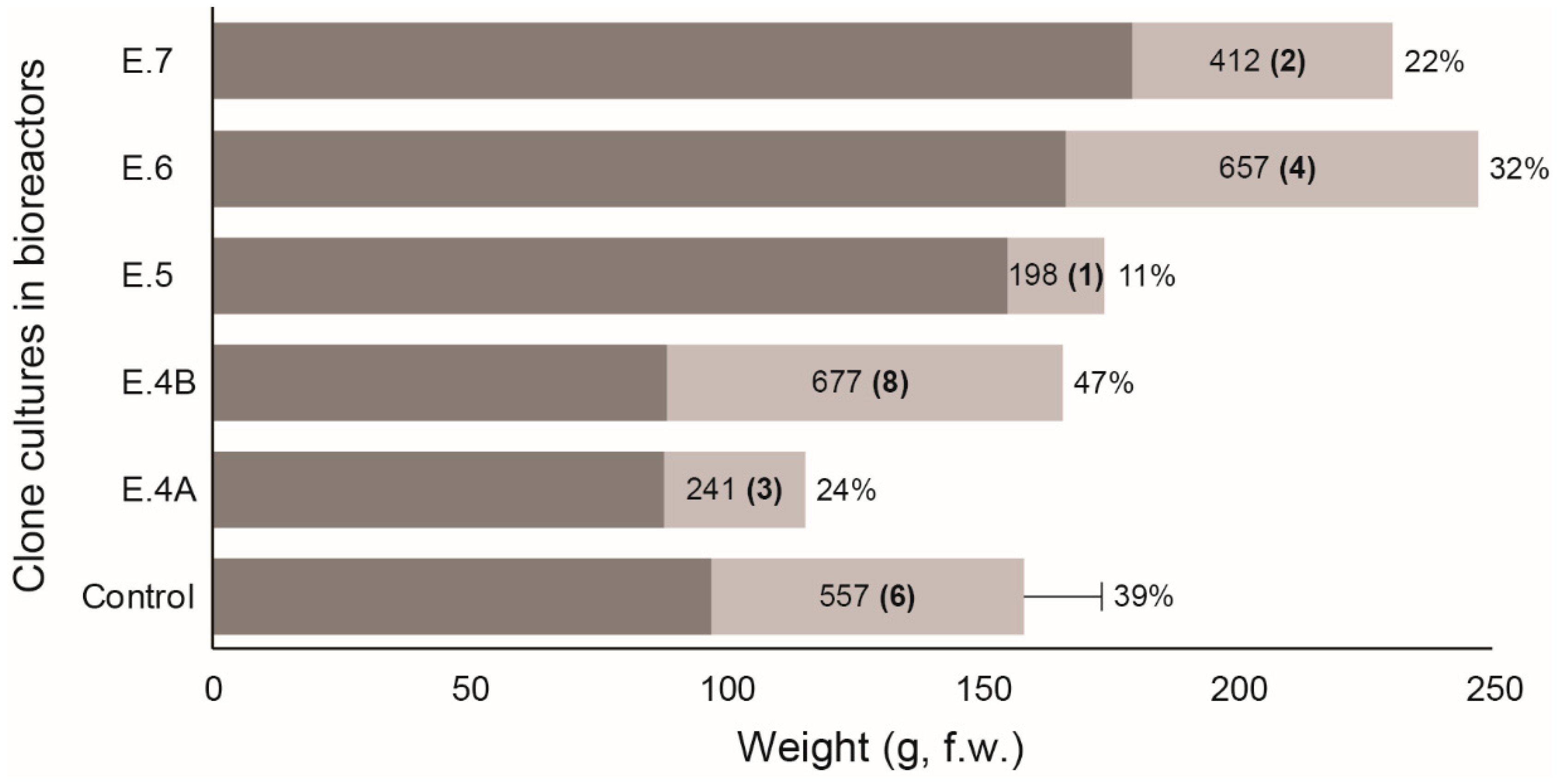
The percentage growth relative to the total weight of the controls in the bioreactors varied between 37% and 39.4% (mean = 39%; n = 4). A slightly higher growth than in the controls was only achieved in treatment B in E.4. (i.e., culture temperature, 20 °C). Growth was lower in the other experiments and was up to 4 times lower in the experiment simulating spring conditions (E.5).
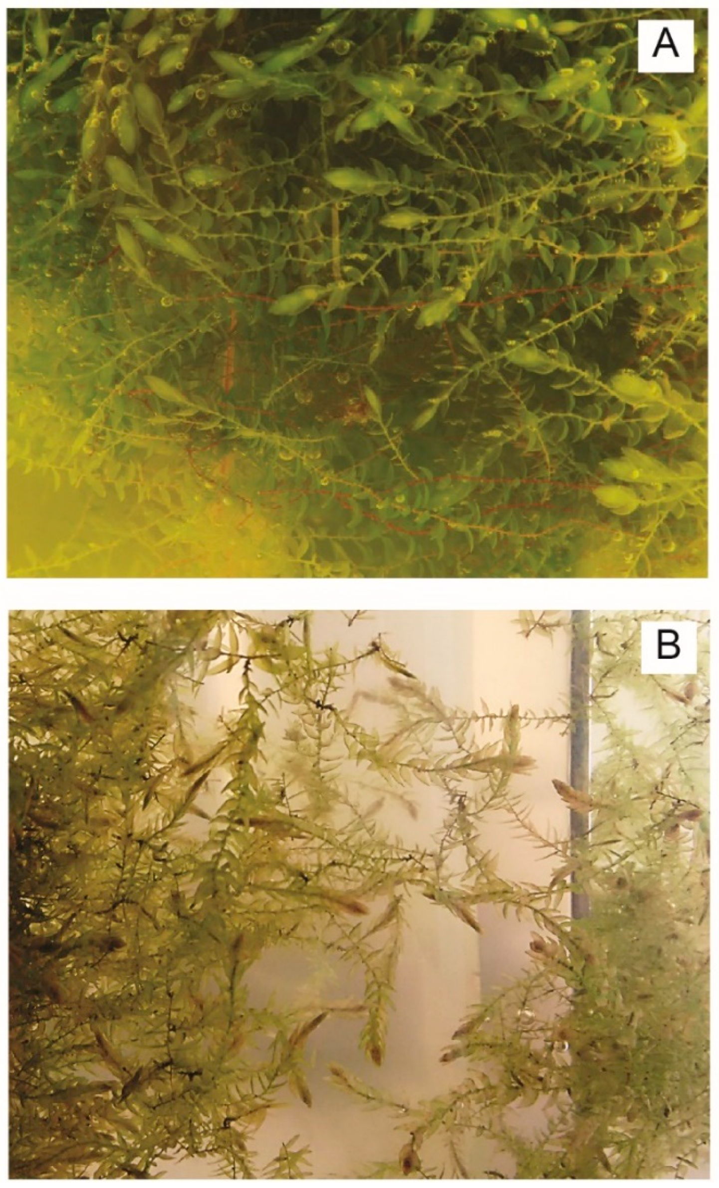
The changes in pH recorded by the bioreactor software during E.4-Tª, E.5-Spring, E.6-CO2 and E.7-Ammonium are shown in Figure 3. The pH of the medium in the control bioreactors varied during the culture period, from an initial value of 5.62 (±0.09, n = 4) to 7.98 (±0.39). The variations in pH during E.6 reflect the intermittent addition of CO2 during development of the experiment. The addition of NH4NO3 led to a decrease in the culture pH to 3.43, which was accompanied by physiological deterioration of the moss and a change in the color of the apical bud (see Figure 4A,B). After changing the culture medium to control conditions (i.e., without NH4NO3), the pH varied as usual, reaching a value of 7.33. Cultivation of the moss at 20 °C led to the acidification of the medium to pH 4.56; however, when shaking was not applied, the pH returned to 7.14.

4. Discussion

Axenic cultures of a clone of the aquatic moss F. antipyretica were successfully maintained. The growth rates were 3 times greater in the bioreactors than in flasks after culture for 4 months, although the growth rate relative to the initial weight was 7 times higher in the flasks (see Figure 1 and Figure 2). Despite the high rate of growth of the clone, the increase in biomass was lower than reported for other bryophytes (e.g., Sphagnum palustre) in which much greater increases in biomass were achieved in only 4 weeks [9]. In order to resolve this problem, we attempted to maximize the in vitro growth of the clone by modifying both the culture medium and the different culture conditions.
One of the techniques used to maximize the growth of S. palustre cultures in bioreactors was to maintain constant illumination [9]. However, as F. antipyretica is a shade-tolerant species, with relatively low compensation and saturation points [13,14], the low growth rates relative to the initial weight of the control cultures in the bioreactors (6 mg·day−1·g−1) and compared with the control cultures in the bioreactors (45 mg·day−1·g−1) may indicate the photoinhibition of growth as a result of the continuous illumination. However, the observed effects of the photoperiod (E.1) did not indicate any changes in growth with a photoperiod of 16/8 or 24/0 h, and thus photoinhibition can be ruled out. The limiting effect of the low density of gametophytes per volume of medium relative to those in the flasks is another possible explanation for the low growth rate in the bioreactors (see [9]) and should be taken into account in future studies.
According to other authors, the provision of an additional source of C has also been shown to be effective for increasing the in vitro growth of bryophytes (e.g., [9,15,16]). These authors demonstrated that the addition of C in the form of sucrose of glucose increased the growth of S. palustre thanks to the ability of peat mosses to assimilate exogenous C in the form of sugars (mixotrophy) in the same way that the addition of CO2 enhanced the growth of F. antipyretica. However, the findings of the present study show that neither the addition of sucrose or of CO2 increased the growth of this moss relative to the controls (71% in E.2 A, 70% in E.2 B relative to 76% in the control flask cultures in and 32% in E.6 relative to 39% in the bioreactor controls; Figure 1 and Figure 2). This appears to indicate that F. antipyretica is not utilizing exogenous sources of C to maximize biomass production. In the case of CO2, a high concentration of this gas and/or its interaction with other variables such as temperature and photoperiod may limit the growth of F. antipyretica as the photosynthetic activity would be increased and the respiration decreased due to the induction of stomatal closure, as suggested by Maberly [17,18]. This would also explain the pH fluctuations observed in E.6, as the addition of CO2 leads to the acidification of the medium due to the addition of carbonates. However, once the CO2 is consumed during photosynthesis, the pH will again increase (Figure 3).
Likewise, we investigated whether the increase in P concentration, the variation in the N:P ratio (E.3) and the addition of NH4NO3 as an additional source of N (E.7) would enhance the growth of the moss clones. N and P are often growth-limiting nutrients in aquatic environments [19], and a higher availability of N will cause a greater imbalance between N and P (possibly causing P to be a limiting nutrient for moss development [20,21]). However, it is not known how these elements affect the growth and development of moss in culture. Some authors have reported that the addition of N and P promotes growth of bryophytes in culture (e.g., [22,23,24]) while others have observed a decrease in biomass after the addition of these nutrients [25]. The results of experiments E.3 (N:P ratio) and E.7 (ammonium) are consistent with the latter findings. In these experiments, the growth of F. antipyretica was not affected by the increased concentration of NH4NO3 (22% compared with 39%, Figure 2), and although the moss showed some deterioration and the apices turned brownish in colour, the appearance improved once the samples were transferred to culture medium without NH4NO3 (with the subsequent return to higher pH) (Figure 4). Conversely, we found that growth decreased on the addition of greater amounts of P (independently of the N:P ratio used) (57–25% compared with 76%, Figure 1).
It is generally established that aquatic bryophytes do not tolerate prolonged exposure to temperatures above 20 °C [26,27] and that optimal growth occurs in the range 5–15 °C [28,29]; more specifically at 10 °C in F. antipyretica [27]. The limited growth at high temperatures may be attributed to the fact that the rapid rate of respiration exceeds the rate of photosynthesis and produces a decrease in pH that influences the growth [30]. However, the results of the present study show that the growth of the culture at 10 °C was half that of the control (i.e., 24% compared with 39%; E.4 A), whereas at 20 °C, the growth was slightly higher than that of the control (i.e., 47% compared with 39%; E.4 B) even at the lowest pH in all of the experiments (Figure 3). Although the F. antipyretica culture was derived from axenic cultures inoculated in the bioreactors, sterility checks usually yielded positive results for bacterial contamination. Temperatures close to 20 °C may favour the growth of this bacterial community, which would act as a biofertilizer, thus favouring the growth of the culture (e.g., [10,31]).
It is possible that some species of Fontinalis may be more productive at the low temperatures that occur during winter. Water flow is greater at this time of year, and nutrients are continually renewed, especially in drought-associated habitats during periods of high temperature [27]. However, it is also possible that low temperatures may limit production [32]. Considering the high rate of growth observed at 20 °C (i.e., E.4 B), as well as the possible effect of nutrient renewal and the behaviour of this species under natural conditions, in which we have observed a growth spurt in the field during spring (personal observation), we simulated spring conditions (E.5) in order to determine whether the change in temperature and renewal of nutrients would favour growth of the clone. The results were unexpected as the growth was almost 4 times lower than that of the controls (11% compared with 39%; E.5) and was lower than in any of the other experiments (Figure 2).
In light of the above findings, and on the basis of our observations of the species under natural conditions, we suspect that there may be some as-yet undetected way of maximizing the production of F. antipyretica in the bioreactors. As commented above, the low density of gametophytes in the volume of medium used in the bioreactors relative to the flasks may be one of the factors limiting production [9]. The factors that may have led to the low production in the bioreactors were the aeration (continual in the bioreactors but absent in the flasks) and the type and intensify of light used in each culture. Nonetheless, despite the observed limitations, the in vitro culture of moss in the liquid medium control (see Table 1) provides a viable alternative to field collection of material. This particularly applies to flask culture, as in addition to the previously mentioned limitations, we must also consider the high cost and maintenance of the bioreactor systems.

5. Conclusions

None of the modifications to the culture conditions greatly increased the in vitro growth of the F. antipyretica clones with respect to the control. However, the growth rate of the clone was sufficiently high to enable the production of large amounts of material for the purpose of biomonitoring water quality. The ability to clone the material will substantially improve the applicability of the moss bag technique.

Author Contributions

Conceptualization, S.D., J.R.A. and J.ÁF.; Formal analysis, S.D., Z.V. and P.G.; Funding acquisition, J.R.A. and J.ÁF.; Investigation, S.D., Z.V., P.G., J.R.A. and J.ÁF.; Methodology, J.R.A. and J.ÁF.; Resources, J.R.A. and J.ÁF.; Validation, S.D.; Visualization, S.D.; Writing—original draft, S.D. and Z.V.; Writing—review & editing, J.R.A. and J.ÁF.

Funding

This work was funded by MINECO project CTM2015-70578-P. The authors belong to the Galician Competitive Research Group GRC/GPC2016-002 and to the CRETUS Strategic Partnership (AGRUP2015/02). All of these programmes are co-funded by FEDER (EU).

Acknowledgments

The authors are very grateful to Veronica Fernández González for valuable assistance in the laboratory.

Conflicts of Interest

The authors declare that there are no conflicts of interest regarding the publication of this paper.

References

  1. Debén, S.; Aboal, J.R.; Carballeira, A.; Cesa, M.; Fernández, J.A. Monitoring river water quality with transplanted bryophytes: A methodological review. Ecol. Ind. 2017, 81, 461–470. [Google Scholar] [CrossRef]
  2. Martínez-Abaigar, J.; García-Álvaro, M.A.; Beaucourt, N.; Núñez-Olivera, E. Combined seasonal and longitudinal variations of element concentrations in two aquatic mosses (Fontinalis antipyretica and F. squamosa). Nova Hedwigia 2002, 74, 349–364. [Google Scholar] [CrossRef]
  3. Ares, A.; Aboal, J.R.; Carballeira, A.; Giordano, S.; Adamo, P.; Fernández, J.A. Moss bag biomonitoring: A methodological review. Sci. Total Environ. 2012, 432, 143–158. [Google Scholar] [CrossRef] [PubMed]
  4. Roy, S.; Sen, C.K.; Hänninen, O. Monitoring of polycyclic aromatic hydrocarbons using ‘moss bags’: Bioaccumulation and responses of antioxidant enzymes in Fontinalis antipyretica Hedw. Chemosphere 1996, 32, 2305–2315. [Google Scholar] [CrossRef]
  5. Hongve, D.; Brittain, J.E.; Bjørnstad, H.E. Aquatic mosses as a monitoring tool for 137Cs contamination in streams and rivers-a field study from central southern Norway. J. Environ. Radioact. 2002, 60, 139–147. [Google Scholar] [CrossRef]
  6. Di Palma, A.; Pardo, D.C.; Spagnuolo, V.; Adamo, P.; Bargagli, R.; Cafasso, D.; Capozzi, F.; Aboal, J.R.; González, A.G.; Pokrovsky, O.; et al. Molecular and chemical characterization of a Sphagnum palustre clone: Key steps towards a standardized and sustainable moss bag technique. Ecol. Ind. 2016, 71, 88–397. [Google Scholar] [CrossRef]
  7. De Traubenberg, C.R.; Ah-Peng, C. A Procedure to Purify and Culture a Clonal Strain of the Aquatic Moss Fontinalis antipyretica for Use as a Bioindicator of Heavy Metals. Arch. Environ. Contam. Toxicol. 2004, 46, 289–295. [Google Scholar] [CrossRef]
  8. Capozzi, F.; Giordano, S.; Aboal, J.R.; Adamo, P.; Bargagli, R.; Boquete, T.; Di Palma, A.; Real, C.; Reski, R.; Spagnuolo, V.; et al. Best options for the exposure of traditional and innovative moss bags: A systematic evaluation in three European countries. Environ. Pollut. 2016, 214, 362–473. [Google Scholar] [CrossRef]
  9. Beike, A.K.; Spagnuolo, V.; Lüth, V.; Steinhart, F.; Ramos-Gómez, J.; Krebs, M.; Adamo, P.; Rey-Asensio, A.I.; Fernández, J.A.; Giordano, S.; et al. Clonal in vitro propagation of peat mosses (Sphagnum L.) as novel green resources for basic and applied research. Plant Cell Tissue Organ Cult. 2015, 120, 1037–1049. [Google Scholar] [CrossRef]
  10. Ares, A.; Duckett, J.G.; Pressel, S. Asexual reproduction and protonemal development in vitro in Fontinalis antipyretica Hedw. J. Bryol. 2014, 36, 122–133. [Google Scholar] [CrossRef]
  11. Hohe, A.; Reski, R. Optimisation of a bioreactor culture of the moss Physcomitrella patens for mass production of protoplasts. Plant Sci. 2002, 163, 69–74. [Google Scholar] [CrossRef]
  12. Schween, G.; Hohe, A.; Koprivova, A.; Reski, R. Effects of nutrients, cell density and culture techniques on protoplast regeneration and early protonema development in a moss, Physcomitrella patens. J. Plant Physiol. 2003, 160, 209–212. [Google Scholar] [CrossRef] [PubMed]
  13. Bowes, G. Pathways of CO2 fixation by aquatic organisms. In Inorganic Carbon Uptake by Aquatic Photosynthetic Organisms; Lucas, W.J., Berry, J.A., Eds.; American Society of Plant Physiologists: Rockville, MD, USA, 1985; pp. 87–210. [Google Scholar]
  14. Carballeira, A.; Díaz, S.; Vázquez, M.D.; López, J. Inertia and resilience in the responses of the aquatic bryophyte Fontinalis antipyretica Hedw. to thermal stress. Arch. Environ. Contam. Toxicol. 1998, 34, 343–349. [Google Scholar] [CrossRef] [PubMed]
  15. Peñuelas, J. HCO3 as an exogenous carbon source for aquatic bryophytes Fontinalis antipyretica and Fissidens grandifrons. J. Exp. Bot. 1985, 36, 441–448. [Google Scholar] [CrossRef]
  16. Decker, E.L.; Reski, R. Current achievements in the production of complex biopharmaceuticals with moss bioreactors. Bioprocess Biosyst. Eng. 2008, 31, 3–9. [Google Scholar] [CrossRef]
  17. Maberly, S.C. Photosynthesis by Fontinalis antipyretica I. IInteraction between photon irradiance concentration of carbon dioxide and temperature. New Phytol. 1985, 100, 127–140. [Google Scholar] [CrossRef]
  18. Maberly, S.C. Photosynthesis by Fontinalis antipyretica II. Assessment of environmental factors limiting photosynthesis and production. New Phytol. 1985, 100, 141–155. [Google Scholar] [CrossRef]
  19. Jarvie, H.P.; Whitton, B.A.; Neal, C. Nitrogen and phosphorus in east coast British rivers: Speciation, sources and biological significance. Sci. Total Environ. 1998, 210/211, 79–109. [Google Scholar] [CrossRef]
  20. Carroll, J.A.; Caporn, S.J.M.; Johnson, D.; Morecroft, M.D.; Lee, J.A. The interactions between plant growth, vegetation structure and soil processes in semi-natural acidic and calcareous grasslands receiving long-term inputs of simulated pollutant nitrogen deposition. Environ. Pollut. 2003, 121, 363–376. [Google Scholar] [CrossRef]
  21. Phoenix, G.K.; Booth, R.E.; Leake, J.R.; Read, D.J.; Grime, J.P.; Lee, J.A. Effects of enhanced nitrogen deposition and phosphorus limitation on nitrogen budgets of semi-natural grasslands. Glob. Chang. Biol. 2003, 9, 1309–1321. [Google Scholar] [CrossRef]
  22. Jauhiainen, J.; Vasander, H.; Silvola, J. Response of Sphagnum fuscum to N deposition and increased CO2. J. Bryol. 1994, 18, 83–95. [Google Scholar] [CrossRef]
  23. Kooijman, A.M.; Bakker, C. Species replacement in the bryophyte layer in mires: The role of water type, nutrient supply and interspecific interactions. J. Ecol. 1995, 83, 1–8. [Google Scholar] [CrossRef]
  24. Gerdol, R.; Bonora, A.; Marchesini, R.; Gualandri, R.; Pancaldi, S. Growth response of Sphagnum capillifolium to nighttime temperature and nutrient level: Mechanisms and implications for global change. Arctic Alpine Res. 1998, 30, 388–395. [Google Scholar] [CrossRef]
  25. Gunnarsson, U.; Rydin, H. Nitrogen fertilization reduces Sphagnum production in bog communities. New Phytol. 2000, 147, 527–537. [Google Scholar] [CrossRef] [Green Version]
  26. Glime, J.M.; Carr, R.E. Temperature survival of Fontinalis novae-angliae Sull. Bryologist 1974, 17–22. [Google Scholar] [CrossRef]
  27. Glime, J.M.; Acton, D.W. Temperature effects on assimilation and respiration in the Fontinalis duriaei -Periphyton Association. Bryologist 1979, 82, 382–392. [Google Scholar] [CrossRef]
  28. Dilks, T.J.K.; Proctor, M.C.F. Comparative experiments on temperature responses of bryophytes: Assimilation, respiration and freezing damage. J. Bryol. 1975, 8, 317–336. [Google Scholar] [CrossRef]
  29. Glime, J.M.; Raeymaekers, G. Temperature effects on branch and rhizoid production in six species of Fontinalis. J. Bryol. 1987, 14, 779–790. [Google Scholar] [CrossRef]
  30. Glime, J.M.; Vitt, D.H. The physiological adaptations of aquatic music. Lindbergia 1984, 10, 41–52. [Google Scholar]
  31. Tani, A.; Akita, M.; Murase, H.; Kimbara, K. Culturable bacteria in hydroponic cultures of moss Racomitrium japonicum and their potential as biofertilizers for moss production. J. Biosci. Bioeng. 2011, 112, 32–39. [Google Scholar] [CrossRef]
  32. Moeller, R.E. The temperature-determined growing season of a submerged hydrophyte: Tissue chemistry and biomass turnover of Utricularia purpurea. Freshw. Biol. 1980, 10, 391–400. [Google Scholar] [CrossRef]
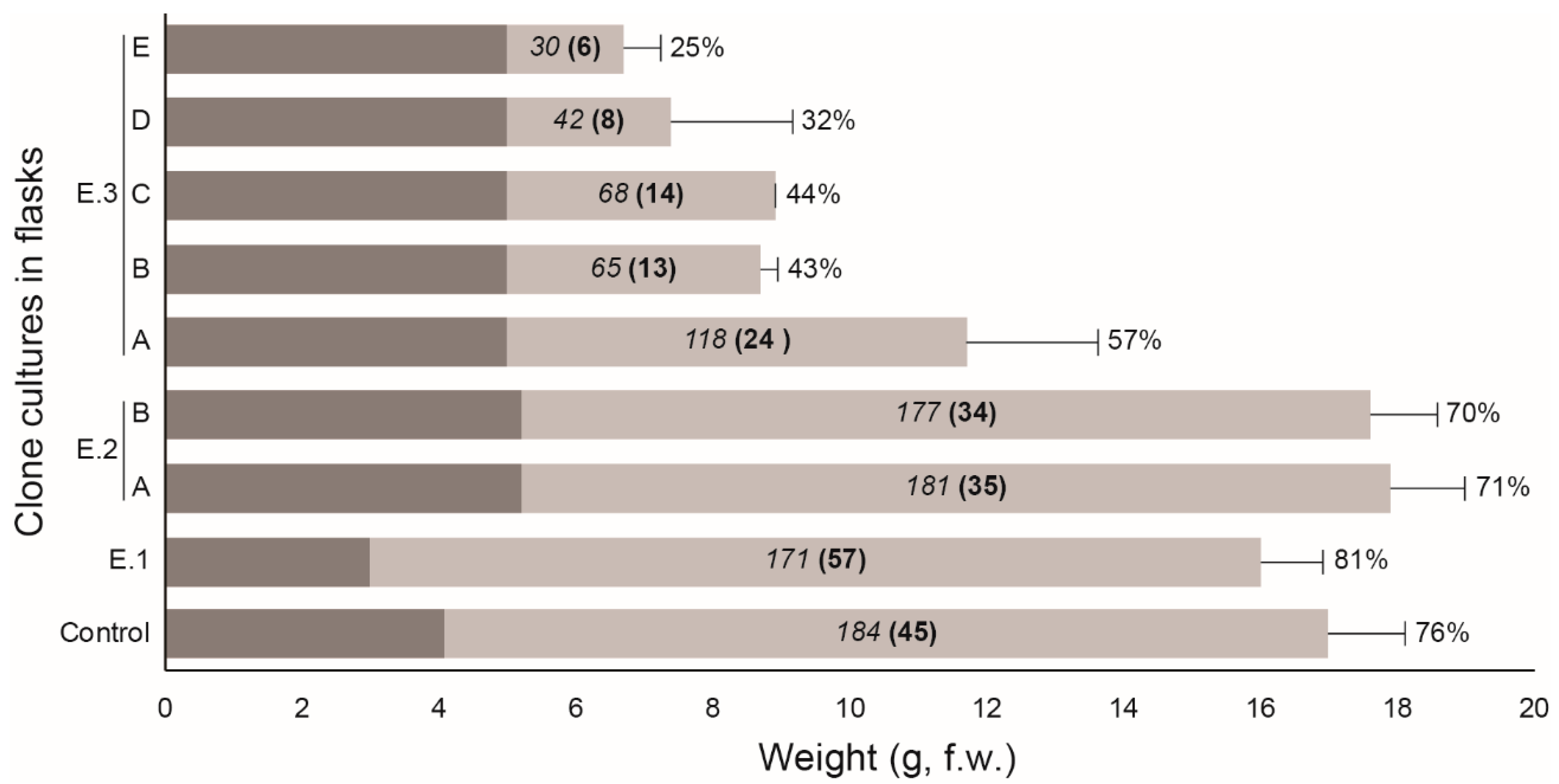
Figure 1. Fresh weight (f.w.) of the moss clone Fontinalis antipyretica cultivated in the different experiments carried out in flasks (E.1–E.3). A, B, …, E = different treatments for each experiment (see text for more details). Growth rates (mg·day−1) are shown in italics, growth rates relative to the initial weight (mg·day−1·g−1) are shown between brackets and bold type, and the proportion of growth relative to the total weight (initial and growth) are shown in regular type.
Water 11 00145 g001
Figure 2. Fresh weight of the moss clone Fontinalis antipyretica cultivated in the different experiments carried out in bioreactors (E.4–E.7). A, B = different treatments of the experiment (see text for more details). Growth rates (mg·day−1) are shown in italics, growth rates relative to the initial weight (mg·day−1·g−1) are shown between brackets and bold type, and the proportion of growth relative to the total weight (initial and growth) are shown in regular type.
Water 11 00145 g002
Figure 3. Evolution in pH of the culture medium of the moss clone Fontinalis antipyretica for the different experiments carried out in bioreactors (E.4–E.7, see text for more details) and recorded by the bioreactor software. Black line and gray shading: mean and interval in controls; blue line: E.4 A; red line: E.4 B; brown line: E.5; green dashed line: E.6 and green line: E.7. Reds points are shown for E.5 and E.7 when the culture conditions were changed.
Water 11 00145 g003
Figure 4. Appearance of the moss clone Fontinalis antipyretica cultivated in bioreactors: (A) in the control culture medium and (B) physiological deterioration observed after the addition of NH4NO3 in the experiment 7.
Water 11 00145 g004
Table 1. Composition of the culture medium and variation in the Fontinalis antipyretica culture conditions in the experiments (Exp.) carried out in flasks (E.1–E.3) and bioreactors (E.4–E.7). A, B, …, E = different treatments (Treatm.) for each experiment (see text for more details). n: number of replicates; T: temperature.
Type of CultureExp.Treatm.nTime (days)T (°C)Volume (L)Photoperiod (h light/dark)Sucrose (%)N:P RatioP (mg·L−1)CO2NH4 (g·L−1)
FlasksControl1357–76150.516/80.32.1250--
E.1776150.524/00.32.1250--
E.2A370150.516/822.1250--
B370150.516/842.1250--
E.3A357150.516/80.323250--
B357150.516/80.346250--
C357150.516/80.32.15200--
D357150.516/80.323500--
E357150.516/80.346500--
BioreactorsControl497–135151224/00.32.1250--
E.4A1113101224/00.32.1250--
B1114201224/00.32.1250--
E.519715 + 204 + 824/00.32.1250--
E.61122151224/00.32.12500.1-
E.71123151224/00.32.1250-0.1