Review

Can a Century Old Experiment Reveal Hidden Properties of Water?

Wetsus—Centre of Excellence for Sustainable Water Technology, Leeuwarden, The Netherlands
Water 2010, 2(3), 381-410; https://doi.org/10.3390/w2030381
Submission received: 30 June 2010 / Revised: 3 August 2010 / Accepted: 3 August 2010 / Published: 10 August 2010
(This article belongs to the Special Issue Water: Facts without Myths)

Abstract

:
In 1893 Sir William Armstrong placed a cotton thread between two wine glasses filled with chemically pure water. After applying a high voltage, a watery connection formed, and after some time, the cotton thread was pulled into one of the glasses, leaving a rope of water suspended between the two glasses. Although being a very simple experiment, it is of special interest since it comprises a number of phenomena currently tackled in modern water science like electrolysis-less charge transport and nanobubbles. This work gives some background information about water research in general and describes the water bridge phenomenon from the viewpoint of different fields such as electrohydrodynamics and quantum field theory. It is shown that the investigation of the floating water bridge led to new discoveries about water, both in the macroscopic and microscopic realm – but these were merely “hidden” in that sense that they only become evident upon application of electric fields.

1. Introduction: Water Properties and Structure

Water is undoubtedly the most important chemical substance of the world. Despite this, and in spite of the fact that it is practically ubiquitous, it still represents one of the best explored [1,2] and yet least understood substances [3,4,5], as its so-called "anomalies" are famous (e.g., [6,7,8]). Many attempts have been made in order to measure or calculate the structure of water beyond the H2O molecule. This is a difficult task due to the hydrogen bond (H-bond) network which can be formed by two or more water molecules. These bonds are characterized by a binding energy between 1 and 50 kJ/mol, depending on and the type of strength of interaction and the local geometry [9], and their network is nowadays held responsible for many of water’s weird properties [10] and is also the reason why water cannot be treated as a "simple liquid" [11]. From a theoretical point of view, the difficulties in understanding liquid water can probably be attributed to two features: cooperative hydrogen bonding (the fact that the binding energy of two H-bonded molecules is modified by the presence of a third molecule [10,12,13,14,15,16]) and nuclear quantum effects. Such effects occur because the proton is so light that classical mechanics can no longer adequately describe properties like spatial dispersion of the hydrogen positions, nuclear tunneling, zero-point energy and, naturally, quantization of nuclear motions. Much work has been done recently toward the simulation of liquid water [17,18,19,20,21,22,23,24,25], its intrinsic ions H+ and OH- [26] and other ions in solution [27] using ab initio electronic-structure methods, sometimes together with quantum dynamics methods [25,28,29,30,31,32,33], but still more work is called for in order to get a more complete and accurate picture of the liquid. So obviously, the description of this substance by its chemical formula—H2O—reflects the reductionism concept of the current scientific paradigm, and in this special case it fails to do justice to the structure and the properties of water. If one considers, e.g., phosphorus pentoxide, whose formula is given as P4O10 according to its dimeric structure, a proposal for the formula of water could be (H2O)n. However, as pointed out above, in this case, the situation is much more complex. Numerous structural models of water proposed by many authors can be found in the book of Eisenberg and Kauzman [1]. The X-ray studies of water and ice by Hajdu et al. and Hadju [34,35,36] are mentioned here as a representative example of the many investigations succeeding that book. For detailed up-to-date structural analyses of water based on neutron scattering and/or X-ray diffraction, reference is made to the works of Teixeira and Luzar [37] and Soper (e.g., [38,39]), exemplary studies on supercooled water as well as a possible liquid-liquid phase transition were published by Stanley et al. [40,41,42], Mishima and Stanley [43] and Yamada et al. [44] (and references quoted therein). In his recent paper, Soper [39] explains that when using an empirical potential structure refinement (EPSR) to form a single atomistic structural model which was simultaneously consistent with both X-ray and neutron diffraction data, X-ray data for water provide a powerful constraint on possible structural models. However, it is not possible to rigorously define the precise position and height of the first peak in the OO radial distribution function; and different neutron datasets on water give rise to further small uncertainties in the position of the hydrogen bond peaks. According to Soper, one general conclusion from the combined use of neutron and X-ray data is that many of the classical water potentials may have a core which is strongly repulsive at short distances producing too sharp a peak in r-space at too short a distance. Thus, a softer core potential is proposed [39].
Leetmaa et al. recently reported on the consistency of such models with infrared/Raman and X-ray absorption spectra [45]. They claim that the overall agreement of calculated spectra based upon established structural models is still unsatisfactory, and that no water model exists that can equally well describe IR/Raman, X-ray absorption spectroscopy, and diffraction data [45]. In a subsequent work [46], they furthermore show that there is no strict proof of tetrahedral water based on diffraction and IR/Raman data, and that the tetrahedral structure model must, to fit diffraction data, be less structured than most models obtained from molecular dynamics simulations [46].
An interesting interpretation of Raman spectra of water revealing a non-homogeneity of the H-bond network related to a (bi-, tri-)furcation of H-bonds was published by Chumaevskii and Rodnikova [47]. They claim this to be due to the presence of defects in the H-bond network which correspond to different conformations of fully hydrogen-bonded five-molecule structural units [47]. For an excellent review about Raman, IR and vibrational spectroscopy of water in general, the reader is referred to the paper of Bakker and Skinner [48] and the references quoted therein.
A novel approach in water modelling was recently presented by Molinero and Moore [49], where water is simulated as a fictional intermediate element between carbon and silicon exploiting their common feature of forming tetrahedrally coordinated units. This model departs from the prevailing paradigm in water modeling which uses long-ranged electrostatic forces to produce a short-ranged hydrogen-bonded structure since it comprises only short-range interactions. Yet it reproduces energetics, density and most anomalies and phase transitions of liquid water with comparable or better accuracy than the most popular atomistic models of water [49].
A very large number of papers which present evidence for the presence of specific molecular arrangements have been published recently (2004) in Science. Miyazaki et al. [50] showed infrared spectroscopic evidence for oligomers of different shape and sizes from n = 4–27 in (H2O)n. Shin et al. [51] present intriguing IR data near the 3.7 µ O-H stretching band in oligomers from n = 6–27, around the "magic number" of n = 21. About six months after these publications, the October issues of Science contained several exquisitely detailed papers on water from senior authors. They discuss the energetics and dynamics of electron binding and transport in various cluster sizes, some of it in vapor samples. These processes are extremely rapid, on the order of tens of femtoseconds, as discussed in a recent work by Nibbering and Elsaesser [52]. Wernet et al. [53] supported the view favouring only ring and chain molecules using XRD and Raman spectroscopy, which was partly confirmed by Odelius et al. [54]; while J.D. Smith et al. [55] used their total electron yield near-edge X-ray absorption time structure (TEY-NEXAFS) technique to come to very different conclusions: according to them, the water and ice H-bonds are very similar, and the usually accepted H-bond strength is consistent with their data. Also, according to Head-Gordon et al. [56], the XAS data is most compatible with water as a tetrahedral hydrogen-bonded liquid. Tse et al. [57] report on the X-ray absorption spectra of high-density amorphous (HDA) ice, low-density amorphous ice Ic, ice Ih, normal and deuterated liquid water by in situ heating of an HDA ice sample where they observed the previously reported distinct pre-edge structure in all spectra. The authors did not wish to directly address or attempt to delineate the controversy between the conventional and novel views of water structure; however, they indicate that a complete quantitative description of the XAS of water and ices is beyond the currently employed theoretical methods, partly due to small but significant variations between the reported experimental spectra and incomplete description of the continuum states [57].

2. Explanatory Models: Interaction with Magnetic and Electric Fields

The interaction of water with electric or magnetic fields also reveals many interesting aspects. The effect of magnetic fields seems extraordinary [58,59,60,61], its “magnetic memory” has been investigated [62]. The effect of magnetic fields on the formation of scale in boilers has been established in a huge amount of data (see [63,64,65] and references quoted therein). In the laboratory, the influence of modest DC magnetic fields on the nucleation and growth of CaCO3 (phases, sizes, morphology) in dilute aqueous solutions have been thoroughly studied and demonstrated by Higashitani et al. and Pach et al. [64,65]. The former demonstrates a strong memory effect in the constituent solutions exposed to a magnetic field. Roy et al. [66] have shown the remarkable effect of a static magnetic field on the pH of water in a conditioned volume. On the other hand, Cowan et al. [67] report that there is no magnetic memory of water at all.
The interaction of water with electric fields has become more and more explored over the last years because of its importance for "electrospinning", "electrowetting" and "electrospray" methods for mass spectometry ("ESMS") or the production of nanofibres [68,69,70,71,72,73,74,75,76,77,78,79,80,81,82,83], but unusual phenomena have also been recently reported like—e.g.,—the electric field driven self-propulsion of a water droplet on a solid surface [84].
The water bridge was first discovered by Lord Armstrong in 1893 (see Figure 1). In his speech before the Newcastle Literary and Philosophical Society, Armstrong describes his experiment: “(...) Amongst other experiments I hit upon a very remarkable one. Taking two wine-glasses filled to the brim with chemically pure water, I connected the two glasses by a cotton thread coiled up in one glass, and having its shorter end dipped into the other glass. On turning on the current, the coiled thread was rapidly drawn out of the glass containing it, and the whole thread deposited in the other, leaving, for a few seconds, a rope of water suspended between the lips of the two glasses. This effect I attributed at that time to the existence of two water currents flowing in opposite directions and representing opposite electric currents, of which the one flowed within the other and carried the cotton with it. It required the full power of the machine to produce this effect, but, unfortunately when it went to London, and was fitted up in the lecture-room, I could not get the full power on account of the difficulty of effecting as good insulation in a room as in the outside air. I therefore failed in getting this result, after announcing that I could do it, and I daresay I got the credit of romancing.”[85].
Figure 1. 1st Lord Armstrong of Cragside, by Mary Lemon Waller (1871–1916), Cragside, The Armstrong Collection (acquired through the National Land Fund and transferred to The National Trust in 1977), ©NTPL/Derrick E. Witty.
Water 02 00381 g001
It should be noted here that, naturally, His Lordship was not romancing, but successfully reproduced the phenomenon later on. However, no additional investigation of this phenomenon was published for 104 years. The knowledge about this experiment was only passed down orally, finally reaching Prof. Uhlig from the ETH Zürich, who put a video of this experiment online in the World Wide Web [86]. Then, in 2006, the author of this paper started an interdisciplinary co-operation which, in 2007, produced a paper about this phenomenon in the Journal of Physics D: Applied Physics [87], which was later featured by the magazine Nature [88]. An exemplary image of the bridge is given in Figure 2.
Figure 2. A floating water bridge produced with glass beakers, deionized water, Pt electrodes, 15kV (see also [87]).
Water 02 00381 g002
Three more papers were published in cooperation with the author of this paper in the Journal of Physics D in 2008, 2009 and 2010 [89,90,91], one in the journal Experiments in Fluids in 2010 [92], and one in the journal Water [93] which could be considered as a first step towards a comprehensive explanation of the phenomenon. Meanwhile, more groups have picked up the topic, giving first results on ion transportation [94], Raman scattering [95] and a derivation of the Maxwell pressure tensor within the water bridge [96]. Next to that, an investigation of mass and charge transfer processes [97] has recently been presented.
There is a long history of research on the effects of high voltage on dielectric fluids, from many points of view: classically physical, which comprises the huge field of (di-) electrophoresis, electrowetting and electrospray; quantum-physical or even quantum-electrodynamical.

2.1. Dielectrophoresis/Electrospray

The fact that a jet flow can be induced by applying a DC high voltage is well known as an electrohydrodynamic (EHD) effect and is called "dielectrophoresis", which is defined as the motion of a dielectric fluid caused by polarization effects. The fluid tends to polarize in the field, therefore, a dipole in the medium experiences a force towards higher field strength. Even on reversal of the polarity of the field, the dipole experiences the force in the same direction. Thus, this effect is independent of the polarity of the field and depends on its non-uniformity only. "Electrophoresis" on the other hand results from the electrostatic attraction of charged particles towards electrodes. The electrophoretic forces, therefore, depend on the polarity of the electrodes and hence are electric field dependent. A good overview of the topic is the 1998 book “Electrohydrodynamics” by Castellanos [98]. Since the main flow direction of the water bridge is polarity dependent but there is also always a flow in both directions [87,89], both effects have to be taken into consideration.
In 1955, Sumoto found that dielectric fluids climb up electrodes if the applied voltage is high enough [99]. Pohl [100] as well as Parmar and Jalaluddin [101] assumed that the dielectrophoretic forces result in electrohydrodynamic motion producing non-uniformity in the applied electric field. The pumping action of strong divergent electric fields [102] and the electroconvection effects [103] have been explained as due to strong electric stresses on liquids in non-uniform fields. According to Maxwell's treatise on the theory of electricity and magnetism [104] and to the theory of liquid hydrodynamics under electric fields by Landau and Lifshitz [105], there are pressure forces perpendicular and tension forces parallel to the electric field lines. This tension can be considered as a thermodynamic reason for the stability of the water bridge [96].
Dielectrophoresis was found responsible for anomalous effects in liquids before, like the methylmethacrylate anomalies which were explained in 1985 by Parmar and Labroo [106]. In 1996 Hughes et al. calculated both trapping and a translational dielectrophoretic forces on particles travelling in electric fields [107], and in 2006, Techaumnat et al. calculated the electric field and the dielectrophoretic force on spherical particles in a chain in a dielectric fluid [108] and found stable regimes, which could be an important basis for similar considerations concerning the floating water bridge.
From the viewpoint of electrospraying, the bridge can be considered as a water jet (a “cone-jet” [81,82,83]) whose break-up is halted by the electric force acting on it. Such jets are normally being produced when generating an electro-spray. In this process a liquid is pumped through a nozzle at a low flow rate, a droplet is formed at the tip of the nozzle. An electric field is applied. This electric field induces a surface charge in the droplet. As a result of the electric stress, the droplet is transformed into a conical shape. The applied electric field accelerates the surface charge toward the cone apex. Due to this acceleration, a jet with a high charge density occurs at the cone apex. This jet breaks up into a number of primary or main droplets and a number of secondary droplets and satellites as described by Cloupeau and Prunet-Foch [109], Gomez and Tang [110], Chen et al. [111] and Hartman et al. [81]. A similar effect can be observed when the water bridge is torn apart [87]. An example of an aqueous Taylor cone is shown with in Figure 3.
Figure 3. Cone (a) and droplet ejection (b-f) in a water electrospray recorded with a Photron SA-1 high speed camera. Nozzle inner/outer diameter 0.84/1.27 mm diameter; +4.5 kV potential; 1.5cm distance to counter electrode; flow rate 2mL/h; time interval between the images 2 ms (from [112]).
Water 02 00381 g003
The shape of the liquid cone is a result of the balance of liquid pressure, liquid surface tension, gravity, electric stresses in the liquid surface, the liquid inertia, and the liquid viscosity. Hartman et al. ([81,82,113]) presented a physical numerical model which is able to accurately calculate the shape of the liquid cone and jet, the current through the liquid cone, and the surface charge distribution on the cone and jet.
According to Hartman, every liquid has a minimum flow rate, below which a stable cone-jet mode cannot exist. At this minimum flow rate the jet breaks up due to axisymmetric instabilities. These instabilities are also called varicose instabilities. At higher flow rates, the current through the liquid cone increases. With increasing current, the surface charge on the jet increases. Above a certain surface charge the jet break-up will also be influenced by lateral or azimuthal instabilities of the jet. These instabilities are also called "kink" instabilities. Gomez and Tang [114] describe the kink instabilities as a whipping motion of the jet, and are presumably these instabilities are responsible for the lateral and azimuthal vibrations of the water bridge. Gomez also noticed that the occurrence of the whipping motion depended on the applied potential difference.
The break-up of a jet has been studied by many people. For instance, in 1878 Rayleigh [115] presented a theory for small varicose instabilities on a liquid jet. In his calculations, the electric stresses were equal to zero, and the velocity of the jet was constant. This theory is able to predict the wavelength of the fastest growing varicose instability. In 1931, Weber [116] included the liquid viscosity into the theory: The higher the viscosity, the longer the "dominant" wavelength. The fastest growing wavelength is often called the dominant wavelength, because the jet always breaks up through instabilities with a wavelength close to the fastest growing wavelength. In 1968, Yuen [117] included non-linear effects in the jet break-up theory. His theory showed that a longer dominant wavelength means a relatively larger volume of secondary droplets and satellites. In 1970, Rutland and Jameson [118] used Yuen's theory to estimate the size of these droplets as function of the wavelength.
In electrohydrodynamic atomization, electric stresses also play an important role. Among others, Melcher [119], Parkin [120], and Turnbull [121] have studied the influence of charge on the jet break-up. Turnbull assumed an insulating liquid, where Melcher assumed the jet to be a perfect conductor. Parkin measured the growth rate of perturbations on a water jet, which could be assumed to be a perfect conductor. His results showed that the results of Melcher were not far from reality. However, due to scattering in the data, low electric stresses and relatively long wavelengths, only a qualitative confirmation could be given of Melcher's results.
Jones [122] reported in 2002 the relationship of dielectrophoresis and electrowetting where he points out that the force responsible for the movement of droplets or mass is not electrowetting but can be evaluated using a capacitive model or the Maxwell stress tensor. A comprehensive review over the field of electrowetting was published in 2005 by Mugele and Baret [123].
For leaky dielectrics, Saville suggested a stability condition for fluid cylinders with axial electric fields of relatively high field strengths [124]. For the special case of the water bridge, Widom et al. [96] published in 2009 their detailed calculation of the water bridge tension terms of the Maxwell pressure tensor in a dielectric fluid.
Cloupeau and Prumet-Foch [109] as well as Gomez and Tang [114] both claimed that the break-up of the jet in the cone-jet mode was not influenced by surface charge. Gomez found theoretical conformation in the results of Schneider et al. [125] and Neukermans [126]. Cloupeau and Gomez both measured the ratio of the droplet diameter over the jet diameter. This ratio was for low viscous liquids within the measurement accuracy equal to 1.89, a value following from Rayleigh's theory of an uncharged jet with a constant liquid velocity.
In 1994, Fernández de la Mora and Loscertales [127] gave relations which can be used to estimate the droplet diameter and the current through the liquid cone which are often called the scaling laws for electrohydrodynamic atomization in the cone-jet mode. They were slightly revised by Gañán-Calvo [128], Gañán-Calvo et al. [129] and Hartman et al. [81].
Nowadays, electrohydrodynamics is a vast field of science, ranging from sophisticated applications like nano-dispensing by electrospray for biotechnology [130], the local delivery of anticancer drugs to treat C6 glioma in vitro [131] or the development of an EHD motor [132] to fundamental investigations like image analysis of the flow in EHD pumps [133] or microgravity experiments with liquid bridges aboard a space shuttle [134].

2.2. Quantum Mechanical/Quantumelectrodynamical Viewpoint

From a quantum mechanical point of view, density functional theory indicates that an electric field would stretch the intermolecular hydrogen bonds in the water network, eventually breaking the three dimensional morphologies to form linear, branched, or netlike structures, resulting in dipolar water monomers aligning along the field axis [135,136] which coincides with the water bridge axis. However, the calculated field strength necessary in order to achieve such chains could are considerably higher than the ones applied in the water bridge experiment. Nevertheless, the existence of similar water chains (even without an external electric field) has been recently suggested [56].
From the viewpoint of quantum field theory (QFT), many features of the water bridge can be explained [137]. Since such an approach seems unusual, it is presented below in more detail with exemplary applications not only to the water bridge, but different phenomena which are discussed nowadays. A full paper on this subject was recently published in the journal Water [93].
To begin with, there is some experimental proof of quantum mechanical coupling in liquid water at room temperature: in 1995 Chatzidimitriou-Dreismann et al. did Raman light-scattering experiments [138] on liquid H2O-D2O mixtures which provided experimental evidence for the quantum entanglement of the fermionic OH- (and bosonic OD) vibrational states, and in 1997 a first experimental proof of nuclear quantum entanglement in liquid water [139] was published, again, by Chatzidimitriou-Dreismann et al., by the means of inelastic neutron scattering. The interpretation of these results is, however, disputed [140,141].
In the frame of the theory of liquids the model of liquid helium proposed by Landau [142] can be compared to the suggested model. Within this model the liquid appears as made up of two phases, one coherent (namely having components oscillating in phase), the other non-coherent (namely having independent components as in a gas). There is no sharp space separation between the two phases since a continuous crossover of molecules occurs between them. This dynamical feature makes the experimental detection of the two phase structure a delicate task, indeed. As a matter of fact, an experimental probe having a resolution time longer than the typical period of the particle oscillation between the two phases produces a picture which is an average of the conformations assumed by the system during this time, and produces the appearance of a homogeneous liquid. On the contrary, the two phase structure would be completely revealed by an instantaneous measurement only. In a realistic situation, an observation lasting a time short enough could give evidence of the chunks of the coherent phase which succeeded to remain coherent during the whole time of the measurement. This kind of observation would give some evidence of the existence of a two phase structure, but would be not enough to give the full instantaneous extension of the (vibrationally) coherent regions. Until recently, experiments performed at room temperature did not produce evidence of the existence of a two phase structure of water [143,144]. However, very recently, two articles [145,146] in favor of the proposed model have appeared. In ref. [145] evidence of two phases of water having different densities and orderings is presented, whereas [146] presents a comprehensive account of the experimental data supporting the existence in liquid water of aggregates quite larger than those accountable in terms of customary electrostatic theories. Recently, a description of liquid water in the frame of QFT exhibiting two interspersed phases in agreement with these last experimental findings has been worked out [147,148,149]. The two phases are a coherent phase made up of extended regions, the so called “coherence domains” (CDs) where all water molecules oscillate in phase between two configurations, and a non-coherent phase made up of independent molecules trapped in the interstices among the CDs. Signatures of coherent vibrational energy transfer in IR and Raman line shapes for liquid water were reported recently [150]. The coherent oscillations of the molecules belonging to the first phase are maintained by the electromagnetic field self-produced and self-trapped within the CD, and they occur between two definite molecule states. The amount of the coherent fraction in the liquid is decreasing with temperature. At room temperature the two fractions are approximately equal [147]. In the bulk water, molecules are subjected to two opposite dynamics: the electrodynamical attraction produced by coherence and the disruptive effect of the thermal collisions, so that, whereas in the average the relative fractions are time independent, at a local microscopic level each molecule is oscillating between the coherent and the non-coherent regime. The coherent structure is thus a flickering one, so that an experiment having a duration longer than the life time of a CD probes water as an homogeneous medium.
In the coherent state in the fundamental configuration all the electrons are tightly bound, whereas in the excited configuration there is one quasi-free electron. Consequently a CD contains a reservoir of quasi-free electrons. Del Giudice et al. [152] have shown that this reservoir can be excited producing cold vortices of quasi-free electrons confined in the CD, the energy spectrum of these vortices can be estimated following the mathematical scheme outlined therein. Similarly, it can be seen that the lowest lying excited state has a rotational frequency of a few kHz and the spacing of the levels has the same order of magnitude. The life-time of these vortices can be extremely long because coherence prevents random (thermal) fluctuations and because the conservation of the topological charge prevents the decay of the vortex in a topologically trivial state.
In normal water, CDs are not necessarily correlated amongst each other. In Ref. [155] the possibility of the onset of a coherence among CDs induced by the tuning of the phases of the oscillations of the CDs is discussed. This tuning of the different CDs can be induced also by the application of an external e.m. field. This prediction could account for the experimental observation of a so called “neowater” produced by Katsir et al. [156]. The stabilization of the array of water CDs implies the stabilization of the corresponding ensemble of nano bubbles, which will therefore form a stable ordered array like reported in [156] and [157]. Katsir et al. connect the ordered nature of the neowater structure to the appearance of the ordered network of nanobubbles, they report a typical bubble size comparable to the CD size. Again, QFT provides a possible rationale for this surprising phenomenon.
In case of the water bridge the external field applied can align the CDs to form super-domains. This effect can be considered an electrical analogue to the Barkhausen-jumps of Weiss domains in ferromagnetic materials when a magnetic field is applied, where order in the microscopic range suddenly creates a macroscopic effect. For this reason, Widom et al. considered water to be an “electric ferrofluid” , and they did not hesitate to compare the water bridge’s rheology to that of superfluids, and concluded that “…considering water as an electric ferrofluid subject to high electric fields allows for structures that are more than just a bit unusual…” [96].
Furthermore, it can be shown [93] that the application of a high voltage, through the complex dynamics mentioned above, gives rise to two levitations, electric and magnetic along the wall of the vessel with the positive electrode, and to one levitation, electric only, in the grounded vessel. This prediction is in agreement with the observation of a larger probability of formation of water-columns in the high voltage beaker than in the grounded one. The levitating drops of water, being coherent, are surrounded by the electromagnetic evanescent field, filtering out of the coherent cores. The tail of the evanescent field spans for a length of the same order of the CD radius, so that it could act as an interaction field among the droplets. Their distance is in the order of the droplet radii, namely some microns. Thus, the possibility of the formation of a string of interacting droplets of water emerges that eventually give rise to the water bridge.
When addressing the properties of the water bridge, QFT can predict many of its features, such as a macroscopic vortex formation upon applying a potential, the occurrence of a cold spot prior to the rising of the water, the asymmetric rising of the water in the beakers [93], the stability of the bridge due to the formation of superdomains, the formation of micro and nano bubbles and consequently the findings by optical [89,92] and neutron scattering [90,91].
Finally, it should be noted here that the phenomenon is not restricted to water, but also works with, e.g., methanol [158]. The formation dynamics and the required voltages are slightly different to those of the water bridge. Apart from that, the phenomenon is very similar; an example is shown in Figure 4. A thorough investigation of alcoholic bridges in comparison to the water bridge is currently being conducted [158].
Figure 4. Comparison between a water (a) (9 kV) and a methanol (b) (12 kV) bridge [158].
Water 02 00381 g004

3. Investigated Properties and Features

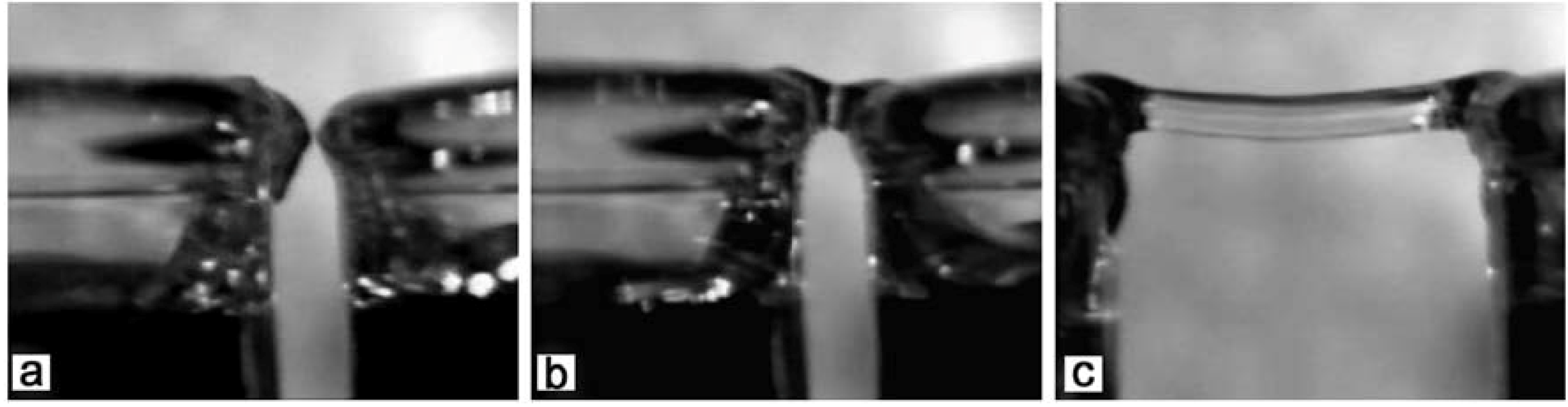
3.1. Bridge Formation

Whereas Lord Armstrong [85] used a cotton thread to create his water ropes, the bridge can easily be created without those, as first demonstrated by Uhlig in his World Wide Webpresentation [86] and then by Fuchs et al. who investigated the bridge formation using high speed visualization. This was done in glass beakers [87] (see Figure 5), Teflon beakers [89] (see Figure 6); and Woisetschläger et al. visualized the process using fringe projection [92] (see Figure 7).
Figure 5. Water bridge formation: (a) rise of water in both beakers after a high voltage was applied and a first ignition spark was observed, (b) spontaneous formation of a connection which remains stable after (c) pulling the beakers apart (from [87]).
Water 02 00381 g005
Figure 6. Formation of a water bridge between two Teflon beakers. (a) No current applied, (b) rise of the water surface mainly in the anodic beaker at approximately 15 kV dc, (c) spark, (d) water jet ejection leading to (e) a stable connection between the beakers (left cathode, right anode beaker) (from [89]).
Water 02 00381 g006
Figure 7. Bridge formation between two Teflon beakers using fringe projection. The shape of the water surface is contoured by the distortions of the otherwise parallel line reflections of an illuminated grid reflected by the surface (for a detailed description of this technique, see e.g., [159].) In (b) the high voltage was applied to the electrodes, in (e) a watery connection formed, in (f) the bridge was stabilized (from [92]).
Water 02 00381 g007
The formation can be summarized as follows: when the high voltage is applied, the water surfaces tower towards the opposite beaker (Figure 5a), particularly in the anodic beaker (Figure 6b and Figure 7b). Finally, small water jets are ejected with and without spark discharge (Figure 6c and Figure 7c) when the voltage exceeds approximately 15 kV. One of these jets eventually forms a watery connection (Figure 5c, Figure 6e and Figure 7f) which stabilizes while increasing in diameter (Figure 7f). This process can be compared [89] to a Taylor cone [160,98] formation. The water bridge can be considered as a result from the interaction of two counteracting cone jets. However, in electrohydrodynamics, such a cone-jet is normally the first stage of a droplet formation process.
After the Taylor cones establish a connection, it is typically very small (see Figure 7e). At this stage, the water bridge resembles a capillary bridge normally achieved with AC voltages by electrowetting (low frequencies) or dielectrophoreses (high frequencies). Such bridges made of water and aqueous solutions were recently studied by Jones et al. [151], a comprehensive review is given by Mugele and Baret [123]. They are normally established between two isolated electrodes and retain their capillary shape and proportions, whereas the water bridge undergoes a further stabilization and growth (see Figure 7f) before reaching its final equilibrium.
Observations have shown [10,51] that there is an asymmetry accompanying the bridge formation, the water in the high voltage (HV) beaker rises higher and forms more massive jets than the water from the grounded beaker. As shortly mentioned in the last chapter, Del Giudice et al. [93] proposed that this is due to the different levitation forces in each beaker. The authors also report on two other features during the bridge’s formation: The formation of a vortex around the electrodes and the cooling of the water surface along the line connecting the electrodes. The first observation is attributed to a macroscopic electronic vortex formation, the second to a symmetry break of the system due to the presence of the second beaker, respectively. The vortices are shown in Figure 8, while the cooling along the joining line prior to the bridge formation is depicted in Figure 9.
Figure 8. Thermographic vizualisation of the macroscopic Vortex in the beakers under application of high voltage before the water bridge is formed. The time interval between the images is 5 sec, the temperature scale is calibrated to the emissivity of water (0.96) (from [93]).
Water 02 00381 g008
Figure 9. Thermographic visualization of the bridge formation mechanism. First, the macroscopic vortices appear (a), then the water cools down at the joining line of the two electrodes (b). With the first sparks (c), the water heats up, and finally forms a water bridge (d). The time interval between the images is ~1 s, the temperature scale is calibrated to the emissivity of water (0.96). The dark spots on the electrodes in (c) and (d) are water droplets which were ejected during the process (from [93]).
Water 02 00381 g009

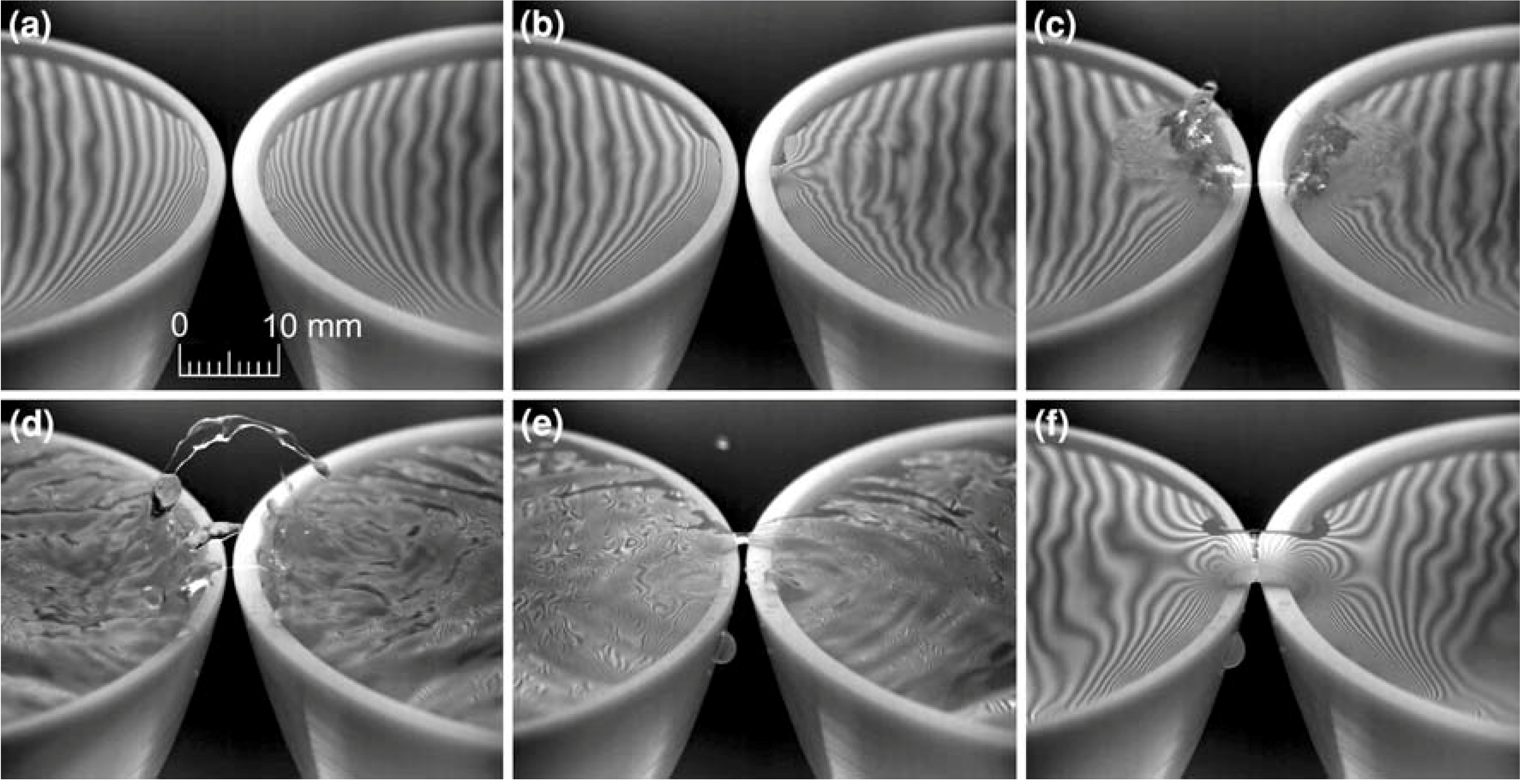
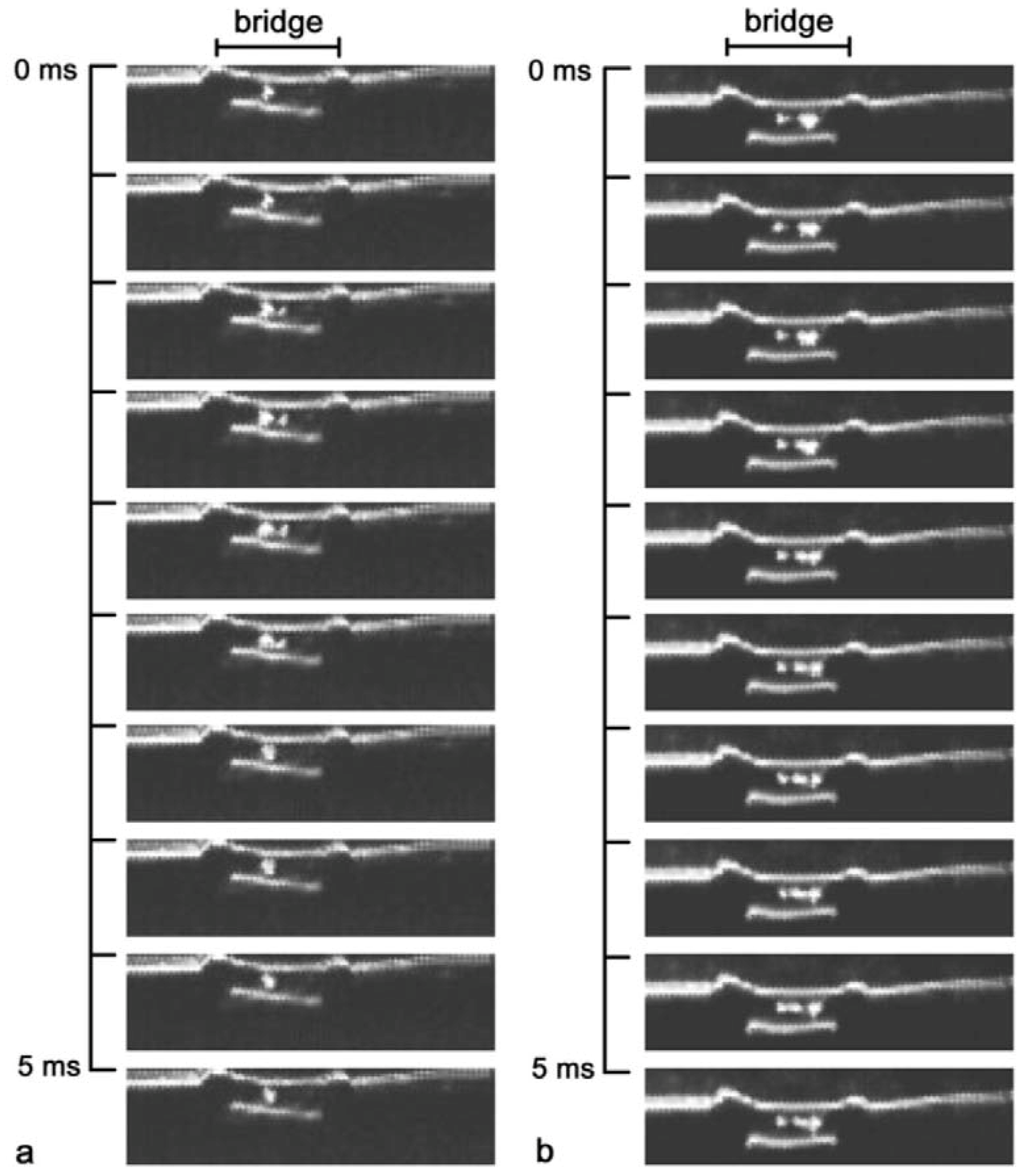
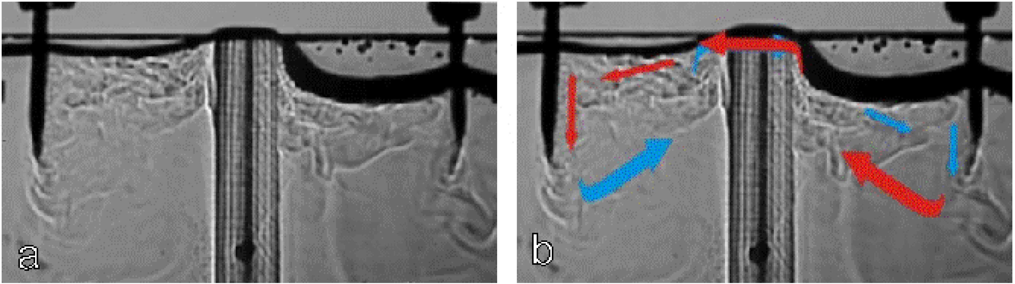
3.2. Bridge Properties

The most outstanding property of the bridge is, as indicated by its name, its stability, the fact that it floats, thereby seemingly defying gravity [88]. In order to understand this property Widom et al. [96] showed that the forces responsible for holding up the water bridge stem from the Maxwell electric field pressure tensor in dielectric polar fluids. In particular, the water bridge viewed as a flexible cable has an electric field induced tension sufficiently large so as to explain its support. The need for de-ionized water in the experiment is evidently due prohibiting conductivity effects from masking the insulating dielectric effects. The Maxwell pressure tensor inside of the water bridge is denoted by:
P i j = P g i j ε 4 π E i E j
Therefrom the authors successfully derived the catenary shape of the bridge. Moreover, they point out that in a polar liquid acting as a ferrofluid, the tension arises out of long ordered chains of low entropy aligned coherent dipolar domains. Based upon their work, Del Giudice et al. [93] discussed these domains in detail and gave qualitative explanations of previously experimentally reported features such as heliocoidal motion of the outer layer, the correlation of charge and mass transfer [87,92], the formation of micro and nanobubbles [90,92] and the somewhat counter-intuitive temperature dependence of the bridge’s stability: whereas cooling with ice destabilizes the bridge, it is easily formed using water close to the boiling point.
Amongst the optical properties of the bridge, already Lord Armstrong observed that “the water in passing the aperture becomes a little heated, and is rendered visible by a flicker” [85]. Fuchs et al. identified this flicker as density fluctuations [87] which they visualized with laser schlieren and fringe projection [89] techniques (see Figure 10). Since the local structure and therefore the local density of the bridge water are apparently the same as in the bulk liquid [90], the density differences are attributed to nano and micro bubbles [92].
Moreover, Lord Armstrong noticed a water flow in both directions, which he associated with a charge transport: “…the facts of the case seem to demonstrate that the negative current flows inside of the positive …” [85]. So far, this statement could not be verified; but a coupled bidirectional mass/charge transfer mechanism resulting in a lemniscate flow pattern has been reported [97] (see Figure 11). The nature of a possible charge carrier in this electrolysis-less system is compared to charged droplets in electrospray, where this issue is also still discussed [161].
When tracer particles were added for flow measurements using a laser Doppler anemometer and for visualization, the floating water bridge revealed a rotating outer layer, made visible in Figure 12 by particle tracks. Details on these measurement techniques can be found in the books by Merzkirch and Albrecht et al. [162,163]. Recorded with a laser Doppler anemometer, a tangential velocity of approximately 0.3 m s−1 was found at mid-length, the axial velocities changed between ±0.2 m s−1, depending on the main direction of mass transfer in the outer layer towards the cathode or the anode beaker.
Figure 10. Density gradients within the water bridge, recorded with a high speed camera at a 10 kHz frame rate (1/20,000 s exposure time), every 5th image is depicted. (a) shows a single structure typical for a ‘young’ bridge (5 min operation time). (b) shows multiple structures (after 30 min operation time). In these images, the length of the bridge was approximately 10 mm. (from [87]).
Water 02 00381 g010
Figure 11. Schlieren image of the flow inside the dual beaker configuration (a). In (b) arrows are added to demonstrate the lemniscate shaped flow from anode to cathode (red) and back (blue) (from [97])
Water 02 00381 g011
For Figure 12, eight consecutive images recorded with a frame rate of 1 kHz were added, revealing similar tangential velocities as recorded by the laser Doppler anemometer. Even without tracer particles, the rotation of the outer shell can be observed with a high-speed camera, because occasionally, single tiny gas bubbles crossed the bridge, rotating with the same speed as the tracer particles [92].
Figure 12. Tracking of particles on the surface of a 1.6 mm thick water bridge. The interval between the single images was 1 ms. Particles imaged through the bridge appear as large dots. The rotational direction was clockwise when looking towards the cathode beaker (left) (from [92]).
Water 02 00381 g012
The water bridge reveals a multilayered structure with mass transport mainly form the anode to the cathode beaker, a backflow preventing the cathodic beaker from overflowing thereby stabilizing the phenomenon for hours. The rotating outer shell reveals optical polarization effects (see Figure 13).
Figure 13. The water bridge between two crossed linear polarizer plates (a,b). P indicates the direction of the polarizer, A the direction of the analyzer. (c) Focused laser beam shining through the length of the bridge (from [92]).
Water 02 00381 g013
These polarization effects could be caused by electrically enforced birefringence in the outer layer, commonly known as the electro-optical Kerr effect. But due to the small Kerr constant of water the high-voltage field strength used for the floating water bridge is too low to observe a Kerr effect (e.g., [165]). Another possible cause could be reflection at the gas/water interfaces in a bubble network in the outer layer of the bridge [92]. To test that possibility, a laser beam was focused into the bridge from the side. This beam was guided along the inner surface of the bridge, similar to the behavior of an optical fiber (Figure 13c), and a strong scatter was observed only in the outer layer of the bridge, supporting this hypothesis. However, a recent two-dimensional neutron scattering showed a possible anisotropy of the first structural peak of D2O, which would probably also manifest itself by partial birefringence. Therefore, at this time, both birefringence [91] and scatter from micrometer-sized bubbles, slightly elongated in the electric field [92], are possible explanations for the observed optical activity.

3.3. Spectroscopic Investigations

So far, neutron [90,91] and Raman [95] scattering experiments have been successfully performed on the water bridge. Raman scattering was done with both DC and AC water bridges, and differences to bulk water were found. According to Pontiero et al. [95], one can assume that the Raman spectrum of a water bridge is given by the superposition of two distinct spectral features, which can be ascribed to the structure of bulk water and to a polarized structure induced by the applied electric field.
The authors attempted to extract the spectral contribution of polarized water by the difference between the water-bridge spectrum, at a given temperature, and the normal water spectrum, at the same temperature, and found that their analysis revealed that in any case the difference reduces to the same, roughly Gaussian shaped, spectral contribution. The interpretation of this contribution is not straight forward due, since the separating the temperature and polarization effects would require additional experiments [95]. The observed change could perhaps be compared to the one observed in the Raman spectra for methanol/acetone mixtures depending on the methanol concentration [164] which is due to vibrational coupling between the molecules, a model similar to the coherent domains proposed by Del Giudice et al. [93].
The neutron scattering measurements on a D2O bridge showed that the first structural peak overlaps with that of bulk D2O [90], thus showing that any density difference must be due to density changes on a larger scale, e.g., nano bubbles. A scattering increase at low Q values was interpreted to be possibly caused by such bubbles. In a 2D scattering experiment [91], a slight anisotropy of the first structural peak was found, indicating a preferred direction of a part of the D2O molecules or their tetrahedral clusters, respectively. This discovery can be considered corroborating the finding of the polarized structure found by Raman scattering [95]; in both cases, the contribution of the polarized/anisotropic fraction is very small. Clearly, more experiments are called for in order to clearly identify the novel structural features and thus to assess their contribution to the whole phenomenon.

3.4. Chemical Investigations

The investigations about if and to which extend electrochemistry plays a role in this phenomenon are in a very early stage. Most probably, if pure water is used, it can be neglected, since no electrolysis can be observed. Preliminary UV-Raman scattering (355 nm) investigations only revealed water [166], and addition of luminol in order to look for possible peroxide formation showed that this was indeed happening—but only if and when discharges appeared during the formation of the bridge (see Figure 14), there was no light emission once the bridge was running [167].
Figure 14. Discharge before the water bridge formation with diluted luminol solution. 12–14 kV dc, 0.5 mA [167].
Water 02 00381 g014
Nishiumi and Honda [94] reported that NaCl is rapidly transported through the bridge from cathode to anode. Moreover, they found that the addition of the soluble electrolytes, NaCl, NaOH and NH4Cl decreased the length of the water bridge from ~12 mm to 1mm when the concentration was increased from 0.0 to 1.5–2 × 10⁻¹ mol/L, respectively, whereas the addition of insoluble electrolyte Al2O3 showed (like the trace particles used by Woisetschläger et al. [92]) little effects on the length of water bridge. The most thoroughly discussed electrochemistry hitherto is the addition of a pH dye to visualize the charge distribution in the system [92] as shown in Figure 15.
In a recent paper [97] we further investigated the role of a pH indicator in this system, and found that one must be very careful when using it as charge visualization tool for the water bridge set-up, because the addition of a pH dye does not directly visualize charge. It raises the conductivity and induces electrochemical reactions.
Figure 15. Visualization of the pH value during bridge operation from red (pH 4) to violet (pH 10) for (a) a two beaker and (b) a three beaker arrangement. With orange pH 5, yellow pH 6, green pH 7, cyan pH 8 and blue pH 9. Before bridge operation the water had a pH value of 5 (orange). The color impression also depends on the thickness of the water layer (from [92]).
Water 02 00381 g015
Thus, it can be used as a tracer substance indicating the origin of the flow, but cannot be seen as a noninvolved indicator. The charges visualized by the dye are produced due to its presence and are mostly absent when pure water is used (see Figure 16).
Furthermore, in that paper, the authors conclude that with regard to the latest discussion about charge storage and transport in water [168,169,170,171], at least for the water bridge experiment, two statements can be made: First, there is a charge transport mechanism next to the electrochemical one, and second, these charges cannot be visualized by chemical means. The findings are still too preliminary to estimate possible repercussions of this statement; more experiments are called for in order to corroborate this claim.
Figure 16. Comparison between bridge operation (~10kV, 30 min) with dye (a,b) and bridge operation without dye (c) and the addition of the dye after shut down (d). (from [97]).
Water 02 00381 g016

4. Conclusions

The understanding of water and its interaction with electric fields seems so basic that one might wonder why any such research is at all necessary nowadays. However, when looking deeper into the field, it turns out that there is much special but little general knowledge. For example, there are many theories for each of the various anomalies of water—but rarely one theory that explains more than a few of these anomalies.
The water bridge is an example of the above cited paradox. Its formation can be described and very well understood by the methods developed for electrohydrodynamic atomization [98], its initial stage—the capillary bridge made of two droplets with a double funnel connection—has thoroughly been investigated—albeit for AC voltages—in the field of electrowetting [123]. However, its final stage—a macroscopic, rotating cylinder with special optical properties [92] through which two water currents run—still represents an interesting opportunity to further study and learn about the interactions of water with electric fields.
The question about the scale in which the phenomenon should be investigated is an interesting one, too. For instance, Pontiero et al. [95] write that they cannot confirm that the microscopic change in the local arrangement of water they found with Raman scattering is related to structure that sustains the bridge, it might just be a secondary effect playing no fundamental role. Thus, although finding peculiarities in the microscopic scale, the authors express their doubts about whether there is an important relation between those and the macroscopic properties of the bridge. On the other hand, Del Giudice et al. [93] claim that many bridge properties originate form the microscopic scale – a change of the local dynamics induced by the external electric field is said to be the basis wherefrom macroscopic properties are derived. Hopefully, future experiments and analyses will be able to validate these controversial approaches.
Another interesting issue is that of charge and mass transfer [97]. Naturally, there are a number of theories explaining conduction and liquid motion in high voltage electric fields [98]. Castellanos [98] generally considers only ions as charge carriers since electrons are normally very short-lived in liquids (see e.g., page 22 in reference [98]). Thus, when water is used, charge transport is accompanied by electrolysis (see e.g., page 83ff in ref. [98]). The fact that this process is absent when a floating water bridge is created raises the question about the nature of the charge transfer in that system. In ref. [97] it is compared it to the charging in electrohydrodynamic atomization processes. However, in their excellent review about this topic, Gañán-Calvo and Montanero point out that “…the role played by the liquid polarity and the behavior of ionic species in the liquid are not satisfactorily described…” and that “… These issues still stir controversy (…) and require clarification. …” (from [161], see also papers quoted therein). Thus it seems that this matter, too, is worthy of further studying, especially when considering a recent discussion about charge storage and transport in water [168,169,170,171].
Summarizing, a great deal about the floating water bridge has been understood so far. The formations of liquid jets and of a capillary bridge are phenomena well known not only for water but for polarizable liquids in general (see e.g., [98] and [123]). Whereas it still has to be determined whether new microscopic effects found by neutron [90,91] and Raman scattering [95] are essential for its macroscopic, especially its optical properties [92], the bridge certainly provided the grounds for such investigations and therefore lead to the quoted discoveries. Therefore, an answer to the question entitling this paper should be carefully phrased. Certainly, the investigation of the floating water bridge lead to new discoveries about water, both macroscopic and microscopic—but these were merely “hidden” in that sense that they only became evident under the application of electric fields. Moreover, there is nothing mythical about these effects—many of them can be unambiguously explained, and there is a number of applicable established theories for those which are still discussed. Therefore, a closer look at Armstong’s experiment did reveal hitherto “hidden” properties of water—but facts only, no myths.

Acknowledgments

First of all, very special acknowledgments go to my wife Astrid H. Paulitsch-Fuchs (Wetsus, Centre of Excellence for Sustainable Water Technology) and to Jakob Woisetschläger (Institute of Thermal Turbomachinery and Machine Dynamics, TU Graz, Austria) for their invaluable and ongoing support. Moreover, with great pleasure, I would like to thank Marie-Claire Bellissent-Funel (Laboratoire Léon Brillouin, Saclay), Eshel Ben-Jacob (Tel Aviv University), Mariano Bizzarri (Università La Sapienza, Roma), Harry Bruning (Wetsus—Centre of Excellence for Sustainable Water Technology), Cees Buisman (Wetsus—Centre of Excellence for Sustainable Water Technology), Gert-Jan Euverink (Wetsus—Centre of Excellence for Sustainable Water Technology), Friedemann Freund (NASA Ames Research Center, California), Karl Gatterer (Graz University of Technology), Emilio Del Giudice (Universitá di Milano), Ferenc Hajdu (Central Research Institute for Chemistry, Hungarian Academy of Sciences, Budapest), Franz Heitmeir (Graz University of Technology), Jan C. M. Marijnissen (Delft University of Technology), Hideo Nishiumi (Chem. Eng. Lab., Hosei University, Japan), Laurence Noirez (Laboratoire Léon Brillouin, CEA-CNRS/IRAMIS, CEA-Saclay), Gerald H. Pollack (University of Washington), Alan Soper (Rutherford Appleton Laboratories, ISIS, Oxford), Piergiorgio Spaggiari (Istituti Ospitalieri Di Cremona, Milano, Italy), José Teixeira (Laboratoire Léon Brillouin, CEA-CNRS/IRAMIS, CEA/Saclay), Giuseppe Vitiello (Universitá degli studi di Salerno), Vladimir Voeikov (M.V. Lomonosov Moscow State University), as well as Luewton L.F. Agostinho, Cees Kamp, Ingo Leusbrock, Martina Sammer and Adam Wexler (Wetsus, Centre of Excellence for Sustainable Water Technology) for the ongoing discussion on the water bridge phenomenon (in alphabetic order).

References

  1. Eisenberg, D.; Kauzman, W. The Structure and Properties of Water; Clarendon Press: Oxford, UK, 1969. [Google Scholar]
  2. Franks, F. Water: A Comprehensive Treatise; Plenum: New York, NY, USA; pp. 1972–1982.
  3. Ball, P. Life’s Matrix: a Biography of Water; Farrar, Straus, and Giroux: New York, NY, USA, 1999. [Google Scholar]
  4. Ball, P. Water: Water—an enduring mystery. Nature 2008, 452, 291–292. [Google Scholar]
  5. Ball, P. Water as an Active Constituent in Cell Biology. Chem. Rev. 2008, 108, 74. [Google Scholar]
  6. Chaplin, M.F. Thirty eight anomalies of water Part 1. Homeopath. Med. Panorama 2003, 11, 12–19. [Google Scholar]
  7. Chaplin, M.F. Thirty eight anomalies of water Part II. Homeopath. Med. Panorama 2003, 11, 22–28. [Google Scholar]
  8. Cho, C.H.; Singh, S.; Robinson, G.W. Understanding all of water's anomalies with a nonlocal potential. J. Chem. Phys. 1997, 107, 7979–7988. [Google Scholar] [CrossRef]
  9. The Hydrogen Bond: Recent Developments in Theory and Experiments; Schuster, P.; Zundel, G.; Sandorfy, C. (Eds.) North-Holland: Amsterdam, The Netherlands, 1976; Vols. I-III.
  10. Xantheas, S.S. Cooperativity and hydrogen bonding network in water clusters. Chem. Phys. 2000, 258, 225–231. [Google Scholar]
  11. Stanley, H.E. Liquid water: A very complex fluid. Pramana-J. Phys. 1999, 53, 53–83. [Google Scholar] [CrossRef]
  12. Reed, A.E.; Weinhold, F.; Curtiss, L.A.; Pochatko, D.J. Natural bond orbital analysis of molecular interactions: Theoretical studies of binary complexes of HF, H2O, NH3, N2, O2, F2, CO, and CO2 with HF, H2O, and NH3. J. Chem. Phys. 1986, 84, 5687–5705. [Google Scholar] [CrossRef]
  13. Ojamäe, L.; Hermansson, K. Ab Initio Study of Cooperativity in Water Chains: Binding Energies and Anharmonic Frequencies. J. Phys. Chem. 1994, 98, 4271–4282. [Google Scholar] [CrossRef]
  14. Pedulla, J. M.; Vila, F.; Jordan, K. D. Binding energy of the ring form of (H2O)6: Comparison of the predictions of conventional and localized‐orbital MP2 calculations. J. Chem. Phys. 1996, 105, 11091–11099. [Google Scholar] [CrossRef]
  15. Kumar, R.; Skinner, J.L. Water Simulation Model with Explicit Three-Molecule Interactions. J. Phys. Chem. 2008, 112, 8311–8318. [Google Scholar] [CrossRef]
  16. Reed, A.E.; Curtiss, L.A.; Weinhold, F. Intermolecular interactions from a natural bond orbital, donor-acceptor viewpoint. Chem. Rev. 1988, 88, 899–926. [Google Scholar] [CrossRef]
  17. Car, R.; Parrinello, M. Unified approach for molecular dynamics and density-functional theory. Phys. Rev. Lett. 1985, 55, 2471–2474. [Google Scholar] [CrossRef] [PubMed]
  18. Izvekov, S.; Voth, G.A. Car-Parrinello molecular dynamics simulation of liquid water: New results. J. Chem. Phys. 2002, 116, 10372–10376. [Google Scholar] [CrossRef]
  19. Lee, H.-S.; Tuckerman, M.E. Dynamical properties of liquid water from ab initio molecular dynamics performed in the complete basis set limit. J. Chem. Phys. 2007, 126, 164501–164516. [Google Scholar] [CrossRef] [PubMed]
  20. Schwegler, E.; Grossman, J.C.; Gygi, F.; Galli, G. Towards an assessment of the accuracy of density functional theory for first principles simulations of water. II. J. Chem. Phys. 2004, 121, 5400–5409. [Google Scholar] [CrossRef] [PubMed]
  21. Mantz, Y.A.; Chen, B.; Martyna, G.J. Structural Correlations and Motifs in Liquid Water at Selected Temperatures: Ab Initio and Empirical Model Predictions. J. Phys. Chem. B 2006, 110, 3540–3554. [Google Scholar] [CrossRef] [PubMed]
  22. Todorova, T.; Seitsonen, A.P.; Hutter, J.; Kuo, I.-F.W.; Mundy, C.J. Molecular Dynamics Simulation of Liquid Water: Hybrid Density Functionals. J. Phys Chem. B 2006, 110, 3685–3691. [Google Scholar] [CrossRef] [PubMed]
  23. van de Vondele, J.; Mohamed, F.; Krack, M.; Hutter, J.; Sprik, M.; Parrinello, M. The influence of temperature and density functional models in ab initio molecular dynamics simulation of liquid water. J. Chem. Phys. 2005, 122, 14515. [Google Scholar] [CrossRef] [PubMed] [Green Version]
  24. Bukowski, R.; Szalewicz, K.; Groenenboom, G.C.; van der Avoird, A. Predictions of the properties of water from first principles. Science 2007, 315, 1249–1252. [Google Scholar] [CrossRef] [PubMed]
  25. Fanourgakis, G.S.; Xantheas, S.S. The Flexible, Polarizable, Thole-Type Interaction Potential for Water (TTM2-F) Revisited. J. Phys. Chem. A 2006, 110, 4100–4106. [Google Scholar] [CrossRef] [PubMed]
  26. Tuckerman, M.; Laasonen, K.; Sprik, M.; Parrinello, M. Ab Initio Molecular Dynamics Simulation of the Solvation and Transport of H3O+ and OH Ions in Water. J. Phys. Chem. 1995, 99, 5749–5752. [Google Scholar] [CrossRef]
  27. Hofer, T.S.; Tran, H.T.; Schwenk, C.F.; Rode, B.M. Characterization of dynamics and reactivities of solvated ions by ab initio simulations. J. Comput. Chem. 2004, 25, 211–217. [Google Scholar] [CrossRef] [PubMed]
  28. Kuharski, R.A.; Rossky, P.J. A quantum mechanical study of structure in liquid H2O and D2O. J. Chem. Phys. 1985, 82, 5164–5177. [Google Scholar] [CrossRef]
  29. Poulsen, J.A.; Nyman, G.; Rossky, P.J. Feynman-Kleinert Linearized Path Integral (FK-LPI) Algorithms for Quantum Molecular Dynamics, with Application to Water and He(4). J. Chem. Theory Comput. 2006, 2, 1482–1491. [Google Scholar] [CrossRef]
  30. Stern, H.A.; Rittner, F.; Berne, B.J.; Friesner, R.A. Combined fluctuating charge and polarizable dipole models: Application to a five-site water potential function. J. Chem. Phys. 2001, 115, 2237–2251. [Google Scholar] [CrossRef]
  31. Paesani, F.; Iuchi, S.; Voth, G.A. Quantum effects in liquid water from an ab initio-based polarizable force field. J. Chem. Phys. 2007, 127, 074506–074514. [Google Scholar] [CrossRef] [PubMed]
  32. de la Peña, L.H.; Kusalik, P.G. Quantum effects in light and heavy liquid water: A rigid-body centroid molecular dynamics study. J. Chem. Phys. 2004, 121, 5992–6002. [Google Scholar] [CrossRef] [PubMed]
  33. Paesani, F.; Voth, G.A. The Properties of Water: Insights from Quantum Simulations. J. Phys. Chem. B 2009, 113, 5702–5719. [Google Scholar] [CrossRef] [PubMed]
  34. Hadju, F.; Lengyel, S.; Pálinkás, G. X-ray scattering and radial distribution function of liquid water. J. Appl. Cryst. 1976, 9, 134–138. [Google Scholar] [CrossRef]
  35. Hadju, F. A Model of liquid water—Tetragonal Clusters: Description and determination of parameters. Acta Chim. 1977, 93, 371–394. [Google Scholar]
  36. Hadju, F. The structural model of water II—the structure of amorphous ice and structural relations between water and some ice polymorphs on the basis of the tetragonal cluster model. Acta Chim. Acad. Sci. Hung. 1978, 96, 355–371. [Google Scholar]
  37. Teixeira, J.; Luzar, A. Physics of Liquid Water. Structure and Dynamics. In Hydration Processes in Biology: Theoretical and Experimental Approaches (NATO ASI series A); Bellissent-Funel, M.C., Ed.; IOS Press: Amsterdam, The Netherlands, 1999; pp. 35–65. [Google Scholar]
  38. Soper, A.K. An asymmetric model for water structure. J. Phys. Condens. Matter 2005, 17, S3273–S3282. [Google Scholar] [CrossRef]
  39. Soper, A.K. Joint structure refinement of X-ray and neutron diffraction data on disordered materials: application to liquid water. J. Phys. Condens. Matter 2007, 19, 335206:1–335206:18. [Google Scholar]
  40. Stanley, H.E.; Buldyrev, S.V.; Franzese, G.; Giovambattista, N.; Starr, F.W. Static and dynamic heterogeneitities in water. Phil. Trans. R. Soc. A 2005, 363, 509–523. [Google Scholar] [CrossRef] [PubMed]
  41. Stanley, H.E.; Teixeira, J. Interpretation of the unusual behavior of H2O and D2O at low temperature: tests of a percolation model. J. Chem. Phys. 1980, 73, 3404–3422. [Google Scholar] [CrossRef]
  42. Stanley, H.E.; Teixeira, J.; Geiger, A.; Blumberg, R.L. Interpretation of the unusual behavior of H2O and D2O at low temperature: Are concepts of percolation relevant to the “puzzle of liquid water”? Phyisca 1981, 106, 260–277. [Google Scholar]
  43. Mishima, O.; Stanley, H.E. The relationship between liquid, supercooled and glassy water. Nature 1998, 396, 329–335. [Google Scholar]
  44. Yamada, M.; Mossa, S.; Stanley, H.E.; Sciortino, F. Interplay between time-temperature transformation and the liquid-liquid phase transition in water. PRL 2002, 88, 195701–195704. [Google Scholar] [CrossRef]
  45. Leetmaa, M.; Ljungberg, M.; Ogasawara, H.; Odelius, M.; Näslund, L.-Å.; Nilsson, A.; Pettersson, L.G.M. Are recent water models obtained by fitting diffraction data consistent with infrared/Raman and x-ray absorption spectra? J. Chem. Phys. 2006, 125, 244510:1–244510:12. [Google Scholar] [CrossRef]
  46. Leetmaa, M.; Wikfeldt, K.T.; Ljungberg, M.P.; Odelius, M.; Swenson, J.; Nilsson, A.; Pettersson, L.G.M. Diffraction and IR/Raman data do not prove tetrahedral water. J. Chem. Phys. 2008, 129, 084502:1–084502:13. [Google Scholar] [CrossRef]
  47. Chumaevskii, N.A.; Rodnikova, M.N. Some peculiarities of liquid water structure. J. Mol. Liquids 2003, 106, 167–177. [Google Scholar] [CrossRef]
  48. Bakker, H.J.; Skinner, J.L. Vibrational Spectroscopy as a Probe of Structure and Dynamics in Liquid Water. Chem. Rev. 2010, 110, 1498–1517. [Google Scholar] [CrossRef]
  49. Molinero, V.; Moore, E.B. Water Modeled As an Intermediate Element between Carbon and Silicon. J. Phys. Chem. B 2009, 113, 4008–4016. [Google Scholar] [CrossRef] [PubMed]
  50. Miyazaki, M.; Fujii, A.; Ebata, T.; Mikami, N. Infrared spectroscopic evidence for protonated water clusters forming nanoscale cages. Science 2004, 304, 1134–1137. [Google Scholar] [CrossRef] [PubMed]
  51. Shin, J.-W.; Hammer, N.I.; Diken, E.G.; Johnson, M.A.; Walters, R.S.; Jaeger, T.D.; Duncan, M.A.; Christie, R.A.; Jordan, K.D. Infrared Signature of Structures Associated with the H+(H2O)n (n = 6 to 27) Clusters. Science 2004, 304, 1137–1140. [Google Scholar] [CrossRef] [PubMed]
  52. Nibbering, E.T.J.; Elsaesser, T. Ultrafast Vibrational Dynamics of Hydrogen Bonds in the Condensed Phase. Chem. Rev. 2004, 104, 1887–1914. [Google Scholar] [CrossRef] [PubMed]
  53. Wernet, Ph.; Nordlund, D.; Bergmann, U.; Cavalleri, M.; Odelius, M.; Ogasawara, H.; Näslund, L.-Å.; Hirsch, T. K.; Ojamäe, L.; Glatzel, P.; Pettersson, L.G.M.; Nilsson, A. The structure of the first coordination shell in liquid water. Science 2004, 304, 995–999. [Google Scholar] [CrossRef] [PubMed]
  54. Odelius, M.; Cavalleri, M.; Nilsson, A.; Pettersson, L.G.M. X-ray absorption spectrum of liquid water from molecular dynamics simulations: Asymmetric model. Phys. Rev. B 2006, 73, 024205:1–024205:6. [Google Scholar] [CrossRef]
  55. Smith, J.D.; Cappa, C.D.; Wilson, K.R.; Messer, B.M.; Cohen, R.C.; Saykally, R.J. Energetics of Hydrogen Bond Network Rearrangements in Liquid Water. Science 2004, 306, 851–853. [Google Scholar] [CrossRef] [PubMed]
  56. Head-Gordon, T.; Johnson, M.E. Tetrahedral structure or chains for liquid water. PNAS 2006, 103, 7973–7977. [Google Scholar] [CrossRef] [PubMed]
  57. Tse, J.S.; Shaw, D.M.; Klug, D.D.; Patchkovskii, S.; Vankó, G.; Monaco, G.; Krisch, M. X-Ray Raman Spectroscopic Study of Water in the Condensed Phases. Phys. Rev. Lett. 2008, 100, 095502:1–095502:4. [Google Scholar] [CrossRef]
  58. Lin, I.J.; Yotvat, J. Exposure of irrigation and drinking water to a magnetic field with controlled power and direction. J. Magn. Magn. Mater. 1990, 83, 525–526. [Google Scholar] [CrossRef]
  59. Sisken, B.; Walker, J. Therapeutic aspects of electromagnetic fields for soft tissue healing. In Electromagnetic Fields: Biological Interactions and Mechanisms; Blank, M., Ed.; American Chemical Society: Washington, DC, USA, 1995. [Google Scholar]
  60. Basset, C.A. Bioelectromagnetics in the service of medicine. In Electromagnetic Fields: Biological Interactions and Mechanisms; Blank, M., Ed.; American Chemical Society: Washington, DC, USA, 1995. [Google Scholar]
  61. Walleczek, J. Magnetokinetic effects on radical pairs: A paradigm for magnetic field interactions with biologic systems at lower than thermal energy. In Electromagnetic Fields: Biological Interactions and Mechanisms; Blank, M., Ed.; American Chemical Society: Washington, DC, USA, 1995. [Google Scholar]
  62. Colic, M.; Morse, D. The elusive mechanism of the magnetic ‘memory’ of water. Colloid. Surfaces A 1999, 154, 167–174. [Google Scholar] [CrossRef]
  63. Alimi, F.; Tlili, M.; Amor, M.B.; Gabrielli, C.; Maurin, G. Influence of magnetic field on calcium carbonate precipitation. Desalination 2007, 206, 163–168. [Google Scholar] [CrossRef]
  64. Higashitani, K.; Kage, A.; Katamura, S.; Imai, K.; Hatade, A. Effects of a Magnetic Field on the Formation of CaCO3 Particles. J. Colloid Interf. Sci. 1993, 156, 90–95. [Google Scholar] [CrossRef]
  65. Pach, L.; Duncan, S.; Roy, R.; Komarneni, S. Effects of a magnetic field on the precipitation of calcium carbonate. J. Materials Sci. Letters 1996, 15, 613–615. [Google Scholar] [CrossRef]
  66. Roy, R.; Tiller, W.A.; Bell, I.; Hoover, M.R. The structure of liquid water; novel insights from materials research; potential relevance to homeopathy. Mater. Res. Innov. 2005, 9, 577–608. [Google Scholar]
  67. Cowan, M.L.; Bruner, B.D.; Huse, N.; Dwyer, J.R.; Chugh, B.; Nibbering, E.T.J.; Elsaesser, T.; Miller, R.J.D. Ultrafast memory loss and energy redistribution in the hydrogen bond network of liquid H2O. Nature 2005, 434, 199–202. [Google Scholar] [CrossRef] [PubMed]
  68. Juraschek, R.; Röllgen, F.W. Pulsation phenomena during electrospray ionization. Int. J. Mass. Spectrom. 1998, 177, 1–15. [Google Scholar] [CrossRef]
  69. Yu, J.H.; Fridrikh, S.V.; Rutledge, G.C. The role of elasticity in the formation of electrospun fibers. Polymer 2006, 47, 4789–4797. [Google Scholar] [CrossRef]
  70. He, J.-H.; Wan, Y.-Q.; Yu, J.-Y. Critical length of straight jet in electrospinning. Polymer 2005, 46, 12637–12640. [Google Scholar] [CrossRef]
  71. Spivak, A.E.; Dzenis, Y.A.; Reneker, D.H. A model of steady state jet in the electrospinning process. Mech. Res. Commun. 2000, 27, 37–42. [Google Scholar] [CrossRef]
  72. Feng, J. Stretching of a straight electrically charged viscoelastic jet. J. Non-Newtonian Fluid Mech. 2003, 116, 55–70. [Google Scholar] [CrossRef]
  73. Marginean, I.; Parvin, L.; Heffernan, L.; Vertes, A. Flexing the electrified meniscus: the birth of a jet in electrosprays. Anal. Chem. 2004, 76, 4202–4207. [Google Scholar] [CrossRef] [PubMed]
  74. Gañán-Calvo, A.M. On the theory of electrohydrodynamically driven capillary jets. J. Fluid Mech. 1997, 335, 165–188. [Google Scholar] [CrossRef]
  75. Jaworek, A.; Krupa, A. Classification of the modes of EHD spraying. J. Aerosol Sci. 1999, 30, 873–893. [Google Scholar] [CrossRef]
  76. Cherney, L.T. Electrohydrodynamics of electrified liquid menisci and emitted jets. J. Aerosol Sci. 1999, 30, 851–862. [Google Scholar] [CrossRef]
  77. Yan, F.; Farouk, B.; Ko, F. Numerical modeling of an electrostatically driven liquid meniscus in the cone-jet mode. J. Aerosol Sci. 2003, 34, 99–116. [Google Scholar] [CrossRef]
  78. Lopez-Herrera, J. M.; Gañán-Calvo, A.M.; Perez-Saborid, M. One-dimensional simulation of the breakup of capillary jets of conducting liquids. Application to E.H.D. spraying. J. Aerosol Sci. 1999, 30, 895–912. [Google Scholar]
  79. He, J.-H.; Wan, Y.-Q.; Yu, J.-Y. Scaling law in electrospinning: relationship between electric current and solution flow rate. Polymer 2005, 46, 2799–2801. [Google Scholar] [CrossRef]
  80. Wei, J.; Shui, W.; Zhou, F.; Lu, Y.; Chen, K.; Xu, G.; Yang, P. Naturally and externally pulsed electrospray. Mass Spectrometry Rev. 2002, 2, 148–162. [Google Scholar] [CrossRef]
  81. Hartman, R.P.A.; Brunner, D.J.; Camelot, D.M.A.; Marijnissen, J.C.M.; Scarlett, B. Jet break-up in electrohydrodynamic atomization in the cone-jet mode. J. Aerosol Sci. 2000, 31, 65–95. [Google Scholar] [CrossRef]
  82. Hartman, R.P.A.; Brunner, D.J.; Camelot, D.M.A.; Marijnissen, J.C.M.; Scarlett, B. Electrohydrodynamic atomization in the cone-jet mode. Physical modelling of the liquid cone and jet. J. Aerosol Sci. 1999, 30, 823–849. [Google Scholar] [CrossRef]
  83. Hartman, R.P.A.; Borra, J.-P.; Brunner, D.J.; Marijnissen, J.C.M.; Scarlett, B. The evolution of electrohydrodynamic sprays produced in the cone-jet mode, a physical model. J. Electrostat. 1999, 47, 143–170. [Google Scholar] [CrossRef]
  84. Gunji, M.; Washizu, M. Self-propulsion of a water droplet in an electric field. J. Phys.-D-Appl. Phys. 2005, 38, 2417–2423. [Google Scholar] [CrossRef]
  85. Armstrong, Lord W. Electrical Phenomena. The Electrical Engineer 1893, 10 February, 154–155. [Google Scholar]
  86. Uhlig, W.; Laboratory of Inorganic Chemistry, ETH Hönggerberg—HCI, Zürich, Switzerland. Personal communication, 2005.
  87. Fuchs, E.C.; Woisetschläger, J.; Gatterer, K.; Maier, E.; Pecnik, R.; Holler, G.; Eisenkölbl, H. The floating water bridge. J. Phys.-D-Appl. Phys. 2007, 40, 6112–6114. [Google Scholar] [CrossRef]
  88. Hand, E. Water doesn’t mind the gap. Nature 2007, 449, 517. [Google Scholar] [CrossRef] [PubMed]
  89. Fuchs, E.C.; Gatterer, K.; Holler, G.; Woisetschläger, J. Dynamics of the floating water bridge. J. Phys.-D-Appl. Phys. 2008, 41, 185502–185507. [Google Scholar] [CrossRef]
  90. Fuchs, E.C.; Bitschnau, B.; Woisetschläger, J.; Maier, E.; Beuneu, B.; Teixeira, J. Neutron Scattering of a Floating Heavy Water Bridge. J. Phys.-D-Appl. Phys. 2009, 42, 065502:1–065502:4. [Google Scholar] [CrossRef]
  91. Fuchs, E.C.; Baroni, P.; Bitschnau, B.; Noirez, L. Two-Dimensional Neutron Scattering in a Floating Heavy Water Bridge. J. Phys.-D-Appl. Phys. 2010, 43, 105502:1–105502:5. [Google Scholar] [CrossRef]
  92. Woisetschläger, J.; Gatterer, K.; Fuchs, E.C. Experiments in a floating water bridge. Exp. Fluids 2010, 48-1, 121–131. [Google Scholar]
  93. Del Giudice, E.; Fuchs, E.C.; Vitiello, G. Collective molecular dynamics of a floating water bridge. Water(Seattle) 2010, 2, 69–82. [Google Scholar]
  94. Nishiumi, H.; Honda, F. Effects of Electrolyte on Floating Water Bridge. Res. Let. Phys. Chem. 2009, 2009, 371650:1–371650:3. [Google Scholar]
  95. Ponterio, R.C.; Pochylski, M.; Aliotta, F.; Vasi, C.; Fontanella, M. E.; Saija, F. Raman scattering measurements on a floating water bridge. J. Phys.-D-Appl. Phys. 2010, 43, 175405:1–175405:8. [Google Scholar] [CrossRef]
  96. Widom, A.; Swain, J.; Silverberg, J.; Sivasubramanian, S.; Srivastava, Y.N. Theory of the Maxwell pressure tensor and the tension in a water bridge. Phys. Rev. E 2009, 80, 016301:1–016301:7. [Google Scholar] [CrossRef]
  97. Fuchs, E.C.; Eisenhut, M.; Agostinho, L.L.F.; Woisetschläger, J. Mass and Charge Transfer within a Floating Water Bridge. In SPIE Conference Proceedings of the XII. International Conference on Laser Applications in Life Sciences, Oulu, Finland, June 2010. submitted.
  98. Castellanos, A. Electrohydrodynamics: No. 380 (Courses and Lectures); Springer: Vienna, Austria, 1998. [Google Scholar]
  99. Sumoto, I. An interesting Phenomenon Observed on Some Dielectrics. J. Phys. Soc. Jpn. 1955, 10, 494. [Google Scholar] [CrossRef]
  100. Pohl, H.A. Some effects of nonuniform fields on dielectrics. J. Appl. Phys. 1958, 29, 1182–1188. [Google Scholar] [CrossRef]
  101. Parmar, D.S.; Jalaluddin, A.K. Dielectrophoretic forces in liquids. Jpn. J. Appl. Phys. 1974, 13, 793–798. [Google Scholar] [CrossRef]
  102. Pickard, W.F. Electrical force effects in dielectric liquids. In Progress in Dielectrics Vol. 6; Birks, J.B., Hart, J., Eds.; Academic Press: New York, NY, USA, 1965; pp. 1–39. [Google Scholar]
  103. Krasucki, Z. Breakdown of liquid dielectrics. Proc. Roy. Soc. London Ser. A. 1966, 294, 393–404. [Google Scholar] [CrossRef]
  104. Maxwell, J.C. A Treatise on Electricity and Magnetism, 3rd ed.; Clarendon Press: Oxford, UK, 1891; Vol. 1, Chapter V; pp. 159–162. [Google Scholar]
  105. Landau, L.D.; Lifshitz, E.M. Electrodynamics of Continuous Media; Pergamon Press: Oxford, UK, 1960; Chapter II; pp. 66–67. [Google Scholar]
  106. Parmar, D.S.; Labroo, B. Dielectrophoretic transients effects on liquid stability. Pramana-J. Phys. 1985, 25, 81–94. [Google Scholar] [CrossRef]
  107. Hughes, M.P.; Pething, R.; Wang, X.-B. Dielectrophoretic forces on particles in travelling electric fields. J. Phys.-D-Appl. Phys. 1996, 29, 474–482. [Google Scholar] [CrossRef]
  108. Techaumnat, B.; Eua-arporn, B.; Takuma, B. Calculation of electric field and dielectrophoretic force on spherical particles in chain. J. Appl. Phys. 2004, 95, 1586–1593. [Google Scholar] [CrossRef]
  109. Cloupeau, M.; Prunet-Foch, B. Electrostatic spraying of liquids in cone-jet mode. J. Electrostatics 1989, 22, 135–159. [Google Scholar] [CrossRef]
  110. Gomez, A.; Tang, K. Charge and fission of droplets in electrostatic sprays. Phys. Fluids 1994, 6, 404–414. [Google Scholar] [CrossRef]
  111. Chen, D.-R.; Pui, D.Y.H.; Kaufman, S.L. Electrospraying of conducting liquids for monodisperse aerosol generation in the 4 nm to 1.8 µm diameter range. J. Aerosol Sci. 1995, 26, 963–977. [Google Scholar] [CrossRef]
  112. Agostinho, L.L.F.; Wetsus Centre of Excellence for Sustainable Water Technology, Agora 1, 8900 CC Leeuwarden, the Netherlands. Personal communication, 2010.
  113. Hartman, R.P.A.; Marijnissen, J.C.M.; Scarlett, B. Electrohydrodynamic atomization in the cone-jet mode. A physical model of the liquid cone and jet. J. Aerosol Sci. 1997, 28, S527–S528. [Google Scholar] [CrossRef]
  114. Gomez, A.; Tang, K. Atomization and dispersion of quasi-monodisperse electrostatic sprays of heptane. In Proceedings of the 5th International Conference on Liquid Atomization and Spray Systems, 15–18 July, 1991; US Department of Commerce, National Institute of Standards and Technology: Gaithersburg, MD, USA, 1991; pp. 805–812. [Google Scholar]
  115. Rayleigh, L.F. On the instability of jets. Proc. London Math. Soc. 1878, 11, 4–13. [Google Scholar] [CrossRef]
  116. Weber, C. Zum Zerfall eines Flüssigkeitsstrahles. Z. Für Angew. Math. und Mech. 1931, 11, 136. [Google Scholar] [CrossRef]
  117. Yuen, M.C. Non-linear capillary instability of a liquid jet. J. Fluid Mech. 1968, 33, 151–163. [Google Scholar] [CrossRef]
  118. Rutland, D.F.; Jameson, G.J. Theoretical prediction of the sizes of drops formed in the break-up of capillary jets. Chem. Eng. Sci. 1970, 25, 1689–1698. [Google Scholar] [CrossRef]
  119. Melcher, J.R. Field-Coupled Surface Waves; MIT Press: Cambridge, MA, USA, 1963. [Google Scholar]
  120. Parkin, C.S. The Production of Droplets from Liquid Jets by Capillary and Electrohydrodynamic Instabilities. PhD Thesis, Loughborough University of Technology, Leicestershire, UK, 1973. [Google Scholar]
  121. Turnbull, R.J. On the instability of an electrostatically sprayed liquid jet. IEEE Trans. Ind. Appl. 1992, 28, 1432–1438. [Google Scholar] [CrossRef]
  122. Jones, T.B. On the relationship of dielectrophoresis and electrowetting. Langmuir 2002, 18, 4437–4443. [Google Scholar] [CrossRef]
  123. Mugele, F.; Baret, J.-C. Electrowetting: from basics to applications. J. Phys. Condens. Matter 2005, 17, R705–R774. [Google Scholar] [CrossRef]
  124. Saville, D.A. ELECTROHYDRODYNAMICS: The Taylor-Melcher Leaky Dielectric Model. Annu. Rev. Fluid Mech. 1997, 29, 27–64. [Google Scholar] [CrossRef]
  125. Schneider, J.M.; Lindblad, N.R.; Hendricks, C.D.; Crowley, J.M. Stability of an electrified liquid jet. J. Appl. Phys. 1967, 38, 2599–2605. [Google Scholar] [CrossRef]
  126. Neukermans, A. Stability criteria of an electrified liquid jet. J. Appl. Phys. 1973, 44, 4769–4770. [Google Scholar] [CrossRef]
  127. Fernández de la Mora, J.; Loscertales, I.G. The current emitted by highly conducting Taylor cones. J. Fluid Mech. 1994, 260, 155–184. [Google Scholar] [CrossRef]
  128. Gañán-Calvo, A.M. Cone-jet analytical extension of Taylor's electrostatic solution and the asymptotic universal scaling laws in electrospraying. Phys. Rev. Lett. 1997, 79, 217–220. [Google Scholar] [CrossRef]
  129. Gañán-Calvo, A.M.; Dávila, J.; Barrero, A. Current and droplet size in the electrospraying of liquids. Scaling Laws. J. Aerosol Sci. 1997, 28, 249–275. [Google Scholar] [CrossRef]
  130. Kuli, M.E.; Abrahams, J.P.; Marijnissen, J.C.M. Nano-dispensing by electrospray for biotechnology. Biotechnol. J. 2006, 1, 969–975. [Google Scholar] [CrossRef] [PubMed]
  131. Xie, J.; Marijnissen, J.C.M.; Wang, C.-H. Microparticles developed by electrodydrodynamic atomization for the local delivery of anticancer drug to treat C6 glioma in vitro. Biomaterials 2006, 27, 3321–3332. [Google Scholar] [CrossRef] [PubMed]
  132. Sugiyama, H.; Ogura, H.; Otsubo, Y. Development of EHD Motor of Water Solution Utilizing Electrohydrodynamics. In Proceedings of the XV International Congress on Rheology: The Society of Rheology 80th Annual Meeting, Monterey, CA, USA, 3–8 August 2008; Co, A., Leal, G., Colby, R., Giacomin, A.J., Eds.; American Institute of Physics: New York, NY, USA, 2008; pp. 1453–1455. [Google Scholar]
  133. Ohyama, R.; Kumeta, M.; Ueda, A.; Watson, A.; Chang, J.S. A fundamental characteristic and image analysis of liquid flow in an AW type EHD pump. J. Visualization 2005, 8, 339–346. [Google Scholar] [CrossRef]
  134. Burcham, C.L.; Saville, D.A. The electrohydrodynamic stability of a liquid bridge: microgravity experiments on a bridge suspended in a dielectric gas. J. Fluid Mech. 2000, 405, 37–56. [Google Scholar] [CrossRef]
  135. Rai, D.; Kulkarni, A.D.; Gejji, S.P.; Pathak, R.K. Water clusters (H2O)n, n = 6–8, in external electric fields. J. Chem. Phys. 2008, 128, 34310:1–34310:14. [Google Scholar] [CrossRef]
  136. Choi, Y.C.; Pak, C.; Kim, K.S. Electric field effects on water clusters (n = 3–5): Systematic ab initio study of structures, energetics, and transition states. J. Chem. Phys. 2006, 124, 94308:1–94308:4. [Google Scholar]
  137. Del Giudice, E.; Istituto Nazionale Fisica Nucleare (INFN), Via Celoria 16, 20133 Milano, Italy. Personal communication, 2008.
  138. Chatzidimitriou-Dreismann, C.A.; Krieger, U.K.; Möller, A.; Stern, M. Evidence of Quantum Correlation Effects of Protons and Deuterons in the Raman Spectra of Liquid H2O-D2O. Phys. Rev. Lett. 1995, 75, 3008–3011. [Google Scholar] [CrossRef] [PubMed]
  139. Chatzidimitriou-Dreismann, C.A.; Redah, T.A.; Streffer, R.M.F.; Mayers, J. Anomaleous deep inelastic neutron scattering from liquid H2O–D2O: evidence of nuclear quantum entanglement. Phys. Rev. Lett. 1997, 79, 2839–2842. [Google Scholar] [CrossRef]
  140. Torii, H. Comment on “Evidence of Quantum Correlation Effects of Protons and Deuterons in Raman Spectra of Liquid H2O-D2O”. Phys. Rev. Lett. 2000, 84, 5236. [Google Scholar] [CrossRef] [PubMed]
  141. Chatzidimitriou-Dreismann, C.A.; Abdul-Redah, T.; Kolaric, B.; Juranic, I. Reply to Comment on “Evidence of Quantum Correlation Effects of Protons and Deuterons in Raman Spectra of Liquid H2O-D2O”. Phys. Rev. Lett. 2000, 84, 5237. [Google Scholar] [CrossRef] [PubMed]
  142. Cyrot, M. Ginzburg-Landau theory for superconductors. Rep. Prog. Phys. 1973, 36, 103–158. [Google Scholar] [CrossRef]
  143. Hendricks, R.W.; Mardon, P.G.; Schaffer, L.B. X-ray zero-angle scattering cross section of water. J. Chem. Phys. 1974, 61, 319–322. [Google Scholar] [CrossRef]
  144. Bosio, L.; Teixeira, J.; Stanley, H.E. Enhanced density fluctuations in supercooled H2O, D2O, and ethanol-water solutions: evidence from small-angle X-ray scattering. Phys. Rev. Lett. 1981, 46, 597–600. [Google Scholar] [CrossRef]
  145. Huang, C.; Wikfeldt, K.T.; Tokushima, T.; Nordlund, D.; Harada, Y.; Bergmann, U.; Niebuhr, M.; Weiss, T.M.; Horikawa, Y.; Leetmaa, M.; Ljungberg, M.P.; Takahashi, O.; Lenz, A.; Ojamäe, L.; Lyubartsev, A.P.; Shin, S.; Pettersson, L.G.M.; Nilsson, A. The inhomogeneous structure of water at ambient conditions. PNAS 2009, 106, 15214–15216. [Google Scholar] [CrossRef] [PubMed]
  146. Yinnon, C.A.; Yinnon, T.A. Domains in aqueous solutions: theory and experimental evidence. Mod. Phys. Lett. B 2009, 23, 1959–1973. [Google Scholar] [CrossRef]
  147. Arani, R.; Bono, I.; Del Giudice, E.; Preparata, G. QED Coherence and the thermodynamics of water. Int. J. Mod. Phys. B 1995, 9, 1813–1841. [Google Scholar] [CrossRef]
  148. Del Giudice, E.; Preparata, G.; Vitiello, G. Water as a free electric dipole laser. Phys. Rev. Let. 1988, 61, 1085–1088. [Google Scholar] [CrossRef]
  149. Del Giudice, E.; Vitiello, G. Role of the electromagnetic field in the formation of domains in the process of symmetry-breaking phase transitions. Phys. Rev. A 2006, 74, 022105:1–022105:9. [Google Scholar]
  150. Yanga, M.; Skinner, J.L. Signatures of coherent vibrational energy transfer in IR and Raman line shapes for liquid water. Phys. Chem. Chem. Phys. 2010, 12, 982–991. [Google Scholar] [CrossRef] [PubMed]
  151. Jones, T.B.; Wang, K.L.; Yao, D.J. Frequency-dependent electromechanics of aqueous liquids: Electrowetting and dielectrophoresis. Langmuir 2004, 20, 2813–2818. [Google Scholar] [CrossRef]
  152. Del Giudice, E.; Preparata, G. A new QED picture of water. In Macroscopic Quantum Coherence; Sassaroli, E., Srivasta, Y., Swain, J., Widom, A., Eds.; World Scientific: Singapore, 1998; pp. 108–129. [Google Scholar]
  153. Zheng, J.-M.; Chin, W.-C.; Khijniak, E.; Khijniak, E., Jr.; Pollack, G.H. Surfaces and interfacial water: Evidence that hydrophilic surfaces have long-range impact. Ad. Colloid. Interface Sci. 2006, 23, 19–27. [Google Scholar] [CrossRef]
  154. Henniker, J.C. The depth of the surface zone of a liquid. Rev. Mod. Phys. 1949, 21, 322–341. [Google Scholar] [CrossRef]
  155. Del Giudice, E.; Tedeschi, A. Water and Autocatalysis in Living Matter. Electromagn. Biol. Med. 2009, 28, 46–52. [Google Scholar] [CrossRef] [PubMed]
  156. Katsir, Y.; Miller, L.; Aharonov, Y.; Ben Jacob, E. The effect of rf-irradiation on electrochemical deposition and its stabilization by nanoparticle doping. J. Electrochem. Soc. 2007, 154, D249–D259. [Google Scholar] [CrossRef]
  157. Ben Jacob, E.; Aharonov, Y.; Shapira, Y. Bacteria harnessing complexity. Biofilms 2004, 1, 239–263. [Google Scholar] [CrossRef]
  158. Woisetschläger, J.; Fuchs, E.C. Nonaqueous floating bridges. unpublished. 2010. [Google Scholar]
  159. Chen, X.B.; Xi, J.T.; Jiang, T.; Jin, Y.X. Research and development of an accurate 3D shape measurement system based on fringe projection: model analysis and performance evaluation. Precision Eng. 2008, 32, 215–221. [Google Scholar] [CrossRef]
  160. Taylor, F.R.S. Disintegration of water drops in an electric field. Proc. Roy. Soc. London Ser. A 1964, 280, 383–397. [Google Scholar] [CrossRef]
  161. Gañán-Calvo, A.M; Montanero, J. Revision of capillary cone-jet physics: Electrospray and flow focusing. Phys. Rev. E. 2009, 79, 066305:1–066305:18. [Google Scholar]
  162. Merzkirch, W. Flow Visualization, 2nd ed.; Academic Press: Orlando, FL, USA, 1987. [Google Scholar]
  163. Albrecht, H.-W.; Borys, M.; Damaschke, N.; Tropea, C. Laser Doppler and phase Doppler measurement techniques; Springer: Heidelberg, Germany, 2003. [Google Scholar]
  164. Musso, M.; Giorgini, M. G.; Torii, H. The effect of microscopic inhomogeneities in acetone/methanol binary liquid mixtures observed through the Raman spectroscopic noncoincidence effect. J. Mol. Liquids 2009, 147, 37–44. [Google Scholar] [CrossRef]
  165. Zahn, M.; Takada, T. High voltage electric field and space charge distributions in highly purified water. J. Appl. Phys. 1983, 54, 4762–4775. [Google Scholar] [CrossRef]
  166. Woisetschläger, J.; Institute for Thermal Turbomachinery and Machine Dynamics, Graz University of Technology, Inffeldgasse 25A, 8010 Graz, Austria. Personal communication, 2008.
  167. Fuchs, E.C.; Woisetschläger, J. The properties of a luminol solution in a floating bridge set-up. unpublished. 2007. [Google Scholar]
  168. Ovchinnikova, K.; Pollack, G.H. Can Water Store Charge? Langmuir 2009, 25, 542–547. [Google Scholar] [CrossRef]
  169. Corti, H.R. Do Concentration Cells Store Charge in Water? Comment on: Can Water Store Charge? Langmuir 2009, 25, 6587–6589. [Google Scholar] [CrossRef] [PubMed]
  170. Ovchinnikova, K.; Pollack, G.H. Reply to: Comment on: Can Water Store Charge? Langmuir 2009, 25, 11202. [Google Scholar] [CrossRef] [PubMed]
  171. Corti, H.R. Response to: Reply to: Comment on: Can Water Store Charge? Langmuir 2009, 25, 11203. [Google Scholar] [CrossRef]

