Article

Detection of Viable Bacteria during Sludge Ozonation by the Combination of ATP Assay with PMA-Miseq Sequencing

1
Beijing Key Laboratory of Water Quality Science and Water Environment Recovery, Beijing University of Technology, Beijing 100124, China
2
Key Laboratory of Drinking Water Science and Technology, Research Center for Eco-Environmental Sciences, Chinese Academy of Sciences, Beijing 100085, China
3
State Key Laboratory of Environmental Aquatic Chemistry, Research Center for Eco-Environmental Sciences, Chinese Academy of Sciences, Beijing 100085, China
4
University of Chinese Academy of Sciences, Beijing 100049, China
*
Author to whom correspondence should be addressed.
Water 2017, 9(3), 166; https://doi.org/10.3390/w9030166
Submission received: 6 December 2016 / Revised: 6 February 2017 / Accepted: 21 February 2017 / Published: 26 February 2017

Abstract

:
Using sludge obtained from municipal sewage treatment plants, the response of viable bacterial populations during the sludge ozonation process was investigated by a combination of adenosine triphosphate (ATP) assay and propidium monoazide (PMA)-Miseq sequencing. The ATP assay was first optimized for application on sludge samples by adjusting the sludge solid contents and reaction time. PMA-modified polymerase chain reaction (PCR) was also optimized by choosing the suitable final PMA concentration. The quantity and composition of viable bacterial populations during sludge ozonation were further elucidated using the optimized ATP and PMA-modified PCR methods. The results indicated that after the sludge was exposed to ozone (O3) at 135 mg·O3/g total suspended solids (TSS), the viable biomass displayed a substantial decrease, with a reduction rate reaching 70.89%. The composition of viable bacterial communities showed a faster succession, showing that an ozone dosage of 114 mg·O3/g TSS is enough to significantly change the viable bacterial population structure. Floc-forming genera, such as Zoogloea, Ferruginibacter, Thauera and Turneriella, are sensitive to ozonation, while the relative abundances of some functional bacterial genera, including SM1A02, Nitrospira and Candidatus Accumulibacter, remained constant or increased in the viable bacterial population during sludge ozonation, indicating that they are more resistant to ozonation.

1. Introduction

Excess sludge produced by biological wastewater treatments has become a big problem. Treatment and disposal of excess sludge may account for up to 60% of the total operational costs of a wastewater treatment plant [1]. For this reason, techniques for reducing excess sludge have gained much attention, and one of the promising techniques is the sludge ozonation process [2,3,4].
Ozone (O3), as a strong chemical oxidant, can destroy the cell structure of microorganisms and oxidize bio-macromolecules such as proteins and DNA, resulting in the appearance of large numbers of dead cells [5]. The interaction between bacterial cells and ozone has only been studied for Escherichia coli in pure cultures, and the results proved that membrane components are the primary targets of O3 damage [6]. The activated sludge process mainly depends on its viable bacterial community [7], which is responsible for the removal of COD and nitrogen and has a very high phylogenetic diversity [8,9]. Different bacteria with variable cell structures in activated sludge might show different responses to ozonation treatment. However, the changes in the quantity and composition of viable biomass in sludge during ozonation are not well understood.
Counting colony-forming units (CFU) of bacteria on a solid agar medium is a traditional method for estimating the number of viable bacteria cells in a sample. Yan et al. [5] used this method to monitor bacterial concentrations during the ozonation process and found that with the increase of ozone dosage from 0 to 0.10 g·O3/g total suspended solids (TSS), the culturable bacteria decreased gradually from 4.5 × 10⁶ to 4.3 × 10⁴ CFU. Considering the low culturability of bacteria in activated sludge (1%–15%) [10], and long incubation time for obtaining a result (usually two days) [5], the culture-based method is time-consuming and not accurate. On the contrary, the adenosine triphosphate (ATP) bioluminescence assay, based on the direct proportionality of ATP to viable biomass, can provide a more accurate estimate of the total active biomass in a sample than the CFU method [7,11,12]. The ATP assay, mainly used in monitoring microbiological drinking water quality [12,13,14], allows an estimation of bacterial populations within minutes and can be done on-site, which is particularly suitable for assessing the reduction of viable bacteria during activated sludge ozonation, but the use of the ATP assay for this application has not yet been reported. As for the change in bacterial community composition, a loss of bacterial diversity was found with the increase of ozone dosage by polymerase chain reaction (PCR)-denaturing gradient gel electrophoresis (DGGE) [5]. However, PCR amplification cannot distinguish the DNA templates from live or dead cells, which account for considerable portions of total cells in ozonated sludge, leading to misunderstanding of the responses of complex bacterial populations to ozonation. Propidium monoazide (PMA), a DNA-binding dye that can easily penetrate dead or membrane-compromised cells, is widely used for the differentiation of live cells [15,16]. Recently, its combination with the newly developed high-throughput sequencing technique has been applied to detect viable microbial communities in the sputum of adult cystic fibrosis patients and some environmental samples such as seawater and heated sludge [17,18,19]. PMA treatment in combination with high-throughput sequencing might also give new insights into the viable bacterial population evolution during activated sludge ozonation.
Previous studies evaluating sludge reduction by ozone have primarily focused on operating conditions and process parameters [20,21,22,23]. In this study, we focus on the quantity and composition of viable bacteria in activated sludge during the ozonation process. To accomplish this, we optimized the application of the ATP assay and PMA treatment for activated sludge analysis, and determined the response of activated sludge bacteria to different dosages of ozone by combining the ATP assay with PMA-modified Miseq sequencing. The results will be helpful for designing and optimizing an economical sludge reduction process.

2. Materials and Methods

Activated sludge and ozonation. The activated sludge used in this study was collected from the aerobic tank of the Gaobeidian municipal wastewater treatment plant in Beijing, China. The plant adopts an inverted A2/O (anoxic–anaerobic–oxic) process to treat sewage containing around 10% industrial wastewater. The removal rates of ammonia nitrogen and total phosphorus of the A2/O process were over 80% and around 70%, respectively, during the sampling campaign. The TSS concentration of the sludge was 3700 mg/L, with a 78% volatile suspended solids concentration.
Two liters of sludge were fed into the ozonation reactor, which was operated in semi-batch mode. The concentration of ozone in the gas phase was analyzed by the iodometric method with KI solution [24]. Prior to this experiment, a three-way valve was used to bypass the ozone-containing gas into a KI solution to determine the inlet ozone concentration and to ensure the stability of the gas. Then the ozone gas was introduced into the reactor by feeding the ozone gas continuously. Samples of the ozonated sludge were periodically collected. In addition, the ozone concentration in the off-gas was detected during the ozonation. Ozone gas was fed continuously into the reactor at a concentration of about 40 mg/L using an ozone generator (ED-OG-R4, Eco Design, Kameoka, Japan). All the experiments were performed in triplicate to ensure precision. In order to compare the results obtained from different ozonation time, the ozone dosage (mg·O3/g TSS) (initial TSS concentration) calculated according to Chu et al. [21] was used.
ATP Assay. Before ATP measurement, sludge flocs were first disaggregated following the procedure of Abzazou et al. [25], which permitted the recovery of total bacterial cells from sludge flocs without affecting bacterial viability. ATP was measured using the BacTiter-GloTM Microbial Cell Viability Assay (G8231; Promega Corporation, Dűbendorf, Switzerland) and a GloMax® 20/20 Luminometer (Turner BioSystems, Sunnyvale, CA, USA). The BacTiter-GloTM reagent containing the ATP releasing agents and the luciferase enzymes was prepared according to the manufacturer’s guidelines. The protocol developed for detecting ATP concentrations in water samples [26] was adopted in this study, which prescribes reagent-to-sample volumes of 50 µL:500 µL, 38 °C for sample incubation, and 20 s incubation for the reaction time. Two aspects of this protocol (sludge concentration and incubation time) were investigated individually: (a) the impact of increased sludge concentration on the sensitivity of the assay was tested by diluting the activated sludge to different TSS values (740, 1480, 2220, 2960 and 3700 mg/L) in a set volume (500 µL) of sample and measuring the light emission at a set temperature (38 °C) and incubation time (20 s); and (b) the optimal incubation time was investigated by using a selected reagent-to-sample ratio (50 µL:500 µL) at different incubation times (20, 40, 60, 80, 100, 120, 180, 240 and 300 s) with the sludge concentration set at 2000 mg/L (see below). From these data, we derived an optimized protocol for the ATP assay in sludge. Total ATP was directly measured as described above, while extracellular ATP was first separated from microbial ATP by filtration of the sample with 0.1 mm filters (Millex®-VV, Millipore, Darmstadt, Germany) and then measured with the same protocol. Microbial ATP was calculated by subtracting extracellular ATP from total ATP. The calibration curves were obtained using a pure ATP standard (Promega Corporation, Madison, WI, USA). All ATP measurements were carried out in triplicate, and ATP concentrations recorded as luminescence were expressed in relative light units (RLU).
PMA Stain and DNA Extraction. In order to ensure the successful application of PMA-modified PCR to activated sludge and ozonated sludge, the TSS concentration of sludge was set at 1000 mg/L as recommended by Taskin et al. [27]. Heat-shocked sludge obtained by heating AS at 95 °C for 15 min was used as a positive control that was mainly composed of dead cells. PMA staining was conducted following a previous report [16]. Briefly, 1-mL aliquots of sludge were transferred into light-transparent 2-mL microcentrifuge tubes. PMA (Biotium Inc., Hayward, CA, USA) dissolved in 20% dimethyl sulfoxide was added to sludge samples at different final concentrations (50, 70 and 100 µM). Following 10 min of incubation in the dark, samples were exposed for 10 min to a 650-W halogen light source at a distance of 15 to 20 cm from the light source. The microcentrifuge tubes were placed on ice during the light exposure to avoid excessive heating. After exposure, sludge samples were centrifuged at 14,000 rpm for 5 min, and the liquid was decanted. The pellets without water were stored at −20 °C until DNA extraction was performed, within 1 day.
DNA extraction was performed by using the FastDNA Spin Kit for soil (MP Biomedicals, Solon, Ohio, USA) according to the manufacturer’s instructions. The extracted DNA was visualized in gel and quantified by a spectrophotometer (ND-1000, NanoDrop, Wilmington, DE, USA).
Quantitative PCR assay. The 16S rRNA gene for domain Bacteria was quantified for the stained samples by using primer set (341F and 534R) and SYBR-Green real-time PCR (RT-PCR). The RT-PCR analysis was conducted as described previously [28]. Simply, standard plasmid-carrying target genes were obtained by TA clones and extracted using a TIANpure Mini Plasmid kit (Tiangen, Beijing, China). Concentrations of the standard plasmids (ng·µL−1) were determined with the Nanodrop spectrometer, and their copy concentrations (copies·µL−1) were then calculated [29]. A 25-μL reaction system was used typically containing 1 × Sybr Green I, 1 × Dye (Takara), 200 nM each primer, 0.5 mg·mL−1 BSA, and 2 μL DNA templates, and Real-time PCR was run on an ABI7300 apparatus (ABI, Waltham, MA, USA). The specificity was assured by the melting curves and gel electrophoresis. Triplicate real-time PCR assays were performed for the decimally diluted standard plasmids to obtain the standard curves, by which the quantity of target DNA in the samples was calculated. The detailed temperature program of RT-PCR and criteria for quality control of the quantitative results were discussed in detail in our previous work [28].
Illumina Miseq sequencing and bioinformation analysis. To explore the composition of the viable bacterial communities in ozonated sludge, gene amplicon sequencing was conducted using barcoded primers (515f/907r) designed to target the V4-V5 region of the 16S rRNA gene [30]. The PCR program was set as follows: 94 °C for 5 min, 30 cycles of 94 °C for 30 s, 50 °C for 45 s, 72 °C for 1 min, and finally 72 °C for 5 min. Triplicate PCR products were combined into a composite sample, and 240 ng of each composite sample was purified using a QIAquick PCR Purification Kit (Qiagen, Hilden, Germany). Sequencing was conducted at the Novogene Co. Ltd. (Beijing, China) on an Illumina MiSeq using a 250-cycle paired-end protocol. Processing of reads was conducted using the QIIME pipeline [31] and annotation against the Greengenes database (Release 13.5) [32]. After quality filtering, between 33,140 and 42,789 reads were obtained per sample and all samples were rarefied to 33,140 randomly selected reads. Hierarchical cluster analysis was performed using R 3.0.3 (http://www.r-project.org/) with the gplots package. Raw sequencing data that obtained from this study were deposited to the NCBI Sequence Read Archive (SRA, http://trace.ncbi.nlm.nih.gov/Traces/sra/sra.cgi?) with accession no. SRS1837815 to SRS1837820.
Statistical analysis. Pair-sample t-tests were conducted using Origin 8.0 (OriginLab, Northampton, MA, USA) to determine whether ATP-based viable biomass quantity was significantly different between samples. A p-value of <0.05 was considered significant.

3. Results and Discussion

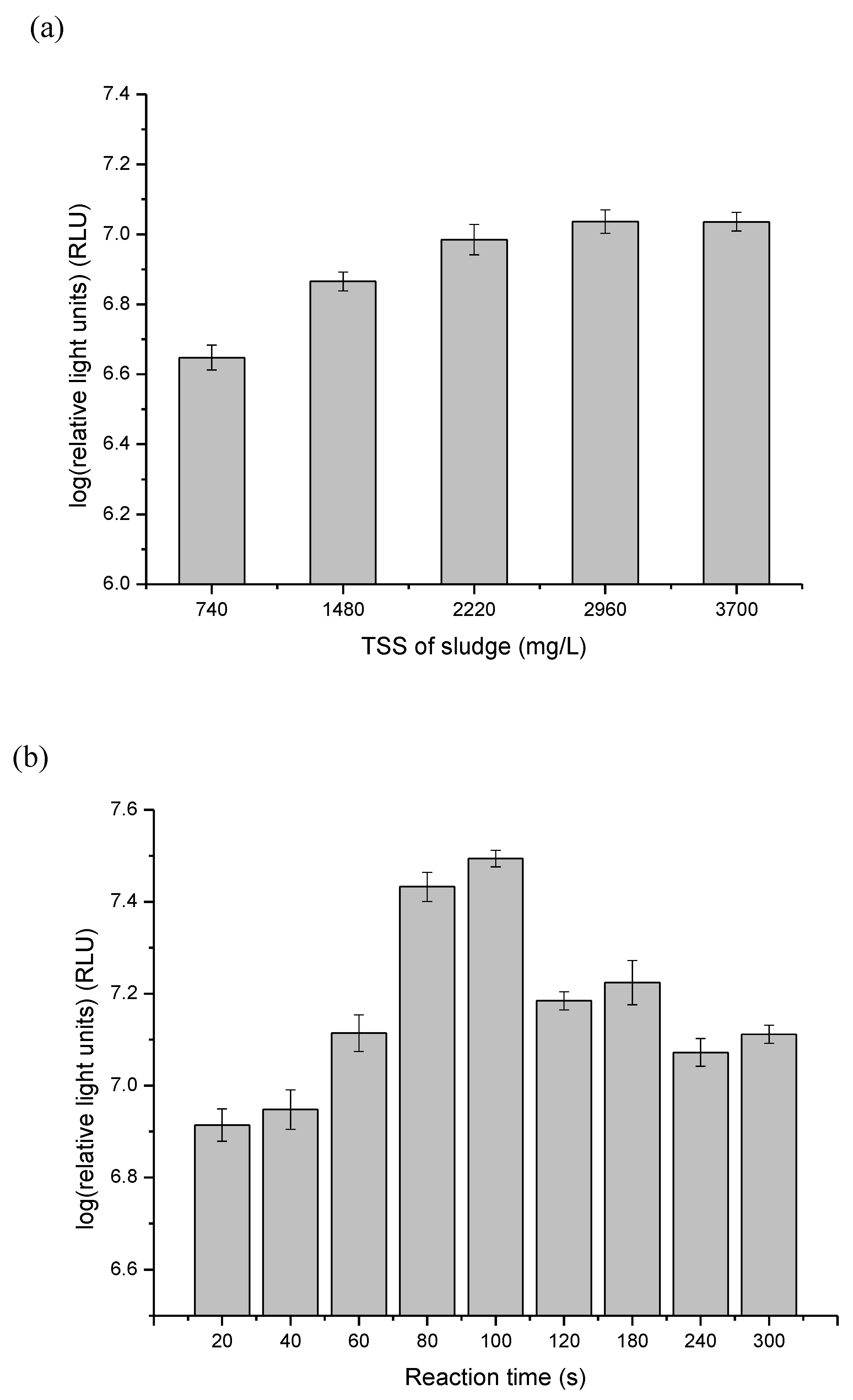
3.1. Optimize ATP Assay for Fast Detection of the Quantity of Viable Biomass during Sludge Ozonation

Because the ATP method has mainly been applied in monitoring the microbiological quality of the drinking water [12,14,33,34], its recommended protocol [26] should be optimized when used in detecting ATP in sludge. In this study, the effects of sludge concentration and incubation time were investigated. Figure 1a shows the positive impact of greater solid content on the luminescence detected from an activated sludge sample when the TSS value was below 2220 mg/L. The luminescence did not increase proportionally to the solid content when it was increased to 2960 mg/L, and remained almost constant when the TSS value increased from 2960 to 3700 mg/L, with the highest light signal around 107, which is three-log higher than that in drinking water [26]. The 50 µL BacTiter-GloTM reagent used in the present study might not be sufficient to completely release ATP from the activated sludge (500 µL sample volume) when its TSS concentration is higher than 2220 mg/L. Thus, to further ensure the accurate determination of ATP, 2000 mg/L was adopted as the sludge solid concentration in the optimized protocol. Since the ATP assay is based on an enzymatic reaction, the reaction time of the assay is important. Figure 1b demonstrates that 20 s, which is usually used in ATP detection in drinking water [11,26], is not sufficient in the luciferase-based assay of activated sludge. The larger cell number present in the sludge required about 100 s to reach the maximum luminescence signal (Figure 1b), and therefore this time was adopted as the reaction time of the assay. The luminescence signal increased about 3.8-fold with the optimized protocol for measuring microbial ATP, making the protocol more sensitive and accurate for activated sludge determination.

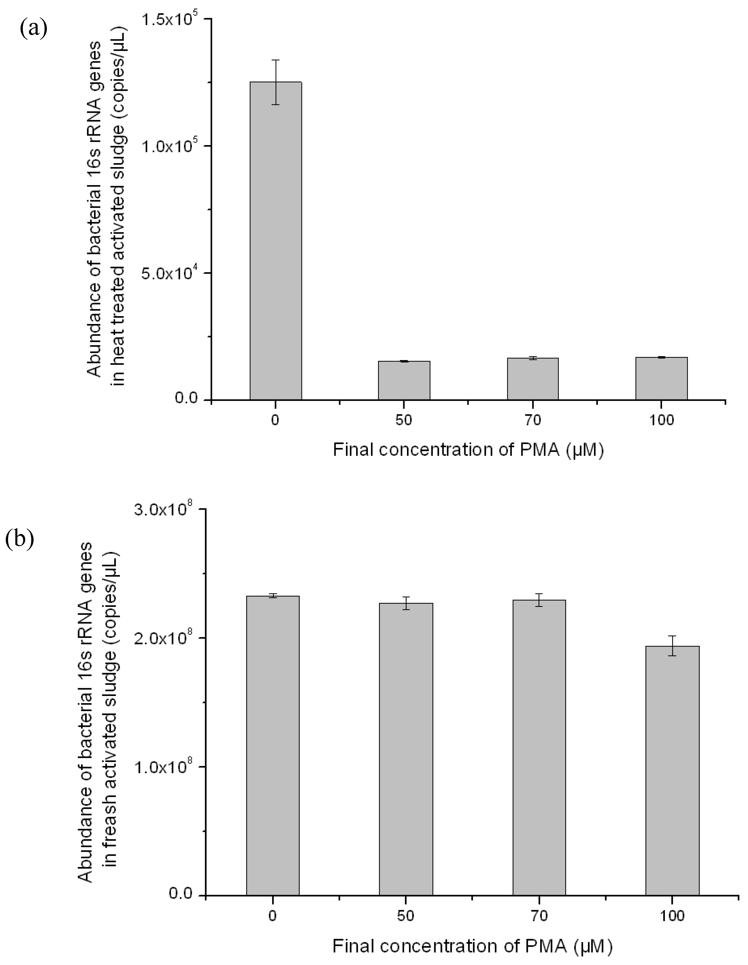
3.2. Optimization of PMA-Modified PCR for Selectively Amplifying the 16S rRNA Genes of Viable Bacteria in Activated Sludge

As for detecting the viable bacterial population compositions, Yan et al. used the conventional PCR-DGGE method for revealing the diversity of bacterial populations in activated sludge during ozonation [5]. However, prolonged persistence of DNA after cell death is a concern for the application of such methods, as DNA from dead cells can result in false-positive identification and inaccurate estimation of diversity. Considering that the cell membrane is the first victim of ozonation [6] and membrane integrity is one of the most reliable criteria for differentiating viable and dead bacteria cells [16,35], PMA was adopted in this study to selectively detect DNA from the viable cells. PMA can preferentially bind to double-stranded DNA, and its photoreactive azido group will be converted to a highly reactive nitrene radical upon photolysis, which readily reacts with any hydrocarbon moiety at the binding site to form a stable covalent nitrogen–carbon bond, resulting in permanent DNA modification [16]. The dye is nearly completely cell membrane impermeable and thus can be selectively used to modify exposed DNA only from dead cells, while leaving DNA from viable cells intact, which results in blocking the subsequent amplification of modified target DNA sequences by PCR. This feature makes the dye highly useful in the selective detection and quantification of viable cells by high-throughput sequencing and quantitative real-time PCR. In addition, PMA-modified PCR methods have been successfully used in detecting viruses, pathogens and heat-tolerant bacteria [36,37,38,39]. In this study, the PMA stain was conducted according to Taskin’s method for biosolids [27], whose solid contents are about 10- to 100-fold higher than that in activated sludge. Therefore, before its application in activated sludge, the PMA method was optimized, mainly focusing on the effect of final PMA concentration on removal of DNA from dead cells. We found that decreasing the final PMA concentration from the value of 100 µM used in Taskin’s method to 50 µM is sufficiently effective to eliminate PCR amplification of genomic DNA from dead cells (Figure 2) no matter whether for fresh sludge or heat-shocked sludge, which might be due to the simpler chemical and biological matrices in activated sludge [27].

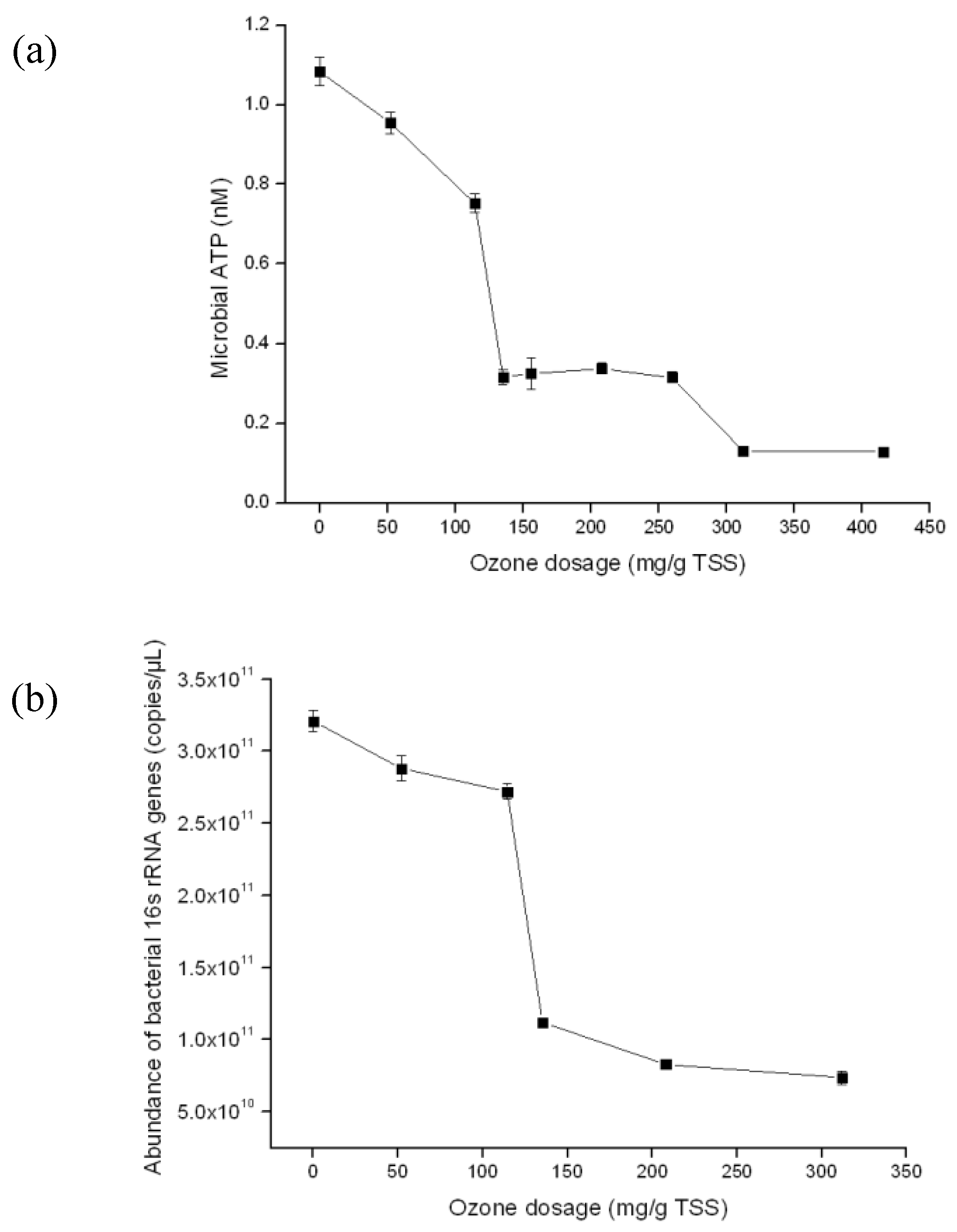
3.3. Detection of the Quantity of Viable Bacterial Populations during Sludge Ozonation Using Optimized ATP Assay and PMA-qPCR

After optimizing the applicability and effectiveness of both methods, the decrease of viable biomass in activated sludge was first detected by the ATP assay. As shown in Figure 3, with the increase of ozone dosage, the microbial ATP concentration decreased. It is interesting to note that the viable biomass displayed a substantial decrease (p < 0.01) when the ozone dose was increased from 114 to 135 mg/g TSS, and thereafter remained constant until the ozone dosage was further increased to 312 mg/g TSS (Figure 3a). Similar results were obtained by using the PMA-modified qPCR analysis (Figure 3b). These results indicate that more concrete and cutting-edge ozone dosages with quantified effects for the destruction of activated sludge might be determined by further refining the sampling points. The sludge ozonation process has been suggested to consist of the sequential decomposition processes of suspended solids disintegration, solubilization of the solids (cells) and mineralization of the soluble organic matter released from the microbial cells [20]. Therefore, the change pattern of viable bacterial populations during the ozonation process suggested that: (1) disintegration was the main process when ozone dosage was less than 114 mg/g TSS, and bacterial cells underwent fast solubilization when the ozone dosage increased to 135 mg/g TSS, with more than 70% reduction in total bacterial cells; and (2) there are some ozone-tolerant bacteria in activated sludge, which require more ozone to break their membranes.

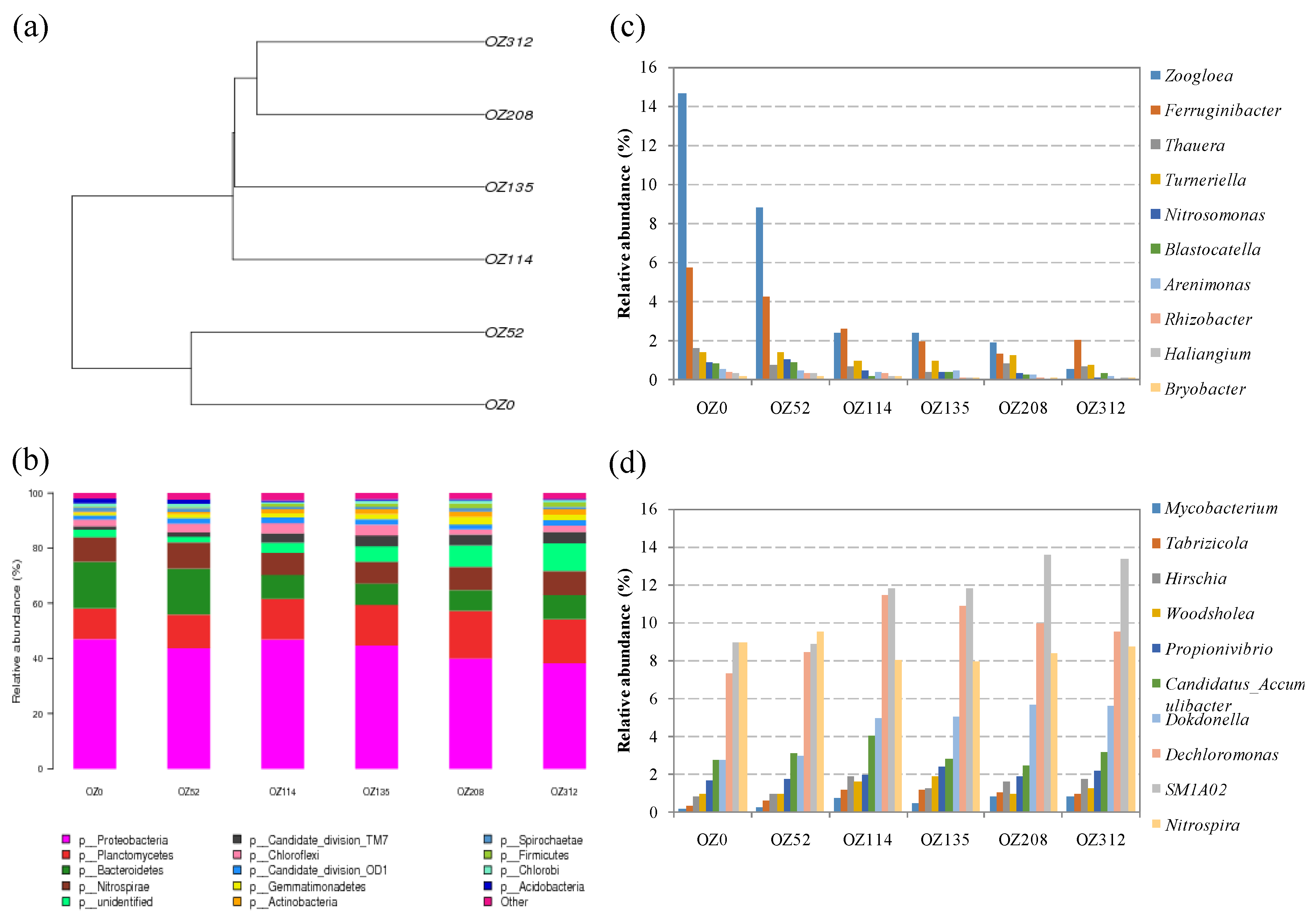
3.4. Detection of the Composition of Viable Bacterial Populations during Sludge Ozonation Using Optimized PMA-Miseq Sequencing

Based on the change pattern of viable biomass during ozonation, sludge samples corresponding to ozone dosages 0 (OZ0), 52 (OZ52), 114 (OZ114), 135 (OZ135), 208 (OZ208) and 312 (OZ312) mg/g TSS, respectively, were extracted and sequenced by using the PMA-modified Miseq sequencing method for analysis of the live bacterial populations. Recently, the combination of Miseq sequencing with PMA treatment was also successfully used to reveal the shifts of live bacterial community in secondary effluent during chlorine disinfection [40]. To our knowledge, this is the first investigation on the behaviors of viable bacterial populations in activated sludge during ozonation. A total of 260 genera affiliated to 25 phyla were identified, indicating a higher diversity of the viable bacteria community in activated sludge than that in secondary effluent or canal water [19,40]. Hierarchical cluster analysis based on the OTU level showed that the viable bacterial communities could be divided into two groups: one group for samples with ozone dosages of 0 and 52 mg/g TSS, and another group for the rest (Figure 4a). The cluster result suggested that an ozone dosage at least at 114 mg/g TSS is enough to completely disintegrate the sludge flocs and significantly change the viable bacterial populations in activated sludge, and the fast decrease in viable biomass when ozone dosage increased from 114 to 135 mg/g TSS was due to the oxidation of all bacterial cells. Yan et al. [5] also found that the DGGE fingerprint of ozonated sludge became simplified when the level of ozone was above 60 mg/g TSS. The viable bacterial community was dominant, with the phyla Proteobacteria (47.01%), Bacteroidetes (16.91%), Planctomycetes (11.12%), Nitrospirae (8.96%), Chloroflexi (2.47%), Acidobacteria (1.73%) Spirochaetae (1.51%) and two Candidate division (OD1 and TM7) (<1.50%) in the raw activated sludge used in this study (OZ0). The relative abundances of the phyla Proteobacteria, Bacteroidetes, Acidobacteria and Spirochaetae decreased, while those of the phyla Planctomycetes and Candidate division TM7 increased during ozonation. Although there was a decrease in relative abundance, Proteobacteria was always the most dominant phylum through the experiment. The phyla of Nitrospirae, Chloroflexi and Candidate division TM7 maintained a stable relative abundance during the ozonation process (Figure 4b).
To further infer the functional change of the viable bacterial community, we ranked the identified OTUs at the genus level, and twenty bacterial genera whose relative abundances significantly changed during the transition period were selected (Figure 4c,d). Zoogloea (14.65%), Ferruginibacter (5.70%), Thauera (1.54%), Turneriella (1.37%), Candidatus Accumulibacter (2.74%), Dokdonella (2.74%), Dechloromonas (7.31%), SM1A02 (8.96%), Nitrosomonas (0.87%) and Nitrospira (8.96%) were the dominant viable bacteria in raw activated sludge. Zoogloea, Ferruginibacter, Thauera, Turneriella, Dokdonella and Dechloromonas are important floc-forming bacteria and are responsible for degradation of organic compounds [41,42,43,44]. The relative abundances of genera Zoogloea, Ferruginibacter, Thauera and Turneriella in the viable bacterial population were decreased during ozonation, indicating they are more vulnerable compared with other bacterial genera, while that of genera Dokdonella and Dechloromonas increased. SM1A02, Nitrospira, and Nitrosomonas are three bacterial genera related to ammonia transformation in the activated sludge process. SM1A02 has been found in many activated sludges with good nitrifying performance, and is thought have potential as a novel anammox strain [45,46,47]. In this study, its relative abundance increased from 8.96% in the original viable bacterial population to 13.35% in the ozonated sludge with ozone dosage as high as 312 mg/g TSS, indicating that bacterial species in this genus might resist ozonation. Further studies involving isolation of pure bacterial strains of this genus would be required to positively affirm the resistance of this species. Nitrosomonas can oxidize ammonia into nitrite, which is then used as a substrate by Nitrospira to produce nitrate. These two common ammonia-oxidizing genera showed different tolerance to ozonation: Nitrosomonas decreased with increasing ozone dosage, while Nitrospira maintained almost constant relative abundance during activated sludge ozonation (Figure 4c,d). Candidatus Accumulibacter is an unclassified group of Betaproteobacteria and only contains a single member, Candidatus Accumulibacter Phosphatis, which is a common bacterial community member in wastewater treatment plants performing enhanced biological phosphorus removal [48]. Our result showed that this genus has a certain level of resistance to ozonation because its relative abundance in the viable bacterial communities remained almost constant during the experiment (Figure 4d). Up to now, ozonation has been usually combined with biological wastewater treatment processes to enhance the removal of nitrogen and phosphorus [22,23,49]. Therefore, the persistence of Candidatus Accumulibacter during ozonation might promote biological phosphorus removal in an activated sludge process fed with ozonated sludge by providing additional functional bacteria, besides the addition of an organic carbon source.
The results of this study showed that ozone exerted different effects on the different activated sludge bacteria. Prabakaran et al. [50] also found that pathogens Klebsiella pneumoniae and Salmonella typhi are more resistant to ozone treatment than another two pathogenic bacteria Pseudomonas fluorescens and Escherichia coli. The varied resistance of different microbes to ozonation might be related with their different characteristics of cell structures, such as lipid and protein components in cell membrane, content of the extracellular polymeric substances and assembling morphology in the sludge. For example, Candidatus Accumulibacter Phosphatis usually existed in clusters [51], which might protect their DNA from the ozone stress [5]. Besides the characteristics of cell structures, it has also been reported that ozone diffusion into the deep zones of the flocs may be a limiting factor of sludge ozonation [52]. Additional research is needed to better understand the mechanism of the different responses of different bacteria to ozonation.

4. Conclusions

The changes in the quantity and composition of viable bacterial populations in activated sludge during ozonation treatment were investigated using the optimized ATP assay and PMA-modified Miseq sequencing. The results indicated that the viable biomass of activated sludge was substantially decreased after being exposed to ozone at 135 mg·O3/g TSS, and the composition of viable bacterial communities showed a significant change when the ozone dosage reached 114 mg·O3/g TSS. Floc-forming genera, including Zoogloea, Ferruginibacter, Thauera and Turneriella, are sensitive to ozonation, while ammonia- or phosphate-transforming bacterial genera, including SM1A02, Nitrospira and Candidatus Accumulibacter, are more resistant. These results will be helpful for designing and optimizing the sludge ozonation process. On the other hand, to better understand the biological response to operation conditions during sludge treatment, the resistance mechanism of different bacteria to ozonation should be explored in the future.

Acknowledgments

This research is financially supported by the National Natural Scientific Foundation of China (No. 21590814).

Author Contributions

Zhe Tian, Min Yang and Yu Zhang conceived and designed the experiments; Shaonan Tian performed the experiments; Zhe Tian analyzed the data; Hong Yang contributed reagents/materials/analysis tools; and Shaonan Tian and Yu Zhang wrote the paper.

Conflicts of Interest

The authors declare no conflict of interest.

References

  1. Spellman, F. Wastewater Biosolids to Compost; Technomic Publishing Company: Lancaster, PA, USA, 1997; pp. 223–235. [Google Scholar]
  2. Wei, Y.S.; Van Houten, R.T.; Borger, A.R.; Eikelboom, D.H.; Fan, Y.B. Minimization of excess sludge production for biological wastewater treatment. Water Res. 2003, 37, 4453–4467. [Google Scholar] [CrossRef]
  3. Yan, S.; Miyanaga, K.; Xing, X.-H.; Tanji, Y. Succession of bacterial community and enzymatic activities of activated sludge by heat-treatment for reduction of excess sludge. Biochem. Eng. J. 2008, 39, 598–603. [Google Scholar] [CrossRef]
  4. Yasui, H.; Shibata, M. An innovative approach to reduce excess sludge production in the activated-sludge process. Water Sci. Technol. 1994, 30, 11–20. [Google Scholar]
  5. Yan, S.-T.; Chu, L.-B.; Xing, X.-H.; Yu, A.-F.; Sun, X.-L.; Jurcik, B. Analysis of the mechanism of sludge ozonation by a combination of biological and chemical approaches. Water Res. 2009, 43, 195–203. [Google Scholar] [CrossRef] [PubMed]
  6. Komanapalli, I.R.; Lau, B.H.S. Ozone-induced damage of Escherichia coli K-12. Appl. Microbiol. Biotechnol. 1996, 46, 610–614. [Google Scholar] [CrossRef] [PubMed]
  7. Hysert, D.W.; Knudsen, F.B.; Morrison, N.M.; Vangheluwe, G.; Lom, T. Application of a bioluminescence ATP assay in brewery wastewater-treatment studies. Biotechnol. Bioeng. 1979, 21, 1301–1314. [Google Scholar] [CrossRef]
  8. Yang, C.; Zhang, W.; Liu, R.; Li, Q.; Li, B.; Wang, S.; Song, C.; Qiao, C.; Mulchandani, A. Phylogenetic diversity and metabolic potential of activated sludge microbial communities in full-scale wastewater treatment plants. Environ. Sci. Technol. 2011, 45, 7408–7415. [Google Scholar] [CrossRef] [PubMed]
  9. Gonzalez-Martinez, A.; Rodriguez-Sanchez, A.; Lotti, T.; Garcia-Ruiz, M.-J.; Osorio, F.; Gonzalez-Lopez, J.; van Loosdrecht, M.C.M. Comparison of bacterial communities of conventional and A-stage activated sludge systems. Sci. Rep. 2016, 6, 18786. [Google Scholar] [CrossRef] [PubMed]
  10. Amann, R.I.; Ludwig, W.; Schleifer, K.H. Phylogenetic identification and in-situ detection of individual microbial-cells without cultivation. Microbiol. Rev. 1995, 59, 143–169. [Google Scholar] [PubMed]
  11. Vang, O.K.; Corfitzen, C.B.; Smith, C.; Albrechtsen, H.-J. Evaluation of ATP measurements to detect microbial ingress by wastewater and surface water in drinking water. Water Res. 2014, 64, 309–320. [Google Scholar] [CrossRef] [PubMed] [Green Version]
  12. Van der Wielen, P.W.J.J.; van der Kooij, D. Effect of water composition, distance and season on the adenosine triphosphate concentration in unchlorinated drinking water in The Netherlands. Water Res. 2010, 44, 4860–4867. [Google Scholar] [CrossRef] [PubMed]
  13. Deininger, R.A.; Lee, J. Rapid determination of bacteria in drinking water using an ATP assay. Field Anal. Chem. Technol. 2001, 5, 185–189. [Google Scholar] [CrossRef]
  14. Delahaye, E.; Welte, B.; Levi, Y.; Leblon, G.; Montiel, A. An ATP-based method for monitoring the microbiological drinking water quality in a distribution network. Water Res. 2003, 37, 3689–3696. [Google Scholar] [CrossRef] [PubMed]
  15. Zhang, T.; Shao, M.-F.; Ye, L. 454 pyrosequencing reveals bacterial diversity of activated sludge from 14 sewage treatment plants. ISME J. 2012, 6, 1137–1147. [Google Scholar] [CrossRef] [PubMed]
  16. Nocker, A.; Cheung, C.-Y.; Camper, A.K. Comparison of propidium monoazide with ethidium monoazide for differentiation of live vs. dead bacteria by selective removal of DNA from dead cells. J. Microbiol. Methods 2006, 67, 310–320. [Google Scholar] [CrossRef] [PubMed]
  17. Rogers, G.; Cuthbertson, L.; Hoffman, L.; Wing, P.; Pope, C.; Hooftman, D.; Lilley, A.; Oliver, A.; Carroll, M.; Bruce, K.; et al. Reducing bias in bacterial community analysis of lower respiratory infections. ISME J. 2013, 7, 697–706. [Google Scholar] [CrossRef] [PubMed] [Green Version]
  18. Guo, F.; Zhang, T. Detecting the nonviable and heat-tolerant bacteria in activated sludge by minimizing DNA from dead cells. Microbial Ecol. 2014, 67, 829–836. [Google Scholar] [CrossRef] [PubMed]
  19. Nocker, A.; Richter-Heitmann, T.; Montijn, R.; Schuren, F.; Kort, R. Discrimination between live and dead cells in bacterial communities from environmental water samples analyzed by 454 pyrosequencing. Int. Microbiol. 2010, 13, 59–65. [Google Scholar] [PubMed]
  20. Ahn, K.H.; Park, K.Y.; Maeng, S.K.; Hwang, J.H.; Lee, J.W.; Song, K.G.; Choi, S. Ozonation of wastewater sludge for reduction and recycling. Water Sci. Technol. 2002, 46, 71–77. [Google Scholar] [PubMed]
  21. Chu, L.-B.; Yan, S.-T.; Xing, X.-H.; Yu, A.-F.; Sun, X.-L.; Jurcik, B. Enhanced sludge solubilization by microbubble ozonation. Chemosphere 2008, 72, 205–212. [Google Scholar] [CrossRef] [PubMed]
  22. Dytczak, M.A.; Londry, K.L.; Siegrist, H.; Oleszkiewicz, J.A. Ozonation reduces sludge production and improves denitrification. Water Res. 2007, 41, 543–550. [Google Scholar] [CrossRef] [PubMed]
  23. Goel, R.; Komatsu, K.; Yasui, H.; Harada, H. Process performance and change in sludge characteristics during anaerobic digestion of sewage sludge with ozonation. Water Sci. Technol. 2004, 49, 105–113. [Google Scholar] [PubMed]
  24. Pires, M.; Carvalho, L. An artifact in air carbonyls sampling using C-18 DNPH-coated cartridge. Anal. Chim. Acta 1998, 367, 223–231. [Google Scholar] [CrossRef]
  25. Abzazou, T.; Salvado, H.; Bruguera-Casamada, C.; Simon, P.; Lardin, C.; Araujo, R.M. Assessment of total bacterial cells in extended aeration activated sludge plants using flow cytometry as a microbial monitoring tool. Environ. Sci. Pollut. Res. 2015, 22, 11446–11455. [Google Scholar] [CrossRef] [PubMed]
  26. Hammes, F.; Goldschmidt, F.; Vital, M.; Wang, Y.; Egli, T. Measurement and interpretation of microbial adenosine tri-phosphate (ATP) in aquatic environments. Water Res. 2010, 44, 3915–3923. [Google Scholar] [CrossRef] [PubMed]
  27. Taskin, B.; Gozen, A.G.; Duran, M. Selective quantification of viable Escherichia coli bacteria in biosolids by quantitative PCR with propidium monoazide modification. Appl. Environ. Microbiol. 2011, 77, 4329–4335. [Google Scholar] [CrossRef] [PubMed]
  28. Liu, M.; Zhang, Y.; Yang, M.; Tian, Z.; Ren, L.; Zhang, S. Abundance and distribution of tetracycline resistance genes and mobile elements in an oxytetracycline production wastewater treatment system. Environ. Sci. Technol. 2012, 46, 7551–7557. [Google Scholar] [CrossRef] [PubMed]
  29. Pei, R.; Kim, S.-C.; Carlson, K.H.; Pruden, A. Effect of river landscape on the sediment concentrations of antibiotics and corresponding antibiotic resistance genes (ARG). Water Res. 2006, 40, 2427–2435. [Google Scholar] [CrossRef] [PubMed]
  30. Xiong, J.; Liu, Y.; Lin, X.; Zhang, H.; Zeng, J.; Hou, J.; Yang, Y.; Yao, T.; Knight, R.; Chu, H. Geographic distance and pH drive bacterial distribution in alkaline lake sediments across Tibetan Plateau. Environ. Microbiol. 2012, 14, 2457–2466. [Google Scholar] [CrossRef] [PubMed]
  31. Caporaso, J.G.; Kuczynski, J.; Stombaugh, J.; Bittinger, K.; Bushman, F.D.; Costello, E.K.; Fierer, N.; Pena, A.G.; Goodrich, J.K.; Gordon, J.I.; et al. Qiime allows analysis of high-throughput community sequencing data. Nat. Methods 2010, 7, 335–336. [Google Scholar] [CrossRef] [PubMed]
  32. DeSantis, T.Z.; Hugenholtz, P.; Larsen, N.; Rojas, M.; Brodie, E.L.; Keller, K.; Huber, T.; Dalevi, D.; Hu, P.; Andersen, G.L. Greengenes, a chimera-checked 16S rRNA gene database and workbench compatible with ARB. Appl. Environ. Microbiol. 2006, 72, 5069–5072. [Google Scholar] [CrossRef] [PubMed] [Green Version]
  33. Boe-Hansen, R.; Albrechtsen, H.J.; Arvin, E.; Jorgensen, C. Bulk water phase and biofilm growth in drinking water at low nutrient conditions. Water Res. 2002, 36, 4477–4486. [Google Scholar] [CrossRef]
  34. Ikonen, J.; Pitkanen, T.; Miettinen, I.T. Suitability of optical, physical and chemical measurements for detection of changes in bacterial drinking water quality. Int. J. Environ. Res. Public Health 2013, 10, 5349–5363. [Google Scholar] [CrossRef] [PubMed]
  35. Kramer, M.; Obermajer, N.; Matijasic, B.B.; Rogelj, I.; Kmetec, V. Quantification of live and dead probiotic bacteria in lyophilised product by real-time PCR and by flow cytometry. Appl. Microbiol. Biotechnol. 2009, 84, 1137–1147. [Google Scholar] [CrossRef] [PubMed]
  36. Cawthorn, D.M.; Witthuhn, R.C. Selective PCR detection of viable enterobacter sakazakii cells utilizing propidium monoazide or ethidium bromide monoazide. J. Appl. Microbiol. 2008, 105, 1178–1185. [Google Scholar] [CrossRef] [PubMed]
  37. Fittipaldi, M.; Pino Rodriguez, N.J.; Codony, F.; Adrados, B.; Penuela, G.A.; Morato, J. Discrimination of infectious bacteriophage t4 virus by propidium monoazide real-time PCR. J. Virol. Methods 2010, 168, 228–232. [Google Scholar] [CrossRef] [PubMed]
  38. Kobayashi, H.; Oethinger, M.; Tuohy, M.J.; Hall, G.S.; Bauer, T.W. Improving clinical significance of PCR: Use of propidium monoazide to distinguish viable from dead staphylococcus aureus and staphylococcus epidermidis. J. Orthop. Res. 2009, 27, 1243–1247. [Google Scholar] [CrossRef] [PubMed]
  39. Rawsthorne, H.; Dock, C.N.; Jaykus, L.A. PCR-based method using propidium monoazide to distinguish viable from nonviable bacillus subtilis spores. Appl. Environ. Microbiol. 2009, 75, 2936–2939. [Google Scholar] [CrossRef] [PubMed]
  40. Pang, Y.-C.; Xi, J.-Y.; Xu, Y.; Huo, Z.-Y.; Hu, H.-Y. Shifts of live bacterial community in secondary effluent by chlorine disinfection revealed by Miseq high-throughput sequencing combined with propidium monoazide treatment. Appl. Microbiol. Biotechnol. 2016, 100, 6435–6446. [Google Scholar] [CrossRef] [PubMed]
  41. Liu, Y.; Jin, J.-H.; Liu, H.-C.; Liu, Z.-P. Dokdonella immobilis sp. nova, isolated from a batch reactor for the treatment of triphenylmethane dye effluent. Int. J. Syst. Evolut. Microbiol. 2013, 63, 1557–1561. [Google Scholar] [CrossRef] [PubMed]
  42. Shin, Y.K.; Hiraishi, A.; Sugiyama, J. Molecular systematics of the genus Zoogloea and emendation of the genus. Int. J. Syst. Bacteriol. 1993, 43, 826–831. [Google Scholar] [CrossRef] [PubMed]
  43. Thomsen, T.R.; Kong, Y.; Nielsen, P.H. Ecophysiology of abundant denitrifying bacteria in activated sludge. Fems Microbiol. Ecol. 2007, 60, 370–382. [Google Scholar] [CrossRef] [PubMed]
  44. Pal, P.; Khairnar, K.; Paunikar, W.N. Causes and remedies for filamentous foaming in activated sludge treatment plant. Glob. Nest J. 2014, 16, 762–772. [Google Scholar]
  45. Chu, Z.-R.; Wang, K.; Li, X.-K.; Zhu, M.-T.; Yang, L.; Zhang, J. Microbial characterization of aggregates within a one-stage nitritation-anammox system using high-throughput amplicon sequencing. Chem. Eng. J. 2015, 262, 41–48. [Google Scholar] [CrossRef]
  46. Wei, C.; He, W.; Wei, L.; Li, C.; Ma, J. The analysis of a microbial community in the UV/O3-anaerobic/aerobic integrated process for petrochemical nanofiltration concentrate (NFC) treatment by 454-pyrosequencing. PLoS ONE 2015, 10, e0139991. [Google Scholar] [CrossRef] [PubMed]
  47. Cui, Y.-W.; Zhang, H.-Y.; Lu, P.-F.; Peng, Y.-Z. Effects of carbon sources on the enrichment of halophilic polyhydroxyalkanoate-storing mixed microbial culture in an aerobic dynamic feeding process. Sci. Rep. 2016, 6, 30766. [Google Scholar] [CrossRef] [PubMed]
  48. Seviour, R.J.; Mino, T.; Onuki, M. The microbiology of biological phosphorus removal in activated sludge systems. Fems Microbiol. Rev. 2003, 27, 99–127. [Google Scholar] [CrossRef]
  49. Lee, J.W.; Cha, H.Y.; Park, K.Y.; Song, K.G.; Ahn, K.H. Operational strategies for an activated sludge process in conjunction with ozone oxidation for zero excess sludge production during winter season. Water Res. 2005, 39, 1199–1204. [Google Scholar] [CrossRef] [PubMed]
  50. Prabakaran, M.; Tamil Selvi, S.; Merinal, S.; Panneerselvam, A. Effect of ozonation on pathogenic bacteria. Adv. Appl. Sci. Res. 2012, 3, 299–302. [Google Scholar]
  51. Crocetti, G.; Hugenholtz, P.; Bond, P.L.; Schuler, A.; Keller, J.; Jenkins, D.; Blackall, L. Identification of polyphosphate-accumulating organisms and design of 16S rRNA-directed probes for their detection and quantitation. Appl. Environ. Microbiol. 2000, 66, 1175–1182. [Google Scholar] [CrossRef] [PubMed]
  52. Dziurla, M.A.; Salhi, M.; Leroy, P.; Paul, E.; Ginestet, P.; Block, J.C. Variations of respiratory activity and glutathione in activated sludges exposed to low ozone doses. Water Res. 2005, 39, 2591–2598. [Google Scholar] [CrossRef] [PubMed]
Figure 1. Optimization of the ATP detection for the sludge samples: (a) effect of sludge solid contents on the luminescence; and (b) effect of reaction time on the luminescence.
Water 09 00166 g001
Figure 2. Effect of final PMA concentrations on quantitative polymerase chain reaction (qPCR) quantification of bacterial 16S rRNA gene: (a) fresh activated sludge; and (b) heat treated activated sludge.
Water 09 00166 g002
Figure 3. Viable bacterial biomass in activated sludge under different ozone dosages: (a) microbial ATP concentration; and (b) bacterial 16S rRNA gene copies.
Water 09 00166 g003
Figure 4. Succession of viable bacterial population: (a) Hierarchical cluster analysis of six bacterial communities. The analysis is the clustering of all OTUs (3% distance). Sample communities were clustered based on the average linkage method. (b) Bacterial community structures at the phylum level in the sludge samples monitored using Miseq sequencing. Relative abundance was defined as the number of sequences affiliated with that phylum divided by the total number of sequences per sample. (c) Ten bacterial genera whose abundances decreased during ozonation. (d) Ten bacterial genera whose abundances increased or not changed during ozonation.
Water 09 00166 g004

