Review

Preparation of Knockout Extract by Immunoaffinity Column and Its Application

Department of Pharmacognosy, Faculty of Pharmaceutical Sciences, Nagasaki International University, 2825-7 Huis Ten Bosch-Cho, Sasebo, Nagasaki 859-3298, Japan
*
Author to whom correspondence should be addressed.
Antibodies 2012, 1(3), 294-307; https://doi.org/10.3390/antib1030294
Submission received: 28 August 2012 / Revised: 30 November 2012 / Accepted: 4 December 2012 / Published: 6 December 2012

Abstract

:
Importance of herbal medicines have recently increased owing to rising interest in their health benefits. However, medicinal plant extracts are complex mixtures of phytochemicals that act synergistically or additively on specific and/or multiple molecular and cellular targets. Thus, it is difficult to examine the actual pharmacological roles of active compounds in plant extracts. This review describes a new strategy for isolating target compounds from plant extracts using immunoaffinity columns coupled with monoclonal antibodies (mAbs) against natural compounds. Through one-step purification using mAb-coupled immunoaffinity columns, we succeeded in preparing a knockout (KO) extract, which contains all components except the target compound. Furthermore, we investigated the pharmacological effects of the KO extract to reveal the actual effects of a bioactive compound in the crude extract. This approach may help determine the potential function of target compounds in herbal medicines.

1. Introduction

Traditional herbal medicines have been used for thousands of years in Asian countries such as China, Japan, and Korea. Recently, global demand for herbal medicines has increased owing to rising interest in the associated health benefits. Studies of bioactive compounds derived from medicinal plant extracts have revealed their molecular actions in vivo and in vitro. However, plant extracts are complex mixtures of effective phytochemicals that act synergistically or additively on specific and/or multiple molecular and cellular targets. Hence, pharmacological evidence of interactions between these natural compounds is required to identify their actual pharmacological activity.
Immunoassay using monoclonal antibodies (mAbs) against small molecular-weight-compounds such as synthetic drugs and natural compounds is a very important tool for investigating receptor binding, enzyme reactions, and quantitative and/or qualitative analysis in both animals and plants. Immunoaffinity purification using mAbs is also commonly used to purify larger molecules, such as peptides and proteins, for research and commercial purposes. However, few studies have targeted small-molecular-weight compounds such as natural bioactive compounds. In previous studies, we established various types of mAbs against natural compounds, including terpenoids [1,2,3], saponins [4,5,6,7,8], alkaloids [9,10], and phenolics [11,12,13,14], and developed several applications using these mAbs. One of the applications is an immunoaffinity column coupled with anti-natural compound-specific mAbs, which can capture target compounds and specifically remove them from crude mixtures. We previously prepared immunoaffinity columns against the terpenoids forskolin [15], solasodine glycosides [16], ginsenoside Rb1 [17], and glycyrrhizin [18].
In this review, we introduce and describe the preparation of a knockout (KO) extract, which is prepared by eliminating one target compound from the crude extract using immunoaffinity columns. Application of the KO extract is critical to such pharmacological investigations for clarifying the actual effects of the bioactive compound and elucidating interactions between the target compound and the other compounds in the component mixture, such as plant extracts and traditional Chinese medicines (TCMs).

2. Preparation of the Immunoaffinity Column against Ginsenoside Rb1 (G-Rb1), and the G-Rb1-KO Extract

Ginseng, the root of Panax ginseng C.A. Meyer (Araliaceae), has been one of the most important medicinal plants in Asian countries for >1,000 years [19]. The pharmacological and therapeutic effects of ginseng are mainly attributed to ginsenosides, which are triterpernoid saponin glycosides (dammarane-type saponins) [19,20]. Recently, 60 additional ginsenosides have been isolated from different ginseng species [21,22]. Ginsenoside Rb1 (G-Rb1) is one of the main ginsenosides obtained from ginseng [23] and reportedly exerts various pharmaceutical actions, such as facilitating acquisition and retrieval of memory [24], scavenging free radicals [25], inhibiting calcium over-influx into neurons [26], and preserving the structural integrity of neurons [27]. In our previous work, we established anti-G-Rb1 mAb and applied it to enzyme-linked immunosorbent assay (ELISA) and a new immunostaining method, named Eastern blotting [4,28]. Furthermore, we prepared an immunoaffinity column coupled with anti-G-Rb1 mAb and performed one-step purification using ginseng extract [17,28]. We here report the preparation of the anti-G-Rb1 mAb-coupled immunoaffinity column and its applications.
Ginseng, the root of Panax ginseng C.A. Meyer (Araliaceae), has been used as one of the most important medicinal plants in Asian countries for more than thousand years [19]. The pharmacological and therapeutic effects of ginseng are mainly attributed to ginsenosides, which are triterprnoid saponin glycosides (dammarane-type saponins) [19,20]. Recently, more than 60 ginsenosides have been isolated from different species of ginseng [21,22]. Ginsenoside Rb1 (G-Rb1) is one of the main ginsenosides of ginseng [23], and has been reported to exert many pharmaceutical actions such as facilitating acquisition and retrieval of memory [24], scavenging free radicals [25], inhibition of calcium over-influx into neurons [26], and preserving the structural integrity of the neurons [27]. In our previous work, we have established anti-G-Rb1 MAb and its application including enzyme-linked immunosorbent assay (ELISA) and a new immunostaining method named Eastern blotting [4,28]. Furthermore, we prepared an immunoaffinity column combined with anti-G-Rb1 MAb, and then performed the one-step isolation from ginseng extract by this column [17,28]. Herein we report the preparation of anti-G-Rb1 immunoaffinity column and it applications.

2.1. Preparation of Anti-G-Rb1mAb

Small compounds such as synthetic drugs and natural compounds are insufficiently complex to induce an immune response and antibody production. For antibody production against small compounds, a hapten must be chemically conjugated to carrier proteins such as bovine serum albumin (BSA). The first step for production of such mAbs is the preparation of a hapten–carrier protein conjugate, which directly combines an immune antigen with the carrier protein. Thus, G-Rb1 was oxidized with NaIO4 solution to generate aldehyde groups on the sugar moiety, allowing the aldehyde group to react with BSA (Figure 1a) [4]. Subsequently, it was necessary to characterize the synthesized antigen G-Rb1-BSA conjugate. However, direct and appropriate methods to determine the hapten-conjugated carrier proteins without differential UV analysis, radiochemical, or chemical methods were not reported. To determine the hapten density of immunoconjugates, Wengatez et al. used matrix-assisted UV laser desorption/ionization (MALDI) mass spectrometry [29]. We also performed direct analysis of hapten–carrier protein conjugates by MALDI-TOF mass spectrometry [1,2,3,4,5,6,7,8,9,10,11,12,13,14,30,31,32]. Figure 1b indicates the MALDI-TOF mass spectra of the G-Rb1-BSA conjugate. A broad peak coinciding with the G-Rb1-BSA conjugate appeared between 70,000 and 90,000 m/z and centered at approximately 79,469 m/z. The molecular weight of BSA is 66,433, and the calculated molecular mass of the G-Rb1 moiety is 1,109. These data suggest that most BSA was effectively conjugated to G-Rb1. The experimental results and molecular weight of BSA revealed that the calculated molecular mass of the conjugated G-Rb1 is between 3,327 and 23,289, suggesting that 3–21 (average of 12) molecules of G-Rb1 are conjugated to BSA [4]. This method is therefore suitable for the characterization of hapten–carrier protein conjugates.
Figure 1. (a) Synthesis of G-Rb1-BSA conjugate. (b) Direct detection of G-Rb1-BSA conjugate by MALDI-TOF mass spectrometry.
Antibodies 01 00294 g001
The synthesized G-Rb1-BSA conjugate was injected into mice [4]. Briefly, BALB/c female mice were injected intraperitoneally with G-Rb1-BSA dissolved in phosphate buffer saline (PBS) four times. The first immunization (50 μg protein) was injected as a 1:1 emulsion in Freund’s complete adjuvant. The second and third immunization (50 μg protein in each injection) were injected as a 1:1 emulsion in Freund’s incomplete adjuvant. On the third day after the final immunization (100 μg protein), splenocytes were isolated and fused with a HAT-sensitive mouse myeloma cell line, P3-X63-Ag8-653, by the polyethylene glycol (PEG) method. After cell fusion, we assessed the binding activity against carrier protein BSA, and the hybridomas secreting antibody against BSA were removed. Hybridomas producing mAb reactive to G-Rb1 were cloned by the limited dilution method [33]. Established hybridomas were cultured in eRDF medium supplemented with 10 μg/mL insulin, 35 μg/mL of transferrin, 20 μM ethanolamine and 25 nM selenium (ITES) [34]. The mAb was purified using a Protein G FF column [4]. The cultured medium containing the IgG was adjusted to pH 7 with 1 M Tris solution and subjected to the column, and washed the column with 10 mM phosphate buffer (pH 7). Absorbed IgG was eluted with 100 mM citrate buffer (pH 3). The eluted IgG was neutralized with 1 M Tris solution, then dialyzed against PBS (pH 7.4) three times, and finally lyophilized. To further examine the binding specificity of this mAb against G-Rb1, but not the carrier protein BSA, we immobilized G-Rb1 to human serum albumin (HSA) and adsorbed the G-Rb1-HSA conjugate onto a polystyrene microtiter plate. Subsequently, the specificity of IgG-type mAb, 9G7, for G-Rb1 was examined by competitive ELISA using a microtiter plate coated with the G-Rb1-HSA conjugate. Under these conditions, the free mAb bound to free G-Rb1 or the G-Rb1-HSA conjugate and the full measurement range of the assay using this anti-G-Rb1 mAb extended from 20 to 400 ng/mL. Since cross-reactivity is the most important factor in determining the utility of antibodies, assay specificity was tested by determining cross-reactivity of mAb with various related compounds. Cross-reactivity against G-Rc and G-Rd, which possess diglucose moieties attached to C-3 hydroxy groups, was weak compared with that against G-Rb1 (0.024% and 0.020%, respectively), and G-Re and G-Rg1 showed negligible cross-reactivity (<0.005%). Therefore, it is clear that this mAb reacts predominantly with G-Rb1 and not with its structurally related compounds or the carrier protein BSA.

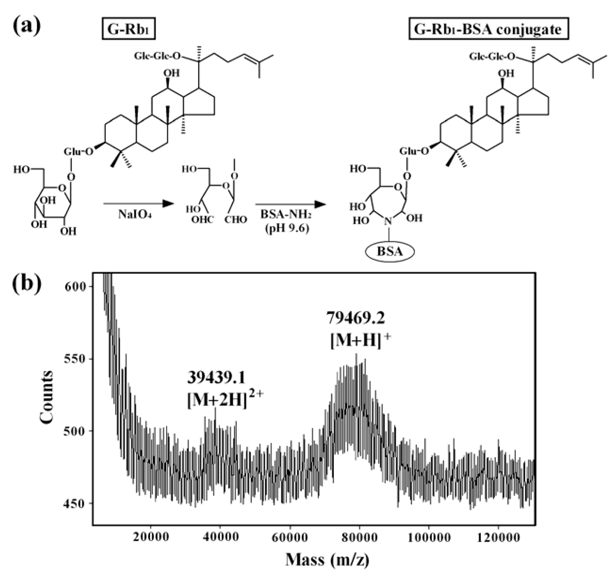
2.2. Preparation of the Anti-G-Rb1 mAb-Coupled Immunoaffinity Column

To couple mAb to the Affi-Gel Hz gel, the sugar moiety of the purified anti-G-Rb1 mAb was oxidized by NaIO4 to give a dialdehyde group on the sugar moiety. The oxidized anti-G-Rb1 mAb was then coupled to the Affi-Gel Hz hydrazide gel to form a hydrozone-type immunoaffinity gel [28]. The immunoaffinity gel was poured into a plastic mini-column (Figure 2).
Figure 2. Scheme of preparation of the anti-G-Rb1 mAb-coupled immunoaffinity column
Antibodies 01 00294 g002
To determine the best conditions for adsorption and elution, 400 μg of G-Rb1 was dissolved in phosphate buffer (PB) and loaded into the immunoaffinity column. After washing with 20 mM PB containing 0.5 M NaCl, different elution buffers were loaded into the column and the recovery efficiency of G-Rb1 was examined by ELISA. Recovery of G-Rb1 was somewhat increased by elution with 20 mM PB containing 10% MeOH and 0.5 M potassium thiocyanate (KSCN). Elution was optimal with 100 mM AcOH buffer (pH 4), which was used as a substitute for 20 mM PB. Although 20% MeOH enhanced the elution of G-Rb1, the MeOH concentration >20% was ineffective. Taken together, elution with 100 mM AcOH buffer containing 20% MeOH and 0.5 M KSCN gave the best recovery of G-Rb1.

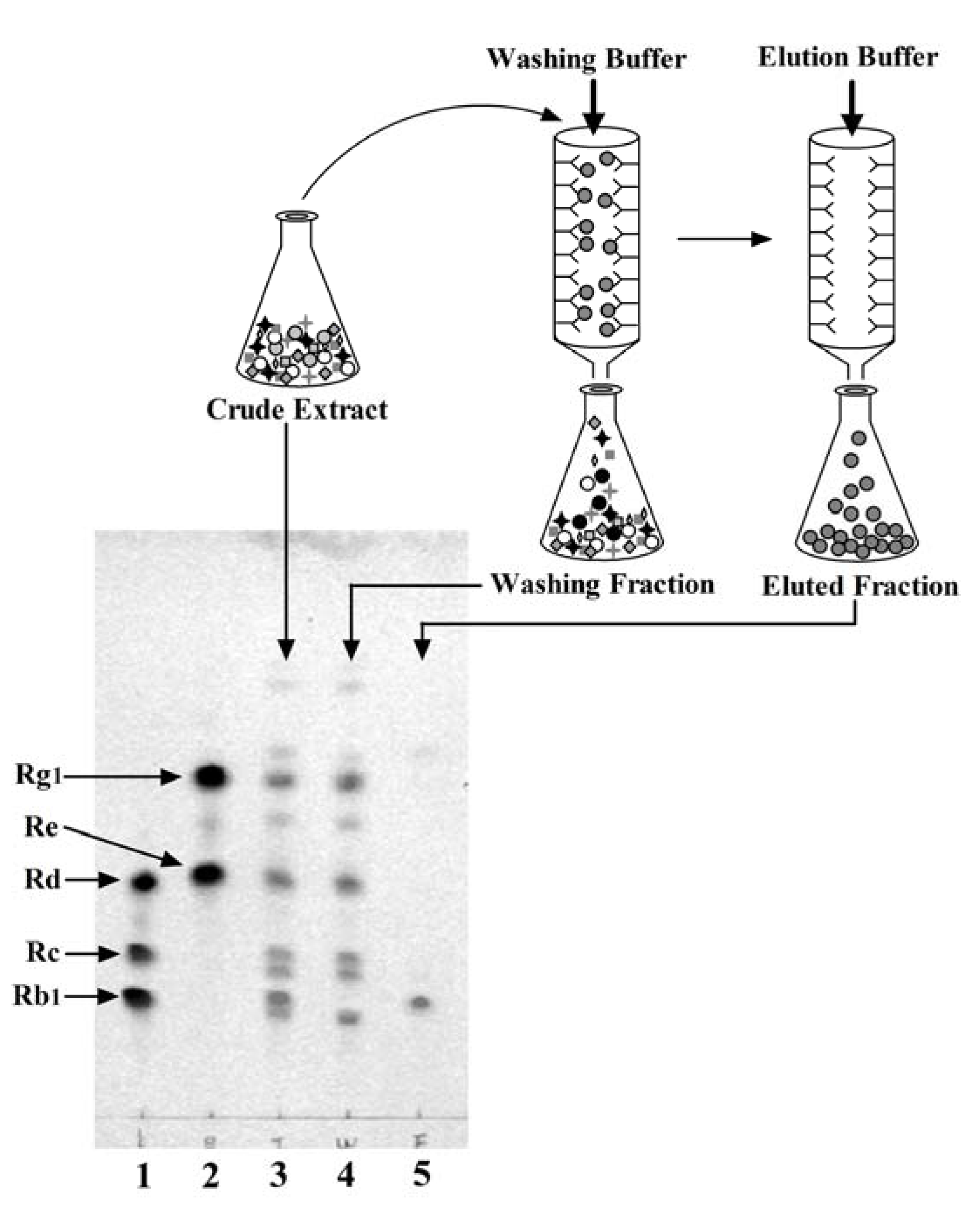
2.3. One-Step Purification of G-Rb1 from Ginseng Extract Using the Anti-G-Rb1 mAb-Coupled Immunoaffinity Column

To examine the anti-G-Rb1 mAb-coupled immunoaffinity column, a crude extract of P. ginseng roots was loaded into the column. After washing the column with washing buffer to remove the unbound compounds, the bound compounds were eluted with elution buffer. Figure 3 shows that fractions 1–8 contained the overloaded G-Rb1, as determined by ELISA. The Eastern blotting procedure, a highly sensitive on-membrane quantitative analysis of anti-G-Rb1 mAb, indicated that these fractions contained other ginsenosides such as G-Rg1, Rc, Re, and Rd. After washing, a single sharp peak was detected in the eluted fractions 21–24, which contained G-Rb1. However, these eluted fractions were contaminated with a small amount of malonyl-G-Rb1, as detected by Eastern blotting, which indicated that anti-G-Rb1 mAb has almost the same reactivity with malonyl-G-Rb1 [28]. Therefore, to convert malonyl-G-Rb1 to pure G-Rb1, these fractions were treated with a mild alkaline solution (0.1% KOH in MeOH) at room temperature [17]. Overloaded G-Rb1 in washing buffer was repeatedly loaded, and purified G-Rb1 was finally obtained. The activity of the anti-G-Rb1 mAb-coupled immunoaffinity column was stable during all procedures. The capacity of the immunoaffinity column was approximately 20 μg of G-Rb1/mL of gel, which did not decrease after 10 repeated purification cycles under the same conditions, as reported in one-step purification of forskolin from a crude extract of Coleus forslohlii root [15].
Figure 3. Elution profile of the P. ginseng crude extract separated using the anti-G-Rb1 mAb-coupled immunoaffinity column. The G-Rb1 concentration in each fraction was monitored by ELISA using anti-G-Rb1 mAb. ∗Inhibition = (A0 − A)/A0; A0 is the absorbance in the absence of the test compounds, and A is the absorbance in the presence of the test compounds.
Antibodies 01 00294 g003
This methodology made it possible to purify G-Rb1 from the crude extract without using complicated procedures and may be applicable to related studies of other families of saponins for which an acceptable one-step purification method has not yet been developed. In addition, it may be possible to separate the total ginseng saponins using a widely cross-reactive mAb against ginsenosides, such as anti-G-Re mAb [35]. A combination of immunoaffinity column chromatography, ELISA, and Eastern blotting could be used for high-sensitivity detection of G-Rb1 from plants and/or experimental animals and humans. Using this combination of methods, we detected G-Rb1 from Kalopanax pictus Nakai, which was not previously reported to contain ginsenosides [36].

2.4. Preparation of the G-Rb1-KO Extract Using the Anti-G-Rb1 mAb-Coupled Immunoaffinity Column

We separated G-Rb1 from the crude ginseng extract using the anti-G-Rb1 mAb-coupled immunoaffinity column. The capacity of the immunoaffinity column is 20 μg of G-Rb1/mL of gel [28]. Upon loading the crude extract into the immunoaffinity column without exceeding the G-Rb1 binding capacity, the column specifically eliminated G-Rb1 from the crude ginseng extract. After loading the ginseng extract into the immunoaffinity column, the washing and eluted fractions were collected. Figure 4 indicates the TLC profile of each fraction stained by H2SO4. A standard of ginsenosides was spotted on lanes 1 (G-Rd, G-Rc, and G-Rb1) and 2 (G-Rg1 and G-Re). The crude extract, washing fraction, and eluted fraction were spotted on lanes 3, 4, and 5, respectively. In the crude extract (lane 3), all spots of ginsenosides were clearly detected. In contrast, the washing fraction (lane 4) contained all ginsenosides, except G-Rb1, and G-Rb1 was detected in the eluted fraction (lane 5). These data clearly demonstrate that the G-Rb1 molecule from the ginseng extract was captured by the anti-G-Rb1 mAb-coupled immunoaffinity column. Thus, the washing fraction was referred to as the G-Rb1-KO extract because it lacked G-Rb1 [37,38]. This G-Rb1-KO extract may be useful for pharmacological characterization of G-Rb1 in ginseng extracts and traditional medicines such as Kampo medicines and TCMs that prescribe ginseng.
Figure 4. Preparation of the KO extract by eliminating G-Rb1 from the P. ginseng crude extract using the anti-G-Rb1 mAb-coupled immunoaffinity column. Lane 1: G-Rd, G-Rc, and G-Rb1, Lane 2: G-Rg1 and G-Re, Lane 3: crude extract, Lane 4: washing fraction, Lane 5: eluted fraction.
Antibodies 01 00294 g004

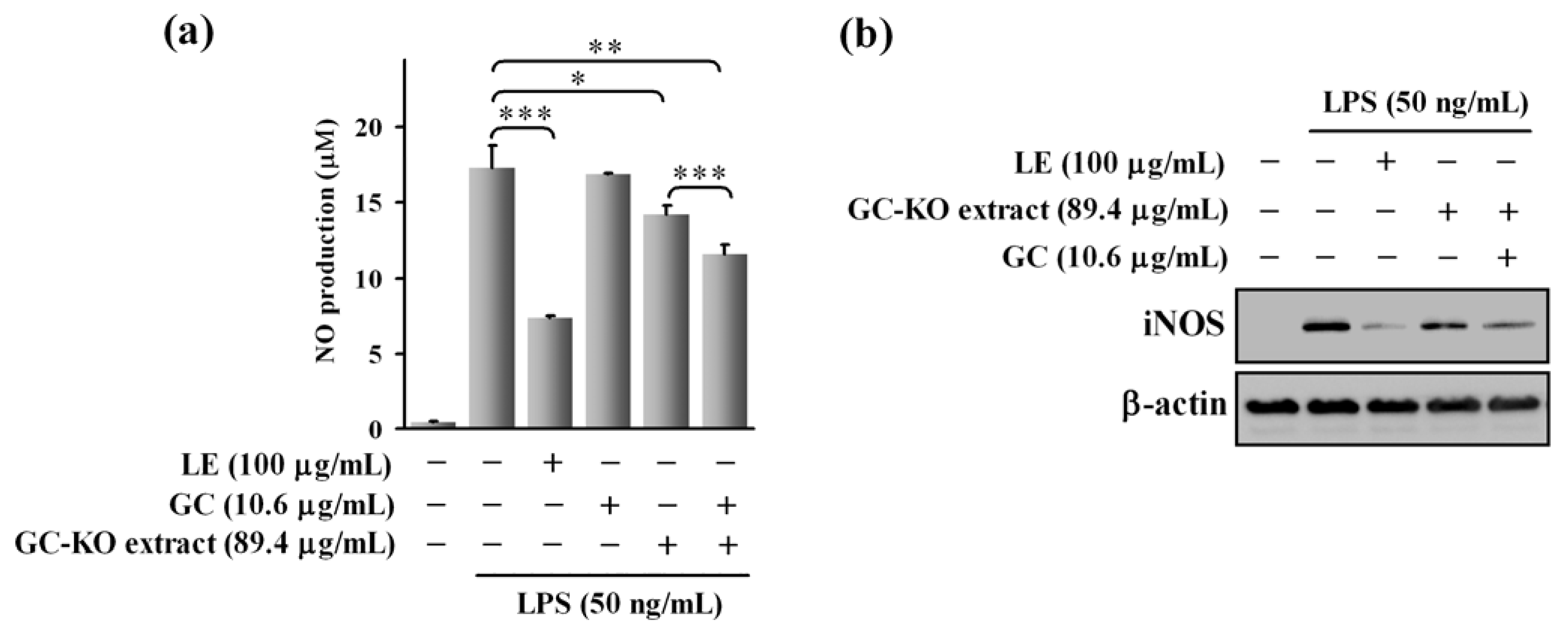
3. Glycyrrhizin (GC)-KO Extract and its Application in Cell-Based Analysis

Licorice (Glycyrrhiza spp.) is an important medicinal plant used in >70% of Kampo medicines and TCMs. Licorice is prescribed with other herbal medicines as a demulcent in the treatment of sore throats, an anti-tussive, an expectorant for coughs and bronchial catarrh, an anti-inflammatory agent for anti-allergic reactions, rheumatism, and arthritis, a prophylactic for liver disease, tuberculosis, and adrenocorticoid insufficiency [39,40,41]. A number of phytochemical investigations showed that licorice contains at least 470 constituents, including triterpenes, saponins, flavonoids, isoflavonoids, chalcones, polysaccharides, simple sugars, amino acids, and other substances [42]. Accumulated evidence indicates that GC is a major bioactive triterpene glycoside in licorice and has numerous pharmacological uses. It exhibits anti-inflammatory, anti-ulcer, anti-tumor, anti-allergic, and hepato-protective activities [41,43]. In addition, it was reported that various licorice constituents, such as flavonoid glycosides and their aglycones, exhibit anti-inflammatory, anti-oxidative, anti-microbial, superoxide scavenging, and anti-carcinogenic activities [41,44]. Although the functional effects of individual compounds in licorice were analyzed in vitro and in vivo, the potential function of GC in licorice extract (LE) and interactions between GC and other components in LE were unclear because it was technically difficult to prepare GC-free LE. Previously, we purified GC from LE using an anti-GC mAb-coupled immunoaffinity column and then prepared a GC-KO extract [18]. In this section, we review the preparation of this GC-KO extract and its application in in vitro assays.

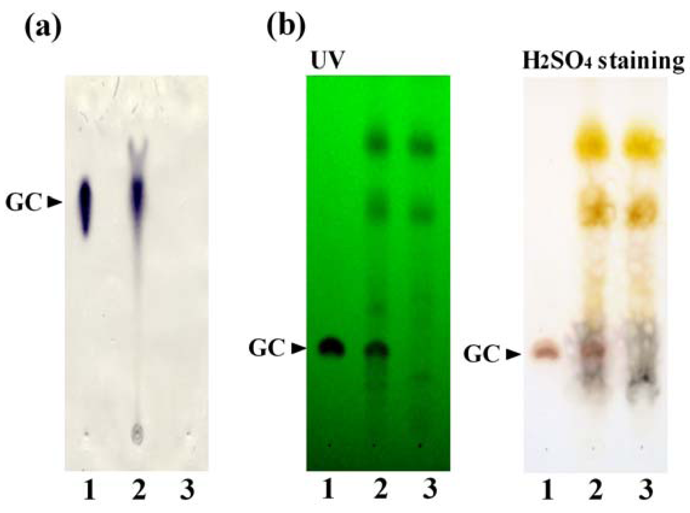
3.1. Preparation of the GC-KO Extract and its Characterization

The anti-GC mAb was prepared using the same procedure as for G-Rb1 [7]. Cross-reactivity of the anti-GC mAb against glycyrrhetic acid-3-O-glucuronide and glycyrrhetic acid was 0.585% and 1.865%, respectively. Cross-reactivity with other related compounds, including deoxycholic acid, ursolic acid, and oleanolic acid, was <0.005%. Thus, we established competitive ELISA and Eastern blotting using anti-GC mAb to specifically detect GC [7,45]. In addition, we used the anti-GC mAb-coupled immunoaffinity column to prepare the GC-KO extract. After purifying 60 mg of anti-GC mAb and coupling it to 25 mL of an Affi-Gel Hz gel, the immunoaffinity gel was packed into a plastic column [18]. To eliminate GC from LE, 12 mg of LE, which contained 1275.0 μg of GC in loading buffer (5% MeOH) was applied to the immunoaffinity column. After circulation of the loading buffer to enhance the binding efficiency of GC, the unbound fraction was collected. The column was washed with washing buffer (5% MeOH) and then eluted with elution buffer (20 mM PB containing 30% MeOH). After elution of bound compounds, each fraction was deionized, the solvent was lyophilized, and the GC concentration was determined by ELISA. The unbound fraction contained 3.50 μg of GC (0.27% of the loaded GC), while the bound fraction contained 1269.26 μg of GC (99.55% of the loaded GC), suggesting that the anti-GC mAb-coupled column eliminated 99.55% of the loaded GC. In the unbound fraction, 3.50 μg of GC (0.27% of the applied GC) was detected. In contrast, 1269.26 μg of GC (99.55% of the applied GC) was found in the bound fraction. These data suggest that the anti-GC mAb-coupled column eliminated 99.55% of the loaded GC and produced a GC-KO extract [46].
To characterize this GC-KO extract, we performed Eastern blotting and TLC analysis [46]. As shown in Figure 5a, Eastern blotting using anti-GC mAb demonstrated that GC was detected in LE (lane 2), but the spot corresponding to GC was not present in the GC-KO extract (lane 3). Furthermore, TLC analysis (Figure 5b) showed that many compounds, including GC, were present in LE (lane 2), whereas GC was completely absent in the GC-KO extract (lane 3). Taken together, these data clearly demonstrate that GC molecules in LE can be eliminated using the anti-GC mAb-coupled immunoaffinity column and that the unbound fraction was indeed a GC-KO extract.
Figure 5. (a) Eastern blotting using anti-GC mAb and (b) TLC profiles of the GC-KO extract. Lane 1: GC, Lane 2: LE, Lane 3: GC-KO extract.
Antibodies 01 00294 g005

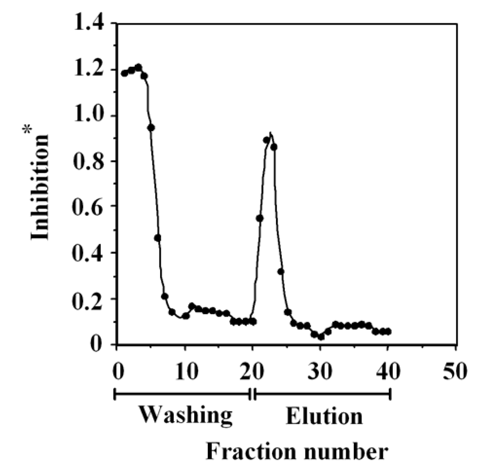
3.2. Cell-Based Assay Using the GC-KO Extract

The GC-KO extract is a useful tool for clarifying the interaction between GC and other compounds contained in LE. To investigate the potential function of GC in LE, we used macrophages to compare the effects of LE and the GC-KO extract on the production of nitric oxide (NO). The free radical NO has multiple effects on various systems, including host defense against pathogens, inhibition of tumor cells, and neurotransmission [47,48,49]. However, excess production of NO, which is synthesized by the inducible NOS (iNOS), can be harmful and can trigger rheumatoid arthritis, gastritis, bowel inflammation, neuronal cell death, and bronchitis [50,51]. Macrophages produce large quantities of NO after stimulation with bacterial lipopolysaccharide (LPS), and concomitantly produced inflammatory cytokines participate in the pathogenesis of inflammatory diseases [52]. Overproduction of NO by iNOS appears to be involved in the pathogenesis of various inflammatory diseases [53]. Therefore, inhibition of NO production by blocking iNOS expression may be useful in treating NO overexpression-mediated diseases.
To examine the inhibitory effect of LE on NO production, we used murine RAW264 macrophage cells, which produce NO upon stimulation with LPS. NO induction by LPS was blocked by treatment with LE in a dose-dependent manner [46]. Furthermore, Western blotting and RT-PCR showed that iNOS protein and mRNA expression was downregulated in a dose-dependent manner by LE and was completely suppressed in the presence of 100 μg/mL of LE. ELISA using anti-GC mAb demonstrated that 100 μg of LE contains 10.6 ± 0.618 μg of GC. However, significant inhibition of NO production and iNOS protein expression was not observed with GC at approximately 10.6 μg/mL.
Figure 6a shows that LE treatment (100 μg/mL) markedly inhibits NO production (inhibition ratio (IR) = 57.7%) compared with LPS treatment alone. In contrast, treatment with GC (10.6 μg/mL) did not suppress NO production. Subsequently, we treated cells with the GC-KO extract (89.4 μg/mL) or the combination of GC-KO extract (89.4 μg/mL) and GC (10.6 μg/mL). Interestingly, although GC alone did not block NO production, the inhibitory effect of the GC-KO extract (IR = 17.8%) was weaker than that of LE. Moreover, co-treatment with the GC-KO extract and GC significantly improved inhibition (IR = 33.5%). To determine whether these effects depended on iNOS expression, we performed Western blotting. As shown in Figure 6b, inhibition by the GC-KO extract was weaker than that by LE and addition of GC to the GC-KO extract rescued the inhibition. These data suggest that GC alone cannot suppress iNOS expression; however, in combination with the other constituents of LE, GC suppresses iNOS expression. Thus, in vitro and in vivo analyses using KO extracts can elucidate the functions of target natural compounds in crude extracts. Moreover, we have compared activity using GC contained in elution fraction and commercially available GC when the cells were treated with GC-KO extract. Since the activity was almost same level, the function of purified GC was not lost during the purification step. In future studies, we will investigate the detailed mechanisms including the differences between the original LE and GC-KO extract, the interactions between GC and GC-KO extract, and the action of GC in LE.
Figure 6. Comparison of the effects of the GC-KO extract and the combination of the GC-KO extract and GC on LPS-induced (a) NO production and (b) iNOS expression. Cells were treated with LE (100 μg/mL), GC-KO extract (89.4 μg/mL), GC (10.6 μg/mL), or the combination of GC-KO extract (89.4 μg/mL) and GC (10.6 μg/mL). * p < 0.05, ** p < 0.01, and *** p < 0.001 indicate significant differences from the effects of LPS alone.
Antibodies 01 00294 g006

4. Conclusions

This review describes a unique strategy involving one-step purification of target compounds from crude extracts using anti-natural compound-specific mAb-coupled immunoaffinity columns. A G-Rb1-KO extract, which contains all compounds except G-Rb1, was prepared from the P. ginseng crude extract using the anti-G-Rb1 mAb-coupled immunoaffinity column. Furthermore, the GC-KO extract was prepared from LE using an anti-GC mAb-coupled immunoaffinity column. Our laboratory has previously prepared mAbs against natural bioactive compounds such as ginsenosides [5,54,55], berberine [10], crocin [2], sennosides [12], and forskolin [1]. Moreover, the immunoaffinity columns coupled with anti-G-Rb1 [17], anti-forskolin [15], anti-solasodine glycoside [16], and anti-GC mAbs [18] achieved one-step purification of target compounds from crude extracts. Therefore, these mAbs and techniques are easily applicable to the preparation of KO extracts that lack only the target compound from crude plant extracts, Kampo medicines, and TCMs.
We also developed an in vitro assay using the GC-KO extract. This assay demonstrated that the GC-KO extract alone did not suppress iNOS expression, but inhibited iNOS when present in combination with the other constituents of LE. Thus, KO extracts may be useful for determining the potential functions of target compounds in crude extracts and Kampo medicines in vitro and in vivo.

Acknowledgments

This work was supported in part by “Science and Technology Research Partnership for Sustainable Development (SATREPS)” funded by the Japan Science and Technology Agency (JST) and the Japan International Cooperation Agency (JICA). The authors would like to thank Enago (www.enago.jp) for the English language review.

References

  1. Sakata, R.; Shoyama, Y.; Murakami, H. Production of monoclonal antibodies and enzyme immunoassay for typical adenylate cyclase activator, Forskolin. Cytotechnology 1994, 16, 101–108. [Google Scholar]
  2. Xuan, L.; Tanaka, H.; Xu, Y.; Shoyama, Y. Preparation of monoclonal antibody against crocin and its characterization. Cytotechnology 1999, 29, 65–70. [Google Scholar]
  3. Lu, Z.; Morinaga, O.; Tanaka, H.; Shoyama, Y. A quantitative ELISA using monoclonal antibody to survey paeoniflorin and albiflorin in crude drugs and traditional Chinese herbal medicines. Biol. Pharm. Bull. 2003, 26, 862–866. [Google Scholar]
  4. Tanaka, H.; Fukuda, N.; Shoyama, Y. Formation of monoclonal antibody against a major ginseng component, ginsenoside Rb1 and its characterization. Cytotechnology 1999, 29, 115–120. [Google Scholar]
  5. Fukuda, N.; Tanaka, H.; Shoyama, Y. Formation of monoclonal antibody against a major ginseng component, ginsenoside Rg1 and its characterization. Monoclonal antibody for a ginseng saponin. Cytotechnology 2000, 34, 197–204. [Google Scholar]
  6. Zhu, S.; Shimokawa, S.; Tanaka, H.; Shoyama, Y. Development of an assay system for saikosaponin a using anti-saikosaponin a monoclonal antibodies. Biol. Pharm. Bull. 2004, 27, 66–71. [Google Scholar]
  7. Shan, S.J.; Tanaka, H.; Shoyama, Y. Enzyme-linked immunosorbent assay for glycyrrhizin using anti-glycyrrhizin monoclonal antibody and an eastern blotting technique for glucuronides of glycyrrhetic acid. Anal. Chem. 2001, 73, 5784–5790. [Google Scholar]
  8. Ishiyama, M.; Shoyama, Y.; Murakami, H.; Shinohara, H. Production of monoclonal antibodies and development of an ELISA for solamargine. Cytotechnology 1996, 18, 153–158. [Google Scholar]
  9. Shoyama, Y.; Fukada, T.; Murakami, H. Production of monoclonal antibodies and ELISA for thebaine and codeine. Cytotechnology 1996, 19, 55–61. [Google Scholar]
  10. Kim, J.S.; Tanaka, H.; Shoyama, Y. Immunoquantitative analysis for berberine and its related compounds using monoclonal antibodies in herbal medicines. Analyst 2004, 129, 87–91. [Google Scholar] [CrossRef]
  11. Morinaga, O.; Tanaka, H.; Shoyama, Y. Production of monoclonal antibody against a major purgative component, sennoside A, its characterization and ELISA. Analyst 2000, 125, 1109–1113. [Google Scholar]
  12. Morinaga, O.; Nakajima, S.; Tanaka, H.; Shoyama, Y. Production of monoclonal antibodies against a major purgative component, sennoside B, their characterization and use in ELISA. Analyst 2001, 126, 1372–1376. [Google Scholar]
  13. Tanaka, H.; Goto, Y.; Shoyama, Y. Monoclonal antibody based enzyme immunoassay for marihuana (cannabinoid) compounds. Immunoassay 1996, 17, 321–342. [Google Scholar]
  14. Loungratana, P.; Tanaka, H.; Shoyama, Y. Production of monoclonal antibody against ginkgolic acids in Ginkgo biloba Linn. Am. J. Chin. Med. 2004, 32, 33–48. [Google Scholar]
  15. Yanagihara, H.; Sakata, R.; Minami, H.; Shoyama, Y.; Murakami, H. Immunoaffinity column chromatography against forskolin using an anti-forskolin monoclonal antibody and its application. Anal. Chim. Acta 1996, 335, 63–70. [Google Scholar]
  16. Putalun, W.; Tanaka, H.; Yukihira, S. Rapid separation of solasodine glycosides by an immunoaffinity column using anti-solamargine monoclonal antibody. Cytotechnology 1999, 31, 151–156. [Google Scholar]
  17. Fukuda, N.; Tanaka, H.; Shoyama, H. Isolation of the pharmacologically active saponin ginsenoside Rb1 from ginseng by immunoaffinity column chromatography. J. Nat. Prod. 2000, 63, 283–285. [Google Scholar]
  18. Xu, J.; Tanaka, H.; Shoyama, Y. One-step immunochromatographic separation and ELISA quantification of glycyrrhizin from traditional Chinese medicines. J. Chromatogr. B 2007, 850, 53–58. [Google Scholar] [CrossRef]
  19. Yun, T.K. Brief introduction of Panax ginseng C.A. Meyer. J. Korean Med. Sci. 2001, 16, S3–S5. [Google Scholar]
  20. Park, J.D.; Rhee, D.K.; Lee, Y.H. Biological activities and chemistry of saponins from Panax ginseng C. A. Meyer. Phytochem. Rev. 2005, 4, 159–175. [Google Scholar]
  21. Choi, K.T. Botanical characteristics, pharmacological and medicinal components of Korean Panax ginseng C.A. Meyer. Acta Pharmacol. Sin. 2008, 29, 1109–1118. [Google Scholar]
  22. Qi, L.W.; Wang, C.Z.; Yuan, C.S. Isolation and analysis of ginseng: Advances and challenges. Nat. Prod. Rep. 2011, 28, 467–495. [Google Scholar]
  23. Washida, D.; Kitanaka, S. Determination of polyacetylenes and ginsenosides in Panax species using high performance liquid chromatography. Chem. Pharm. Bull. 2003, 51, 1314–1317. [Google Scholar] [CrossRef]
  24. Mook-Jung, I.; Hong, H.S.; Boo, J.H.; Lee, K.H.; Yun, S.H.; Cheong., M.Y.; Joo, I.; Huh, K.; Jung, M.W. Ginsenoside Rb1 and Rg1 improve spatial learning and increase hippocampal synaptophysin level in mice. J. Neurosci. Res. 2001, 63, 509–515. [Google Scholar]
  25. Lim, J.H.; Wen, T.C.; Matsuda, S.; Tanaka, J.; Maeda, N.; Peng, H.; Aburaya, J.; Ishihara, K.; Sakanaka, M. Protection of ischemic hippocampal neurons by ginsenoside Rb1, a main ingredient of ginseng root. Neurosci. Res. 1997, 28, 191–200. [Google Scholar] [CrossRef]
  26. Liu, M.; Zhang, J. Effects of ginsenoside Rb1 and Rg1 on synaptosomal free calcium level, ATPase and calmodulin in rat hippocampus. Chin. Med. J. (Engl.) 1995, 108, 544–547. [Google Scholar]
  27. Jiang, K.Y.; Qian, Z.N. Effects of Panax notoginseng saponins on posthypoxic cell damage of neurons in vitro. Zhongguo Yao Li Xue Bao 1995, 16, 399–402. [Google Scholar]
  28. Fukuda, N.; Tanaka, H.; Shoyama, Y. Applications of ELISA, western blotting and immunoaffinity concentration for survey of ginsenosides in crude drugs of Panax species and traditional Chinese herbal medicines. Analyst 2000, 125, 1425–1429. [Google Scholar] [CrossRef]
  29. Wengatz, I.; Schmid, R.D.; Kreißig, S.; Wittmann, C.; Hock, B.; Ingendoh, A.; Hillenkamp, F. Determination of the hapten density of immuno-conjugates by matrix-assisted UV laser desorption/ionization mass spectrometry. Anal. Lett. 1992, 25, 1983–1997. [Google Scholar] [CrossRef]
  30. Shoyama, Y.; Sakata, R.; Isobe, R.; Murakami, H. Direct determination of forskolin-bovine serum albumin conjugate by matrix-assisted laser desorption ionization mass spectrometry. Org. Mass Spect. 1993, 28, 987–988. [Google Scholar] [CrossRef]
  31. Shoyama, Y.; Fukada, T.; Tanaka, T.; Kusai, A.; Nojima, K. Direct determination of opium alkaloid-bovine serum albumin conjugate by matrix-assisted laser desorption ionization mass spectrometry. Biol. Pharm. Bull. 1993, 16, 1051–1053. [Google Scholar] [CrossRef]
  32. Goto, Y.; Shima, Y.; Morimoto, S.; Shoyama, Y.; Murakami, H.; Kusai, A.; Nojima, K. Determination of tetrahydrocannabinolic acid-carrier protein conjugate by matrix-assisted laser desorption ionization mass spectrometry and antibody formation. Org. Mass Spectr. 1994, 29, 668–671. [Google Scholar] [CrossRef]
  33. Galfrè, G.; Milstein, C. Preparation of monoclonal antibodies: Strategies and procedures. Methods Enzymol. 1981, 73, 3–46. [Google Scholar]
  34. Goding, J.W. Antibody production by hybridomas. J. Immunol. Methods 1980, 39, 285–308. [Google Scholar] [CrossRef]
  35. Morinaga, O.; Tanaka, H.; Shoyama, Y. Detection and quantification of ginsenoside Re in ginseng samples by a chromatographic immunostaining method using monoclonal antibody against ginsenoside Re. J. Chromatogr. B 2006, 830, 100–104. [Google Scholar] [CrossRef]
  36. Tanaka, H.; Fukuda, N.; Yahara, S.; Isoda, S.; Yuan, C.S.; Shoyama, Y. Isolation of ginsenoside Rb1 from Kalopanax pictus by eastern blotting using anti-ginsenoside Rb1 monoclonal antibody. Phytother. Res. 2005, 19, 255–258. [Google Scholar] [CrossRef]
  37. Tanaka, H.; Fukuda, N.; Shoyama, Y. Eastern blotting and immunoaffinity concentration using monoclonal antibody for ginseng saponins in the field of traditional Chinese medicines. J. Agric. Food Chem. 2007, 55, 3783–3787. [Google Scholar] [CrossRef]
  38. Wang, C.A.; Shoyama, Y. Herbal medicine: Identification, analysis, and evaluationstrategies. In Textbook of Complementary and Alternative Medicine, 2nd ed.; Yuan, C.S., Bieber, E.J., Bauer, B.A., Eds.; Informa Healthcare: London, UK, 2006; pp. 51–70. [Google Scholar]
  39. Kim, S.C.; Byun, S.H.; Yang, C.H.; Kim, C.Y.; Kim, J.W.; Kim, S.G. Cytoprotective effects of Glycyrrhizae radix extract and its active component liquiritigenin against cadmium-induced toxicity (effects on bad translocation and cytochrome c-mediated PARP cleavage). Toxicology 2004, 197, 239–251. [Google Scholar] [CrossRef]
  40. Fuchikami, J.; Isohama, Y.; Sakaguchi, M.; Matsuda, M.; Kucota, T.; Akie, Y.; Fujino, A.; Miyata, T. Effect of glycyrrhizin on late asthmatic responses induced by antigen inhalation in guinea pigs. J. Pharmacol. Sci. 2004, 94, 251. [Google Scholar]
  41. Asl, M.N.; Hosseinzadeh, H. Review of pharmacological effects of Glycyrrhiza sp. and its bioactive compounds. Phytother. Res. 2008, 22, 709–724. [Google Scholar] [CrossRef]
  42. Zhang, Q.; Ye, M. Chemical analysis of the Chinese herbal medicine Gan-Cao (licorice). J. Chromatogr. A 2009, 1216, 1954–1969. [Google Scholar] [CrossRef]
  43. Wang, Z.Y.; Nixon, D.W. Licorice and cancer. Nutr. Cancer 2001, 39, 1–11. [Google Scholar]
  44. Chin, Y.W.; Jung, H.A.; Liu, Y.; Su, B.N.; Castoro, J.A.; Keller, W.J.; Pereira, M.A.; Kinghorn, A.D. Anti-oxidant constituents of the roots and stolons of licorice (Glycyrrhiza glabra). J. Agric. Food Chem. 2007, 55, 4691–4697. [Google Scholar] [CrossRef]
  45. Morinaga, O.; Fujino, A.; Tanaka, H.; Shoyama, Y. An on-membrane quantitative analysis system for glycyrrhizin in licorice roots and traditional Chinese medicines. Anal. Bioanal. Chem. 2005, 383, 668–672. [Google Scholar] [CrossRef]
  46. Uto, T.; Morinaga, O.; Tanaka, H.; Shoyama, Y. Analysis of the synergistic effect of glycyrrhizin and other constituents in licorice extract onlipopolysaccharide-induced nitric oxide production using knock-out extract. Biochem. Biophys. Res. Commun. 2012, 417, 473–478. [Google Scholar] [CrossRef]
  47. Bogdan, C. Nitric oxide and the immune response. Nat. Immunol. 2001, 2, 907–916. [Google Scholar] [CrossRef]
  48. Bogdan, C. Nitric oxide and the regulation of gene expression. Trends Cell Biol. 2001, 11, 66–75. [Google Scholar] [CrossRef]
  49. Vincent, S.R. Nitric oxide neurons and neurotransmission. Prog. Neurobiol. 2010, 90, 246–255. [Google Scholar] [CrossRef]
  50. Abramson, S.B.; Amin, A.R.; Clancy, R.M.; Attur, M. The role of nitric oxide in tissue destruction. Best Pract. Res. Clin. Rheumatol. 2001, 15, 831–845. [Google Scholar] [CrossRef]
  51. Gassull, M.A. The role of nutrition in the treatment of inflammatory bowel disease. Aliment. Pharmacol. Ther. 2004, 20, 79–83. [Google Scholar] [CrossRef]
  52. Blantz, R.C.; Munger, K. Role of nitric oxide in inflammatory conditions. Nephron 2002, 90, 373–378. [Google Scholar] [CrossRef]
  53. Kröncke, K.D.; Fehsel, K.; Kolb-Bachofen, V. Inducible nitric oxide synthase in human diseases. Clin. Exp. Immunol. 1998, 113, 147–156. [Google Scholar] [CrossRef]
  54. Fukuda, N.; Tanaka, H.; Shoyama, Y. Western blotting for ginseng saponins, ginsenosides using anti-ginsenoside Rb1 monoclonal antibody. Biol. Pharm. Bull. 1999, 22, 219–220. [Google Scholar] [CrossRef]
  55. Sritularak, B.; Morinaga, O.; Yuan, C.S.; Shoyama, Y.; Tanaka, H. Quantitative analysis of ginsenosides Rb1, Rg1, and Re in American ginseng berry and flower samples by ELISA using monoclonal antibodies. J. Nat. Med. 2009, 63, 360–363. [Google Scholar]

