
Progress in the Application of Biomimetic Mineralization for Tooth Repair

1
Clinical Research Center for Oral Diseases of Zhejiang Province, Stomatology Hospital, School of Stomatology, Zhejiang University School of Medicine, Hangzhou 310000, China
2
Key Laboratory of Oral Biomedical Research of Zhejiang Province, Cancer Center of Zhejiang University, Hangzhou 310000, China
3
Engineering Research Center of Oral Biomaterials and Devices of Zhejiang Province, Hangzhou 310000, China
*
Authors to whom correspondence should be addressed.
†
These authors contributed equally to this work.
Minerals 2023, 13(11), 1433; https://doi.org/10.3390/min13111433
Submission received: 7 October 2023
/
Revised: 7 November 2023
/
Accepted: 8 November 2023
/
Published: 12 November 2023
(This article belongs to the Special Issue Mineral Nucleation and Growth across Multiple Scales: Current Research and Future Goals)
Abstract
:The tooth, including enamel and dentin, is a prominent biomineral that is produced by the biomineralization of living organisms. Although the mechanical performance of the tooth is outstanding, caries easily develop in a complex oral environment. The analysis of the chemical composition and the relationship between the mechanical properties and the structure is of great importance in solving caries. In this review, the multilevel structure and mechanical properties of enamel and dentin are briefly introduced, along with caries formation and the limitations of clinical dental restoration. Furthermore, the progress of the application of a wide range of biomimetic strategies for tooth remineralization is highlighted, including the use of calcium phosphate ionic clusters to construct the mineralization front, ensuring the oriented epitaxial growth of enamel crystals and replicating the complex structure of the enamel. Moreover, compared with the current clinical treatment, in which the resin composite and glass ionomer cement are the main repair materials and the high incidence of secondary caries leads to imperfect restorations, the remineralization tactics could achieve excellent repair effectiveness in reconstructing the complicated structure, restoring mechanical strength and gaining permanent repair. A basic understanding of enamel and dentin, their potential for restoration, and hopeful prospects for tooth repair that can be applied in the clinical setting, not just in the laboratory, is provided by this review.
1. Introduction
The tooth is a prominent biomineral, a highly mineralized tissue consisting of enamel, dentin, and pulp from the outside to the inside. With the development and application of advanced technologies, the chemical composition and complicated structure of the tooth have been investigated in depth, allowing us to grasp the relationship between the mechanical properties and the structure. The tooth, in particular the enamel, has a robust material strength that can withstand hundreds of thousands of chewing movements. However, teeth are susceptible to chemical attack when exposed to various acidic environments, leading to caries [1,2]. Dental caries is considered the oral disease with the highest prevalence rate, and dental caries is ranked by the World Health Organization (WHO) as one of the three major diseases threatening human health, along with cancer and cardiovascular diseases [3]. Oral diseases such as pulpitis and periapical periodontitis can be caused by untreated dental caries, and they can also exacerbate systemic chronic diseases such as cardiovascular and cerebrovascular disorders, posing a threat to overall health [4]. Therefore, achieving long-term stable dental restoration has become an increasingly urgent goal in this field. Given the structure and properties of teeth, the most promising approach is to achieve a unity of materials, structure, and mechanical properties through biomimetic mineralization.
2. Structure and Properties of Teeth
The hard tissue of the tooth, a fundamental biological mineral structure, consists of dentin, enamel, and cementum. Dentin is the main structure of the tooth, and enamel, as the hardest tissue in the body, covers its surface. Enamel and dentin are discussed specifically below.
2.1. Enamel
Enamel is composed of 96% minerals (mainly hydroxyapatite (HAP)), 3% water, and 1% organic matter [5]. Because of its highly organized structure and superior material performance [6], enamel serves as a protective layer to protect the tooth from the harsh conditions of the oral environment, including mechanical stress [7], temperature changes [8], and acid attacks [9].
2.1.1. Hierarchical Structure of Enamel
Enamel has a complex hierarchical structure from the nanoscale to the micron scale (Figure 1A), which is currently divided into seven levels [10,11]. At the nanoscale, the basic unit is highly ordered, with parallel HAP nanowire crystals that are 50–70 nm in width, 20–35 nm in thickness, and very long, with the longest ones spanning the entire thickness of the enamel.
At the micron scale, HAP nanowire crystals (on the order of 104) bind tightly together to self-assemble into an enamel rod with a diameter of approximately 4–6 μm. These enamel rods are further arranged in an orderly manner and woven together with the internal enamel rods (interrods) to form the basic assembly unit of the enamel. Both the enamel rods and the interrods are primarily composed of HAP crystals, but the orientation of the c-axis of the crystals varies, with an angle of approximately 60 degrees [12].
Organic matter, mainly the product of protein hydrolysis [13], exists between the enamel rods and the interrods, forming the enamel rod sheath, which plays a critical role in the mechanical properties [14]. Under a scanning electron microscope (SEM), the enamel shows a lock-hole-like structure [11] and a fish-scale-like structure [12] after acid etching (using acids such as 30%–37% phosphoric acid, citric acid, lactic acid, and 3% nitric acid [15]). The formation of these unique structures is mainly due to the different acid resistance levels of the crystals in the enamel rods and the interrods, which is mainly because the gaps between the enamel rods contain higher protein content [11].
In addition, the orientation of the enamel rods varies within the enamel layer. For example, in the outer one-third of the surface layer of the enamel, the enamel rods are parallel to each other, arranged in a radiating manner, and perpendicular to the occlusal surface of the tooth [16]; in the remaining two-thirds of the inner layer, the enamel rods are arranged in a curved and intersecting pattern.
2.1.2. Mechanical Properties of Enamel
Enamel, as a representative biomineral in nature, has a unique multilevel structure that gives it excellent mechanical strength and wear resistance, and it is the hardest biocomposite found thus far in biominerals to date [13,17,18]. The hardness of enamel ranges from 3 to 6 GPa, and the elastic modulus is between 70 and 115 GPa [19]. The hardness and elastic modulus of the enamel vary at different locations, with the enamel closest to the surface having the highest hardness and elastic modulus (Figure 1B,C). From the outer surface to the junction between the enamel and dentin (dentin-enamel junction, DEJ), the hardness and elastic modulus gradually decrease [20].
This variation occurs because the chemical composition of the enamel affects both the structure and the mechanical properties of the enamel. Differences in the levels of proteins [21,22,23] and inorganic ions (such as magnesium ions [24], carbonate ions [25], and fluoride ions [26]) can lead to differences in the mechanical properties of the enamel. Hoffman et al. found that hypomineralized enamel has significantly lower hardness and elastic modulus than normal enamel due to the higher protein content in hypomineralized enamel [22]. Because the enamel rod sheath contains a higher protein content than that of the enamel rod, the enamel rod has a higher hardness and elastic modulus [23].
Figure 1.
(A) Schematic illustration of the hierarchical assembly of the enamel structure, from the millimeter to the nanometer scale, reprinted with permission from Ref. [11]. 2007, John Wiley and Sons. Hardness (B) and elastic modulus (C) of the enamel as determined by nanoindentation, reprinted with permission from Ref. [19]. 2002, Elsevier.

2.2. Dentin
2.2.1. The Structure of Dentin
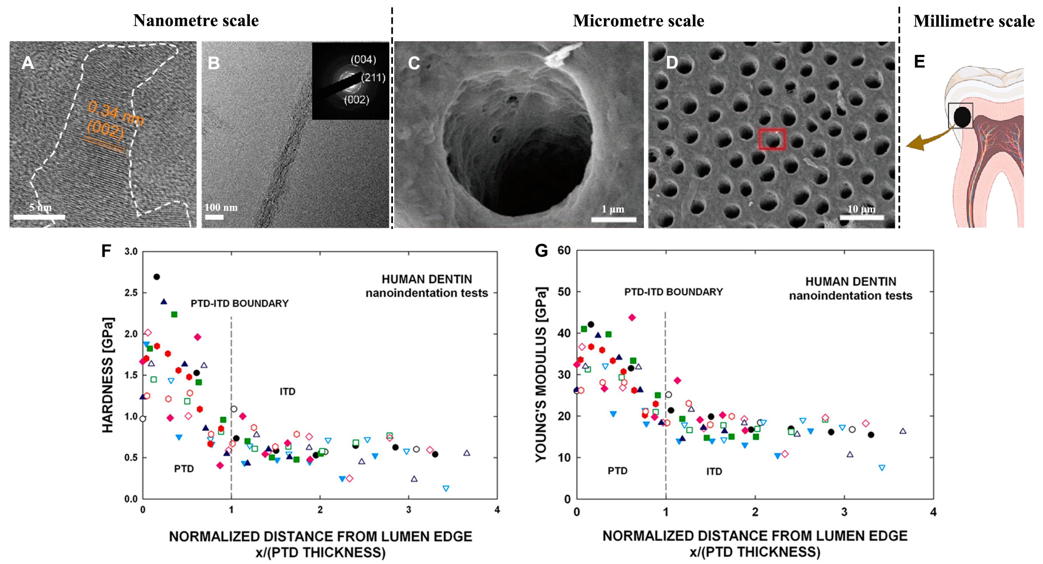
Dentin, the main hard tissue of the teeth, is covered by enamel in the crown and cementum in the root. Dentin is mainly composed of dentin tubules, odontoblastic processes, and intercellular substance [27,28]; it consists of approximately 70% inorganic substance, primarily HAP, 20% organic substance, and 10% water by weight. The dentin tubules and odontoblastic processes traverse the entire dentin, while the mineralized collagen fibers within the intercellular substance intertwine to form a network that constructs the dentin scaffold (Figure 2A–E). Non-collagenous proteins (NCPs) such as dentin phosphoprotein (DPP), dentin sialoprotein (DSP), and dentin matrix protein-1 (DMP-1) play a crucial role in dentin formation [29,30,31,32,33]. Under the regulation of NCPs, the multi-level ordered organic–inorganic composite structure is formed as HAP crystals grow along the c-axis of the collagen fibers [34,35].
2.2.2. Mechanical Properties of Dentin
Enamel and dentin have different mechanical properties and complement each other in maintaining the mechanical stability of teeth. The hardness of dentin is lower than that of enamel due to its high organic content and the presence of water in the dentin tubules. This endows dentin with a certain degree of elasticity that provides a favorable buffering environment for enamel [36,37].
Dentin has an average hardness of approximately 0.8 GPa and an elastic modulus of nearly 20 GPa [38]. The mechanical properties of dentin are anisotropic and are influenced by several factors, including the location, density, and direction of the dentin tubules, the orientation of the collagen fibers, and the average density of the mineral phases [39]. Under evaluation with atomic force microscope (AFM), the microstructural mechanical properties of different parts of dentin vary (Figure 2F,G); the elastic modulus of highly mineralized peritubular dentin reached 40 GPa, while that of weakly mineralized intertubular dentin was lower [40]. Near the pulp, the hardness of peritubular dentin exhibited a noticeable gradient as the mineral content changed.
The mechanical properties of dentin vary depending on its mineral content. Microscopically, the mineralization of dentin can be categorized into intrafibrillar mineralization and extrafibrillar mineralization based on the relative position of HAP and the collagen matrix. Intrafibrillar mineralization plays a crucial role in determining the mechanical properties of dentin at the nanoscale [41,42]. It also helps to prevent the degeneration and degradation of collagen when exposed to external stimuli, which is essential for maintaining the integrity of dentin.
Figure 2.
(A) High-resolution transmission electron microscopy (HRTEM) image of mineralized collagen fibrils. (B) TEM image of mineralized collagen fibrils. Inset: selected area electron diffraction (SAED) pattern, reprinted with permission from Ref. [43]. 2023, Elsevier. (C) High-magnification SEM images of the regions marked with red squares in (D). (D) SEM image of dentin surface and dentin tubules, reprinted with permission from Ref. [44]. 2022, Elsevier. (E) Schematic image showing the anatomy of the tooth. Hardness (F) and modulus (G) results for dentin plotted vs. distance from lumen (PTD, peritubular dentin; ITD, intertubular dentin), where the different colors and shapes represent different dental tubules, reprinted with permission from Ref. [40]. 2011, Elsevier.
3. Tooth Restoration
3.1. Dental Caries
Dental caries is mainly caused by an imbalance between the mineralization and demineralization of tooth minerals [1]. Normally, the gain and loss of minerals are balanced. However, acid-producing bacteria form dental plaque on the tooth surface and produce large amounts of organic acids. Since the acids are not easily diluted by saliva, the pH value of localized areas of the enamel surface drops significantly. When the pH falls to a certain level (below 5.5, according to in vitro experiments [45]), the calcium phosphate in the dental enamel dissolves, leading to demineralization.
Ca10(PO4)6(OH)2(s) + 8H+ (aq) ⇋ 10Ca2+(aq) + 6HPO4 2−(aq) + 2H2O
According to the equilibrium chemical equation for the dissolution of HAP, the dissolution of HAP crystals is related not only to the pH but also to the concentrations of calcium and phosphate ions. As acidic substances dissolve the enamel, these ions are gradually depleted, causing the pH to rise. Saliva is oversaturated with calcium and phosphate ions and then forms calcium phosphate crystals on the enamel surface, a process known as remineralization. If the amount of demineralization is equal to the amount of remineralization, dental caries will not form in the enamel. However, if the amount of demineralization exceeds the amount of remineralization, dental caries will slowly begin to form.
3.2. Clinical Therapeutic Strategies and Their Limitations
The main materials in clinical practice for the direct restoration of dental hard tissues are glass ionomer cement and composite resin. Glass ionomer cement is primarily composed of aluminosilicate glass, calcium fluoride, polyacrylic acid, and others, and has excellent adhesion and biocompatibility. It is commonly used in clinics for the repair of root caries and deciduous caries defects, and its fluoride content allows for the slow release of fluoride ions, which helps to prevent secondary caries [46]. However, its mechanical properties are not suitable, and it lacks relative aesthetics. Therefore, it is less commonly used on teeth that are subject to masticatory pressure or require high aesthetics [47]. Composite resin consists of two phases: an organic resin matrix and an inorganic/organic filler. The organic resin matrix phase is made from a mixture of multifunctional monomers and light-sensitive initiators, while the inorganic/organic filler phase contains micro/nano-sized fillers, which are mainly used as reinforcement [48]. Due to its excellent aesthetics, mechanical properties, and wear resistance, it has become the material of choice for the direct restoration of dental hard tissue defects in clinical practice, but the issue of mechanical properties remains [49].
Despite the widespread use of composite resin materials for the direct restoration of dental hard tissue defects, flaws such as microleakage still pose challenges for the permanent restoration of dental hard tissues in clinical practice. Evidence-based medical studies show that the success rate of direct composite resin restorations after 10 years is only 68.7% [50], with the main reasons for failure being secondary caries and fractures of the restoration caused by microleakage [51]. This is mainly because these materials differ greatly from teeth (whose main component is calcium phosphate minerals) in terms of their chemical composition and structure, resulting in significant differences in physical and chemical properties (such as the coefficient of expansion, hardness, and elastic modulus). As a result, current materials are unable to meet the clinical demand for the long-term stable restoration of dental hard tissue defects, necessitating the development of new restorative materials that have no microleakage and tightly bonded interfaces.
4. Biomimetic Mineralization for the Remineralization of Enamel and Dentin
In clinical dentistry, traditional remineralization is achieved by using fluoride to enhance the deposition of calcium phosphate on the enamel surface, which is called an acid-resistant layer of fluorapatite. Despite this method having been successful in hardening enamel, the structure of this mineral layer is disordered and loose, and distinct from the natural enamel [52]. Inspired by the process of natural enamel formation, strategies of biomimetic remineralization have been proposed and developed for several decades [53]. These strategies include simulating in-body mineralization conditions, such as the enamel disks immersed in simulated body fluid, to mimic the function of the proteins involved in the biomineralization of the tooth [54,55], and constructing a mineralization front similar to that observed during the formation of calcified tissue [56]. These methods allow the regrowth of HAP crystals on the enamel surface in an attempt to replicate the complex structure of the tooth and restore its mechanical properties.
During enamel remineralization, controlling the nucleation and growth of enamel crystals is of great importance and full of challenges. In this process, the crystallization of HAP follows the rules of the classical nucleation theory (CNT), which are well described in ref. [57]. Normally, researchers could add some active molecules or inorganic ions to regulate the kinetic pathway, creating a condition to ensure the oriented crystallization and obtain a layer of minerals with a well-organized structure on the enamel surface. This would involve the heterogeneous nucleation of HAP, where the calcium and phosphate ions nucleate on an existing crystal and have a lower nucleation barrier than homogeneous nucleation. An important factor, the interfacial interaction parameter, m, which is strongly dependent on the supersaturation, is crucial for the formation of ordered crystals [58,59]. Based on these mechanistic understandings, many remineralization approaches have been developed and some have achieved great feats.
4.1. Enamel Remineralization
4.1.1. Amelogenin and Amelogenin-Inspired Peptides
Amelogenin, the major protein associated with enamel formation, is secreted by the ameloblasts and accounts for 80%–90% of the proteins in the developing enamel matrix. It consists of 178 amino acids and is rich in proline, glutamic acid, histidine, and leucine. Amelogenin is a hydrophobic macromolecule, but it contains a series of hydrophilic amino acid residues at its C-terminus. In an aqueous solution, the adjustment of the pH and ion strength of the solution can induce amelogenin molecules to assemble into nanospheres [60,61], with a hydrophobic center and a hydrophilic surface. These nanospheres can further self-assemble to form microfibers [62]. These amelogenin assemblies play a key role in controlling crystal orientation and crystal-oriented growth during the enamel formation [63].
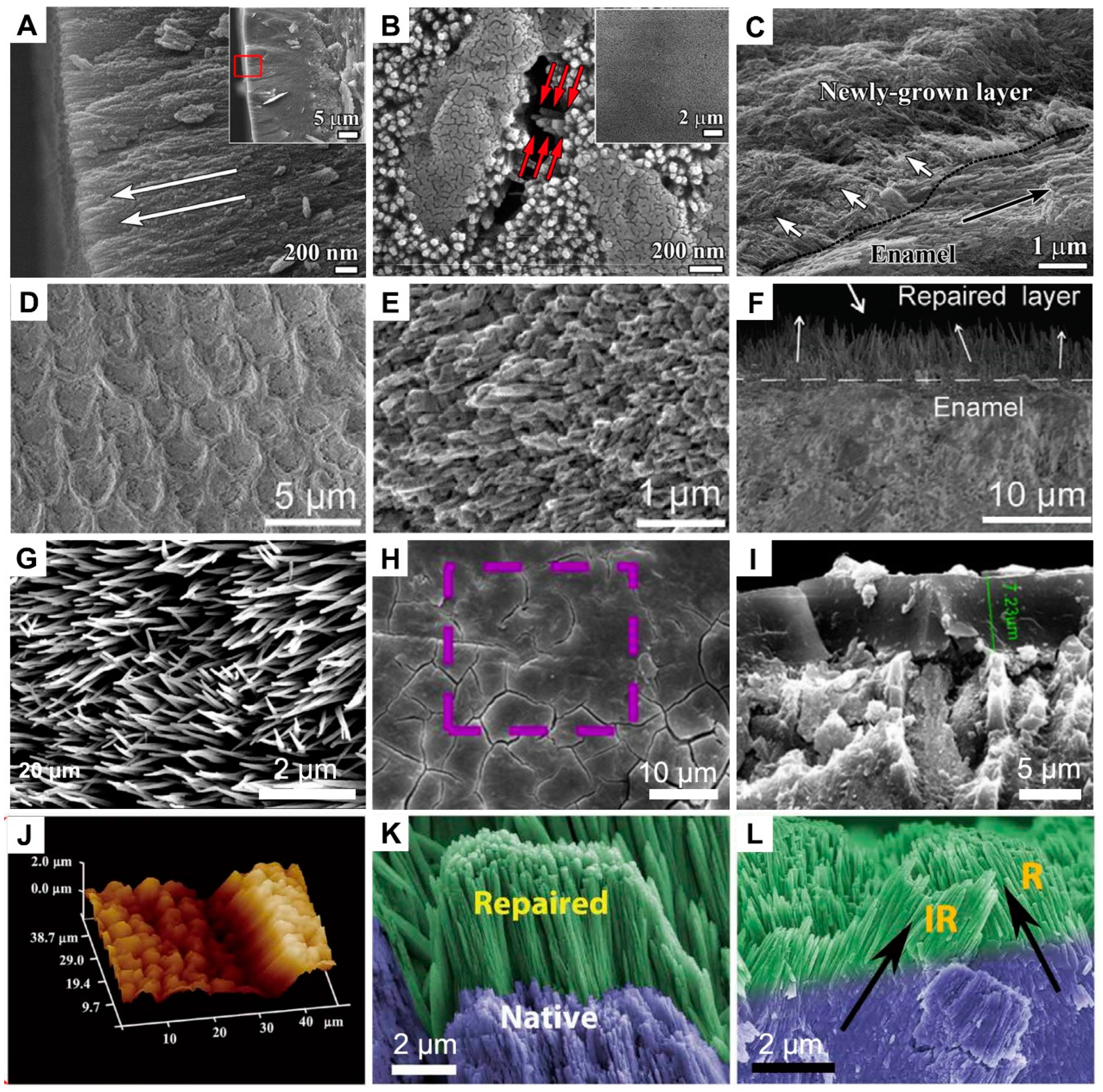
Ruan et al. used an amelogenin–chitosan hydrogel to form a mineral layer approximately 15 μm thick on the surface of the enamel (Figure 3A,B). This mineral layer was tightly bonded to the tooth surface and arranged in a bundle-like structure [55]. However, the growth of enamel crystals does not follow the c-axis of natural enamel (Figure 3C). The elastic modulus and hardness of the repair layer differed significantly from those of naturally healthy enamel. Prajapati et al. found that by adding matrix metalloproteinase-20 (MMP-20), the newly grown crystals in the repair layer could have a more uniform orientation and higher crystallinity, significantly improving the elastic modulus and hardness of the repaired enamel [64].
Since the synthesis process of amelogenin and MMP-20 is complex and costly, amelogenin-based peptides are utilized in the biomimetic mineralization of enamel. Ren et al. added hydrophilic segments (TKREEVD) to five Gln-Pro-X repeat sequences (QPYQPVQPHQPMQPQ) to form an enamel protein-derived peptide (QP5). QP5 can promote the long-term remineralization of early dental caries [54]. Ding et al. reported that the combined use of fluoride and QP5 has a potential synergistic effect on the remineralization of dental caries, and the microhardness of the repair layer is significantly improved [65]. However, the shape, size, and orientation of the mineralized layer crystals exhibit notable differences compared to natural enamel. Further research into the logic and efficacy of peptide design is of considerable importance. Inspired by the biocompatible amelogenin-inspired matrix, Wang et al. developed a phase-transited lysozyme film that mimics an N-terminal amelogenin with a central domain combined with a synthetic peptide based on the functional domains of the C-terminal amelogenin. This technology can achieve the growth of well-ordered HAP crystals both in vitro and in vivo, yielding an aligned structure that is nearly identical to that of natural enamel (Figure 3D–F) [66].
Due to the difficulty of obtaining amelogenin, there are significant limitations to its research and application. Currently, a synthetic protein polyamidoamine-amine-type dendrimer (PAMAM) can be successfully prepared and applied to the repair of the enamel. This kind of dendritic molecule can self-assemble into spherical macromolecules in solution and then convert into linear macromolecules, a behavior similar to that of amelogenin [67,68]. Based on this, Chen et al. used carboxyl-terminated PAMAM dendritic polymers (PAMAM-COOH) as templates and incorporated fluoride ions to obtain relatively ordered rod-shaped HAP crystals on the enamel surface (Figure 3G), parallel to the long axis of the enamel rods [69]. Similarly, Wu et al. used alendronate-PAMAM-carboxyl polymer (ALN-PAMAM-COOH) to repair the enamel and conducted experiments in mice. Although a large amount of rod-shaped HAP grows on the surface of the enamel, it does not have an ordered structure (Figure 3H,I) [70]. These artificial proteins are adsorbed on the enamel surface and further self-assembled, and then induce the deposition of calcium and phosphorus in the solution to achieve ordered growth. However, there is still a significant gap compared to natural enamel, and in these experiments, the repair effect is better with the synergistic participation of fluoride ions than with PAMAM polymers alone.
4.1.2. Calcium Phosphate Particle-Based Systems
Investigations into nature have shown that the growth of the zebrafish skeleton involves an integral compound known as amorphous calcium phosphate (ACP) [71]. This intermediate, amorphous substance plays a pivotal role in the formation of these integral minerals [72]. In addition, ACP is the initial deposition mineral in the early stages of enamel biogenesis [73,74].
In an attempt to mimic the biomineralization process, a variety of composite materials containing ACP nanoparticles have been extensively used in enamel mineralization efforts [75,76,77,78,79,80]. Notably, these ACP nanoparticles significantly stimulate enamel remineralization, while their instability limits their practical applicability in clinical treatment remarkably [80]. ACP has a higher solubility than calcium phosphate crystals and is easily transformed into HAP in solution. Several composites capable of stabilizing ACP have found applications in enamel restoration procedures. Phosphorylated chitosan-stabilized ACP can achieve the remineralization of enamel [76]. Casein phosphopeptide-amorphous calcium phosphate (CPP-ACP) can deposit a layer of calcium phosphate mineral on the surface, which has no structure and poor mechanical properties.
Further regulation of the mineralization process is required to obtain an ordered structure. Li et al. demonstrated that the biologically inspired cooperative effect between glutamic acid and nano-apatite particles can result in the regeneration of enamel-like structures under physiological conditions. Importantly, this feasible method of enamel remodeling ensures that the mechanical properties of the repaired enamel are well preserved [81]. Wang et al. utilized carboxymethyl chitosan (CMC) and ALN to stabilize ACP into nanoparticles. Sodium hypochlorite degraded the CMC-ALN matrix into HAP@ACP core–shell nanoparticles. Finally, these nanoparticles evolved into rod-like ordered apatite crystals under the guidance of 10 mM glycine [82]. However, both oriented and ordered mineral crystals perpendicular to the enamel surface and those parallel to the enamel surface, indicated by red arrows, were found.
Shao et al. introduced an innovative calcium phosphate ion cluster (CPIC) to construct a biomimetic mineralization front and successfully replicated the complex hierarchical structure of enamel [56]. Triethanolamine can stabilize CPICs of approximately 1.5 nm in size in ethanol. It facilitates the construction of a continuous crystalline-amorphous mineralization front on the enamel surface, thus prompting the epitaxial growth of enamel crystals along the c-axis of natural enamel (Figure 3J–L). Remarkably, the repaired enamel exhibits mechanical properties similar to those of natural enamel, with a hardness value of 3.84 ± 0.20 GPa and an elastic modulus of 87.26 ± 3.73 GPa. However, this process requires strict non-aqueous conditions to allow the formation of the crystalline-amorphous mineralization front, which limits its practical application in the complex oral environment.
Figure 3.
(A,B) SEM images of an enamel-like layer formed with amelogenin-chitosan hydrogel showed the cross-section and surface of the repaired enamel. White arrows in (A) indicate the HAP orientations in the newly grown layer, and the red square rectangle in inset of (A) corresponding to (A). The red arrows in (B) indicate a typical bundle of parallel crystals inside the newly grown layer. (C) A newly-grown layer that are marked with white arrows fused to the surface of the natural enamel, reprinted with permission from Ref. [55]. 2014, Elsevier. (D,E) SEM images of enamel regeneration via PTL/C-AMG and (F) the corresponding cross-section, in which the white arrows indicate the orientation of the newly formed minerals, reprinted with permission from Ref. [66]. 2020, John Wiley and Sons. (G) SEM images of crystals grown on acid-etched enamel with the PAMAM-COOH and (H,I) the ALN-PAMAM-COOH, reprinted with permission from Refs. [69,70]. 2013, Elsevier. (J) A three-dimensional AFM image and (K) an SEM image showing both acid-etched enamel and enamel repaired with CPIC. (L) Cross-section of the final repaired enamel, where the black arrows mark the orientation of the enamel crystals, reprinted with permission from Ref. [56]. 2019, AAAS.
4.2. Dentin Remineralization
4.2.1. Proteins or Peptides for Dentin Remineralization
According to previous studies, the deposition of HAP within and between collagen fibers is regulated by NCPs, sharing a common characteristic of high serine and aspartic acid content. These amino acids are rich in carboxyl groups and contribute to the overall negative charge of proteins, which enables them to bind to calcium phosphate via interaction with calcium ions, thereby stabilizing amorphous precursors [33,83]. Consequently, proteins play a crucial role in maintaining the stability of the solution and providing a suitable environment for the subsequent crystallization of the apatite.
The use of natural NCPs is limited by the challenges associated with their extraction, storage, and high cost. However, importantly, the specific sequences present in NCPs play a crucial role in their functionality. Inspired by the structure of NCPs, many biomimetic polypeptide molecules have been designed and synthesized.
The regulatory role of DPP in HAP nucleation and growth is believed to be primarily due to its abundant Asp-Ser-Ser (DSS) repeats. Li et al. used the 8DSS peptide to achieve the remineralization of demineralized dentin [84]. 8DSS can interact with dentin collagen, resulting in the formation of mineral precipitates on the surface and within the dentin tubules. The application of 8DSS has a significant positive effect on the elastic modulus and hardness of demineralized dentin and reduces dentin permeability [85].
DMP-1 can stabilize calcium and phosphorus ions in solution and generate nucleation precursors. Previous research has shown that the amino acid sequence responsible for collagen adsorption in DMP-1 is linked to certain amino acid sequences responsible for HAP adsorption [86]. This association results in the formation of polypeptides that exhibit strong adsorption to type Ⅰ collagen, regulating mineral deposition, stabilizing amorphous precursors formed by calcium and phosphorus ions, and promoting the nucleation and growth of HAP. Additionally, the intermolecular assembly of functional domains within a β-sheet template is essential in the process of mineral nucleation induced by DMP-1 [87]. A calcium-responsive self-assembled β-tablet peptide, ID8, has been designed to preprocess collagen [88]. The ID8 peptide not only enhances the intermolecular hydrogen bonding but also improves the hydrophilicity of collagen and aids in the retention of calcium within collagen. It has the potential to help regulate collagen mineralization and facilitate the biomimetic mineralization process of early caries [89].
During biomineralization, amelogenin in enamel and NCPs in dentin affect the organization and growth of HAP crystals. The bioactivity of amelogenin at the DEJ interface exhibits a significant level of activity. The amelogenin-inspired design of a self-assembled peptide (P26) has a unique dual remineralization function of enamel and dentin [90]. P26 assembles densely packed spherical nanoparticles along collagen fibrils, facilitating the process of remineralization in dentin defects (Figure 4A–D). The dense accumulation of HAP crystals blocked the tubules and encased the exposed collagen fibril, enhancing mineral recovery and restoring the biological and biomechanical properties of demineralized dentin.
4.2.2. NCP Surrogate
Synthetic analogs of natural NCPs are frequently employed to address the limitations associated with natural NCPs. These analogs are designed to mimic the function of natural NCPs and facilitate mineralization within collagen fibers. The theory of polymer-induced liquid precursors (PILP) is an important approach in the fabrication of mineralized collagen fibers. According to the theory of PILP, the inclusion of these charged polymers in the solution can effectively stabilize the calcium phosphate clusters during the early stages of remineralization, preventing their transformation into HAP crystals [34,91]. The PILP can infiltrate into the collagen fibers via capillary action and subsequently interact with the binding sites on the collagen fibers, and over time the occurrence of crystallization produces the well-organized mineralized collagen fibers. The use of NCPs analogs commonly includes polyaspartic acid (p-Asp), polyacrylic acid (PAA), CMC, polyglutamic acid(p-Glu), PAMAM, and poly(allylamine) hydrochloride (PAH), etc.
Gower et al. used p-Asp to stabilize ACP and achieve the intrafibrillar mineralization of collagen fibers and proposed the PILP mechanism by which collagen mineralization occurs [92]. By using p-Asp as an analog of NCPs, the process of the remineralization of artificial caries was successfully achieved [93], resulting in an increase in mineral content within the collagen fiber, improved crystallinity, and the restoration of mechanical properties in the damaged dentin area [94]. HAP crystals overgrew on the surface of the mineralized collagen and firmly adhered to the tubule wall, resulting in a compact occlusion of the dentin tubules (Figure 4E–H) [44].
Another polyelectrolyte, PAA, which is one of the most widely used in the study of collagen mineralization, can mimic the functions of NCPs and is relatively low in cost. Its molecular weight and concentration could affect the mineralization process; as the concentration of PAA increases and the molecular weight of PAA decreases, the crystallization rate of HAP decreases, thus affecting the mineralization degree of collagen fibers. This is mainly because of the variations in the efficiency of PAA/Ca complexation, as well as the influence of PAA molecular weight migration on precursor adsorption efficiency [95,96,97].
These polyelectrolytes could interact strongly with calcium phosphate minerals, stabilizing ACP and inhibiting the nucleation of HAP, thereby inducing intrafibrillar mineralization. It seems unlikely that intrafibrillar mineralized collagen fibrils with a higher degree of mineralization could be obtained in a mineralized system without NCP or its surrogates. At present, these organic additives are necessary in the field to ensure that we obtain well-mineralized collagen similar to that observed in bone.
4.2.3. Based on the Promotion of Collagen Remineralization for the Repair of Dentin
In the process of collagen mineralization, in addition to emphasizing the stabilizing effect of NCPs on ACP precursors, attention is also given to the ability of NCPs to bind calcium and collagen, attract ACP precursors, promote their penetration into the fiber, and initiate nucleation at specific collagen sites. Matrix phosphoproteins irreversibly bind to collagen in hard tissue, providing a template function by which negatively charged phosphate esters bind to positively charged interstitial regions in collagen, forming a negatively charged surface [98,99,100]. The increased local ion oversaturation created on this surface facilitates the nucleation and growth of HAP.
Several phosphorus-containing reagents, such as polyvinylphosphonic acid (PVPA), sodium trimetaphosphate (STMP), and sodium tripolyphosphate (STPP), are used as template analogs to anchor collagen fibers and induce intrafibrillar mineralization. Using PAA and PVPA as dual biomimetic analogues in Portland cement/phosphate-containing solution systems, the remineralization of etched-dentin to a depth of approximately 5 μm has been achieved with interfibrillar and intrafibrillar remineralization [101], as well as the remineralization of the resin–dentin interface [102]. STMP and STPP can be fixed to collagen by chemical phosphorylation and irreversible binding sites for phosphate groups on the dentin surface [103]. When dentin is treated with STMP, HAP forms in the phosphorylated collagen matrix within the fibers, inducing collagen mineralization and achieving artificial caries recrystallization [104].
Other organic biomolecules can also modify collagen fibers and regulate the mineralization of collagen fibers and dentin. Citrate can enhance the wetting effect of ACP on collagen fibers by significantly reducing the interfacial energy between them, and therefore the pretreatment with citrate effectively improved the process of collagen mineralization and dentin remineralization (Figure 4I–L) [105]. Wu et al. found that aminoglycan molecules such as hyaluronic acid can accelerate mineralization by providing more nucleation sites and shortening the induction time of ACP-mediated HAP crystallization [43]. In addition, there are many organic molecules, such as dopamine acrylamide [106], PAA [107], polydopamine (PDA) [108], and CMC [109], which contain chemical groups that can coordinate with calcium ions or have a high affinity for calcium phosphate minerals. This strategy paves the way for researchers to search for reagents that can enhance the mineralization of collagen fibers, improve dentin remineralization, and have the potential to develop as a clinical therapeutic drug.
Figure 4.
(A–D) Peptide P26 assembly aligned along the collagen fibril (indicated with yellow arrows in (A)) and induced collagen and dentin remineralization (indicated with yellow arrows in (B) and red arrows in (D)), reprinted with permission from Ref. [90]. 2020, American Chemical Society. The yellow square in (B) indicates the area where the SAED data were recorded. High magnification of SEM images of (D) corresponding to the regions marked by rectangles in (C), where new crystals grew along the tubule walls (yellow arrows), the diameter of the collagen increased over time, resulting in a “beaded string-like” appearance due to HAP precipitation (red arrows). (E–H) PILP-induced dentin remineralization and occlusion of dentin tubules, reprinted with permission from Ref. [44]. 2022, Royal Society of Chemistry. High magnification of SEM images of (F,H) correspond to the regions marked by rectangles in (E,G), respectively. (I–L) Remineralization of dentin without (J) or with (L) citrate pretreatment, reprinted with permission from Ref. [105]. 2018, John Wiley and Sons. (M–P) TEM images of PAH-ACP@hmZrO2-mediated collagen mineralization, reprinted with permission from Ref. [110]. 2018, Elsevier. The open arrowheads in (O) indicated the partially mineralized collagen fibrils.

4.2.4. ACP Nanoparticles for Dentin Remineralization
The mineralization theory based on PILP has been widely recognized and studied. However, its application in the real oral environment is challenging due to the requirement of saturated calcium and phosphorus ion concentration. It is difficult to continuously provide a sufficient concentration of calcium and phosphate for mineralization in the oral cavity. As a precursor to mineralization, ACP is prone to phase transition in solution. Constructing a proper stabilization and transport system for ACP is an excellent strategy for remineralization, allowing for the backfilling of ACP into demineralized dentin and collagen.
The delivery system utilizes a range of materials, such as polycaprolactone, chitosan, PLGA, and mesoporous silica. Several studies have loaded ACP into mesoporous nanoparticles [111,112]. For example, amine-functionalized enlarged pore silica nanoparticles (AF-eMSN) attached to PAA-ACP successfully achieved mineralization, which is the first attempt to deliver ACP by using enlarged pore silica for mineralization in collagen fibers [113]. Using zirconia, a biocompatible ceramic commonly used in dentistry, to synthesize mesoporous nanocapsules loaded with PAH-ACP, the released PAH-ACP still retains its ability to penetrate and mineralize collagen fibers (Figure 4M–P) [110].
Considering the process of composite resin adhesion and the issues of collagen degradation and microleakage at the demineralized collagen interface, the incorporation of ACP particles into self-etching adhesives or resins is a strategy that has been extensively studied. The use of self-etching adhesives as a carrier of ACP nanoprecursors facilitates continuous biomimetic remineralization [114]. Core-shell chlorhexidine/ACP (CHX/ACP) nanoparticles have been synthesized and utilized to enhance dental resin composites, providing them with exceptional mechanical strength, antimicrobial activity, and remineralization capabilities [115]. These strategies could further release calcium and phosphate ions to create supersaturation relative to the HAP crystals, and could also infiltrate directly into the fibers, leading to intrafibrillar mineralization, which shows promise for clinical applications.
5. Conclusions and Outlook
In summary, the intricate structure of teeth endows them with superior mechanical properties, but also increases the difficulty of their repair. Existing clinical restorative materials differ significantly from natural dental tissues in terms of chemical composition, structure, and performance. This difference usually results in an inability to achieve durable, long-term restorative results. The strategy of biomimetic mineralization for repairing dental hard tissues presents a highly promising avenue. Recent studies have demonstrated the satisfactory remineralization of both enamel and dentin under specific experimental conditions. Nevertheless, further exploration is needed for this approach to be successfully implemented in clinical applications.
Given the complexity of the oral environment, the effectiveness of biomimetic mineralization technology in clinical applications may be influenced by several factors, including the oral microbial population and oral enzyme activity of each patient. Therefore, it is crucial to carry out more in-depth in vivo animal studies and clinical research, which are essential to optimize the application of this technology and to develop biomimetic mineralization materials that are more adaptable to the oral environment.
At present, the repair layers of biomimetic mineralization materials are mostly at the micron level and large-scale repairs at the millimeter level have not yet been achieved. Therefore, we can consider applying biomimetic mineralization materials in the form of oral cleaning products (such as toothpaste and mouthwash), aiming to inhibit demineralization, promote remineralization, and prevent dental caries. In addition, biomimetic mineralization materials can be used in combination with existing dental restorative materials to improve their durability and functionality.
Author Contributions
Conceptualization, C.S. and Z.T.; writing—original draft preparation, Z.T. and S.S.; writing—review and editing, C.S. and Z.C.; supervision, C.S.; funding acquisition, C.S. and Z.C. All authors have read and agreed to the published version of the manuscript.
Funding
This work was supported by the National Science Foundation for Young Scientists of China (Grant no. 22205202) and the Pioneer and Leading Goose R&D Program of Zhejiang (Grant no. 2022C03164).
Data Availability Statement
Data are contained within the article.
Conflicts of Interest
The authors declare no competing interests.
References
- Selwitz, R.H.; Ismail, A.I.; Pitts, N.B. Dental caries. Lancet 2007, 369, 51–59. [Google Scholar] [CrossRef] [PubMed]
- Kruzic, J.J.; Hoffman, M.; Arsecularatne, J.A. Fatigue and wear of human tooth enamel: A review. J. Mech. Behav. Biomed. Mater. 2023, 138, 21. [Google Scholar] [CrossRef]
- Frencken, J.E.; Sharma, P.; Stenhouse, L.; Green, D.; Laverty, D.; Dietrich, T. Global epidemiology of dental caries and severe periodontitis—A comprehensive review. J. Clin. Periodontol. 2017, 44, S94–S105. [Google Scholar] [CrossRef]
- Park, S.Y.; Kim, S.H.; Kang, S.H.; Yoon, C.H.; Lee, H.J.; Yun, P.Y.; Youn, T.J.; Chae, I.H. Improved oral hygiene care attenuates the cardiovascular risk of oral health disease: A population-based study from Korea. Eur. Heart J. 2019, 40, 1138–1145. [Google Scholar] [CrossRef] [PubMed]
- Ungar, P.S. Mammalian dental function and wear: A review. Biosurface Biotribol. 2015, 1, 25–41. [Google Scholar] [CrossRef]
- Zolotarev, V.M.; Grisimov, V.N. Architectonics and Optical Properties of Dentin and Dental Enamel. Opt. Spectrosc. 2001, 90, 753–759. [Google Scholar] [CrossRef]
- Zhao, X.; O’Brien, S.; Shaw, J.; Abbott, P.; Munroe, P.; Habibi, D.; Xie, Z. The origin of remarkable resilience of human tooth enamel. Appl. Phys. Lett. 2013, 103, 241901. [Google Scholar] [CrossRef]
- Gavrilovski, D.S.; Blagojevic, N.S.; Gavrilovski, M.P. Modelling glass-ceramic enamel properties. J. Serb. Chem. Soc. 2002, 67, 135–142. [Google Scholar] [CrossRef]
- Lubarsky, G.V.; Lemoine, P.; Meenan, B.J.; Deb, S.; Mutreja, I.; Carolan, P.; Petkov, N. Enamel proteins mitigate mechanical and structural degradations in mature human enamel during acid attack. Mater. Res. Express 2014, 1, 025404. [Google Scholar] [CrossRef]
- Palmer, L.C.; Newcomb, C.J.; Kaltz, S.R.; Spoerke, E.D.; Stupp, S.I. Biomimetic Systems for Hydroxyapatite Mineralization Inspired by Bone and Enamel. Chem. Rev. 2008, 108, 4754–4783. [Google Scholar] [CrossRef]
- Cui, F.-Z.; Ge, J. New observations of the hierarchical structure of human enamel, from nanoscale to microscale. J. Tissue Eng. Regen. Med. 2007, 1, 185–191. [Google Scholar] [CrossRef]
- White, S.N.; Luo, W.; Paine, M.L.; Fong, H.; Sarikaya, M.; Snead, M.L. Biological organization of hydroxyapatite crystallites into a fibrous continuum toughens and controls anisotropy in human enamel. J. Dent. Res. 2001, 80, 321–326. [Google Scholar] [CrossRef] [PubMed]
- He, L.H.; Swain, M.V. Understanding the mechanical behaviour of human enamel from its structural and compositional characteristics. J. Mech. Behav. Biomed. Mater. 2008, 1, 18–29. [Google Scholar] [CrossRef] [PubMed]
- Yoon, Y.J.; Kim, I.H.; Han, S.Y. The Reason why a Sheath Exists in Enamel. Int. J. Precis. Eng. Manuf. 2015, 16, 807–811. [Google Scholar] [CrossRef]
- Cao, C.; Mei, M.; Li, Q.-l.; Lo, E.; Chu, C. Methods for Biomimetic Mineralisation of Human Enamel: A Systematic Review. Materials 2015, 8, 2873–2886. [Google Scholar] [CrossRef]
- Popowics, T.E.; Rensberger, J.M.; Herring, S.W. Enamel microstructure and microstrain in the fracture of human and pig molar cusps. Arch. Oral Biol. 2004, 49, 595–605. [Google Scholar] [CrossRef]
- Chai, H.; Lee, J.J.W.; Constantino, P.J.; Lucas, P.W.; Lawn, B.R. Remarkable resilience of teeth. Proc. Natl. Acad. Sci. USA 2009, 106, 7289–7293. [Google Scholar] [CrossRef]
- Lawn, B.R.; Lee, J.J.W.; Chai, H. Teeth: Among Nature’s Most Durable Biocomposites. Annu. Rev. Mater. Res. 2010, 40, 55–75. [Google Scholar] [CrossRef]
- Cuy, J.L.; Man, A.B.; Livi, K.J.; Teaford, M.F.; Weihs, T.P. Nanoindentation mapping of the mechanical properties of human molar tooth enamel. Arch. Oral Biol. 2002, 47, 281–291. [Google Scholar] [CrossRef]
- Imbeni, V.; Kruzic, J.J.; Marshall, G.W.; Marshall, S.J.; Ritchie, R.O. The dentin–enamel junction and the fracture of human teeth. Nat. Mater. 2005, 4, 229–232. [Google Scholar] [CrossRef] [PubMed]
- Baldassarri, M.; Margolis, H.C.; Beniash, E. Compositional Determinants of Mechanical Properties of Enamel. J. Dent. Res. 2008, 87, 645–649. [Google Scholar] [CrossRef] [PubMed]
- Xie, Z.H.; Swain, M.V.; Swadener, G.; Munroe, P.; Hoffman, M. Effect of microstructure upon elastic behaviour of human tooth enamel. J. Biomech. 2009, 42, 1075–1080. [Google Scholar] [CrossRef] [PubMed]
- Ge, J.; Cui, F.Z.; Wang, X.M.; Feng, H.L. Property variations in the prism and the organic sheath within enamel by nanoindentation. Biomaterials 2005, 26, 3333–3339. [Google Scholar] [CrossRef] [PubMed]
- Gordon, L.M.; Cohen, M.J.; MacRenaris, K.W.; Pasteris, J.D.; Seda, T.; Joester, D. Amorphous intergranular phases control the properties of rodent tooth enamel. Science 2015, 347, 746–750. [Google Scholar] [CrossRef]
- Xu, C.; Reed, R.; Gorski, J.P.; Wang, Y.; Walker, M.P. The distribution of carbonate in enamel and its correlation with structure and mechanical properties. J. Mater. Sci. 2012, 47, 8035–8043. [Google Scholar] [CrossRef]
- Alkattan, R.; Lippert, F.; Tang, Q.; Eckert, G.J.; Ando, M. The influence of hardness and chemical composition on enamel demineralization and subsequent remineralization. J. Dent. 2018, 75, 34–40. [Google Scholar] [CrossRef] [PubMed]
- Tjäderhane, L.; Carrilho, M.R.; Breschi, L.; Tay, F.R.; Pashley, D.H. Dentin basic structure and composition—An overview. Endod. Top. 2009, 20, 3–29. [Google Scholar] [CrossRef]
- Carda, C.; Peydró, A. Ultrastructural patterns of human dentinal tubules, odontoblasts processes and nerve fibres. Tissue Cell 2006, 38, 141–150. [Google Scholar] [CrossRef] [PubMed]
- Qin, C.; D’Souza, R.; Feng, J.Q. Dentin matrix protein 1 (DMP1): New and important roles for biomineralization and phosphate homeostasis. J. Dent. Res. 2007, 86, 1134–1141. [Google Scholar] [CrossRef]
- Butler, W.T. Dentin matrix proteins and dentinogenesis. Connect Tissue Res. 1995, 33, 59–65. [Google Scholar] [CrossRef] [PubMed]
- Yamamoto, R.; Oida, S.; Yamakoshi, Y. Dentin Sialophosphoprotein-derived proteins in the dental pulp. J. Dent. Res. 2015, 94, 1120–1127. [Google Scholar] [CrossRef]
- Suzuki, S.; Sreenath, T.; Haruyama, N.; Honeycutt, C.; Terse, A.; Cho, A.; Kohler, T.; Mueller, R.; Goldberg, M.; Kulkarni, A.B. Dentin sialoprotein and dentin phosphoprotein have distinct roles in dentin mineralization. Matrix Biol. 2009, 28, 221–229. [Google Scholar] [CrossRef] [PubMed]
- Deshpande, A.S.; Fang, P.-A.; Zhang, X.; Jayaraman, T.; Sfeir, C.; Beniash, E. Primary Structure and phosphorylation of dentin matrix protein 1 (DMP1) and dentin phosphophoryn (DPP) uniquely determine their role in biomineralization. Biomacromolecules 2011, 12, 2933–2945. [Google Scholar] [CrossRef]
- Nudelman, F.; Pieterse, K.; George, A.; Bomans, P.H.H.; Friedrich, H.; Brylka, L.J.; Hilbers, P.A.J.; de With, G.; Sommerdijk, N.A.J.M. The role of collagen in bone apatite formation in the presence of hydroxyapatite nucleation inhibitors. Nat. Mater. 2010, 9, 1004–1009. [Google Scholar] [CrossRef]
- Wang, Y.; Azais, T.; Robin, M.; Vallee, A.; Catania, C.; Legriel, P.; Pehau-Arnaudet, G.; Babonneau, F.; Giraud-Guille, M.-M.; Nassif, N. The predominant role of collagen in the nucleation, growth, structure and orientation of bone apatite. Nat. Mater. 2012, 11, 724–733. [Google Scholar] [CrossRef]
- Chen, Y.; Wu, R.; Shen, L.; Yang, Y.; Wang, G.; Yang, B. The multi-scale meso-mechanics model of viscoelastic dentin. J. Mech. Behav. Biomed. Mater. 2022, 136, 105525. [Google Scholar] [CrossRef] [PubMed]
- Thompson, V.P. The tooth: An analogue for biomimetic materials design and processing. Dent. Mater. 2020, 36, 25–42. [Google Scholar] [CrossRef]
- Carreon, A.H.; Funkenbusch, P.D. Nanoscale properties and deformation of human enamel and dentin. J. Mech. Behav. Biomed. Mater. 2019, 97, 74–84. [Google Scholar] [CrossRef]
- Zhang, Y.-R.; Du, W.; Zhou, X.-D.; Yu, H.-Y. Review of research on the mechanical properties of the human tooth. Int. J. Oral Sci. 2014, 6, 61–69. [Google Scholar] [CrossRef] [PubMed]
- Ziskind, D.; Hasday, M.; Cohen, S.R.; Wagner, H.D. Young’s modulus of peritubular and intertubular human dentin by nano-indentation tests. J. Struct. Biol. 2011, 174, 23–30. [Google Scholar] [CrossRef] [PubMed]
- Milazzo, M.; Jung, G.S.; Danti, S.; Buehler, M.J. Mechanics of mineralized collagen fibrils upon transient loads. ACS Nano 2020, 14, 8307–8316. [Google Scholar] [CrossRef] [PubMed]
- Al-Qudsy, L.; Hu, Y.-W.; Xu, H.; Yang, P.-F. Mineralized collagen fibrils: An essential component in determining the mechanical behavior of cortical bone. ACS Biomater. Sci. Eng. 2023, 9, 2203–2219. [Google Scholar] [CrossRef]
- Wu, H.; Shao, C.; Shi, J.; Hu, Z.; Zhou, Y.; Chen, Z.; Tang, R.; Xie, Z.; Jin, W. Hyaluronic acid-mediated collagen intrafibrillar mineralization and enhancement of dentin remineralization. Carbohydr. Polym. 2023, 319, 121174. [Google Scholar] [CrossRef] [PubMed]
- Chen, Z.; Duan, Y.; Shan, S.; Sun, K.; Wang, G.; Shao, C.; Tang, Z.; Xu, Z.; Zhou, Y.; Chen, Z.; et al. Deep and compact dentinal tubule occlusion via biomimetic mineralization and mineral overgrowth. Nanoscale 2022, 14, 642–652. [Google Scholar] [CrossRef] [PubMed]
- Jones, F.H. Teeth and bones: Applications of surface science to dental materials and related biomaterials. Surf. Sci. Rep. 2001, 42, 75–205. [Google Scholar] [CrossRef]
- Ulrich, L. Dental glass ionomer cements as permanent filling materials?—Properties, limitations and future trends. Materials 2009, 3, 76–96. [Google Scholar]
- Olegário, I.; Ladewig, N.; Hesse, D.; Bonifácio, C.; Braga, M.; Imparato, J.; Mendes, F.; Raggio, D. Is it worth using low-cost glass ionomer cements for occlusal ART restorations in primary molars? 2-year survival and cost analysis of a Randomized clinical trial. J. Dent. 2020, 101, 103446. [Google Scholar] [CrossRef]
- Rueggeberg, F.A. From vulcanite to vinyl, a history of resins in restorative dentistry. J. Prosthet. Dent. 2002, 87, 364–379. [Google Scholar] [CrossRef]
- Brunthaler, A.; König, F.; Lucas, T.; Schedle, W.S. Longevity of direct resin composite restorations in posterior teeth: A review. Clin. Oral Investig. 2003, 7, 63–70. [Google Scholar] [CrossRef]
- Kanzow, P.; Wiegand, A. Retrospective analysis on the repair vs. replacement of composite restorations. Dent. Mater. 2020, 36, 108–118. [Google Scholar] [CrossRef]
- Sarrett, D. Clinical challenges and the relevance of materials testing for posterior composite restorations. Dent. Mater. 2005, 21, 9–20. [Google Scholar] [CrossRef] [PubMed]
- Nimbalkar, S.; Lim, L.H.; Lee, Z.T.; Lim, K.H.; Sia, S.Y. Efficacy of three enamel protecting agents on shear bond strength of orthodontic brackets bonded to demineralised enamel with conventional adhesive. J. Clin. Diagn. Res. 2021, 15, ZC18–ZC21. [Google Scholar] [CrossRef]
- Zafar, M.S.; Amin, F.; Fareed, M.A.; Ghabbani, H.; Riaz, S.; Khurshid, Z.; Kumar, N. Biomimetic aspects of restorative dentistry biomaterials. Biomimetics 2020, 5, 34. [Google Scholar] [CrossRef] [PubMed]
- Ren, Q.; Li, Z.; Ding, L.; Wang, X.; Niu, Y.; Qin, X.; Zhou, X.; Zhang, L. Anti-biofilm and remineralization effects of chitosan hydrogel containing amelogenin-derived peptide on initial caries lesions. Regen. Biomater. 2018, 5, 69–76. [Google Scholar] [CrossRef]
- Ruan, Q.C.; Moradian-Oldak, J. An amelogenin–chitosan matrix promotes assembly of an enamel-like layer with a dense interface. Acta Biomater. 2013, 9, 7289. [Google Scholar] [CrossRef] [PubMed]
- Shao, C.Y.; Jin, B.A.; Mu, Z.; Lu, H.; Zhao, Y.Q.; Wu, Z.F.; Yan, L.M.; Zhang, Z.S.; Zhou, Y.C.; Pan, H.H.; et al. Repair of tooth enamel by a biomimetic mineralization frontier ensuring epitaxial growth. Sci. Adv. 2019, 5, eaaw9569. [Google Scholar] [CrossRef] [PubMed]
- Wang, L.; Nancollas, G.H. Calcium orthophosphates: Crystallization and dissolution. Chem. Rev. 2008, 108, 4628–4669. [Google Scholar] [CrossRef] [PubMed]
- Jiang, H.; Liu, X.Y. Principles of mimicking and engineering the self-organized structure of hard tissues. J. Biol. Chem. 2004, 279, 41286–41293. [Google Scholar] [CrossRef]
- Jiang, H.D.; Liu, X.Y.; Zhang, G.; Li, Y. Kinetics and template nucleation of self-assembled hydroxyapatite nanocrystallites by chondroitin sulfate. J. Biol. Chem. 2005, 280, 42061–42066. [Google Scholar] [CrossRef]
- Moradian-Oldak, J.; Simmer, J.P.; Lau, E.C.; Sarte, P.E.; Fincham, A.G. Detection of monodisperse aggregates of a recombinant amelogenin by dynamic light scattering. Biopolymers 2010, 34, 1339–1347. [Google Scholar] [CrossRef]
- Engelberth, S.A.; Bacino, M.S.; Sandhu, S.; Li, W.; Bonde, J.; Habelitz, S. Progression of self-assembly of amelogenin protein supramolecular structures in simulated enamel fluid. Biomacromolecules 2018, 19, 3917–3924. [Google Scholar] [CrossRef]
- Du, C.; Falini, G.; Fermani, S.; Abbott, C.; Moradian-Oldak, J. Supramolecular Assembly of Amelogenin Nanospheres into Birefringent Microribbons. Science 2005, 307, 1450–1454. [Google Scholar] [CrossRef] [PubMed]
- Fincham, A.G.; Moradian-Oldak, J.; Diekwisch, T.G.H.; Lyaruu, D.M.; Wright, J.T.; Bringas, P.; Slavkin, H.C. Evidence for amelogenin “Nanospheres” as functional components of secretory-stage enamel matrix. J. Struct. Biol. 1995, 115, 50–59. [Google Scholar] [CrossRef] [PubMed]
- Prajapati, S.; Ruan, Q.; Mukherjee, K.; Nutt, S.; Moradian-Oldak, J. The presence of MMP-20 reinforces biomimetic enamel regrowth. J. Dent. Res. 2018, 97, 84–90. [Google Scholar] [CrossRef] [PubMed]
- Ding, L.J.; Han, S.L.; Wang, K.; Zheng, S.N.; Zheng, W.Y.; Peng, X.; Niu, Y.M.; Li, W.; Zhang, L.L. Remineralization of enamel caries by an amelogenin-derived peptide and fluoride in vitro. Regen. Biomater. 2020, 7, 283–292. [Google Scholar] [CrossRef] [PubMed]
- Wang, D.; Deng, J.J.; Deng, X.L.; Fang, C.Q.; Zhang, X.; Yang, P. Controlling enamel remineralization by Amyloid-Like amelogenin mimics. Adv. Mater. 2020, 32, e2002080. [Google Scholar] [CrossRef]
- Yang, S.; He, H.; Wang, L.; Jia, X.; Feng, H. Oriented crystallization of hydroxyapatite by the biomimetic amelogenin nanospheres from self-assemblies of amphiphilic dendrons. Chem. Commun. 2011, 47, 10100–10102. [Google Scholar] [CrossRef]
- Yang, J.; Cao, S.; Li, J.; Xin, J.; Chen, X.; Wu, W.; Xu, F.; Li, J. Staged self-assembly of PAMAM dendrimers into macroscopic aggregates with a microribbon structure similar to that of amelogenin. Soft Matter 2013, 9, 7553–7559. [Google Scholar] [CrossRef]
- Chen, L.; Liang, K.N.; Li, J.S.; Wu, D.; Zhou, X.D.; Li, J.Y. Regeneration of biomimetic hydroxyapatite on etched human enamel by anionic PAMAM template in vitro. Arch. Oral Biol. 2013, 58, 975–980. [Google Scholar] [CrossRef]
- Wu, D.; Yang, J.; Li, J.; Chen, L.; Tang, B.; Chen, X.; Wu, W.; Li, J. Hydroxyapatite-anchored dendrimer for in situ remineralization of human tooth enamel. Biomaterials 2013, 34, 5036–5047. [Google Scholar] [CrossRef]
- Mahamid, J.; Sharir, A.; Addadi, L.; Weiner, S. Amorphous calcium phosphate is a major component of the forming fin bones of zebrafish: Indications for an amorphous precursor phase. Proc. Natl. Acad. Sci. USA 2008, 105, 12748–12753. [Google Scholar] [CrossRef] [PubMed]
- Steve, W.; Julia, M.; Yael, P.; Yurong, M.A.; Lia, A. Overview of the amorphous precursor phase strategy in biomineralization. Front. Mater. Sci. China 2009, 3, 104–108. [Google Scholar]
- Lowenstam, H.A.; Weiner, S. Transformation of amorphous calcium phosphate to crystalline dahillite in the radular teeth of chitons. Science 1985, 227, 51–53. [Google Scholar] [CrossRef]
- Beniash, E.; Metzler, R.A.; Lam, R.S.K.; Gilbert, P. Transient amorphous calcium phosphate in forming enamel. J. Struct. Biol. 2009, 166, 133–143. [Google Scholar] [CrossRef] [PubMed]
- Zhao, J.; Liu, Y.; Zhang, S.H. Amorphous calcium phosphate and its application in dentistry. Chem. Cent. J. 2011, 5, 7. [Google Scholar] [CrossRef] [PubMed]
- Zhang, X.; Li, Y.; Sun, X.; Kishen, A.; Deng, X.; Yang, X.; Wang, H.; Cong, C.; Wang, Y.; Wu, M. Biomimetic remineralization of demineralized enamel with nano-complexes of phosphorylated chitosan and amorphous calcium phosphate. J. Mater. Sci. Mater. Med. 2014, 25, 2619–2628. [Google Scholar] [CrossRef]
- Fletcher, J.; Walsh, D.; Fowler, C.E.; Mann, S. Electrospun mats of PVP/ACP nanofibres for remineralization of enamel tooth surfaces. CrystEngComm 2011, 13, 3692–3697. [Google Scholar] [CrossRef]
- Iafisco, M.; Degli Esposti, L.; Ramirez-Rodriguez, G.B.; Carella, F.; Gomez-Morales, J.; Ionescu, A.C.; Brambilla, E.; Tampieri, A.; Delgado-Lopez, J.M. Fluoride-doped amorphous calcium phosphate nanoparticles as a promising biomimetic material for dental remineralization. Sci. Rep. 2018, 8, 17016. [Google Scholar] [CrossRef]
- Xiao, Z.H.; Que, K.H.; Wang, H.R.; An, R.; Chen, Z.; Qiu, Z.J.; Lin, M.L.; Song, J.H.; Yang, J.; Lu, D.Y.; et al. Rapid biomimetic remineralization of the demineralized enamel surface using nano-particles of amorphous calcium phosphate guided by chimaeric peptides. Dent. Mater. 2017, 33, 1217–1228. [Google Scholar] [CrossRef]
- Weir, M.D.; Chow, L.C.; Xu, H.H.K. Remineralization of Demineralized Enamel via Calcium Phosphate Nanocomposite. J. Dent. Res. 2012, 91, 979–984. [Google Scholar] [CrossRef]
- Li, L.; Mao, C.Y.; Wang, J.M.; Xu, X.R.; Pan, H.H.; Deng, Y.; Gu, X.H.; Tang, R.K. Bio-inspired enamel repair via Glu-directed assembly of apatite nanoparticles: An approach to biomaterials with optimal characteristics. Adv. Mater. 2011, 23, 4695–4701. [Google Scholar] [CrossRef] [PubMed]
- Wang, H.R.; Xiao, Z.H.; Yang, J.; Lu, D.Y.; Kishen, A.; Li, Y.Q.; Chen, Z.; Que, K.H.; Zhang, Q.; Deng, X.L.; et al. Oriented and ordered biomimetic remineralization of the surface of demineralized dental enamel using HAP@ACP nanoparticles guided by glycine. Sci. Rep. 2017, 7, 40701. [Google Scholar] [CrossRef]
- Gericke, A.; Qin, C.; Sun, Y.; Redfern, R.; Redfern, D.; Fujimoto, Y.; Taleb, H.; Butler, W.T.; Boskey, A.L. Different forms of DMP1 play distinct roles in mineralization. J. Dent. Res. 2010, 89, 355–359. [Google Scholar] [CrossRef]
- Liang, K.; Xiao, S.; Shi, W.; Li, J.; Yang, X.; Gao, Y.; Gou, Y.; Hao, L.; He, L.; Cheng, L.; et al. 8DSS-promoted remineralization of demineralized dentin in vitro. J. Mater. Chem. B 2015, 3, 6763–6772. [Google Scholar] [CrossRef]
- Liang, K.; Xiao, S.; Liu, H.; Shi, W.; Li, J.; Gao, Y.; He, L.; Zhou, X.; Li, J. 8DSS peptide induced effective dentinal tubule occlusion in vitro. Dent. Mater. 2018, 34, 629–640. [Google Scholar] [CrossRef] [PubMed]
- Padovano, J.D.; Ravindran, S.; Snee, P.T.; Ramachandran, A.; Bedran-Russo, A.K.; George, A. DMP1-derived peptides promote remineralization of human dentin. J. Dent. Res. 2015, 94, 608–614. [Google Scholar] [CrossRef]
- He, G.; Dahl, T.; Veis, A.; George, A. Nucleation of apatite crystals in vitro by self-assembled dentin matrix protein 1. Nat. Mater. 2003, 2, 552–558. [Google Scholar] [CrossRef]
- Li, Z.; Ren, Q.; Han, S.; Ding, L.; Qin, X.; Hui, D.; Het, T.; Tian, T.; Lu, Z.; Zhang, L. Promoting effect of a calcium-responsive self-assembly β-sheet peptide on collagen intrafibrillar mineralization. Regen. Biomater. 2022, 9, rbac059. [Google Scholar] [CrossRef] [PubMed]
- Li, Z.C.; Qin, X.; Ren, Q.; Hui, D.; Tian, T.; He, T.; Li, W.; Zhang, L.L. Rational design of β-sheet peptides with self-assembly into nanofibres on remineralisation of initial caries lesions. Chin. J. Dent. Res. 2020, 23, 131–141. [Google Scholar] [PubMed]
- Mukherjee, K.; Visakan, G.; Phark, J.-H.; Moradian-Oldak, J. Enhancing collagen mineralization with amelogenin peptide: Toward the restoration of dentin. ACS Biomater. Sci. Eng. 2020, 6, 2251–2262. [Google Scholar] [CrossRef] [PubMed]
- Olszta, M.J.; Cheng, X.; Jee, S.S.; Kumar, R.; Kim, Y.-Y.; Kaufman, M.J.; Douglas, E.P.; Gower, L.B. Bone structure and formation: A new perspective. Mater. Sci. Eng. R. Rep. 2007, 58, 77–116. [Google Scholar] [CrossRef]
- Gower, L.B.; Odom, D.J. Deposition of calcium carbonate films by a polymer-induced liquid-precursor (PILP) process. J. Cryst. Growth 2000, 210, 719–734. [Google Scholar] [CrossRef]
- Burwell, A.K.; Thula-Mata, T.; Gower, L.B.; Habeliz, S.; Kurylo, M.; Ho, S.P.; Chien, Y.-C.; Cheng, J.; Cheng, N.F.; Gansky, S.A.; et al. Functional remineralization of dentin lesions using polymer-induced liquid-precursor process. PLoS ONE 2012, 7, e38852. [Google Scholar] [CrossRef] [PubMed]
- Bacino, M.; Girn, V.; Nurrohman, H.; Saeki, K.; Marshall, S.J.; Gower, L.; Saeed, E.; Stewart, R.; Le, T.; Marshall, G.W.; et al. Integrating the PILP-mineralization process into a restorative dental treatment. Dent. Mater. 2019, 35, 53–63. [Google Scholar] [CrossRef]
- Qi, Y.; Ye, Z.; Fok, A.; Holmes, B.N.; Espanol, M.; Ginebra, M.-P.; Aparicio, C. Effects of molecular weight and concentration of poly(Acrylic Acid) on biomimetic mineralization of collagen. ACS Biomater. Sci. Eng. 2018, 4, 2758–2766. [Google Scholar] [CrossRef] [PubMed]
- Shen, L.; Bu, H.; Zhang, Y.; Tang, P.; Li, G. Molecular weight and concentration of poly (acrylic acid) dual-responsive homogeneous and intrafibrillar collagen mineralization using an in situ co-organization strategy. Polym. Compos. 2021, 42, 4448–4460. [Google Scholar] [CrossRef]
- Chen, L.; Zeng, Z.; Li, W. Poly(acrylic acid)-assisted intrafibrillar mineralization of type I collagen: A review. Macromol. Rapid Commun. 2023, 44, e2200827. [Google Scholar] [CrossRef]
- Butler, W.T. Dentin matrix proteins. Eur. J. Oral Sci. 1998, 106, 204–210. [Google Scholar] [CrossRef] [PubMed]
- Gulseren, G.; Tansik, G.; Garifullin, R.; Tekinay, A.B.; Guler, M.O. Dentin phosphoprotein mimetic peptide nanofibers promote biomineralization. Macromol. Biosci. 2019, 19, e1800080. [Google Scholar] [CrossRef] [PubMed]
- Prasad, M.; Butler, W.T.; Qin, C. Dentin sialophosphoprotein in biomineralization. Connect. Tissue Res. 2010, 51, 404–417. [Google Scholar] [CrossRef] [PubMed]
- Tay, F.R.; Pashley, D.H. Guided tissue remineralisation of partially demineralised human dentine. Biomaterials 2008, 29, 1127–1137. [Google Scholar] [CrossRef]
- Mai, S.; Kim, Y.K.; Toledano, M.; Breschi, L.; Ling, J.Q.; Pashley, D.H.; Tay, F.R. Phosphoric acid esters cannot replace polyvinylphosphonic acid as phosphoprotein analogs in biomimetic remineralization of resin-bonded dentin. Dent. Mater. 2009, 25, 1230–1239. [Google Scholar] [CrossRef] [PubMed]
- Gu, L.-S.; Kim, J.; Kim, Y.K.; Liu, Y.; Dickens, S.H.; Pashley, D.H.; Ling, J.-Q.; Tay, F.R. A chemical phosphorylation-inspired design for Type I collagen biomimetic remineralization. Dent. Mater. 2010, 26, 1077–1089. [Google Scholar] [CrossRef] [PubMed]
- Liu, Y.; Li, N.; Qi, Y.; Niu, L.-N.; Elshafiy, S.; Mao, J.; Breschi, L.; Pashley, D.H.; Tay, F.R. The use of sodium trimetaphosphate as a biomimetic analog of matrix phosphoproteins for remineralization of artificial caries-like dentin. Dent. Mater. 2011, 27, 465–477. [Google Scholar] [CrossRef] [PubMed]
- Shao, C.; Zhao, R.; Jiang, S.; Yao, S.; Wu, Z.; Jin, B.; Yang, Y.; Pan, H.; Tang, R. Citrate improves collagen mineralization via Interface Wetting: A physicochemical understanding of biomineralization control. Adv. Mater. 2018, 30, 1704876. [Google Scholar] [CrossRef]
- Wu, L.; Wang, Q.; Li, Y.; Yang, M.; Dong, M.; He, X.; Zheng, S.; Cao, C.Y.; Zhou, Z.; Zhao, Y.; et al. A dopamine acrylamide molecule for promoting collagen biomimetic mineralization and regulating crystal growth direction. ACS Appl. Mater. Interfaces 2021, 13, 39142–39156. [Google Scholar] [CrossRef] [PubMed]
- Song, Q.; Jiao, K.; Tonggu, L.; Wang, L.G.; Zhang, S.L.; Yang, Y.D.; Zhang, L.; Bian, J.H.; Hao, D.X.; Wang, C.Y.; et al. Contribution of biomimetic collagen-ligand interaction to intrafibrillar mineralization. Sci. Adv. 2019, 5, eaav9075. [Google Scholar] [CrossRef]
- Amornkitbamrung, U.; In, Y.; Wang, Z.; Song, J.; Oh, S.H.; Hong, M.-H.; Shin, H. Axis-Oriented platelets of crystalline hydroxyapatite in biomimetic intrafibrillar mineralization of polydopamine-functionalized collagen type I. ACS Omega 2022, 7, 4821–4831. [Google Scholar] [CrossRef]
- Li, Z.; Zeng, Y.; Ren, Q.; Ding, L.; Han, S.; Hu, D.; Lu, Z.; Wang, L.; Zhang, Y.; Zhang, L. Mineralization promotion and protection effect of carboxymethyl chitosan biomodification in biomimetic mineralization. Int. J. Biol. Macromol. 2023, 234, 123720. [Google Scholar] [CrossRef]
- Huang, X.-Q.; Yang, H.-Y.; Luo, T.; Huang, C.; Tay, F.R.; Niu, L.-N. Hollow mesoporous zirconia delivery system for biomineralization precursors. Acta Biomater. 2018, 67, 366–377. [Google Scholar] [CrossRef]
- Cuylear, D.L.; Elghazali, N.A.; Kapila, S.D.; Desai, T.A. Calcium phosphate delivery systems for regeneration and biomineralization of mineralized tissues of the craniofacial complex. Mol. Pharm. 2023, 20, 810–828. [Google Scholar] [CrossRef] [PubMed]
- Wei, J.; Shi, J.; Wu, Q.; Yang, L.; Cao, S. Hollow hydroxyapatite/polyelectrolyte hybrid microparticles with controllable size, wall thickness and drug delivery properties. J. Mater. Chem. B 2015, 3, 8162–8169. [Google Scholar] [CrossRef] [PubMed]
- Luo, X.-J.; Yang, H.-Y.; Niu, L.-N.; Mao, J.; Huang, C.; Pashley, D.H.; Tay, F.R. Translation of a solution-based biomineralization concept into a carrier-based delivery system via the use of expanded-pore mesoporous silica. Acta Biomater. 2016, 31, 378–387. [Google Scholar] [CrossRef] [PubMed]
- Abuna, G.; Feitosa, V.P.; Correr, A.B.; Cama, G.; Giannini, M.; Sinhoreti, M.A.; Pashley, D.H.; Sauro, S. Bonding performance of experimental bioactive/biomimetic self-etch adhesives doped with calcium-phosphate fillers and biomimetic analogs of phosphoproteins. J. Dent. 2016, 52, 79–86. [Google Scholar] [CrossRef]
- Yang, Y.; Xu, Z.; Guo, Y.; Zhang, H.; Qiu, Y.; Li, J.; Ma, D.; Li, Z.; Zhen, P.; Liu, B.; et al. Novel core-shell CHX/ACP nanoparticles effectively improve the mechanical, antibacterial and remineralized properties of the dental resin composite. Dent. Mater. 2021, 37, 636–647. [Google Scholar] [CrossRef]
Disclaimer/Publisher’s Note: The statements, opinions and data contained in all publications are solely those of the individual author(s) and contributor(s) and not of MDPI and/or the editor(s). MDPI and/or the editor(s) disclaim responsibility for any injury to people or property resulting from any ideas, methods, instructions or products referred to in the content. |
© 2023 by the authors. Licensee MDPI, Basel, Switzerland. This article is an open access article distributed under the terms and conditions of the Creative Commons Attribution (CC BY) license (https://creativecommons.org/licenses/by/4.0/).
Share and Cite
MDPI and ACS Style
Tang, Z.; Shan, S.; Chen, Z.; Shao, C. Progress in the Application of Biomimetic Mineralization for Tooth Repair. Minerals 2023, 13, 1433. https://doi.org/10.3390/min13111433
AMA Style
Tang Z, Shan S, Chen Z, Shao C. Progress in the Application of Biomimetic Mineralization for Tooth Repair. Minerals. 2023; 13(11):1433. https://doi.org/10.3390/min13111433
Chicago/Turabian StyleTang, Zhenhang, Songzhe Shan, Zhuo Chen, and Changyu Shao. 2023. "Progress in the Application of Biomimetic Mineralization for Tooth Repair" Minerals 13, no. 11: 1433. https://doi.org/10.3390/min13111433
Note that from the first issue of 2016, this journal uses article numbers instead of page numbers. See further details here.