Article

Comparison of Heterotrophic Bioleaching and Ammonium Sulfate Ion Exchange Leaching of Rare Earth Elements from a Madagascan Ion-Adsorption Clay

by
Megan J. Barnett
*,
Barbara Palumbo-Roe
and
Simon P. Gregory
British Geological Survey, Nicker Hill, Keyworth NG12-5GG, UK
*
Author to whom correspondence should be addressed.
Minerals 2018, 8(6), 236; https://doi.org/10.3390/min8060236
Submission received: 18 April 2018 / Revised: 23 May 2018 / Accepted: 25 May 2018 / Published: 30 May 2018
(This article belongs to the Collection Bioleaching)

Abstract

:
Rare earth elements (REE) are considered to be a critical resource, because of their importance in green energy applications and the overdependence on Chinese imports. REE rich ion-adsorption deposits (IAD) result from tropical weathering of REE enriched igneous rocks. Commercial REE leaching from IAD, using salt solutions occurs via an ion-exchange mechanism. Bioleaching of IAD by Aspergillus or Bacillus, was compared to Uninoculated Control and Salt leaching (0.5 M ammonium sulfate) over 60 days. Salt leaching was most effective, followed by Aspergillus, Bacillus then Uninoculated Control. Most of the REE and major elements released by Salt leaching occurred before day 3. With bioleaching, REE and major elements release increased with time and had a greater heavy to light REE ratio. Similar total heavy REE release was observed in Salt leaching and Aspergillus (73.1% and 70.7% Lu respectively). In bioleaching experiments, pH was inversely correlated with REE release (R2 = 0.947 for Lu) indicating leaching by microbially produced acids. These experiments show the potential for bioleaching of REE from IAD, but dissolution of undesirable elements could cause problems in downstream processing. Further understanding of the bioleaching mechanisms could lead to optimization of REE recovery.

1. Introduction

The rare earth elements (REE) are a group of metals that include the lanthanides and sometimes yttrium and/or scandium. The heavy REE (HREE) and light REE (LREE) are among 20 raw materials, or groups of materials, that are considered a critical resource [1]. The HREE are considered more critical than LREE. Criticality is based on economic importance and nature of the supply chain, in the case of REE this risk is due to China dominating world production (91% in 2015) [2]. Thus, effort is put into seeking out new sources of REE, and into the development of cost effective, environmentally friendly extraction and separation techniques.
REE are not rare in terms of global abundance, but do not concentrate into metal-rich ores as do iron or aluminum. They are found in a variety of different geological settings and, with up to 8.2% rare earth oxides (REO), carbonatites are the most common geological resource. Alkaline igneous rocks are also mined for REE and have typical concentration of 1.5% REO [3].
Ion adsorption deposits (IAD) have gained interest as a source of REE, and they currently account for about 35% of China’s production [4]. IAD were first identified in southern China and then in Madagascar, Laos, Brazil, and other tropical and subtropical regions. They are formed by intense weathering of REE enriched basement. Weathering mobilizes REE and can cause them to move through the weathering profile and concentrate at certain horizons [5]. Although their REE content is typically low (0.3–0.035% REO), they are attractive as they have low radioactivity and their REE are easily extracted. Typically, Chinese IAD have a relatively high HREE content [3]. IAD are considered to be easily leachable deposits and can typically be leached using salt solutions such as ammonium sulfate or sodium chloride. In this process, the ammonium or sodium ions exchange with REE cations, which, in these ore deposits, are mainly sorbed to permanent negatively charged sites on the clays via electrostatic bonds [6]. These exchange reactions are fast, stoichiometric and reversible [7].
The term bioleaching is applied to biomining techniques where the target metal is solubilized by the action of microorganisms or by products of microbial metabolism. Bioleaching has been tested on various rocks and waste material to mobilize REE (summarized by [8]). The majority of these studies used heterotrophic microorganisms and report highly variable REE recoveries, ranging from less than 1% to nearly 90%. Heterotrophic microorganisms solubilize the metal ore by the formation of organic acids such as citric, oxalic, malic and gluconic acids from the metabolic conversion of sugar compounds (sucrose, glucose). The organic acid metabolites have multiple effects of enhancing dissolution by lowering the pH and increasing the metal load in solution by complexing/chelating the metal into soluble organo-metallic complexes [9].
The ability of bioleaching of REE by heterotrophic microorganisms has been tested for the phosphate mineral monazite and waste streams from monazite processing [10,11,12], REE bearing carbonaceous shale [13] and red mud [14,15]. Complexation reactions are expected to influence speciation and mobility of REE and a number of studies have evaluated the REE solution complexation with a variety of organic ligands [16,17,18,19]. The available thermodynamic data for REE complexes with organic ligands compiled in [20] indicate stability constants of REE with organic ligands, e.g., citrate, oxalate, gluconate and EDTA, increase with REE atomic number. The potential of a microbial leaching process to cause fractionation of heavy from light REE and enriching the leachates in HREE, as a result of stronger HREE-organic ligand complexes, could make heterotrophic leaching a suitable and effective leaching strategy, given the significantly higher price and criticality of HREE than LREE. Enhanced leaching of HREE has already been observed during leaching of REE from red mud, with up to 70% Lu being leached compared to maximum of 27% for La [15].
Laboratory studies investigating leaching of REE by heterotrophs have not been applied to IAD. Neither has bioleaching been directly compared with conventional salt leaching. We aimed to test whether bioleaching can release REE from IAD and how this compared to standard salt leaching. We tested whether REE are fractionated differently (using as fractionation index the heavy (Lu)/light (La) REE ratio) in bioleaching compared to salt leaching. A potential commercial IAD was used for the evaluation of the acid- and ligand-promoted mechanisms of REE release by microorganisms versus the ion exchange/desorption mechanisms of the salt leaching. One fungal and one bacterial strain were compared to an uninoculated control and ammonium sulfate salt solution. Total REE released and their changes with leaching time were obtained. The data show comparable REE released in bioleaching and salt leaching experiments and a slightly higher heavy to light REE leaching ratio with bioleaching. The data will direct and support future work, looking towards an economically and environmentally viable alternative to current approaches.

2. Materials and Methods

Two IAD profiles from the Ambohimirahavavy Alkaline Complex, NW Madagascar, were used as a source of microorganisms [21]. Samples were collected from an IAD profile overlying volcanic breccia within an alkaline ring dyke (sample intervals were 0–0.8 m, 0.8–3.8 m, 3.8–4.8 m and 4.8–5.8 m) and from a profile overlaying the country rock outside the ring dyke (sample intervals 0.9–1.1 m, 3.4–3.6 m, 4.3–4.5 m and 5.8–6.0 m). The samples were not handled under sterile conditions during collection and storage prior to transfer to the laboratory. Thus, some of the isolates could be introduced after collection. However, as the primary aim for the isolation of microorganisms was their ability to leach REE, aseptic handling was not considered essential.
To isolate a range of microorganisms, each sample was spread onto eight different microbial agar plates or agar plates modified with HCl or NaOH: 5% peptone, tryptone, yeast and glucose (PTYG), 5% PTYG + HCl, 5% PTYG + NaOH, Actinomyces isolation agar (AIA), AIA + HCl, Horikoshi agar, Rose Bengal agar and Urease. Forty colonies were selected for further characterization of potentially useful characteristics: pH decrease (via the addition of bromophenol purple to 5% PTYG), urease activity, inorganic phosphate utilization and production of siderophores. Details of microbial broths and agars are included in Supplementary Material. As the production of organic acids has been implicated as a lixiviant in other bioleaching applications [9], pH decrease was the primary selection criterion. The ability to solubilize inorganic phosphate avoided the need to add phosphate to the growth medium, which was desirable as the addition of as excess phosphate in growth medium could cause the precipitation of REE phosphates [22]. Limiting phosphate in the medium also limited excessive growth, which was desirable because it has been reported that biomass can sorb REE [23].
Bacterial isolates were identified by 16S rRNA gene sequencing and fungal isolates were identified by combination of sequencing 18S rRNA gene and the internally transcribed sequence between the 18S and 5.8S subunits (ITS1). Details of primers and PCR conditions are contained with Supplementary Material. Sequencing was conducted by Eurofins Genomics (Wolverhampton, UK) on PCR products.
To choose a suitable growth medium both isolates were tested for pH change, without the addition of IAD, over 31 days in five solutions (PTYG, 5% PTYG, citric acid broth (CAB), 15% cane molasses and deionized (DI) water). A modified CAB medium composition was then selected and used in subsequent batch reactors and consisted of CAB medium excluding phosphate.
Two microbial isolates, Aspergillus and Bacillus, were compared to Uninoculated Control and 0.5 M ammonium sulfate Salt leaching (Table 1); each condition was performed in triplicate. 0.5 M ammonium sulfate was selected as a typical salt and concentration used to leach REE IAD ores [24]. The microbial isolates were incubated in modified CAB for 48 h at 25 °C before the addition of IAD (day 0). Each sample contained 3 g of IAD (Table 2), crushed and sieved to <0.5 mm, 150 mL fluid and was incubated at 25 °C and 150 rpm giving 2% w/v IAD. The Uninoculated Control, consisting of the same modified CAB medium, but without the inoculum, was treated under the same conditions.
The IAD is a ferruginous, poorly sorted clay soil and major and trace element composition was completed by inductively-coupled plasma mass spectrometry (ICP-MS) (Agilent 7500CX series, Agilent, Santa Clara, CA, USA) following fusion with sodium peroxide and acid digestion. Elements analyzed were: Ag, Al, As, B, Ba, Be, Bi, Ca, Cd, Ce, Co, Cr, Cs, Cu, Dy, Er, Eu, Fe, Ga, Gd, Hf, Ho, K, La, Li, Lu, Mg, Mo, Mn, Na, Nb, Nd, Ni, P, Pb, Pr, Rb, S, Sb, Se, Si, Sm, Sn, Sr, Ta, Tb, Th, Ti, Tl, Tm, U, V, W, Y, Yb, Zn and Zr.
Aliquots (0.5 mL) of fluid were taken every 2–7 days from each experiment to monitor pH; the pH measurements of the leachates were taken using a daily calibrated pH probe using pH 4, 7 and 10 calibration buffers. 25 mL subsamples of leachate suspension were taken after 3, 10, 21 and 60 days. The subsamples were centrifuged at 3910× g for 5 min: the supernatant was filtered and acidified before chemical analysis. The major and trace elements were determined by ICP-MS, while the suspended particle mass, after air drying, was recorded to account for loss of solid material during the sampling. All ICP data were corrected for sample removal and water loss due to evaporation. On day 60, subsamples of the fluid were also taken for analysis of organic ligands (oxalate, citrate, acetate, gluconate, succinate, malate, pyruvate, itaconate) by ion chromatography (Dionex ICS5000 Capillary Ion Exchange I.C., Thermo Fisher Scientific, Waltham, MA, USA). Eh measurements were carried of the final leachate solutions; the Pt electrode Eh measured values were corrected to the Standard Hydrogen Electrode.

3. Results

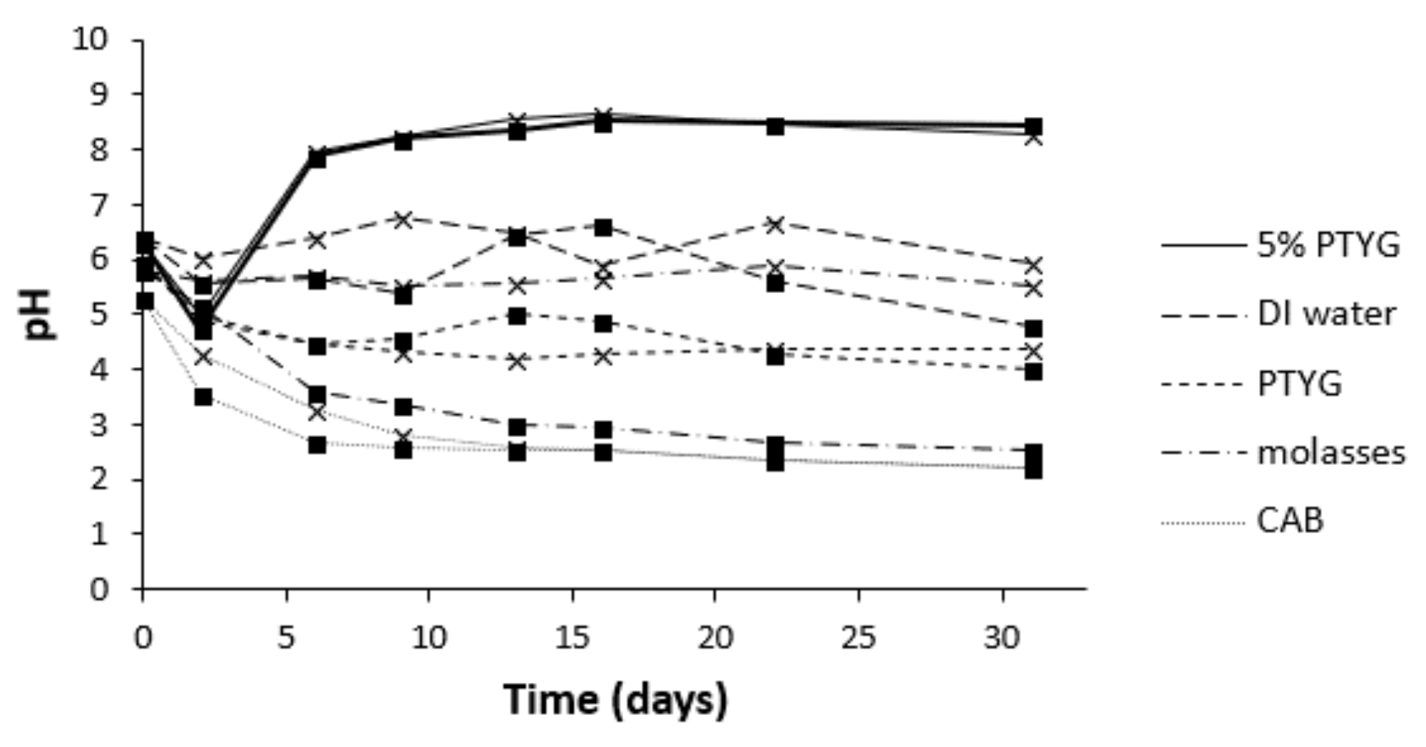
3.1. Selection of Isolates, Growth Medium and IAD

Of the 40 isolates chosen for further screening, 31 distinct isolates were identified and 16 of these were selected for identification by DNA sequencing (Table 3). Three isolates were fungi (TD25, TD47 and BP32), and the remainder were bacteria. Two of the isolates were able to reduce the pH and solubilize inorganic phosphate and these were used in bioleaching studies. They were identified as Aspergillus sp. (TD25) and Bacillus sp. (TD29) (Table 3). Both isolates came from the profile above the country rock, about 4 m depth, however due to large volume of material available and higher REE concentration, the IAD from the profile overlying volcanic breccia was used, at 3.8–4.8 m depth.
Of the tested growth media, CAB medium showed the greatest pH decrease, reaching pH 2.2 (Figure 1). A greater initial pH decrease was seen in the Aspergillus culture. The pH in the Bacillus culture reached a similar value in CAB by day 13. A similar decrease was seen with molasses inoculated with Aspergillus, to pH 2.6, however molasses inoculated with Bacillus varied from pH 5.5 to 5.9. A small decrease in pH was observed by both strains in PTYG, with a minimum of 4. The pH in DI water fluctuated between 4.8 and 6.8. The pH initially decreased with 5% PTYG, then increased after day 2. Given that CAB demonstrated the greatest pH decrease, and the decrease was observed with both isolates, it was selected for further leaching studies.

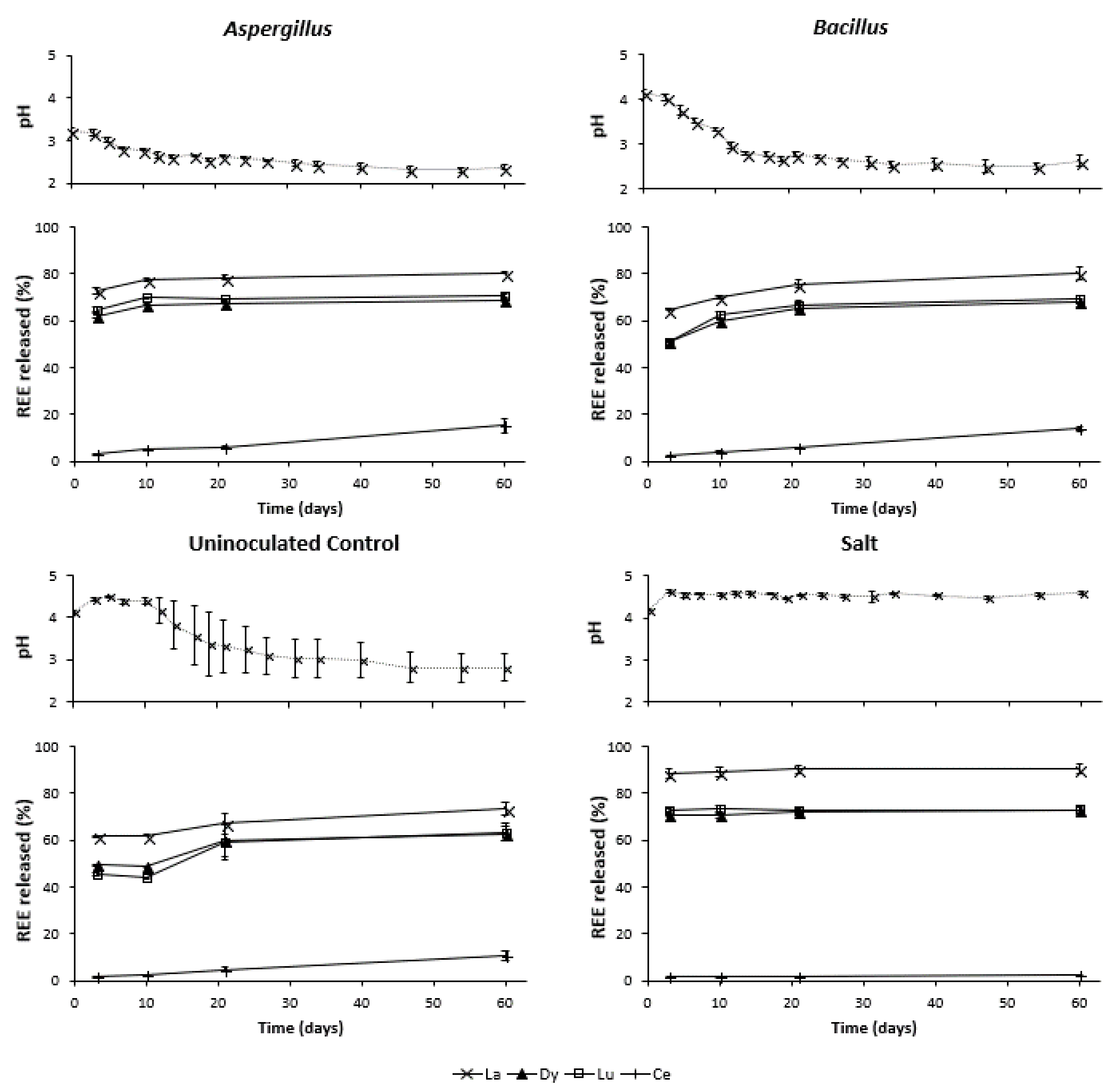
3.2. Changes in pH during Leaching

The pH changes throughout the experiments are illustrated in top sections of Figure 2. At day 0 the pH of the salt leachates (mean of three replicates) was 4.2 and reached a steady state by day 3 at pH~4.5. At day 0 the pH of the Aspergillus bioleachates was 3.2, while the pH of the Bacillus and Uninoculated Control were both higher, pH 4.1. By day 10, the pH in both Aspergillus and Bacillus leachates decreased below pH of 3, then the pH decreased at a slower rate to about day 40; after this the pH fluctuated between 2.3 and 2.7. The pH of the Uninoculated Control leachates showed an initial increase to a maximum of 4.5 on day 5. After day 10 the pH began to decrease, dropping below pH 3 on day 40 and reached a low of 2.8 by day 47. The large error bars seen in Figure 2 in the Uninoculated Control experiment reflects a consistently higher pH of one of the replicates, and the pH decrease did not start until day 17.
To confirm the presence and type of organic acids produced, the final leaching solutions were analyzed for organic ligand composition and concentration by ion chromatography (Table 4). Gluconate dominated Aspergillus, Bacillus and two of three Uninoculated Control leachate replicates; in the other replicate citrate dominated, this is the replicate that was responsible for the higher pH after day 10. The Aspergillus bioleachates also showed production of citrate, at approximately half the gluconate concentration. Much lower concentrations of oxalate were detected and was highest in Uninoculated Controls. No other major organic acids were detected.

3.3. REE Released during Bioleaching and Salt Leaching

To describe the REE leaching behavior, the REE released (element leached/total element) of La, Dy, Lu and Ce are presented (Figure 2). Data for all other REE are in Supplementary Material. More La was released than other REE. For the Salt leaching, La was leached to 88.3% after 3 days, whilst 72.6%, 64.8% and 61.6% was leached by the Aspergillus and Bacillus isolates and the Uninoculated Control, respectively. The amount of La released in the bioleachates increased with time, up to 80.4% for both isolates, while the leaching of La in the Salt leaching experiments was rapid and nearly complete by the first sampling point. Leaching of Dy and Lu showed the same increase in REE leaching as observed for La; however, after 3 days REE released was lower in the Aspergillus bioleachate, 62.2% and 64.8% for Dy and Lu, respectively. In the Bacillus leachate 51.2% Dy and 50.8% Lu were released and in the Uninoculated Control 49.2% Dy and 45.6% Lu were released (Figure 2).
Ce demonstrated different leaching behavior to the other REE. Although the amount of Ce released increased with time, it remained low and bioleaching experiments considerably outperformed the salt leaching experiments (15.3% and 14.0% leached for Aspergillus and Bacillus, respectively, compared to 2.28% for the salt leach). An increase in the Ce leached by the Uninoculated Control was observed on day 21, which coincides with the decrease in pH and increase leaching of the other REE.
Throughout the experiment Aspergillus leachates had a higher Lu/La ratio than the Salt leachates, approximately 3.0 × 10⁻³ and 2.7 × 10⁻³, respectively (Table 5). This higher heavy to light REE ratio in bioleaching is also seen when comparing Aspergillus experiments to the Uninoculated Control experiments on day 3 and day 10.
The increase in REE released with time in each of the bioleaching experiments was accompanied by a decrease in pH (Figure 2). This trend is maintained when combining the data for all bioleaching experiments for the same element. A strong correlation is observed between pH and REE released: La (R2 = 0.865), Dy (R2 = 0.963) and Lu (R2 = 0.947). Much poorer correlation was seen with pH and Ce (R2 = 0.536). R2 values are Pearson correlation coefficients of pH and REE released in mg∙kg−1. The Salt leaching data clustered with a small range of higher pHs and REE released for each element. There was no dependence on pH for any element.

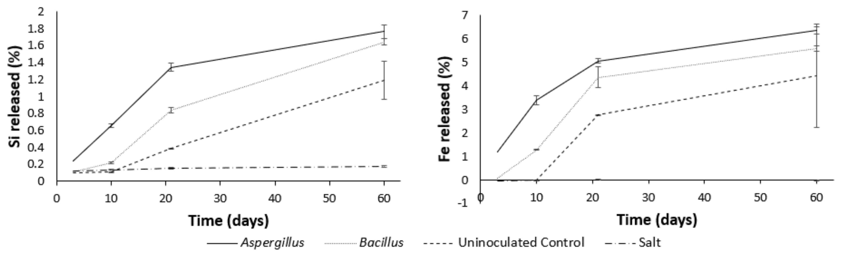
3.4. Major Elements during Leaching

Figure 3, representing the Si and Fe element released (%) versus time, shows the similarities between the dissolution rate of Si and Fe (as indicated by the curve slope) in the Aspergillus and Bacillus bioleaching experiments, compared to the salt leaching experiment. It is noticeable how the latter significantly differs from the bioleachates, as it shows minimal leaching of these elements, regarded as indicative of the mineral matrix composed by clays and iron oxyhydroxides (kaolinite and goethite). The Uninoculated Control experiment shows no leaching until day 10 and an increase in Si and Fe leached thereafter.

4. Discussion

In this study, bioleaching of a potential Madagascan IAD was compared to the current standard mining method for this type of deposit (Salt leaching). Although several studies have demonstrated the bioleaching potential of various heterotrophic microorganisms, none have carried out a comparison to the current industry standard methods for that type of deposit. Such a comparison is essential for any new technology to be adopted instead of, or complementary to, current techniques.
The results demonstrated that the Salt leaching yielded the greatest amount of REE released, followed by Aspergillus, Bacillus and then Uninoculated Control experiments. Nevertheless, total yields were overall comparable between experiments, in particular for Aspergillus HREE bioleaching, where 71% Lu was leached compared to 73% in the Salt leaching. The timescale over which this was achieved was, however, longer with the bioleaching experiment.
The release of REE was very rapid in the Salt leaching experiments, with almost all of the REE released by the first sampling point. The rapid release of REE observed in Salt leachates is attributable to the fast kinetics involved in ion exchange as observed by others [6,24,25]. Salt leaching of five Madagascan REE ores by [26], demonstrated rapid release of REE with a similar range of REE released. In contrast, the more gradual release of REE observed during the Aspergillus and Bacillus bioleaching experiments indicates the contribution of a different acid- and ligand-promoted mechanism of release, due to metabolic organic acid production during the experiment. This is supported by the strong negative correlation between REE extracted and the solution pH. The concomitant increase in dissolved Si and Fe as the pH decreases, seen in the Aspergillus and Bacillus bioleaching, but not in the Salt leaching experiments, is the evidence of the instability and partial dissolution of the minerals in the IAD (kaolinite and goethite as the main mineral phases, as seen in IAD from the same area [27]). At pH < 3, which was reached by day 10 in both the Aspergillus and Bacillus bioleaching experiments, dissolution is common upon exposure of minerals to organic acids [28]. It needs to be noted, however, that, as the lower pH coincides with longer incubations, there could be an additional kinetic effect to account for enhanced REE release, as the detachment of the metal during metal oxide/silicate dissolution is rate limiting and slow [29].
Careful evaluation of the pH and REE leaching pattern for the Uninoculated Control experiments gave further insights into the extraction mechanisms. The Uninoculated Control showed no change of pH until day 12, with, a decrease thereafter, suggesting that some microbial growth occurred in these controls at this time. Due to concerns over potential modification of clay minerals and their surface chemistry, standard sterilization procedures, such as autoclaving [30], were not deemed appropriate and the IAD was not sterilized. Based on these observations, the flasks after day 12 were no longer considered to represent the controls. However, by limiting the observations between day 3 and day 10 in the Uninoculated Control experiments, the lack of pH change, combined with minimal Si and Fe release, were indicative of minimal microbial growth in this period and any leaching observed at this time was likely to be due to the effects of the growth medium alone. As such, the growth medium was able to extract up to 62% La. This is in contrast to other leaching experiments with heterotrophs where the growth medium does not show significant leaching of REE [15,31], however not surprising, as in these studies the target REE is generally associated with the mineral structure and not in easily exchangeable sites on the surface of IAD minerals. Despite the significant release of REE by the growth medium only, REE recovery was enhanced in the inoculated samples, in support of the ability of the heterotroph to enhance the leaching.
One element (Ce) showed different behavior to the other REE. Ce leaching was enhanced in the bioleaching compared to the Salt leaching experiments, and, unlike the other REE, did not show strong correlation with pH (R2 = 0.766). In contrast to other REE, oxidation of Ce3+ to Ce4+ occurs under oxidizing conditions during the weathering of parent rock and soil formation, leading to the precipitation of insoluble Ce4+ oxides in the lateritic profiles [5]. As such, Ce is unavailable for simple ion-exchange extractions. More acidic conditions obtained in the bioleaching experiments compared to the salt leaching experiments are likely to enhance Ce oxide dissolution, and likely responsible for the different fractionation of Ce to the other REE between the Salt and bioleaching experiments. Ce oxide dissolution can also be enhanced by reducing conditions, given Ce3+ is many orders of magnitude more soluble than Ce4+ [32]. Although the range of Eh in the bioleaching experiments indicates oxidizing conditions at the end of the experiment, there may be local Eh differences within biomass. Based on these observations bioleaching is more effective to extract Ce in IAD than salt leaching.
The important feature of a potential fractionation of HREE from LREE under leaching of IAD by Aspergillus and Bacillus, based on stronger complexation of HREE than LREE by organic acids, has been tested in the bioleaching experiments of this study. This fractionation is potentially attractive to industry as it would produce a leachate with a higher proportion of the more economically valuable HREE. Albeit small, a fractionation towards HREE during leaching, with a higher Lu/La ratio, was observed in the bioleachates compared to salt leachates. In order to confirm the significance of complexing properties of the metabolic organic acids produced by Aspergillus and Bacillus in fractionating HREE from LREE, further tests are needed. It is in fact possible that what observed is not only due to complexation, but the combined result of both the acidic and complexing effects of organic acids. If the HREE are preferably locked in the ore mineral matrix rather than in the surface exchangeable sites, the acidification of the medium, due to the organic acids, would indeed free the HREE locked in the mineral matrix more than a salt leaching, thus enriching the HREE fraction of the organic leachate compare to a salt leachate. A comparison with extraction by HCl to test only the proton-induced dissolution on this material is part of a future experimental plan.
Organic acid production by Aspergillus was not unexpected as strains are used for the commercial production of citric acid [33]. The type and concentration of organic acid produced by a microbial strain depends on growth conditions, with the main controls including ambient pH of growth medium and limitation of particular nutrients [34,35]. Phosphate limitation in growth media has previously been linked to gluconate production in A. niger [35], as observed in this study in the Aspergillus, Bacillus and two of the Uninoculated Control bioleachates. All samples contained a microbial community representative of the material in situ, however the Uninoculated Control experiments contained only the natural community and the variation in organic acid production and pH in these experiments suggests that the establishment of the mixed microbial community may have been susceptible to small changes in initial conditions or stochastic processes. Consequently, in one of the replicates a microbial community developed that did not produce gluconate and showed higher pH than the other replicates. DNA extraction from clay rich soils is notoriously difficult [36] and it was not possible to obtain community composition information from these samples to characterize any differences.
Although this study has shown some potential for a bioleaching approach to REE release from IAD, two improvements would be required to make such a process economically attractive. Firstly, the 60-day bioleaching used in this experiment is not practical for commercial operations when compared to the fast kinetics of salt leaching. As REE released was closely linked to pH, separation of acid production and leaching steps could reduce leaching times. One approach to reduce contact time would be to apply two-step bioleaching method. In this approach the organism is cultured in the absence of rock, so that it produces an organic acid rich spent medium, which can be applied to the rock to be leached, either as it is, or after filtration to remove cells. The first leaching step could be optimized for organic acid production, as a faster pH drop was observed without presence of IAD in unmodified CAB, or for exploration of the use of waste resources for organic acid production [37]. The second step could then be optimized for leaching. Although the use of two-step bioleaching method was not shown to increase in REE leaching when applied to bioleaching of REE in monazite [11], the two-step approach including the Aspergillus cells did improve bioleaching of heavy metals from red mud [14].
Secondly, the increased concentration of Si observed in the bioleaching suggests that the clay minerals are being dissolved. This is accompanied by large quantities of Fe leached, up to 6.4% in Aspergillus bioleachates. Iron in the leachate is a concern for separation of REE during downstream processing [38], and is therefore an undesirable consequence of this bioleaching approach. As there is a kinetic effect of clay, reducing contact time could reduce impurities in leachate [29]. This impurity combined with the long timescale of bioleaching observed in this study are likely to be barriers to the adoption of a bioleaching approach as described here, although provides a foundation for future work.

5. Conclusions

Directly comparing bioleaching of the studied IAD material with a standard salt leaching solution shows the following order of REE released: Salt > Aspergillus > Bacillus > Uninoculated Control, with the exception of Ce where bioleaching enhanced Ce release.
When considering total REE leached in the bioleaching experiments, Aspergillus sp. initially leached more REE but, given enough time, Bacillus sp. leached a similar amount of REE.
Bioleaching of REE and major elements increased with time and had a greater heavy to light REE ratio than Salt leaching.
Bioleaching enhanced leaching over the growth medium alone by the production of organic (predominantly gluconic) acids.
Reducing contact time and the co-leaching of impurities, such Fe, remain as challenges that need to be addressed in order for bioleaching to be considered as an alternative to salt leaching for IAD.

Supplementary Materials

The following are available online at https://www.mdpi.com/2075-163X/8/6/236/s1. Document S1: Details for screening of microbial isolated from IAD, containing details of microbial broths, agars, PCR conditions and sequencing primers. Table S1: Leaching data of all leachates and timepoints, and elemental composition of original IAD in mg∙kg−1. Errors are plus and minus one standard deviation from triplicate experiments.

Author Contributions

M.J.B., B.P.-R. and S.P.G. conceived and designed the experiments; M.J.B. and S.P.G. performed the experiments; M.J.B. and B.P.-R. analyzed the data; M.J.B., S.P.G. and B.P.-R. wrote the paper

Acknowledgments

This work was funded by the Natural Environment Research Council as part of the SoS RARE project (NE/M011429/1). The authors would like to thank Umwelt- und Ingenieurtechnik GmbH (Germany), Tantalus Rare Earths, Madagascar and University of Brighton, UK for the donation of starting material. Thomas Barlow and Michael Watts of The British Geological Survey Analytical Chemistry Laboratories are gratefully acknowledged for carrying out the ICP chemical analysis. Alastair Bewsher at The University of Manchester is thanked for organic acid analysis. The paper is published with the permission of the Executive Director of the British Geological Survey.

Conflicts of Interest

The authors declare no conflict of interest.

References

  1. EU Commission (EC). Report on Critical Raw Materials for the EU; European Commission: Brussels, Belgium, 2014. [Google Scholar]
  2. Brown, T.J.; Idoine, N.E.; Raycraft, E.R.; Shaw, R.A.; Deady, E.A.; Hobbs, S.F.; Bide, T. World Mineral Production 2011–2015; British Geological Survey: Keyworth, UK; Nottingham, UK, 2017. [Google Scholar]
  3. Wall, F. Rare earth elements. In Critical Metals Handbook; Gunn, G., Ed.; John Wiley & Sons Ltd.: New York, NY, USA, 2013; pp. 312–339. [Google Scholar]
  4. Yang, X.J.; Lin, A.; Li, X.L.; Wu, Y.; Zhou, W.; Chen, Z. China’s ion-adsorption rare earth resources, mining consequences and preservation. Environ. Dev. 2013, 8, 131–136. [Google Scholar] [CrossRef]
  5. Sanematsu, K.; Watanabe, Y. Characteristics and genesis of ion adsorption-type rare earth element deposits. Rev. Econ. Geol. 2016, 18, 55–79. [Google Scholar]
  6. Moldoveanu, G.A.; Papangelakis, V.G. Recovery of rare earth elements adsorbed on clay minerals: I. Desorption mechanism. Hydrometallurgy 2012, 117–118, 71–78. [Google Scholar] [CrossRef]
  7. Bradbury, M.H.; Baeyens, B. Sorption of Eu on Na- and Ca-montmorillonites: Experimental investigations and modelling with cation exchange and surface complexation. Geochim. Cosmochim. Acta 2002, 66, 2325–2334. [Google Scholar] [CrossRef]
  8. Barmettler, F.; Castelberg, C.; Fabbri, C.; Brandl, H. Microbial mobilization of rare earth elements (REE) from mineral solids—A mini review. AIMS Microbiol. 2016, 2, 190–204. [Google Scholar] [CrossRef]
  9. Burgstaller, W.; Schinner, F. Leaching of metals with fungi. J. Biotechnol. 1993, 27, 91–116. [Google Scholar] [CrossRef]
  10. Brisson, V.L.; Zhuang, W.Q.; Alvarez-Cohen, L. Bioleaching of rare earth elements from monazite sand. Biotechnol. Bioeng. 2016, 113, 339–348. [Google Scholar] [CrossRef] [PubMed]
  11. Hassanien, W.A.G.; Desouky, O.A.N.; Hussien, S.S.E. Bioleaching of some rare earth elements from Egyptian monazite using Aspergillus ficuum and Pseudomonas aeruginosa. Walailak J. Sci. Technol. 2014, 11, 809–823. [Google Scholar]
  12. Shin, D.; Kim, J.; Kim, B.; Jeong, J.; Lee, J. Use of phosphate solubilizing bacteria to leach rare earth elements from monazite-bearing ore. Minerals 2015, 5, 189–202. [Google Scholar] [CrossRef]
  13. Hewedy, M.A.; Rushdy, A.A.; Kamal, N.M. Bioleaching of rare earth elements and Uranium from Sinai soil, Egypt using Actinomycetes. Egypt. J. Hosp. Med. 2013, 53, 909–917. [Google Scholar] [CrossRef]
  14. Qu, Y.; Lian, B.; Mo, B.; Liu, C. Bioleaching of heavy metals from red mud using Aspergillus niger. Hydrometallurgy 2013, 136, 71–77. [Google Scholar] [CrossRef]
  15. Qu, Y.; Lian, B. Bioleaching of rare earth and radioactive elements from red mud using Penicillium tricolor RM-10. Bioresour. Technol. 2013, 136, 16–23. [Google Scholar] [CrossRef] [PubMed]
  16. Pourret, O.; Davranche, M.; Gruau, G.; Dia, A. Rare earth elements complexation with humic acid. Chem. Geol. 2007, 243, 128–141. [Google Scholar] [CrossRef] [Green Version]
  17. Tang, J.; Johannesson, K.H. Speciation of rare earth elements in natural terrestrial waters: Assessing the role of dissolved organic matter from the modeling approach. Geochim. Cosmochim. Acta 2003, 67, 2321–2339. [Google Scholar] [CrossRef]
  18. Wood, S.A. The aqueous geochemistry of the rare-earth elements: Critical stability constants for complexes with simple carboxylic acids at 25 °C and 1 bar and their application to nuclear waste management. Eng. Geol. 1993, 34, 229–259. [Google Scholar] [CrossRef]
  19. Yamamoto, Y.; Takahashi, Y.; Shimizu, H. Systematic change in relative stabilities of REE-humic complexes at various metal loading levels. Geochem. J. 2010, 44, 39–63. [Google Scholar] [CrossRef]
  20. Martell, A.E.; Smith, R.M. Critical Stability Constants. Other Organic Ligands; Plenum: New York, NY, USA, 1977; Volume 3. [Google Scholar]
  21. Estrade, G.; Salvi, S.; Béziat, D.; Williams-Jones, A.E. The origin of skarn-hosted rare-metal mineralization in the Ambohimirahavavy alkaline complex, Madagascar. Econ. Geol. 2015, 110, 1485–1513. [Google Scholar] [CrossRef]
  22. Byrne, R.H.; Kim, K.-H. Rare earth precipitation and coprecipitation behavior: The limiting role of PO43− on dissolved rare earth concentrations in seawater. Geochim. Cosmochim. Acta 1993, 57, 519–526. [Google Scholar] [CrossRef]
  23. Takahashi, Y.; Châtellier, X.; Hattori, K.H.; Kato, K.; Fortin, D. Adsorption of rare earth elements onto bacterial cell walls and its implication for REE sorption onto natural microbial mats. Chem. Geol. 2005, 219, 53–67. [Google Scholar] [CrossRef]
  24. Moldoveanu, G.A.; Papangelakis, V.G. Recovery of rare earth elements adsorbed on clay minerals: II. Leaching with ammonium sulfate. Hydrometallurgy 2013, 131–132, 158–166. [Google Scholar] [CrossRef]
  25. Tian, J.; Yin, J.; Chi, R.; Rao, G.; Jiang, M.; Ouyang, K. Kinetics on leaching rare earth from the weathered crust elution-deposited rare earth ores with ammonium sulfate solution. Hydrometallurgy 2010, 101, 166–170. [Google Scholar]
  26. Moldoveanu, G.A.; Papangelakis, V.G. An overview of rare-earth recovery by ion-exchange leaching from ion-adsorption clays of various origins. Mineral. Mag. 2016, 80, 63–76. [Google Scholar] [CrossRef]
  27. Gilbertson, J. A Competent Persons Report on the Tantalus Project, Northern Madagascar; Prepared by Tantalus Rare Earths AG; SRK Exploration Services Ltd.: Cardiff, UK, 2015. [Google Scholar]
  28. Lazo, D.E.; Dyer, L.G.; Alorro, R.D. Silicate, phosphate and carbonate mineral dissolution behaviour in the presence of organic acids: A review. Miner. Eng. 2017, 100, 115–123. [Google Scholar] [CrossRef]
  29. Stumm, W.; Morgan, J.J. Kinetics at the solid-water interface: Adsoprtion, dissolution of minerals, nucleation, and crystal growth. In Aquatic Chemistry, Chemical Equilibria and Rates in Natural Waters, 3rd ed.; Stumm, W., Morgan, J.J., Eds.; John Wiley & Sons Inc.: New York, NY, USA, 1996; p. 1022. [Google Scholar]
  30. Jenneman, G.E.; McInerney, M.J.; Crocker, M.E.; Knapp, R.M. Effect of sterilization by dry heat or autoclaving on bacterial penetration through Berea sandstone. Appl. Environ. Microbiol. 1986, 51, 39–43. [Google Scholar] [PubMed]
  31. Behera, S.K.; Panda, P.P.; Singh, S.; Pradhan, N.; Sukla, L.B.; Mishra, B.K. Study on reaction mechanism of bioleaching of nickel and cobalt from lateritic chromite overburdens. Int. Biodeterior. Biodegrad. 2011, 65, 1035–1042. [Google Scholar] [CrossRef]
  32. Arenas, L.F.; de León, C.P.; Walsh, F.C. Electrochemical redox processes involving soluble cerium species. Electrochim. Acta 2016, 205, 226–247. [Google Scholar] [CrossRef]
  33. Papagianni, M. Advances in citric acid fermentation by Aspergillus niger: Biochemical aspects, membrane transport and modeling. Biotechnol. Adv. 2007, 25, 244–263. [Google Scholar] [CrossRef] [PubMed]
  34. Andersen, M.R.; Lehmann, L.; Nielsen, J. Systemic analysis of the response of Aspergillus niger to ambient pH. Genome Biol. 2009, 10, R47. [Google Scholar] [CrossRef] [PubMed] [Green Version]
  35. Kubicek, C.P.; Schreferl-Kunar, G.; Wöhrer, W.; Röhr, M. Evidence for a cytoplasmic pathway of oxalate biosynthesis in Aspergillus niger. Appl. Environ. Microbiol. 1988, 54, 633–637. [Google Scholar] [PubMed]
  36. Young, J.M.; Rawlence, N.J.; Weyrich, L.S.; Cooper, A. Limitations and recommendations for successful DNA extraction from forensic soil samples: A review. Sci. Justice 2014, 54, 238–244. [Google Scholar] [CrossRef] [PubMed]
  37. Bakhiet, S.E.A.; Al-Mokhtar, E.A.I. Production of citric acid by Aspergillus niger using sugarcane molasses as substrate. Jordan J. Biol. Sci. 2015, 8, 211–215. [Google Scholar]
  38. Sui, N.; Huang, K.; Lin, J.; Li, X.; Wang, X.; Xiao, C.; Liu, H. Removal of Al, Fe and Si from complex rare-earth leach solution: A three-liquid-phase partitioning approach. Sep. Purif. Technol. 2014, 127, 97–106. [Google Scholar] [CrossRef]
Figure 1. Changes in pH in five microbiological media over 31 days. Aspergillus (squares) and Bacillus (crosses).
Minerals 08 00236 g001
Figure 2. Changes in pH (top section of graphs) and release of La, Dy, Lu and Ce (bottom sections of graphs) over the duration of the experiment. Day zero pH measurement was taken 1 h after mixing IAD. The chemical analysis starts from day three as first data point. Error bars are standard deviation of triplicate measurements.
Minerals 08 00236 g002
Figure 3. Changes in leaching of Si and Fe with time. Error bars are standard deviation of triplicates.
Minerals 08 00236 g003
Table 1. Set up of batch leaching tests.
DescriptionInoculumFluid
AspergillusAspergillus sp. IsolateModified CAB
BacillusBacillus sp. IsolateModified CAB
Uninoculated ControlNoneModified CAB
SaltNone0.5 M ammonium sulfate
Table 2. Composition of IAD determined in mg·kg−1.
Element (mg·kg−1)SiAlFeLaCeDyLu
IAD188,569175,29837,358159438753.35.32
Table 3. Characterization of isolates from Madagascan IAD. BP isolated are from profile overlying the volcanic breccia and TD isolates are from profile overlying the country rock. Closest matching sequences were obtained from GenBank in June (BP46, BP57, TD02 and TD05) and remainder in July 2016.
IsolateIAD Sample DepthpH DropSolubilize Inorganic PhosphateUrease ActivitySiderophoreClosest Match (Accession Number)
BP180–0.8 m, 3.8–4.8 m+Bacillus thuringiensis (KP813752)
BP230–0.8 m+Burkholderia sp. (AB911072)
BP250.8–3.8 m++Leifsonia sp. (LN876290)
BP320–0.8, 0.8–3.8 m++Cunninghamella bainieri (KF201293)
BP380.8–3.8, 3.8–4.8 m+Shewanella sp. (GU143896)
BP463.8–4.8 m+Uncultured bacterium (AM180664)
BP574.8–5.8 m++Bacillus aryabhattai (AB9304966)
TD010.9–1.1 m+*Bacillus thuringiensis (HQ83480)
TD020.9–1.1 m+Bacillus sp. (KC893975)
TD170.9–1.1 mBacillus sp. (KU315821)
TD200.9–1.1 m++Bacterium (KT692625)
TD253.4–3.6 m+++Aspergillus niger (KM516789)
TD293.4–3.6 m++Bacillus sp. (KJ584025)
TD313.4–3.6 m++Staphlococcus sp. (KX079771)
TD374.3–4.5 m++Bacillus sp. (KP670302)
TD473.4–3.6 m+Curvularia verruculosa (HF934909)
* No growth.
Table 4. Concentration of oxalate, citrate and gluconate and pH and Eh at the end of the bioleaching experiment. The individual organic acid concentrations in the Salt leachates were below the detection limit of 0.1 g·L−1. Eh of salt leachates was 584 ± 13 mV. Errors are standard deviation of triplicates.
Organic Ligand (g·L1)pHEh
OxalateCitrateGluconate mV
Aspergillus0.695 ± 0.31810.2 ± 1.321.1 ± 1.22.38 ± 0.07579 ± 5
Bacillus0.563 ± 0.4643.19 ± 3.7321.7 ± 1.72.62 ± 0.13577 ± 53
Uninoculated Control1.03 ± 0.180.858 ± 0.8889.45 ± 7.922.81 ± 0.34640 ± 100
Original medium<0.05<0.05<0.054.384.38
Table 5. Lu to La ratio. Errors are plus and minus one standard deviation from triplicate leaching experiments.
Aspergillus (×10−3)Bacillus (×10−3)Uninoculated Control (×10−3)Salt (×10−3)
Day 32.98 ± 0.012.61 ± 0.042.47 ± 0.042.75 ± 0.05
Day 103.01 ± 0.022.99 ± 0.052.36 ± 0.012.74 ± 0.00
Day 212.94 ± 0.032.94 ± 0.022.95 ± 0.402.68 ± 0.03
Day 602.93 ± 0.052.88 ± 0.042.88 ± 0.132.69 ± 0.01

