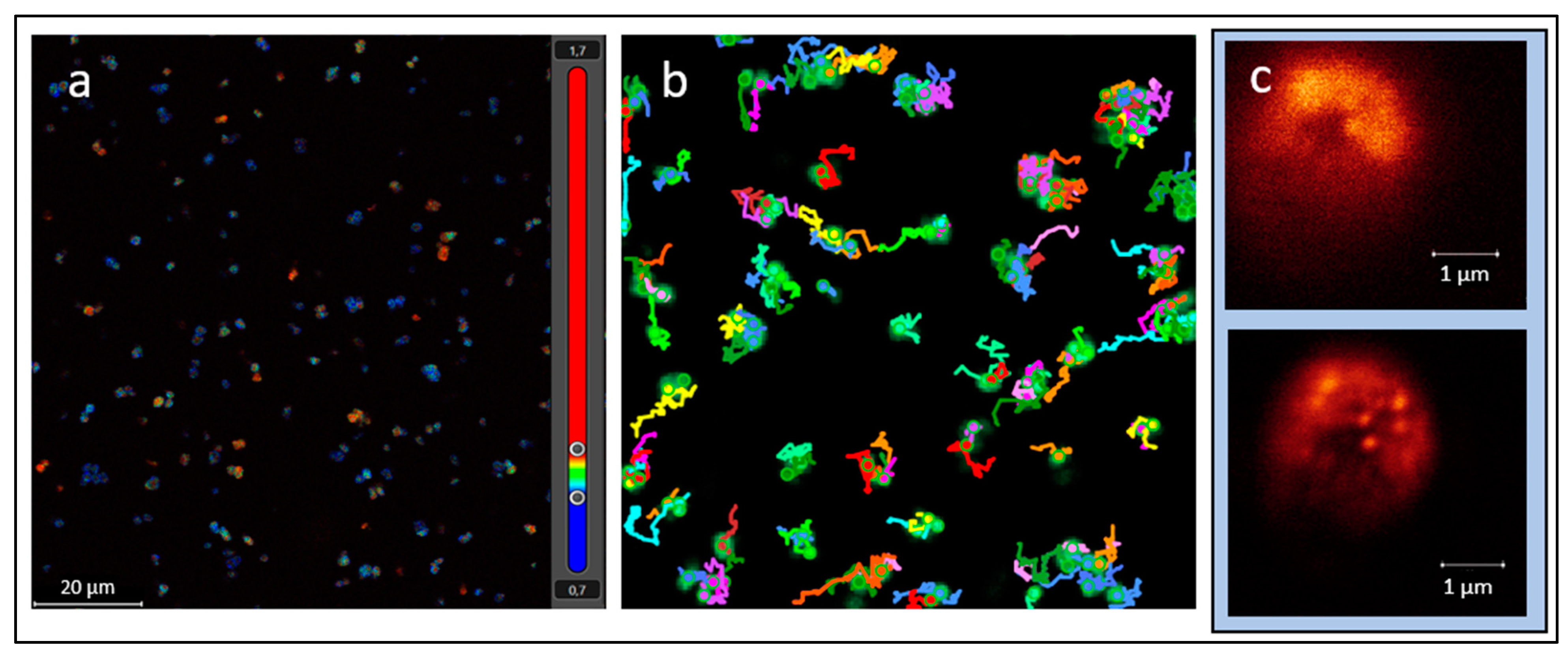
The Hypothesis of a “Living Pulse” in Cells
Abstract
:1. Introduction
2. The Hypothesis
3. The Question of Detection
4. The Significance of Detecting a “Living Pulse”
5. Conclusions
Author Contributions
Funding
Institutional Review Board Statement
Informed Consent Statement
Data Availability Statement
Acknowledgments
Conflicts of Interest
References
- Nadeau, J.; Bedrossian, M.; Lindensmith, C. Imaging technologies and strategies for detection of extant extraterrestrial microorganisms. Adv. Phys. 2018, 3, 1424032. [Google Scholar] [CrossRef] [Green Version]
- Miyata, M.; Robinson, R.C.; Uyeda, T.Q.P.; Fukumori, Y.; Fukushima, S.; Haruta, S.; Homma, M.; Inaba, K.; Ito, M.; Kaito, C.; et al. Tree of motility—A proposed history of motility systems in the tree of life. Genes Cells 2022, 25, 6–21. [Google Scholar] [CrossRef] [Green Version]
- Riekeles, M.; Schirmack, J.; Schulze-Makuch, D. Machine learling algorithms applied to identify microbial species by their motility. Life 2021, 11, 44. [Google Scholar] [CrossRef] [PubMed]
- Jarrell, F.; Albers, S.-V. The archaellum: An old motility structure with a new name. Trends Microbiol. 2012, 20, 307–312. [Google Scholar] [CrossRef]
- Uyeda, J.C.; Harmon, L.J.; Blank, C.E. A comprehensive study of cyanobacterial morphological and ecological evolutionary dynamics through deep geologic time. PLoS ONE 2016, 11, e0162539. [Google Scholar] [CrossRef] [Green Version]
- Khan, S.; Scholey, J.M. Assembly, functions and evolution of archaella, flagella and cilia. Curr. Biol. 2018, 28, R278–R292. [Google Scholar] [CrossRef] [Green Version]
- Lingam, M. Theoretical constraints imposed by gradient detection and dispersial on microbial size in astrobiological environments. Astrobiology 2021, 21, 813–830. [Google Scholar] [CrossRef]
- Snyder, C.; Centlivre, J.P.; Bhute, S.; Shipman, G.; Friel, A.D.; Viver, T.; Palmer, M.; Konstantinidis, K.T.; Sun, H.J.; Ro-sello-Mora, R.; et al. Microbial Motility at the Bottom of North America: Digital Holographic Microscopy and Genomic Motility Signatures in Badwater Spring, Death Valley National Park. Astrobiology 2023, 23, 295–307. Available online: https://www.liebertpub.com/doi/full/10.1089/ast.2022.0090 (accessed on 25 June 2023). [CrossRef]
- Lindensmith, C.; Nadeau, J.L.; Bedrossian, M.; Sumrall, L.; Wallace, J.K.; Serabyn, E. Microscopic object classification through passive motion observations with holographic microscopy. In Proceedings of the 2020 IEEE Aerospace Conference, Big Sky, MT, USA, 7–14 March 2020; pp. 1–7. [Google Scholar] [CrossRef]
- Lindensmith, C.A.; Rider, S.; Bedrossian, M.; Wallace, J.K.; Serabyn, E.; Showalter, G.M.; Deming, J.W.; Nadeau, J.L. A Submersible, Off-Axis Holographic Microscope for Detection of Microbial Motility and Morphology in Aqueous and Icy Environments. PLoS ONE 2016, 11, e0147700. Available online: https://journals.plos.org/plosone/article?id=10.1371/journal.pone.0147700 (accessed on 25 June 2023). [CrossRef]
- Venturelli, L.; Harrold, Z.R.; Murray, A.E.; Villialba, M.I.; Lundin, E.M.; Dietler, G.; Kasas, S.; Foschia, R. Nanomechanical bio-sensing for fast and reliable detection of viability and susceptibility of microorganisms. Sens. Actuators B. Chem. 2021, 348, 130650. [Google Scholar] [CrossRef]
- Kasas, S.; Ruggeri, F.S.; Benadiba, C.; Maillard, C.; Stupar, P.; Tournu, H.; Dietler, G.; Longo, G. Detecting nanoscale vibrations as signature of life. Proc. Nat. Acad. Sci. USA 2014, 112, 378–381. [Google Scholar] [CrossRef]
- Boisen, A.; Dohn, S.; Keller, S.S.; Schmid, S.; Tenie, M. Cantilever-like micromechanical sensors. Rep. Prog. Phys. 2011, 74, 036101. [Google Scholar] [CrossRef]
- Waggoner, P.S.; Craighead, H.G. Micro- and nanomechanical sensors for environmental, chemical, and biological detection. Lab Chip 2007, 7, 1238–1255. [Google Scholar] [CrossRef] [PubMed]
- Alvarez, M.; Lechuga, L.M. Microcantilever-based platforms as biosensing tools. Analyst 2010, 135, 827–836. [Google Scholar] [CrossRef]
- Hansen, K.M.; Thundat, T. Microcantilever biosensors. Methods 2005, 37, 57–64. [Google Scholar] [CrossRef]
- Schneider, S.W.; Egan, M.E.; Jena, B.P.; Guggino, W.B.; Oberleithner, H.; Geibel, J. Continuous detection of extracellular ATP on living cells by using atomic force microscopy. Proc. Natl. Acad. Sci. USA 1999, 96, 12180–12185. [Google Scholar] [CrossRef]
- Pelling, A.E.; Schati, S.; Gralla, E.B.; Valentine, J.S.; Gimzewski, J.K. Local nanomechanical motion of the cell wall of Saccharomyces cerevisiae. Science 2004, 305, 1147–1150. [Google Scholar] [CrossRef]
- Pelling, A.E.; Schati, S.; Gralla, E.B.; Gimzewski, J.K. Time dependence of the frequency and amplitude of the local nanomechanical motion of yeast. Nanomedicine 2005, 1, 178–183. [Google Scholar] [CrossRef] [PubMed]
- Lissandrello, C.; Inci, F.; Francom, M.; Paul, M.R.; Demirci, U.; Ekinci, K.L. Nanomechanical motion of Escherichia coli adhered to a surface. Appl. Phys. Lett. 2014, 105, 113701. [Google Scholar] [CrossRef] [Green Version]
- Lloyd, A.C. The regulation of cell size. Cell 2013, 154, 1194–1205. [Google Scholar] [CrossRef] [Green Version]
- Tamayo, J.; Kosaka, P.M.; Ruz, J.J.; San Paulo, A.; Calleja, M. Biosensors based on nanomechanical systems. Chem. Soc. Rev. 2013, 42, 1287–1311. [Google Scholar] [CrossRef] [PubMed] [Green Version]
- Willaert, R.G.; Vanden Boer, P.; Malovichko, A.; Alioscha-Perez, M.; Radotić, K.; Bartolić, D.; Kalauzi, A.; Villalba, M.I.; Sanglard, G.; Dietler, G.; et al. Single yeast cell nanomotions correlate with cellular activity. Sci. Adv. 2020, 6, 3139. [Google Scholar] [CrossRef] [PubMed]
- Nowacki, L.; Follet, J.; Vayssade, M.; Vigneron, P.; Rotellini, L.; Cambay, F.; Egles, C.; Rossi, C. Real-time QCM-D monitoring of cancer cell death early events in a dynamic context. Biosens. Bioelectron. 2015, 64, 469–476. [Google Scholar] [CrossRef] [PubMed]
- Wang, G.; Dewilde, A.H.; Zhang, J.; Pal, A.; Vashist, M. A living cell quartz crystal microbalance biosensor for continuous monitoring of cytotoxic responses of macrophages to single-walled carbon nanotubes. Part. Fibre Toxicol. 2011, 8, 4. [Google Scholar] [CrossRef] [Green Version]
- Braunhut, S.J.; McIntosh, D.; Vorotnikova, E.; Zhou, T.; Marx, K.A. Detection of apoptosis and drug resistance of human breast cancer cells to taxane treatments using quartz crystal microbalance biosensor technology. Assay Drug Dev. Technol. 2005, 3, 77–88. [Google Scholar] [CrossRef]
- Burg, T.P.; Godin, M.; Knudsen, S.M.; Shen, W.; Carlson, G.; Foster, J.S.; Babcock, K.; Manalis, S.R. Weighing of biomolecules, single cells and single nanoparticles in fluid. Nature 2007, 446, 1066–1069. [Google Scholar] [CrossRef] [Green Version]
- Martínez-Martín, D.; Fläschner, G.; Gaub, B.; Martin, S.; Newton, R.; Beerli, C.; Mercer, J.; Gerber, C.; Müller, D.J. Inertial picobalance reveals fast mass fluctuations in mammalian cells. Nature 2017, 550, 500–505. [Google Scholar] [CrossRef] [Green Version]
- Cermak, N.; Olcum, S.; Delgado, F.F.; Wasserman, S.C.; Payer, K.R.; Murakami, M.A.; Knudsen, S.M.; Kimmerling, R.J.; Stevens, M.M.; Kikuchi, Y.; et al. High-throughput measurement of single-cell growth rates using serial microfluidic mass sensor arrays. Nat. Biotechnol. 2016, 34, 1052–1059. [Google Scholar] [CrossRef] [Green Version]
- Lang, F.; Busch, G.L.; Ritter, M.; Völkl, H.; Waldegger, S.; Gulbins, E.; Häussinger, D. Functional significance of cell volume regulatory mechanisms. Physiol. Rev. 1998, 78, 247–306. [Google Scholar] [CrossRef]
- Martínez, N.F.; Kosaka, P.M.; Tamayo, J.; Ramírez, J.; Ahumada, O.; Mertens, J.; Hien, T.D.; Rijn, C.V.; Calleja, M. High throughput optical readout of dense arrays of nanomechanical systems for sensing applications. Rev. Sci. Instrum. 2010, 81, 125109. [Google Scholar] [CrossRef] [Green Version]
- Johnson, W.L.; France, D.C.; Rentz, N.S.; Cordell, W.T.; Walls, F.L. Sensing bacterial vibrations and early response to antibiotics with phase noise of a resonant crystal. Sci. Rep. 2017, 7, 12138. [Google Scholar] [CrossRef] [PubMed] [Green Version]
- Irwin, L.N.; Schulze-Makuch, D. Strategy for modeling putative ecosystems on Europa. Astrobiology 2003, 3, 813–821. [Google Scholar] [CrossRef] [PubMed]
- Alvarez, L.A.J.; Schwarz, U.; Friedrich, L.; Foelling, J.; Hecht, F.; Roberti, M.J. TauSTED: Pushing STED beyond Its Limits with Lifetime. Nat. Methods. 2021. Available online: https://www.nature.com/articles/d42473-021-00241-0 (accessed on 25 June 2023).
- Tognoni, E. High-speed multifunctional scanning ion conductance microscopy: Innovative strategies to study dynamic cellular processes. Curr. Opin. Electrochem. 2021, 28, 100738. [Google Scholar] [CrossRef]
- Johnson, C.H.; Golden, S.S.; Ishiura, M.; Kondo, T. Circadian clocks in prokaryotes. Mol. Microbiol. 1996, 21, 5–11. [Google Scholar] [CrossRef]
- Huang, T.C.; Grobbelaar, N. The circadian clock in the prokaryote Synechococcus RF-1. Microbiology 1995, 141, 535–540. [Google Scholar] [CrossRef] [Green Version]
- Sweeney, B.M.; Borgese, M.B. A circadian rhythm in cell division in a prokaryote, the cyanobacterium Synechococcus WH7803. J. Phycol. 1989, 25, 183–186. [Google Scholar] [CrossRef]
- Lin, R.F.; Huang, T.C. Circadian rhythm of Cyanothece RF-1 (Synechococcus RF-1). In Bacterial Circadian Programs; Ditty, J.L., Mackey, S.R., Johnson, C.H., Eds.; Springer: Berlin/Heidelberg, Germany, 2009; pp. 39–61. [Google Scholar]
- Ouyang, Y.; Andersson, C.R.; Kondo, T.; Golden, S.S.; Johnson, C.H. Resonating circadian clocks enhance fitness in cyanobacteria. Proc. Natl. Acad. Sci. USA 1998, 95, 8660–8664. [Google Scholar] [CrossRef]
- Ma, P.; Mori, T.; Zhao, C.; Thiel, T.; Johnson, C.H. Evolution of KaiC-dependent timekeepers: A photo-circadian timing mechanism confers adaptive fitness in the purple bacterium Rhodopseudomonas palustris. PLoS Genet. 2016, 12, e1005922. [Google Scholar] [CrossRef] [Green Version]
- Eelderink-Chen, Z.; Bosman, J.; Sartor, F.; Dodd, A.N.; Kovacs, A.T.; Merrow, M. A circadian clock in a nonphotosynthetic prokaryote. Sci. Adv. 2021, 7, eabe2086. [Google Scholar] [CrossRef]
- Johnson, C.H.; Zhao, C.; Xu, Y.; Mori, T. Timing the day: What makes bacterial clocks tick? Nat. Rev. Microbiol. 2017, 15, 232–242. [Google Scholar] [CrossRef] [Green Version]
- Clarke, S.; Koshland, D.E. Membrane receptors for aspartate and serine in bacterial chemotaxis. J. Biol. Chem. 1979, 254, 9695–9702. [Google Scholar] [CrossRef] [PubMed]
- Dahlquist, F.W.; Elwell, R.A.; Lovely, P.S. Studies of bacterial chemotaxis in defined concentration gradients. A model for chemotaxis toward L-serine. J. Supramol. Struct. 1976, 4, 329–342. [Google Scholar] [CrossRef] [PubMed]
- Schulze-Makuch, D.; Wagner, D.; Kounaves, S.P.; Mangelsdorf, K.; Devine, K.D.; de Vera, J.-P.; Schmitt-Kopplin, P.; Grossart, H.-P.; Parro, V.; Kaupenjohann, M.; et al. Transitory habitat for microorganisms in the hyperarid Atacama Desert. Proc. Natl. Acad. Sci. USA 2018, 115, 2670–2675. [Google Scholar] [CrossRef] [Green Version]
- Azua-Bustos, A.; Fairén, A.G.; González-Silva, C.; Prieto-Ballesteros, O.; Carrizo, D.; Sánchez-García, L.; Parro, V.; Fernández-Martínez, M.Á.; Escudero, C.; Muñoz-Iglesias, V.; et al. Dark microbiome and extremely low organics in Atacama fossil delta unveil Mars life detection limits. Nat. Commun. 2023, 14, 808. [Google Scholar] [CrossRef] [PubMed]
- Siegel, B.Z.; McMurty, G.; Siegel, S.M.; Chen, J.; Larock, P. Life in the calcium chloride environment of Don Juan Pond, Antarctica. Nature 1979, 280, 828–829. [Google Scholar] [CrossRef]
- Samarkin, V.A.; Madigan, M.T.; Casciotti, K.L.; Priscu, J.C.; McKay, C.P.; Joye, S.B. Abiotic nitrous oxide emission from the hypersaline Don Juan Pond in Antarctica. Nat. Geosci. 2010, 3, 341–344. [Google Scholar] [CrossRef]
- Cavalazzi, B.; Babieri, R.; Gómez, F.; Capaccioni, B.; Olsson-Francis, K.; Pondrelli, M.; Rossi, A.P.; Hickman-Lewis, K.; Agangi, A.; Gasparotto, G.; et al. The Dallol Geothermal Area, Northern Afar (Ethiopia)—An exceptional planetary field analog on Earth. Astrobiology 2019, 19, 553–578. [Google Scholar] [CrossRef] [Green Version]
- Bellila, J.; Moreira, D.; Jardillier, L.; Reboul, G.; Benzerara, K. Hyperdiverse archaea near life limits at the polyextreme geothermal Dallol area. Nat. Ecol. Evol. 2019, 3, 1552–1561. [Google Scholar] [CrossRef] [Green Version]
- Cockell, C.S.; Blame, M.; Bridges, C.; Davila, A.; Schwenzer, S.P. Uninhabited habitats on Mars. Icarus 2012, 217, 184–193. [Google Scholar] [CrossRef]
- Houtkooper, J.M.; Schulze-Makuch, D. A possible biogenic origin for hydrogen peroxide on Mars: The Viking results reinterpreted. Int. J. Astrobiol. 2007, 6, 147–152. [Google Scholar] [CrossRef] [Green Version]
- Quinn, R.C.; Zent, A.P. Peroxide-modified titanium dioxide: A chemical analog of putative martian soil oxidants. Orig. Life Evol. Biosph. 1999, 29, 59–72. [Google Scholar] [CrossRef]
- Klein, H.P. Did Viking discover life on Mars? Orig. Life Evol. Biosph. 1999, 29, 625–631. [Google Scholar] [CrossRef]
- NASA. NASA Science, Mars Sample Return Mission. Available online: https://mars.nasa.gov/msr/ (accessed on 10 April 2023).
- Jones, A. Tianwen-3: China’s Mars Sample Return Mission. The Planetary Science Society. 2022. Available online: https://www.planetary.org/articles/tianwen-3-china-mars-sample-return-mission (accessed on 15 May 2023).
- Anderson, A.W.; Nordan, H.C.; Cain, R.F.; Parrish, G.; Duggan, D. Studies on a radio-resistant micrococcus. I. Isolation, morphology, cultural characteristics, and resistance to gamma radiation. Food Technol. 1956, 10, 575–577. [Google Scholar]

| Type of Movement | Description | Example Organisms |
|---|---|---|
| Swimming | Movement of an individual organism, powered by rotating flagella or archaella | Many bacteria and archaea such as Escherichia coli, Bacillus subtilis, Vibrio cholerae, Halobacterium salinarum, Methanococcus voltae |
| Twitching | A form of crawling to move over a surface using a type IV pilus to pull a cell forward, similar to throwing a hook and pulling the organisms in that direction | Acinetobacter calcoaceticus, Pseudomonas aeruginosa, Shewanella putrefaciens, Vibirio cholerae |
| Gliding | Movement along the surface of aqueous films without the aid of external appendages such as flagella, cilia, or pili | Certain rod-shaped bacteria, e.g., myxobacteria such as Myxococcus xanthus |
| Sliding | Passive movement along for example a concentration gradient or by the presence of surfactants | B. subtilis, Serratia marcescens, P. aeruginosa |
| Non-motile | Growing only along a stab line when cultured | Pathogenic bacteria, such as Streptococcus sp., Klebsiella pneumoniae, and Yersinia pestis, but also for example Deinococcus radiodurans |
| Reproduction | Cell duplication | All life forms |
| Swarming | Rapid (2–10 μm/s) and coordinated translocation of a bacterial population across a solid or semi-solid surface | Proteus mirabilis, E. coli, B. subtilis, P. aeruginosa |
Disclaimer/Publisher’s Note: The statements, opinions and data contained in all publications are solely those of the individual author(s) and contributor(s) and not of MDPI and/or the editor(s). MDPI and/or the editor(s) disclaim responsibility for any injury to people or property resulting from any ideas, methods, instructions or products referred to in the content. |
© 2023 by the authors. Licensee MDPI, Basel, Switzerland. This article is an open access article distributed under the terms and conditions of the Creative Commons Attribution (CC BY) license (https://creativecommons.org/licenses/by/4.0/).
Share and Cite
Walther-Antonio, M.; Schulze-Makuch, D. The Hypothesis of a “Living Pulse” in Cells. Life 2023, 13, 1506. https://doi.org/10.3390/life13071506
Walther-Antonio M, Schulze-Makuch D. The Hypothesis of a “Living Pulse” in Cells. Life. 2023; 13(7):1506. https://doi.org/10.3390/life13071506
Chicago/Turabian StyleWalther-Antonio, Marina, and Dirk Schulze-Makuch. 2023. "The Hypothesis of a “Living Pulse” in Cells" Life 13, no. 7: 1506. https://doi.org/10.3390/life13071506


